
Recruitment of Tyrosine Phosphatase HCP by the Killer Cell Inhibitory Receptor
22
Recruitment of Tyrosine Phosphatase HCP (SHP-1) by the Killer Cell Inhibitory Receptor Deborah N. Burshtyn * , Andrew M. Scharenberg †,§ , Nicolai Wagtmann * , Sumati Rajagopalan * , Karim Berrada ‡ , Taolin Yi ‡ , Jean-Pierre Kinet †,§ , and Eric O. Long * * Laboratory of Immunogenetics, National Institutes of Health, 12441 Parklawn Drive, Rockville, Maryland 20852 † Molecular Allergy and Immunology Section, National Institute of Allergy and Infectious Diseases, National Institutes of Health, 12441 Parklawn Drive, Rockville, Maryland 20852 ‡ Department of Cancer Biology, The Cleveland Clinic Foundation Research Institute, 9500 Euclid Avenue, Cleveland, Ohio 44195 Summary Cytolysis of target cells by natural killer (NK) cells and by some cytotoxic T cells occurs unless prevented by inhibitory receptors that recognize MHC class I on target cells. Human NK cells express a p58 inhibitory receptor specific for HLA-C. We report association of the tyrosine phosphatase HCP with the p58 receptor in NK cells. HCP association was dependent on tyrosine phosphorylation of p58. Phosphotyrosyl peptides corresponding to the p58 tail bound and activated HCP in vitro. Furthermore, introduction of an inactive mutant HCP into an NK cell line prevented the p58-mediated inhibition of target cell lysis. These data imply that the inhibitory function of p58 is dependent on its tyrosine phosphorylation and on recruitment and activation of HCP. Introduction Natural killer (NK) cells recognize various target cells, including tumor cells and cells infected by viruses. The ability of most cells to resist assault by NK cells is due to recognition of major histocompatibility complex (MHC) class I molecules expressed on target cells by inhibitory receptors on NK cells (reviewed by Yokoyama, 1995; Raulet and Held, 1995). The first such receptors to be described at the molecular level belong to the mouse Ly-49 family of type II transmembrane molecules with lectin-like domains (reviewed by Yokoyama and Seaman, 1993). Ly-49 molecules recognize H-2 class I molecules and are involved in the delivery of an inhibitory signal (Karlhofer et al., 1992; Correa et al., 1994; Daniels et al., 1994). In contrast, human NK cells express a family of immunoglobulin-related molecules called p58 and p70 that provide specific recognition of HLA-C and HLA-B molecules, respectively (Wagtmann et al., 1995a; Colonna and Samaridis, 1995; D’Andrea et al., 1995). p58 and p70 molecules also function as inhibitory receptors in a subset of T cells (Ferrini et al., 1994; Mingari et al., 1995; Phillips et al., 1995). Functional transfers of individual p58 and p70 killer cell inhibitory receptors (KIRs) into NK clones demonstrated that a single receptor confers both the specificity for HLA molecules on target cells and the ability to receive a signal that prevents target cell lysis (Wagtmann et al., 1995b). The specific distinction between HLA-Cw3 and HLA-Cw4 alleles by NK cells (Colonna et al., 1993; Vitale et al., 1995) is determined at the level of direct binding by different members of the p58 family (Wagtmann et al., 1995b). What is not known is how KIRs prevent activation of the cytotoxic response. § Present address: Department of Pathology, Beth Israel Hospital and Harvard Medical School, Boston, Massachusetts 02215. NIH Public Access Author Manuscript Immunity. Author manuscript; available in PMC 2008 June 10. Published in final edited form as: Immunity. 1996 January ; 4(1): 77–85. NIH-PA Author Manuscript NIH-PA Author Manuscript NIH-PA Author Manuscript
-
Upload
independent -
Category
Documents
-
view
7 -
download
0
Transcript of Recruitment of Tyrosine Phosphatase HCP by the Killer Cell Inhibitory Receptor

Recruitment of Tyrosine Phosphatase HCP (SHP-1) by the KillerCell Inhibitory Receptor
Deborah N. Burshtyn*, Andrew M. Scharenberg†,§, Nicolai Wagtmann*, SumatiRajagopalan*, Karim Berrada‡, Taolin Yi‡, Jean-Pierre Kinet†,§, and Eric O. Long*
* Laboratory of Immunogenetics, National Institutes of Health, 12441 Parklawn Drive, Rockville, Maryland20852
† Molecular Allergy and Immunology Section, National Institute of Allergy and Infectious Diseases, NationalInstitutes of Health, 12441 Parklawn Drive, Rockville, Maryland 20852
‡ Department of Cancer Biology, The Cleveland Clinic Foundation Research Institute, 9500 Euclid Avenue,Cleveland, Ohio 44195
SummaryCytolysis of target cells by natural killer (NK) cells and by some cytotoxic T cells occurs unlessprevented by inhibitory receptors that recognize MHC class I on target cells. Human NK cells expressa p58 inhibitory receptor specific for HLA-C. We report association of the tyrosine phosphatase HCPwith the p58 receptor in NK cells. HCP association was dependent on tyrosine phosphorylation ofp58. Phosphotyrosyl peptides corresponding to the p58 tail bound and activated HCP in vitro.Furthermore, introduction of an inactive mutant HCP into an NK cell line prevented the p58-mediatedinhibition of target cell lysis. These data imply that the inhibitory function of p58 is dependent onits tyrosine phosphorylation and on recruitment and activation of HCP.
IntroductionNatural killer (NK) cells recognize various target cells, including tumor cells and cells infectedby viruses. The ability of most cells to resist assault by NK cells is due to recognition of majorhistocompatibility complex (MHC) class I molecules expressed on target cells by inhibitoryreceptors on NK cells (reviewed by Yokoyama, 1995; Raulet and Held, 1995). The first suchreceptors to be described at the molecular level belong to the mouse Ly-49 family of type IItransmembrane molecules with lectin-like domains (reviewed by Yokoyama and Seaman,1993). Ly-49 molecules recognize H-2 class I molecules and are involved in the delivery ofan inhibitory signal (Karlhofer et al., 1992; Correa et al., 1994; Daniels et al., 1994). In contrast,human NK cells express a family of immunoglobulin-related molecules called p58 and p70that provide specific recognition of HLA-C and HLA-B molecules, respectively (Wagtmannet al., 1995a; Colonna and Samaridis, 1995; D’Andrea et al., 1995). p58 and p70 moleculesalso function as inhibitory receptors in a subset of T cells (Ferrini et al., 1994; Mingari et al.,1995; Phillips et al., 1995). Functional transfers of individual p58 and p70 killer cell inhibitoryreceptors (KIRs) into NK clones demonstrated that a single receptor confers both the specificityfor HLA molecules on target cells and the ability to receive a signal that prevents target celllysis (Wagtmann et al., 1995b). The specific distinction between HLA-Cw3 and HLA-Cw4alleles by NK cells (Colonna et al., 1993; Vitale et al., 1995) is determined at the level of directbinding by different members of the p58 family (Wagtmann et al., 1995b). What is not knownis how KIRs prevent activation of the cytotoxic response.
§Present address: Department of Pathology, Beth Israel Hospital and Harvard Medical School, Boston, Massachusetts 02215.
NIH Public AccessAuthor ManuscriptImmunity. Author manuscript; available in PMC 2008 June 10.
Published in final edited form as:Immunity. 1996 January ; 4(1): 77–85.
NIH
-PA Author Manuscript
NIH
-PA Author Manuscript
NIH
-PA Author Manuscript
Some members of the p58 receptor family have a long cytoplasmic tail, while others have ashort form (Wagtmann et al., 1995a). The long form, containing two tyrosine residues in thecontext of YxxL motifs, is present in p58 receptors that can deliver the inhibitory signal(Wagtmann et al., 1995b). The configuration of tyrosine residues in p58 is reminiscent of theimmune receptor tyrosine-based activation motif (ITAM) associated with the Fc, B, and T cellreceptors (Chan et al., 1994). ITAMs (YxxL(x)6–8YxxL) transduce activation signals uponreceptor cross-linking by serving as substrates for src family tyrosine kinases such as lck orlyn, and by subsequent association with SH2 domains contained in tyrosine kinases such asZAP-70 and syk (Chan et al., 1994). The YxxL motifs of p58 may serve to recruit proteinscontaining SH2 domains. However, the greater spacing between the two tyrosine residues(YxxL(x)26YxxL), as compared with that in ITAMs, suggests a different role for p58.
Understanding how p58 receptors inhibit NK cells is complicated because the receptors on NKcells responsible for inducing lysis of tumor and virus-infected cells are not defined. However,several known surface molecules can activate NK cells, including the low affinity Fc receptorCD16 (Lanier et al., 1983), CD2 (Siliciano et al., 1985), and receptors belonging to the C-typelectin family, such as NKR-P1 (Chambers et al., 1989) and CD69 (Moretta et al., 1991; Lopez-Cabrera et al., 1993). The ability to activate lysis through CD16 has been exploited to developassays for p58-mediated inhibition, such as the redirected lysis of FcR+ cells coated withantibodies specific for CD16. Simultaneous antibody-mediated engagement of both p58 andCD16 at the site of contact with the target cell overrides the activation signals and preventstarget cell lysis (Vitale et al., 1995). Therefore, engagement of p58 molecules somehow blocksthe biochemical cascade elicited through an activating receptor. The CD16 activation pathwayis dependent on tyrosine phosphorylation events (Einspahr et al., 1991), as is the lytic activityof NK cells directed at tumor cells (Einspahr et al., 1991; Ting et al., 1991; Stahls et al.,1992). Hence, recruitment of proteins that can disrupt tyrosine kinase–mediated activationwould be a plausible mechanism by which an inhibitory receptor could interfere with activationsignals.
Association of the hematopoietic cell-specific tyrosine phosphatase (HCP) (Yi et al., 1992;Shen et al., 1991; Matthews et al., 1992; Plutzky et al., 1992) with several receptors hassuggested a role for this phosphatase in either modulating or blocking activation signals(reviewed by Thomas, 1995). Association of HCP with CD22 results in the down-regulationof activation signals by the B cell receptor (Doody et al., 1995). The lower antigen thresholdfor B cell activation in mice that are deficient in HCP suggested that signals transmitted by theB cell receptor are modulated by HCP (Cyster and Goodnow, 1995). The only case in whicha substrate for HCP has been defined is in the recruitment of HCP by the erythropoietin receptor(EPO-R): the HCP-mediated termination of EPO-R signaling occurs throughdephosphorylation of Jak2 kinase (Klingmuller et al., 1995). A function of the FcγRIIB on Bcells is to block activation signals completely through the B cell receptor when these tworeceptors are cocross-linked (Amigorena et al., 1992; Muta et al., 1994). A short sequence inthe cytoplasmic tail of FcγRIIB, including a YxxL motif, is critical for this inhibitory effect(Muta et al., 1994) and for association with HCP (D’Ambrosio et al., 1995). Althoughfunctional evidence for a role of HCP in blocking activation signals is still lacking, thesefindings provided a potential mechanism for an inhibitory signal. We therefore tested whetherHCP was involved in the inhibition of target cell lysis by KIR. We report here that HCPassociates with tyrosine-phosphorylated p58 and we provide functional evidence that HCPplays a role in the delivery of a negative signal that prevents target cell lysis by NK cells.
Burshtyn et al. Page 2
Immunity. Author manuscript; available in PMC 2008 June 10.
NIH
-PA Author Manuscript
NIH
-PA Author Manuscript
NIH
-PA Author Manuscript

ResultsTyrosine Phosphorylation Induced by Antibody-Mediated Cross-Linking of p58 Molecules
The presence of an ITAM-related sequence in the p58 cytoplasmic tail (Figure 1) suggestedthat the p58 receptor may become tyrosine phosphorylated. The status of tyrosinephosphorylation of p58 receptors in NK cells was determined by immunoprecipitation of p58from NK clones followed by Western blotting with phosphotyrosine-specific antibodies. Thereceptor was not detectably phosphorylated in untreated NK cells (Figure 2). To explore thepossibility that p58 could become phosphorylated when engaged by its ligand on a target cell,aggregation of p58 molecules was induced by antibody-mediated cross-linking. Unlikeinteractions with target cells, such treatment engages a large fraction of the receptors at thesame time, which may facilitate detection of phosphorylation events in the limited number ofcells available from individual NK clones. As previously reported (Melero et al., 1994),antibody cross-linking of p58 led to increases in tyrosine phosphorylation of cellular substrates(Figure 2). This was also observed with F(ab′)2-fragments, ruling out a requirement for acontribution from Fc receptors expressed by the NK cells. These results indicate that p58 aloneis able to engage a cascade of tyrosine phosphorylation. Antibody cross-linking induceddetectable tyrosine phosphorylation of a protein corresponding in size and migration to p58 inthe p58 immunoprecipitations (Figure 2). Treatment with intact GL183 antibody led to amarked reduction in the amount of p58 recovered in the immunoprecipitation. Since such aloss of p58 protein is not observed upon F(ab′)2 cross-linking, it is possible that cross-linkingwith intact antibody leads to cocross-linking with Fc receptors. Fc receptor cross-linking isknown to induce attachment with cytoskeletal structures and result in detergent insolubility ofthe complex (see Beaven and Metzger, 1993). In addition, since Fc receptor cross-linkingactivates lck (Salcedo et al., 1993;Cone et al., 1993;Pignata et al., 1993), it is possible that aportion of the tyrosine phosphorylation observed after antibody cross-linking is due to Fcreceptor cocross-linking. A tyrosine-phosphorylated protein of about 30 kDa was detected inthe p58 precipitations independently of p58 cross-linking. Attempts to study proteins that mayassociate with p58 in NK clones were hindered by the limited number of cells available.
HCP Association with p58 in NK CellsTo determine whether p58 associates with the phosphatase HCP, populations of NK cells fromtwo donors were lysed under gentle conditions. Western blot analysis with anti-HCP antiserawas performed on material immunoprecipitated with anti-p58. HCP association with p58 wasobserved in the absence of Ab-mediated cross-linking (Figure 3a). The only readily detectabletyrosine-phosphorylated protein in the anti-p58 immunoprecipitations from these NK cells wasa 58 kDa protein comigrating with p58 (Figure 3b). The detection of this phosphorylated 58kDa protein in this experiment (as opposed to the experiment with NK clones) is possibly dueto the greater number of cells (3 × 10⁷ cells per lane in Figure 3 and 3 × 10⁶ cells per lane inFigure 2), or the prior activation of p58 receptors in mixed populations of NK cells that maynot have occurred in NK clones, or both. The relationship between p58 phosphorylation andHCP association was evaluated by inducing further phosphorylation by monoclonal antibody(MAb)-mediated p58 cross-linking. Despite a drastic decrease in the amount of p58 recoveredunder these conditions, the strength of the phosphotyrosine signal was maintained or evenincreased. Correction for p58 loss by densitometric analysis of the data in Figure 3 indicatedthat tyrosine phosphorylation of p58 after cross-linking was increased 8- and 18-fold in NKcells from each donor, respectively. Similarly, the amount of coprecipitating HCP increased.These results suggested that, provided the phosphorylated 58 kDa protein is p58, the smallamount of p58 recovered after cross-linking is heavily phosphorylated and that HCPassociation correlates with tyrosine phosphorylation of the p58 receptor.
Burshtyn et al. Page 3
Immunity. Author manuscript; available in PMC 2008 June 10.
NIH
-PA Author Manuscript
NIH
-PA Author Manuscript
NIH
-PA Author Manuscript

Tyrosine-Phosphorylated p58 Recruits HCPTo establish directly whether p58 receptors can be tyrosine phosphorylated and whether thisphosphorylation leads to HCP association, we reconstituted these interactions by vacciniavirus–driven expression of each component in fibroblast cells. Fibroblasts do not normallyexpress p58, hematopoietic cell-specific src family kinases such as lyn, or the phosphataseHCP. The YxxL motifs of p58, and the reported association of p58 with lck (Bottino et al.,1994), suggested that src family kinases may phosphorylate p58. Indeed, tyrosinephosphorylation of p58 was observed after coexpression of the src family kinase lyn (Figure4). In addition, a band of lower molecular mass (~48 kDa) phosphorylated in the presence oflyn was also detected by the anti-p58 antisera. It corresponds in size to the core glycosylatedp58 (Wagtmann et al., 1995b) and may represent an immature form of p58 that is moreprevalent during vaccinia-driven expression than in normal NK cells.
This engineered phosphorylation of p58 was used to test whether phosphorylation was requiredfor HCP association. HCP did not coimmunoprecipitate with lyn (data not shown) and did notcoprecipitate with non-phosphorylated p58 (Figure 4). Coimmunoprecipitation of HCP withp58 was observed only when the fibroblast cells were also infected with a vaccinia virusencoding lyn. Interestingly, the presence of HCP associated with p58 did not decrease thetyrosine phosphorylation of the mature p58 (Figure 4). The reduced phosphorylation of the 48kDa protein in the presence of HCP was not observed in all experiments. The reason for thedifference is unclear. The failure of HCP to dephosphorylate mature p58 indicates that p58 isnot a substrate for the phosphatase activity of HCP. It is likely that phosphotyrosine residuesassociated with SH2 domains become protected from phosphatase activity (Scharenberg et al.,1995).
HCP Binds to and Is Activated by p58 PeptidesHCP contains two SH2 domains that regulate HCP activity. Removal of the SH2 domains oroccupancy of the SH2 binding sites with peptides increases HCP catalytic activity (Townleyet al., 1993; Pei et al., 1994). Therefore, we tested phosphotyrosyl peptide sequences of p58for direct binding with HCP and assessed the effect on HCP enzymatic activity. Tyrosine-phosphorylated peptides corresponding to each YxxL motif of p58 bound cellular HCP inJurkat cell lysates (Figure 5A). Using a direct binding assay with GST fusion proteins carryingeither one of the two HCP SH2 domains, we determined that the peptides interacted specificallywith the second SH2 domain (SH2c) of HCP (Figure 5B). The GST fusion proteins were usedin excess and therefore did not provide information about the relative affinity of each peptidefor the SH2c domain. The p58 phosphotyrosyl peptides also activated the enzymatic activityof HCP in vitro (Figure 5C). The membrane-proximal peptide sequence (pY1) was a morepotent activator of HCP than the membrane-distal peptide (pY2) (Figure 5C).
An Inactive Mutant of HCP Blocks p58 FunctionThe phosphotyrosine-dependent association of HCP with p58 and the association of HCP withp58 in NK cells suggests that this association is important for p58 function. To address thispossibility, the functional reconstitution of p58-mediated inhibition of target cell lysis invaccinia virus–infected NK clones (Wagtmann et al., 1995b) was used to express a catalyticallyinactive form of HCP (HCP-C453S) in NK cells. The NK cell line NK-92 was used becauseit is highly active in killing assays, it does not express detectable endogenous p58 (data notshown), and it has a detectable but low level of endogenous HCP as compared with NK clones(Figure 6). Therefore, we reasoned that competition with HCP-C453S would be more effectivein NK-92 than in NK clones. HCP-C453S has no phosphatase activity in vitro (K. B. and T.Y., unpublished data).
Burshtyn et al. Page 4
Immunity. Author manuscript; available in PMC 2008 June 10.
NIH
-PA Author Manuscript
NIH
-PA Author Manuscript
NIH
-PA Author Manuscript

NK-92 cells were infected with recombinant vaccinia viruses encoding the p58-cl6 receptoror the p58-cl42 receptor, specific for HLA-Cw3 and HLA-Cw4, respectively (Wagtmann etal., 1995b). As shown previously in NK clones (Wagtmann et al., 1995b), these receptorsconferred to NK-92 cells the ability to recognize HLA-C alleles specifically and to receive aninhibitory signal (Figure 7). Vaccinia virus infections also caused some nonspecific reductionin the lytic activity of NK-92 cells, which was particularly evident with the .221-Cw3 targetcells and with infected NK-92 expressing high levels of p58-cl42. Nevertheless, despite thesereductions in lysis caused by vaccinia virus, coexpression of HCP-C453S reversed the specificprotection mediated by p58 and restored lysis of target cells. The effect of HCP-C453S wasspecific because it reversed the specific p58-mediated protection but not the vaccinia virus–induced non-specific inhibition of lysis (i.e., with cl42 plus HCP-C453S on .221-Cw3 targets).
DiscussionLysis of target cells by NK cells and by a subset of cytotoxic T cells is inhibited by KIRs thatrecognize MHC class I molecules on target cells. Here, we have provided evidence that theinhibitory function of the p58 KIR involves tyrosine phosphorylation of p58 and therecruitment of the tyrosine phosphatase HCP.
Functional Interaction of HCP with Tyrosine-Phosphorylated p58 KIRIn contrast with antigen receptors that initiate activation signals through associated moleculescarrying ITAMs, the p58 receptor carries potential tyrosine phosphorylation motifs in its owncytoplasmic tail. Aggregation of p58 receptors on NK cells with MAbs caused tyrosinephosphorylation of many proteins, including a molecule that comigrated with p58 in anti-p58immunoprecipitations. The tyrosine phosphorylation dependence of HCP association with p58suggests that the interaction of p58 and HCP may be regulated by kinases that are activatedupon contact with target cells. Although the kinase responsible for tyrosine phosphorylationof p58 in NK cells has not been identified, it is likely to be a member of the src family ofkinases, because tyrosine phosphorylation of p58 by lyn in fibroblasts reproduced theassociation with HCP. NK cells express several src family kinases (Eiseman and Bolen,1990). An obvious candidate for p58 phosphorylation is lck, which is found in association withp58 in NK cells (Bottino et al., 1994). The tyrosine phosphorylation of p58 implies that p58has the potential to transduce signals by interacting directly with proteins containing SH2domains.
HCP was found associated with p58 in normal NK cells. The amount of HCP associated withp58 was increased by p58 cross-linking with antibodies, suggesting a role for this interactionin the mechanism of p58-mediated signaling. Tyrosine phosphorylation–dependentrecruitment of catalytic molecules containing SH2 domains is a common mechanism for signaltransduction by lymphocyte receptors. Therefore, it was not surprising that HCP bound onlyto tyrosine-phosphorylated p58 in fibroblasts and to tyrosine-phosphorylated peptidescorresponding to the p58 cytoplasmic tail in vitro. Interestingly, phosphorylated peptides fromp58 bound only to the second SH2 domain of HCP and this binding stimulated the phosphataseactivity of HCP. These observations indicated that HCP bound to p58 in NK cells may becatalytically active and suggested that the role of the interaction with p58 is to recruit andactivate HCP, as opposed to sequester HCP.
Evidence that the interaction between HCP and p58 is necessary for p58 function was obtainedthrough a dominant-negative mutant approach and by use of an expression system in NK cells.Vaccinia virus–driven expression of individual p58 receptors (Wagtmann et al., 1995b) in thehighly lytic cell line NK-92 led to specific recognition of HLA-C molecules expressed on targetcells and to inhibition of lysis. Coexpression of a dominant-negative mutant of HCP (HCP-C453S), which is defective for catalysis but has normal SH2 substrate binding activity, resulted
Burshtyn et al. Page 5
Immunity. Author manuscript; available in PMC 2008 June 10.
NIH
-PA Author Manuscript
NIH
-PA Author Manuscript
NIH
-PA Author Manuscript

in reversal of the p58-mediated inhibition of lysis. HCP-C453S did not augment the lyticcapacity of the infected NK-92 cells, except with respect to target cells protected by HLA-C-mediated engagement of the p58 receptor. Therefore, HCP-C453S interfered specifically withthe p58 inhibitory function, most likely by competing with endogenous HCP for the bindingsite on p58. The high abundance of HCP and its association with p58 in NK cells suggest thatHCP is the main protein forming functional complexes with tyrosine-phosphorylated p58 inits natural environment.
An important conclusion from this study is that tyrosine phosphorylation of p58 is required forthe p58-mediated negative signal. As HCP bound to tyrosine-phosphorylated but notunphosphorylated p58, the ability of the dominant-negative mutant HCP to interfere with p58-mediated signaling in NK-92 cells must be through an interaction with tyrosine-phosphorylatedp58.
A Motif for Interaction with HCP?The EPO, interleukin-2 (IL-2), IL-3 and c-kit receptors all bind the amino-terminal SH2 domain(SH2n) of HCP and contain the [pY]Fx(F/P/L/Y) motif, as determined by direct binding ofSH2n to a degenerate peptide library in which the three residues following the phosphotyrosinevaried (Songyang et al., 1994). At least in the case of the EPO-R, recruitment of HCP is knownto be involved in terminating signals that are initiated by the receptor itself (Klingmuller et al.,1995). In contrast, for several other receptors, such as FcγRIIB (D’Ambrosio et al., 1995),CD22 (Doody et al., 1995), and p58, HCP recruitment serves to block or to down-regulatesignals initiated by another receptor. FcγRIIB and CD22 both negatively regulate activationthrough the B cell receptor, whereas p58 regulates activation signals in cytotoxic T and NKcells. Interestingly, we found that, in contrast with the first group of receptors, phosphotyrosylpeptides from p58 bound to the second SH2 domain (SH2c) of HCP.
To test whether the binding motif for the SH2c domain of HCP may be different from that forthe SH2n domain, and whether the p58 binding motif may be shared by other receptors, wealigned the sequences flanking the tyrosine residues from KIR (p58 and p70) and fromFcγRIIB and CD22. The FcγRIIB has a single tyrosine in the cytoplasmic tail, whereas CD22has six, three of which can interact with HCP (Doody et al., 1995). The only common featurethat was not shared with other SH2 binding proteins was the presence of V or I at position −2,relative to the YxxL (Figure 8). This motif differs from the (T/S)xx[pY]xxL motif proposedearlier for peptides that interact with HCP (Thomas, 1995) and from the motif for binding tothe SH2n domain of HCP (Songyang et al., 1994). The significance of the conserved (V/I)xYxxL putative immunoreceptor tyrosine-based inhibitory motif (ITIM, see Cambier, 1995)was strengthened by its absence in over 35 sequences reported to interact with SH2 domainsand in many more putative SH2-binding sequences (Songyang et al., 1993). In addition, theconserved V/I at −2 was found in two other NK-specific molecules: the Ly-49 receptor familyand NKG2-A. Even though these are type II transmembrane molecules with an amino-terminalcytoplasmic tail, they both carry a QE(V/I)TY sequence identical to that in KIR (Figure 8).Ly-49 serves as an MHC class I–specific NK inhibitory receptor in mouse (Karlhofer et al.,1992), whereas NKG2-A is a human NK-specific molecule of unknown function (Yabe et al.,1993). It will be interesting to test whether these two receptors inhibit NK activation by amechanism similar to that used by p58 KIR.
A Potential Mechanism for InhibitionThe KIR–HCP complex could act at various points along the activation pathway of NK cellsby directly inactivating a specific protein required for cytolysis, or by activating proteins thatinterfere with signal transduction. Recent data suggest that inhibition of NK cells by their MHCclass I–specific receptors occurs upstream of calcium fluxes and inositol phosphate metabolism
Burshtyn et al. Page 6
Immunity. Author manuscript; available in PMC 2008 June 10.
NIH
-PA Author Manuscript
NIH
-PA Author Manuscript
NIH
-PA Author Manuscript

(Kaufman et al., 1995). Furthermore, when mouse NK cells are presented with susceptible andMHC class I–protected target cells at the same time, protected targets do not interfere with thekilling of susceptible cells (Ljunggren et al., 1988). Assuming the human and mouse KIR aresimilar in this respect, p58-mediated inhibition is unlikely to use long-lived inhibitory factorsthat diffuse throughout the NK cell. It is more likely that p58 prevents early and localizedevents that occur at the site of contact with target cells. Recruitment and activation of thephosphatase HCP by p58 could easily fulfill these criteria.
It has been proposed that the inhibition of B cell activation by FcγRIIB on B cells is mediatedby recruitment of HCP to phosphorylated FcγRIIB (D’Ambrosio et al., 1995). The inhibitionby FcγRIIB and tyrosine phosphorylation of FcγRIIB are dependent on cocross-linking ofFcγRIIB with the B cell receptor via antigen–antibody complexes. In comparison, we haveobserved that clustering of p58 alone leads to increased tyrosine phosphorylation of p58 in NKcells. However, in the context of an NK–target interaction, kinases activated by other receptorson the NK cell may be involved in phosphorylation of p58. A reasonable prediction is that, aslong as MHC class I molecules that bind p58 are expressed by the target cells, p58 receptorsand activating receptors cocluster at the interface of the NK and target cells. In this manner,p58 would serve to recruit and activate HCP specifically at the effector–target contact site.This localized activation of HCP could dephosphorylate and inactivate a triggering receptoror a kinase recruited by that receptor and thereby stop transduction of the activation signal.Therefore, in a subsequent interaction with a cell that does not express p58 ligands, the NKcell would be able to receive the activation signal and lyse this susceptible target. Identificationof the substrates for the phosphatase activity of HCP in NK cells will shed light on themechanism by which p58 inhibits NK-mediated lysis of target cells.
Experimental ProceduresMAbs, Antisera, and Peptides
The MAb Leu-19 (immunoglobulin G1 [IgG1]) (Becton Dickinson) is specific for humanCD56; MOPC-21 (Sigma), of unknown specificity, served as an IgG1 control. GL183 and EB6(Moretta et al., 1990) ascites and purified GL183 (IgG1) specific for p58 were provided by A.Moretta and C. Bottino (University of Genova, Italy). GL183 reacts with p58-cl6 and EB6 withp58-cl42. AffiniPure goat anti-mouse IgG and F(ab′)2 goat anti-mouse IgG, mouse anti-goatIgG, peroxidase-conjugated goat anti-rabbit IgG, and fluorescein isothiocyanate–conjugatedgoat anti-mouse IgG were purchased from Jackson Research Laboratories. Theantiphosphotyrosine-specific antibody 4G10 (IgG2b), biotin-conjugated 4G10, and polyclonalanti-SH-PTP1 (αHCP) were purchased from Upstate Biotechnology, Incorporated. αcyt6 wasgenerated against peptides corresponding to the carboxy-terminal residues of p58-cl6,CVYTELPNAES. The antiserum was affinity purified on the immunizing peptide coupled toSulfoLink (Pierce). αcyt6 reacts also with p58 molecules having a very similar but slightlylonger tail. αcyt49 was generated against the cl-49 peptide CEQDHQEVSYA and suppliedaffinity purified (Research Genetics). αcyt49 reacts with the protein corresponding to the cl6sequence. F(ab′)2 fragments of GL183 were generated by digestion with pepsin (Calbiochem)and purified by depletion on protein G–agarose or by high pressure liquid chromatography ona DEAE column. Purity was determined by SDS–PAGE and Coomassie staining of reducedand nonreduced samples. A microbicinchoninic acid assay (Pierce) was usedto determine theprotein concentration.
The peptides Y1 (EQDPQEVTYAQLNH), pY1 (EQDPQEVT[pY]AQL NH), Y2(KTPPTDIIVYTELPNA), and pY2 (KTPPTDIIV[pY]TELPNA) were purchased fromQuality Controlled Biochemicals, Inc. and correspond to the sequences flanking each of thetwo tyrosines in the cytoplasmic tail of p58-cl6. [pY] denotes synthesis with a phosphotyrosineresidue.
Burshtyn et al. Page 7
Immunity. Author manuscript; available in PMC 2008 June 10.
NIH
-PA Author Manuscript
NIH
-PA Author Manuscript
NIH
-PA Author Manuscript

Cell Lines and CultureHuman NK populations were enriched from human PBL using the MACS NK isolation kit(Miltenyi Biotec, Incorporated, California) and analyzed for purity by flow cytometry.Populations were between 90%–99% CD3 negative and 90%–99% CD56 positive. The cellswere seeded in microtiter plates at 105 cells/well with 105 irradiated autologous feeder cellsand 0.5 μg/ml phytohemagglutinin. Fresh feeder cells were added on day 5. The cultures weremaintained in Iscove’s media supplemented with 10% O+ serum, L-glutamine, 5% purifiedIL-2 (Schiaparelli), and 100 U/ml recombinant IL-2 (a gift from Hoffmann–La Roche). NKcells were used for experiments between days 10 and 17 of culture. Human NK clones weregenerated and maintained as previously described (Malnati et al., 1993). The cell line 3T3Eexpresses the human FcεR (Scharenberg et al., 1995). The human NK cell line NK-92 (a giftfrom H.-G. Klingemann) (Gong et al., 1994) was maintained in MyeloCult H5100 (StemCellTechnologies) supplemented with 100 U/ml recombinant IL-2. Cell lines .221, .221-Cw3, and .221-Cw4 (a gift from J. Gumperz and P. Parham) were maintained as described (Wagtmannet al., 1995b).
Receptor Cross-Linking, Immunoprecipitations, and Western BlottingUnless otherwise indicated in the figure legends, the cells were treated by the followingprocedure. Cells were washed once in cold Iscove’s media, resuspended at 5 × 10⁷–5 × 10⁸
cells/ml, divided into 100 μl aliquots, and maintained on ice. Cells were preincubated for 5min with primary antibodies (1 μg for intact antibodies and 0.69 μg of GL183 F(ab′)2).Secondary antibodies, F(ab′)2 (5.2 μg), or intact goat anti-mouse IgG (5.4 μg) were added justprior to incubating at 37°C for 1 min. Control samples received only the secondary antibody.Cross-linking was stopped by addition of 0.4 ml cold lysis buffer (1% Triton X-100, 0.15 MNaCl, 20 mM Tris–HCl [pH 8], 5 mM iodoacetamide, and 2 mM NaVO3) or Lysis-L (0.5%Triton-X 100, 75 mM NaCl, 20 mM Tris–HCl [pH 8], 5 mM iodoacetamide, 0.5 mM PMSF,2 mM NaVO3, and 5 mM NaF). MAbs were added where appropriate for control precipitationsprior to centrifugation at 14,000 × g for 10–15 min. Immune complexes were collected byaddition of 12–20 μl of protein G–agarose beads (GIBCO BRL). The pellets were washed fivetimes with lysis buffer and eluted by boiling in SDS–PAGE sample buffer with 5% 2-mercaptoethanol. The proteins were separated by SDS–PAGE and transferred to Immobilon-P membranes (Millipore). Membranes were blocked in phosphate-buffered saline containing5% bovine serum albumin, 0.1% Tween-20, or in SuperBlock (Pierce). Western blotting wasperformed with the appropriate antisera and peroxidase-conjugated goat anti-rabbit IgGantisera or biotinylated 4G10 and peroxidase-conjugated avidin (Amersham) and detected byenhanced chemiluminescence (Amersham or NEN-Dupont). Membranes were stripped byincubation at 60°C for 1 hr in 2% SDS, 100 mM 2-mercaptoethanol, 62.5 mM Tris–HCl (pH8). Jurkat lysate (Transduction Laboratories) was used a migration marker for HCP.
Vaccinia Virus InfectionsRecombinant vaccinia Vac–c was generated with the plasmid pSC-65 (S. Chakrabarti and B.Moss, personal communication). Vac–lyn containing murine lyn (Scharenberg et al., 1995),and Vac–6 and Vac–42, containing p58-cl6 and p58-cl42, respectively, (Wagtmann et al.,1995b) have been described. Vac–HCP and Vac–HCP-C453S were generated as described(Earl and Moss, 1988), using cDNA of mouse HCP or HCP with the cysteine residue 453mutated to serine. 3T3E monolayers were infected in DMEM containing 0.1% bovine serumalbumin, 25 mM HEPES, and L-glutamine for 6 hr. Cells were collected in 1 mM EDTA, 0.1%bovine serum albumin in phosphate-buffered saline, washed once, and lysed in 0.4 ml of Lysis-L buffer. Immunoprecipitations were performed with 1 μl of GL183 ascites essentially asdescribed above. NK-92 cells (3 × 10⁵) were infected for 1.5 hr with purified viruses essentiallyas previously described for NK clones (Wagtmann et al., 1995b). The infection with
Burshtyn et al. Page 8
Immunity. Author manuscript; available in PMC 2008 June 10.
NIH
-PA Author Manuscript
NIH
-PA Author Manuscript
NIH
-PA Author Manuscript

recombinant vaccinia virus Vac–6 was at 5 pfu/cell and for all other viruses at 10 pfu/cell.Under these conditions, approximately 80% of the cells expressed p58-cl6 at the surface of thecell, and greater than 95% expressed p58-cl42.
Peptide Conjugation, Binding Assay, and Phosphatase AssayThe peptide conjugation and binding assay were performed as previously described (Yi et al.,1995). In brief, the peptides were conjugated to Affigel 10 (BioRad Laboratories), followingthe instructions of the manufacturer. For each binding assay, 20 μl of peptide conjugated beads(approximately 0.2 μg of peptide) were incubated with 200 μl of Jurkat cell lysate at 4°C for90 min. The samples were washed gently two times with cold lysis buffer, eluted by boilingin SDS sample buffer, analyzed by SDS–PAGE and Western blotting with antibody againstHCP. The preparation of GST fusion protein of HCP has been described previously (Yi et al.,1992). The phosphatase (PTPase) activity of the GST–HCP fusion protein was determinedusing pNPP (Sigma) as a substrate. The PTPase assay was carried out at 30°C for 5 min in 50μl of reaction mixture (100 mM HEPES [pH 7.4], 150 mM NaCl, 1 mM EDTA, 1 mM DTT,10 mM pNPP, 20 mM GST–HCP). The reaction was terminated by adding 950 μl of 1 N NaOH.The reaction product p-nitrophenolate was quantified by measuring absorbance at 405 nm.
Acknowledgements
We thank S. Rojo for assistance in generating the NK populations, A. Moretta and C. Bottino for antibodies, J. Gumperzand P. Parham for transfected cells, H.-G. Klingemann for NK-92, B. Moss for plasmid pSC65, the National Institutesof Health Blood Bank for human blood samples, Hoffmann–La Roche for rIL-2, D. Wiest for assistance withdensitometry, and J. O’Shea for helpful discussions. This work was supported in part by American Cancer Societygrant DB-74554 and American Heart Association grant NEO-94-074-GIA to T. Y.
ReferencesAmigorena S, Bonnerot C, Drake JR, Choquet D, Hunziker W, Guillet JG, Webster P, Sautes C, Mellman
I, Fridman WH. Cytoplasmic domain heterogeneity and functions of IgG Fc receptors in Blymphocytes. Science 1992;256:1808–1812. [PubMed: 1535455]
Beaven MA, Metzger H. Signal transduction by Fc receptors: the Fc epsilon RI case. Immunol Today1993;14:222–226. [PubMed: 8517921]
Bottino C, Vitale M, Olcese L, Sivori S, Morelli L, Augugliaro R, Ciccone E, Moretta L, Moretta A. Thehuman natural killer cell receptor for major histocompatibility complex class I molecules: surfacemodulation of p58 molecules and their linkage to CD3 zeta chain, FcepsilonRI gamma chain and thep56lck kinase. Eur J Immunol 1994;24:2527–2534. [PubMed: 7523145]
Cambier JC. New nomenclature for the Reth motif (or ARH1/TAM/ARAM/YXXL). Immunol Today1995;16:110. [PubMed: 7888063]
Chambers WH, Vujanovic NL, DeLeo AB, Olszowy MW, Herberman RB, Hiserodt JC. Monoclonalantibody to a triggering structure expressed on rat natural killer cells and adherent lymphokine-activated killer cells. J Exp Med 1989;169:1373–1389. [PubMed: 2466943]
Chan AC, Desai DM, Weiss A. The role of protein tyrosine kinases and protein tyrosine phosphatases inT cell antigen receptor signal transduction. Annu Rev Immunol 1994;12:555–592. [PubMed: 8011291]
Colonna M, Samaridis J. Cloning of immunoglobulin-superfamily members associated with HLA-C andHLA-B recognition by human natural killer cells. Science 1995;268:405–408. [PubMed: 7716543]
Colonna M, Borsellino G, Falco M, Ferrara GB, Strominger JL. HLA-C is the inhibitory ligand thatdetermines dominant resistance to lysis by NK1- and NK2-specific natural killer cells. Proc Natl AcadSci USA 1993;90:12000–12004. [PubMed: 8265660]
Cone JC, Lu Y, Trevillyan JM, Bjorndahl JM, Phillips CA. Association of the p56lck protein tyrosinekinase with the FcgammaRIIIA/CD16 complex in human natural killer cells. Eur J Immunol1993;23:2488–2497. [PubMed: 8405050]
Correa I, Corral L, Raulet DH. Multiple natural killer cell–activating signals are inhibited by majorhistocompatibility complex class I expression in target cells. Eur J Immunol 1994;24:1323–1331.[PubMed: 8206092]
Burshtyn et al. Page 9
Immunity. Author manuscript; available in PMC 2008 June 10.
NIH
-PA Author Manuscript
NIH
-PA Author Manuscript
NIH
-PA Author Manuscript

Cyster JG, Goodnow CC. Protein tyrosine phosphatase 1C negatively regulates antigen receptor signalingin B lymphocytes and determines thresholds for negative selection. Immunity 1995;2:13–24.[PubMed: 7600299]
D’Ambrosio D, Hippen KL, Minskoff SA, Mellman I, Pani G, Siminovitch KA, Cambier JC. Recruitmentand activation of PTP1C in negative regulation of antigen receptor signaling by FcgammaRIIB1.Science 1995;268:293–297. [PubMed: 7716523]
D’Andrea A, Chang C, Franz-Bacon K, McClanahan T, Phillips JH, Lanier LL. Molecular cloning ofNKB1: a natural killer cell receptor for HLA-B allotypes. J Immunol 1995;155:2306–2310.[PubMed: 7650366]
Daniels BF, Nakamura MC, Rosen SD, Yokoyama WM, Seaman WE. Ly-49A, a receptor for H-2Dd,has a functional carbohydrate recognition domain. Immunity 1994;1:785–792. [PubMed: 7895167]
Doody GM, Justement LB, Delibrias CC, Matthews RJ, Lin J, Thomas ML, Fearon DT. A role in B cellactivation for CD22 and the protein tyrosine phosphatase SHP. Science 1995;269:242–244.[PubMed: 7618087]
Earl, PL.; Moss, B. Generation of recombinant vaccinia viruses. In: Ausubel, FM.; Brent, R.; Kingston,RE.; Moore, DD.; Seidman, JG.; Smith, JA.; Struhl, K., editors. Current Protocols in MolecularBiology. New York: John Wiley and Sons; 1988. p. 16.17.1-16.17.16.
Einspahr KJ, Abraham RT, Binstadt BA, Uehara Y, Leibson PJ. Tyrosine phosphorylation provides anearly and requisite signal for the activation of natural killer cell cytotoxic function. Proc Natl AcadSci USA 1991;88:6279–6283. [PubMed: 2068107]
Eiseman E, Bolen JB. src-related tyrosine protein kinases as signaling components in hematopoietic cells.Cancer Cells 1990;2:303–310. [PubMed: 1704247]
Ferrini S, Cambiaggi A, Meazza R, Sforzini S, Marciano S, Mingari MC, Moretta L. T cell clonesexpressing the natural killer cell–related p58 receptor molecule display heterogeneity in phenotypicproperties and p58 function. Eur J Immunol 1994;24:2294–2298. [PubMed: 7925558]
Gong JH, Maki G, Klingemann HG. Characterization of a human cell line (NK-92) with phenotypicaland functional characteristics of activated natural killer cells. Leukemia 1994;8:652–658. [PubMed:8152260]
Karlhofer FM, Ribaudo RK, Yokoyama WM. MHC class I alloantigen specificity of Ly-49+ IL-2-activated natural killer cells. Nature 1992;358:66–70. [PubMed: 1614533]
Kaufman DS, Schoon RA, Robertson MJ, Leibson PJ. Inhibition of selective signaling events in naturalkiller cells recognizing major histocompatibility complex class I. Proc Natl Acad Sci USA1995;92:6484–6488. [PubMed: 7604018]
Klingmuller U, Lorenz U, Cantley LC, Neel BG, Lodish HF. Specific recruitment of SH-PTP1 to theerythropoietin receptor causes inactivation of JAK2 and termination of proliferative signals. Cell1995;80:729–738. [PubMed: 7889566]
Lanier LL, Le AM, Phillips JH, Warner NL, Babcock GF. Subpopulations of human natural killer cellsdefined by expression of the Leu-7 (HNK-1) and Leu-11 (NK-15) antigens. J Immunol1983;131:1789–1796. [PubMed: 6225799]
Ljunggren HG, Ohlen C, Hoglund P, Yamasaki T, Klein G, Kärre K. Afferent and efferent cellularinteractions in natural resistance directed against MHC class I deficient tumor grafts. J Immunol1988;140:671–678. [PubMed: 3335786]
Lopez-Cabrera M, Santis AG, Fernandez-Ruiz E, Blacher R, Esch F, Sanchez-Mateos P, Sanchez-MadridF. Molecular cloning, expression, and chromosomal localization of the human earliest lymphocyteactivation antigen AIM/CD69, a new member of the C-type animal lectin superfamily of signal-transmitting receptors. J Exp Med 1993;178:537–547. [PubMed: 8340758]
Malnati MS, Lusso P, Ciccone E, Moretta A, Moretta L, Long EO. Recognition of virus-infected cellsby natural killer cell clones is controlled by polymorphic target cell elements. J Exp Med1993;178:961–969. [PubMed: 8394407]
Matthews RJ, Bowne DB, Flores E, Thomas ML. Characterization of hematopoietic intracellular proteintyrosine phosphatases: description of a phosphatase containing an SH2 domain and another enrichedin proline-, glutamic acid-, serine-, and threonine-rich sequences. Mol Cell Biol 1992;12:2396–2405.[PubMed: 1373816]
Burshtyn et al. Page 10
Immunity. Author manuscript; available in PMC 2008 June 10.
NIH
-PA Author Manuscript
NIH
-PA Author Manuscript
NIH
-PA Author Manuscript

Melero I, Salmeron A, Balboa MA, Aramburu J, Lopez-Botet M. Tyrosine kinase–dependent activationof human NK cell functions upon stimulation through a 58-kDa surface antigen selectively expressedon discrete subsets of NK cells and T lymphocytes. J Immunol 1994;152:1662–1673. [PubMed:8120376]
Mingari MC, Vitale C, Cambiaggi A, Schiavetti F, Melioli G, Ferrini S, Poggi A. Cytolytic T lymphocytesdisplaying natural killer (NK)-like activity: expression of NK-related functional receptors for HLAclass I molecules (p58 and CD94) and inhibitory effect on the TCR-mediated target cell lysis orlymphokine production. Int Immunol 1995;7:697–703. [PubMed: 7547697]
Moretta A, Bottino C, Pende D, Tripodi G, Tambussi G, Viale O, Orengo A, Barbaresi M, Merli A,Ciccone E, Moretta L. Identification of four subsets of human CD3−CD16+ natural killer (NK) cellsby the expression of clonally distributed functional surface molecules: correlation between subsetassignment of NK clones and ability to mediate specific alloantigen recognition. J Exp Med1990;172:1589–1598. [PubMed: 2147946]
Moretta A, Poggi A, Pende D, Tripodi G, Orengo AM, Pella N, Augugliaro R, Bottino C, Ciccone E,Moretta L. CD69-mediated pathway of lymphocyte activation: anti-CD69 monoclonal antibodiestrigger the cytolytic activity of different lymphoid effector cells with the exception of cytolytic Tlymphocytes expressing T cell receptor alpha/beta. J Exp Med 1991;174:1393–1398. [PubMed:1720808]
Muta T, Kurosaki T, Misulovin Z, Sanchez M, Nussenzweig MC, Ravetch JV. A 13-amino-acid motifin the cytoplasmic domain of Fc gamma RIIB modulates B-cell receptor signaling. Nature1994;368:70–73. [PubMed: 8107887]
Pei D, Lorenz U, Klingmuller U, Neel BG, Walsh CT. Intramolecular regulation of protein tyrosinephosphatase SH-PTP1: a new function for Src homology 2 domains. Biochemistry 1994;33:15483–15493. [PubMed: 7528537]
Phillips JH, Gumperz JE, Parham P, Lanier LL. Superantigen-dependent, cell-mediated cytotoxicityinhibited by MHC class I receptors on T lymphocytes. Science 1995;268:403–405. [PubMed:7716542]
Pignata C, Prasad KVS, Robertson MJ, Levine H, Rudd CE, Ritz J. FcgammaRIIIA-mediated signalinginvolves src-family lck in human natural killer cells. J Immunol 1993;151:6794–6800. [PubMed:8258691]
Plutzky J, Neel BG, Rosenberg RD. Isolation of a src homology 2-containing tyrosine phosphatase. ProcNatl Acad Sci USA 1992;89:1123–1127. [PubMed: 1736296]
Raulet DH, Held W. Natural killer cell receptors: the offs and ons of NK cell recognition. Cell1995;82:697–700. [PubMed: 7671299]
Salcedo TW, Kurosaki T, Kanakaraj P, Ravetch JV, Perussia B. Physical and functional association ofp56lck with Fc gamma RIIIA (CD16) in natural killer cells. J Exp Med 1993;177:1475–1480.[PubMed: 8478617]
Scharenberg AM, Lin S, Cuenod B, Yamamura H, Kinet JP. Reconstitution of interactions betweentyrosine kinases and the high affinity IgE receptor which are controlled by receptor clustering. EMBOJ 1995;14:3385–3394. [PubMed: 7628439]
Shen SH, Bastien L, Posner BI, Chretien P. A protein–tyrosine phosphatase with sequence similarity tothe SH2 domain of the protein–tyrosine kinases. Nature 1991;352:736–739. [PubMed: 1652101]
Siliciano RF, Pratt JC, Schmidt RE, Ritz J, Reinherz EL. Activation of cytolytic T lymphocyte and naturalkiller cell function through the T11 sheep erythrocyte binding protein. Nature 1985;317:428–430.[PubMed: 2864636]
Songyang Z, Shoelson SE, Chaudhuri M, Gish G, Pawson T, Haser WG, King F, Roberts T, RatnofskyS, Lechleider RJ. SH2 domains recognize specific phosphopeptide sequences. Cell 1993;72:767–778. [PubMed: 7680959]
Songyang Z, Shoelson SE, McGlade J, Olivier P, Pawson T, Bustelo XR, Barbacid M, Sabe H, HanafusaH, Yi T. Specific motifs recognized by the SH2 domains of Csk, 3BP2, fps/fes, GRB-2, HCP, SHC,Syk, and Vav. Mol Cell Biol 1994;14:2777–2785. [PubMed: 7511210]
Stahls A, Heiskala M, Mustelin T, Andersson LC. Activation of natural killer cells via the Fc gammaRIII (CD16) requires initial tyrosine phosphorylation. Eur J Immunol 1992;22:611–614. [PubMed:1371474]
Burshtyn et al. Page 11
Immunity. Author manuscript; available in PMC 2008 June 10.
NIH
-PA Author Manuscript
NIH
-PA Author Manuscript
NIH
-PA Author Manuscript

Thomas ML. Of ITAMs and ITIMs: turning on and off the B cell antigen receptor. J Exp Med1995;181:1953–1956. [PubMed: 7539033]
Ting AT, Einspahr KJ, Abraham RT, Leibson PJ. Fc gamma receptor signal transduction in natural killercells: coupling to phospholipase C via a G protein–independent, but tyrosine kinase–dependentpathway. J Immunol 1991;147:3122–3127. [PubMed: 1833464]
Townley R, Shen SH, Banville D, Ramachandran C. Inhibition of the activity of protein tyrosinephosphate 1C by its SH2 domains. Biochemistry 1993;32:13414–13418. [PubMed: 7504950]
Vitale M, Sivori S, Pende D, Moretta L, Moretta A. Coexpression of two functionally independent p58inhibitory receptors in human natural killer cell clones results in the inability to kill all normalallogeneic target cells. Proc Natl Acad Sci USA 1995;92:3536–3540. [PubMed: 7724594]
Wagtmann N, Biassoni R, Cantoni C, Verdiani S, Malnati M, Vitale M, Bottino C, Moretta L, MorettaA, Long EO. Molecular clones of the p58 natural killer cell receptor reveal immunoglobulin-relatedmolecules with diversity in both the extra-and intracellular domains. Immunity 1995a;2:439–449.[PubMed: 7749980]
Wagtmann N, Rajagopalan S, Winter CC, Peruzzi M, Long EO. Killer cell inhibitory receptors specificfor HLA-C and HLA-B identified by direct binding and by functional transfer. Immunity 1995b;3:801–809. [PubMed: 8777725]
Yabe T, McSherry C, Bach FH, Fisch P, Schall RP, Sondel PM, Houchins JP. A multigene family onhuman chromosome 12 encodes natural killer-cell lectins. Immunogenetics 1993;37:455–460.[PubMed: 8436421]
Yi T, Zhang J, Miura O, Ihle JN. Hematopoietic cell phosphatase associates with erythropoietin (Epo)receptor after Epo-induced receptor tyrosine phosphorylation: identification of potential bindingsites. Blood 1995;85:87–95. [PubMed: 7528577]
Yi TL, Cleveland JL, Ihle JN. Protein tyrosine phosphatase containing SH2 domains: characterization,preferential expression in hematopoietic cells, and localization to human chromosome 12p12-p13.Mol Cell Biol 1992;12:836–846. [PubMed: 1732748]
Yokoyama WM. Natural killer cell receptors specific for major histocompatibility complex class Imolecules. Proc Natl Acad Sci USA 1995;92:3081–3085. [PubMed: 7724519]
Yokoyama WM, Seaman WE. The Ly-49 and NKR-P1 gene families encoding lectin-like receptors onnatural killer cells: the NK gene complex. Annu Rev Immunol 1993;11:613–635. [PubMed:8476574]
Burshtyn et al. Page 12
Immunity. Author manuscript; available in PMC 2008 June 10.
NIH
-PA Author Manuscript
NIH
-PA Author Manuscript
NIH
-PA Author Manuscript

Figure 1. Tyrosine Residues in Cytoplasmic Tails of the p58 NK Receptor FamilySchematic representation of two types of cytoplasmic tails found in members of the p58receptor family expressed on NK cells. The hatched and open boxes represent thetransmembrane and cytoplasmic domains, respectively. Amino acid residues are shown insingle letter code. Spacing between the amino acid residues is shown to scale. The ITAM asfound in the CD3 subunits of the TCR is aligned with the first YxxL of the p58 tail. Theunderlined regions correspond to peptides used for generation of antisera.
Burshtyn et al. Page 13
Immunity. Author manuscript; available in PMC 2008 June 10.
NIH
-PA Author Manuscript
NIH
-PA Author Manuscript
NIH
-PA Author Manuscript

Figure 2. Cross-Linking p58 with Antibody in NK Clones Induces Tyrosine Phosphorylation of a58 kDa ProteinThree NK clones expressing the GL183 determinant (SR70, SR47, and SR64) were pooled.The cells (3 × 10⁶ per condition) were preincubated in the absence of primary antibody (minus),with F(ab′)2–GL183 (F–GL183), or intact GL183 (GL183). Following addition of F(ab′)2–goat anti-mouse IgG, cells were incubated for 1 min at 37°C and lysed. GL183 was added tothe control samples (minus) after lysis. The GL183 immune complexes (αp58) were collectedwith mouse anti-goat IgG and the supernatants were subjected to a second immunoprecipitationwith anti-phosphotyrosine MAb 4G10 (αptyr). The immunoprecipitated samples wereelectrophoresed on the same gel and analyzed by Western blot with anti-phosphotyrosine(αptyr). The right panel represents a sequential probing of the membrane in the middle panelusing antisera αcyt49. Molecular mass markers are indicated in kilodaltons on the left.
Burshtyn et al. Page 14
Immunity. Author manuscript; available in PMC 2008 June 10.
NIH
-PA Author Manuscript
NIH
-PA Author Manuscript
NIH
-PA Author Manuscript

Figure 3. p58 Association with HCP in NK CellsNK cell populations derived from two donors were treated with MAbs specific for CD56(Leu19) and p58 (GL183) and cross-linked for 1 min at 37°C with goat anti-mouse IgG. Thecells were lysed in Lysis-L buffer and the associated proteins immunoprecipitated (the controlprecipitation is with MOPC-21) and Western blotted for HCP (a). The samples correspond to4 × 107 cells/lane from donor A and 3 × 10⁷ cells/lane from donor B. The far-left lane containstotal cell lysate from Jurkat cells and indicates the position of HCP. The membrane wasreprobed with anti-phosphotyrosine (b). The membrane was then stripped and reprobed todetermine the amount of p58 reactive with αcyt6 (c). The same region of the gel is presentedin each panel, and the position of a 66 kDa molecular mass marker is indicated by a bar on theright.
Burshtyn et al. Page 15
Immunity. Author manuscript; available in PMC 2008 June 10.
NIH
-PA Author Manuscript
NIH
-PA Author Manuscript
NIH
-PA Author Manuscript

Figure 4. Tyrosine Phosphorylation of p58 and Phosphorylation-Dependent Association with HCPReconstituted in NonhematopoieticCells 3T3E cells (5 × 10⁶) were infected for 6 hr with 5 pfu/cell of recombinant vaccinia virusesin combination with the appropriate dose of the control virus (Vac–c) to normalize theinfections to 15 pfu/cell. The recombinant viruses express lyn tyrosine kinase (Vac–lyn), HCP(Vac–HCP), p58-cl6 (Vac–6). Following lysis with Lysis-L buffer, all samples were subjectedto immunoprecipitation with GL183. The samples were divided into two lanes of the same gelsuch that, following transfer to a membrane, they were probed in parallel with anti-phosphotyrosine (αptyr) or anti-HCP (αHCP). The amount of p58 in each sample wasdetermined by reprobing with αcyt6 the membrane that had been first probed with αHCP. The
Burshtyn et al. Page 16
Immunity. Author manuscript; available in PMC 2008 June 10.
NIH
-PA Author Manuscript
NIH
-PA Author Manuscript
NIH
-PA Author Manuscript

position corresponding to the migration of p58 is indicated on the right, along with those ofmolecular mass markers.
Burshtyn et al. Page 17
Immunity. Author manuscript; available in PMC 2008 June 10.
NIH
-PA Author Manuscript
NIH
-PA Author Manuscript
NIH
-PA Author Manuscript

Figure 5. p58-Derived Phosphotyrosyl Peptides Bind and Activate HCP(A) Binding of cellular HCP to p58 phosphotyrosyl peptides. Peptides corresponding to eachof the two p58 YxxL sequences were synthesized with a tyrosine (Y1 and Y2) or aphosphotyrosine (pY1 and pY2). Bead-conjugated peptides were incubated with a lysate ofJurkat cells. Cellular proteins associated with the peptides were analyzed by SDS–PAGE andWestern blotting with antibody against HCP.(B) HCP interacts via its SH2c domain with the p58 peptides. Beads bearing the indicatedpeptides were incubated with GST fusion proteins containing either one of the two SH2domains of HCP as indicated in the diagram. Proteins associated with the peptides wereanalyzed by SDS–PAGE and Western blotting with an antibody against GST.
Burshtyn et al. Page 18
Immunity. Author manuscript; available in PMC 2008 June 10.
NIH
-PA Author Manuscript
NIH
-PA Author Manuscript
NIH
-PA Author Manuscript

(C) Activation of phosphatase activity by p58 peptides. The PTPase activity of a completeHCP–GST fusion protein was determined in the presence of 0–240 μM of peptide as indicated.The data represent the mean ± SD values of duplicate samples from three independentexperiments.
Burshtyn et al. Page 19
Immunity. Author manuscript; available in PMC 2008 June 10.
NIH
-PA Author Manuscript
NIH
-PA Author Manuscript
NIH
-PA Author Manuscript
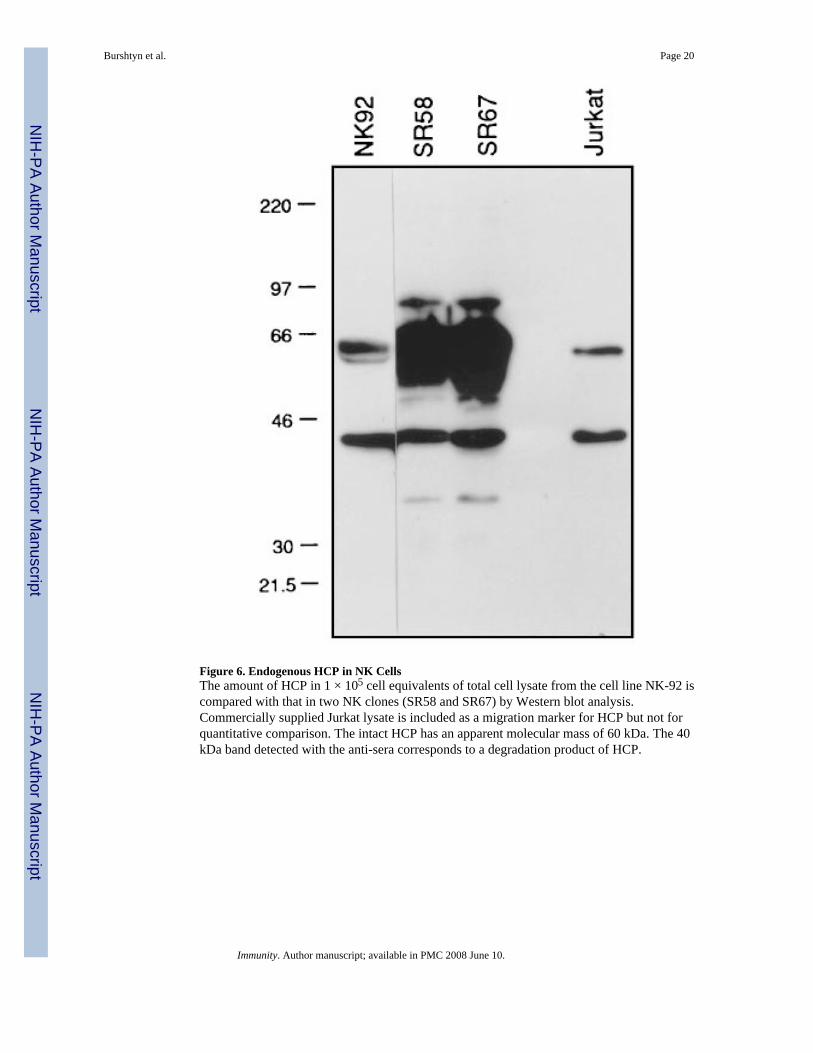
Figure 6. Endogenous HCP in NK CellsThe amount of HCP in 1 × 10⁵ cell equivalents of total cell lysate from the cell line NK-92 iscompared with that in two NK clones (SR58 and SR67) by Western blot analysis.Commercially supplied Jurkat lysate is included as a migration marker for HCP but not forquantitative comparison. The intact HCP has an apparent molecular mass of 60 kDa. The 40kDa band detected with the anti-sera corresponds to a degradation product of HCP.
Burshtyn et al. Page 20
Immunity. Author manuscript; available in PMC 2008 June 10.
NIH
-PA Author Manuscript
NIH
-PA Author Manuscript
NIH
-PA Author Manuscript
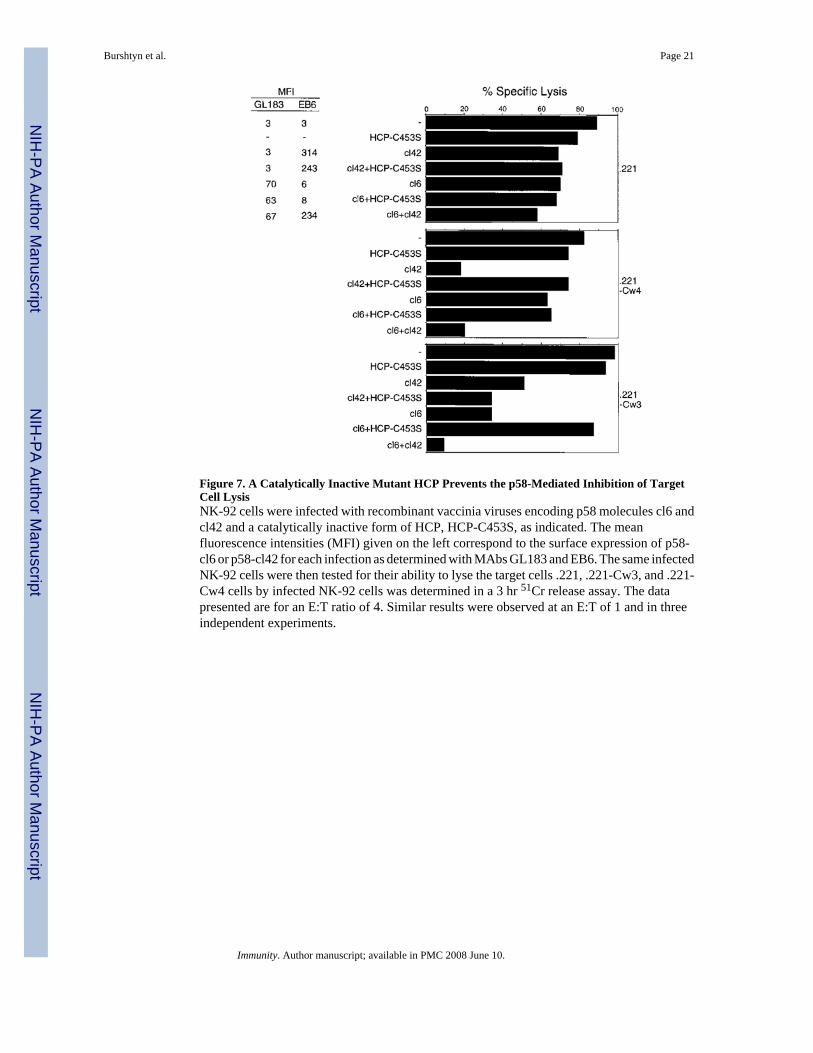
Figure 7. A Catalytically Inactive Mutant HCP Prevents the p58-Mediated Inhibition of TargetCell LysisNK-92 cells were infected with recombinant vaccinia viruses encoding p58 molecules cl6 andcl42 and a catalytically inactive form of HCP, HCP-C453S, as indicated. The meanfluorescence intensities (MFI) given on the left correspond to the surface expression of p58-cl6 or p58-cl42 for each infection as determined with MAbs GL183 and EB6. The same infectedNK-92 cells were then tested for their ability to lyse the target cells .221, .221-Cw3, and .221-Cw4 cells by infected NK-92 cells was determined in a 3 hr 51Cr release assay. The datapresented are for an E:T ratio of 4. Similar results were observed at an E:T of 1 and in threeindependent experiments.
Burshtyn et al. Page 21
Immunity. Author manuscript; available in PMC 2008 June 10.
NIH
-PA Author Manuscript
NIH
-PA Author Manuscript
NIH
-PA Author Manuscript

Figure 8. NK Receptors and HCP Binding Proteins Share a Unique Immunoreceptor Tyrosine-based Inhibition Motif (ITIM)Amino acid sequences (in single letter code) surrounding the YxxL motifs of the indicatedproteins are aligned. Dashes indicate identity with the sequence around the membrane-proximal tyrosine of the p58 cytoplasmic tail, p58 (Y1). Sequences were taken from the NKinhibitory receptors p58-cl6 (Wagtmann et al., 1995a), p70 (D’Andrea et al., 1995; Wagtmannet al., 1995b), and Ly-49 (Yokoyama and Seaman, 1993). NKG2-A is a molecule of unknownfunction expressed in human NK cells (Yabe et al., 1993). The single tyrosine residue in mouseFcγRIIB (D’Ambrosio et al., 1995) and three of the six tyrosines (i.e., Y2, Y5, and Y6) in CD22(Doody et al., 1995) are known to interact with HCP.
Burshtyn et al. Page 22
Immunity. Author manuscript; available in PMC 2008 June 10.
NIH
-PA Author Manuscript
NIH
-PA Author Manuscript
NIH
-PA Author Manuscript