The Phosphoramidase Compentency of Prototypical Phosphatase ...

600
Braz J Med Biol Res 41(7) 2008
A. Mota et al.
www.bjournal.com.br
Brazilian Journal of Medical and Biological Research (2008) 41: 600-609ISSN 0100-879X
Characterization of rat heart alkalinephosphatase isoenzymes and modulationof activityA. Mota1, P. Silva2, D. Neves3, C. Lemos1, C. Calhau1, D. Torres1, F. Martel1, H. Fraga1,L. Ribeiro1, M.N.M.P. Alçada1, M.J. Pinho1, M.R. Negrão1, R. Pedrosa1, S. Guerreiro1,J.T. Guimarães1, I. Azevedo1 and M.J. Martins1
1Departamento de Bioquímica (U38-FCT), Faculdade de Medicina, 2Departamento de Patologia,Faculdade de Medicina and Instituto de Patologia e Imunologia Molecular, 3Laboratório de BiologiaCelular e Molecular, Faculdade de Medicina and Instituto de Biologia Molecular e Celular,Universidade do Porto, Porto, Portugal
Correspondence to: M.J. Martins, Departamento de Bioquímica, Faculdade de Medicina (U38-FCT),Universidade do Porto, Alameda Professor Hernâni Monteiro, 4200-319 Porto, PortugalFax: +351-225513624. E-mail: [email protected]
Alkaline phosphatase (ALP) is important in calcification and its expression seems to be associated with the inflammatoryprocess. We investigated the in vitro acute effects of compounds used for the prevention or treatment of cardiovascular diseaseson total ALP activity from male Wistar rat heart homogenate. ALP activity was determined by quantifying, at 410 nm, the p-nitrophenol released from p-nitrophenylphosphate (substrate in Tris buffer, pH 10.4). Using specific inhibitors of ALP activity andthe reverse transcription-polymerase chain reaction, we showed that the rat heart had high ALP activity (31.73 ± 3.43 nmol p-nitrophenol·mg protein-1·min-1): mainly tissue-nonspecific ALP but also tissue-specific intestinal ALP type II. Both ALPisoenzymes presented myocardial localization (striated pattern) by immunofluorescence. ALP was inhibited a) strongly by 0.5mM levamisole, 2 mM theophylline and 2 mM aspirin (91, 77 and 84%, respectively) and b) less strongly by 2 mM L-phenylalanine, 100 µL polyphenol-rich beverages and 0.5 mM progesterone (24, 21 to 29 and 11%, respectively). ß-estradiol andcaffeine (0.5 and 2 mM) had no effect; 0.5 mM simvastatin and 2 mM atenolol activated ALP (32 and 36%, respectively).Propranolol (2 mM) tended to activate ALP activity and corticosterone activated (18%) and inhibited (13%) (0.5 and 2 mM,respectively). We report, for the first time, that the rat heart expresses intestinal ALP type II and has high total ALP activity. ALPactivity was inhibited by compounds used in the prevention of cardiovascular pathology. ALP manipulation in vivo may constitutean additional target for intervention in cardiovascular diseases.
Key words: Heart; Alkaline phosphatase; Polyphenol-rich beverages; Steroid hormones; Methylxanthines
A preliminary report of this manuscript was presented at the “Experimental Biology” Congress, San Francisco, CA, USA, April1-5, 2006 in abstract form: Mota A, Calhau C, Martel F, Martins MJ. “Modulation of rat heart alkaline phosphatase activity bydrugs, hormones and nutrients”. Faseb J 2006; 20: A897 (Abstract 588.12).
Research supported by FCT (POCI, FEDER and Programa Comunitário de Apoio).
Received September 28, 2007. Accepted June 23, 2008
Introduction
Alkaline phosphatase (ALP; EC 3.1.3.1.; orthophos-phoric-monoester phosphohydrolase, alkaline optimum)is ubiquitous in nature (from bacteria to humans); however,
its physiological role and natural substrates remain largelyunknown (1,2). The first established function of ALP is itsrole in bone mineralization (1-3). More recently, it has beenproposed that ALP regulates lipid transport (4). ALP ex-pression in vascular endothelia of small arterioles in the

601
Braz J Med Biol Res 41(7) 2008
Characterization of rat heart alkaline phosphatase
www.bjournal.com.br
brain and heart, which normally do not mineralize, couldcontribute to vascular hardening and calcification that could,in turn, be related to vascular aging and vascular disease(1,5).
Vascular calcification, including coronary artery calcifi-cation and aortic valve calcification, is a common andclinically significant component of atherosclerosis and car-diac valvular disease. The amount of coronary calcificationcorrelates with the overall coronary plaque burden and anincreased risk of myocardial infarction. The degree ofaortic valve calcification is a strong predictor of both theprogression and the outcome of aortic stenosis. Thesecalcified lesions contain not only various components as-sociated with bone mineralization, such as ALP, but alsoinflammatory cells such as macrophages and lympho-cytes. Although the mechanisms underlying valve calcifi-cation have not been established, recent data suggest thatvalve calcification is an active process, much like athero-sclerosis, that is preceded by inflammation, lipid deposi-tion and the accumulation of extracellular bone matrixproteins (1,6-11).
ALP represents a family of phosphomonoesterases,which, in humans, are expressed by four different geneloci : tissue-nonspecific (Tn-ALP), intestinal (Int-ALP), pla-cental (P-ALP) and germ-cell isoenzymes (Gc-ALP) (2).Tn-ALP is present in a large number of tissues (mainlylocated in the external side of plasma membranes via aglycosyl-phosphatidylinositol anchor linkage) and is mostabundant in liver, kidney and bone. Int-ALP, P-ALP andGc-ALP are tissue-specific ALPs (Ts-ALP). Ts-ALPs arenamed for the tissue with highest expression and activity(none are expressed in just one tissue). Only humans andgreat apes have P-ALP; all other mammals have insteadInt-ALP (2,12). Tissue differential processing of each of theALP gene products, for example through differential glyco-sylation, gives rise to tissue-specific isoforms (2,13). De-pending on the physiological/pathological conditions ALPcan be released into intestinal lumen, plasma or bile invarious isoforms: with or without its anchor and in theformer case associated or not with membrane fragments(2). ALP isoenzymes (and isoforms) can be distinguishedby their heat stability, optimum pH, substrate affinity oreffect of several specific inhibitors or activators (2,13-19).
The variety of ALP forms led our group to characterizethe effect (in vitro and/or in vivo) of several endogenousmetabolites, drugs, hormones, beverages and food com-ponents on ALP isoenzyme and/or isoform activity and/orexpression. In vitro data from our group regarding ALPactivity modulation will be presented shortly in the discus-sion of this article, since some of our previous results will beuseful for comparison with the present data (13,14,17-19).
In order to further clarify the physiological role of ALP,we have determined in vitro the acute effect of compounds,drugs, hormones and polyphenol-rich beverages, with re-ported cardiovascular protective effects (for the preventionor treatment of cardiovascular diseases) and/or knowneffects on ALP activity from other sources (cell lines,tissues and species) on total ALP activity from rat heart.We hypothesize that the modulation of ALP activity may beanother process contributing to explain the protection givenby aspirin, steroid hormones, statins, ß-blockers andpolyphenol-rich beverages against cardiovascular dis-eases.
Material and Methods
Beverages and their preparationLager-type beer (SuperBock®), stout-type beer (Super-
Bock Stout®) and alcohol-free beer (Cheers®) were pur-chased at the local market (Portuguese beers produced byUNICER, Portugal). Green and black teas (both fromTetley®) were also from the local market. Red wine (Vinhadas Garças®) was produced in Palmela region (Terras doSado, South of Portugal) by UNICER and provided to us bythis company. Green and black teas were prepared ac-cording to the recommendations of supplier, by infusingone tea bag (green tea: 1.75 g for 5 min; black tea: 1.5 g for2 min) in 250 mL boiling water. Gas was removed frombeers before use. All beverages were used at pH 10.4.
AnimalsSixteen male adult Wistar rats (free from liver and bone
diseases, purchased from Harlan Interfauna Ibèrica,Barcelona, Spain), weighing 300-340 g, were used. Theanimals were maintained under controlled environmentalconditions (12-h light/dark cycle, at 24°C room tempera-ture), in groups of 3, fed a suitable commercial diet (Harlan)with water ad libitum. Animal procedures were accordingto the European Community guidelines (86/609/EEC) andthe Portuguese Act (129/92) for the use of experimentalanimals [the corresponding author holds a Portugueseauthorization for working with laboratory animals (accord-ing to category C of the recommendations of the Federa-tion of European Laboratory Animal Science Associa-tions)]. Animals were anesthetized with pentobarbital (60mg/kg) and the heart was perfused in situ with ice-coldisotonic NaCl to wash out blood. The organ was thenremoved and rinsed in ice-cold isotonic NaCl. For reversetranscription-polymerase chain reaction (rt-PCR) analy-sis, a segment from the left ventricle was rinsed withdiethylpyrocarbonate-water and stored at -80°C until themoment of the experiment. Rat heart was frozen at -80°C

602
Braz J Med Biol Res 41(7) 2008
A. Mota et al.
www.bjournal.com.br
until the immunofluorescence experiment. Rat heart wasplaced in two times (mL/g) homogenization solution [phos-phate-buffered saline (PBS, pH 7.4) + 0.5% Triton X-100;4:1], with no homogenization or other form of cell disrup-tion, and stored at -80°C until the moment of ALP activityassay performance.
Reverse transcription-polymerase chain reactionAll molecular biology reagents used were purchased
from Sigma (Sigma Alcobendas, Spain) unless otherwisestated. After total RNA extraction with Tripure® (Roche,Germany), 20 µg total RNA was incubated at 37°C for 30min with 23 units of RNase-free DNase I in 100 µL 5 mMMgCl2, 50 mM triethanolamine-HCl, pH 7.5, to degradeany residual DNA. The RNA was extracted with phenol-chloroform, precipitated with ethanol and dissolved in wa-ter. For cDNA synthesis, 5 µg of the RNA thus preparedwas incubated at 45°C for 1 h in a total volume of 20 µL with200 units of Superscript™ II Reverse Transcriptase (RT;Gibco BRL, Life Technologies, Gaithersburg, MD, USA), in10 µM random hexamers, 0.375 mM per dNTP, 3 mMMgCl2, 75 mM KCl, 50 mM Tris-HCl, pH 8.3 (25°C), 10 mMdithiothreitol and 40 units RNase inhibitor (RNaseOUT™;Gibco BRL). For paired negative controls RT was omitted.Following heat-inactivation of the proteins (10 min at 95°C)and addition of 5 µL 0.5 mg/mL DNase-free RNase A in 10mM Tris-HCl, pH 8.0, and 50% (v/v) glycerol, the cDNAwas incubated at 37°C for 30 min to degrade unreactedmRNA. PCR was performed on 4 µL of this preparation.The PCR product mixture (50 µL) contained 0.5 µM perprimer, 0.2 mM per dNTP, 2.3 mM MgCl2 and 2 units of TaqDNA polymerase in the buffer provided (Gibco BRL). Thefollowing primers for rat Int-ALP type I, Int-ALP type II, Tn-ALP and glyceraldehyde-3-phosphate dehydrogenase(GAPDH) were used: 5'-CCA GCC AGC TTA CCA ATGAGA-3' (forward primer Int-ALP type I), 5'-CTG CCT GCTGCT TGT AGT TGA-3' (reverse primer Int-ALP type I), 5'-CCT GGA GCC CTA CAC CGA CTG-3' (forward primerInt-ALP type II), 5'-GCC CCC AGT AGT AGC ATC AGC-3'(reverse primer Int-ALP type II), 5'-CAG TGT CAG CCGTTA ATT GAC-3' (forward primer Tn-ALP), 5'-ATC GTGCTG ACC TTG CCA CA-3' (reverse primer Tn-ALP), 5'-ACT GGC GTC TTC ACC ACC AT-3' (forward primerGAPDH), 5'-TCC ACC ACC CTG TTG CTG TA-3' (reverseprimer GAPDH). Thermocycling consisted of 1 cycle of 2min at 94°C, followed by 35 cycles of 1 min at 94°C, 2 minat 26°C and 2 min at 72°C for Int-ALP type I, Int-ALP typeII and GAPDH primers or 1 cycle of 2 min at 94°C, followedby 40 cycles of 0.5 min at 94°C, 0.5 min at 26°C and 1.5 minat 72°C for Tn-ALP and GAPDH. The predicted sizes (inbp) of the PCR products were: 249 (Int-ALP type I), 218
(Int-ALP type II), 246 (Tn-ALP) and 682 (GAPDH). Tenmicroliters of each individual PCR product was then run on1.6% agarose gel and visualized with an ultraviolet trans-illuminator (Vilber Lourmat, France) using ethidium bro-mide staining, a COHU CCD camera and the appropriatefilters for UV light (Cohu, USA).
ImmunofluorescenceThe frozen rat heart was included in ornithine carbamyl
transferase solution (Bright, England) and 5-µm thick sec-tions were cut with a Leica cryostat (Leica MicrosystemsGmbH, Germany) and placed onto poly-L-lysine coatedmicroscopy slides (Polysine™, Menzel-Glaser, Germany).Immunofluorescence was performed using primary anti-bodies (Santa Cruz Biotechnology, USA) against Int-ALP[goat polyclonal antibody (PLAP L-19: sc-15065), diluted1/25] and Tn-ALP [goat polyclonal antibody (TNAP N-18:sc-23430), diluted 1/25], for 60 min at room temperature,after a 10-min blocking period at room temperature [10%bovine serum albumin (Sigma) w/v in PBS]. PBS was usedinstead of the primary antibodies for the negative controls.Although we have used a polyclonal antibody against P-ALP and Int-ALP, only Int-ALP was detected since rats donot express P-ALP (2,5). A 60-min incubation was per-formed with the secondary antibody (donkey anti-goatIgG-FITC, sc-2024, from Santa Cruz Biotechnology, di-luted 1/200) at room temperature. Washings between in-cubations were performed twice for 5 min each with PBS +0.1% Tween 20 (Sigma). Antibody dilutions were madeusing UltraAb diluent (LabVision Corporation, USA). Nu-clei counterstaining was achieved with DAPI (4'-6-diamidino-2-phenylindole, diluted 1/100, Sigma) for 15min at room temperature. Slides were mounted withVectashield (mounting medium for fluorescence; VectorLaboratories, Inc., USA) and observed with an ApoTomeMicroscope (AxioImager.Z1, Zeiss System, Germany), witha light source of fixed wavelength of 488 and 350 nm, forIgG-FITC (green) and DAPI (blue), respectively. TheAxionVision 3.0 program (Zeiss) was used to acquireimages and photographs.
Alkaline phosphatase activityp-Nitrophenylphosphate (p-NPP), p-nitrophenol (p-
NPL), levamisole, L-phenylalanine, theophylline, caffeine,acetylsalicylic acid, propranolol, atenolol, progesterone,corticosterone, ß-estradiol and simvastatin were purchasedfrom Sigma. On the day of ALP quantification, tissuesamples diluted further in homogenization solution (to 1 gtissue + 12 mL homogenization solution) were homog-enized and kept on ice. ALP activity assays were carriedout in triplicate, as previously described (13,15). The reac-

603
Braz J Med Biol Res 41(7) 2008
Characterization of rat heart alkaline phosphatase
www.bjournal.com.br
Figure 1.Figure 1.Figure 1.Figure 1.Figure 1. Effect of alkaline phosphatase (ALP) classic inhibitors,levamisole and L-phenylalanine, on ALP activity from rat hearthomogenate. Results are reported as mean ± SEM and repre-sent a percent of the corresponding control activity (first columnof each set of results, 100%). The ALP-specific activity in thecontrol was 31.73 ± 3.43 nmol p-nitrophenol·mg protein-1·min-1.The horizontal lines indicate significant differences between ef-fects of inhibitors. *P ≤ 0.005 (paired or unpaired Student t-test).
Figure 2.Figure 2.Figure 2.Figure 2.Figure 2. Detection of Int-ALP type I (Int-ALP-I) and Int-ALP typeII (Int-ALP-II) mRNA in total RNA from rat heart homogenateusing rt-PCR. rt-PCR analysis with specific primers for Int-ALP-I(lane 1), Int-ALP-II (lane 3) and GAPDH (lanes 1 and 3) wasperformed with total RNA prepared from rat heart homogenate(left ventricle). GAPDH mRNA detection was used as control forintactness of mRNA. Lanes (+) and (-) correspond to samplesgenerated in the presence and absence of RT enzyme duringreverse transcriptase reaction, respectively; lanes 2 and 4 corre-spond to negative controls for Int-ALP-I and Int-ALP-II, respec-tively. PCR products were separated by agarose gel electropho-resis, followed by staining with ethidium bromide. GAPDH =glyceraldehyde-3-phosphate dehydrogenase; Int-ALP = intesti-nal alkaline phosphatase.
tion mixtures, with a final volume of 500 µL, contained: 80mM Tris buffer, pH 10.4, 0.4 mM MgCl2, 0.376 mg p-NPP,the compound (0.1 to 2 mM) or the beverage (50 or 100 µL)to be tested and heart homogenate. The reactions werestarted by the addition of the tissue enzyme sample: 20 µLheart homogenate in a final dilution of 1/13 in homogeniza-tion solution. Phosphate concentration in the incubationmedium was 0.353 mM, similarly to other studies by ourgroup on ALP activity from tissue homogenates (13,15).Levamisole and L-phenylalanine were dissolved in water.Theophylline, caffeine and simvastatin were dissolved anddiluted in dimethylsulfoxide. Acetylsalicylic acid (aspirin),propranolol, atenolol, progesterone, corticosterone and ß-estradiol were dissolved and diluted in ethanol. Controlsfor drugs were run in the presence of a correspondingvolume of solvent. Ethanol 5, 5.6 and 13% solutions wereused as solvent controls for stout-type beer, lager-typebeer and red wine, respectively. Water was used as sol-vent control for green and black teas. Incubations werecarried out at 37°C for 30 min and the enzymatic reactionwas stopped by adding 2 mL 20 mM NaOH (4°C). Theabsorbance of the p-NPL produced by ALP activity wasmeasured at 410 nm (Spectronic Genesys 5, Milton Roy,USA). A p-NPL calibration curve was constructed in orderto calculate ALP activity. The effect of each compound orbeverage on ALP activity was calculated as the percent ofthe activity of the appropriate ALP control.
Protein determinationProtein concentration was determined as described by
Bradford (20), with bovine serum albumin as standard.
All data are reported as mean ± SEM and represent thepercent of the corresponding control activity (N = numberof cases). The significance of differences between meanswas assessed by paired or unpaired Student t-test.
Results
The effect of eleven different compounds (belonging todifferent chemical and/or pharmacological classes) andsix different beverages (with high and distinct polyphenolcontent) was evaluated on total ALP activity from rat hearthomogenate. Assay conditions allowed linearity of theenzymatic reaction (data not shown).
Levamisole and L-phenylalanine significantly inhibitedALP activity from rat heart homogenate, although leva-misole had a significantly stronger effect (Figure 1). While0.1 and 0.5 mM levamisole inhibited more than 80% of ALPactivity (in a concentration-dependent way) 2 mM L-phen-
604
Braz J Med Biol Res 41(7) 2008
A. Mota et al.
www.bjournal.com.br
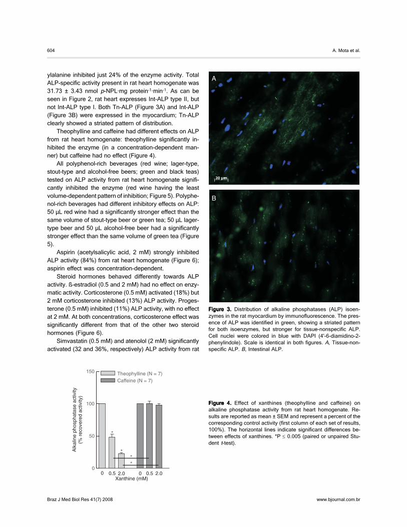
ylalanine inhibited just 24% of the enzyme activity. TotalALP-specific activity present in rat heart homogenate was31.73 ± 3.43 nmol p-NPL·mg protein-1·min-1. As can beseen in Figure 2, rat heart expresses Int-ALP type II, butnot Int-ALP type I. Both Tn-ALP (Figure 3A) and Int-ALP(Figure 3B) were expressed in the myocardium; Tn-ALPclearly showed a striated pattern of distribution.
Theophylline and caffeine had different effects on ALPfrom rat heart homogenate: theophylline significantly in-hibited the enzyme (in a concentration-dependent man-ner) but caffeine had no effect (Figure 4).
All polyphenol-rich beverages (red wine; lager-type,stout-type and alcohol-free beers; green and black teas)tested on ALP activity from rat heart homogenate signifi-cantly inhibited the enzyme (red wine having the leastvolume-dependent pattern of inhibition; Figure 5). Polyphe-nol-rich beverages had different inhibitory effects on ALP:50 µL red wine had a significantly stronger effect than thesame volume of stout-type beer or green tea; 50 µL lager-type beer and 50 µL alcohol-free beer had a significantlystronger effect than the same volume of green tea (Figure5).
Aspirin (acetylsalicylic acid, 2 mM) strongly inhibitedALP activity (84%) from rat heart homogenate (Figure 6);aspirin effect was concentration-dependent.
Steroid hormones behaved differently towards ALPactivity. ß-estradiol (0.5 and 2 mM) had no effect on enzy-matic activity. Corticosterone (0.5 mM) activated (18%) but2 mM corticosterone inhibited (13%) ALP activity. Proges-terone (0.5 mM) inhibited (11%) ALP activity, with no effectat 2 mM. At both concentrations, corticosterone effect wassignificantly different from that of the other two steroidhormones (Figure 6).
Simvastatin (0.5 mM) and atenolol (2 mM) significantlyactivated (32 and 36%, respectively) ALP activity from rat
Figure 3.Figure 3.Figure 3.Figure 3.Figure 3. Distribution of alkaline phosphatases (ALP) isoen-zymes in the rat myocardium by immunofluorescence. The pres-ence of ALP was identified in green, showing a striated patternfor both isoenzymes, but stronger for tissue-nonspecific ALP.Cell nuclei were colored in blue with DAPI (4'-6-diamidino-2-phenylindole). Scale is identical in both figures. A, Tissue-non-specific ALP. B, Intestinal ALP.
Figure 4.Figure 4.Figure 4.Figure 4.Figure 4. Effect of xanthines (theophylline and caffeine) onalkaline phosphatase activity from rat heart homogenate. Re-sults are reported as mean ± SEM and represent a percent of thecorresponding control activity (first column of each set of results,100%). The horizontal lines indicate significant differences be-tween effects of xanthines. *P ≤ 0.005 (paired or unpaired Stu-dent t-test).

605
Braz J Med Biol Res 41(7) 2008
Characterization of rat heart alkaline phosphatase
www.bjournal.com.br
Figure 5.Figure 5.Figure 5.Figure 5.Figure 5. Effect of polyphenol-rich beverages, red wine (13% ethanol), beer [lager-type (5.6% ethanol), stout-type (5.0% ethanol) andalcohol-free] and tea (green and black) on alkaline phosphatase activity from rat heart homogenate. Results are reported as mean ±SEM and represent a percent of the corresponding control activity (first column of each set of results, 100%). The horizontal linesindicate significant differences between effects of polyphenol-rich beverages. *P ≤ 0.05, **P ≤ 0.005 (paired or unpaired Student t-test).
Figure 6.Figure 6.Figure 6.Figure 6.Figure 6. Effect of acetylsalicylic acid (aspirin), steroid hormones (ß-estradiol, corticosterone and progesterone), simvastatin and ß-blockers (propranolol and atenolol) on alkaline phosphatase activity from rat heart homogenate. Results are reported as mean ± SEMand represent a percent of the corresponding control activity (first column of each set of results, 100%). The horizontal lines indicatesignificant differences between effects of compounds. *P ≤ 0.05, **P ≤ 0.005 (paired or unpaired Student t-test).
heart homogenate (Figure 6). Atenolol (2 mM) induced asignificantly stronger activation (36%) than 2 mM propran-olol; propranolol presented a tendency to activate ALP(effects of both ß-blockers were concentration-dependent;Figure 6).
Discussion
To our knowledge, this is the first report concerning ratheart ALP isoenzyme identification (in terms of its mRNAexpression as well as protein localization and percentage

606
Braz J Med Biol Res 41(7) 2008
A. Mota et al.
www.bjournal.com.br
of activity), total ALP activity quantification in the rat heartand its in vitro acute modulation by compounds and polyphe-nol-rich beverages reported in the literature to have cardio-vascular beneficial effects.
The inhibitory effects of levamisole and L-phenylala-nine showed that both Tn- and Ts-ALPs were present in ratheart (with Tn-ALP presenting a greater activity than Int-ALP), since these compounds are, respectively, specificinhibitors of these two ALP groups (2,13,19). Our resultsare in agreement with those reported by Van Belle (16)regarding the effects of levamisole and L-phenylalanine onALP extracted from normal rat heart (without any treat-ment). Additionally, we showed, by rt-PCR analysis, thatInt-ALP, namely Int-ALP type II, was indeed expressed inthis tissue.
The rat has two Int-ALP genes (Int-ALP type I and Int-ALP type II) coding for two Int-ALPs with different primarystructures (79% amino acid homology), temporal postnatalexpression, substrate specificity, tissue localization andresponse to fat feeding and to cortisone. After an acute fatfeeding, Int-ALP type II expression is increased in liver(where Int-ALP type I is not expressed) and Int-ALP type IIexpression increases five times more than Int-ALP type I inthe duodenum (4,21,22). It is interesting to note that ratliver and heart, tissues that can use fat as an energysource in several physiological states, express the sameInt-ALP gene.
We hypothesized that the high total ALP activity ob-served in the normal rat heart (without any treatment)homogenate could not be accounted for only by the pres-ence of ALP in the vascular compartment and, by immuno-fluorescence, we showed that both Int- and Tn-ALPs arepresent in rat cardiomyocytes, which is in agreement withour results of total ALP activity in the rat heart and of ratheart ALP activity inhibition by levamisole and L-phenylal-anine. Autofluorescence of vascular tissue did not allow usto visualize ALP in blood vessels. Nevertheless, the pres-ence of Int-ALP in the human vascular compartment ofdifferent organs has already been reported by Domar et al.(12) and the presence of Tn-ALP in both the heart vascularcompartment and muscle has already been reported bySchultz-Hector et al. (5). Müller et al. (23) reported anincrease of ALP activity, after a single injection of iso-prenaline into the right atrium, particularly on the Schwanncell and its membranes, membranes enveloping nerveaxons, the sarcolemma of the muscle cell and the fibrocytecell surface. These investigators (23) suggested that theSchwann cell is responsible for the production of ALP.Here we report the presence of Int-ALP in cardiomyocytesand the presence of both ALP isoenzymes in normal ratheart (without any treatment). Although speculative, we
suggest that the distribution of specific fluorescence alongthe myofibrils of myocardial cells is compatible with thepresence of ALP in the sarcoplasmic reticulum.
Several lines of evidence suggest ALP as a putativetarget for prevention and treatment of cardiovascular dis-eases. ALP is important for the initiation of calcification andits inhibition prevents calcification, either normal or ectopic(1-3,6-8,10,11). ALP activity has been associated withhuman valve calcification in vitro and in vivo (7,8,11), withcalcification of bioprosthetic heart valves from bovine peri-cardium in vitro and in vivo models (10) and with in vitrosheep aortic valve interstitial cell calcification induced bytransforming growth factor-ß1 (11). These in vitro resultsare supported by the rt-PCR studies of gene expressionpattern differences between human calcific aortic stenosisand human normal aortic valve cusps demonstrating thatcalcific aortic cusps have increased ALP and transforminggrowth factor-ß1 expression (11). An association betweencardiac valve calcification and inflammation has been re-ported (9,11,24). ALP seems to be associated with inflam-matory processes and their modulation/regulation (25,26).In vitro and in vivo, ALP activity is modulated by oxidativestress (25,27,28). The ingestion of nutrients/beverageswith anti-oxidant properties has positive effects on cardio-vascular health (29-31).
In the present study, we sought to understand thereported “usefulness” of various compounds, hormonesand beverages for prevention and/or treatment of cardio-vascular diseases. A marked ALP inhibition was observedwith theophylline and aspirin, and some inhibition withwine, beers and teas. ß-estradiol and caffeine had noeffect; simvastatin and atenolol activated ALP (propranololalso showing an activation tendency). Corticosterone pre-sented a mixed behavior on ALP and progesterone slightlyinhibited it.
An inhibitory effect on rat heart ALP activity would beacceptable as a similar background for the cardioprotec-tive effects of the compounds, hormones and beveragestested. By reducing rat heart ALP activity they would con-tribute to reduce or stop progression of cardiomyocyte,vascular, valvular and/or aorta calcification improving orstabilizing the cardiovascular system; however, that didnot occur for all of them. We concluded that only the agentswe have tested with reported preventive effects againstcardiovascular pathology have ALP inhibitory activity. Themeaning of this association is presently unknown but theimportance of cardiovascular diseases strongly recom-mends further exploration of this link.
Methylxanthines, caffeine and theophylline, have beenincluded in this study because a) caffeine is probably themost frequently ingested pharmacologically active sub-

607
Braz J Med Biol Res 41(7) 2008
Characterization of rat heart alkaline phosphatase
www.bjournal.com.br
stance in the world and recent research indicates thatmoderate coffee intake may be associated with beneficialeffects on cardiovascular health (32); b) theophylline isdescribed as an inhibitor of ALP: Tn-ALP isoforms and Ts-ALP isoenzymes are inhibited/modulated at different de-grees (13). The theophylline inhibitory effect on rat heartALP was similar to its effect on rat kidney cortex ALP butstronger than that on rat liver ALP (13).
Tea, wine and beer were used in this research becauseof their extensive worldwide consumption, anti-oxidantcapacity (related to their polyphenol content) and describedhealth benefits in preventing cardiovascular diseases (29-31). The evolution of these pathologies seems to exacer-bate in estrogen-deficient women, thus, our interest in ß-estradiol (33). Wine, beer and tea were tested directly invitro (in an acute treatment) upon ecto-ALP activity from ahuman vascular smooth muscle (AALTR) cell line for thefirst time by our group (19). A much stronger inhibitoryeffect of these beverages had been observed on AALTRecto-ALP activity (19) than on rat heart ALP. These AALTRresults are largely and positively correlated with the polyphe-nol content of the beverages tested (19,29), what did nothappen in the case of rat heart ALP.
Progesterone, ß-estradiol and/or corticosterone effectson rat liver and kidney cortex ALP activity as well as on ratbrain microvessel endothelial (RBE4) cell line (13,14) aredifferent from those presented here. However, similarly toour present results, in AALTR cell line ß-estradiol had noeffect while corticosterone (and progesterone) activatedthe enzyme (18).
Aspirin has been reported to have a protective role inthe cardiovascular system, exhibiting anti-platelet, anti-thrombotic, anti-inflammatory and anti-oxidant properties.It also inhibits calcification of bovine pericardium used forbioprosthetic heart valves (10,34). Our results with aspirinare in accordance with published data concerning the invivo and in vitro effects of aspirin upon ALP activity. Aspirininhibits, in vitro and in vivo, the calcium deposition and/orALP activity in bioprosthetic heart valves from bovinepericardium (10). Aspirin, in a therapeutic concentrationrange-dependent manner, inhibits ALP secretion and thestimulatory insulin effect upon ALP production by humanosteoblasts in vitro (35).
It is interesting to note that two drugs sharing anti-oxidant and anti-inflammatory properties have oppositeeffects on rat heart ALP: aspirin and simvastatin (34,36).
Statins have well-known protective effects on cardio-vascular health, mainly ascribed to their hypolipidemicactivity. Beyond this, statins exert pleiotropic effects onvascular wall cells, including improvement of endothelialdysfunction, stabilization of atherosclerotic plaque, de-
crease of oxidative stress and vascular inflammation(17,36,37). On the other hand, they are reported to in-crease bone density (17,36). We have recently reported anincrease on ALP activity in rat heart homogenate forlovastatin (17), but not as intense as with simvastatin.These ALP activating effects may explain the lack of statinprotection against aortic valve calcification reported byCowell et al. (for a discussion of this subject, see Ref. 17).Simvastatin inhibits rat liver and kidney cortex ALP activity(17).
ß-blockers are currently used to treat cardiovascularproblems (38) and have been reported to have positiveeffects on bone mineralization and/or osteoporosis (39).The ALP activating effects of propranolol and atenololobserved here fit well with their putative beneficial effectson bone.
Epidemiological studies show a positive correlationbetween osteoporosis and vascular calcification suggest-ing that when bone does not mineralize properly the ves-sels do and vice versa (6). For most of the compounds andbeverages tested there are data about beneficial effectson bone mineralization and/or osteoporosis (17,30,31,36,39,40).
Glycosylation can influence/interfere with ALP cata-lytic activity and ALP activity modulation (2,13,19). Differ-ential glycosylation of the Tn- and Int-ALPs and/or distinctlevel of expression of Tn- and Int-ALPs (assuming theirdistinct modulation by the compounds and beveragestested) can explain the distinct ALP modulation resultsobtained with xanthines, polyphenol-rich beverages, ste-roid hormones and simvastatin on the various cell linesand/or tissues used so far as ALP sources. Thus, weconcluded that it is fundamental that ALP activity studiestake the enzyme source into account.
To our knowledge this is the first time that ALP was invitro acutely treated with aspirin, simvastatin, atenolol andpropranolol (simvastatin results are discussed in Ref. 17),and there are no data in the literature relating caffeine,theophylline, ß-blockers (atenolol and propranolol) andpolyphenol-rich beverages (wine, beer and tea) and car-diomyocyte/vascular/valvular/aorta valve calcification. Morestudies, namely involving chronic treatments (measuringALP expression and performing mineralization assays)with these compounds and beverages on cardiovascular(and bone) tissues or cell lines are needed and stronglyencouraged by our present results.
Both Tn- and Int-ALPs are associated with membranesurfaces characterized by significant transport activity (1,2,4,12,14,17,22,23,25). The apparent Tn-ALP localization inthe sarcoplasmic reticulum leads us to raise the hypo-thesis of ALP being involved in the modulation of the

608
Braz J Med Biol Res 41(7) 2008
A. Mota et al.
www.bjournal.com.br
crucial transport of Ca2+ across the sarcoplasmic reticulummembrane, what should be investigated in the future. Also,evaluation of a putative participation of Int-ALP type II inlipid transport and deposition in rat heart muscle andvascular compartment would be interesting, since lipiddeposition contributes to atherosclerosis and Int-ALP isinvolved in regulating intestinal lipid transport (4). Further-more, exploration of this hypothesis (Int-ALP and lipidtransport at the heart) may help to clarify the pathogenesis
of lipid overstorage in cardiac myocytes in type 2 diabetesmellitus and the development of heart failure and/or thereduction in triacylglycerol storage and mobilization insituations of early heart failure.
Acknowledgments
We gratefully acknowledge red wine Vinha das Garças®
to UNICER, Portugal.
References
1. Hui M, Tenenbaum HC. New face of an old enzyme: alkalinephosphatase may contribute to human tissue aging by in-ducing tissue hardening and calcification. Anat Rec 1998;253: 91-94.
2. Van Hoof V, De Broe ME. Interpretation and clinical signifi-cance of alkaline phosphatase isoenzyme patterns. CritRev Clin Lab Sci 1994; 31: 197-293.
3. Fedde KN, Blair L, Silverstein J, Coburn SP, Ryan LM,Weinstein RS, et al. Alkaline phosphatase knock-out micerecapitulate the metabolic and skeletal defects of infantilehypophosphatasia. J Bone Miner Res 1999; 14: 2015-2026.
4. Narisawa S, Huang L, Iwasaki A, Hasegawa H, Alpers DH,Millan JL. Accelerated fat absorption in intestinal alkalinephosphatase knockout mice. Mol Cell Biol 2003; 23: 7525-7530.
5. Schultz-Hector S, Balz K, Bohm M, Ikehara Y, Rieke L.Cellular localization of endothelial alkaline phosphatase re-action product and enzyme protein in the myocardium. JHistochem Cytochem 1993; 41: 1813-1821.
6. Chen NX, Moe SM. Arterial calcification in diabetes. CurrDiab Rep 2003; 3: 28-32.
7. Mathieu P, Voisine P, Pepin A, Shetty R, Savard N,Dagenais F. Calcification of human valve interstitial cells isdependent on alkaline phosphatase activity. J Heart ValveDis 2005; 14: 353-357.
8. Rajamannan NM, Subramaniam M, Rickard D, Stock SR,Donovan J, Springett M, et al. Human aortic valve calcifica-tion is associated with an osteoblast phenotype. Circulation2003; 107: 2181-2184.
9. Kizu A, Shioi A, Jono S, Koyama H, Okuno Y, Nishizawa Y.Statins inhibit in vitro calcification of human vascular smoothmuscle cells induced by inflammatory mediators. J CellBiochem 2004; 93: 1011-1019.
10. Vasudev SC, Chandy T, Sharma CP, Mohanty M, Umasan-kar PR. Synergistic effect of released aspirin/heparin forpreventing bovine pericardial calcification. Artif Organs2000; 24: 129-136.
11. Clark-Greuel JN, Connolly JM, Sorichillo E, Narula NR,Rapoport HS, Mohler ER III, et al. Transforming growthfactor-beta1 mechanisms in aortic valve calcification: in-creased alkaline phosphatase and related events. AnnThorac Surg 2007; 83: 946-953.
12. Domar U, Nilsson B, Baranov V, Gerdes U, Stigbrand T.
Expression of intestinal alkaline phosphatase in human or-gans. Histochemistry 1992; 98: 359-364.
13. Martins MJ, Negrao MR, Hipolito-Reis C. Alkaline phos-phatase from rat liver and kidney is differentially modulated.Clin Biochem 2001; 34: 463-468.
14. Calhau C, Martel F, Pinheiro-Silva S, Pinheiro H, Soares-da-Silva P, Hipolito-Reis C, et al. Modulation of insulin trans-port in rat brain microvessel endothelial cells by an ecto-phosphatase activity. J Cell Biochem 2002; 84: 389-400.
15. Martins MJ, Negrao MR, Hipolito-Reis C, Azevedo I. Physi-ologic concentrations of bile salts inhibit rat hepatic alkalinephosphatase but not the intestinal isoenzyme. Clin Biochem2000; 33: 611-617.
16. Van Belle H. Kinetics and inhibition of rat and avian alkalinephosphatases. Gen Pharmacol 1976; 7: 53-58.
17. Negrao MR, Mota A, Azevedo I, Martins MJ. Statins andtissue mineralization: putative involvement of alkaline phos-phatase. Med Hypotheses 2006; 67: 524-528.
18. Keating E, Negrão MR, Martins MJ, Azevedo I. Humanvascular smooth muscle cells ecto-ALP activity: modulationby steroids and phytochemicals. FASEB J 2004; 18: A285(Abstract).
19. Negrao MR, Keating E, Faria A, Azevedo I, Martins MJ.Acute effect of tea, wine, beer, and polyphenols on ecto-alkaline phosphatase activity in human vascular smoothmuscle cells. J Agric Food Chem 2006; 54: 4982-4988.
20. Bradford MM. A rapid and sensitive method for the quantita-tion of microgram quantities of protein utilizing the principleof protein-dye binding. Anal Biochem 1976; 72: 248-254.
21. Goseki-Sone M, Oida S, Iimura T, Yamamoto A, Matsu-moto HN, Omi N, et al. Expression of mRNA encodingintestinal type alkaline phosphatase in rat liver and its in-crease by fat-feeding. Liver 1996; 16: 358-364.
22. Xie Q, Alpers DH. The two isozymes of rat intestinal alkalinephosphatase are products of two distinct genes. PhysiolGenomics 2000; 3: 1-8.
23. Muller E, Van Noorden S, Pearse AG. Ultrastructural local-ization of alkaline phosphatase in rat atrium. J Mol CellCardiol 1971; 3: 209-212.
24. Torun D, Sezer S, Baltali M, Adam FU, Erdem A, OzdemirFN, et al. Association of cardiac valve calcification andinflammation in patients on hemodialysis. Ren Fail 2005;27: 221-226.

609
Braz J Med Biol Res 41(7) 2008
Characterization of rat heart alkaline phosphatase
www.bjournal.com.br
25. Sanchez de Medina F, Martinez-Augustin O, Gonzalez R,Ballester I, Nieto A, Galvez J, et al. Induction of alkalinephosphatase in the inflamed intestine: a novel pharmacolo-gical target for inflammatory bowel disease. Biochem Phar-macol 2004; 68: 2317-2326.
26. Kapojos JJ, Poelstra K, Borghuis T, van den Berg A, BaeldeHJ, Klok PA, et al. Induction of glomerular alkaline phos-phatase after challenge with lipopolysaccharide. Int J ExpPathol 2003; 84: 135-144.
27. Santos A, Alçada MN, Mota A, Martins MJ. Effect of beer,orange juice and soft drink on rat serum alkaline phos-phatase. FASEB J 2005; 19: A1476 (Abstract).
28. Mody N, Parhami F, Sarafian TA, Demer LL. Oxidativestress modulates osteoblastic differentiation of vascular andbone cells. Free Radic Biol Med 2001; 31: 509-519.
29. Bravo L. Polyphenols: chemistry, dietary sources, metabo-lism, and nutritional significance. Nutr Rev 1998; 56: 317-333.
30. Kondo K. Beer and health: preventive effects of beer com-ponents on lifestyle-related diseases. Biofactors 2004; 22:303-310.
31. Scalbert A, Manach C, Morand C, Remesy C, Jimenez L.Dietary polyphenols and the prevention of diseases. CritRev Food Sci Nutr 2005; 45: 287-306.
32. Sudano I, Binggeli C, Spieker L, Luscher TF, Ruschitzka F,Noll G, et al. Cardiovascular effects of coffee: is it a risk
factor? Prog Cardiovasc Nurs 2005; 20: 65-69.33. Teede HJ. Sex hormones and the cardiovascular system:
effects on arterial function in women. Clin Exp PharmacolPhysiol 2007; 34: 672-676.
34. Maree AO, Fitzgerald DJ. Aspirin and coronary artery dis-ease. Thromb Haemost 2004; 92: 1175-1181.
35. Khokher MA, Dandona P. The effect of indomethacin andaspirin on alkaline phosphatase secretion and [3H]thymidineincorporation by human osteoblasts. Br J Rheumatol 1988;27: 291-294.
36. Horiuchi N, Maeda T. Statins and bone metabolism. OralDis 2006; 12: 85-101.
37. Ii M, Losordo DW. Statins and the endothelium. VasculPharmacol 2007; 46: 1-9.
38. Gilman AG, Rall TW, Nies AS, Taylor P. The pharmacologi-cal basis of therapeutics. 8th edn. New York: PergamonPress; 1990.
39. Bonnet N, Gadois C, McCloskey E, Lemineur G, Lespes-sailles E, Courteix D, et al. Protective effect of beta blockersin postmenopausal women: influence on fractures, bonedensity, micro and macroarchitecture. Bone 2007; 40: 1209-1216.
40. Wu CH, Yang YC, Yao WJ, Lu FH, Wu JS, Chang CJ.Epidemiological evidence of increased bone mineral den-sity in habitual tea drinkers. Arch Intern Med 2002; 162:1001-1006.
Copyright © 2022 FDOKUMEN