
"Solubility of Magnetite in Heat Transfer Agent of Nuclear Power ...
Upload
khangminh22Category
view
1download
0
Formation of Magnetite in Highly Alkaline Media in the Presenceof Small Amounts of Ruthenium*
Stjepko Krehula** and Svetozar Musi}
Division of Materials Chemistry, Ru|er Bo{kovi} Institute, P.O. Box 180, HR-10002 Zagreb, Croatia
RECEIVED DECEMBER 5, 2006; REVISED APRIL 3, 2007; ACCEPTED APRIL 5, 2007
The effect of small amounts of ruthenium on the formation of magnetite in highly alkaline mediawas investigated using X-ray powder diffraction (XRD), Mössbauer and FT-IR spectroscopies,field emission scanning electron microscopy (FE-SEM) and energy dispersive X-ray spectro-scopy (EDS). Acicular a-FeOOH particles precipitated in a highly alkaline medium with theaddition of tetramethylammonium hydroxide (TMAH) were used as a reference material. Ini-tial addition of small amounts of Ru(NO)(NO3)3 to that precipitation system had a drastic ef-fect on the formation of iron oxide phases and their properties. The addition of Ru(NO)(NO3)3
favoured the formation of stoichiometric Fe3O4. With an increase of the initial Ru(NO)(NO3)3
concentration in the precipitation systems less time was needed for the formation of Fe3O4 as asingle Fe-bearing phase in the precipitates. Ruthenium ions made solid solutions a-(Fe,Ru)OOH;however, there was no indication of the formation of solid solutions with a-Fe2O3 and Fe3O4.Mössbauer and FT-IR spectroscopies supported the conclusion on the formation of solid solu-tions a-(Fe,Ru)OOH. FE-SEM showed the formation of octahedral Fe3O4 particles of a mmrange size. Ruthenium particles (≈ 20 nm in size) were deposited onto the surfaces of Fe3O4
particles. They were also present in the form of clusters containing octahedral Fe3O4 particlesin the nanosize range (≈ 100 nm or less). The formation of Fe3O4 was interpreted as a com-bining effect of the thermal decomposition products of TMAH under autoclaving conditionsand the catalytic action of ruthenium. In such a way strong reductive conditions in the investi-gated precipitation system were created.
Keywords
rutheniumTMAH
magnetiteXRD
MössbauerFT-IR
FE-SEMEDS
CROATICA CHEMICA ACTACCACAA 80 (3-4) 517¿527 (2007)
ISSN-0011-1643CCA-3194
Original Scientific Paper
INTRODUCTION
Magnetite (Fe3O4) shows unique magnetic and electricalproperties and due to that it has been the subject of manyinvestigations. Verwey and De Boer1 solved the crystalstructure of magnetite and Fleet2,3 further refined it. Mag-netite has the inverse-spinel structure with space groupFd3m and lattice constant a = 8.394(5) Å.4 Fleet2 meas-ured a = 8.3941(7) Å for natural magnetite. Iron ionsoccupy two distinct structural positions in magnetite: Fe3+
ions are at tetrahedral sites, whereas Fe2+ and Fe3+ ionsare equally distributed at octahedral sites of the spinel-type structure. Fe3O4 undergoes a sharp phase transitionat near ≈ 120 K,5 which phenomenon is often cited in re-ference literature as the Verwey transition. This transitionis related to an abrupt decrease in electric conductivityand a specific heat anomaly. 57Fe Mössbauer spectrumof Fe3O4 is characterized by two hyperfine magnetic fields(two sextets) at room temperature (RT). In the region of
* Dedicated to Professor Nikola Kallay on the occasion of his 65th birthday.
** Author to whom correspondence should be addressed. (E-mail: [email protected])
Verwey transition and temperatures below it, the Möss-bauer spectrum starts to be very complicated. Hargroveand Kündig6 reported more than two sextets present in theregion of Verwey transition, and later investigations7–10
confirmed this finding. Moreover, the Mössbauer spec-trum of Fe3O4 was interpreted as the superposition of sev-eral sextets. However, there are still uncertainties aboutthe origin of these sextets.
Synthetic magnetite is a very important material invarious applications where its chemical or magnetic pro-perties are exploited. Fe3O4 and substoichiometric Fe3–xO4
are the typical products of iron (steel) rusting under dif-ferent conditions.11,12 Magnetite can be prepared in thelaboratory using different synthesis methods. The com-mon way to prepare Fe3O4 particles is the oxidation of anaqueous Fe(OH)2 suspension precipitated from FeII-saltsolutions.13,14 Magnetite can be obtained by adding alka-li to the aqueous solutions of FeII/FeIII-salts.15 The phasecomposition of thus obtained precipitates depends on thepH and temperature. Magnetite particles were also form-ed by a reaction of ferric oxyhydroxides with ferrousspecies,16,17 or by the reduction of hematite in a H2 streamat higher temperature, with the retention of the originalhematite particle shape.18 Thermal decomposition of iron--organic salts is a very simple method for the prepara-tion of magnetite.19 On the other hand, the disadvantagesof this method are a difficult control of the size and mor-phology of the particles and the presence of additionalunwanted phases such as maghemite (g-Fe2O3) and he-matite (a-Fe2O3). A similar situation occurs in the pyro-lysis of aerosol droplets containing an Fe-bearing com-ponent, where additional phases, g-Fe2O3 and a-Fe2O3,can also be formed. Very small magnetite particles canbe obtained by combining the microemulsion method withg-irradiation.20 The product of these syntheses methodsis usually substoichiometric magnetite (Fe3-xO4). Recent-ly, Krehula and Musi}21 have found that stoichiometricmagnetite can precipitate in alkaline aqueous media whenruthenium ions are present. In this work we have extend-
ed our investigations to much lower concentrations ofruthenium ions than those used in the previous work,21
with an aim to obtain more data about that precipitationsystem, as well as about the corresponding precipitationmechanism.
EXPERIMENTAL
Preparation of Samples
Analytical reagents FeCl3 · 6H2O and Ru(NO)(NO3)3 wereused. Tetramethylammonium hydroxide solution (25 %, massfraction, w, electronic grade 99.9999 %) supplied by Alfa
Aesar was used. Twice-distilled water prepared in our ownlaboratory was used in all experiments. The experimental con-ditions for the preparation of G- and R-samples are given inTable I. In the preparation of R-samples a predeterminedvolume of the TMAH solution (10 mL) was added to themixed FeCl3 + Ru(NO)(NO3)3 solutions. The total volumeof each precipitation system was 40 mL. The suspensionsso formed were vigorously shaken for approximately 10 min,then heated at 160 °C, using the general-purpose bomb byParr (model 4744), comprising the vessel and cup made ofTeflon. Reference sample G was synthesised without theRu(NO)(NO3)3 component. After a proper heating time theprecipitates were cooled to room temperature (mother liquidpH ≈ 13.5–13.8). The precipitates were separated from themother liquid using an ultra-speed centrifuge Sorvall RC2-B,then subsequently washed with twice-distilled water to re-move the »neutral electrolyte«.
Instrumentation
57Fe Mössbauer spectra were recorded at RT in the trans-mission mode using a standard WissEl (Starnberg, Germany)instrumental configuration. The 57Co/Rh Mössbauer sourcewas used. The velocity scale and all data refer to the me-tallic a-Fe absorber at RT. A quantitative analysis of the re-corded spectra was made using the MossWinn program.
Fourier transform infrared (FT-IR) spectra were record-ed at RT using a Perkin-Elmer spectrometer (model 2000).
518 S. KREHULA AND S. MUSI]
Croat. Chem. Acta 80 (3-4) 517¿527 (2007)
TABLE I. Conditions for the preparation of the samples by autoclaving at 160 °C (total volume of the each precipitation system is 40 mL)
Sample [FeCl3] /
mol dm–3
[Ru(NO)(NO3)3] /
mol dm–3
r = [Ru] / ([Ru] + [Fe]) Volume 25 % w TMAH /
mL
Autoclaving time /
hours
G 0.1 0 0 10 2
G1 0.1 0 0 10 72
R1 0.1 1.0 ×10⁻⁵ 1.0 ×10⁻⁴ 10 72
R2 0.1 2.5 ×10⁻⁵ 2.5 ×10⁻⁴ 10 24
R3 0.1 1.0 ×10⁻⁴ 1.0 ×10⁻³ 10 24
R4 0.1 2.5 ×10⁻⁴ 2.5 ×10⁻³ 10 2
R5 0.1 2.5 ×10⁻⁴ 2.5 ×10⁻³ 10 24
R6 0.1 5.0 ×10⁻⁴ 5.0 ×10⁻³ 10 2
R7 0.1 5.0 ×10⁻⁴ 5.0 ×10⁻³ 10 24
The FT-IR spectrometer was linked to a PC with an installedIRDM (IR Data Manager) program to process the recordedspectra. The specimens were pressed into small discs usinga spectroscopically pure KBr matrix.
X-ray powder diffractometer APD 2000 (Cu Ka radia-tion, graphite monochromator, NaI-Tl detector) manufac-tured by ItalStructures (Riva Del Garda, Italy) was used.
A thermal field emission scanning electron microscope(FE-SEM, model JSM-7000F, manufactured by JEOL Ltd.)was used. FE-SEM was linked to the EDS/INCA 350 (energydispersive X-ray analyser) manufactured by Oxford Instru-
ments Ltd. The specimens were not coated with an electri-cally conductive surface layer.
RESULTS AND DISCUSSION
X-ray Powder Diffraction
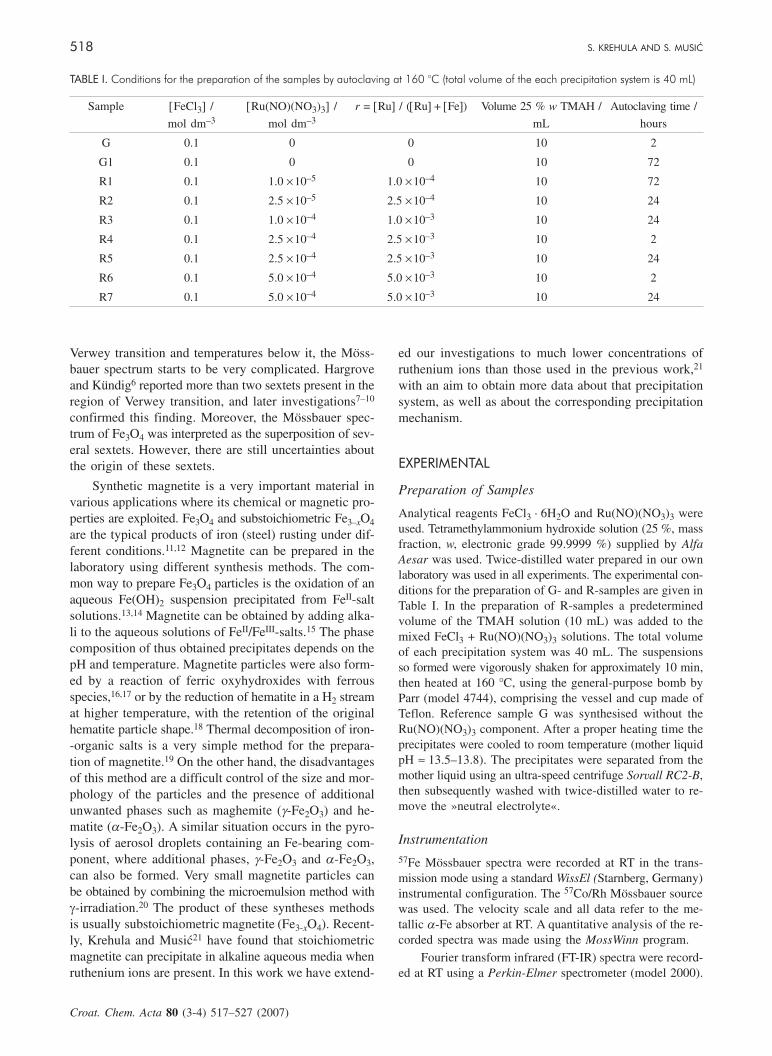
The results of XRD phase analysis are given in Table II,whereas selected XRD patterns are shown in Figures 1 and2. Crystal phases were determined from XRD patterns usingJCPDS PDF cards No. 29–713 for a-FeOOH, No. 13–534for a-Fe2O3 and No. 19–629 for Fe3O4.22
a-FeOOH was obtained as a single phase by hydrother-mal heating at 160 °C for 2 h (sample G) and 72 h (sampleG1) of the suspensions without ruthenium ions. However,we have found that the adding of small amounts of ruthe-nium in the form of Ru(NO)(NO3)3 had a drastic effect onthat precipitation process. Ru(NO)(NO3)3 was chosen as avery convenient ruthenium salt which does not undergo hy-drolysis in the solid state and dissolves into a transparentsolution. On the other hand, ageing of the ruthenium saltRuCl3 · xH2O for a prolonged time results in oxidation andthe formation of a Ru(IV)-(hydrous)oxide. At the concen-tration ratio r = 1.0 × 10⁻⁴ (sample R1), where r = [Ru] /([Ru] + [Fe]), a significant change in the precipitation sys-tem was observed upon 72 h of autoclaving. A mixture ofa-FeOOH + a-Fe2O3 was obtained. With an increase in the
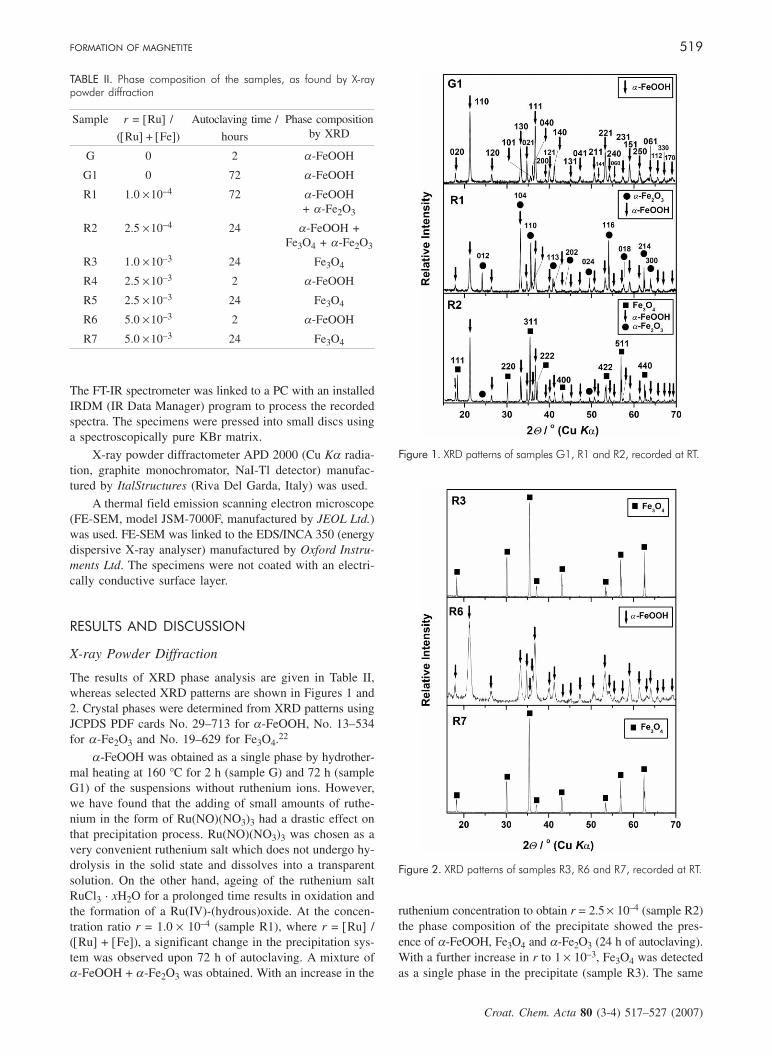
ruthenium concentration to obtain r = 2.5 × 10⁻⁴ (sample R2)the phase composition of the precipitate showed the pres-ence of a-FeOOH, Fe3O4 and a-Fe2O3 (24 h of autoclaving).With a further increase in r to 1 × 10⁻³, Fe3O4 was detectedas a single phase in the precipitate (sample R3). The same
FORMATION OF MAGNETITE 519
Croat. Chem. Acta 80 (3-4) 517¿527 (2007)
TABLE II. Phase composition of the samples, as found by X-raypowder diffraction
Sample r = [Ru] /
([Ru] + [Fe])
Autoclaving time /
hours
Phase compositionby XRD
G 0 2 a-FeOOH
G1 0 72 a-FeOOH
R1 1.0 ×10⁻⁴ 72 a-FeOOH+ a-Fe2O3
R2 2.5 ×10⁻⁴ 24 a-FeOOH +Fe3O4 + a-Fe2O3
R3 1.0 ×10⁻³ 24 Fe3O4
R4 2.5 ×10⁻³ 2 a-FeOOH
R5 2.5 ×10⁻³ 24 Fe3O4
R6 5.0 ×10⁻³ 2 a-FeOOH
R7 5.0 ×10⁻³ 24 Fe3O4
Figure 1. XRD patterns of samples G1, R1 and R2, recorded at RT.
Figure 2. XRD patterns of samples R3, R6 and R7, recorded at RT.
phase composition was found for samples formed after 24 hof autoclaving in the presence of a higher ruthenium con-centration (samples R5 and R7). Samples formed after 2 hof autoclaving (R4 and R6) showed only the lines characte-ristic of a-FeOOH. XRD analysis of the precipitates show-ed a very strong effect of the small amounts of rutheniumions, initially added as a Ru(NO)3+ complex. The presenceof ruthenium created reductive conditions in the precipita-tion system, and as a result Fe3O4 could precipitate as asingle Fe-bearing phase.
57Fe Mössbauer Spectroscopy
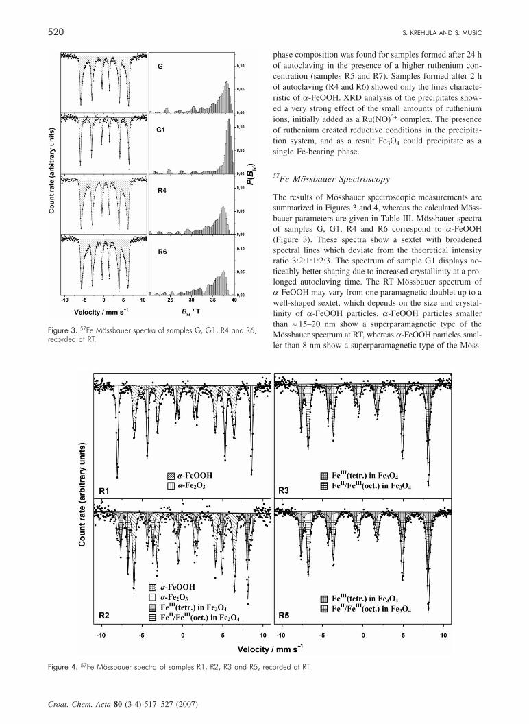
The results of Mössbauer spectroscopic measurements aresummarized in Figures 3 and 4, whereas the calculated Möss-bauer parameters are given in Table III. Mössbauer spectraof samples G, G1, R4 and R6 correspond to a-FeOOH(Figure 3). These spectra show a sextet with broadenedspectral lines which deviate from the theoretical intensityratio 3:2:1:1:2:3. The spectrum of sample G1 displays no-ticeably better shaping due to increased crystallinity at a pro-longed autoclaving time. The RT Mössbauer spectrum ofa-FeOOH may vary from one paramagnetic doublet up to awell-shaped sextet, which depends on the size and crystal-linity of a-FeOOH particles. a-FeOOH particles smallerthan ≈ 15–20 nm show a superparamagnetic type of theMössbauer spectrum at RT, whereas a-FeOOH particles smal-ler than 8 nm show a superparamagnetic type of the Möss-
520 S. KREHULA AND S. MUSI]
Croat. Chem. Acta 80 (3-4) 517¿527 (2007)
Figure 3. 57Fe Mössbauer spectra of samples G, G1, R4 and R6,recorded at RT.
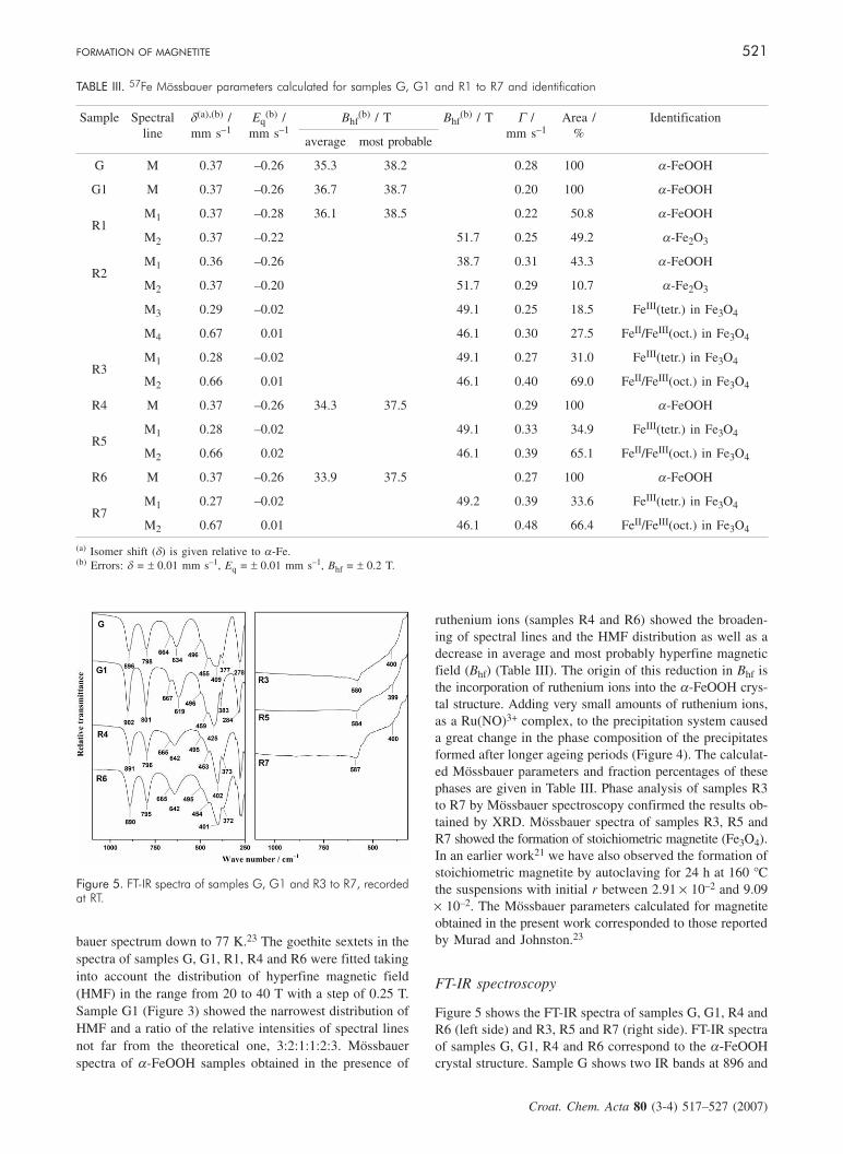
Figure 4. 57Fe Mössbauer spectra of samples R1, R2, R3 and R5, recorded at RT.
bauer spectrum down to 77 K.23 The goethite sextets in thespectra of samples G, G1, R1, R4 and R6 were fitted takinginto account the distribution of hyperfine magnetic field(HMF) in the range from 20 to 40 T with a step of 0.25 T.Sample G1 (Figure 3) showed the narrowest distribution ofHMF and a ratio of the relative intensities of spectral linesnot far from the theoretical one, 3:2:1:1:2:3. Mössbauerspectra of a-FeOOH samples obtained in the presence of
ruthenium ions (samples R4 and R6) showed the broaden-ing of spectral lines and the HMF distribution as well as adecrease in average and most probably hyperfine magneticfield (Bhf) (Table III). The origin of this reduction in Bhf isthe incorporation of ruthenium ions into the a-FeOOH crys-tal structure. Adding very small amounts of ruthenium ions,as a Ru(NO)3+ complex, to the precipitation system causeda great change in the phase composition of the precipitatesformed after longer ageing periods (Figure 4). The calculat-ed Mössbauer parameters and fraction percentages of thesephases are given in Table III. Phase analysis of samples R3to R7 by Mössbauer spectroscopy confirmed the results ob-tained by XRD. Mössbauer spectra of samples R3, R5 andR7 showed the formation of stoichiometric magnetite (Fe3O4).In an earlier work21 we have also observed the formation ofstoichiometric magnetite by autoclaving for 24 h at 160 °Cthe suspensions with initial r between 2.91 × 10⁻² and 9.09× 10⁻². The Mössbauer parameters calculated for magnetiteobtained in the present work corresponded to those reportedby Murad and Johnston.23
FT-IR spectroscopy
Figure 5 shows the FT-IR spectra of samples G, G1, R4 andR6 (left side) and R3, R5 and R7 (right side). FT-IR spectraof samples G, G1, R4 and R6 correspond to the a-FeOOHcrystal structure. Sample G shows two IR bands at 896 and
FORMATION OF MAGNETITE 521
Croat. Chem. Acta 80 (3-4) 517¿527 (2007)
TABLE III. 57Fe Mössbauer parameters calculated for samples G, G1 and R1 to R7 and identification
Sample Spectralline
d(a),(b) /mm s–1
Eq(b) /
mm s–1Bhf
(b) / T Bhf(b) / T G /
mm s–1Area /
%Identification
average most probable
G M 0.37 –0.26 35.3 38.2 0.28 100 a-FeOOH
G1 M 0.37 –0.26 36.7 38.7 0.20 100 a-FeOOH
R1M1 0.37 –0.28 36.1 38.5 0.22 50.8 a-FeOOH
M2 0.37 –0.22 51.7 0.25 49.2 a-Fe2O3
R2M1 0.36 –0.26 38.7 0.31 43.3 a-FeOOH
M2 0.37 –0.20 51.7 0.29 10.7 a-Fe2O3
M3 0.29 –0.02 49.1 0.25 18.5 FeIII(tetr.) in Fe3O4
M4 0.67 0.01 46.1 0.30 27.5 FeII/FeIII(oct.) in Fe3O4
R3M1 0.28 –0.02 49.1 0.27 31.0 FeIII(tetr.) in Fe3O4
M2 0.66 0.01 46.1 0.40 69.0 FeII/FeIII(oct.) in Fe3O4
R4 M 0.37 –0.26 34.3 37.5 0.29 100 a-FeOOH
R5M1 0.28 –0.02 49.1 0.33 34.9 FeIII(tetr.) in Fe3O4
M2 0.66 0.02 46.1 0.39 65.1 FeII/FeIII(oct.) in Fe3O4
R6 M 0.37 –0.26 33.9 37.5 0.27 100 a-FeOOH
R7M1 0.27 –0.02 49.2 0.39 33.6 FeIII(tetr.) in Fe3O4
M2 0.67 0.01 46.1 0.48 66.4 FeII/FeIII(oct.) in Fe3O4
(a) Isomer shift (d) is given relative to a-Fe.(b) Errors: d = ± 0.01 mm s–1, Eq = ± 0.01 mm s–1, Bhf = ± 0.2 T.
Figure 5. FT-IR spectra of samples G, G1 and R3 to R7, recordedat RT.
798 cm–1 which can be assigned to Fe-O-H bending vibra-tions in a-FeOOH. These bands are usually used for theidentification of a-FeOOH in a qualitative phase analysisof iron oxide mixtures. The IR band at 634 cm–1 with ashoulder at 664 cm–1, very strong and broad IR bands at409 cm–1 with a shoulder at 496 cm–1, and small intensityIR bands at 455 and 377 cm–1 were present in the spectrumof sample G. Verdonck et al.24 studied the IR spectrum ofa-FeOOH using the normal coordinate analysis, and com-pared the results with the experimental spectra of a-FeOOHand a-FeOOD. The IR bands recorded at 630, 495 and 270cm–1 were rather insensitive to deuteration, so the authorsconcluded that these IR bands were due to Fe-O stretchingvibrations in a-FeOOH. A similar interpretation of IR bandsbelow 650 cm–1 was given by Cambier,25 who also reportedthat an intense IR band around 630 cm–1 was influenced bythe shape of a-FeOOH particles. Weckler and Lutz26 alsoinvestigated the IR spectrum of a-FeOOH.
Main spectral features of samples G and G1 were thesame; in the spectra of these two samples, however, therewere shifts of IR bands. The most pronounced shifts fromsample G to sample G1 were 634 → 619 cm–1 (a lattice bandwith the transition moment parallel to the a-axis) and 409 to
425 cm–1 (a lattice band with the transition moment parallel tothe c-axis). The shoulder at 664 cm–1 for sample G changedto a band of weak intensity at 667 cm–1 for sample G1 andthe weak band at 377 cm–1 for sample G was better pro-nounced at 383 cm–1 for sample G1. The IR band at 425 cm–1
(sample G1) was also less broadened. These differences canbe assigned to a better crystallinity of a-FeOOH particlesin sample G1 than in sample G, an increase in particle thick-ness (the a-axis direction) and a decrease in their shape ani-sotropy at a longer ageing time.
a-FeOOH particles precipitated in the presence of ruthe-nium ions (samples R4 and R6) showed lattice vibration bandsat 642 and 402 cm–1 for sample R4, and 642 and 401 cm–1
for sample R6. The shifts in the positions of those bands, incomparison with the bands in the spectrum of sample G, canbe assigned to the formation of solid solutions a-(Fe,Ru)OOHand to an elongation of particles in the c-axis direction.Moreover, the positions of OH bending bands at 891 and796 cm–1 for sample R4 and 890 and 795 cm–1 for sampleR6 were slightly different from those recorded for sample G(896 and 798 cm–1). These results show that the most influ-enced IR bands in a-FeOOH doped with ruthenium ions arethose related to Fe-O stretching (lattice) vibrations, thus con-firming the incorporation of ruthenium ions in the a-FeOOHstructure. It can be concluded that a lower degree of crys-tallinity of a-FeOOH and Ru substitution in a-FeOOH had asimilar effect on the corresponding IR spectrum.
FT-IR spectra of samples R3, R5 and R7 showed an IRband centred between 580 and 587 cm–1 and a very broadshoulder (band) at ≈ 400 cm–1. These spectra are consistentwith the spectrum of magnetite quoted in literature.27
FE-SEM
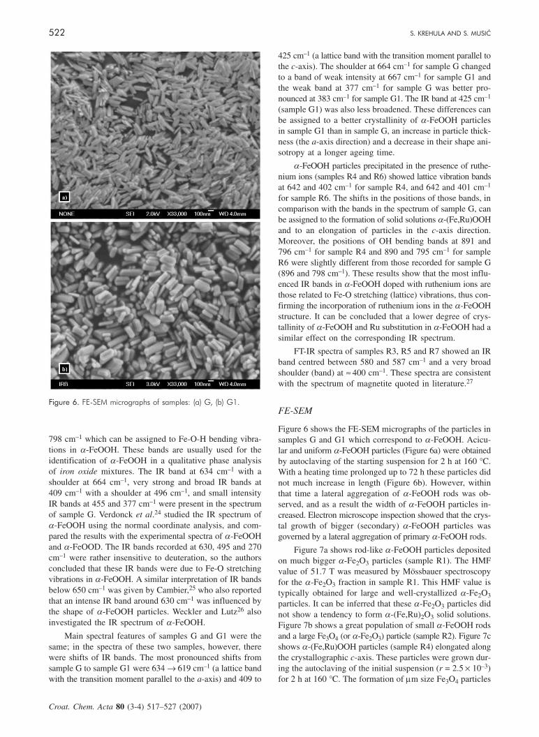
Figure 6 shows the FE-SEM micrographs of the particles insamples G and G1 which correspond to a-FeOOH. Acicu-lar and uniform a-FeOOH particles (Figure 6a) were obtainedby autoclaving of the starting suspension for 2 h at 160 °C.With a heating time prolonged up to 72 h these particles didnot much increase in length (Figure 6b). However, withinthat time a lateral aggregation of a-FeOOH rods was ob-served, and as a result the width of a-FeOOH particles in-creased. Electron microscope inspection showed that the crys-tal growth of bigger (secondary) a-FeOOH particles wasgoverned by a lateral aggregation of primary a-FeOOH rods.
Figure 7a shows rod-like a-FeOOH particles depositedon much bigger a-Fe2O3 particles (sample R1). The HMFvalue of 51.7 T was measured by Mössbauer spectroscopyfor the a-Fe2O3 fraction in sample R1. This HMF value istypically obtained for large and well-crystallized a-Fe2O3particles. It can be inferred that these a-Fe2O3 particles didnot show a tendency to form a-(Fe,Ru)2O3 solid solutions.Figure 7b shows a great population of small a-FeOOH rodsand a large Fe3O4 (or a-Fe2O3) particle (sample R2). Figure 7cshows a-(Fe,Ru)OOH particles (sample R4) elongated alongthe crystallographic c-axis. These particles were grown dur-ing the autoclaving of the initial suspension (r = 2.5 × 10⁻³)for 2 h at 160 °C. The formation of mm size Fe3O4 particles
522 S. KREHULA AND S. MUSI]
Croat. Chem. Acta 80 (3-4) 517¿527 (2007)
Figure 6. FE-SEM micrographs of samples: (a) G, (b) G1.

with characteristic octahedral shape was observed upon a pro-longed heating time (24 h) at 160 °C (Figure 7d; sample R5).
Figure 8a shows a-(Fe,Ru)OOH particles in sample R6.These particles are elongated along the crystallographic c-axis,and the decreased HMF value indicates the incorporation ofruthenium ions into the a-FeOOH crystal structure. The for-mation of mm range Fe3O4 particles was observed upon aprolonged heating time (24 h) at 160 °C (Figure 8b). Figure8c shows one plane of a Fe3O4 crystal at high optical mag-nification, covered with nanosize particles some of whichare aggregated. With an aim to obtain more data about theseparticles we analysed them by EDS. Generally, the popu-lation of these small particles increased in the precipitationsystems with an increase in the starting Ru(NO)(NO3)3concentration. Figures 9a,c show the FE-SEM micrograph ofsample R3 (r = 1.0 × 10⁻³) precipitated for 24 h at 160 °C.Two selected areas were analysed by EDS. One (Figure 9a)represents a part of one plane of the Fe3O4 crystal whichwas not covered with small particles (light spots). EDS ana-lysis of this selected area showed only iron and oxygen. EDSanalysis of the other one (Figure 9c) within a cluster ofsmall particles (light spots) showed a significant amount of
ruthenium (Figure 9d). FE-SEM of this cluster under highoptical magnification showed both ruthenium particles andsmall Fe3O4 particles. The size of these Fe3O4 particles was≈ 100 nm or less. EDS results are consistent with Mössbau-er measurements which did not prove the incorporation ofruthenium in ionic state into the Fe3O4 crystal structure.
Effect of Ruthenium Ions on the Precipitation
Mechanism
The present work has indicated a complex mechanism of theprecipitation of iron oxides in the presence of ruthenium. Areference sample of a-FeOOH was obtained using the pro-cedure described in our previous work.28 Under the preci-pitation conditions utilised in that work as well as in thisone, very high pH values ≈ 13.5–13.8 can be obtained usingTMAH as a precipitating agent. In the case of TMAH ap-plication the initially formed precipitate (ferrihydrite) can bedissolved on strong shaking, as observed by the naked eye.a-FeOOH is formed after some ageing time under homo-genous precipitation conditions. Due to that, by autoclavingof the precipitation system at 160 °C it was possible to obtain
FORMATION OF MAGNETITE 523
Croat. Chem. Acta 80 (3-4) 517¿527 (2007)
Figure 7. FE-SEM micrographs of samples: (a) R1, (b) R2, (c) R4, (d) R5.

acicular a-FeOOH particles with narrow size distribution.Thus obtained a-FeOOH particles were structurally verystable and did not transform into other iron oxides in thecorresponding suspensions.
The addition of a very small amount of ruthenium inthe form of a Ru(NO)3+ complex to the starting solution
(before adding the TMAH solution) had a drastic effect onthe precipitation systems investigated in the present work.At the beginning of the precipitation process ruthenium ionswere incorporated into ferrihydrite substituting Fe3+ ions, oras a co-gel (ferrihydrite + ruthenium (hydrous)oxide). Sub-stitution of ruthenium for Fe3+ ions was possible due tosimilarities in the ionic radii of Fe3+ (0.645 Å) and Ru4+
(0.62 Å).29 Crystallization of a-FeOOH as a single phasewas observed for shorter times (2h in the present work),whereas the incorporation of ruthenium ions into thea-FeOOH structure was proved by Mössbauer spectro-scopy. The incorporation of ruthenium ions delayed the crystal-lization of a-FeOOH, then favoured the crystallization ofa-Fe2O3. Factors such as the Fe3+ concentration in the solu-tion, the incorporation of ruthenium ions into the a-FeOOHcrystal structure, as well as different nucleation rates betweena-FeOOH and a-Fe2O3 were responsible for the formationof a-(Fe,Ru)OOH and a-Fe2O3 phases. Upon a prolongedautoclaving time Fe3O4 was formed. Under given experimen-tal conditions Fe3O4 can be obtained as a single Fe-bearingphase, as shown in the present work. Mössbauer spectro-scopy showed the formation of stoichiometric magnetite, thussuggesting reductive conditions created in the precipitationsystems (suspensions). In that part of the overall precipita-tion mechanism the role of ruthenium is dominant, whichcan be explained by the catalytic action of ruthenium. Ge-nerally, ruthenium dioxide, ruthenium black and rutheniumsupported on various carriers are well-known catalysts. Therole of ruthenium in our precipitation systems can be asso-ciated with the behaviour of TMAH, which decomposes atelevated temperatures while yielding a dominant NH3, vo-latile amines and CH3OH. In the presence of the rutheniumcatalyst, NH3 decomposes to N2 and H2, whereas CH3OHdecomposes to CO and H2. In the present experiments itshould be the formation of nascent hydrogen which easilyreduces Fe3+ to Fe2+. In such a way the conditions for theformation of Fe3O4 at high pH are created. Mössbauerspectroscopy did not provide evidence of the incorporationof ruthenium ions into the Fe3O4 crystal structure.
The effects of ruthenium catalysts were also observ-ed in some other works. For example, the formation ofFe3O4/g-Fe2O3 by a reduction of a-Fe2O3 was observedduring the investigation of the activity of the rutheniumcatalyst in a water-gas shift reaction (WGSR).30 Berry et
al.31 used in situ 57Fe Mössbauer spectroscopy to in-vestigate the effects of pre-treatment of titania-supportediron-ruthenium and iron-iridium catalysts in a hydrogenatmosphere. Musi} et al.32 used a ruthenium catalyst inthe denitration of simulated highly radioactive liquid waste(HRLW) compositions using the formic acid as a reduc-ing agent. Ruthenium was added as the Ru(NO)(NO3)3
salt or as the so-called soluble RuO2 · xH2O. The influ-ence of rhodium and palladium on denitration reactionswas also investigated. Ruthenium and rhodium were goodcatalysts for denitration reactions during which they creat-ed reductive conditions, so the reduction of Fe3+ to Fe2+
was observed. Fe2+ ions were reoxidised by a careful ad-
524 S. KREHULA AND S. MUSI]
Croat. Chem. Acta 80 (3-4) 517¿527 (2007)
Figure 8. FE-SEM micrographs of samples: (a) R6, (b) R7 at loweroptical magnification, and (c) R7 at higher optical magnificationof one plane of the Fe3O4 particle.

dition of the concentrated H2O2 solution. a-FeOOH andan amorphous fraction were the principal phases in theprecipitates upon Fe2+ oxidation by H2O2.
In the present work the concentrations of rutheniumin the precipitation systems have been very small, so thatthe ruthenium phase could not be detected with certainty.For that reason we prepared the precipitation system withan increased ruthenium concentration (r = 3.333 × 10⁻¹).XRD of the precipitate formed upon 24 h of autoclavingat 160 °C showed the presence of a-Fe2O3 and 2-lineferrihydrite with ruthenium ions incorporated. The pres-ence of metallic ruthenium was not detected by XRD.On the other hand, Fe3O4 and metallic ruthenium weredetected in the precipitate obtained upon 72 h of auto-claving at 160 °C. Furthermore, the precipitation system(r = 4.76 × 10⁻²) prepared with NaOH alkali by auto-claving for 24 h did not show the formation of Fe3O4.Finally, taking into account these results it can be con-cluded that the formation of Fe3O4 in highly alkaline pHmedia is a combining effect of the thermal decompo-sition of TMAH and the catalytic action of ruthenium.
CONCLUSION
• Acicular and uniform a-FeOOH particles wereprecipitated in line with the procedure described by Kre-
hula et al.28 TMAH was used as a precipitating agent.The phase composition of the a-FeOOH precipitate didnot change up to 72 h of autoclaving at 160 °C. The crys-tal growth of a-FeOOH particles was governed by a la-teral aggregation of these particles.
• Adding small amounts of ruthenium in the form ofa Ru(NO)(NO3)3 complex to the starting FeCl3 solutionhad a drastic effect on the above-mentioned precipitationprocess at 160 °C. A mixture of a-FeOOH and a-Fe2O3
was obtained at an initial concentration ratio r = 1.0 × 10⁻⁴
and 72 h of autoclaving. At r = 2.5 × 10⁻⁴ and 24 h ofautoclaving a mixture of a-FeOOH, Fe3O4 and a-Fe2O3
was obtained. Fe3O4 as a single Fe-bearing phase was ob-tained at r = 1.0 × 10⁻³, and higher initial concentrationsof Ru(NO)(NO3)3 after 24 h of autoclaving.
• Addition of ruthenium favoured the formation ofstoichiometric Fe3O4. The formation of solid solutions wasnot proved. Mössbauer spectroscopy did not prove the for-mation of solid solutions between ruthenium and Fe3O4 ora-Fe2O3, but did prove the formation of a-(Fe,Ru)OOHsolid solutions. The phase composition of samples deter-mined by Mössbauer spectroscopy was in line with XRDmeasurements.
• FT-IR measurements showed that the most influen-ced IR-bands in the spectra of a-FeOOH particles doped
FORMATION OF MAGNETITE 525
Croat. Chem. Acta 80 (3-4) 517¿527 (2007)
Figure 9. EDS analyses of sample R3.

with ruthenium were those related to Fe-O stretching (lat-tice) vibrations, thus confirming a conclusion about theincorporation of ruthenium ions into the a-FeOOH crys-tal structure.
• FE-SEM showed the formation of octahedral Fe3O4
particles (crystals) within the mm range size. Rutheniumparticles (≈ 20 nm in size) were deposited on the surfaceof Fe3O4 particles. They were also present in the formof clusters. These clusters contained octahedral Fe3O4
particles; their size, however, was in the nanosize range(≈ 100 nm or less).
• The formation of Fe3O4 was explained by a combi-ning effect of the products of TMAH decomposition andthe catalytic activity of ruthenium under autoclaving con-ditions. This conclusion was supported by additional ex-periments. By using NaOH instead of TMAH under thesame experimental conditions Fe3O4 was not formed. Ithas not been possible to prove with certainty the forma-tion of metallic ruthenium at small concentrations of ad-ded Ru(NO)(NO3)3 and a prolonged autoclaving time.However, at the initial concentrations of Ru(NO)(NO3)3
much higher than those reported in the present work theformation of metallic ruthenium was confirmed by XRD.
Acknowledgement. – The authors thank to the Ministry ofScience, Education and Sport of the Republic of Croatia forfinancial support (contract no. 0982904-2952).
REFERENCES
1. E. J. W. Verwey and J. H. de Boer, Recl. Trav. Chim. Pays-
Bas 55 (1936) 531–540.2. M. E. Fleet, Acta Crystallogr., Sect. B 37 (1981) 917–920.3. M. E. Fleet, J. Solid State Chem. 62 (1986) 75–82.4. S. Musi} and S. Popovi}, J. Radioanal. Nucl. Chem. 111
(1987) 27–41.5. J. García and G. Subías, J. Phys.: Condens. Matter 16 (2004)
R145–R178.6. R. S. Hargrove and W. Kündig, Solid State Commun. 8 (1970)
303–308.7. G. Galeczki and A. A. Hirsch, J. Magn. Magn. Mater. 7 (1978)
230–233.8. S. Umemura and S. Iida, J. Phys. Soc. Jpn. 47 (1979) 458–
467.9. C. M. Srivastava, S. N. Shringi, and M. V. Babu, Phys. Status
Solidi A 65 (1981) 731–735.
10. S. J. Harker and R. J Pollard, Nucl. Instrum. Methods Phys.
Res., Sect. B 76 (1993) 61–63.
11. S. Musi}, I. Nowik, M. Risti}, Z. Orehovec, and S. Popo-vi}, Croat. Chem. Acta 77 (2004) 141–151.
12. S. Musi}, \. Drag~evi}, and S. Popovi}, Croat. Chem. Acta
66 (1993) 469–478.
13. T. Ishikawa, H. Nakazaki, A. Yasukawa, K. Kandori, andM. Seto, Mater. Res. Bull. 33 (1998) 1609–1619.
14. T. Ishikawa, H. Nakazaki, A. Yasukawa, K. Kandori, andM. Seto, Corros. Sci. 41 (1999) 1665–1680.
15. Z. H. Zhou, J. Wang, X. Liu, and H. S. O. Chan, J. Mater.
Chem. 11 (2001) 1704–1709.
16. T. Ishikawa, Y. Kondo, A. Yasukawa, and K. Kandori, Cor-
ros. Sci. 40 (1998) 1239–1251.
17. M. Mohapatra, S. Anand, R. P. Das, C. Upadhyay, and H.C. Verma, Hydrometallurgy 66 (2002) 125–134.
18. H. Itoh and T. Sugimoto, J. Colloid Interface Sci. 265 (2003)283–295.
19. B. Gr`eta, M. Risti}, I. Nowik, and S. Musi}, J. Alloys Compd.
334 (2002) 304–312.
20. M. Goti}, T. Jurkin, and S. Musi}, Colloid Polym. Sci. 285(2007) 793–800.
21. S. Krehula and S. Musi}, J. Alloys Compd. 416 (2006) 284–290.
22. R. M. Cornell and U. Schwertmann, The Iron Oxides, Struc-
ture, Properties, Reactions, Occurrence and Uses, Wiley-VCH, Weinheim, Germany, 2003.
23. E. Murad and J. H. Johnston, Iron Oxides and Oxyhydro-
xides, in: G. J. Long (Ed.), Mössbauer Spectroscopy Applied
to Inorganic Chemistry, Vol. 2, Plenum Publishing Cor-poration 1987, pp. 507–582.
24. L. Verdonck, S. Hoste, F. F. Roelandt, and G. P. van derKelen, J. Mol. Struct. 79 (1982) 273–279.
25. P. Cambier, Clay Miner. 21 (1986) 191–200.
26. B. Weckler and H. D. Lutz, Eur. J. Solid State Inorg. Chem.
35 (1998) 531–544.
27. M. S. Ellid, Y. S. Murayed, M. S. Zoto, S. Musi}, and S.Popovi}, J. Radioanal. Nucl. Chem. 258 (2003) 299–305.
28. S. Krehula, S. Popovi}, and S. Musi}, Mater. Lett. 54 (2002)108–113.
29. R. D. Shannon, Acta Crystallogr., Sect. A 32 (1976) 751–767.
30. A. Basinska, L. Kepinski, and F. Domka, Appl. Catal., A
183 (1999) 143–153.
31. F. J. Berry, X. Changhai, and S. Jobson, J. Chem. Soc., Fa-
raday Trans. 86 (1990) 165–169.
32. S. Musi}, M. Risti}, and S. Popovi}, J. Radioanal. Nucl.
Chem. 134 (1989) 353–365.
526 S. KREHULA AND S. MUSI]
Croat. Chem. Acta 80 (3-4) 517¿527 (2007)
SA@ETAK
Nastajanje magnetita u jako lu`natom mediju u prisutnosti malih koli~ina rutenija
Stjepko Krehula i Svetozar Musi}
Istra`ivan je utjecaj malih koli~ina rutenija na nastajanje magnetita u jako lu`natom mediju primjenomrentgenske difrakcije u prahu, Mössbauerove spektroskopije, FT-IR spektroskopije, visokorezolucijske pretra`neelektronske mikroskopije (FE-SEM) i spektroskopije karakteristi~nog rentgenskog zra~enja (EDS). Izdu`ene~estice a-FeOOH, pripravljene u jako lu`natom mediju dodatkom tetrametilamonijeva hidroksida (TMAH), kori-{tene su kao referentni materijal. Prisutnost malih koli~ina Ru(NO)(NO3)3 u talo`nom sustavu sna`no je utjecalana nastajanje `eljezovih oksida i njihova svojstva. Dodatak Ru(NO)(NO3)3 uzrokovao je nastajanje stehiometrij-skog Fe3O4. S pove}anjem po~etne koncentracije Ru(NO)(NO3)3 u talo`nom je sustavu smanjeno vrijeme po-trebno za nastajanje Fe3O4 kao jedine faze u talogu koja sadr`i `eljezo. Rutenijevi su ioni ugra|eni u ~vrste oto-pine a-(Fe,Ru)OOH, me|utim, nije bilo naznaka o nastajanju ~vrstih otopina s a-Fe2O3 i Fe3O4. Mössbauerova iFT-IR spektroskopija podr`ale su tvrdnju o nastajanju ~vrstih otopina a-(Fe,Ru)OOH. Tehnika FE-SEM pokazalaje nastajanje oktaedarskih ~estica Fe3O4 veli~ine nekoliko mm. Na povr{ini ~estica Fe3O4 deponirane su ~esticerutenija (veli~ine ≈ 20 nm). Ove ~estice su tako|er prisutne u obliku nakupina koje sadr`e i oktaedarske ~esticeFe3O4 veli~ine ≈ 100 nm ili manje. Nastajanje Fe3O4 obja{njeno je kombiniranim efektom produkata termi~kerazgradnje TMAH pri hidrotermi~kim uvjetima i kataliti~kog djelovanja rutenija, ~ime se stvaraju jaki redukcij-ski uvjeti u istra`ivanom talo`nom sustavu.
FORMATION OF MAGNETITE 527
Croat. Chem. Acta 80 (3-4) 517¿527 (2007)
Copyright © 2022 FDOKUMEN