Novel conopeptides of the I-superfamily occur in several clades of cone snails
Upload
independentCategory
view
1download
0
Divergence of Chemical Function in the Alkaline PhosphataseSuperfamily: Structure and Mechanism of the P-C Bond CleavingEnzyme Phosphonoacetate Hydrolase+
Alexander Kim1, Matthew M. Benning2, Sang OkLee1, John Quinn3, Brian M. Martin4, HazelM. Holden2,*, and Debra Dunaway-Mariano1,*
1 Department of Chemistry and Chemical Biology, University of New Mexico, Albuquerque, NewMexico 87131 2 Department of Biochemistry, University of Wisconsin, 433 Babcock Dr., Madison,Wisconsin 53706-1544 3 School of Biology and Biochemistry and Questor Centre, The Queen’sUniversity of Belfast, Medical Biology Centre, 97, Lisburn Road, BT9 7BL Belfast, NorthernIreland 4 Molecular Structure Unit, Laboratory of Neurotoxicology, NIMH Building 10, Room3N309, 10 Center Drive, MSC 1262 Bethesda, MD 20892-1262
AbstractPhosphonates constitute a class of natural products that mimic the properties of the more commonorganophosphate ester metabolite, yet are not readily degraded owing to the direct linkage of thephosphorus atom to the carbon atom. Phosphonate hydrolases have evolved to allow bacteria toutilize environmental phosphonates as a source of carbon and phosphorus. The work reported inthis paper examines one such enzyme, phosphonoacetate hydrolase. By using a bioinformaticapproach we circumscribed the biological range of phosphonoacetate hydrolase to a select groupof bacterial species from different classes of Proteobacteria. In addition, using gene context weidentified a novel 2-aminoethylphosphonate degradation pathway in which phosphonoacetatehydrolase is a participant. The X-ray structure of phosphonoformate-bound phosphonoacetatehydrolase was determined to reveal that this enzyme is most closely related to nucleotidepyrophosphatase/diesterase, a promiscuous two-zinc ion metalloenzyme of the alkalinephosphatase enzyme superfamily. The X-ray structure and metal ion specificity tests showed thatphosphonoacetate hydrolase is also a two-zinc ion metalloenzyme. By using site-directedmutagenesis and 32P-labeling strategies, the catalytic nucleophile was shown to be Thr64. Astructure-guided, site-directed mutation based inquiry of the catalytic contributions of active siteresidues identified Lys126 and Lys128 as the most likely candidates for stabilization of the aci-carboxylate dianion leaving group. A catalytic mechanism is proposed which combines Lys12/Lys128 leaving group stabilization with zinc ion activation of the Thr64 nucleophile and thesubstrate phosphoryl group.
+This work was supported by N.I.H. grant GM 28688 (D. D-M), NIH grant DK47814 (HMH) and BBSRC (UK) grant 81/P11488(J.P.Q.) and intramural funding from the IRP, NIMH (B.M.M.).*To whom correspondence should be addressed regarding X-ray structure determination (H.M.H.) or kinetic and bioinformaticanalyses (D.D.-M.). Debra Dunaway-Mariano, Department of Chemistry and Chemical Biology, University of New Mexico,Albuquerque, NM 87111, Tel: (505) 277–3383, [email protected], Fax: (505) 277–6202. Hazel Holden, Department of Biochemistry,433 Babcock Dr., University of Wisconsin, Madison, Wisconsin, 53706–1544, Tel: (608) 262-4988,[email protected]., Fax: (608) 262-1319.Supporting Information AvailableThe supporting information contains Figure SI1, the multiple alignment of phosphonoacetate hydrolase amino acid sequences. Thismaterial is available free of charge via the Internet at http://pubs.acs.org.
NIH Public AccessAuthor ManuscriptBiochemistry. Author manuscript; available in PMC 2012 May 3.
Published in final edited form as:Biochemistry. 2011 May 3; 50(17): 3481–3494. doi:10.1021/bi200165h.
NIH
-PA Author Manuscript
NIH
-PA Author Manuscript
NIH
-PA Author Manuscript
KeywordsPhosphonoacetate hydrolase; enzyme mechanism; alkaline phosphatase; phosphonate; divergentevolution; enzyme superfamily; phosphoryl transfer; nucleotide pyrophosphatase/diesterase; 2-aminoethylphosphonate; phosphonoacetaldehyde; phosphonoformate; nucleophilic catalysis; zincion
Environmental phosphonates are both synthetic and biogenic in origin (for recent reviewssee references 1–4). Phosphonates differ from the more predominant organophosphates inthat the phosphorus atom is bonded directly to a carbon atom. The unique properties ofphosphonates derive from their C-P bond that, unlike the P-O-C linkage, is stable to acidhydrolysis, base hydrolysis, and to the action of enzymes that catalyze phosphoryl transferfrom phosphate esters and anhydrides (5,6). Phosphonates are used commercially asdetergent additives, agrochemicals, lubricant additives, antioxidants, adhesives, antiviralagents, antibiotics, and insecticides, and thus, they widely distributed in the environment (7–10).
Biogenic phosphonates are produced by specialized microbes and invertebrates for nicheadaptation, signaling, phosphorus storage or chemical warfare (7). 2-Aminoethylphosphonate (AEP)1, which is typically found conjugated to lipids or glycans, isthe most ubiquitous of the phosphonate natural products (11). The P-C bond in AEP, as wellas in many other biosynthetic phosphonates, is formed through the rearrangement ofphosphoenol pyruvate (PEP) to phosphonopyruvate (PPyr) (Scheme 1) catalyzed by theenzyme PEP phosphomutase (12). This thermodynamically unfavorable reaction is drivenforward by a coupling reaction. In the case of AEP biosynthesis this reaction is thedecarboxylation of PPyr to form phosphoacetaldehyde (PAld), catalyzed by the enzymephosphonopyruvate decarboxylase (13). The AEP is ultimately formed by AEPtransaminase-catalyzed ammonia group transfer from L-glutamate to PAld (14).
The nitrogen, carbon and phosphorus atoms of AEP are recycled in the environment byspecialized bacteria, which employ the two-step chemical pathway shown in Scheme 1.PAld, the pathway intermediate, is formed from AEP by AEP transaminase-catalyzedammonium group transfer to pyruvate (15). PAld undergoes hydrolytic cleavage atphosphorus thus liberating inorganic phosphate and acetaldehyde. The enzyme catalyst forthis reaction, phosphonatase, combines covalent electrophilic catalysis (viz. Schiff baseformation with the PAld carbonyl group), and nucleophilic catalysis (viz. formation of anaspartylphosphate intermediate) to mediate the transfer of the phosphoryl group to a watermolecule (16–18) (Scheme 2). Phosphonatase is a member of the haloalkonic aciddehalogenase (HAD) enzyme superfamily, which is comprised primarily ofphosphohydrolases that catalyze the hydrolysis of phosphate monoesters and anhydrides viaan aspartylphosphate intermediate (19). By virtue of its ability to catalyze the hydrolyticcleavage of the PAld P-C bond, phosphonatase adds a novel chemical function to the HADsuperfamily.
The work that we report in this paper examines the chemical specialization of the P-C bondcleaving enzyme phosphonoacetate hydrolase (PAc hydrolase) (20–21), which is a member
1Abbreviations used are: PAc, phosphonoacetate; PAc hydrolase, phosphonoacetate hydrolase; ATP, adenosine 5′-triphosphate; ADP,adenosine 5′-diphosphate; AMP, adenosine 5′-monophosphate; PEP, phosphoenol pyruvate; NADH, dihydronicotinamide adeninedinucleotide; HEPES, N-[2-hydroxyethyl]piperazine-N′-[2 ethanesulfonic acid]; ADA, N-(2-acetamido) iminodiacetic acid; BICINE,N,N-Bis(2-hydroxyethyl)glycine; MOPSO, 3-(N-morpholino)-2-hydroxypropanesulfonic acid; PPyr, phosphonopyruvate; AP,alkaline phosphatase; NPP, nucleotide pyrophosphatase/diesterase; PAld, phosphonoacetaldehyde; AEP, 2-aminoethylphosphonate;HAD, haloalkonic acid dehalogenase; HPLC, high performance liquid chromatography.
Kim et al. Page 2
Biochemistry. Author manuscript; available in PMC 2012 May 3.
NIH
-PA Author Manuscript
NIH
-PA Author Manuscript
NIH
-PA Author Manuscript
of the alkaline phosphatase enzyme superfamily (22). PAc hydrolase was first discovered ina bacterial isolate (Pseudomonas fluorescens 23F) from sludge produced by a laundry wastetreatment plant located near Dunmurry, Northern Ireland (23–24). At the time it seemed thatPAc hydrolase might have evolved to convert commercial PAc, introduced to theenvironment through waste disposal, to usable forms of carbon (acetate/glyoxylate cycle)and phosphorus (orthophosphate). However, later studies revealed the presence of the PAchydrolase gene in bacterial isolates from remote and environmentally diverse geographicalsites, which suggested that PAc might be synthesized in specialized bacteria (25–26).Functional analysis of the genes neighboring the P. fluorescens 23F PAc hydrolase gene didnot, however, identify protein products that might produce PAc (27). Nonetheless, with therecent explosion of sequenced bacterial genomes we succeeded in locating orthologs of theP. fluorescens 23F PAc gene (by bioinformatic analysis) to gene clusters (in several speciesof Proteobacteria), which appear to support a novel 3-step chemical pathway for AEPdegradation (Scheme 1). This pathway shares with the phosphonatase-centered pathway thetransamination of AEP to form PAld (28), but deviates with the oxidation of the PAld toform phosphonoacetate (PAc) (Scheme 1). The hydrolytic cleavage of the P-C bond in PAcforms acetate and orthophosphate as the pathway end products rather than the acetaldehydeand orthophosphate produced by the hydrolytic cleavage of PAld.
In the text below, we examine the biological range and biochemical function of PAchydrolase. Moreover, we report the results from an in depth structure-function analysis,which show that PAc hydrolase combines the catalytic strategies of the phosphohydrolasesof the alkaline phosphatase superfamily, with a unique mechanism of substrate leavinggroup stabilization, to create a novel chemical function that supports a newly discoveredAEP degradation pathway.
Materials and MethodsMaterials
All biological buffers, enzymes, and chemicals used in the enzyme assays were purchasedfrom Sigma. The [γ-32P]ATP was purchased from Amersham. All substrates and inhibitorswere purchased from Sigma with the exception on phosphonopyruvate andphosphonoacetaldehyde, which were synthesized according to published procedure (videinfra).
Preparation of wild-type and mutant PAc hydrolaseThe P. fluorescens 23F PAc hydrolase gene (29) was subcloned into pKK223-3 (Pharmacia)using a PCR based approach. Transformed Escherichia coli JM105 cells were grown in LBmedia at 37 °C for 16 h (OD600 = 1). Following the addition of IPTG (1 mM) the cells wereincubated until the culture OD600 reached 1.8 (< 6 h) and then they were harvested bycentrifugation (18000 g) (yield of 5 g/L). Forty grams of wet cells were suspended in 400mL of ice-cold Buffer A (30 mM K+BICINE and 1 mM ZnCl2; pH 8.5) and then passedthrough a French press at 1000 psi. The lysate was clarified by centrifugation (18000 g for30 min) at 4 °C prior to adding ammonium sulfate to 25% saturation. The resultingprecipitate was removed by centrifugation before adding additional ammonium sulfate to55%. The precipitated protein was harvested and then dissolved in 200 mL of Buffer A anddialyzed overnight at 4 °C against 2 L of Buffer A. The protein solution waschromatographed at 4 °C on a 5 × 45 cm DEAE-cellulose column pre-equilibrated withBuffer A. The column was eluted with 100 mL of Buffer A followed by a 2 L linear gradientof KCl (0 M - 0.5 M) in Buffer A. The PAc hydrolase containing fractions (eluting at 0.25KCl) were identified using a spectrophotometric activity assay (vide infra) and/or SDS-PAGE analysis. The desired fractions were pooled, combined with ammonium sulfate (20%
Kim et al. Page 3
Biochemistry. Author manuscript; available in PMC 2012 May 3.
NIH
-PA Author Manuscript
NIH
-PA Author Manuscript
NIH
-PA Author Manuscript
saturation) and then loaded onto a 2.5 × 30 cm Phenyl Sepharose column pre-equilibratedwith Buffer A/ammonium sulfate (20% saturation). The column was eluted with a 2 L lineargradient 10 - 0% ammonium sulfate in Buffer A. The PAc hydrolase containing fractions(eluted at ca. 0% ammonium sulfate) were pooled and then concentrated using an Amiconultrafiltration apparatus to a concentration of ~20 mg/mL. The concentrated enzyme wasdialyzed against Buffer A overnight at 4 °C and then loaded onto a 2 cm × 20 cmHydroxylapatite column pre-equilibrated with Buffer A. The column was washed with 10mL of Buffer A, followed by 400 mL linear gradient 0 – 0.1 M phosphate in Buffer A. Thedesired fractions (eluted at ca. 0.02 M phosphate) were pooled and concentrated with anAmicon until the enzyme concentration reached ~30 mg/mL. The concentrated enzymesolution (1 mL) was then chromatographed at 4 °C on a 1.5 × 180 cm Sephracryl S-200column using Buffer A as eluant. The desired fractions were combined, concentrated to ~20mg/mL and passed through a 0.2 μm syringe filter before storing at 4 °C. The site-directedmutant genes were prepared from the wild-type gene by PCR using commercial primers andpfu polymerase (Stratagene). The purified DNA was ligated to the linearized pKK223-3plasmid using T4 DNA ligase. The ligation product was used to transform competent E. coliJM105 cells. A positive clone isolated from the LB agar plate (100 μg/ml ampicillin) wasverified by DNA sequencing. The mutant enzymes were prepared as described for wild-typePAc hydrolase.
PAc hydrolase MW determinationThe N-terminal sequence was determined by automated peptide sequencing asTQLISVNSRSYRLS thus showing that the N-terminal Met was removed byposttranslational modification. The theoretical subunit mass of the recombinant hydrolasewas calculated using the EXPASY Compute pI/MW program. The size of native hydrolasewas determined by Sephacryl S-200 column chromatography using protein molecularweight standards to create a plot of log MW vs. elution volume.
Spectrophotometric activity assaysFor all assays, control reactions in which the PAc hydrolase was omitted were carried out.PAc hydrolysis. These reactions were monitored at 25 or 30 °C by measuring the oxidationof β-NADH at 340 nm (ε = 6.22 mM−1cm−1) in 1 mL assay solutions initially containingPAc hydrolase, PAc, 10 u acetate kinase, 10 u pyruvate kinase, 10 u lactate dehydrogenase,5 mM ZnCl2, 10 mM MgCl2, 1 mM PEP, 1 mM ATP, and 0.5 mM β-NADH in 50 mMK+ADA pH 7.0. PAld hydrolysis. This reaction was monitored at 25 °C by measuring theoxidation of β-NADH at 340 nm (ε = 6.22 mM−1cm−1) in a 1 mL assay solutions initiallycontaining PAc hydrolase, PAld (30), 10 u alcohol dehydrogenase, 0.5 mM β-NADH, 5 mMZnCl2, and 5 mM MgCl2 in 50 mM K+ADA (pH 7.0). Bis-p-nitrophenylphosphate, p-nitrophenylphosphate and p-nitrophenylsulfate hydrolysis. These reactions were monitoredat 25 °C by measuring the formation of p-nitrophenolate at 410 nm (ε = 16200 M−1cm−1) in1 mL assay solutions containing reactant and 1 mM ZnCl2 in 100 mM K+TRICINE (pH8.0). ATP hydrolysis. This reaction was monitored at 25 °C by measuring the oxidation ofNADH at 340 nm (ε = 6.22 mM−1cm−1) in 1 mL assay solutions initially containing PAchydrolase, 1 mM ATP, 5 mM ZnCl2, 5 mM MgCl2,1 mM PEP, 10 u pyruvate kinase, 10 ulactate dehydrogenase, and 0.5 mM NADH in 50 mM K+ADA (pH 7.0). 2-Phosphoglycerate, 3-phosphoglycerol, and fosfomycin hydrolysis. The reaction solutionsinitially contained 40 μM PAc hydrolase, 2 mM reactant and 5 mM ZnCl2 in 50 mMK+ADA (25 °C, pH 7.0). The concentration of orthophosphate was measured by adding analiquot of the solution to the Fiske-Subbarow assay solution (5 M H2SO4, 2.5 % V/W(NH4)2 Molybdate and 0.25 % V/W Fiske-Subbarow Reducer (Sigma)), then measuring thesolution OD600 following 30 min of incubation. The orthophosphate concentration wascalculated from a standard curve prepared with commercial KH2PO4.
Kim et al. Page 4
Biochemistry. Author manuscript; available in PMC 2012 May 3.
NIH
-PA Author Manuscript
NIH
-PA Author Manuscript
NIH
-PA Author Manuscript
Determination of PAc hydrolase metal ion activationPAc hydrolase was exhaustively dialyzed at 4 °C against 30 mM K+BICINE (pH 8.0)prepared using deionized H2O (VWR Scientific Products). Reaction solutions initiallycontaining 1 mM PAc, dialyzed PAc hydrolase and 3 mM divalent metal ion in 30 mMK+BICINE (pH 8.0) were incubated at 25 °C for a specified period and then assayed foracetate (using 10 u/ml acetate kinase, 10 u/ml pyruvate kinase, 10 u/ml lactatedehydrogenase, 5 mM ZnCl2, 10 mM MgCl2, 1 mM PEP, 1 mM ATP, and 0.5 mM β-NADH).
Steady-State Kinetic Constant DeterminationsThe Km and kcat values were determined from initial velocity data measured as a function ofsubstrate concentration (0.5 – 10Km). The initial velocity data were fitted to equation 1. Thekcat value was calculated by dividing the Vm value by the concentration of PAc hydrolasedetermined using the Bradford assay kit (Sigma).
Eq. 1
where v is initial velocity, Vm is the maximum velocity, [S] is the substrate concentration,and Km is the Michaelis constant.
The inhibition constant (Ki) values were determined from the initial velocity data measuredas a function of substrate concentration (0.5 – 10Km) and inhibitor concentration ([I] at 0,1Ki, 2Ki). Data were fitted to equation 2 for competitive inhibition.
Eq. 2
pH Rate Profile AnalysisThe Vm and Vm/Km values were determined from initial velocity data measured as afunction of substrate concentration (0.5 – 10Km) at solution pH values that ranged from 6.0to 9.0. The reaction solutions were buffered using 50 mM ADA or 50 mM BICINE. TheVm/Km values were fitted to equation 3 to define the apparent ionization constants K1 andK2 and the Vm values were fitted to equation 4.
Eq. 3
Eq. 4
Where Y is the Vm/Km or Vm value, c is the pH-independent value of Y, [H] is the hydrogenion concentration.
32P-Labeling of phosphonoacetate hydrolase with [γ32P]ATPThe purified PAc hydrolase was labeled using [γ-32P]ATP. The 400 μL reaction mixtureinitially contained 24 μM PAc hydrolase, 1 mM ZnCl2, 50 mM K+MES (pH 6.0) and 110
Kim et al. Page 5
Biochemistry. Author manuscript; available in PMC 2012 May 3.
NIH
-PA Author Manuscript
NIH
-PA Author Manuscript
NIH
-PA Author Manuscript
μM [γ32P]ATP. A 25 μL aliquot was removed at 5 s, 30 s, 1 min, 10 min, and 20 min andmixed with 25 μL of 1 M perchloric acid and stored on ice. For the control reaction, theperchloric acid was added to the enzyme followed by the addition of the [γ32P]ATP. Thequenched samples were centrifuged at 10000 g for 10 min at 4 °C using a microcentrifuge.Each sample was washed with 50 μL 0.5 M K+MES (pH 6.0) twice without disturbing theprotein pellet. The protein pellet was dissolved in 25 μL SDS loading buffer andchromatographed on a SDS-PAGE gel. The gel was washed four times with a 250 mLwashing solution (1: 2 glacial acetic acid/ethanol), then placed on the filter paper andcovered with a gel drying cellulose membrane. The dried gel was placed on the X-ray film(Kodak) with intensifier (Dupont) and stored at −80 °C for 14 h. The film was developedand the SDS-PAGE gel was then stained with Coomassie blue. The labeling reaction wasalso carried out with the T64A PAc hydrolase mutant and with the wild-type enzymeinhibited with 2 mM phosphonoformate.
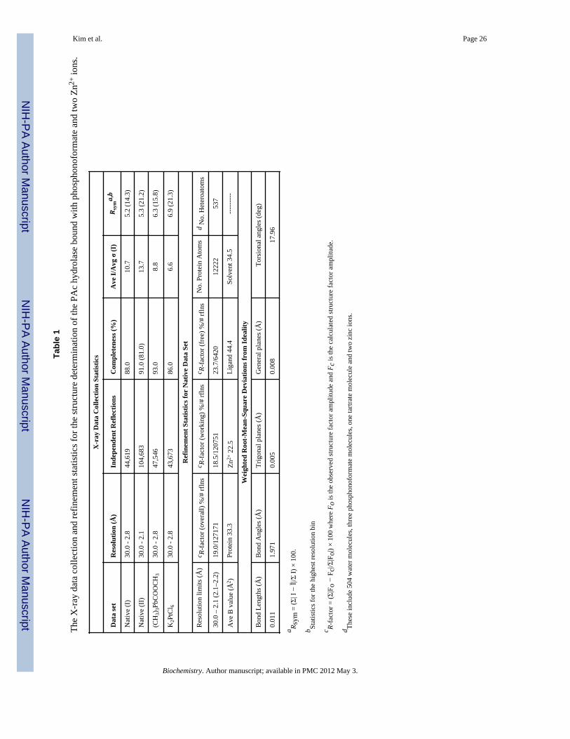
PAc hydrolase crystallization and structure determinationCrystals of phosphonoacetate hydrolase in Buffer A were grown from a solution containing10% poly(ethylene glycol) 8000, 100 mM Mg-acetate, and 5 mM phosphonoformatebuffered at pH 7.0 with 100 mM MOPSO. Batch setups with macroseeding producedcrystals in approximately two weeks with typical dimensions of 0.5 × 0.4 × 0.2 mm. Thecrystals belonged to the space group P21 with unit cell dimensions of a = 57.5 Å, b = 129.5Å, and c = 133.4 Å, β = 96.9°. There were four molecules in the asymmetric unitcorresponding to a crystalline solvent content of 56%. X-ray data from a single nativecrystal were collected to 2.8 Å resolution at 4 °C with a Bruker HiStar area detector system.The X-ray source was nickel-filtered CuKα radiation from a Rigaku RU200 X-ray generatoroperated at 50 kV and 90 mA and equipped with a 300 μm focal cup. The x-ray data wereprocessed with the software package SAINT (Bruker AXS, Inc.) and internally scaled withXCALIBRE (31). The structure of phosphonoacetate hydrolase was solved by multipleisomorphous replacement with two heavy-atom derivatives: trimethyllead acetate andpotassium hexachloroplatinate (II). The heavy atom derivative data sets, including Friedelpairs, were collected and processed to 2.8 Å resolution in a similar manner as described forthe native X-ray data. The locations of the heavy atom binding positions were determined byinspection of difference Patterson maps calculated to 5.0 Å resolution and placed on acommon origin by appropriate difference Fourier maps. Initial phases were subsequentlycalculated with the program SOLVE (32) and improved by solvent flattening with DM (33).The relationships between the four subunits in the asymmetric unit were determined with theprogram MUNCHKINS (34). Four-fold averaging with the program DM was subsequentlyutilized and produced a readily interpretable electron density map. After the model forphosphonoacetate hydrolase was constructed, crystals belonging to the monoclinic spacegroup P21 were grown from 8% poly(ethylene) glycol 8000, 100 mM MgCl2, 250 mMtartrate, and 7 mM phosphonoformate buffered at pH 7.0 with 100 mM MOPSO. Thesecrystals displayed unit cell dimensions of a = 57.8 Å, b = 129.3 Å, and c = 133.5 Å anddiffracted to a nominal resolution of 2.1 Å. Again, a native X-ray data set was collected in asimilar manner to that previously described for the orthorhombic crystal form. The structurewas solved via molecular replacement with the software package, AMORE (35). The modelwas improved with alternating cycles of least squares refinement with the program TNT (36)and manual inspection with the modeling package, Turbo (37). Relevant X-ray datacollection and least squares refinement statistics are presented in Table 1.
Results and DiscussionPAc hydrolase is a member of the alkaline phosphatase enzyme superfamily (22). Thisfunctionally diverse superfamily is comprised of phosphohydrolases, phosphotransferases,
Kim et al. Page 6
Biochemistry. Author manuscript; available in PMC 2012 May 3.
NIH
-PA Author Manuscript
NIH
-PA Author Manuscript
NIH
-PA Author Manuscript
phosphomutases, and sulfatases (38–43). The family members share an α/β/α core domainand the use of metal ion catalysis and nucleophilic catalysis. PAc hydrolase is most closelyrelated to family members nucleotide pyrophosphatase/phosphodiesterase (NPP) andalkaline phosphatase (AP). It is with these two members that PAc hydrolase will becompared in the text that follows.
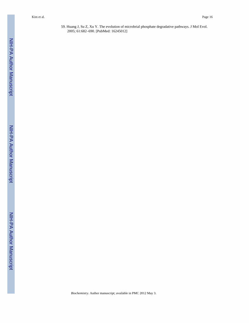
X-ray Structure of the PAc Hydrolase-Phosphonoformate ComplexQuaternary and Tertiary Structure—Recombinant P. fluorescens 23F PAc hydrolasewas crystallized at neutral pH in the presence of Zn2+, Mg2+, phosphonoformate and tartrate.The crystals belonged to the space group P21 with unit cell dimensions of a = 57.5 Å, b =129.5 Å, and c = 133.4 Å, β = 96.9°. The structure was solved, by multiple isomorphousreplacement, at 2.1 Å resolution. The X-ray data collection and refinement statistics arelisted in Table 1. Four subunits are observed in the asymmetric unit (Figure 1A). Two zincions are bound to each subunit. Three of the subunits are each bound with aphosphonoformate ligand and the fourth with a tartrate ligand. The theoretical monomermass (−Met) is 44,239 Da and the mass of the native enzyme determined by gel filtrationchromatography is ~93 kDa. Therefore, the predominant form of PAc hydrolase in solutionis the homodimer. Inspection of the subunit-subunit contacts in the crystal structure suggeststhat residues Arg253, Asp249, Ile251, Thr248, Leu346, Pro346, Arg345, Ala209, Pro210,Glu28, Lys375, Glu30, Gln34, Asn33, Gln37, Ile36, Val53 and Thr48 form the dimerizationsurfaces (Figure 1B).
The PAc hydrolase monomer is comprised of a large α/β/α core domain (residues 1–245 and367–388; 6-stranded mixed β-sheet) and a smaller α/β cap domain (residues 255–360; 4-stranded antiparallel β-sheet) (Figure 2A). The phosphonoformate ligand and two zinc ionsare bound at the core domain (henceforth referred to as the catalytic domain) and arepartially shielded from solvent by the cap domain. The locations of the respective activesites in the two superfamily members E. coli AP (PBD code 1ALK) (44) (Figure 2B) andXanthomonas axonopodis NPP (PDB code 2GSU) (45) (Figure 2C) are conserved.However, an interesting divergence in structure is observed for AP, which unlike the PAchydrolase and NPP does not possess a cap domain to cover the active site of the catalyticdomain.
Active Site—The PAc hydrolase active site contains two zinc ions, one of which (“Zn1”)is coordinated to His206 (2.2 Å), His368 (2.2 Å), Asp202 (1.9 Å) and to one oxygen atom(1.8 Å) of the phosphonoformate ligand phosphoryl group (Figure 2A). The second zinc ion(“Zn2”) is coordinated to His242 (1.8 Å), Asp25 (1.8 Å), Asp241 (2.0 Å) and to Thr64 (1.6Å). X. axonopodis NPP (45) and E. coli AP (44) coordinate their zinc ions using the sameconstellation of His, Asp and Thr (Ser for AP) ligands (Figures 2B and 2C). The two zincions of AP (4.0 Å apart) are located closer together than are the two zinc ions of NPP (4.3 Åapart) and PAc hydrolase (4.6 Å apart). Consequently, the phosphate molecule bound to APcoordinates to both zinc ions whereas the phosphoryl group of the AMP molecule bound toNPP coordinates to only one zinc ion (Zn1), although the coordination to both zinc ions inthe transition state has been proposed (45).
The substrate PAc was manually modeled in place of the phosphonoformate inhibitor in thePAc hydrolase active site. When the two phosphoryl groups are superimposed and theacetate group is overlayed with the phosphonoformate carboxylate group, the potential forsteric clash with neighboring residues is evident. In the crystal structure, thephosphonoformate carboxylate group is positioned (at 2.7 Å) to engage in favorableelectrostatic interaction with one of the two zinc ions (Zn2). We suspect that this interactionmight direct the observed binding orientation of the carboxylate group. The model shown in
Kim et al. Page 7
Biochemistry. Author manuscript; available in PMC 2012 May 3.
NIH
-PA Author Manuscript
NIH
-PA Author Manuscript
NIH
-PA Author Manuscript
Figure 3 retains the position of the phosphoryl group but directs the acetate group towardssolvent. The orientation is consistent with the direction of the adenosine unit of the NPP-bound AMP ligand (45) and with the acetate group of the PAc ligand bound to AP (PDBcode 1EW8) (46). The validity of this model was examined by carrying out the site-directedmutagenesis studies described in a later section. First however, we report the kineticproperties of the wild-type enzyme (vide infra).
PAc Hydrolase pH and Metal Ion DependencepH Dependence—The Vm and Vm/Km pH rate profiles for PAc hydrolase-catalyzed PAchydrolysis were measured in order to determine the optimal pH range for catalysis (Figure4). The bell-shaped Vm/Km pH rate profile defines a narrow optimal pH range centered atpH 7. Activity assays were therefore carried out at pH 7 and only when required by thecoupling assay employed, was a higher pH (pH 8) used. The pH rate profiles were fitted toequation 3 or 4 to define apparent pKa values (see figure legend) for the ionization ofessential groups. Owing to what we view to be the complexities associated with theinterpretation of pH rate profiles, we have not attempted to assign the apparent pKa values toPAc hydrolase catalytic groups.
Metal Ion Dependence—PAc hydrolase that had been exhaustively dialyzed againstmetal-free buffer was used to test the effect of a variety of divalent metal ions (Zn2+, Mg2+,Ca2+, Ni2+, Mn2+, Cr2+, Cu2+, Co2+) on the initial velocity of catalyzed hydrolysis of 1 mMPAc. At a concentration of 3 mM, Zn2+ and Co2+ produced a respective 10-fold and 2-foldincrease in the initial velocity measured in the absence of added metal ion. The other metalions tested did not enhance the initial velocity. An equal mixture of Zn2+ and Mg2+ (1.5 mMeach) was no more effective than Zn2+ alone.2 PAc hydrolase kinetic experiments weretherefore carried out using Zn2+ as the metal ion activator..
PAc Hydrolase Substrate SpecificityThe substrate specificity constant (kcat/Km = 4 × 10⁴ M−1 s−1 at pH 7 and 30 °C; Table 2)for PAc hydrolase-catalyzed PAc hydrolysis falls within the range of substrate specificityconstants measured for other enzymes, which (like PAc hydrolase) function in secondarymetabolism (viz. 1 × 10³ to 1 × 10⁵ M−1 s−1).
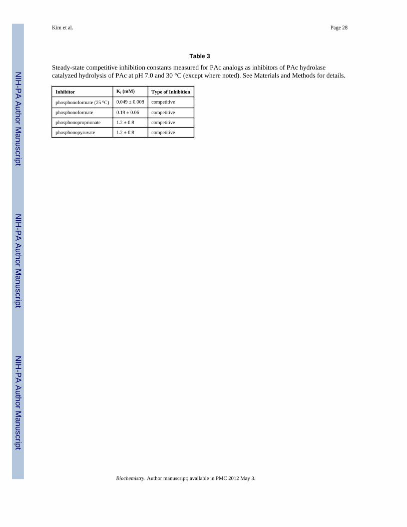
The structural determinants of PAc hydrolase substrate recognition were examined usingstructural analogs of PAc (structures are shown in Chart 1). The kcat and kcat/Km valuesmeasured for catalyzed PAld hydrolysis are 10-fold and 100-fold smaller, respectively thanthe corresponding values measured for PAc hydrolysis (Table 2). Thus, the charged oxygenatom of the PAc carboxylate group is an important, yet nonessential, structural feature forsubstrate recognition. Heterolytic cleavage of the P-C bond in PAld results in the formationof an enolate anion, in contrast to the aci-carboxylate dianion formed from PAc. P-C bondcleavage in PPyr would form a carboxy-enolate intermediate as would P-C bond cleavage infosfomycin, if coupled with epoxide ring-opening. Neither fosfomycin nor PPyr aresubstrates for PAc hydrolase (detection limit for turnover is ~ 1 × 10⁻⁷ s−1). PPyr andphosphonoproprionate proved to be PAc hydrolase competitive inhibitors (vs PAc at pH 7and 30 °C), both with a Ki = 1.2 mM (Table 3). Phosphonoformate was shown to be aconsiderably stronger competitive inhibitor (Ki = 0.19 mM at 30 °C and 0.049 mM at 25 °C)(Table 3). This tighter binding might however, be attributable to the small size of the
2AP binds a magnesium ion cofactor at its active site. The absence of a bound magnesium ion in the PAc hydrolase X-ray structureand the absence of a requirement for magnesium ion for catalysis are evidence that the third metal cofactor used in AP catalysis is notemployed in PAc hydrolase catalysis.
Kim et al. Page 8
Biochemistry. Author manuscript; available in PMC 2012 May 3.
NIH
-PA Author Manuscript
NIH
-PA Author Manuscript
NIH
-PA Author Manuscript
phosphonoformate, which as pointed out earlier allows it to bind with its carboxylate groupfavorably interacting with a zinc ion (Zn2) (Figure 2A).
Next, PAc hydrolase was tested for hydrolytic activity towards substrates that arehydrolyzed by other members of the alkaline phosphatase superfamily, namely AP, NPP andarylsulfatase. PAc hydrolase catalyzed the hydrolysis of 2-phosphoglycerate and 3-phosphoglycerol, but at a very slow rate (kcat ~ 4 × 10⁻⁵ s−1).3 The turnover rate measuredfor these two substrates is four-orders of magnitude lower than that measured for the nativesubstrate, PAc (Table 2). For comparison, the kcat/Km value reported E. coli AP catalyzedhydrolysis of ethylphosphate is 1 × 10⁵ M−1 s−1 (47).
p-Nitrophenylphosphate constitutes an easier target for PAc hydrolase-catalyzed hydrolysisbecause the p-nitrophenolate anion is a good leaving group. Indeed, the kcat value measuredfor p-nitrophenylphosphate is similar to the kcat value measured for PAc (Table 2), howeverthe Km value (17 mM) is quite large. For contrast, we note that the kcat/Km value reportedfor AP catalyzed p-nitrophenylphosphate hydrolysis (3 × 10⁷ M−1 s−1) (47) is six orders ofmagnitude larger than the PAc hydrolase kcat/Km value.
ATP, which also possesses a good leaving group (ADP), proved to be a very slow, yet viablesubstrate for PAc hydrolase (1 × 10⁻¹ M−1 s−1). This allowed us to use [γ-32P]ATP to testnucleophilic catalysis, which is reported in the following section.
Bis-p-nitrophenylphosphate was used as the substrate for testing PAc hydrolasephosphodiesterase activity. NPP and AP catalyze bis-p-nitrophenylphosphate hydrolysiswith the kcat/Km values of 2 × 10³ M−1 s−1 and 5 × 10⁻² M−1 s−1, respectively (45). Incontrast, no activity was detected for PAc hydrolase. Lastly, p-nitrophenylsulfate was testedas a substrate in order to access sulfatase activity. Only a very low level of activity wasdetected: kcat/Km = 2 × 10⁻⁴ M−1 s−1,3 which is on par with that reported for X. axonopodisNPP: kcat/Km = 2 × 10⁻⁵ M−1 s−1 (48).
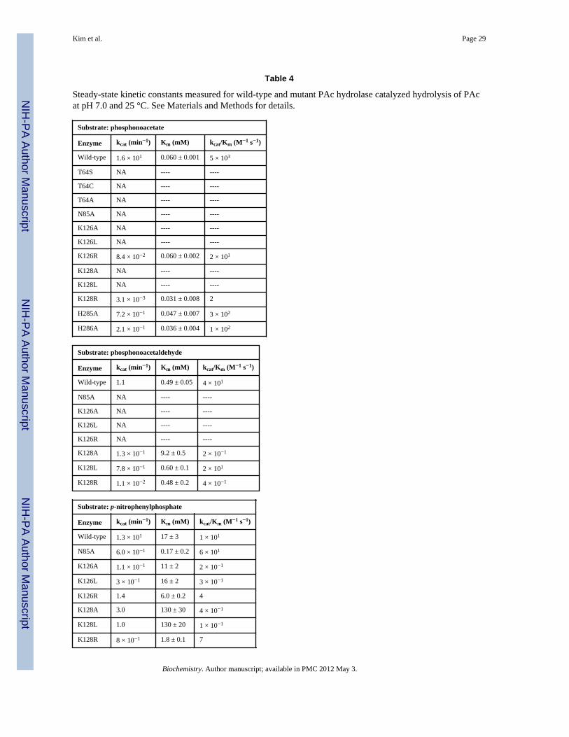
PAc Hydrolase Catalytic MechanismNucleophile—The alkaline phosphatase superfamily catalytic trait is nucleophiliccatalysis, which for the phosphoryl transferases is mediated by a zinc ion (Zn2)-coordinatedSer (AP) (49) or Thr (NPP) (45, 50–51). The PAc hydrolase Thr64, shown positioned for in-line attack of the PAc phosphoryl group in Figure 3, is the likely nucleophile in PAchydrolase catalysis. In order to confirm the role of Thr64, the following set of experimentswere conducted. Firstly, the T64S, T64C and T64A mutant enzymes were prepared andtested for catalytic activity towards PAc hydrolysis. The CD spectra (not shown) andchromatographic behavior of the PAc hydrolase mutants were observed to be consistent withthose of the wild-type enzyme, which indicates that the amino acid replacements had notdisrupted the native fold. All three mutants were found to possess no detectable activity(detection limit 1 × 10⁻⁷ s−1), and thus the Thr64 was shown to be essential for PAchydrolase catalysis (Table 4). Replacement of the Thr132 nucleophile in the Triticumaestivum NPP with Ala or Ser similarly removed all detectable activity (52).
Secondly, 32P-labeling of the enzyme via catalytic turnover of [γ-32P]ATP was tested withwild-type PAc hydrolase and the T64A mutant, in the presence and absence the inhibitorphosphonoformate (Figure 5). ATP is a slow PAc hydrolase substrate (Table 2). Thereaction was carried out at pH 6 in order to retard the phosphoenzyme hydrolysis step, andthereby maximize the accumulation of the phosphorylated enzyme intermediate. The
3 The PAc hydrolase was judged to be pure based on SDS-PAGE analysis, however we did not take additional steps to rule out thepossibility that the low activities observed with these substrates originate in a contaminating, high activity hydrolase.
Kim et al. Page 9
Biochemistry. Author manuscript; available in PMC 2012 May 3.
NIH
-PA Author Manuscript
NIH
-PA Author Manuscript
NIH
-PA Author Manuscript
autoradiogram of the SDS-PAGE gel of the chromatographed [γ-32P]ATP-treated enzymesshows that wild-type PAc hydrolase was 32P-labeled and that the presence of saturatingphosphonoformate precluded 32P-labeling. This result shows that the autophosphorylationoccurs at the active site. The T64A PAc hydrolase mutant was not 32P-labeled. Thisobservation shows that the T64 is required for the PAc hydrolase autophosphorylation.Earlier investigations of nucleophilic catalysis in mammalian NPPs showed, by kineticanalysis and by sequencing the 32P-labeled proteolytic peptide fragment, that the Thr64counterpart (Thr204 in bovine liver NPP and Thr210 in human autotaxin, a NPP) undergoestransient phosphorylation during catalytic turnover of [γ-32P]ATP (50–51). By analogy, weassign Thr64 as the site of phosphorylation during PAc hydrolase catalyzed ATP turnover,and conclude that Thr64 performs the role of nucleophilic catalysis in PAc hydrolysis.
Substrate Activation—The phosphoryl group of the phosphonoformate ligand iscoordinated to one of the two PAc hydrolase active site zinc ions (Zn1). We retained thisinteraction in creating the model for PAc binding shown in Figure 3. The Asn85 side chainthat is positioned across from the PAc phosphoryl group is close (6 Å), yet in this model it isnot close enough for a hydrogen bond interaction. In order to reach the Asn85 side chain, thephosphoryl group would have to lose coordination to the zinc ion (Zn1). Other models wereexamined for the potential of an Asn85 hydrogen bond interaction with the PAc carboxylategroup, while maintaining coordination of the phosphoryl group with the zinc ion and thecorrect positioning for in-line attack by the Thr64, but none was found to satisfy theserequirements. An alignment of PAc hydrolase ortholog sequences (45% or above sequenceidentity) (SI Figure 1) indicates that Asn85 is stringently conserved. The counterpart to thePAc hydrolase Asn85 in X. axonopodis NPP is Asn111 (45) and in E. coli AP it is Arg186(44). The side chains of both residues are engaged in a hydrogen bond interaction with thetransferring phosphoryl group. The kcat/Km value for E. coli AP catalyzed hydrolysis ofethylphosphate is reduced 7000-fold in the R186S mutant, however for hydrolysis of p-nitrophenylphosphate the kcat/Km value of the R186S mutant is reduced only 170-fold (53).In the case of the Triticum aestivum NPP the impact of Ala replacement of Asn165 (counterpart to the X. axonopodis NPP Asn111) on catalytic efficiency is substrate dependent: fornicotinamide adenine dinucleotide and nucleoside triphosphate substrates the activity isreduced ~10-fold, but for p-nitrophenylphosphate it is increased ~100-fold (52). The PAchydrolase N85A mutant showed no significant catalytic activity towards PAc or PAld (Table4). On the other hand, whereas the kcat measured for p-nitrophenolphosphate hydrolysis wasreduced, inexplicably so was the Km (100-fold) for a net 10-fold increase in the kcat/Kmvalue (Table 4). Although Asn85 contributes to catalysis, its role is not clear.
The side chains of stringently conserved PAc hydrolase residues Lys126 and Lys128 arelocated in the vicinity of the carboxylate group of the modeled PAc (Figure 3). Theammonium group of Lys126 is within hydrogen bonding distance of the carboxylate oxygenatom. The conformation of the side chain of Lys128 observed in the structure of the PAchydrolase-phosphonoformate complex can be modeled to place the ammonium group withinhydrogen bond interaction distance of the PAc carboxylate group. Each Lys was separatelyreplaced with Arg, Leu and Ala and the kinetic properties of the site directed mutants weremeasured (Table 4). All three Lys126 mutants are inactive as catalysts of PAc or PAldhydrolysis, but each retained significant activity towards p-nitrophenylphosphate hydrolysis.The PAc hydrolase K128A and K128L mutants do not hydrolyze PAc whereas the kcat/Kmvalue for K128R mutant is 1000-fold lower than that of the wild-type enzyme. The threemutants do, however retain activity towards catalysis of PAld hydrolysis. The kcat values forp-nitrophenylphosphate hydrolysis are similar to those measured for wild-type PAchydrolase (Table 4). Overall, the PAc hydrolysis reaction is most sensitive to Lys126 andLys128 amino acid replacement, which is consistent with the need for two positively chargeresidues to stabilize of the aci-carboxylate dianion displaced by the Thr64.
Kim et al. Page 10
Biochemistry. Author manuscript; available in PMC 2012 May 3.
NIH
-PA Author Manuscript
NIH
-PA Author Manuscript
NIH
-PA Author Manuscript
The alkaline phosphatase enzyme superfamily member cofactor independentphosphoglycerate mutase utilizes a large, dynamic cap domain to bind the substrateglycerate unit as it closes over the catalytic domain (41). The observation that PAc hydrolaseand NPP both possess a cap domain (AP does not) (Figure 2) prompted us to probe theimpact of the replacement of the PAc hydrolase cap domain residues that are directedtowards the catalytic site, on catalytic efficiency. His285 and His286 were targeted becausethey maintain hydrogen bond forming potential, are stringently conserved among PAchydrolase orthologs (SI Figure 1) and are positioned over the substrate (Figure 3). If the capdomain was to clamp down further on the catalytic domain one or both of the His residuesmight be positioned to favorably interact with the PAc carboxylate. However, Alareplacement of either His reduced the kcat by only an order of magnitude (Table 4). Thissmall reduction does not support a direct role for His85 or His86 in catalysis. We thereforeconclude that the snapshot of the cap and catalytic domains observed in the crystal structurerepresent the catalytically active form of the enzyme.
Proposed Model for Catalysis—The model that we propose for the PAc hydrolasecatalytic mechanism is a variation on the mechanism of NPP catalysis proposed byHerschlag and coworkers (45), which in turn is based on the NPP crystal structure (45) andon the large volume of work that has been carried out to define the catalytic mechanism ofthe E. coli AP (47, 49, 53–56). The key difference between the chemical reactions catalyzedby PAc hydrolase and NPP/AP is the first step, which involves P-C bond cleavage ratherthan P-OR bond cleavage. It is on this step that we will focus our discussion.
The cleavage of the P-OR bond catalyzed by AP and NPP occurs by attack the catalytic Seror Thr, respectively. The active form of the nucleophile is the alkoxide anion, which isstabilized and oriented by coordination to one of the two zinc ion cofactors (Zn2) (Figure 2Band 2C). The phosphorus is activated for nucleophilic attack by interaction of its oxygenatoms with one or both of the zinc ions and with the properly positioned Arg (AP) or Asn(NPP) side chain. The stabilization of the alkoxide leaving group is necessary for the P-ORbond cleavage in phosphate ester metabolites, and this role has been assigned to one of thetwo zinc ions (Zn2), which must form a coordination bond with the departing oxygen atom.The zinc ion cannot engage in such an interaction with the PAc methylene group. Therefore,an alternate strategy to leaving group stabilization in PAc hydrolase catalysis must exist.The leaving group is an aci-carboxylate dianion. Aci-carboxylate dianion intermediates areknown to be formed and stabilized in the active sites of members of the enolase enzymesuperfamily (57) and in the active sites of members of the isocitrate lyase/PEP mutasesuperfamily (58). Enzymes from both these families use electropositive groups (metal ioncofactor and/or hydrogen bond donating amino acid residues) to delocalize charge viainteraction with the carboxylate group of aci-carboxulate dianion. The best candidates forthis role in PAc hydrolase are the respective ammonium groups of Lys126 and Lys128.Thus, the proposed model for the PAc hydrolase substrate complex (Scheme 3) incorporatesthe coordination of the substrate phosphoryl group to one zinc ion (Zn1), the coordination ofthe Thr64 nucleophile to the other zinc ion (Zn2), and the hydrogen bond interactions of thePAc carboxylate group with the Lys126 and Lys128 side chains.
It is interesting to contrast the catalytic strategy used by phosphonatase to catalyze P-C bondcleavage in PAld (Scheme 2) with the catalytic strategy depicted in Scheme 3 for the PAchydrolase. Because PAc hydrolase also catalyzes PAld hydrolysis, albeit less efficiently thanPAc hydrolysis (Table 2), in effect PAld hydrolase activity has evolved within the catalyticscaffold of the alkaline phosphatase enzyme superfamily in addition to evolving within thecatalytic scaffold of the HAD enzyme superfamily. The C=O of PAld can in principleengage one of the PAc hydrolase active site Lys side chains in hydrogen bond formation forstabilization of the enolate anion formed with P-C bond cleavage. The PAld hydrolase
Kim et al. Page 11
Biochemistry. Author manuscript; available in PMC 2012 May 3.
NIH
-PA Author Manuscript
NIH
-PA Author Manuscript
NIH
-PA Author Manuscript
activity of PAc hydrolase is considerably less than that of phosphonatase (18) (kcat/Km = 1 ×10⁵ M−1 s−1 vs kcat/Km = 8 × 101 M−1 s−1) and although interesting from an evolution-mechanism perspective, this activity is not expected to be of biological significance.
PAc Hydrolase Biological Range and FunctionTo gain insight into the metabolic function of PAc hydrolase we first carried out a BLASTsearch of the bacterial genomes deposited in NCBI, using the P. fluorescens 23F PAchydrolase amino acid sequence as query, to identify bacteria that produce PAc hydrolase. Inthis manner we were able to trace the biological range of PAc hydrolase within the bacterialspecies represented in the NCBI genome bank. The sequence identity boundary separatingorthologs from paralogs was well defined (> 45% vs < 30%). Interestingly, PAc hydrolasewas found only in Proteobacteria. Within the beta subdivision, Bordetella, Burkholderia,Cupriavidus, Ralstonia, Alicycliphilus, Polaromonas, and Verminephrobacter speciespossess orthologs that share 50–85% sequence identity with the P. fluorescens 23F enzyme.Within the alpha subdivision Mesorhizobium, Oligotropha, Rhodobacterales,Rhodopseudomonas, Roseovarius, Thalassiobium, Agrobacterium, and Sinorhizobiumspecies possess orthologs that share 55–46% sequence identity with the P. fluorescens 23Fenzyme. In the delta subdivision Sorangium, Pseudomonadaceae and Azotobacter are theonly representatives found to contain a PAc hydrolase ortholog (47% sequence identity).
Next, we searched among the annotated genomes for PAc hydrolase gene neighbors thatmight encode proteins that function in the synthesis or breakdown of PAc. We discoveredgene clusters in the genomes of Sinorhizobium medicae and meliloti that encode AEPtransaminase, PAc hydrolase and an aldehyde dehydrogenase. In numerous Burkholderiaspecies the genes annotated AEP transporter, PAc hydrolase and PAld dehydrogenase arejuxtaposed. Together these enzymes might form the components of a novel AEP transportand degradation pathway, as is illustrated in Scheme 1. A BLAST of the genomes of thePAc hydrolase containing bacteria using AEP transaminase and the putative PAlddehydrogenase sequences as queries confirmed that all three of the pathway genes arepresent. The spotty distribution of the PAc hydrolase gene among the various subdivisionsof Proteobacteria is suggestive of its distribution by lateral gene transfer (59). This is alsoindicated by the co-location of a putative transposon as was observed for a few of theinspected genomes. If clustered together, all three of the pathway genes might efficiently betransferred by a single mobile genetic element.
A brief check was made to determine if the phosphonatase gene is present in the genomes ofthe bacteria that harbor the PAc hydrolase gene. With only a few exceptions (Burkholderiagraminus being one) it is not. A BLAST search of the NCBI genome bank using thephosphonatase sequence as query identifies phosphonatase in a wide range of Gram-negative and Gram-positive bacteria. This suggests that the AEP degradation pathway thatproceeds via P-C cleavage in PAld appears to be more wide spread than that which proceedsvia P-C bond cleavage in PAc (Scheme 1).
ConclusionOf the known members of the alkaline phosphatase enzyme superfamily, the PAc hydrolasetertiary structure and active site structure most closely resembles those of the NPP.However, the lack of significant sequence identity between the PAc hydrolase and NPP(~15%) compared to relatively high sequence identity between PAc hydrolase orthologs (>45%) suggests early divergence within this lineage of the alkaline phosphate enzymesuperfamily. Nevertheless, two basic elements of catalysis, namely activation of thesubstrate phosphoryl group by zinc ion coordination and nucleophilic attack by a zinc ionactivated Thr are conserved (Scheme 3). Whereas the active site Asn that binds the substrate
Kim et al. Page 12
Biochemistry. Author manuscript; available in PMC 2012 May 3.
NIH
-PA Author Manuscript
NIH
-PA Author Manuscript
NIH
-PA Author Manuscript
phosphoryl group in NPP is also present in PAc hydrolase, it does not appear to participatein PAc binding. In NPP, a zinc ion stabilizes the oxygen anion leaving group whereas inPAc hydrolase, the carbanion leaving group is stabilized by delocalization to the carboxylateoxygen atoms and hence to the charged Lys residues (Scheme 3).
Supplementary MaterialRefer to Web version on PubMed Central for supplementary material.
References1. White AK, Metcalf WW. Microbial Metabolism of reduced phosphorus compounds. Annu Rev
Microbiol. 2007; 61:379–400. [PubMed: 18035609]2. Metcalf WW, van der Donk WA. Biosynthesis of phosphonic and phosphinic acid natural products.
Ann Rev Biochem. 2009; 78:65–94. [PubMed: 19489722]3. Nair SK, Van der Donk WA. Structure and mechanism of enzymes involved in biosynthesis and
breakdown of the phosphonates fosfomycin, dehydrophos and phosphinothricin. Arch BiochemBiophys. 2011; 505:13–21. [PubMed: 20854789]
4. Quinn JP, Kulakova AN, Cooley NA, McGrath JW. New ways to break an old bond: the bacterialcarbon-phosphorus hydrolases and their roles in biogeochemical phosphorus cycling. EnvironMicrobiol. 2007; 9:2392–2400. [PubMed: 17803765]
5. Kittredge MS, Robert E, Simonsen DG. The occurrence of free 2-aminoethylphosphonic acid in thesea anemone, Anthopleura elegantissima. Biochemistry. 1962; 1:624–628. [PubMed: 14456574]
6. Horiguchi M, Kandatsu M. Isolation of 2-aminoethane phosphonic acid from rumen protozoa.Nature. 1959; 184:901–902. [PubMed: 14403103]
7. Hilderbrand, RL. The role of phosphonates in living systems. Hilderbrand, RL., editor. C.R.C. Press,Inc; Boca Raton, Florida: 1983.
8. Orsini F, Sello G, Sisti M. Aminophosphonic acids and derivatives. Synthesis and biologicalapplications. Curr Med Chem. 2010; 17:264–289. [PubMed: 20214568]
9. De Clercq E, Holy A. Acyclic nucleoside phosphonates: a key class of antiviral drugs. Nat RevDrug Discov. 2005; 4:928–940. [PubMed: 16264436]
10. Falagas ME, Kastoris AC, Kapaskelis AM, Karageorgopoulos DE. Fosfomycin for the treatment ofmulti-drug resistant, including extended-spectrum beta-lactamase producing, Enterobacteriaceaeinfections: a systematic review. Lancet Infect Dis. 2010; 10:43–50. [PubMed: 20129148]
11. Kennedy KE, Thompson GA. Phosphonolipids: Localization in surface membranes ofTetrahymena. Science. 1970; 168:989–991. [PubMed: 5441031]
12. Bowman E, McQueney M, Barry R, Dunaway-Mariano D. Catalysis and thermodynamics of therearrangement of phosphoenol pyruvate to phosphonopyruvate. Entry into the phosphonate classof naturally occurring organophosphonates. J Amer Chem Soc. 1988; 110:5575–5577.
13. Zhang G, Dai J, Lu Z, Dunaway-Mariano D. The phosphonopyruvate decarboxylase fromBacteroides fragilis. J Biol Chem. 2003; 278:41302–41308. [PubMed: 12904299]
14. Kim, J. PhD Thesis. University of Maryland; 1994. Investigations of the 2-aminoethylphosphonatebiosynthetic enzymes in Tetrahymena pyriformis; p. 96-106.
15. Kim AD, Baker AS, Metcalf WW, Wanner BL, Martin BM, Dunaway-Mariano D. The 2-aminoethylphosphonate-specific Transaminase of the 2-aminoethylphosphonate degradationpathway. J Bacteriology. 2002; 184:4134–4140.
16. Olsen DB, Hepburn TW, Moos M, Mariano PS, Dunaway-Mariano D. Investigation of the Bacilluscereus phosphonoacetate hydrolse. Evidence for a Schiff Base Mechanism and sequence analysisof an active site peptide containing the catalytic lysine residue. Biochemistry. 1988; 27:2229–2236. [PubMed: 3132206]
17. Lee S-L, Hepburn TW, Schwartz WH, Ammon HL, Mariano PS, Dunaway-Mariano.Stereochemical probe for the mechanism of P-C bond cleavage catalyzed by the Bacillus cereusphosphonoacetaldehyde hydrolase. J Amer Chem Soc. 1992; 114:7346–7354.
Kim et al. Page 13
Biochemistry. Author manuscript; available in PMC 2012 May 3.
NIH
-PA Author Manuscript
NIH
-PA Author Manuscript
NIH
-PA Author Manuscript
18. Baker AS, Ciocci MJ, Metcalf WW, Kim JB, Babbitt PC, Wanner BL, Martin BM, Dunaway-Mariano D. Insights into the mechanism of catalysis by the P-C bond cleaving enzymephosphonoacetaldehyde hydrolase derived from gene sequence-analysis and mutagenesis.Biochemistry. 1998; 37:9305–9315. [PubMed: 9649311]
19. Allen KN, Dunaway-Mariano D. Phosphoryl group transfer: evolution of a catalytic scaffold.Trends Biochem Sci. 2004; 29:495–503. [PubMed: 15337123]
20. McMullan G, Quinn JP. In vitro characterization of a phosphate starvation-independent carbon-phosphorus bond cleavage activity in Pseudomonas fluorescens 23F. J Bacteriol. 1994; 176:320–324. [PubMed: 8288524]
21. McGrath JW, Wisdom GB, McMullan G, Larkin MJ, Quinn JP. The purification and properties ofphosphonoacetate hydrolase, a novel carbon-phosphorus bond-cleavage enzyme fromPseudomonas fluorescens 23F. Eur J Biochem. 1995; 234:225–30. [PubMed: 8529644]
22. Galperin MY, Bairoch A, Koonin EV. A superfamily of metalloenzymes unifiesphosphopentomutase and cofactor-independent phosphoglycerate mutase with alkalinephosphatases and sulfatases. Protein Science. 1998; 7:1829–1835. [PubMed: 10082381]
23. McCullan G, Harrington F, Quinn JP. Metabolism of phosphonoacetate as the sole carbon andphosphorus source by an environmental bacterial isolate. Appl Environ Microbiol. 1992; 58:1364–1366. [PubMed: 16348700]
24. McCullan G, Quinn JP. Detection of a novel carbon-phosphorus bond cleavage activity in cell-freeextracts of an environmental Pseudomonas fluorescens isolate. Biochem Biophys Res Commun.1992; 184:1022–1027. [PubMed: 1575721]
25. Panas P, Ternan NG, Dooley JS, McMullan G. Detection of phosphonoacetate degradation andphnA genes in soil bacteria from distinct geographical origins suggest its possible biogenic origin.Environ Microbiol. 2006; 8:939–945. [PubMed: 16623750]
26. Thomas S, Burdett H, Temperton B, Wick R, Snelling D, McGrath JW, Quinn JP, Munn C, GilbertJA. Evidence for phosphonate usage in the coral holobiont. ISME J. 2010; 4:459–461. [PubMed:19956272]
27. Kulakova AN, Kulakov LA, Akulenko NV, Ksenzenko VN, Hamilton JT, Quinn JP. Structural andfunctional analysis of the phosphonoacetate hydrolase (phnA) gene region in Pseudomonasfluorescens 23F. J Bacteriol. 2001; 183:3268–3275. [PubMed: 11344133]
28. Kim AD, Baker AS, Metcalf WW, Wanner BL, Martin BM, Dunaway-Mariano D. The 2-aminoethylphosphonate-specific transaminase of the 2-aminoethylphosphonate degradationpathway. J Bacteriology. 2002; 184:4134–4140.
29. Kulakova AN, Kulakov LA, Quinn JP. Cloning of the phosphonoacetate hydrolase gene fromPseudomonas fluorescens 23F encoding a new type of carbon-phosphorus bond cleaving enzymeand its expression in Escherichia coli and Pseudomonas putida. Gene. 1997; 195:49–53.[PubMed: 9300819]
30. Isbell AF, Englert LF, Rosenberg H. Phosohonacetaldehyde. J Org Chem. 1969; 34:755–756.31. Wesenberg G, Rayment I. unpublished results.32. Terwilliger TC, Berendzen J. Automated MAD and MIR structure solution. Acta Crystallogr D.
1999; 55:849–861. [PubMed: 10089316]33. Cowtan K, Main P. Miscellaneous algorithms for density modification. Acta Crystallogr. 1998;
D54:487–493.34. Rypniewski WR, Breiter DR, Benning MM, Wesenberg G, Oh BH, Markley JL, Rayment I,
Holden HM. Crystallization and structure determination to 2.5-A resolution of the oxidized[2Fe-2S] ferredoxin isolated from Anabaena 7120. Biochemistry. 1991; 30:4126–4131. [PubMed:1902376]
35. Navaza J. Amore - an Automated Package for Molecular Replacement. Acta Crystallogr. 1994;A50:157–163.
36. Tronrud DE, Ten Eyck LF, Matthews BW. Acta Crystallogr Sect A. 1987; 43:489–501.37. Roussel, A.; Cambillau, C. Silicon Graphics Geometry Partners Directory. Silicon Graphics; 1991.38. Millan JL. Alkaline Phosphatases. Structure, substrate specificity and functional relatedness to
other members of a large superfamily of enzymes. Purinergic Signalling. 2006; 2:335–341.[PubMed: 18404473]
Kim et al. Page 14
Biochemistry. Author manuscript; available in PMC 2012 May 3.
NIH
-PA Author Manuscript
NIH
-PA Author Manuscript
NIH
-PA Author Manuscript
39. Galperin MY, Jedrzejas MJ. Conserved core structure and active site residues in alkalinephosphatase superfamily enzymes. Proteins. 2001; 45:318–324. [PubMed: 11746679]
40. Jedrzejas MJ, Setlow P. Comparison of the binuclear metalloenzymes diphosphoglycerate-independent phosphoglycerate mutase and alkaline phosphatase:their mechanism of catalysis via aphosphoserine intermediate. Chem Rev. 2001; 101:607–618. [PubMed: 11712498]
41. Rigden DJ, Lamani E, Mello LV, Littlejohn JE, Jedrzejas MJ. Insights into the catalyticmechanism of cofactor-independent phosphoglycerate mutase from X-ray crystallography,simulated dynamics and molecular modeling. J Mol Biol. 2003; 328:909–920. [PubMed:12729763]
42. Gosh D. Human sulfatases: a structural perspective. Cell Mol Life Sci. 2007; 64:2013–2022.[PubMed: 17558559]
43. Bojarova P, Williams SJ. Sulfotransferases, sulfatases and formyglycine-generating enzymes: asulfation fascination. Curr Opin Chem Biol. 2008; 12:573–581. [PubMed: 18625336]
44. Kim EE, Wyckoff HW. Reaction mechanism of alkaline phosphatase based on crystal structures.Two-metal ion catalysis. J Mol Biol. 1991; 218:449–464. [PubMed: 2010919]
45. Zalatan JG, Fenn TD, Brunger AT, Herschlag D. Structural and functional comparisons ofnucleotide pyrophosphatase/phosphodiesterase and alkaline phosphatase: implications formechanism and evolution. Biochemistry. 2006; 45:9788–9803. [PubMed: 16893180]
46. Holtz KM, Stec B, Myers JK, Antonelli SM, Widlanski TS, Kantrowitz ER. Alternate modes ofbinding in two crystal structures of alkaline phosphatase-inhibitor complexes. Protein Sci. 2000;9:907–915. [PubMed: 10850800]
47. O’Brien PJ, Hershlag D. Alkaline phosphatase revisited: Hydrolysis of alkyl phosphates.Biochemistry. 2002; 41:3207–3225. [PubMed: 11863460]
48. Lassila J, Herschlag D. Promiscuous sulfatase activity and thio-effects in a phosphodiesterase ofthe alkaline phosphatase superfamily. Biochemistry. 2008; 47:12853–12859. [PubMed: 18975918]
49. Coleman JE. Structure and mechanism of alkaline phosphatase. Annu Rev Biophys. 1992; 21:441–483.
50. Oda Y, Kuo MD, Huang SS, Huang JS. The major acidic fibroblast growth factor (aFGF)-stimulated phosphoprotein from bovine liver plasma membranes has a aFGF-stimulated kinase,autoadenylation, and alkaline nucleotide phosphodiesterase activities. J Biol Chem. 1993;268:27318–27326. [PubMed: 7505270]
51. Clair T, Lee HY, Lioatta LA, Stracke ML. Autotaxin is an exoenzyme possessing 5′-nucleotidephosphodiesterase/ATP pyrophosphatase and ATPase activities. J Biol Chem. 1997; 272:996–1001. [PubMed: 8995394]
52. Joye IJ, Belien T, Brijis K, Soetaert W, Delcour JA. Mutational analysis of wheat (Triticumaestivum L.) nucleotide pyrophosphatse/phosphodiesterase shows the roles of six amino acids inthe catalytic mechanism. Appl Microbiol Biotechnol. 2010 Published on line Dec 29 2010.
53. O’Brien PJ, Lassila JK, Fenn TD, Zaltan JG, Herschlag D. Arginine coordination in enzymaticphosphoryl transfer: evaluation of the effect of Arg166 mutations in Escherichhia coli alkalinephosphatase. Biochemistry. 2008; 47:7663–7672. [PubMed: 18627128]
54. Stec B, Holtz KM, Kantrowitz ER. A revised mechanism for the alkaline phosphatase reactioninvolving three metal ions. J Mol Biol. 2000; 299:1303–1311. [PubMed: 10873454]
55. Zaltan J, Fenn TD, Herschlag D. Comparative enzymology in the alkaline phosphatase superfamilyto determine the catalytic role of an active site metal ion. J Mol Biol. 2008; 384:1174–1189.[PubMed: 18851975]
56. Wang J, Kantrowitz ER. Trapping the tetrahedral intermediate in the alkaline phosphatase reactionby substitution of the active site serine with threonine. Protein Sci. 2006; 15:2395–2401. [PubMed:17008720]
57. Gerlt JA, Babbitt PC, Rayment I. Divergent evolution in the enolase superfamily: the interplay ofmechanism and specificity. Arch Biochem Biophys. 2005; 433:59–70. [PubMed: 15581566]
58. Liu S, Lu Z, Han Y, Melamud E, Dunaway-Mariano D, Herzberg O. Crystal structures of 2-methylisocitrate lyase in complex with product and with isocitrate inhibitor provide insight intolyase substrate specificity, catalysis and evolution. Biochemistry. 2005; 44:2949–2962. [PubMed:15723538]
Kim et al. Page 15
Biochemistry. Author manuscript; available in PMC 2012 May 3.
NIH
-PA Author Manuscript
NIH
-PA Author Manuscript
NIH
-PA Author Manuscript
59. Huang J, Su Z, Xu Y. The evolution of microbrial phosphate degradative pathways. J Mol Evol.2005; 61:682–690. [PubMed: 16245012]
Kim et al. Page 16
Biochemistry. Author manuscript; available in PMC 2012 May 3.
NIH
-PA Author Manuscript
NIH
-PA Author Manuscript
NIH
-PA Author Manuscript
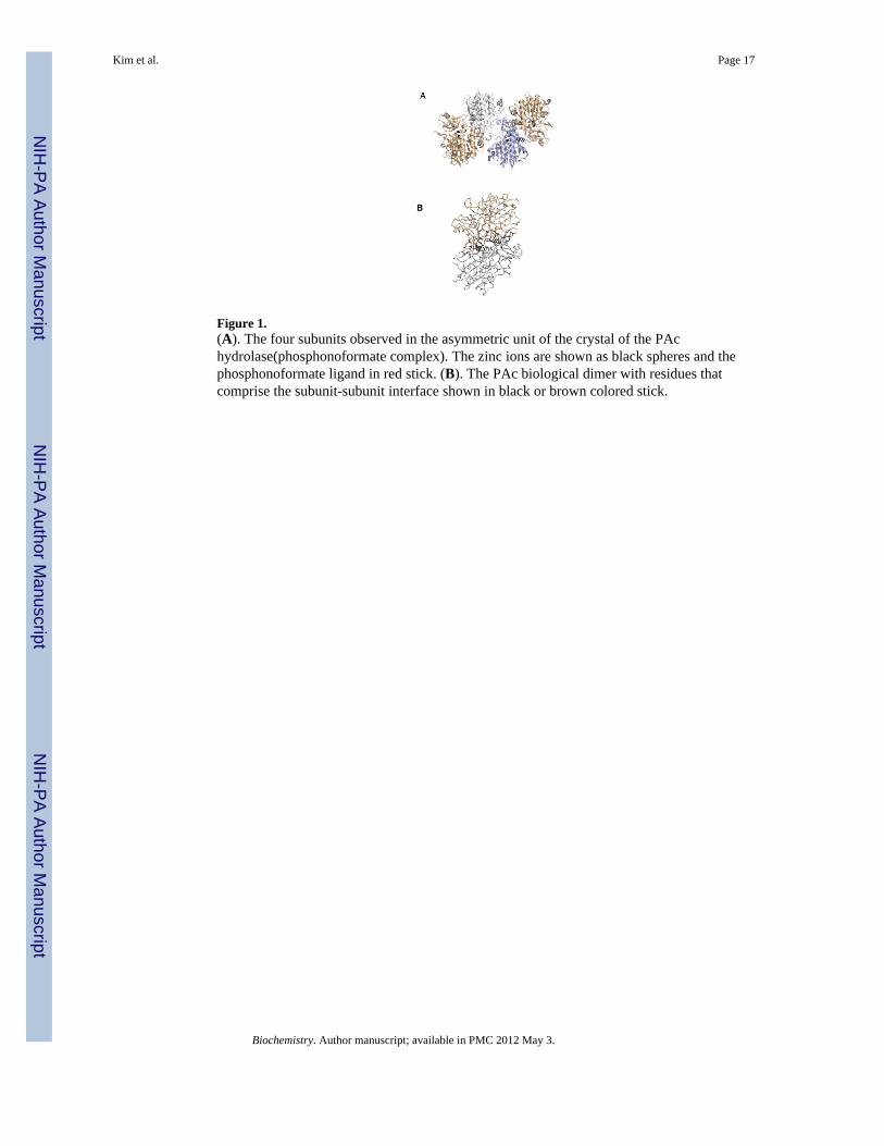
Figure 1.(A). The four subunits observed in the asymmetric unit of the crystal of the PAchydrolase(phosphonoformate complex). The zinc ions are shown as black spheres and thephosphonoformate ligand in red stick. (B). The PAc biological dimer with residues thatcomprise the subunit-subunit interface shown in black or brown colored stick.
Kim et al. Page 17
Biochemistry. Author manuscript; available in PMC 2012 May 3.
NIH
-PA Author Manuscript
NIH
-PA Author Manuscript
NIH
-PA Author Manuscript
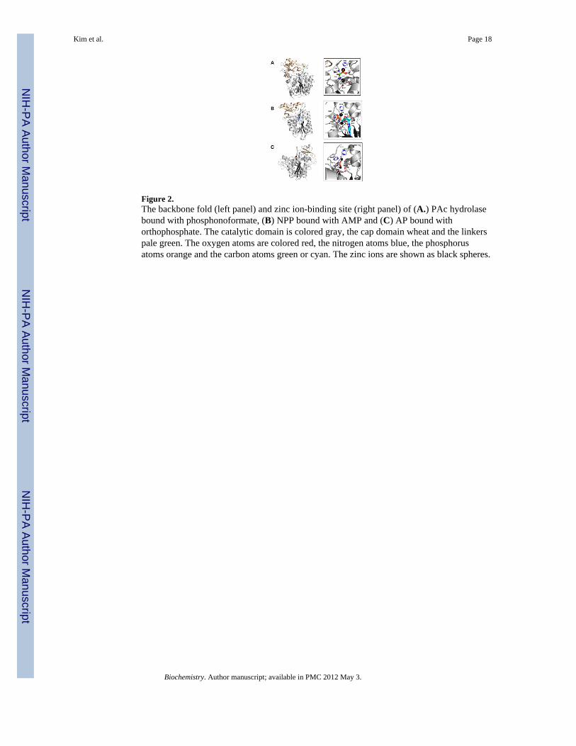
Figure 2.The backbone fold (left panel) and zinc ion-binding site (right panel) of (A.) PAc hydrolasebound with phosphonoformate, (B) NPP bound with AMP and (C) AP bound withorthophosphate. The catalytic domain is colored gray, the cap domain wheat and the linkerspale green. The oxygen atoms are colored red, the nitrogen atoms blue, the phosphorusatoms orange and the carbon atoms green or cyan. The zinc ions are shown as black spheres.
Kim et al. Page 18
Biochemistry. Author manuscript; available in PMC 2012 May 3.
NIH
-PA Author Manuscript
NIH
-PA Author Manuscript
NIH
-PA Author Manuscript
Figure 3.The model of PAc bound to the PAc hydolase active site. The oxygen atoms are colored red,the nitrogen atoms blue, the phosphorus atoms orange and the carbon atoms green. The zincions are shown as black spheres.
Kim et al. Page 19
Biochemistry. Author manuscript; available in PMC 2012 May 3.
NIH
-PA Author Manuscript
NIH
-PA Author Manuscript
NIH
-PA Author Manuscript
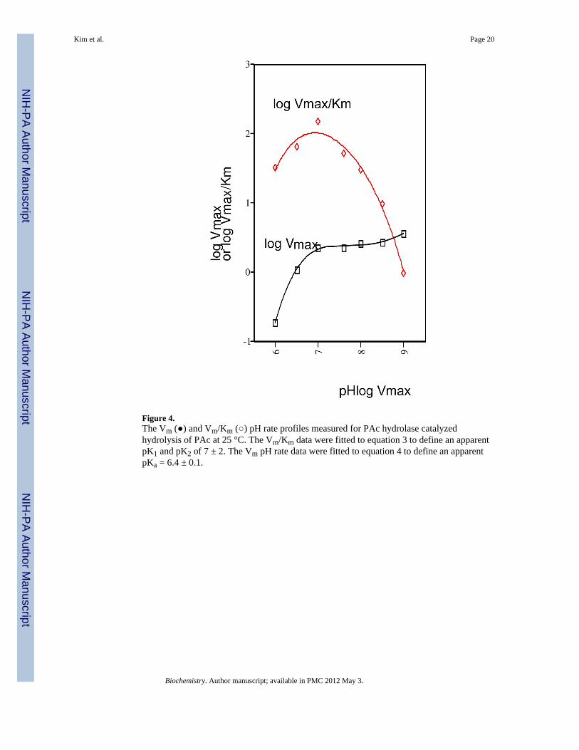
Figure 4.The Vm (●) and Vm/Km (○) pH rate profiles measured for PAc hydrolase catalyzedhydrolysis of PAc at 25 °C. The Vm/Km data were fitted to equation 3 to define an apparentpK1 and pK2 of 7 ± 2. The Vm pH rate data were fitted to equation 4 to define an apparentpKa = 6.4 ± 0.1.
Kim et al. Page 20
Biochemistry. Author manuscript; available in PMC 2012 May 3.
NIH
-PA Author Manuscript
NIH
-PA Author Manuscript
NIH
-PA Author Manuscript
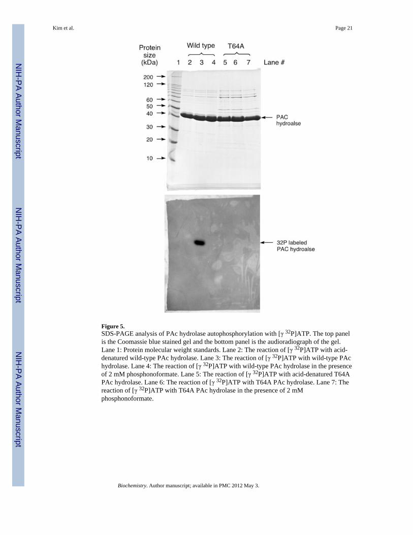
Figure 5.SDS-PAGE analysis of PAc hydrolase autophosphorylation with [γ 32P]ATP. The top panelis the Coomassie blue stained gel and the bottom panel is the audioradiograph of the gel.Lane 1: Protein molecular weight standards. Lane 2: The reaction of [γ 32P]ATP with acid-denatured wild-type PAc hydrolase. Lane 3: The reaction of [γ 32P]ATP with wild-type PAchydrolase. Lane 4: The reaction of [γ 32P]ATP with wild-type PAc hydrolase in the presenceof 2 mM phosphonoformate. Lane 5: The reaction of [γ 32P]ATP with acid-denatured T64APAc hydrolase. Lane 6: The reaction of [γ 32P]ATP with T64A PAc hydrolase. Lane 7: Thereaction of [γ 32P]ATP with T64A PAc hydrolase in the presence of 2 mMphosphonoformate.
Kim et al. Page 21
Biochemistry. Author manuscript; available in PMC 2012 May 3.
NIH
-PA Author Manuscript
NIH
-PA Author Manuscript
NIH
-PA Author Manuscript
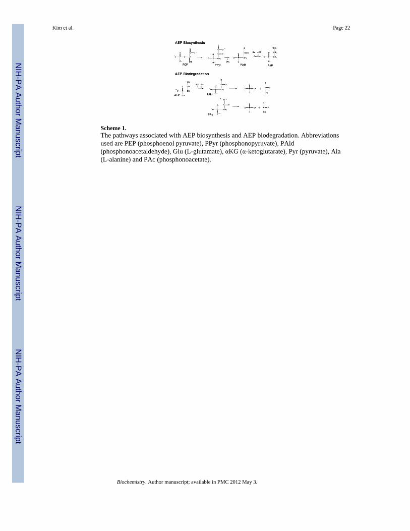
Scheme 1.The pathways associated with AEP biosynthesis and AEP biodegradation. Abbreviationsused are PEP (phosphoenol pyruvate), PPyr (phosphonopyruvate), PAld(phosphonoacetaldehyde), Glu (L-glutamate), αKG (α-ketoglutarate), Pyr (pyruvate), Ala(L-alanine) and PAc (phosphonoacetate).
Kim et al. Page 22
Biochemistry. Author manuscript; available in PMC 2012 May 3.
NIH
-PA Author Manuscript
NIH
-PA Author Manuscript
NIH
-PA Author Manuscript
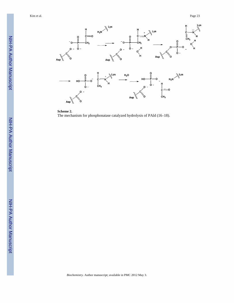
Scheme 2.The mechanism for phosphonatase catalyzed hydrolysis of PAld (16–18).
Kim et al. Page 23
Biochemistry. Author manuscript; available in PMC 2012 May 3.
NIH
-PA Author Manuscript
NIH
-PA Author Manuscript
NIH
-PA Author Manuscript
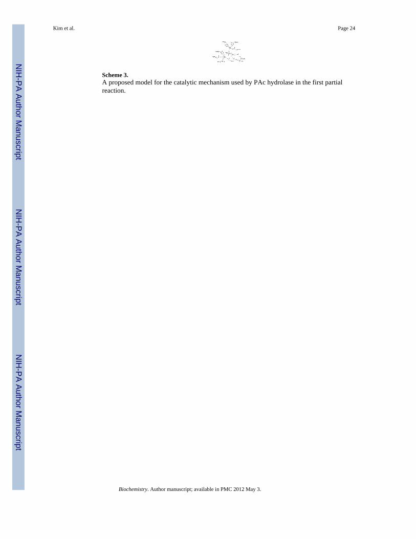
Scheme 3.A proposed model for the catalytic mechanism used by PAc hydrolase in the first partialreaction.
Kim et al. Page 24
Biochemistry. Author manuscript; available in PMC 2012 May 3.
NIH
-PA Author Manuscript
NIH
-PA Author Manuscript
NIH
-PA Author Manuscript
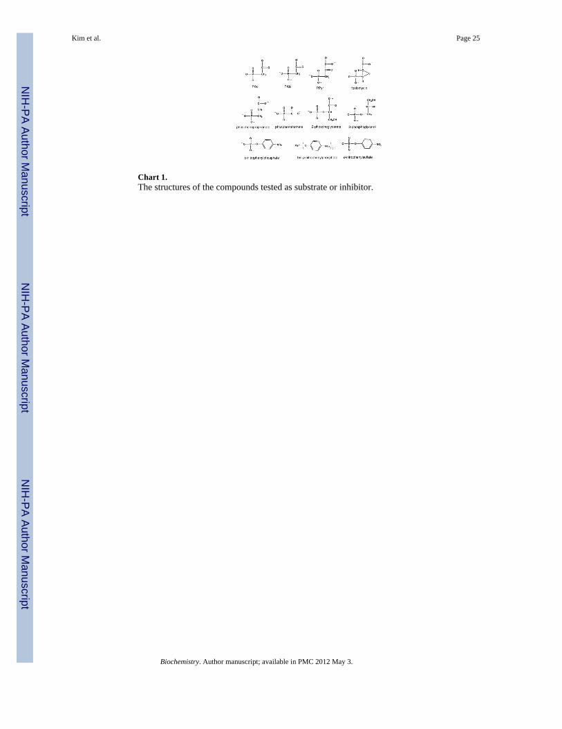
Chart 1.The structures of the compounds tested as substrate or inhibitor.
Kim et al. Page 25
Biochemistry. Author manuscript; available in PMC 2012 May 3.
NIH
-PA Author Manuscript
NIH
-PA Author Manuscript
NIH
-PA Author Manuscript
NIH
-PA Author Manuscript
NIH
-PA Author Manuscript
NIH
-PA Author Manuscript
Kim et al. Page 26
Tabl
e 1
The
X-r
ay d
ata
colle
ctio
n an
d re
finem
ent s
tatis
tics f
or th
e st
ruct
ure
dete
rmin
atio
n of
the
PAc
hydr
olas
e bo
und
with
pho
spho
nofo
rmat
e an
d tw
o Zn
2+ io
ns.
X-r
ay D
ata
Col
lect
ion
Stat
istic
s
Dat
a se
tR
esol
utio
n (Å
)In
depe
nden
t Ref
lect
ions
Com
plet
enes
s (%
)A
ve I/
Avg
σ (I
)R s
yma,
b
Nat
ive
(I)
30.0
- 2.
844
,619
88.0
10.7
5.2
(14.
3)
Nat
ive
(II)
30.0
- 2.
110
4,68
391
.0 (8
1.0)
13.7
5.3
(21.
2)
(CH
3)3P
bCO
OC
H3
30.0
- 2.
847
,546
93.0
8.8
6.3
(15.
8)
K2P
tCl 6
30.0
- 2.
843
,673
86.0
6.6
6.9
(21.
3)
Ref
inem
ent S
tatis
tics f
or N
ativ
e D
ata
Set
Res
olut
ion
limits
(Å)
c R-f
acto
r (ov
eral
l) %
/# rf
lns
c R-f
acto
r (w
orki
ng) %
/# rf
lns
c R-f
acto
r (fr
ee) %
/# rf
lns
No.
Pro
tein
Ato
ms
d N
o. H
eter
oato
ms
30.0
– 2
.1 (2
.1–2
.2)
19.0
/127
171
18.5
/120
751
23.7
/642
012
222
537
Ave
B v
alue
(Å2 )
Prot
ein
33.3
Zn2+
22.
5Li
gand
44.
4So
lven
t 34.
5--
----
---
Wei
ghte
d R
oot-M
ean-
Squa
re D
evia
tions
from
Idea
lity
Bon
d Le
ngth
s (Å
)B
ond
Ang
les (
Å)
Trig
onal
pla
nes (
Å)
Gen
eral
pla
nes (
Å)
Tors
iona
l ang
les (
deg)
0.01
11.
971
0.00
50.
008
17.9
6
a R sym
= (Σ
| I −
Ī|/Σ
I) ×
100
.
b Stat
istic
s for
the
high
est r
esol
utio
n bi
n
c R-fa
ctor
= (Σ
|Fo
− F
c|/Σ|
F o|)
× 10
0 w
here
Fo
is th
e ob
serv
ed st
ruct
ure
fact
or a
mpl
itude
and
Fc
is th
e ca
lcul
ated
stru
ctur
e fa
ctor
am
plitu
de.
d Thes
e in
clud
e 50
4 w
ater
mol
ecul
es, t
hree
pho
spho
nofo
rmat
e m
olec
ules
, one
tartr
ate
mol
ecul
e an
d tw
o zi
nc io
ns.
Biochemistry. Author manuscript; available in PMC 2012 May 3.

NIH
-PA Author Manuscript
NIH
-PA Author Manuscript
NIH
-PA Author Manuscript
Kim et al. Page 27
Table 2
Steady-state kinetic constants for PAc hydrolase-catalyzed hydrolysis of PAc and various analogs at 25 °C andpH 7.0 (except where noted). See Materials and Methods for details.
Substrate kcat (s−1) Km (mM) kcat/Km (M−1 s−1)
phosphonoacetatea 0.27 ± 0.1 0.060 ± 0.009 5 × 10³
phosphonoacetatea (30 °C) 0.67 ± 0.1 0.015 ± 0.001 4 × 10⁴
p-nitrophenylphosphateb 0.22 ± 0.1 14 ± 0.2 2 × 101
2-phosphoglyceratea,c 4 × 10⁻⁵ ------ ------
3-phosphoglycerola,c 4 × 10⁻⁵ ------ ------
p-nitrophenylsulfateb ~2 × 10⁻⁷ ~ 10 ~2 × 10⁻⁴
ATPa 2 ×10⁻⁴ 1 2 × 10⁻¹
phosphonopyruvatea NAd ------ ------
phosphonoacetaldehydea 0.027 ± 0.01 0.33 ± 0.01 8 × 101
fosfomycina,c NAd ------ ------
bis-p-nitrophenylphosphateb,c NAd ------ ------
aThe reaction buffer was 50 mM K+ADA (pH 7.0).
bThe reaction buffer was 50 mM K+TRICINE (pH 8.0).
cTested at a concentration of 2 mM. For 2-phosphoglycerate and 3-phosphoglycerol the ratio of the initial velocity and the enzyme concentration is
used to approximates the kcat value.
dNo activity (NA) was observed within the detection limit of ~ 1 × 10⁻⁷ s−1.
Biochemistry. Author manuscript; available in PMC 2012 May 3.
NIH
-PA Author Manuscript
NIH
-PA Author Manuscript
NIH
-PA Author Manuscript
Kim et al. Page 28
Table 3
Steady-state competitive inhibition constants measured for PAc analogs as inhibitors of PAc hydrolasecatalyzed hydrolysis of PAc at pH 7.0 and 30 °C (except where noted). See Materials and Methods for details.
Inhibitor Ki (mM) Type of Inhibition
phosphonoformate (25 oC) 0.049 ± 0.008 competitive
phosphonoformate 0.19 ± 0.06 competitive
phosphonoproprionate 1.2 ± 0.8 competitive
phosphonopyruvate 1.2 ± 0.8 competitive
Biochemistry. Author manuscript; available in PMC 2012 May 3.
NIH
-PA Author Manuscript
NIH
-PA Author Manuscript
NIH
-PA Author Manuscript
Kim et al. Page 29
Table 4
Steady-state kinetic constants measured for wild-type and mutant PAc hydrolase catalyzed hydrolysis of PAcat pH 7.0 and 25 °C. See Materials and Methods for details.
Substrate: phosphonoacetate
Enzyme kcat (min−1) Km (mM) kcat/Km (M−1 s−1)
Wild-type 1.6 × 101 0.060 ± 0.001 5 × 10³
T64S NA ---- ----
T64C NA ---- ----
T64A NA ---- ----
N85A NA ---- ----
K126A NA ---- ----
K126L NA ---- ----
K126R 8.4 × 10⁻² 0.060 ± 0.002 2 × 101
K128A NA ---- ----
K128L NA ---- ----
K128R 3.1 × 10⁻³ 0.031 ± 0.008 2
H285A 7.2 × 10⁻¹ 0.047 ± 0.007 3 × 10²
H286A 2.1 × 10⁻¹ 0.036 ± 0.004 1 × 10²
Substrate: phosphonoacetaldehyde
Enzyme kcat (min−1) Km (mM) kcat/Km (M−1 s−1)
Wild-type 1.1 0.49 ± 0.05 4 × 101
N85A NA ---- ----
K126A NA ---- ----
K126L NA ---- ----
K126R NA ---- ----
K128A 1.3 × 10⁻¹ 9.2 ± 0.5 2 × 10⁻¹
K128L 7.8 × 10⁻¹ 0.60 ± 0.1 2 × 101
K128R 1.1 × 10⁻² 0.48 ± 0.2 4 × 10⁻¹
Substrate: p-nitrophenylphosphate
Enzyme kcat (min−1) Km (mM) kcat/Km (M−1 s−1)
Wild-type 1.3 × 101 17 ± 3 1 × 101
N85A 6.0 × 10⁻¹ 0.17 ± 0.2 6 × 101
K126A 1.1 × 10⁻¹ 11 ± 2 2 × 10⁻¹
K126L 3 × 10⁻¹ 16 ± 2 3 × 10⁻¹
K126R 1.4 6.0 ± 0.2 4
K128A 3.0 130 ± 30 4 × 10⁻¹
K128L 1.0 130 ± 20 1 × 10⁻¹
K128R 8 × 10⁻¹ 1.8 ± 0.1 7
Biochemistry. Author manuscript; available in PMC 2012 May 3.
Copyright © 2022 FDOKUMEN