
A Leukotriene A 4 Hydrolase-Related Aminopeptidase from Yeast Undergoes Induced Fit upon Inhibitor...
15
A Leukotriene A 4 Hydrolase-Related Aminopeptidase from Yeast Undergoes Induced Fit upon Inhibitor Binding Charlotte Helgstrand 1 , Mahmudul Hasan 1 , Hüseyin Uysal 1 , Jesper Z. Haeggström 2 and Marjolein M. G. M. Thunnissen 1 ⁎ 1 Centre of Molecular Protein Science, Lund University, Getingevägen 60, SE 22100 Lund, Sweden 2 Department of Medical Biochemistry and Biophysics, Division of Chemistry 2, Karolinska Institute, SE 171-77 Stockholm, Sweden Received 31 August 2010; received in revised form 24 November 2010; accepted 30 November 2010 Available online 10 December 2010 Edited by G. Schulz Keywords: leukotriene A4 hydrolase; aminopeptidase; epoxide hydrolase; catalysis; crystallography Vertebrate leukotriene A 4 hydrolases are bifunctional zinc metalloenzymes with an epoxide hydrolase and an aminopeptidase activity. In contrast, highly homologous enzymes from lower organisms only have the aminopeptidase activity. From sequence comparisons, it is not clear why this difference occurs. In order to obtain more information on the evolutionary relationship between these enzymes and their activities, the structure of a closely related leucine aminopeptidase from Saccharomyces cerevisiae that only shows a very low epoxide hydrolase activity was determined. To investigate the molecular architecture of the active site, the structures of both the native protein and the protein in complex with the aminopeptidase inhibitor bestatin were solved. These structures show a more spacious active site, and the protected cavity in which the labile substrate leukotriene A 4 is bound in the human enzyme is partially obstructed and in other parts is more solvent accessible. Furthermore, the enzyme undergoes induced fit upon binding of the inhibitor bestatin, leading to a movement of the C-terminal domain. The main triggers for the domain movement are a conformational change of Tyr312 and a subtle change in backbone conformation of the PYGAMEN fingerprint region for peptide substrate recognition. This leads to a change in the hydrogen- bonding network pulling the C-terminal domain into a different position. Inasmuch as bestatin is a structural analogue of a leucyl dipeptide and may be regarded as a transition state mimic, our results imply that the enzyme undergoes induced fit during substrate binding and turnover. © 2010 Elsevier Ltd. All rights reserved. *Corresponding author. E-mail address: [email protected]. Present addresses: C. Helgstrand, Department of Medicinal Chemistry, Faculty of Pharmaceutical Sciences, University of Copenhagen, Universitetsparken 2, DK-2100 Copenhagen, Denmark; H. Uysal, Department of Medical Biochemistry and Biophysics, Division of Medical Inflammation Research, Karolinska Institute, SE 171-77 Stockholm, Sweden. Abbreviations used: scLTA4H, leukotriene A 4 hydrolase from Saccharomyces cerevisiae; GST, gluthathione S-transferase; PEG, polyethylene glycol; Tris, tris(hydroxymethyl)aminomethane; ES, 2-(N-morpholino)ethanesulfonic acid; MMT buffer, L-malic acid; MES, Tris buffer. doi:10.1016/j.jmb.2010.11.059 J. Mol. Biol. (2011) 406, 120–134 Contents lists available at www.sciencedirect.com Journal of Molecular Biology journal homepage: http://ees.elsevier.com.jmb 0022-2836/$ - see front matter © 2010 Elsevier Ltd. All rights reserved.
Transcript of A Leukotriene A 4 Hydrolase-Related Aminopeptidase from Yeast Undergoes Induced Fit upon Inhibitor...

doi:10.1016/j.jmb.2010.11.059 J. Mol. Biol. (2011) 406, 120–134
Contents lists available at www.sciencedirect.com
Journal of Molecular Biologyj ourna l homepage: ht tp : / /ees .e lsev ie r.com. jmb
A Leukotriene A4 Hydrolase-Related Aminopeptidasefrom Yeast Undergoes Induced Fit upon InhibitorBinding
Charlotte Helgstrand1, Mahmudul Hasan1, Hüseyin Uysal1,Jesper Z. Haeggström2 and Marjolein M. G. M. Thunnissen1⁎1Centre of Molecular Protein Science, Lund University, Getingevägen 60, SE 22100 Lund, Sweden2Department of Medical Biochemistry and Biophysics, Division of Chemistry 2, Karolinska Institute, SE 171-77Stockholm, Sweden
Received 31 August 2010;received in revised form24 November 2010;accepted 30 November 2010Available online10 December 2010
Edited by G. Schulz
Keywords:leukotriene A4 hydrolase;aminopeptidase;epoxide hydrolase;catalysis;crystallography
*Corresponding author. E-mail addPresent addresses: C. Helgstrand,
of Copenhagen, Universitetsparkenand Biophysics, Division of MedicalAbbreviations used: scLTA4H, leu
PEG, polyethylene glycol; Tris, tris(buffer, L-malic acid; MES, Tris buffe
0022-2836/$ - see front matter © 2010 E
Vertebrate leukotriene A4 hydrolases are bifunctional zinc metalloenzymeswith an epoxide hydrolase and an aminopeptidase activity. In contrast,highly homologous enzymes from lower organisms only have theaminopeptidase activity. From sequence comparisons, it is not clear whythis difference occurs. In order to obtain more information on theevolutionary relationship between these enzymes and their activities, thestructure of a closely related leucine aminopeptidase from Saccharomycescerevisiae that only shows a very low epoxide hydrolase activity wasdetermined. To investigate the molecular architecture of the active site, thestructures of both the native protein and the protein in complex with theaminopeptidase inhibitor bestatin were solved. These structures show amore spacious active site, and the protected cavity in which the labilesubstrate leukotriene A4 is bound in the human enzyme is partiallyobstructed and in other parts is more solvent accessible. Furthermore, theenzyme undergoes induced fit upon binding of the inhibitor bestatin,leading to a movement of the C-terminal domain. The main triggers for thedomain movement are a conformational change of Tyr312 and a subtlechange in backbone conformation of the PYGAMEN fingerprint region forpeptide substrate recognition. This leads to a change in the hydrogen-bonding network pulling the C-terminal domain into a different position.Inasmuch as bestatin is a structural analogue of a leucyl dipeptide and maybe regarded as a transition state mimic, our results imply that the enzymeundergoes induced fit during substrate binding and turnover.
© 2010 Elsevier Ltd. All rights reserved.
ress: [email protected] of Medicinal Chemistry, Faculty of Pharmaceutical Sciences, University2, DK-2100 Copenhagen, Denmark; H. Uysal, Department of Medical BiochemistryInflammation Research, Karolinska Institute, SE 171-77 Stockholm, Sweden.kotriene A4 hydrolase from Saccharomyces cerevisiae; GST, gluthathione S-transferase;hydroxymethyl)aminomethane; ES, 2-(N-morpholino)ethanesulfonic acid; MMTr.
lsevier Ltd. All rights reserved.

121LTA4 Hydrolase-Related Aminopeptidase from Yeast
Introduction
The zinc metalloenzyme leukotriene A4 (LTA4)hydrolase (LTA4H) catalyses the hydrolysis ofthe unstable epoxide LTA4 (5S-trans-5,6-oxido-7,9-trans-11,14-cis eicosatetraenoic acid) into LTB4(5S,12R-dihydroxy-6,14-cis-8,10-trans-eicosatetrae-noic acid), a potent chemotactic agent and mediatorof inflammation.1,2 The LTA4Hs found in vertebratesare bifunctional proteins, since they also possess ananion-dependent Arg-aminopeptidase activity.3–5
The zinc atom is essential for both activities, whichtake place in overlapping active sites. The humanenzyme has been studied extensively, and the keyelements for the catalytic activities have beenidentified by a combination of site-directed muta-genesis and crystallography techniques.6
LTA4H is part of a class of multifunctionalproteins, also known as moonlighting proteins,which have more than one distinct function withina single polypeptide chain.7 These proteins maycontain dual catalytic activities or combinations ofnoncatalytic functions with enzymatic activities.Moonlighting proteins are a diverse class of proteinsthat can be found in all phyla, and their functionsaddress a wide range of cellular and physiologicalprocesses.8 To perform multiple functions, theseproteins can utilize separate binding sites fordifferent substrates or assume different roles uponcomplex formation with cofactors or other proteins.Products of gene fusions, splice variants, or proteinsshowing promiscuous enzymatic activity are notconsidered to be part of this class. It has beenspeculated that in order to obtain a moonlightingfunction, a protein needs to have an innatecompatibility to obtain this function.9 According tothis hypothesis, additional mutations are required todevelop the full multifunctional protein. LTA4H is asomewhat unique moonlighting protein, since itstwo activities are exerted via distinct but yetoverlapping active sites. Thus, within one activecentre, certain amino acids are common for the twoactivities, whereas others are specifically used inonly one of the two catalytic mechanisms.6 Thefunction of the epoxide hydrolase activity of LTA4H
Table 1. Homology of selected related proteins to human LTA
Enzyme Species
LTA4 hydrolase18 S. cerevisiaeAminopeptidase 113 C. elegansCold-active aminopeptidase15 C. psychreythraeaAminopeptidase N19 P. falciparumAminopeptidase N20 N. meningtitisAminopeptidase N17,21 E. coliTricorn interacting factor F316 T.acidophilum
Homology comparisons were calculated by using FASTA at the EBI wiHaeggström et al., 1990.3
is well established and provides a potent chemotac-tic factor, LTB4, during the initiation of inflamma-tion. Recently, data were presented, suggesting thatthe aminopeptidase activity of LTA4H cleaves andinactivates a proinflammatory peptide, Pro-Gly-Pro,during the resolution of inflammation.10 Hence, thetwo activities of LTA4H seem to play specific andfunctionally opposite roles during two separatephases of an inflammatory reaction.LTA4H is a member of the M1 family of
metalloproteases,11 characterized by a common Znbinding signature, HEXXH, in which the Hisresidues function as Zn binding ligands and theGlu serves as the general base catalyst in the peptidehydrolysis. The zinc-binding site is completed by aGlu residue located 18 amino acids downstream ofthe HEXXHmotif. All members of the family exhibitthe aminopeptidase activity; however, the epoxidehydrolase activity is not conserved despite consid-erable sequence conservation. This activity can onlybe detected among vertebrates, including birds,frogs, and fish.12–16 Thus, although aminopeptidase1 from Caenorhabditis elegans is highly homologousat the amino acid level to human LTA4H, no LTA4hydrolysis could be detected17 (see Table 1 forhomology percentages). These observations raisequestions on the evolutionary relationship of theseenzymes, how the presence of the epoxide hydrolaseactivity was established in these proteins, andwhether conclusions drawn for other moonlightingproteins play a role for LTA4H.Apart from the structure of human LTA4H,21
structures of other members of the family haverecently become available. The most homologous isthe structure of a cold-active aminopeptidase fromColwellia psychreythraea,20 while more distant struc-tures are represented by those of the Tricorninteracting factor F319 and the aminopeptidases Nfrom Escherichia coli,18,22 Neisseria meningitidis,23 andPlasmodium falciparum.24 All these enzymes are builtfrom several domains coming together in a hook-like formation with the active site positioned in adeep cavity between the domains. They also exhibitsequential and structural conservation for the twoN-terminal domains, while there is considerable
4H
Identity (%) Similarity (%) Residue overlap
40.9 68.8 61636 63.5 63633.9 62.3 62326.5 53.5 46524.8 50.0 46822.9 54.3 49422.0 54.5 451
th BLOSUM50 as similarity matrix. Sequence for humLTA4H from
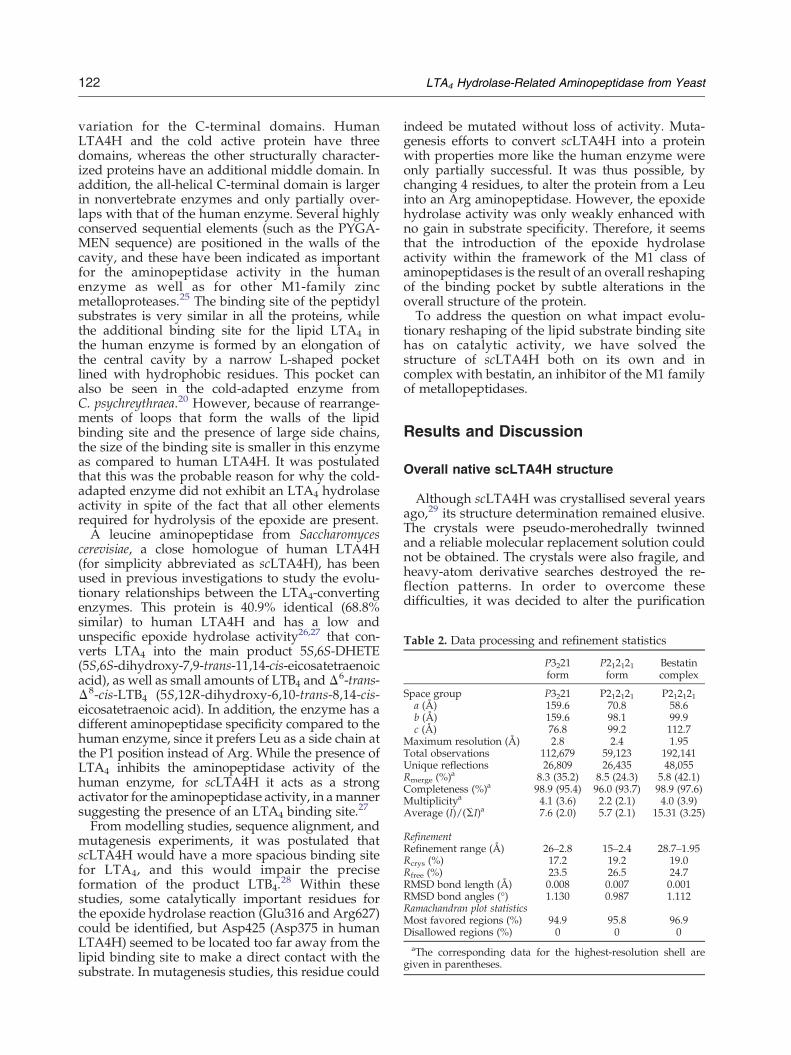
Table 2. Data processing and refinement statistics
P3221form
P212121form
Bestatincomplex
Space group P3221 P212121 P212121a (Å) 159.6 70.8 58.6b (Å) 159.6 98.1 99.9c (Å) 76.8 99.2 112.7
Maximum resolution (Å) 2.8 2.4 1.95Total observations 112,679 59,123 192,141Unique reflections 26,809 26,435 48,055Rmerge (%)a 8.3 (35.2) 8.5 (24.3) 5.8 (42.1)Completeness (%)a 98.9 (95.4) 96.0 (93.7) 98.9 (97.6)Multiplicitya 4.1 (3.6) 2.2 (2.1) 4.0 (3.9)Average (I)/(ΣI)a 7.6 (2.0) 5.7 (2.1) 15.31 (3.25)
RefinementRefinement range (Å) 26–2.8 15–2.4 28.7–1.95Rcrys (%) 17.2 19.2 19.0Rfree (%) 23.5 26.5 24.7RMSD bond length (Å) 0.008 0.007 0.001RMSD bond angles (°) 1.130 0.987 1.112Ramachandran plot statisticsMost favored regions (%) 94.9 95.8 96.9Disallowed regions (%) 0 0 0aThe corresponding data for the highest-resolution shell are
given in parentheses.
122 LTA4 Hydrolase-Related Aminopeptidase from Yeast
variation for the C-terminal domains. HumanLTA4H and the cold active protein have threedomains, whereas the other structurally character-ized proteins have an additional middle domain. Inaddition, the all-helical C-terminal domain is largerin nonvertebrate enzymes and only partially over-laps with that of the human enzyme. Several highlyconserved sequential elements (such as the PYGA-MEN sequence) are positioned in the walls of thecavity, and these have been indicated as importantfor the aminopeptidase activity in the humanenzyme as well as for other M1-family zincmetalloproteases.25 The binding site of the peptidylsubstrates is very similar in all the proteins, whilethe additional binding site for the lipid LTA4 inthe human enzyme is formed by an elongation ofthe central cavity by a narrow L-shaped pocketlined with hydrophobic residues. This pocket canalso be seen in the cold-adapted enzyme fromC. psychreythraea.20 However, because of rearrange-ments of loops that form the walls of the lipidbinding site and the presence of large side chains,the size of the binding site is smaller in this enzymeas compared to human LTA4H. It was postulatedthat this was the probable reason for why the cold-adapted enzyme did not exhibit an LTA4 hydrolaseactivity in spite of the fact that all other elementsrequired for hydrolysis of the epoxide are present.A leucine aminopeptidase from Saccharomyces
cerevisiae, a close homologue of human LTA4H(for simplicity abbreviated as scLTA4H), has beenused in previous investigations to study the evolu-tionary relationships between the LTA4-convertingenzymes. This protein is 40.9% identical (68.8%similar) to human LTA4H and has a low andunspecific epoxide hydrolase activity26,27 that con-verts LTA4 into the main product 5S,6S-DHETE(5S,6S-dihydroxy-7,9-trans-11,14-cis-eicosatetraenoicacid), as well as small amounts of LTB4 and Δ6-trans-Δ8-cis-LTB4 (5S,12R-dihydroxy-6,10-trans-8,14-cis-eicosatetraenoic acid). In addition, the enzyme has adifferent aminopeptidase specificity compared to thehuman enzyme, since it prefers Leu as a side chain atthe P1 position instead of Arg. While the presence ofLTA4 inhibits the aminopeptidase activity of thehuman enzyme, for scLTA4H it acts as a strongactivator for the aminopeptidase activity, in amannersuggesting the presence of an LTA4 binding site.27
From modelling studies, sequence alignment, andmutagenesis experiments, it was postulated thatscLTA4H would have a more spacious binding sitefor LTA4, and this would impair the preciseformation of the product LTB4.
28 Within thesestudies, some catalytically important residues forthe epoxide hydrolase reaction (Glu316 and Arg627)could be identified, but Asp425 (Asp375 in humanLTA4H) seemed to be located too far away from thelipid binding site to make a direct contact with thesubstrate. In mutagenesis studies, this residue could
indeed be mutated without loss of activity. Muta-genesis efforts to convert scLTA4H into a proteinwith properties more like the human enzyme wereonly partially successful. It was thus possible, bychanging 4 residues, to alter the protein from a Leuinto an Arg aminopeptidase. However, the epoxidehydrolase activity was only weakly enhanced withno gain in substrate specificity. Therefore, it seemsthat the introduction of the epoxide hydrolaseactivity within the framework of the M1 class ofaminopeptidases is the result of an overall reshapingof the binding pocket by subtle alterations in theoverall structure of the protein.To address the question on what impact evolu-
tionary reshaping of the lipid substrate binding sitehas on catalytic activity, we have solved thestructure of scLTA4H both on its own and incomplex with bestatin, an inhibitor of the M1 familyof metallopeptidases.
Results and Discussion
Overall native scLTA4H structure
Although scLTA4H was crystallised several yearsago,29 its structure determination remained elusive.The crystals were pseudo-merohedrally twinnedand a reliable molecular replacement solution couldnot be obtained. The crystals were also fragile, andheavy-atom derivative searches destroyed the re-flection patterns. In order to overcome thesedifficulties, it was decided to alter the purification

123LTA4 Hydrolase-Related Aminopeptidase from Yeast
scheme of the protein and recrystallise the proteininto another crystal form. This approach wassuccessful; by recloning the protein, expressing itas a GST fusion protein, and using a differentpurification approach, a new crystal form wasobtained. Molecular replacement using the structureof the human enzyme gave a clear solution, andsubsequent rebuilding and refining yielded a struc-ture with good statistics (see Table 2). Unambiguousdensity can be seen for the full-length protein, apartfrom a surface loop (226–230). During the refine-ment of the crystal form from the GST-affinitypurified protein, a glutathione molecule was iden-tified. It is located in the crystallographic interfacebetween symmetry-related protein molecules, and itis covalently bound to Cys147. One of its carboxylgroups interacts with His97, while the othercarboxyl group makes interactions with Lys601and Lys604 from a crystallographic neighbouringmolecule. Using the P3221 structure as a model formolecular replacement using PHASER,30 it waspossible to obtain a clear solution for the previoustwinned P212121 crystal form using data from acrystal that had a twin fraction of only 0.07. The twostructures superpose with each other with an RMSD
Fig. 1. Cartoon presentation of scLTA4H. (a) Structure of twith the bestatin inhibitor. All figures are produced with the pare coloured as follows: red, N-terminal domain; green, catamolecule is drawn as a stick model in aqua, while the active-
of 0.490 Å for 4412 atom pairs (out of 5092), and onlysmall conformational differences are seen in theloops 144–149 and 96–103. Both loops are involvedin glutathione binding and also make several crystalcontacts in the P3221 form. Loop 96 to 103 is flexiblein the P212121 form. Since data for the P212121 formextend to 2.4 Å instead of 2.8 Å in the case of theP3221 form, and only very minor changes can beseen between the two crystal forms, all followingresults and discussion are based on the P212121 form.ScLTA4 is built from three different domains (an
all-β N-terminal domain, residues 40–252; a mixedα-β catalytic domain, residues 253–506; and an allhelical C-terminal domain, residues 507–671) (Fig. 1).In comparison with the human enzyme, scLTA4Hhas an extension at the N terminus, some smalldeletions (3–4 residues) in the N-terminal domain,and two larger insertions (6 and 12 residues,respectively) located in loop areas between thehelices in the C-terminal domain.The catalytic domain is the most conserved (56%
identity, 76% similarity at the amino acid level) andshows only minor structural changes with an RMSDof 0.80 Å for 210 Cα positions. In contrast, theC-terminal domain is only 26.1% identical (46.0%
he enzyme alone; (b) structure of the enzyme in complexrogram Pymol31 if not indicated differently. Both cartoonslytic domain; and blue, C-terminal domain. The bestatinsite zinc molecules are drawn as grey spheres.
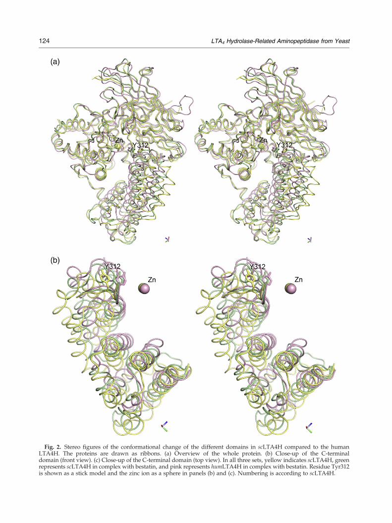
Fig. 2. Stereo figures of the conformational change of the different domains in scLTA4H compared to the humanLTA4H. The proteins are drawn as ribbons. (a) Overview of the whole protein. (b) Close-up of the C-terminaldomain (front view). (c) Close-up of the C-terminal domain (top view). In all three sets, yellow indicates scLTA4H, greenrepresents scLTA4H in complex with bestatin, and pink represents humLTA4H in complex with bestatin. Residue Tyr312is shown as a stick model and the zinc ion as a sphere in panels (b) and (c). Numbering is according to scLTA4H.
124 LTA4 Hydrolase-Related Aminopeptidase from Yeast

Fig. 2 (legend on previous page)
125LTA4 Hydrolase-Related Aminopeptidase from Yeast
similar), and here the RMSD is 1.28 Å for 119 Cα
positions. The larger insertions that are found in thisdomain are situated in the loop areas that connectthe helices. The largest insertion of 12 residuesforms a long meandering loop with no secondarystructure elements. For the N-terminal domain(33.8% identity, 50% similarity), there are onlyminor structural differences, with the RMSD being0.67 Å for 144 Cα positions. The extension at the Nterminus forms an extended loop with a small helixthat sits on top of the C-terminal helical lobe of thecatalytic domain. All further deletions and inser-tions are located in loop areas.The C-terminal domain shows not only the largest
local rearrangements but also its position relative tothe other two domains has been altered in compar-ison with human LTA4H (Fig. 2a and b). While theN-terminal and catalytic domains have a similarposition with respect to each other, the C-terminaldomain has been rotated 25°. The main impact ofthis rotation is that the cavity surrounding thecatalytic Zinc has become wider and more accessi-ble to solvent. The opening of the active site is notdue to a crystallographic artefact, since both crystalforms of the native enzyme show the same rotationof the C-terminal domain, in spite of the fact thatthe observed glutathione molecule in the P3221 formis bound to a residue (Cys147) near the opening ofthe cavity.
Inhibitor binding causes a domain movementand closure of the active site
The large conformational change of the C-terminaldomain suggested that scLTA4H might undergo aninduced fit upon substrate or inhibitor binding. Inorder to explore this possibility, the structure of thecomplex between scLTA4H and the aminopeptidaseinhibitor bestatin was solved. To avoid possiblecrystal-induced bias, crystals of the complex wereobtained by co-crystallisation rather than a soakingprocedure. The structure could be solved using thenative scLTA4H as a model, and after refinement,unambiguous density could be seen for the wholemodel. Fo−Fc difference density maps clearlyshowed the presence of the bestatin molecule inthe active site (Fig. 3a). The conformation of theinhibitor bound to scLTA4H is almost identical towhat is seen in the complex with the humanenzyme,21 with similar interactions to the zinc ionby the hydroxyl and carbonyl oxygen atoms. Thehydroxyl oxygen also makes an interaction toTyr429, while the carbonyl oxygen makes anadditional H-bond to Glu316. The phenyl ring ofbestatin is bound in a hydrophobic pocket linedwith Tyr312, Met315, Phe359, and the aliphatic partof the side chain of Glu186. This pocket is closed offat the bottom by the side chain of Phe424 (Fig. 3b,schematic picture). The main-chain amides of
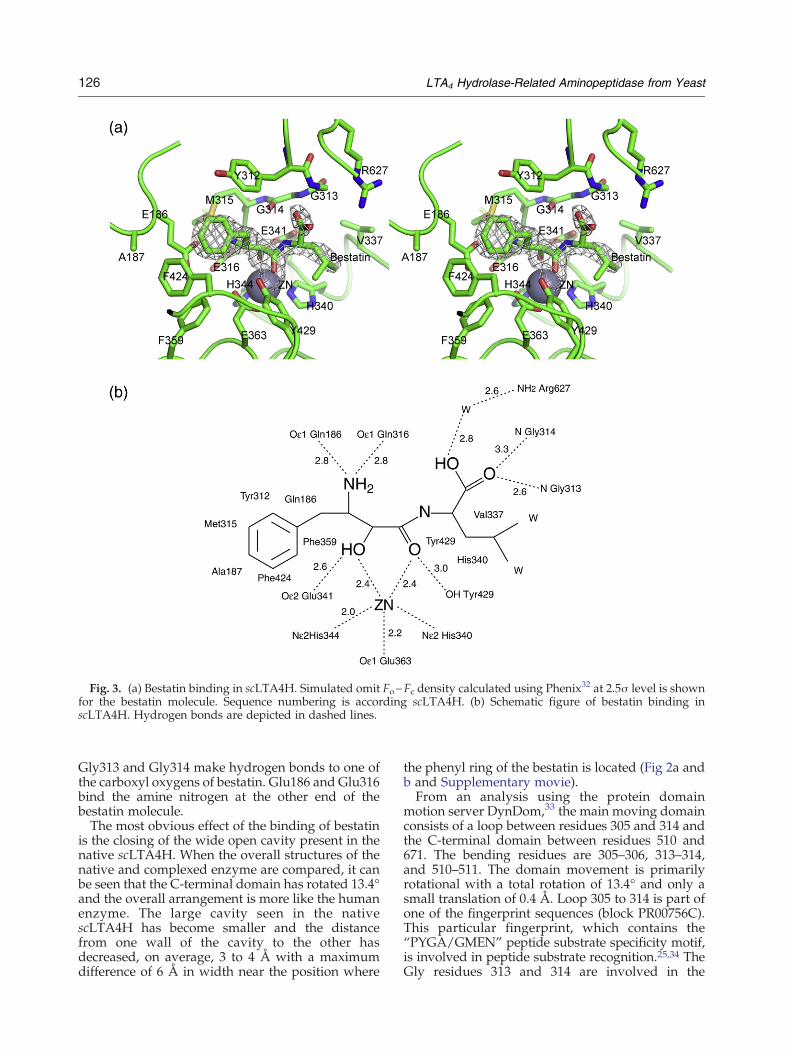
Fig. 3. (a) Bestatin binding in scLTA4H. Simulated omit Fo−Fc density calculated using Phenix32 at 2.5σ level is shownfor the bestatin molecule. Sequence numbering is according scLTA4H. (b) Schematic figure of bestatin binding inscLTA4H. Hydrogen bonds are depicted in dashed lines.
126 LTA4 Hydrolase-Related Aminopeptidase from Yeast
Gly313 and Gly314 make hydrogen bonds to one ofthe carboxyl oxygens of bestatin. Glu186 and Glu316bind the amine nitrogen at the other end of thebestatin molecule.The most obvious effect of the binding of bestatin
is the closing of the wide open cavity present in thenative scLTA4H. When the overall structures of thenative and complexed enzyme are compared, it canbe seen that the C-terminal domain has rotated 13.4°and the overall arrangement is more like the humanenzyme. The large cavity seen in the nativescLTA4H has become smaller and the distancefrom one wall of the cavity to the other hasdecreased, on average, 3 to 4 Å with a maximumdifference of 6 Å in width near the position where
the phenyl ring of the bestatin is located (Fig 2a andb and Supplementary movie).From an analysis using the protein domain
motion server DynDom,33 the main moving domainconsists of a loop between residues 305 and 314 andthe C-terminal domain between residues 510 and671. The bending residues are 305–306, 313–314,and 510–511. The domain movement is primarilyrotational with a total rotation of 13.4° and only asmall translation of 0.4 Å. Loop 305 to 314 is part ofone of the fingerprint sequences (block PR00756C).This particular fingerprint, which contains the“PYGA/GMEN” peptide substrate specificity motif,is involved in peptide substrate recognition.25,34 TheGly residues 313 and 314 are involved in the
127LTA4 Hydrolase-Related Aminopeptidase from Yeast
backbone recognition of the peptidyl substratemolecule,25 while the side chains of residues 312and 316 are part of the binding pocket for the sidechain of the P1 residue of the peptidyl substrate.This S1 pocket is also part of the binding site for thelipid substrate in the epoxide hydrolase active site inhuman LTA4H.The key residue for the conformational changes
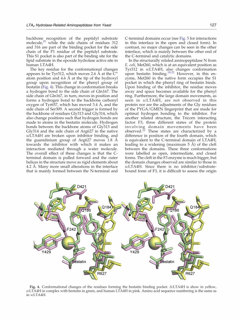
appears to be Tyr312, which moves 2.6 Å at the Cα
atom position and 4.6 Å at the tip of the hydroxylgroup upon recognition of the phenyl group ofbestatin (Fig. 4). This change in conformation breaksa hydrogen bond to the side chain of Gln167. Theside chain of Gln167, in turn, moves in position andforms a hydrogen bond to the backbone carbonyloxygen of Tyr657, which has moved 3.4 Å, and theside chain of Ser309. A second trigger is formed bythe backbone of residues Gly313 and Gly314, whichalso change positions such that hydrogen bonds aremade to atoms in the bestatin molecule. Hydrogenbonds between the backbone atoms of Gly313 andGly314 and the side chain of Arg627 in the nativescLTA4H are broken upon inhibitor binding, andthe guanidinium group of Arg627 moves 1.8 Åtowards the inhibitor with which it makes aninteraction mediated through a water molecule.The overall effect of these changes is that the C-terminal domain is pulled forward and the outerhelices in the structure move as rigid elements about4.2 Å. Many more small alterations in the interfacethat is mainly formed between the N-terminal and
Fig. 4. Conformational changes of the residues forming thscLTA4H in complex with bestatin in green, and human LTA4in scLTA4H.
C-terminal domains occur (see Fig. 5 for interactionsin this interface in the open and closed form). Incontrast, no major changes can be seen in the otherinterface, which is mainly between the other end ofthe C-terminal and catalytic domains.In the structurally related aminopeptidase N from
E. coli, Met260, which is at an equivalent position asTyr312 in scLTA4H, also changes conformationupon bestatin binding.18,22 However, in this en-zyme, Met260 in the native form occupies the S1pocket in which the phenyl ring of bestatin binds.Upon binding of the inhibitor, the residue movesaway and space becomes available for the phenylring. Furthermore, the large domain movements, asseen in scLTA4H, are not observed in thisprotein nor are the adjustments of the Gly residuesof the PYGA/GMEN fingerprint that are made foroptimal hydrogen bonding to the inhibitor. Foranother related structure, the Tricorn interactingfactor F3, three different states of the proteininvolving domain movements have beenobserved.19 These states are characterized by adifference in position of the fourth domain, whichis equivalent to the C-terminal domain of LTA4H,leading to a widening (maximum 5 Å) of the cleftbetween the domains. These three conformationswere labelled as open, intermediate, and closedforms. The cleft in the F3 enzyme is much bigger, butthe domain changes observed are similar to those inscLTA4H. Since there is no inhibitor/substrate-bound form of F3, it is difficult to assess the origin
e bestatin binding pocket. ScLTA4H is show in yellow,H in pink. Amino acid sequence numbering is the same as

Fig. 5. Stereo figure showing the conformational changes in the interface that mediate the domain movements betweenthe open and closed forms of scLTA4H. Open scLTA4H is in yellow, and closed scLTA4H in complex with bestatin is ingreen. The bestatin molecule is shown in green. For key residues, the side chains are depicted as stick models.
128 LTA4 Hydrolase-Related Aminopeptidase from Yeast
of the observed differences. The F3 enzyme containsan insertion right in front of the PYGA/GMENfingerprint, and Tyr312 is replaced by an Ala.However, the position of Tyr351, a putative catalyticresidue (Tyr429 in scLTA4H, Tyr383 in humanLTA4H), is rotated nearly 3 Å away from the zincatom, suggesting that none of the observed struc-tures of the F3 enzyme is productive and that furtherconformational changes have to occur to activatethis protein.19
Absence of critical Pro residues may explainflexibility of the C-terminal domain
For the human enzyme, no changes in the relativeposition of the C-terminal domain have beenobserved in previously solved structures. Althoughmost human LTA4H structures in the PDB databasecontain inhibitors or substrate analogues, the struc-ture of the R563A mutant (PDB code 1SQM)35 wasobtained without the presence of an inhibitor. Thisstructure only shows minimal differences comparedto the human LTA4H in complex with bestatin(RMSD). Some minor rearrangements are observednear the site of mutation, but there are no overallchanges in the domain architecture or significant
changes in conformation of residues involved ininhibitor binding. One reason for the seeminglyincreased rigidity of human LTA4H might be thepresence of two Pro residues in the first bendingregion at the equivalent positions of Val307 andAsp308. The presence of two rigid Pro residues justat the bending sites might prohibit the domainmovement as seen in the yeast protein. The cold-adapted aminopeptidase from C. psychrerythrae hasalso prolines at these positions. The structure of thisprotein without inhibitor is very similar to thehuman enzyme complexed with bestatin, and noopening of the active site can be seen.20 Interesting-ly, in most members of the LTA4H family, a doubleor, in some cases, a single proline is found at thesepositions. Only in LTA4H homologues from S.cerevisiae, C. glabrata, Z. rouxii, and V. polyspora, noproline residues are present at these positions andinstead a Val-Asp is found.
Comparing the substrate binding pocket in thehuman and yeast enzymes
The difference in peptidase specificity betweenhuman LTA4H (Arg) and scLTA4H (Leu) can beexplained by changes in both the size and nature of
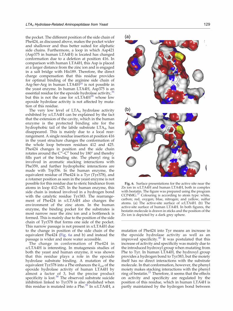
Fig. 6. Surface presentations for the active site near theZn ion in scLTA4H and human LTA4H, both in complexwith bestatin. The figure was prepared using the programCCP4MG.37 Colouring is according to atom type: white,carbon; red, oxygen; blue, nitrogen; and yellow, sulfuratoms. (a) The active-site surface of scLTA4H. (b) Theactive-site surface of human LTA4H. In both figures, thebestatin molecule is drawn in sticks and the position of theZn ion is depicted by a dark grey sphere.
129LTA4 Hydrolase-Related Aminopeptidase from Yeast
the pocket. The different position of the side chain ofPhe424, as discussed above, makes the pocket widerand shallower and thus better suited for aliphaticside chains. Furthermore, a loop in which Asp421(Asp375 in human LTA4H) is located has changedconformation due to a deletion at position 416. Incomparison with human LTA4H, this Asp is placedat a larger distance from the zinc ion and is engagedin a salt bridge with His189. Therefore, the directcharge compensation that this residue providesfor optimal binding of the arginine side chain ofArg-Ser-Arg in human LTA4H25 is not possible inthe yeast enzyme. In human LTA4H, Asp375 is anessential residue for the epoxide hydrolase activity,36
but this is not the case for scLTA4H28 whose lowepoxide hydrolase activity is not affected by muta-tion of this residue.The very low level of LTA4 hydrolase activity
exhibited by scLTA4H can be explained by the factthat the extension of the cavity, which in the humanenzyme is the protected binding site for thehydrophobic tail of the labile substrate LTA4, hasdisappeared. This is mainly due to a local rear-rangement. A single residue insertion at position 416in the yeast structure changes the conformation ofthe whole loop between residues 412 and 425.Phe424 changes in position and the side chainrotates around the Cα–Cβ bond by 180° and therebyfills part of the binding site. The phenyl ring isinvolved in aromatic stacking interactions withPhe359, and further hydrophobic interactions aremade with Trp356. In the human enzyme, theequivalent residue of Phe424 is a Tyr (Tyr378), anda rotamer position as seen in the yeast enzyme is notpossible for this residue due to steric hindrance fromatoms in loop 412–425. In the human enzyme, thisside chain is instead involved in a hydrogen bondwith the catalytic residue Tyr383. The rearrange-ment of Phe424 in scLTA4H also changes theenvironment of the zinc atom. In the humanenzyme, the binding pocket for the substrates ismost narrow near the zinc ion and a bottleneck isformed. This is mainly due to the position of the sidechain of Tyr378 that forms one side of the pocket.This narrow passage is not present in scLTA4H dueto the change in position of the side chain of theequivalent Phe424 (Fig. 6a and b) and instead thepassage is wider and more water accessible.The change in conformation of Phe424 in
scLTA4H is interesting. In mutagenesis studies ofboth the yeast and human enzyme, it was shownthat this residue plays a role in the epoxidehydrolase substrate binding. A mutation of theequivalent Tyr378 into a Phe enhances the kcat of theepoxide hydrolase activity of human LTA4H byalmost a factor of 3, but the precise productspecificity is lost.38 The observed substrate suicideinhibition linked to Tyr378 is also abolished whenthis residue is mutated into a Phe.39 In scLTA4H, a
mutation of Phe424 into Tyr means an increase inthe epoxide hydrolase activity as well as animproved specificity.40 It was postulated that thisincrease of activity and specificity was mainly due tothe introduced hydroxyl group whenmutating fromPhe to Tyr. In human LTA4H, the hydroxyl groupprovides a hydrogen bond to Tyr383, but the moietyitself has no direct interactions with the substratemolecule. In that conformation, however, the phenylmoiety makes stacking interactions with the phenylring of bestatin.21 Therefore, it seems that the effectson activity and specificity are regulated by theposition of this residue, which in human LTA4H ispartly maintained by the hydrogen bond between

130 LTA4 Hydrolase-Related Aminopeptidase from Yeast
the hydroxyl groups of Tyr378 and Tyr383. Asdiscussed above, the conformation of the Tyr378 inthe human enzyme makes the active-site pocketnarrow near the zinc atom, while in the yeast enzymethe channel near the zinc is considerably broader.However, other changes around the active site
play a role as well. In the cold-adapted aminopep-tidase, the equivalent residue to Phe424 is also a Phe,but no large structural differences between theposition of this residue and that of the Tyr378 inthe human LTA4H can be seen20 and the specificityof this protein has not changed. In scLTA4H, newstacking interactions between Phe424 and Phe359occur, while in the cold-adapted protein the residueat position 359 is not a Phe but instead a Leu.Moreover, just like human LTA4H, the cold-adaptedenzyme does not have an insertion at position 416,which means that a similar conformation as theyeast enzyme would also introduce some sterichindrance from residues at the equivalent positionsof 420 to 421.The change in size of the active site pocket is not
only due to local rearrangements and changes inside-chain conformations but also due to the overalldomain movement seen in the open and closedforms of scLTA4H. From an analysis using theDynDom server,33 it can be seen that the C-terminaldomain in scLTA4H complexed with bestatin is notas closed as in human LTA4H (scLTA4H + bestatin,65% closed; human LTA4H + bestatin, 83% closed,in reference to the uncomplexed scLTA4H). Thus, inthe scLTA4H–bestatin complex, the overall confor-mation of the C-terminal domain is in between thepositions it has in the native scLTA4H and thehuman LTA4H. One of the causes for this differencemight be the substitution of a Tyr for a Phe atposition 310 (scLTA4H numbering). Residue 310 ispart of the main rotating areas positioned betweenbending residue 305–306 and 313–314. The addi-tional hydroxyl group requires more space, and thisis provided by the translation of loop 308–312. Theloop 624–629 from the C-terminal domain ofscLTA4H has also been displaced slightly. Thismeans that the S1 pocket is somewhat larger in thescLTA4H and the domains cannot come as close asin the human enzyme.The structures of scLTA4H with and without
bestatin suggest that during evolution, the intro-duction of the epoxide hydrolase activity in thisclass of enzymes has been a process involving subtlestructural changes. The presence of the essentialcatalytic residues for the epoxide hydrolase activitywithin scLTA4H makes it plausible that this class ofrelated aminopeptidases was predisposed to obtaina moonlighting epoxide hydrolase function. This iscollaborated by the fact that scLTA4 has a lowepoxide hydrolase activity. Since all the requiredcatalytic residues for the epoxide hydrolase activityare present in scLTA4H, the main adaptation seems
to be a gradual process in which these residues arepositioned such that their relative positions arecompatible with a turnover of the lipid leukotrienesubstrate. At the same time, the substrate bindingsite has been adapted to provide a shieldedhydrophobic environment in which the labile allylicepoxide of LTA4 is protected from unwantedpremature cleavage resulting in biologically inactiveproducts. In the yeast enzyme, the pocket is both toolimited in size and too accessible to solvent toprovide this environment. Thus, as in the case forthe directed laboratory evolution experiments onserum paraoxonase and carbonic anhydrase II,9 itseems that an innate compatibility combined withgradual accumulation of mutations leads to acquire-ment of an additional function.The fact that scLTA4H displays an induced fit is
an interesting observation. Many members of theM1 family seem to display a certain degree ofinduced fit (see paragraph above), a feature that hasnever been observed for human LTA4H. It isattractive to speculate that a preformed pocketfunctioning through a lock-and-key mechanism isbetter suited to supply the protective and preciseenvironment required for hydrolysis of LTA4 intoLTB4. Further studies are required to fully under-stand the adaption of this intriguing class ofbifunctional enzymes.
Materials and Methods
Materials
Reagents for determination of protein concentrationwere obtained from Bio-Rad, and bestatin hydrochlorideand other chemicals were purchased from Sigma-Aldrichunless indicated otherwise. All chemicals used were of thehighest quality commercially available.
Cloning of GST and His-tagged scLTA4H
The full-length sequence of scLTA4H was cloned intothe expression vectors petM-11 and petM-30 (a kind gift ofDr. G. Stier, EMBL Hamburg), which add an N-terminal(His)6-tag and GST-(His)6-tag, respectively, to theexpressed recombinant proteins. The affinity tags can beremoved by proteolytic cleavage by tobacco etch virusprotease.A standard PCR reaction was carried out for
amplification of the scLTA4H gene with the primers5′-ccatggtgcctctttcaattgagcagag-3′ (forward) and 5′-gcggccgctcaaagacctaaatcttgtt-3′ (reverse) and Pfu TurboDNA polymerase (Promega). The forward primer intro-duced an NcoI restriction site, whereas the reverse primerintroduced a NotI restriction site downstream of thestop codon. The amplified DNA fragment of scLTA4Hwas purified from a 1% agarose gel and cloned into apCR-Blunt II-TOPO cloning vector using the Zero BluntTOPO PCR Cloning Kit (Invitrogen) according to the

131LTA4 Hydrolase-Related Aminopeptidase from Yeast
manufacturer's instructions. Six colonies from an agarplate were amplified and purified by a plasmid miniprep(Qiagen). The plasmids were digested with EcoRI(Fermentas) and analysed on a 1% agarose gel for insert.Positive clones were labelled as pCR-scLTA4H andfurther verified by automated DNA sequencing usinga BigDye Terminator version 3.0 DNA sequencing kit(Applied Biosystems). The plasmids pCR-scLTA4H,pETM-11, and pETM-30 were purified on a midiprepscale and double-digested with NcoI and NotI (Fermentas).Digestion reactions were run on a 1% agarose gel. TheDNA fragment encoding scLTA4H and linear vectorfragments of pETM-11 and pETM-30 were purified fromthe agarose gel using a gel extraction kit (Qiagen). Thepurified fragment of scLTA4H was ligated into pETM-11and pETM-30 using T4 DNA ligase (Fermentas) andtransformed into competent E. coli TOP-10 cells (Invitro-gen). Plasmids from six colonies of each construct wereminiprep purified and analysed by double digestion usingNcoI and NotI. Positive clones were subjected toexpression analysis. All six clones were confirmed tocontain the correct insert and transformed to BL21 (DE3)RIL cells. Small-scale expression tests showed no variationin protein expression among the different clones.
Expression and purification of GST-(His)6 -taggedscLTA4H
In large-scale expressions, both the (His)6-tagged andGST-(His)6-tagged proteins were expressed very well(N25 mg protein purified per liter of culture), but usingGST affinity purification as the first purification step gavehigher-purity protein. For the subsequent work, theGST-(His)6-tagged protein was used. The protein wasexpressed in BL21 (DE3) RIL cells by growing 2×1-litercultures at 37 °C to OD 0.5, lowering the temperature to20 °C and inducing expression with 1 mM IPTG atOD 0.7–0.8, followed by 4–6 h of incubation at 20 °C.The cells were harvested by centrifugation at 6000 rpmand the pellets were frozen.A frozen cell pellet from 2 liters of expression was
resuspended in 50 ml of 60 mM Tris–HCl (pH 7.5),100 mM NaCl, 10 mM NaN3 containing complete miniEDTA-free protease inhibitor (Roche) and lysed bysonication for 2×5 min. The insoluble cell debris wasremoved by centrifugation at 30,000g for 30 min, and thelysate filtered through a 0.45-μm syringe filter.The cleared lysate was mixed with 5 ml of glutathione
Sepharose 4B (GE Healthcare) that had been pre-equili-brated with 30 mMTris–HCl (pH 7.5), 50 mMNaCl, 5 mMNaN3 andwas incubated for 60 min on a shaking platformat room temperature. The lysate/resin mix was trans-ferred to a gravity flow column (BioRad EconoPack), andthe resin was allowed to drain. After washing with severalcolumn volumes of 30 mM Tris–HCl (pH 7.5), 50 mMNaCl, 5 mM NaN3, the bound protein was eluted with 3column volumes of 30 mM Tris–HCl (pH 7.5), 50 mMNaCl, 5 mM Na N3, 10 mM reduced glutathione.The eluted scLTA4H protein was mixed with AcTEV
protease (Invitrogen; 1 μl for each estimated mg ofprotein) and incubated at 4 °C overnight. To remove thecleaved affinity tag and uncleaved protein, the proteinsolution was adjusted to 500 mM NaCl and 10 mMimidazole and mixed with 5 ml of Ni2+ Sepharose FF
(GE healthcare) that had been pre-equilibrated with30 mM Tris–HCl (pH 7.5), 500 mM NaCl, 10 mMimidazole, 5 mM N3. After 60 min of incubation on ashaking platform at room temperature, the mix wastransferred to a gravity flow column and the resin wasallowed to drain. This flow-through contained thecleaved scLTA4H and was dialyzed overnight against2 liters of 20 mM Tris–HCl (pH 8.0).The protein solution was further diluted with 5
volumes of 20 mM Tris–HCl (pH 8.0) and loaded on a5-ml HiTrap Q FF ion exchange column (GE Healthcare)pre-equilibrated in 20 mM Tris–HCl (pH 8.0). The boundprotein was eluted by a steep salt gradient of 0–100%20 mM Tris–HCl (pH 8.0), 500 mM NaCl over 5 ml, andthe eluted fractions were pooled and concentrated toN3 mg/ml. The protein preparation was judged to beN90% pure by SDS-PAGE.ScLTA4Hwas further purified by injection on a Superdex
200 10/300 gel filtration column (GE Healthcare) pre-equlibrated with 20 mM Tris–HCl (pH 7.5) with a flow of0.3 ml/min. It is worth noting that a bimodal sizedistribution was always observed during the gel filtration,with one monomer peak and one multimer peak repre-senting 20–60% of total protein amounts. Additionalaggregation of protein from the monomer peak wasobserved already after 3 days of storage at 4 °C, andgel-filtration was always performed immediately beforesetting up crystallisation drops. Fractions from the mono-mer peak were pooled and concentrated to 2–12 mg/ml.
Crystallisation of the P3221 crystal form
Using the PACT screen (Molecular Dimensions) andvapour diffusion with scLTA4H at a concentration of6.6 mg/ml, initial crystal hits were identified withMMT buffer (L-malic acid, MES, Tris buffer; pH 5–6.5)with 25% polyethylene glycol (PEG) 1500 and in Hepes [4-(2-hydroxyethyl)-1-piperazineethanesulfonic acid; pH 7.5]with 20% PEG 6000. Further refinement of the crystal-lisation conditions revealed that scLTA4H (2–6 mg/ml)would crystallise in Hepes or MMT buffer between pH 7and 9 with 12–18% PEG 6000 or 15–20% PEG 1500.Crystals made this way were extremely small, flaky, andmixed with heavy precipitate. To obtain diffractionquality crystals, hanging drops of 2 μl of protein(6 mg/ml) and 1 μl of mother liquor [0.1 M MMTbuffer (pH 8), 12–14% PEG 6000] were allowed toequilibrate against the mother liquor for 18–24 h beforethe drops were streak seeded from earlier crystals.Crystals of acceptable size and morphology generallyappeared overnight in the seeded drops. All crystal-lisations were performed at 20 °C.
Purification and crystallisation of the P212121crystal form
The purification and crystallisation of the P212121 formhas been described elsewhere.29 The crystals were grownin liquid–liquid diffusion experiments using melting pointcapillaries in which 5 μl of protein (6 mg/ml) in 10 mMTris (pH 8.5) was layered on 5 μl of 14% PEG 8000 and100 mM Tris (pH 7.3). Rod-shaped crystals appeared in8 to 10 weeks at room temperature.

132 LTA4 Hydrolase-Related Aminopeptidase from Yeast
Crystallisation of the complex between scLTA4Hand bestatin
For crystallising the complex between scLTA4H andbestatin, the GST-(His)6 -tagged protein was purified asdescribed above. The protein was crystallised in thepresence of the inhibitor using the hanging drop vapourdiffusion. In the initial experiments, microcrystals werefound in condition #41 [0.085 M Hepes–Na (pH 7.5), 8.5%(v/v) isopropanol, 17% (w/v) PEG 4000, and 15%anhydrous glycerol] from the Crystal Screen Cryo(Hampton Research). After additive and pH screening,good-quality crystals were obtained in 15% glycerol,16.95% PEG 4000, 8.5% isopropanol, and 0.1 M MMTbuffer (pH 7.0) with 0.1 M betaine monohydrate as anadditive. The ratios of protein (9.6 mg/ml) with bestatin(6 mM), reservoir, and additive solutions in the finalcrystallisation drops were 2:1:0.5.
Data collection and processing
Data for the P3221 form of scLTA4H were collected atstation I911-2, and the data for the P212121 scLTA4H atstation I711 at MAX lab, Lund, Sweden, both on Mar165CCD detectors (Rayonix, MarResearch, USA). Data for theenzyme inhibitor complex were collected at beam stationI9I1-3 at MAX lab, Lund, Sweden, on a Mar225 CCDdetector. All data were collected at 100 K. Prior to datacollection, the crystals were transferred to appropriatecryo-solutions before being flash-cooled in a stream ofnitrogen gas at 100 K. The P3221 crystals were transferredto a drop of ultimate cryoprotectant (8% glycerol, 8%ethylene glycol, 9% sucrose, and 2% glucose) for less than1 min and flash frozen in the cryo-cooling stream at thedata collection station.The P212121 crystals were transferred in a stepwise
manner to a cryoprotecting solution consisting of 20%l-(+)-2,3-butanediol, 8% PEG 8000, 50 mM Tris (pH 7.3),and 2.5 mg/ml scLTA4H.29 For the inhibitor complex,the crystals were soaked in a 1:1 mixture of reservoir andUCP solutions and, after 1 min, transferred to thecryogenic stream of nitrogen. Complete data sets wereobtained from single crystals. Data processing andreduction for the P3221 data set was performed withthe program MOSFLM41 and programs from theCCP4-suite42; the data for the P212121 crystal form wereprocessed by Denzo and Scalepack43, while the data forthe complex were processed with XDS.44
Molecular replacement and refinement
In case of the P3121 crystal form of scLTA4H, thephasing problem was solved by molecular replacementusing PHASER30 with the structure of the human enzyme(PDB code 1hs6)21 as the searching model. Only proteinatoms were included in the calculations.Subsequent refinement was carried out using Phenix.32
2Fo−Fc and Fo−Fc electron density maps were used inCOOT45 for model inspection and rebuilding. At positionCys155, clear Fo−Fc density indicated the presence of anattached glutathione molecule remaining from the purifi-cation procedure. The glutathione could be built unam-biguously and was refined using full occupancy.
The structure of the P212121 crystal form was solved bymolecular replacement with PHASER30 using the struc-ture of the P3221 form of scLTA4H as the searching model.The crystal form has cell dimensions in which b and c havealmost the same values, and this gave rise to pseudo-merohedral twinning. Calculations by XTRIAGE from thePhenix package46 made it possible to determine the twinfractions of data sets from different crystals; by using datafrom a crystal with the lowest twin fraction (0.07), asolution for the molecular replacement problem wasobtained. The structure was refined by Phenix.32 For theP212121 form, one l-(+)-2,3-butanediol molecule and onetris(hydroxymethyl)aminomethane molecule were identi-fied in difference density maps.In case of the inhibitor complex, using the P212121
structure as a search model gave a clear signal inmolecular replacement calculations in PHASER.30 Rigid-body refinement with the different domains of the proteinas rigid objects was used to refine the clear domainmovement that had occurred. Subsequent refinement byPhenix32 and inspection and manual model building in2Fo−Fc and Fo−Fc electron density maps in Coot45 werefollowed. Clear difference electron density indicated thepresence of the bestatin molecule near the Zn ion.Validation by MolProbity47 indicated areas of the
structure that needed further rebuilding for all thedifferent structures.Detailed statistics for the refinement of the different
structures can be found in Table 2.
Accession numbers
The coordinates of the structures are deposited at thePDB with the following accession codes: scLTA4H P3121crystal form, 2xpy; scLTA4H P212121 crystal form, 2xpz;and the bestatin–scLTA4H complex, 2xq0.
Acknowledgements
The formulation of a ultimate cryoprotectantsolution recipe was generously provided by MarkPusey. This work was supported by an EMBO Long-Term Fellowship, the Swedish Research Council,CIDaT (Vinnova), and Karolinska Institutet.
Supplementary Data
Supplementary data associated with this articlecan be found, in the online version, at doi:10.1016/j.jmb.2010.11.059
References
1. Samuelsson, B. (1983). Leukotrienes: mediators ofimmediate hypersensitivity reactions and inflam-mation. Science, 220, 568–575.

133LTA4 Hydrolase-Related Aminopeptidase from Yeast
2. Ford-Hutchinson, A. W. (1990). Leukotriene B4 ininflammation. Crit. Rev. Immunol. 10, 1–12.
3. Haeggström, J. Z., Wetterholm, A., Vallee, B. L. &Samuelsson, B. (1990). Leukotriene A4 hydrolase: anepoxide hydrolase with peptidase activity. Biochem.Biophys. Res. Commun. 173, 431–437.
4. Minami, M., Ohishi, N., Mutoh, H., Izumi, T., Bito, H.,Wada, H. et al. (1990). Leukotriene A4 hydrolase is azinc-containing aminopeptidase. Biochem. Biophys.Res. Commun. 173, 620–626.
5. Orning, L., Gierse, J. K. & Fitzpatrick, F. A. (1994). Thebifunctional enzyme leukotriene A4 hydrolase is anarginine aminopeptidase of high efficiency andspecificity. J. Biol. Chem. 269, 11269–11273.
6. Haeggström, J. Z. (2004). Leukotriene A4 hydrolase/Aminopeptidase, the gatekeeper of chemotactic leuko-triene B4 biosynthesis. J. Biol. Chem. 279, 50639–50642.
7. Jeffery, C. J. (2004). Moonlightning proteins: complica-tions and implications for proteomics research. DrugDesign Today: Targets, 3, 71–78.
8. Huberts, D. H. E. W. & van der Klei, I. J. (2010).Moonlightning proteins: an intriguing mode ofmultitasking. Biochim. Biophys. Acta, 1803, 520–525.
9. Aharoni, A., Gaidukov, L., Khersonsky, O., Gould,S. M. c. Q., Roodveldt, C. & Tawfik, D. S. (2005).The ‘evolvability’ of promiscuous protein functions.Nat. Genet. 37, 73–76.
10. Snelgrove, R. J., Jackson, P. L., Hardison, M. T.,Noerager, B. D., Andrew Kinloch, A., Gaggar, A. et al.(2010). A critical role for LTA4H in limiting chronicpulmonary neutrophilic inflammation. Science, 330,70–74.
11. Barret, A. (1998). Family M1 of membrane alanylaminopeptidase. In Handbook of Proteolytic Enzymes(Barret, A. J., Rawlings, N. D. & Woessner, J. F., eds.),pp. 994–996, Academic Press, London.
12. Green, F. A. (1987). In vivo generation of 5-lipox-ygenase products in frogs and toads. Biochem. Biophys.Res. Commun. 148, 1533–1539.
13. Green, F. A., Herman, C. A., Herman, R. P., Claesson,H. E. & Hamberg, M. (1987). Monohydroxyeicosate-traenoic acid and leukotriene production by theinflammatory cells of Xenopus laevis. J. Exp. Zool. 243,211–215.
14. Habenicht, A. J., Goerig, M., Rothe, D. E., Specht, E.,Ziegler, R., Glomset, J. A. & Graf, T. (1989). Earlyreversible induction of leukotriene synthesis in chick-en myelomonocytic cells transformed by a tempera-ture-sensitive mutant of avian leukemia virus E26.Proc. Natl Acad. Sci. USA, 86, 921–924.
15. Pettitt, T. R., Rowley, A. F., Barrow, S. E., Mallet, A. I.& Secombes, C. J. (1991). Synthesis of lipoxins andother lipoxygenase products by macrophages fromthe rainbow trout, Oncorhynchus mykiss. J. Biol. Chem.266, 8720–8726.
16. Knight, J., Holland, J. W., Bowden, L. A., Halliday, K.& Rowley, A. F. (1995). Eicosanoid generatingcapacities of different tissues from the rainbow trout,Oncorhynchus mykiss. Lipids, 30, 451–458.
17. Baset, H. A., Ford-Hutchinson, A. W. & O'Neill, G. P.(1998). Molecular cloning and functional expression ofa Caenorhabditis elegans aminopeptidase structurallyrelated to mammalian leukotriene A4 hydrolases.J. Biol. Chem. 273, 27978–27987.
18. Ito, K., Nakajima, Y., Onohara, Y., Takeo, M.,Nakashima, K., Matsubara, F. et al. (2006).Crystal structure of aminopeptidase N (proteo-bacteria alanyl aminopeptidase) from Escherichiacoli and conformational change of methionine 260involved in substrate recognition. J. Biol. Chem. 281,33664–33676.
19. Kyrieleis, O. J., Goettig, P., Kiefersauer, R., Huber, R.& Brandstetter, H. (2005). Crystal structures of thetricorn interacting factor F3 from Thermoplasmaacidophilum, a zinc aminopeptidase in three differentconformations. J. Mol. Biol. 349, 787–800.
20. Bauvois, C., Jacquamet, L., Huston, A. L., Borel, F.,Feller, G. & Ferrer, J. L. (2008). Crystal structure of thecold-active aminopeptidase from Colwellia psychrer-ythraea, a close structural homologue of the humanbifunctional leukotriene A4 hydrolase. J. Biol. Chem.283, 23315–23325.
21. Thunnissen, M.M. G. M., Nordlund, P. & Haeggström,J. Z. (2001).Crystal structure of human leukotriene A(4)hydrolase, a bifunctional enzyme in inflammation.Nat. Struct. Biol. 8, 131–135.
22. Addlagatta, A., Gay, L. & Matthews, B. W. (2006).Structure of aminopeptidase N from Escherichia colisuggests a compartmentalized, gated active site. Proc.Natl Acad. Sci. USA, 103, 13339–13344.
23. Nocek, B., Mulligan, R., Bargassa, M., Collart, F. &Joachimiak, A. (2008). Crystal structure of aminopep-tidase N from human pathogen Neisseria meningitidis.Proteins, 70, 273–279.
24. McGowan, S., Porter, C. J., Lowther, J., Stack, C. M.,Golding, S. J., Skinner-Adams, T. S. et al. (2009).Structural basis for the inhibition of the essentialPlasmodium falciparum M1 neutral aminopeptidase.Proc. Natl Acad. Sci. USA, 106, 2537–2542.
25. Tholander, F., Muroya, A., Roques, B. P., Fournié-Zaluski, M. C., Thunnissen, M. M. G. M. &Haeggström, J. Z. (2008). Structure-based dissection ofthe active site chemistry of leukotriene A4 hydrolase:implications for M1 aminopeptidases and inhibitordesign. Chem. Biol. 15, 920–929.
26. Nars, F., Bécam, A. M. & Herbert, C. J. (1996). Thesequence of 12.8 kb from the left arm of chromosomeXIV reveals a sigma element, a pro-tRNA and sixcomplete open reading frames, one of which encodesa protein similar to the human leukotriene A4hydrolase. Yeast, 12, 493–499.
27. Kull, F., Ohlson, E. & Haeggström, J. Z. (1999).Cloning and characterization of a bifunctional leuko-triene A4 hydrolase from Saccharomyces cerevisiae.J. Biol. Chem. 274, 34683–34690.
28. Tholander, F., Kull, P., Ohlson, E., Thunnissen,M. M. G. M. & Haeggström, J. Z. (2005).Leukotriene A4 hydrolase: insights to the molecularevolution by homology modeling and mutationalanalysis of enzyme from Saccharomyces cerevisiae.J. Biol. Chem. 280, 33477–33486.
29. Andersson, B., Kull, F., Haeggström, J. Z. &Thunnissen, M. M. G. M. (2003). Crystallizationand X-ray diffraction data analysis of leukotrieneA4 hydrolase from Saccharomyces cerevisiae. ActaCrystallogr., Sect. D: Biol. Crystallogr. 59, 1093–1095.
30. McCoy, A. J., Grosse-Kunstleve, R. W., Adams, P. D.,Winn, M. D., Storoni, L. C. & Read, R. J. (2007). Phaser

134 LTA4 Hydrolase-Related Aminopeptidase from Yeast
crystallographic software. J. Appl. Crystallogr. 40,658–674.
31. DeLano, W. L. (2002). The PyMOL Molecular GraphicsSystem. DeLano Scientific, San Carlos, CA, USA.
32. Adams, P. D., Afonine, P. V., Bunkóczi, G., Chen,V. B., Davis, I. W., Echols, N. et al. (2010). PHENIX: acomprehensive Python-based system for macromo-lecular structure solution. Acta Crystallogr., Sect. D:Biol. Crystallogr. 66, 213–221.
33. Hayward, S. & Berendsen, H. J. C. (1998). Systematicanalysis of domain motions in proteins from confor-mational change; New results on Citrate Synthaseand T4 Lysozyme. Proteins, Structure, Function andGenetics, 30, 144–154.
34. Thunnissen, M. M. G. M., Andersson, B., Samuelsson,B., Wong, C. H. & Haeggstrom, J. Z. (2002). Crystalstructures of leukotriene A4 hydrolase in complexwith captopril and two competitive tight-bindinginhibitors. FASEB J. 16, 1648–1650.
35. Rudberg, P. C., Tholander, F., Andberg, M.,Thunnissen, M. M. G. M. & Haeggstrom, J. Z.(2004). Leukotriene A4 hydrolase: identification of acommon carboxylate recognition site for the epoxidehydrolase and aminopeptidase substrates. J. Biol.Chem. 279, 27376–27382.
36. Rudberg, P. C., Tholander, F., Thunnissen,M.M. G.M.,Samuelsson, B. & Haeggström, J. Z. (2002). LeukotrieneA4 hydrolase: selective abrogation of leukotriene B4formation by mutation of aspartic acid 375. Proc. NatlAcad. Sci. USA, 99, 4215–4220.
37. Potterton, L., McNicholas, S., Krissinel, E., Gruber, J.,Cowtan, K., Emsley, P. et al. (2004). Developments inthe CCP4 molecular-graphics project. Acta Crystallogr.,Sect. D: Biol. Crystallogr. 60, 2288–2294.
38. Mueller, M. J., Andberg, M. B., Samuelsson, B. &Haeggström, J. Z. (1996). Leukotriene A4 hydrolase,mutation of tyrosine 378 allows conversion of leuko-
triene A4 into an isomer of leukotriene B4. J. Biol. Chem.271, 24345–24348.
39. Mueller, M. J., Blomster, M., Oppermann, U. C. T.,Jörnvall, H., Samuelsson, B. & Haeggström, J. Z.(1996). Leukotriene A4 hydrolase: protection frommechanism-based inactivation by mutation of tyro-sine-378. Proc. Natl Acad. Sci. USA, 93, 5931–5935.
40. Kull, F., Ohlson, E., Lind, B. & Haeggström, J. Z.(2001). Leukotriene A4 hydrolase from Saccharomycescerevisiae, formation of leukotriene B4 and identifica-tion of catalytic residues. Biochemistry, 40, 12695–12703.
41. Leslie, A. G. W. (1992). “Recent changes to theMOSFLM package for processing film and imageplate data”, Joint CCP4 + ESF-EAMCB Newsletter onProtein Crystallography, No. 26.
42. Collaborative Computational Project N (1994). TheCCP4 suite—programs for protein crystallography.Acta Crystallogr., Sect. D: Biol. Crystallogr. 50, 760–763.
43. Otwinowski, Z. (1993). In Proceedings of the CCP4Study Weekend Data Collection and Processing (Saywer,L., Isaacs, N & bailey, S, eds.), pp. 80–86, Warrington:Daresbury Laboratories.
44. Kabsch, W. (1993). Automatic processing of rotationdiffraction data from crystals of initially unknownsymmetry and cell constants. J. Appl. Crystallogr. 26,795–800.
45. Emsley, P., Lohkamp, B., Scott, W. & Cowtan, K.(2010). Features and development of Coot. ActaCrystallogr. D, 66, 486–501.
46. Zwart, P. H., Grosse-Kunstleve, R. W. & Adams, P. D.(2005). Characterization of X-ray data sets.CCP4Newsl, 42; contribution 10.
47. Chen, V. B., Arendall, W. B., Headd, J. J., Keedy, D. A.,Immormino, R. M., Kapral, G. J. et al. (2010).MolProbity: all-atom structure validation for macro-molecular crystallography. Acta Crystallogr., Sect. D:Biol. Crystallogr. 66, 12–21.