Delimiting the Origin of a B Chromosome by FISH Mapping, Chromosome Painting and DNA Sequence...
17
Delimiting the Origin of a B Chromosome by FISH Mapping, Chromosome Painting and DNA Sequence Analysis in Astyanax paranae (Teleostei, Characiformes) Duı´lio M. Z. de A. Silva 1 *, Jose ´ Carlos Pansonato-Alves 1 , Ricardo Utsunomia 1 , Cristian Araya-Jaime 1 , Francisco J. Ruiz-Ruano 3 , Sandro Natal Daniel 4 , Diogo Teruo Hashimoto 2 , Cla ´ udio Oliveira 1 , Juan Pedro M. Camacho 3 , Fa ´ bio Porto-Foresti 4 , Fausto Foresti 1 1 Departamento de Morfologia, Instituto de Biocie ˆ ncias, Universidade Estadual Paulista, Distrito de Rubia ˜o Junior, Botucatu, Sa ˜o Paulo, Brazil, 2 CAUNESP, Universidade Estadual Paulista, Campus Jaboticabal, Jaboticabal, Sa ˜o Paulo, Brazil, 3 Departamento de Gene ´ tica, Universidad de Granada, Granada, Spain, 4 Departamento de Cie ˆ ncias Biolo ´ gicas, Faculdade de Cie ˆ ncias, Universidade Estadual Paulista, Campus de Bauru, Bauru, Sa ˜ o Paulo, Brazil Abstract Supernumerary (B) chromosomes have been shown to contain a wide variety of repetitive sequences. For this reason, fluorescent in situ hybridisation (FISH) is a useful tool for ascertaining the origin of these genomic elements, especially when combined with painting from microdissected B chromosomes. In order to investigate the origin of B chromosomes in the fish species Astyanax paranae, these two approaches were used along with PCR amplification of specific DNA sequences obtained from the B chromosomes and its comparison with those residing in the A chromosomes. Remarkably, chromosome painting with the one-arm metacentric B chromosome probe showed hybridization signals on entire B chromosome, while FISH mapping revealed the presence of H1 histone and 18S rDNA genes symmetrically placed in both arms of the B chromosome. These results support the hypothesis that the B chromosome of A. paranae is an isochromosome. Additionally, the chromosome pairs Nos. 2 or 23 are considered the possible B chromosome ancestors since both contain syntenic H1 and 18S rRNA sequences. The analysis of DNA sequence fragments of the histone and rRNA genes obtained from the microdissected B chromosomes showed high similarity with those obtained from 0B individuals, which supports the intraspecific origin of B chromosomes in A. paranae. Finally, the population hereby analysed showed a female-biased B chromosome presence suggesting that B chromosomes in this species could influence sex determinism. Citation: Silva DMZdA, Pansonato-Alves JC, Utsunomia R, Araya-Jaime C, Ruiz-Ruano FJ, et al. (2014) Delimiting the Origin of a B Chromosome by FISH Mapping, Chromosome Painting and DNA Sequence Analysis in Astyanax paranae (Teleostei, Characiformes). PLoS ONE 9(4): e94896. doi:10.1371/journal.pone.0094896 Editor: Igor V. Sharakhov, Virginia Tech, United States of America Received February 3, 2014; Accepted March 19, 2014; Published April 15, 2014 Copyright: ß 2014 Silva et al. This is an open-access article distributed under the terms of the Creative Commons Attribution License, which permits unrestricted use, distribution, and reproduction in any medium, provided the original author and source are credited. Funding: This research was funded by grants from the State of Sa ˜ o Paulo Research Foundation (FAPESP) to DMZAS (2011/16825-3) and CO (2010/17009-2), grants from National Council for Research and Development (CNPq) to FF and by Coordenac ¸a ˜o de Aperfeic ¸oamento de Pessoal de Nı ´vel Superior (CAPES). The funders had no role in study design, data collection and analysis, decision to publish, or preparation of the manuscript. Competing Interests: The authors have declared that no competing interests exist. * E-mail: [email protected] Introduction About 15% of eukaryotic species carry supernumerary (B) chromosomes, i.e. dispensable genomic elements present in only some individuals from a given population. In most cases, B chromosomes behave as parasitic elements prospering in natural populations due to their advantage in transmission (drive) not obeying Mendelian rules [1–6]. In some cases, B chromosomes appear to provide some advantage to the host genome and can thus be considered heterotic [7,8]. Examples of advantageous B chromosomes were already reported in the Nectria haematococca with B chromosomes favouring this fungus pathogenicity [9] and in the chive Allium schenoprassum, in which the B chromosomes increase viability from seed to seedling [10]. Historically, B chromosomes were considered inert genomic elements [11], but recent research has shown the transcription of several types of genes residing in B chromosomes, such as rRNA genes [12,13], repetitive DNA with similarity to mobile elements [14] and protein-coding genes in the Siberian roe deer Capreolus pygargus [15]. Interestingly, B chromosomes in a cichlid fish species have been shown to have a functional effect on female sex determination, with B chromosomes being restricted to females [16]. This effect is similar to the case of the paternal sex ratio (PSR) chromosome in wasps, in which a B chromosome has the opposite effect, i.e., transforming female determining zygotes into males and B chromosomes thus being restricted to males [6,17]. DNA composition of B chromosomes is unknown in most cases, even though this is an interesting topic in B chromosome research. In general, B chromosomes are rich in several classes of repetitive DNA, including 5S and 45S ribosomal DNA (rDNA), satellite DNA, histone genes, small nuclear DNA, mobile elements, and organellar sequences [11,18–25]. Chromosome painting has been used in several species to ascertain whether B chromosomes share DNA sequences with the A chromosomes from the same or other closely related species [20,26–32]. In addition, fluorescent in situ hybridisation (FISH) mapping of repetitive DNA has also provided valuable information in some cases [21,31,33–36]. Recently, next generation sequencing (NGS) technology has been applied to B chromosome research in three species: the fungus Mycosphaerella graminicola [37], the fruit fly Drosophila albomicans [38] and rye Secale PLOS ONE | www.plosone.org 1 April 2014 | Volume 9 | Issue 4 | e94896
-
Upload
independent -
Category
Documents
-
view
1 -
download
0
Transcript of Delimiting the Origin of a B Chromosome by FISH Mapping, Chromosome Painting and DNA Sequence...

Delimiting the Origin of a B Chromosome by FISHMapping, Chromosome Painting and DNA SequenceAnalysis in Astyanax paranae (Teleostei, Characiformes)Duılio M. Z. de A. Silva1*, Jose Carlos Pansonato-Alves1, Ricardo Utsunomia1, Cristian Araya-Jaime1,
Francisco J. Ruiz-Ruano3, Sandro Natal Daniel4, Diogo Teruo Hashimoto2, Claudio Oliveira1, Juan
Pedro M. Camacho3, Fabio Porto-Foresti4, Fausto Foresti1
1 Departamento de Morfologia, Instituto de Biociencias, Universidade Estadual Paulista, Distrito de Rubiao Junior, Botucatu, Sao Paulo, Brazil, 2 CAUNESP, Universidade
Estadual Paulista, Campus Jaboticabal, Jaboticabal, Sao Paulo, Brazil, 3 Departamento de Genetica, Universidad de Granada, Granada, Spain, 4 Departamento de Ciencias
Biologicas, Faculdade de Ciencias, Universidade Estadual Paulista, Campus de Bauru, Bauru, Sao Paulo, Brazil
Abstract
Supernumerary (B) chromosomes have been shown to contain a wide variety of repetitive sequences. For this reason,fluorescent in situ hybridisation (FISH) is a useful tool for ascertaining the origin of these genomic elements, especially whencombined with painting from microdissected B chromosomes. In order to investigate the origin of B chromosomes in thefish species Astyanax paranae, these two approaches were used along with PCR amplification of specific DNA sequencesobtained from the B chromosomes and its comparison with those residing in the A chromosomes. Remarkably,chromosome painting with the one-arm metacentric B chromosome probe showed hybridization signals on entire Bchromosome, while FISH mapping revealed the presence of H1 histone and 18S rDNA genes symmetrically placed in botharms of the B chromosome. These results support the hypothesis that the B chromosome of A. paranae is anisochromosome. Additionally, the chromosome pairs Nos. 2 or 23 are considered the possible B chromosome ancestorssince both contain syntenic H1 and 18S rRNA sequences. The analysis of DNA sequence fragments of the histone and rRNAgenes obtained from the microdissected B chromosomes showed high similarity with those obtained from 0B individuals,which supports the intraspecific origin of B chromosomes in A. paranae. Finally, the population hereby analysed showed afemale-biased B chromosome presence suggesting that B chromosomes in this species could influence sex determinism.
Citation: Silva DMZdA, Pansonato-Alves JC, Utsunomia R, Araya-Jaime C, Ruiz-Ruano FJ, et al. (2014) Delimiting the Origin of a B Chromosome by FISH Mapping,Chromosome Painting and DNA Sequence Analysis in Astyanax paranae (Teleostei, Characiformes). PLoS ONE 9(4): e94896. doi:10.1371/journal.pone.0094896
Editor: Igor V. Sharakhov, Virginia Tech, United States of America
Received February 3, 2014; Accepted March 19, 2014; Published April 15, 2014
Copyright: � 2014 Silva et al. This is an open-access article distributed under the terms of the Creative Commons Attribution License, which permits unrestricteduse, distribution, and reproduction in any medium, provided the original author and source are credited.
Funding: This research was funded by grants from the State of Sao Paulo Research Foundation (FAPESP) to DMZAS (2011/16825-3) and CO (2010/17009-2),grants from National Council for Research and Development (CNPq) to FF and by Coordenacao de Aperfeicoamento de Pessoal de Nıvel Superior (CAPES). Thefunders had no role in study design, data collection and analysis, decision to publish, or preparation of the manuscript.
Competing Interests: The authors have declared that no competing interests exist.
* E-mail: [email protected]
Introduction
About 15% of eukaryotic species carry supernumerary (B)
chromosomes, i.e. dispensable genomic elements present in only
some individuals from a given population. In most cases, B
chromosomes behave as parasitic elements prospering in natural
populations due to their advantage in transmission (drive) not
obeying Mendelian rules [1–6]. In some cases, B chromosomes
appear to provide some advantage to the host genome and can
thus be considered heterotic [7,8]. Examples of advantageous B
chromosomes were already reported in the Nectria haematococca with
B chromosomes favouring this fungus pathogenicity [9] and in the
chive Allium schenoprassum, in which the B chromosomes increase
viability from seed to seedling [10].
Historically, B chromosomes were considered inert genomic
elements [11], but recent research has shown the transcription of
several types of genes residing in B chromosomes, such as rRNA
genes [12,13], repetitive DNA with similarity to mobile elements
[14] and protein-coding genes in the Siberian roe deer Capreolus
pygargus [15]. Interestingly, B chromosomes in a cichlid fish species
have been shown to have a functional effect on female sex
determination, with B chromosomes being restricted to females
[16]. This effect is similar to the case of the paternal sex ratio
(PSR) chromosome in wasps, in which a B chromosome has the
opposite effect, i.e., transforming female determining zygotes into
males and B chromosomes thus being restricted to males [6,17].
DNA composition of B chromosomes is unknown in most cases,
even though this is an interesting topic in B chromosome research.
In general, B chromosomes are rich in several classes of repetitive
DNA, including 5S and 45S ribosomal DNA (rDNA), satellite
DNA, histone genes, small nuclear DNA, mobile elements, and
organellar sequences [11,18–25]. Chromosome painting has been
used in several species to ascertain whether B chromosomes share
DNA sequences with the A chromosomes from the same or other
closely related species [20,26–32]. In addition, fluorescent in situ
hybridisation (FISH) mapping of repetitive DNA has also provided
valuable information in some cases [21,31,33–36]. Recently, next
generation sequencing (NGS) technology has been applied to B
chromosome research in three species: the fungus Mycosphaerella
graminicola [37], the fruit fly Drosophila albomicans [38] and rye Secale
PLOS ONE | www.plosone.org 1 April 2014 | Volume 9 | Issue 4 | e94896

cereale [39]. This approach has shown that the B chromosome
content is a complex mixture of different types of DNA sequences,
including the already known types of repetitive DNAs (see above),
many insertions of organellar DNA and many pseudogenized
single-copy genes, which may be transcribed [39,40].
Although B chromosomes have been reported in a variety of fish
groups [41], few studies using a multiple molecular and
cytogenetic approach have been carried out. Among fishes, the
genus Astyanax represents an interesting model to study B
chromosomes, with occurrence records in ten species [41–47].
However, significant data about origin and evolution of those
chromosomes are restricted to the species A. scabripinnis [31,33].
Aiming to enhance the knowledge about composition, origin,
evolution and possible function of B chromosomes in A. paranae, we
performed chromosome painting with B-DNA microdissected
probes and FISH mapping with different repetitive DNA probes
(5S and 18S rDNA; H1, H3, and H4 histone genes; and Rex1 and
Rex3 transposable elements). Moreover, we analysed the nucleo-
tide sequences of ribosomal and histone multigene families
obtained from microdissected B chromosomes and from genomic
DNA of B-lacking individuals of A. paranae and three other Astyanax
species.
Materials and Methods
Ethics StatementSamples were collected in accordance with Brazilian environ-
mental protection legislation (collection permission MMA/
IBAMA/SISBIO - number 3245), and the procedures for
sampling, maintenance and analysis of the samples were
performed in compliance with the Brazilian College of Animal
Experimentation (COBEA) and was approved (protocol 405) by
the BIOSCIENCE INSTITUTE/UNESP ETHICS COMMIT-
TEE ON USE OF ANIMALS (CEUA).
Origin of samples and karyotypic analysisWe randomly collected 50 individuals (18 males and 32 females)
of the fish species Astyanax paranae Eigenmann, 1914 [48] and some
individuals without B chromosomes of A. bockmanni, A. fasciatus and
A. altiparanae, in the Capivara River belonging to the Tiete River
basin (22u5395799S/48u2391199W), close to Botucatu (Sao Paulo,
Brazil). The specimens were identified and deposited in the fish
collection of the Laboratorio de Biologia e Genetica de Peixes de
Botucatu, Sao Paulo, Brazil, under the numbers LBP13340 (A.
paranae), LBP13342 (A. bockmanni), LBP13344 (A. fasciatus) and
LBP13346 (A. altiparanae). To perform cytogenetic analyses, the
specimens were anesthetised and dissected, and the mitotic
chromosomes were obtained from kidney tissue and gills using
the technique described in Foresti et al. [49]. C-banding was
performed following the protocol described by Sumner [50], and
the nucleolar organiser regions (NORs) were identified according
to Howell and Black [51]. Chromosome morphology was
determined according to the criterion established by Levan et al.
[52], and thus, the morphology was classified as metacentric (m),
submetacentric (sm), subtelocentric (st), and acrocentric (a) and was
arranged in the karyotype in a decreasing size order.
Chromosome microdissectionMicrodissection was performed in an Eppendorf TransferMan
NK2 micromanipulator coupled to a Zeiss Axiovert 100 micro-
scope. We microdissected ten copies of a complete metacentric B
chromosome (Bm) for DNA sequencing (mB-DNA). Moreover, two
different B-derived DNA probes were generated through micro-
dissection for FISH experiments: 1) one arm of one copies of the
Bm chromosome (mBm-probe) (in order to test its isochromosome
nature) and 2) ten copies of a complete submetacentric B
chromosome (Bsm) (mBsm-probe). The microdissected DNAs were
placed in 9 ml of DNase-free ultrapure water and then amplified
using the GenomePlex Single Cell Whole Genome Amplification
Kit (wga4-Sigma) [53]. After the initial amplification, we
generated DNA probes (mBm-probe and mBsm-probe) labelled
with digoxigenin-11-dUTP (Roche Applied Science) using the
GenomePlex Whole Genome Amplification Reamplification Kit
(wga3-Sigma) following the manufacturer’s protocol.
DNA amplification, cloning and sequencingFor gDNA extraction, we used the Wizard Genomic DNA
Purification Kit (Promega) following manufacturer’s instructions.
For taxonomic confirmation of all materials collected, we
performed DNA barcoding by amplifying a partial sequence of
the mitochondrial gene Cytochrome c oxidase subunit 1 (COI),
with the primers: FishF1 59-TCAACCAACCACAAAGA-
CATTGGCAC-39 and FishR1 59-TAGACTTCTGGGTGGC-
CAAAGAATCA-39 [54].
Partial DNA sequences for 5S and 18S rDNA, ITS, H1, H3,
and H4 histone genes, and Rex1 and Rex3 transposable elements
(TEs) were obtained by PCR (Polymerase Chain Reaction) from A.
paranae genomic DNA (gDNA) lacking B chromosomes using the
primers described in Table S1. The reactions were performed in
1X PCR buffer, 1.5 mM of MgCl2, 200 mM of each dNTP
(dATP, dCTP, dGTP, dTTP), 0.5 U of Taq polymerase
(Invitrogen), 0.1 mM of each primer and 50 ng of gDNA. The
basic cycle to amplify these regions consisted of denaturation at
95uC for 5 min, followed by 30 cycles at 95uC for 1 min, 45 s at T
annealing uC (Table S1), 1 min at 72uC and a final extension of
10 min at 72uC.
The 18S rDNA, the ITS regions, and the H1 and H3 histone
genes were amplified on the A. paranae mB-DNA, and we also
amplified them on 0B-gDNA from A. paranae, A. bockmanni, A.
fasciatus and A. altiparanae. To isolate a representative diversity of
copies of these sequences present in the PCR reaction, we cloned
the PCR-obtained bands for these genes by linking them to a
TOPO TA cloning vector and cloning them in One Shot TOP10
Competent Cells. A number of clones were chosen for DNA
sequencing. We then isolated the plasmid DNA with the
Perfectprep Plasmid Mini kit (Eppendorff). For all DNA sequenc-
es, the PCR products were purified using the ExoSAP-IT kit (USB
Corporation) and were sequenced with the Big Dye TM
Terminator v 3.1 Cycle Sequencing Ready Reaction Kit (Applied
Biosystems), following manufacturer’s instructions. We sequenced
each plasmid in both directions using the M13F (59-GTAAAAC-
GACGGCCAG-39) and M13R (59-CAGGAAACAGCTATGAC-
39) primers.
Fluorescent in situ hybridisation (FISH)For FISH experiments, the chromosomes were treated accord-
ing to the procedures described by Pinkel et al. [55], using high
stringency conditions. The probes were labelled by PCR with
biotin-16-dUTP (Roche Applied Science) and the signal was
detected with avidin-FITC (Roche Applied Science), or else they
were labelled with digoxigenin-11-dUTP (Roche Applied Science)
and the signal was detected with anti-digoxigenin-rhodamine
(Roche Applied Science). The images were captured with a digital
camera (Olympus DP70) attached to an Olympus BX61
epifluorescence photomicroscope. Image treatment, including
karyotype mounting and optimisation of brightness and contrast,
was performed using the Adobe Photoshop CS4 program.
Delimiting the Origin of an B Chromosome
PLOS ONE | www.plosone.org 2 April 2014 | Volume 9 | Issue 4 | e94896

Sequence analysisAll DNA sequences were initially analysed with Geneious
version Pro 4.8.5 created by Biomatters (http://www.geneious.
com/) and sequence alignments were performed with Clustal W
[56] implemented in Geneious software. DNA diversity analyses
were performed with the DnaSP software [57]. Calculation of dN
and dS, and selection tests for protein coding DNA sequences were
performed with the MEGA software [58] using the Nei-Gojobori
method [59]. We built maximum likelihood trees for the 18S, H1,
H3 and ITS regions using the PhyML software [60] considering a
GTR + G model and performing a bootstrap analysis with 1000
replicates. A p-distance matrix among groups was performed for
the concatenated ITS sequences using pairwise deletion for
missing data. We inferred a species tree with all obtained
sequences from the gDNA of the Astyanax species and the B
chromosome of A. paranae for all the regions with two or more
clones by group, i.e, H1, H3 and the ITS, using the BEAST
software [61]. We launched three MCMC during 108 generations,
sampling a tree each 1000 generations. We assessed the
convergence of the three runs with Tracer and combined the
resulting trees with LogCombiner after applying a 10% burnin.
In order to confirm their identity, the sequences were used as
queries for BLASTn [62] searches against NCBI’s nr database
(http://www.ncbi.nlm.nih.gov/blast) and then were deposited in
the GenBank with the following accession numbers: KJ129765
(Rex1), KJ129638-KJ129645 (COI sequence), KJ129646-
KJ129668 (H1 histone gene), KJ129669-KJ129692 (H3 histone
gene), KJ129693-KJ129728 (ITS1 sequence), KJ129729-
KJ129764 (ITS2 sequence) and KJ129623-KJ129637 (18S
rDNA). For A. bockmanni species identification, we used COI
sequences deposited in GenBank under the accession numbers
KC153991-KC153994. The COI sequences were submitted to
the identification engine tool from BOLDSYSTEMS (http://
www.barcodinglife.com/index.php/IDS_OpenIdEngine). All se-
quences contaminated with ambient bacterial DNA were carefully
screened and discarded. Statistical comparisons by chi-square and
Student t tests were performed in a spreadsheet program.
Results
The molecular identification engine tool showed that the COI
region amplified from the analysed samples correspond to A.
paranae, A. bockmanni, A. fasciatus and A. altiparanae with 100% of
similarity in all cases. The analysed samples of A. paranae showed
2n = 50 chromosomes (8m+12sm+14st+16a, NF = 84) (Figure 1a).
Moreover, B chromosomes were observed in 100% of the cells in
12 out of 32 females, ten of which carried a metacentric macro B
(Bm) and two carried a submetacentric macro B (Bsm) (Figure 1a).
Only one out of the 18 males analysed carried the Bm
chromosome. No individual carried more than one B chromosome
per cell. A contingency chi-square test showed that B frequency
was nearly significantly higher in females (x2 = 3.67, df = 1,
P = 0.0555).
Chromosome analysisC-banding technique showed two types of heterochromatin:
faint C-positive blocks were present in some A chromosomes and
in the B chromosomes, while strong C-positive blocks were present
in the terminal region of the long arms of the acrocentric pairs
Nos. 18–22 (Figure 1b). Ag-NORs showed intra-population
variation, with pair No. 12 showing the principal NOR activity,
and pair No. 2 sometimes showing Ag-NOR positive signals
(Figure 1b). No Ag-NORs were observed in the B chromosomes.
Consistent with Ag-NOR patterns, chromosome pair No. 12
carried an 18S rDNA cluster in the short arm. Additionally, both
types of B chromosomes carried a small rDNA cluster in the
terminal region of both chromosome arms (Figures 1c,d, 2d,e). We
also observed the variable presence of rDNA clusters in six other A
chromosomes, namely pairs Nos. 1, 2, 7, 11, 18 and 23
(Figures 1c,d, 3); however, likewise Ag-NOR, only the rDNA in
chromosome 2 was observed as an active NOR (Figure 1b, inset).
The 5S rDNA was located in the pericentromeric region of the
pairs Nos. 2 and 20 (Figures 1c, 2a). H1, H3 and H4 histone sites
were found in synteny on the short arms of chromosomes 2 and 23
(Figures 1d, 2a, 2b, 2c). Also, H1 histone genes were detected in
Bm chromosome pericentromeric region distributed symmetrically
in both arms. There was an unequal distribution of the H1 histone
genes in Bsm chromosome, with most of them located in the long
arm (Figure 2a). The Rex1 transposable element was scattered
throughout all A chromosomes (Figure 2d), but Rex3 was
compartmentalised in the heterochromatic regions of the acro-
centric chromosomes no. 18–22 (Figure 2e).
The chromosome painting technique yielded similar patterns
with the two probes assayed (mBm-probe and mBsm-probe), with
the Bm and Bsm chromosomes being completely marked along
with small signals in some A chromosomes (Figure 2f). Remark-
ably, some of these signals were coincident with ribosomal and
histone sites, mainly in the short arms of pairs Nos. 2 and 23
(Figure 2f). All of the information obtained by chromosome
painting and FISH mapping is summarised in the ideogram in
Figure 3.
DNA sequence analysisWe tried to amplify several types of repetitive DNA (18S, ITS1
and ITS2 regions of the 45S rDNA, 5S rDNA and H1, H3 and
H4 histone genes) from 0B genomic DNA (0B-gDNA) and from
microdissected B-DNA (mB-DNA). All these repetitive DNAs were
amplified from 0B-gDNA, but only 18S, ITS H1 and H3 were
amplified from microdissected DNA from B chromosome. We got
the full sequence of all cloned regions. However, due to the length
of ITS region (1800 bp), about 250 bp of the ITS2 region could
not be included in the analysis.
A comparison of nucleotide diversity for the DNA sequences
found in the microdissected B chromosomes with those obtained
from 0B individuals showed significant differences for 18S rDNA,
the ITS1 region and the H3 histone gene, but not for the ITS2
region or the H1 histone gene (Table 1). Whereas ITS1 and H3
showed lower nucleotide diversity in the B chromosome copies,
the 18S rDNA region showed higher diversity in the B
chromosome copies.
Considering the H1 and H3 histone genes, the amplified regions
included codons 14–163 for H1 and codons 4–121 for H3. Six out
of the 150 aminoacid positions analysed in H1 showed variation,
but no variation was found for H3. Additionally, no stop codons
were observed for both genes. We calculated the number of
synonymous and non-synonymous substitutions per synonymous
(dS) and non-synonymous (dN) site, respectively, for each group of
sequences. As shown in Table 2, no significant differences were
observed between the DNA sequences coming from the A
chromosomes (0B-gDNA) or the B chromosomes (mB). Both
synonymous and non-synonymous substitutions were found for the
H1 gene in both types of DNA sequences (0B-gDNA and mB), with
dN/dS ratios being much lower than 1, but being three times
higher for the DNA sequences coming from the B chromosome
(Table 2), suggesting the possibility that purifying selection is
relaxed for the H1 genes located in the B chromosomes. In fact, a
comparison of the A. paranae H1 haplotypes with those obtained in
Delimiting the Origin of an B Chromosome
PLOS ONE | www.plosone.org 3 April 2014 | Volume 9 | Issue 4 | e94896

the Astyanax species, by the Nei–Gojobori neutrality test, provided
significant evidence for purifying selection (Table S2).
Maximum-likelihood trees built with the sequences obtained
from the microdissected B chromosomes, the 0B-gDNA from A.
paranae, and gDNA from A. bockmanni, A. fasciatus and A. altiparanae,
were not resolutive for 18S rDNA and the two histone genes due
to the scarce variation observed (Figures S1–S3). Thus, concate-
nated ITS regions were used and suggested higher similarity
between the sequences obtained from the B chromosomes and
those from 0B-gDNA in A. paranae and A. bockmanni, and lower
similarity with those from A. fasciatus and A. altiparanae (Figure 4).
These conclusions are supported by the average p-distances shown
in the Table S3. Additionally, a species tree performed by the
BEAST method for the histone genes and the ITS regions
indicated that the DNA sequences contained in the B chromosome
of A. paranae are most similar to those in the A chromosomes of this
same species, thus pointing to an intraspecific origin for the B
chromosome (Figure 5).
Discussion
B chromosomes in the genus Astyanax were first reported in A.
scabripinnis as a large metacentric B chromosome similar to the
largest A chromosome pair [63,64]. Also, several B chromosomes
have been found in different species within this genus, presenting
variation in morphology, size and DNA composition [41,42,47].
In A. paranae, large B chromosomes have also been reported from
the Tiete River basin [64,65], but no compositional information
had been reported yet.
Our results from chromosome painting with the DNA probes
obtained from the Bm and Bsm chromosomes suggest that A and
B chromosomes share similar DNA sequences. This finding
suggests an intraspecific origin for this B chromosome (see 11) and
points towards two A chromosomes (Nos. 2 and 23) as possible
candidates to have been the B ancestor. However, this detected
similarity may be attributed to the presence of large 18S rDNA
and H1 histone clusters in these chromosomes. Although the
anonymity of DNA probes used in the chromosome painting
technique does not give great consistency to this conclusion, the
combination of this technique with FISH mapping of repetitive
DNAs, and the comparative analysis of such sequences from the
microdissected B chromosomes, with those in the A chromosomes
of A. paranae and other closely related species, has given strong
support to the intraspecific origin for this B chromosome.
In fish, only satellite DNA, 45S rDNA and transposable
elements had previously been found in the B chromosomes of
Astyanax scabripinnis [33], Prochilodus lineatus [66], Haplochromis
obliquidens [36], Astatotilapia latifasciata [67], Alburnus alburnus [68],
while active nucleolar sites were observed in the B chromosomes of
Moenkausia sanctaefilomenae [69]. Here, we show the first evidence of
histone protein genes allocated in a fish B chromosome, a fact that
had previously been reported only in the grasshoppers Locusta
migratoria [21] and Rhammatocerus brasiliensis [22]. Also, both B
Figure 1. Karyotypes of Astyanax paranae from the Capivara River after conventional staining (A), C-banding (B), and double FISHwith 5S and 18S rDNA probes (C) and 18S rDNA and H1 histone (D). Ag-NORs are represented in the box. Bar = 10 mm.doi:10.1371/journal.pone.0094896.g001
Delimiting the Origin of an B Chromosome
PLOS ONE | www.plosone.org 4 April 2014 | Volume 9 | Issue 4 | e94896

chromosome types (Bm and Bsm) found in A. paranae carried 18S
rDNA in the terminal region of both chromosome arms. The
symmetric distribution of 18S rDNA and H1 histone genes in both
arms suggests that the B chromosome is an isochromosome, as
previously shown for a very similar macro B chromosome in A.
scabripinnis [33]. Indeed, chromosome painting with the mBm-
probe (which was obtained from a single B arm) painted the whole
Bm and Bsm chromosomes, thus suggesting the homology
between both B arms and the homology between both B types.
In addition, the differences between the relative numbers of H1
histone genes in both arms of Bsm related to the Bm chromosome
suggest that the origin of the former Bsm was via a pericentric
inversion with asymmetrical breakpoints in the H1 regions of both
arms. Remarkably, similar Bm and derived Bsm chromosomes
have been reported in A. scabripinnis [31,33,42,70–72].
Figure 2. Metaphases of Astyanax paranae from the Capivara River after double-FISH with 5S rDNA and H1 histone (A), H1 and H3histone (B), H1 and H4 histone (C), Rex1 and 18S rDNA (D), Rex3 and 18S rDNA (E) and after chromosome painting with mBm-probe(F). Bar = 10 mm.doi:10.1371/journal.pone.0094896.g002
Figure 3. Ideogram of Astyanax paranae from the Capivara River showing the main cytogenetic markers localisation used in thispaper. In the box, a summary of the FISH markers and chromosome painting associated with B chromosome in chromosomes 2, 23, Bm and Bsm.doi:10.1371/journal.pone.0094896.g003
Delimiting the Origin of an B Chromosome
PLOS ONE | www.plosone.org 5 April 2014 | Volume 9 | Issue 4 | e94896

The B chromosome origin through isochromosome in A. paranae
implies that its two arms are homologous. Therefore, any
explanation of B origin, in this case, needs to be limited to one
arm only, as the final step of B origin is the centromeric
misdivision of an acrocentric proto-B chromosome yielding a
metacentric iso-B. In fact, the painting probe we generated from
microdissection of a single chromosome arm of the metacentric B
chromosome actually painted the two arms, thus demonstrating its
isochromosome nature. We did not find useful information for 5S
rDNA or Rex retrotransposons among the mapped repetitive DNA
probes but our results with the 18S rDNA and histone genes
clearly pointed to two A chromosomes as the most likely A
chromosome ancestors of B chromosomes in this species. These
were chromosomes Nos. 2 and 23 based on the remarkable fact
that they are the only A chromosomes carrying the two repetitive
DNAs (detected by FISH) found in the B chromosomes, i.e., 18S
rDNA and H1 histone genes. These genes occupy distant locations
in the B chromosomes: H1 being near the centromeric region (i.e.,
proximal) but 18S rDNA being close to the long arm telomere (i.e.,
distal). The comparison with these repetitive DNAs locations in
chromosomes Nos. 2 and 23 indicates that the origin of the B
chromosomes was not simple.
If the ancestor was chromosome 2, the B chromosomes could
have derived from the short arm region containing the H1 and
18S rDNA, however, some additional DNA should have been
placed in between these two sequences and the centromeric 5S
rDNA should have been lost. Alternatively, the B chromosome
could have derived from the long arm of chromosome 23,
provided that it contains H1 histone genes in its proximal region;
which would explain the presence of distal 18S rDNA and
proximal H1 histone genes in the B chromosome, and the
presence of DNA of unknown nature (the so-called B heterochro-
matin) between the two repetitive DNAs. This would suggest
chromosome 23 as the most parsimonious hypothesis on the origin
of B chromosomes. The absence of visible H3 and H4 histone sites
in the B chromosomes, despite their syntenic condition with H1 in
chromosomes 2 and 23 could be attributed to the occurrence of
orphon genes (solitary genetic elements derived from tandem
multigene families) that spread from this element [73]. Remark-
ably, a very similar A chromosome (no. 24) was suggested to be
large metacentric B ancestor chromosome in A. scabripinnis
[31,33,70], which suggests the possibility of a common origin for
B chromosomes in these two species [74].
The higher dN/dS ratio of mB-DNA sequences compared with
the A-chromosomes sequences (Table 2) indicates the possibility
that purifying selection is relaxed for the H1 genes located in the B
chromosomes, as expected if they were genetically inactive and
could explain their differential amplification in the B chromosome.
In fact, the lack of selective pressure on B chromosomes of rye
allows the accumulation of Revolver repetitive DNA on these
elements [75]. Conversely, the number of H3 copies in the B
chromosome is probably lower, due to the absence of visible FISH
signals. This could explain the lower nucleotide diversity observed
in this gene of B chromosome sequences compared with those in
the A chromosomes (Table 1).
The majority of the H1 and H3 histone sequences isolated from
A. paranae B chromosomes showed the same putative amino acid
sequence as those obtained from 0B genomic DNA, thus
Table 1. Nucleotide diversity (p) for the DNA sequences analysed, and Student (t) tests comparing A chromosome (0B-gDNA) andmicrodissected B chromosome (mB) sequences.
Gene Sample n Sites S Hap p SE t df P
18S 0B-gDNA 8 1338 8 6 0.0019 0.0002
mB 5 1338 21 5 0.0072 0.0007 27.48 11 0.00001
ITS1 0B-gDNA 5 428 20 4 0.0250 0.0033
mB 10 422 10 8 0.0051 0.0003 5.98 13 0.00005
ITS2 0B-gDNA 5 384 3 3 0.0032 0.0006
mB 10 384 6 5 0.0031 0.0004 0.04 13 0.97173
H1 0B-gDNA 8 451 22 7 0.0230 0.0020
mB 8 451 29 6 0.0172 0.0027 1.71 14 0.10860
H3 0B-gDNA 9 355 25 5 0.0174 0.0032
mB 7 355 8 5 0.0093 0.0008 2.43 14 0.02887
Where n = number of sequences, S = number of segregating sites, Hap = number of haplotypes, SE = standard error, df = degrees of freedom, P = probability.doi:10.1371/journal.pone.0094896.t001
Table 2. Number of synonymous and non-synonymous substitutions per synonymous (dS) and non-synonymous (dN) site,respectively, observed in the DNA sequences of the H1 and H3 histone genes obtained from 0B genomic DNA (0B-gDNA) and Bmicrodissected DNA (mB).
Gene Sample n dN SE t df P dS SE t df P dN/dS
H1 0B-gDNA 8 0.004 0.002 0.077 0.016 0.052
mB 8 0.007 0.002 21.06 14 0.307 0.045 0.01 1.70 14 0.112 0.156
H3 0B-gDNA 9 0 0 0.068 0.011 0
mB 7 0 0 0.036 0.012 1.97 14 0.069 0
doi:10.1371/journal.pone.0094896.t002
Delimiting the Origin of an B Chromosome
PLOS ONE | www.plosone.org 6 April 2014 | Volume 9 | Issue 4 | e94896

Figure 4. Maximum likelihood tree built with the concatenated ITS rDNA sequences. The sequences were obtained from themicrodissected B chromosomes, gDNA from 0B A. paranae individuals, and gDNA from A. bockmanni, A. fasciatus and A. altiparanae (Aalti) specimens.Note the high similarity of the B chromosome sequences (uBpar) and the sequences obtained from 0B-gDNA in A. paranae (Apar) and A. bockmanni(Abock), and the lower similarity to those from A. fasciatus (Afasc).doi:10.1371/journal.pone.0094896.g004
Figure 5. Species tree built with the histone genes and ITS regions by BEAST. Note that the DNA sequences of A. paranae obtained fromthe microdissected B chromosomes (mB-DNA) are most similar to those in the host genome (0B-gDNA).doi:10.1371/journal.pone.0094896.g005
Delimiting the Origin of an B Chromosome
PLOS ONE | www.plosone.org 7 April 2014 | Volume 9 | Issue 4 | e94896

suggesting that they are potentially active. Teruel et al. [21]
reported the presence of H3 and H4 histone sequences with the
same putative amino acid sequences in the L. migratoria B
chromosomes as those obtained from 0B genomic DNA indicating
that they are potentially active. Ruiz-Estevez et al. [76] detected B-
derived rRNA sequences in grasshopper Eyprepocnemis plorans
confirming that B sequences are potentially active and can
contribute to the host genome functions. Pseudogenic sequences of
B chromosomes in rye are expressed and have potential to regulate
the expression of A-located genes [40]. These examples show that
B chromosomes are not always inert genomic elements, highlight-
ing the necessity of future investigations about the potential
activity of genes in A. paranae B chromosome.
The nucleotide diversity observed in the rDNA obtained from
the B chromosome, compared with that shown by the same
sequences in the A chromosomes unveiled some interesting facts.
First, the 18S rDNA showed higher diversity in B chromosome
than in A chromosomes, which would be consistent with the
inactivity of B-rDNA. In fact, the Ag-NOR technique suggests that
it is always inactive. On this basis, it should be expected that
nucleotide diversity were also higher in B chromosome at ITS
regions. However, it was about similar in the ITS2 and
significantly lower in the ITS1 region (see Table 1). A possible
explanation would be that the distal location of rDNA in both B
chromosome arms, and the fact that iso-B-chromosomes frequent-
ly show distal chiasmata between the two homologous arms (which
in the A. paranae B chromosome would involve the rDNA), would
lead to the homogenization of the rDNA in both B chromosome
arm ends through crossover, assuming that the B chromosome of
A. paranae has the same behaviour on the meiosis as the B
chromosome of A. scabripinnis [33]. However, in this work we did
not analyse the meiosis behaviour of B chromosome due to the
scarcity of male samples bearing B chromosomes. Moreover, this
technique is a laborious task to apply in Teleostei females, since
the pachytene phase occur in the early stages of the development
[77]. Another possible explanation to the ITS nucleotide diversity
in the B chromosome is based on the remarkable spreading of 18S
rDNA in different chromosomes (Figures 1c, d, 2d, e). Thus,
considering that interchromosomal homogenization rates are
lower than intrachromosomal events [78,79], the B-sequences
(isolated from a single chromosome) might present a lower
diversity when compared with 0B-gDNA (with several 18S rDNA
A-chromosomes sites). Although these possibilities are very
interesting, we realize that the limited number of DNA sequences
analysed could also be the responsible for former contradictions;
therefore further research with larger samples is needed.
The species tree built with the sequences of H1, H3 and ITS
regions (Figure 5) was more informative than the gene trees
(Figures 4, S1–S3) because it clearly separated the species A.
bockmanni and A. paranae from the two other species (A. altiparanae
and A. fasciatus), with posterior branch probability being higher
than 0.95. In addition, this tree shows that A. paranae mB-DNA is
closer to A. paranae 0B-gDNA indicating an intraspecific origin for
this B chromosome. However, the posterior probability for the
branch separating the 0B and mB sequences in A. paranae from
those in A. bockmanni was only 0.61, thus casting some doubts about
the topology obtained. This is probably due to the fact that A.
paranae and A. bockmanni belong to the A. scabripinnis species
complex which includes several closely related species sharing a
very recent common ancestor, and this latter could already
harbour the B chromosomes which could have be inherited by the
descent species [74].
B chromosomes that are restricted to one sex have already been
described in Astyanax scabripinnis, such as a micro B chromosome
restricted to males [80] or a macro B restricted to females [81].
These cases were interpreted as the result of possible chromosome
elimination from the somatic tissues analysed [80]. Of course, this
possibility cannot be excluded without analysing the germ line in
all of these cases. However, there is another interesting explana-
tion focused on the possible effects of gene content in B
chromosomes. The A. paranae sample collected in the Capivara
River, analysed here showed a significant female biased sex ratio
distortion because 32 individuals were female but only 18 were
male (x2 = 3.92, df = 1, P = 0.048). A previous sample of this same
species by Maistro et al. [64] in the Araqua River (another
tributary of the Tiete River) failed to find B chromosomes in
males. Although Porto-Foresti et al. [65] found a female biased sex
ratio showing B chromosomes present in females 57.1% and males
8.7% at the same collection site. A similar scenario has been
observed for the large B chromosomes in A. scabripinnis [70]. The
female-biased B chromosome presence in A. paranae shown here
suggests the possibility that B chromosomes in this species
influence sex determining. Remarkably, Yoshida et al. [16] have
recently provided evidence that B chromosomes in a cichlid fish
species exert functional effects on sex determination. Therefore, it
would be interesting to investigate the presence of possible genes
related with sex determinism in the A. paranae B chromosomes, and
their possible activity during the embryo stages where sex
determination is occurring.
Supporting Information
Figure S1 Maximum likelihood tree built with the 18S rDNA
sequences. The sequences were obtained from the microdissected
B chromosomes (Apar_B), gDNA from 0B A. paranae individuals
(Apar_gen), gDNA from A. fasciatus specimens (Afasc), with A.
altiparanae (Aalti) as outgroup.
(TIF)
Figure S2 Maximum likelihood tree built with the H1 gene
sequences. The sequences were obtained from the microdissected
B chromosomes (Apar_B), gDNA from 0B A. paranae individuals
(Apar_gen), gDNA from A. bockmanni (Abock) and A. fasciatus
specimens (Afasc), using A. altiparanae (Aalti) as outgroup.
(TIF)
Figure S3 Maximum likelihood tree built with the H3 gene
sequences. The sequences were obtained from the microdissected
B chromosomes (Apar_B), gDNA from 0B A. paranae individuals
(Apar_gen), gDNA from A. bockmanni (Abock) and A. fasciatus
specimens (Afasc), using A. altiparanae (Aalti) as outgroup.
(TIF)
Table S1 DNA sequence of the primers employed for PCR
amplification of the different repetitive DNAs assayed.
(DOCX)
Table S2 Codon-based test of neutrality, positive selection and
purifying selection for the H1 histone gene partial sequence.
(DOCX)
Table S3 Genetic divergence values among species. Note the
higher similarity between the sequences obtained from the B
chromosomes (Apar_B) and those from 0B-gDNA in A. paranae
(Apar_gen) and A. bockmanni (Abock), and lower similarity with
those from A. fasciatus (Afasc) and A. altiparanae (Aalti).
(DOCX)
Acknowledgments
We thank Renato Devide for support during sample collection and the
anonymous reviewers for their substantial contributions.
Delimiting the Origin of an B Chromosome
PLOS ONE | www.plosone.org 8 April 2014 | Volume 9 | Issue 4 | e94896

Author Contributions
Conceived and designed the experiments: DMZAS JCPA RU SND DTH
CO JPMC FF FPF. Performed the experiments: DMZAS JCPA RU CAJ
FJRR SND FPF. Analyzed the data: DMZAS JCPA RU CAJ FJRR SND
DTH CO JPMC FPF FF. Contributed reagents/materials/analysis tools:
FJRR CO JPMC FF. Wrote the paper: DMZAS JCPA RU FJRR DTH
CO JPMC FF.
References
1. Ostergren G (1945) Parasitic nature of extra fragment chromosomes. Bot Notiser
2: 157–163.
2. Nur U (1966) Harmful B chromosomes in a mealy bug population. Genetics 54:
1225–1238.
3. Nur U (1977) Maintenance of a ‘parasitic’ B chromosome in the grasshopper
Melanoplus femur-rubrum. Genetics 87: 499–512.
4. Jones RN (1985) Are B chromosomes selfish? In The evolution of genome size
(ed.T . Cavalier-Smith), pp. 397–425. London:Wiley.
5. Jones RN (1991) B-chromosome drive. Am. Nat. 137, 430–442.
6. Werren JH, Nur U, Eickbush DG (1987) An extrachromosomal factor causing
loss of paternal chromosomes. Nature 327: 75–76.
7. Darlington CD (1958) ‘‘Evolution of genetic systems’’. Oliver and Boyd,
Edinburgh and London.
8. White MJD (1973) Animal Cytology and Evolution. 3rd ed. London: Cambridge
University Press.
9. Miao VP, Covert SF, VanEtten HD (1991) A fungal gene antibiotic resistance
on a dispensable (‘‘B’’) chromosome. Science 254(5039): 1773–1776.
10. Plowman AB, Bougourd SM (1994) Selectively advantageous effects of B
chromosomes on germination behavior in Allium schoenoprasum L. Heredity 72:
587–593.
11. Camacho JPM (2005) B Chromosomes. In: Gregory TR editor: The evolution of
the genome. Elsevier, San Diego pp. 223–286.
12. Leach CR, Houben A, Bruce F, Pistrick K, Demidov D, et al. (2005) Molecular
evidence for transcription of genes on a B chromosome in Crepis capillaris.
Genetics 171: 269–278.
13. Ruız-Estevez M, Lopez-Leon MD, Cabrero J, Camacho JPM (2012) B-
Chromosome Ribosomal DNA Is Functional in the Grasshopper Eyprepocnemis
plorans. PLoS ONE 7(5): e36600.
14. Carchilan M, Kumke K, Mikolajewski S, Houben A (2009) Rye B chromosomes
are weakly transcribed and might alter the transcriptional activity of A
chromosome sequences. Chromosoma 118: 607–616.
15. Trifonov VA, Dementyeva PV, Larkin DM, O’Brien PCM, Perelman PL, et
al.(2013) Transcription of a protein-coding gene on B chromosomes of the
Siberian roe deer (Capreolus pygargus). BMC Biology 11: 90.
16. Yoshida K, Terai Y, Mizoiri S, Aibara M, Nishihara H, et al. (2011) B
Chromosomes Have a Functional Effect on Female Sex Determination in Lake
Victoria Cichlid Fishes. PLoS Genet 7(8): e1002203.
17. Werren JH, Stouthamer R (2003) PSR (paternal sex ratio) chromosomes: the
ultimate selfish genetic elements. Genetica 117: 85–101.
18. Friebe B, Jiang J, Gill B (1995) Detection of 5S rDNA and other repeated DNA
on supernumerary B chromosomes of Triticum species (Poaceae). Pl. Syst. Evol.
196: 131–139.
19. Cabrero J, Bakkali M, Bugrov A, Warchalowska-Sliwa E, Lopez-Leon MD, et
al. (2003) Multiregional origin of B chromosomes in the grasshopper
Eyprepocnemis plorans. Chromosoma 112: 207–211.
20. Bugrov AG, Karamysheva TV, Perepelov EA, Elisaphenko EA, Rubtsov DN, et
al. (2007) DNA content of the B chromosomes in grasshopper Podisma kanoi
Storozh. (Orthoptera, Acrididae). Chromosome Res. 15: 315–325.
21. Teruel M, Cabrero J, Perfectti F, Camacho JPM (2010) B chromosome ancestry
revealed by histone genes in the migratory locust. Chromosoma 119: 217–225.
22. Oliveira NL, Cabral-de-Mello DC, Rocha MF, Loreto V, Martins C, et al.
(2011) Chromosomal mapping of rDNAs and H3 histone sequences in the
grasshopper Rhammatocerus brasiliensis (acrididae, gomphocerinae): extensive
chromosomal dispersion and co-localization of 5S rDNA/H3 histone clusters
in the A complement and B chromosome. Molecular Cytogenetics 4: 24.
23. Bueno D, Palacios-Gimenez OM, Cabral-de-Mello DC (2013) Chromosomal
Mapping of Repetitive DNAs in the Grasshopper Abracris flavolineata Reveal
Possible Ancestry of the B Chromosome and H3 Histone Spreading. PLosONE
8(6): e66532.
24. Kour G, Kaul S, Dhar MK (2014) Molecular Characterization of Repetitive
DNA Sequences from B Chromosome in Plantago lagopus L. Cytogenet Genome
Res 142: 121–128.
25. Houben A, Banaei-Moghaddam AM, Klemme S, Timmis JN (2013) Evolution
and biology of supernumerary B chromosomes. Cell Mol Life Sci 71: 467–478.
26. Long H, Qi ZX, Sun XM, Chen CB, Li XL, et al. (2008) Characters of DNA
constitution in the rye B chromosome. J. Integr Plant Biol. 50: 183–189.
27. Rubtsov NB, Karamysheva TV, Andreenkova OV, Bochkaerev MN, Kartavt-
seva IV, et al. (2004) Comparative analysis of micro and macro B chromosomes
in the Korean field mouse Apodemus peninsulae (Rodentia, Murinae) performed by
chromosome microdissection and FISH. Cytogenet Genome Res. 106: 289–294.
28. Bugrov AG, Karamysheva TV, Pyatkova MS, Rubtsov DN, Andreenkova OV,
et al. (2003) B chromosomes of the Podisma sapporensis Shir (Orthoptera,
Acrididae) analyzed by chromosome microdissection and FISH. Folia Biol
Krakow. 51: 1–11.
29. Teruel M, Cabrero J, Montiel EE, Acosta MJ, Sanchez A, et al. (2009)
Microdissection and chromosome painting of X and B chromosomes in Locusta
migratoria. Chromosome Research 17: 11–18.
30. Teruel M, Cabrero J, Perfectti F, Acosta MJ, Sanchez A, et al. (2009)
Microdissection and chromosome painting of X and B chromosomes in the
Grasshopper Eyprepocnemis plorans. Cytogenet Genome Res 125: 286–291.
31. Vicari MR, Pistune HFM, Castro JP, Almeida MC, Bertollo LAC, et al. (2011)
New insights on the origin of B chromosomes in Astyanax scabripinnis obtained by
chromosome painting and FISH. Genetica 139: 1073–1081.
32. Martins CCC, Diniz D, Sobrinho-Scudeler PE, Foresti F, Campos LAO, et al.
(2013) Investigation of Partamona helleri (Apidae, Meliponini) B chromosome
origin. An approach by microdissection and whole chromosome painting.
Apidologie 44: 75–81.
33. Mestriner CA, Galetti PM Jr, Valentini SR, Ruiz IRG, Abel LDS, et al. (2000)
Structural and functional evidence that a B chromosome in the characid fish
Astyanax scabripinnis is an isochromosome. Heredity 85: 1–9.
34. Stitou S, Dıaz de la Guardia R, Jimenez R, Burgos M (2000) Inactive ribosomal
cistrons are spread throughout the B chromosomes of Rattus rattus (Rodentia,
Muridae). Implications for their origin and evolution. Chromosome Res 8: 305–
311.
35. Dhar MK, Friebe B, Koul AK, Gill B (2002) Origin of an apparent B
chromosome by mutation, chromosome fragmentation an specific DNA
sequence amplification. Chromosoma 111: 332–340.
36. Poletto AB, Ferreira IA, Martins C (2010) The B chromosomes of the African
cichlid fish Haplochromis obliquidens harbour 18S rRNA gene copies. BMC
Genetics 11: 1.
37. Goodwin SB, Ben M’Barek S, Dhillon B, Wittenberg AHJ, Crane CF, et al.
(2011) Finished Genome of the Fungal Wheat Pathogen Mycosphaerella graminicola
Reveals Dispensome Structure, Chromosome Plasticity, and Stealth Pathogen-
esis. PLoS Genet 7(6): e1002070.
38. Zhou Q, Hong-mei Z, Quang-fei, Zhao L, Zhang GJ, et al. (2012) Deciphering
neo-sex and B chromosome evolution by the draft genome of Drosophila
albomicans. BMC Genomics 13: 109.
39. Martis MM, Klemme S, Banaei-Moghaddam AM, Blattner FR, Macas J, et al.
(2012) Selfish supernumerary chromosome reveals its origin as a mosaic of host
genome and organellar. PNAS 109(33): 13343–13346.
40. Banaei-Moghaddam AM, Meier K, Karimi-Ashtiyani R, Houben A (2013)
Formation and expression of pseudogenes on the B chromosome of rye. Plant
Cell 25(7): 2536.
41. Oliveira C, Foresti F, Hilsdorf AWS (2009) Genetics of neotropical fishes: from
chromosomes to populations. Fish Physiol Biochem 35: 81–100.
42. Moreira-Filho O, Galetti PM Jr, Bertollo LAC (2004) B chromosomes in the fish
Astyanax scabripinnis (Characidae, Tetragonopterinae) An overview in natural
populations. Cytogenet Genome Res 106: 230–234.
43. Fernandes CA, Martins-Santos IC (2005) Sympatric occurrence of three
cytotypes and four morphological types of B chromosomes of Astyanax scabripinnis
(Pisces, Characiformes) in the River Ivaı Basin, state of Parana, Brazil. Genetica
124: 301–306.
44. Carvalho RA, Martins-Santos IC, Dias AL (2008) B chromosomes: an update
about their occurrence in freshwater Neotropical fishes (Teleostei). Journal of
Fish Biology 72: 1907–1932.
45. Hashimoto DT, Porto-Foresti F (2010) Chromosome polymorphism and
heterochromatin and nucleolar regions in two populations of the fish Astyanax
bockmanni (Teleostei: Characiformes). Neotrop ichth 8: 861–866.
46. Daniel SN, Hashimoto DT, Pansonato-Alves JC, Foresti F, Porto-Foresti F
(2012) Cytogenetic characterization of distinct B chromosomes in a population
of the fish Astyanax bockmanni (Teleostei, Characiformes). Caryologia 65(3): 228–
233.
47. Santos LP, Castro JP, Francisco CM, Vicari MR, Almeida MC, et al. (2013)
Cytogenetic analysis in the neotropical fish Astyanax goyacensis Eigenmann, 1908
(Characidae, incertae sedis): karyotype description and occurrence of B
microchromosomes. Molecular Cytogenetics 6: 48.
48. Eigenmann CH (1914) Some results from studies of South American fishes. IV.
New genera and species of South American fishes. Indiana University Studies,
20: 44–48.
49. Foresti F, Toledo-Filho AS, Almeida-Toledo LF (1981) Polymorphic nature of
the nucleolus organizer regions in fishes. Cytogenet Cell Genet 31: 134–141.
50. Sumner AT (1972) A simple technique for demonstrating centromeric
heterochromatin. Expl. Cell Res. 75: 304–306.
51. Howell WM, Black DA (1980) Controlled silver-staining of nucleolus organizer
regions with a protective colloidal developer: a 1-step method. Experientia 36:
1014–1015.
52. Levan A, Fredga K, Sandberg AA (1964) Nomenclature for centromeric position
of chromosomes. Hereditas 52: 201–220.
Delimiting the Origin of an B Chromosome
PLOS ONE | www.plosone.org 9 April 2014 | Volume 9 | Issue 4 | e94896

53. Gribble S, Ng BL, Prigmore E, Burford DC, Carter NP (2004) Chromosome
paints from single copies of chromosomes. Chromosome Res 12: 143–151.
54. Ward RD, Zemlak TS, Innes BH, Last PR, Hebert PD (2005) DNA Barcoding
of Australia’s fish species. Phil. Trans. R. Soc. B. 360: 1847–1857.
55. Pinkel D, Straume T, Gray JW (1986) Cytogenetic analysis using quantitative,
high sensitivity, fluorescence hybridization. Proc. Natl. Acad. Sci 83: 2934–2938.
56. Thompson JD, Higgins DG, Gibson TJ (1994) CLUSTAL W: improving the
sensitivity of progressive multiple sequence alignment through sequence
weighting, position specific gap penalties and weight matrix choice. Nucl Acids
Res 22: 4673–4680.
57. Librado P, Rozas J (2009) DnaSP v5: a software for comprehensive analysis of
DNA polymorphism data. Bioinformatics 25: 1451–1452.
58. Tamura K, Dudley J, Nei M, Kumar S (2007) MEGA4: molecular evolutionary
genetics analysis (MEGA) software version 4.0. Mol Biol Evol 24: 1596–1599.
59. Nei M, Gojobori T (1986) Simple methods for estimating the numbers of
synonymous and nonsynonymous nucleotide substitutions. Mol Biol Evol 3:
418–426.
60. Guindon S, Dufayard JF, Lefort V, Anisimova M, Hordijk W, et al. (2010) New
algorithms and methods to estimate maximum-likelihood phylogenies: assessing
the performance of PhyML 3.0. Systematic biology 59(3):307–321.
61. Heled J, Drummond AJ (2010). Bayesian inference of species trees from
multilocus data. Molecular biology and evolution 27(3):570–580.
62. Altschul SF, Gish W, Miller W, Myers EW, Lipman DJ (1990) Basic local
alignment search tool. Journal of Molecular Biology 215: 403–410.
63. Salvador LB, Moreira-Filho O (1992) B chromosomes in Astyanax scabripinnis
(Pisces, Characidae) Heredity 69: 50–56.
64. Maistro EL, Foresti F, Oliveira C, Almeida-Toledo LF (1992) Occurrence of
macro B chromosomes in Astyanax scabripinnis paranae (Pisces, Characiformes,
Characidae). Genetica 87: 101–106.
65. Porto-Foresti F, Oliveira C, Maistro EL, Foresti F (1997) Estimated frequency of
B-chromosomes and population density B chromosome in of Astyanax scabripinnis
paranae in a small stream. Brazil J Genet 20: 377–380.
66. Artoni RF, Vicari MR, Endler AL, Cavallaro ZI, Jesus CM, et al. (2006)
Banding pattern of A and B chromosome of Prochilodus lineatus (Characiformes,
Prochilodontidae), with comments on B chromosome evolution. Genetica 127:
277–284.
67. Fantinatti BE, Mazzuchelli J, Valente GT, Cabral-de-Mello DC, Martins C
(2011) Genomic content and new insights on the origin of the B chromosome of
the cichlid fish Astatotilapia latifasciata. Genetica 139: 273–282.
68. Ziegler CG, Lamatsch DK, Steinlein C, Engel W, Schartl M, et al.(2003) The
giant B chromosome of the cyprinid fish Alburnus alburnus harbours aretrotransposon-derived repetitive DNA sequence. Chromosome Res 11: 23–35.
69. Hashimoto DT, Voltolin TA, Paes ADNVA, Foresti F, Bortolozzi J, et al. (2012)
Cytogenetic analysis of B chromosomes in one population of the fish Moenkhausia
sanctaefilomenae (Steindachner, 1907) (Teleostei, Characiformes). Comp Cytogen
6(2): 141–151.70. Vicente VE, Moreira-Filho O, Camacho JPM (1996) Sex-ratio distortion
associated with the presence of a B chromosome in Astyanax scabripinnis (Teleostei,
Characidae). Cytogenet Cell Genet 74: 70–75.71. Neo DM, Bertollo LAC, Moreira-Filho O (2000) Morphological differentiation
and possible origin of B chromosomes in natural Brazilian populations ofAstyanax scabripinnis (Pisces, Characidae). Genetica 108: 211–215.
72. Ferro DAM, Moreira-Filho O, Bertollo LAC (2003) B chromosome polymor-phism in the fish, Astyanax scabripinnis. Genetica 119: 147–153.
73. Childs G, Maxson R, Cohn RH, Kedes L (1981) Orphons: dispersed genetic
elements derived from tandem repetitive genes of eucaryotes. Cell 23: 651–66.74. Moreira-Filho O, Fenocchio AS, Pastori MC, Bertollo LAC (2001) Occurrence
of a metacentric macrochromosome B in different species of the genus Astyanax
(Pisces, Characidae, Tetragonopterinae). Cytologia 66: 59–64.
75. Klemme S, Banaei-Moghaddam AM, Macas J, Wicker T, Novak P, et al. (2013)
High-copy sequences reveal distinct evolution of the rye B chromosome. NewPhytologist 199: 550–558.
76. Ruız-Estevez M, Lopez-Leon M, Cabrero J, Camacho JPM (2013) RibosomalDNA is active in different B chromosome variants of the grasshopper
Eyprepocnemis plorans. Genetica 141: 337–345.77. Grier H (2000) Ovarian germinal epithelium and folliculogenesis in the common
snook, Centropomus undecimalis (Teleostei: centropomidae). Journal Of Morphol-
ogy 243(3):265–81.78. Kuhn GC, Kuttler H, Moreira-Filho O, Heslop-Harrison JS (2012) The 1.688
repetitive DNA of Drosophila: concerted evolution at different genomic scalesand association with genes. Mol Biol Evol. 29(1):7–11.
79. Teruel M, Ruız-Ruano FJ, Marchal JA, Sanchez A, Cabrero J, et al. (2013)
Disparate molecular evolution of two types of repetitive DNAs in the genome ofthe grasshopper Eyprepocnemis plorans. Heredity 1–12.
80. Rocon-Stange EA, Almeida-Toledo LF (1993) Supernumerary B chromosomesrestricted to males in Astyanax scabripinnis (Pisces, Characidae). Braz. J. Genet. 16:
601–615.81. Mizoguchi SMHN, Martins-Santos IC (1997) Macro- and microchromosomes B
in females of Astyanax scabripinnis (Pisces, Characidae). Hereditas 127: 249–253.
Delimiting the Origin of an B Chromosome
PLOS ONE | www.plosone.org 10 April 2014 | Volume 9 | Issue 4 | e94896


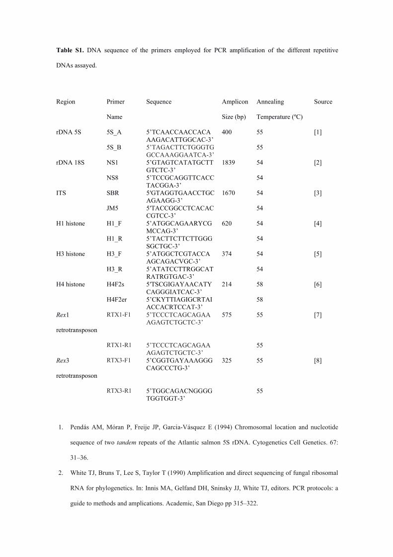
Table S1. DNA sequence of the primers employed for PCR amplification of the different repetitive
DNAs assayed.
Region Primer
Name
Sequence Amplicon
Size (bp)
Annealing
Temperature (ºC)
Source
rDNA 5S 5S_A 5’TCAACCAACCACAAAGACATTGGCAC-3’
400 55 [1]
5S_B 5’TAGACTTCTGGGTGGCCAAAGGAATCA-3’
55
rDNA 18S NS1 5’GTAGTCATATGCTTGTCTC-3’
1839 54 [2]
NS8 5’TCCGCAGGTTCACCTACGGA-3’
54
ITS SBR 5′GTAGGTGAACCTGCAGAAGG-3’
1670 54 [3]
JM5 5′TACCGGCCTCACACCGTCC-3’
54
H1 histone H1_F 5’ATGGCAGAARYCGMCCAG-3’
620 54 [4]
H1_R 5’TACTTCTTCTTGGGSGCTGC-3’
54
H3 histone H3_F 5’ATGGCTCGTACCAAGCAGACVGC-3’
374 54 [5]
H3_R 5’ATATCCTTRGGCATRATRGTGAC-3’
54
H4 histone H4F2s 5′TSCGIGAYAACATYCAGGGIATCAC-3’
214 58 [6]
H4F2er 5’CKYTTIAGIGCRTAIACCACRTCCAT-3’
58
Rex1
retrotransposon
RTX1-F1 5’TCCCTCAGCAGAAAGAGTCTGCTC-3’
575 55 [7]
RTX1-R1 5’TCCCTCAGCAGAAAGAGTCTGCTC-3’
55
Rex3
retrotransposon
RTX3-F1 5’CGGTGAYAAAGGGCAGCCCTG-3’
325 55 [8]
RTX3-R1 5’TGGCAGACNGGGGTGGTGGT-3’
55
1. Pendás AM, Móran P, Freije JP, Garcia-Vásquez E (1994) Chromosomal location and nucleotide
sequence of two tandem repeats of the Atlantic salmon 5S rDNA. Cytogenetics Cell Genetics. 67:
31–36.
2. White TJ, Bruns T, Lee S, Taylor T (1990) Amplification and direct sequencing of fungal ribosomal
RNA for phylogenetics. In: Innis MA, Gelfand DH, Sninsky JJ, White TJ, editors. PCR protocols: a
guide to methods and amplications. Academic, San Diego pp 315–322.

3. Montoya-Burgos JI (2003) Historical biogeography of the catfish genus Hypostomus (Siluriformes:
Loricariidae), with implications on the diversification of Neotropical ichthyofauna. Molecular
Ecology 12:1855–1867.
4. Hashimoto DT, Ferguson-Smith MA, Rens W, Foresti F, Porto-Foresti F (2011) Chromosome
mapping of H1 histone and 5S RNA gene clusters in three species of Astyanax (Teleostei:
Characiformes). Cytogenet Genome Res 134: 64–71.
5. Colgan D, McLauchlan A, Wilson G, Livingston S (1998) Histone H3 and U2 snRNA DNA
sequences and arthropod molecular evolution. Aust J Zool. 46: 419–43.
6. Pineau P, Henry M, Suspène R et al. (2005)A universal primer set for PCR amplification of nuclear
histone H4 genes from all animal species. Mol Biol Evol. 22: 582–588.
7. Volff JN, Körting C, Schartl M (2000) Multiple lineages of the non-LTR retrotransposon Rex1 with
varying success in invading fish genomes. Mol Biol Evol 17: 1673–1684.
8. Volff JN, Körting C, Sweeney K, Schartl M (1999) The non-LTR retrotransposon Rex3 from the
fish Xiphophorus is widespread among teleosts. Mol Biol Evol 16: 1427–1438.

Table S2. Codon-based test of neutrality, positive selection and purifying selection for the H1 histone
gene partial sequence.
Hypothesis Z P
Neutrality (dN = dS) -4.391 <0.001
Positive selection (dN > dS) -4.184 1
Purifying selection (dN < dS) 4.317 <0.001

Table S3. Genetic divergence values among species. Note the higher similarity between the sequences
obtained from the B chromosomes (Apar_B) and those from 0B-gDNA in A. paranae (Apar_gen)
and A. bockmanni (Abock), and lower similarity with those from A. fasciatus (Afasc) and A.
altiparanae (Aalti).
Apar_gen Apar_B Aalti AbockApar_B 0.017Aalti 0.105 0.099Abock 0.035 0.032 0.109Afasc 0.048 0.046 0.113 0.051