The origin of human chromosome 2 analyzed by comparative chromosome mapping with a DNA microlibrary
Upload
khangminh22Category
view
0download
0
The Force for Poleward Chromosome Motion in Haemanthus Cells Acts along the Length of the Chromosome during Metaphase but Only at the Kinetochore during Anaphase Alexey Khodjakov,* Richard W. Cole,* Andrew S. Bajer, ~ and Conly L. Rieder**
*Laboratory of Cell Regulation, Wadsworth Center for Laboratories and Research, Albany, New York 12201-0509; ~Department of Biomedical Sciences, State University of New York, Albany, New York 12222; and§Department of Biology, University of Oregon, Eugene, Oregon 97403
Abstract. The force for poleward chromosome motion during mitosis is thought to act, in all higher organisms, exclusively through the kinetochore. We have used time-lapse, video-enhanced, differential interference contrast light microscopy to determine the behavior of kinetochore-free "acentric" chromosome fragments and "monocentric" chromosomes containing one kine- tochore, created at various stages of mitosis in living higher plant (Haemanthus) cells by laser microsurgery. Acentric fragments and monocentric chromosomes generated during spindle formation and metaphase both moved towards the closest spindle pole at a rate (~1.0 txm/min) similar to the poleward motion of anaphase chromosomes. This poleward transport of chromosome fragments ceased near the onset of anaphase and was replaced, near midanaphase, by an-
other force that now transported the fragments to the spindle equator at 1.5-2.0 tzm/min. These fragments then remained near the spindle midzone until phrag- moplast development, at which time they were again transported randomly poleward but now at ~3/xm/min. This behavior of acentric chromosome fragments on anastral plant spindles differs from that reported for the astral spindles of vertebrate cells, and demonstrates that in forming plant spindles, a force for poleward chromosome motion is generated independent of the kinetochore. The data further suggest that the three stages of non-kinetochore chromosome transport we observed are all mediated by the spindle microtubules. Finally, our findings reveal that there are fundamental differences between the transport properties of forming mitotic spindles in plants and vertebrates.
URING mitosis the two sister kinetochores on each replicated chromosome acquire a bundle of mi- crotubules (Mt), 1 known as a kinetochore fiber,
that securely tethers them to opposing spindle poles (for review see Salmon, 1989; Rieder, 1990). These kineto- chore fibers are required for producing the forces that move chromosomes poleward throughout mitosis and, as a result, all current models envision that the poleward force on a chromosome acts exclusively on the kinetochore (for review see Mitchison, 1989; McIntosh and Hering, 1991; Ault and Rieder, 1994; Inoue and Salmon, 1995). Cur- rently, the most popular mechanism envisions that this
Address all correspondence to Conly L. Rieder, Laboratory of Cell Regu- lation, Wadsworth Center for Laboratories and Research, P.O. Box 509, Albany, NY 12201-0509. Tel.: (518) 474-6774. Fax: (518) 486-4901. e-mail: [email protected].
1. Abbreviat ions used in this paper: ACF, acentric chromosome fragment; Mt, microtubule.
force is produced by kinetochore-associated molecular motors (for review see Mclntosh and Pfarr, 1991; Sawin and Endow, 1993; Rieder and Salmon, 1994) acting on ki- netochore Mts, which themselves are coordinately disas- sembling within the kinetochore (for review see Mitchison and Salmon, 1992; Desai and Mitchison, 1995).
In animal somatic cells the equatorial alignment of chro- mosomes on the forming spindle also appears to involve another force that acts on the chromosome arms to coun- terbalance the sister kinetochore-based poleward-directed forces (for review see Rieder and Salmon, 1994; Cassi- meris et al., 1994; Fuller, 1995). This force, referred to as the polar ejection force or "polar wind," transports chro- mosome arms away from the closest pole during spindle formation (i.e., prometaphase). The presence of this ejec- tion force can be easily demonstrated by severing chromo- some arms from the kinetochore region with a laser micro- beam during prometaphase. Under this condition the kinetochore-free (acentric) chromosome fragments (ACF) are transported away from the closest pole at ,'-~2 ixm/min (Rieder et al., 1986; Rieder and Salmon, 1994).
© The Rockefeller University Press, 0021-9525/96/03/1093/12 $2.00 The Journal of Cell Biology, Volume 132, Number 6, March 1996 1093-1104 1093

The molecular mechanism behind the polar ejection force is unknown. The most popular hypothesis is that chromosome arms contain Mt plus-end motors that inter- act with astral and half-spindle Mts growing from the pole (Salmon, 1989; Ault et al., 1991; Carpenter, 1991). This hy- pothesis is supported circumstantially by Wang and Adler's (1995) recent finding that chromosome arms in chick cells become decorated with a kinesin-like molecule (chromo- kinesin) as the cell enters mitosis. It is also supported by functional studies in Drosophila (Afshar et al., 1995; Mur- phy and Karpen, 1995) and Xenopus (Vernos et al., 1995) that implicate chromosome-associated kinesin-like pro- teins (NOD and Xklp 1) in chromosome positioning on the meiotic (oocyte) spindle. Alternatively, the fact that the strength of the ejection force is related to Mt density, and depends on the normal behavior of Mt plus ends, sug- gests that it arises from the constant growth of dynamically unstable polar-nucleated Mt ends as they impact and push on the chromosome (Ault et al., 1991; Cassimeris et al., 1994; Rieder and Salmon, 1994).
An important consideration in mitosis research is the extent to which mechanistic findings in one system are ap- plicable to other systems (Rieder et al., 1993). In this re- gard it is currently unknown whether the polar ejection force is a feature only of the astral mitosis of vertebrate cells, or whether it is also present during spindle formation in other cell types or organisms. Included here are the as- tral meiotic spindles of animal spermatocytes and the anastral spindles of many animal oocytes (e.g., mouse, Drosophila, Xenopus) and most plants. Unlike astral spin- dies, which are formed from two radial (astral) arrays of centrosomal Mts, the anastral spindles of some oocytes and higher plants are organized primarily by the chromo- somes from random Mt arrays (for review see Smirnova and Bajer, 1992; Rieder et al., 1993; Vernos and Karsenti, 1995). Since forming anastral spindles lack well-developed arrays of polar Mrs, and since the polar ejection force has only been demonstrated for spindles formed from astral Mt arrays, it is possible that these two distinct pathways of spindle morphogenesis produce spindles that utilize fun- damentally different mechanisms to position chromo- somes, i.e., that astral and anastral spindles exert different non-kinetochore-based forces on the chromosomes.
To evaluate this possibility, we have examined the be- havior of ACFs and monocentric chromosomes containing a single kinetochore, created at various stages of mitosis by laser microsurgery in Haemanthus endosperm. We chose this higher plant system because, unlike the anastral spindles found in some animal oocytes, chromosome be- havior on the forming anastral spindle can be dearly fol- lowed by video-light microscopy in living Haemanthus cells. Our observations reveal that Haemanthus spindles possess stage-specific non-kinetochore-based chromatin transport systems that are neither present during astral mi- tosis nor previously demonstrated for any other animal spindle. Unlike during astral mitosis, we show clearly for the first time that a poleward force is exerted along the en- tire length of Haemanthus chromosomes during spindle formation and chromosome congression. Then, at the on- set of anaphase, this force disappears and is replaced by an opposite force that is similar to the polar ejection force seen in animal somatic cells. From our results we conclude
that there are fundamental differences in how chromo- somes become positioned on the forming mitotic spindle in plant and animal cells.
Materials and Methods
Endosperm Preparation Endosperm from the African blood lily, Haemanthus katherinae Bak, were extruded onto agar-coated coverslips and mounted for live cell time- lapse video microscopy as described by Bajer and Mol~-Bajer (1986). Some preparations were fixed and stained for the immunogold localiza- tion of spindle microtubules by light microscopy as described by De Mey et al. (1982).
Laser Microsurgery and Video-enhanced Microscopy The differential interference contrast video-enhanced light microscopic/ laser microsurgery system, described in detail by Cole et al. (1995), was used to sever chromosome arms and to follow their behavior in living Haemanthus endosperm cells. This system is centered around an inverted differential interference contrast light microscope (Diaphot 200; Nikon Inc., Garden City, NY) equipped with a motorized stage (Ludl MAC 2000; Ludl Electronics, Ltd., Hawthorne, NY). In brief, Haemanthus cells were followed with heat-filtered and shuttered 546-nm light using a ×60 1.4 NA differential interference contrast objective and a 0.85 NA con- denser. Sequential images were obtained every 4 s by integrating two video frames directly onto a chip (P100 CCD; Paultek, Princeton, NJ). This integrated frame was then routed through Image I (Universal Imag- ing Corp., West Chester, PA) for image processing before storage on a la- ser videodisk recorder (LVR-3300M; Sony Corp. of America, Montvale, NJ). Electronic and optical noise within the system was reduced by back- ground subtraction and by recording an eight-frame jumping average.
The chromosome microsurgery system used in our study was based on a pulsed (5 ns) Neodymium-YAG (yttrium-aluminum-garnet) laser (Sure- lite II; Continuum, Santa Clara, CA). The 1064-rim output of this laser was frequency doubled to 532 nm and filtered to remove stray 1064-nm light. This beam was then steered into the epiport of the Diaphot 200 where it was reflected, via a custom-made dichroic mirror (Omega Opti- cal, Brattleboro, VT), through the Wollaston prism and onto the back ap- erture of the 1.4 NA objective. The objective then focused the beam to a diffraction-limited spot of ~0.5qxm diam (Cole et al., 1995). As demon- strated by Berns and others (for review see Berns et al., 1991; Cole et al., 1995; Rieder et al., 1995), pulsed 532-nm laser light can be used to selec- tively destroy chromatin and chromatin-associated organelles (e.g., kine- tochores, nucleolar organizers) in living mitotic cells without deleteriously affecting spindle Mts or the process of mitosis.
The focused laser spot at the specimen plane was set near the center of a video screen, and its exact position was determined by irradiating a dried film of RBC5. Once located, the position of the laser was marked with cross-hairs on the video monitor. Cutting was then achieved by using the Ludl motorized stage to slowly pass the specimen through the fixed la- ser beam path. In our system optimal chromosome cutting was achieved by operating the laser at 10 Hz with 5-ns pulses, and each pulse contained ~500 nJ of power as measured at the focal point of the objective lens. In practice it took ~4--8 s, or 40-80 laser pulses, to completely sever a Hae- manthus chromosome across its short axis.
Tracking Chromosomes and Chromosome Fragments Distance vs. time plots were generated using software contained in the Im- age I system that calculates the distance between two movable cursors su- perimposed on sequential video frames. For this process time-lapse video- disk images were rerouted into Image I through a time-base corrector (For.A Corp., Natick, MA). Since dividing Haemanthus cells do not nor- mally have focused spindle poles but are nonmotile and stuck to the agar, a stationary point on the video screen was selected as a reference point for each sequence analyzed. The distance between this point, and either the edge of a primary constriction or a distinguishable feature on a chromo- some fragment edge (e.g., a barb of chromatin or a sharp edge), was then calculated for each frame.
The Journal of Cell Biology, Volume 132, 1996 1094

Resul ts
Both Kinetochores Are Required to Keep a Metaphase Chromosome Positioned on the Haemanthus Spindle Equator
When fully formed in highly flattened Haemanthus cells, the spindle contains two diffuse and broad spindle poles (Fig. 1 A). At this time it consists of conspicuous kineto- chore fiber Mt bundles interspersed within an irregular meshwork of other Mts. By metaphase the sister kineto- chore regions on all chromosomes are aligned near the spindle equator, but because of the irregularly shaped po- lar regions, the kinetochores do not necessarily define a linear metaphase plate across the broad axis of the spindle (Fig. 1 A). It is noteworthy that the arms of long chromo- somes in these cells are bent near the kinetochore region and project towards one or both spindle poles (Fig. 1 A).
When one kinetochore on a bioriented chromosome is destroyed with a laser in animal somatic cells, the now monocentric chromosome moves to the pole to which the nonirradiated kinetochore is attached with a velocity simi- lar to anaphase chromosomes (for review see Rieder et al.,
1995). To determine whether this is also true for chromo- somes on plant spindles, we selectively ablated one of the kinetochores on 14 bioriented metaphase chromosomes in seven Haemanthus cells. This operation produced a mono- centric chromosome containing a single kinetochore that, in every case, moved from the metaphase plate into the polar region to which the nonirradiated kinetochore was attached (Fig. 2). At our temporal resolution of 4 s, which can resolve a lateral displacement of ~0.1 Ixm per frame, the kinetics of this motion was smooth and linear for 10-15 ~m (Fig. 3) until the monocentric chromosome reached the end of the clear zone delineating the spindle pole. The velocity of this motion varied between monocentric chro- mosomes created in the same cell and ranged from 0.5 to 1.1 p~m/min with an average of 0.8 ixm/min. This is approx- imately the same poleward velocity exhibited by Haeman- thus chromosomes during anaphase (our data; see also Ba- jer, 1966). After a monocentric chromosome reached the edge of the clear zone, which roughly defines the bound- ary of the spindle pole, its directed motion was replaced by a Brownian "drift" (Fig. 3). By behavioral criteria the irra- diated kinetochore on laser-generated monocentric chro- mosomes was no longer functional since the chromatid
Figure 1. (A and B) Light micrographs of metaphase (A) and late anaphase (B) Haemanthus spindles stained for the immunogold local- ization of microtubules (red) and counterstained with toluidine blue for the localization of chromosomes. Note that during metaphase (A) the chromosome arms are bent in the direction of one or both of the spindle poles, while the kinetochore regions are positioned on the spindle equator. In anaphase (B) the arms now trail the kinetochore regions as they move poleward. Bar in B, 10 Ixm.
Khodjakov et al. Non-kinetochore-based Chromosome Transport 1095

Figure 2. (A-l) Selected frames from a time-lapse video sequence of a Haemanthus cell in which one of two sister kinetochores was de- stroyed by the laser on two metaphase chromosomes. The arrowhead in A notes a kinetochore region on a bioriented chromosome that is subsequently destroyed by the laser in B. As a result of this operation, the now monocentric chromosome initiates motion towards the (right-hand) pole to which the nonirradiated kinetochore (arrow in C-l) is attached. Note that this chromosome rotates as it moves poleward (C-E) so that its long axis is parallel to the direction of poleward motion. The arrowhead in E notes one of the kinetochore re- gions on a second metaphase chromosome, which was subsequently destroyed by the laser in F. Again, after laser microsurgery, this now monocentric chromosome began moving towards the (left-hand) pole to which its nonirradiated kinetochore (lower arrow in G-l) was attached, but it did not rotate significantly during this motion. A kinetic plot for these two chromosomes is presented in Fig. 3. Time in min:s is shown at the lower right corner of each frame. Bar in L 10 i~m.
containing the irradiated kinetochore showed no kineto- chore-based motility after disjoining from its sister during the ensuing anaphase (see below).
In the majority of cases, monocentric chromosomes ro- tated around their long axis as they moved poleward so that one arm led the motion; i.e., the kinetochore lost its poleward orientation during poleward motion (chromo- some 1 in Fig. 2). The rate of poleward movement did not change during or after this rotational movement. This ob- servation suggests that the force responsible for the pole- ward motion of monocentric chromosomes in metaphase cells is not generated solely at the kinetochore but also along the chromosome arms.
A Poleward Force Constantly Acts Along the Arms of Metaphase Chromosomes in Haemanthus
To determine if a pole-directed force acts along the arms
of chromosomes in forming Haemanthus spindles, inde- pendent of that acting on the kinetochores, we cut the arms away from the centromeric region of 30 bioriented chromosomes in 20 late prometaphase/metaphase cells. Such an operation in animal somatic cells produces kineto- chore-free ACFs that are expelled towards the spindle equator where they remain throughout the duration of mi- tosis (Rieder et al., 1986, 1995). However, in contrast to animal cells, we found that all ACFs created in prometaphase or metaphase Haemanthus cells were transported away from the metaphase plate and toward the closest pole (Fig. 4). The parameters of this poleward motion, including the average velocity (0.8 vLm/min; range 0.5-1.8 o~m/min), were similar to that of monocentric chromosomes formed in metaphase ceils (see above and below).
To compare the poleward movement of both monocen- tric chromosomes and ACFs more accurately, we used the
The Journal of Cell Biology, Volume 132, 1996 1096

35
30
25
E 20
. 15 i::5
10
5
0
¢
_ I r ~ '
I t i
0 10 20 30 40
Time (rain)
Figure 3. Kinetic plot for the upper (1) and lower (2) chromo- somes described in Fig. 2. The times shown in Fig. 2 are coinci- dent with points along the time axis of this plot. Note that once created by laser irradiation (arrows), both of these monocentric chromosomes initiated poleward motion with constant but differ- ent velocities (1 = 0.5 ixm/min; 2 = 0.9 p~m/min). After reaching the polar areas the chromosomes stopped moving and exhibited Brownian drift.
laser to generate, from the same chromosome, a monocen- tric fragment and multiple ACFs (Fig. 4). Under this con- dition both the monocentric and ACFs exhibited the same kinetic parameters during poleward motion (Fig. 5). From these observations we conclude that pole-directed forces are generated throughout the metaphase Haemanthus spindle, independent of the kinetochores that act along the length of the chromosomes.
The Poleward Force Acting on Haemanthus Chromosome Arms during Metaphase Is Abolished at Anaphase Onset
We next asked if the force that transports ACFs poleward during spindle formation in Haemanthus persists through- out mitosis. To address this question, we used the laser to sever one or both arms from the centromere region of 17 chromosomes, all positioned near the spindle equator, as anaphase was initiated in eight cells. By the time that the operation was complete, both the ACFs and the cen- tromere-containing chromosome fragment had disjoined into independent sister component parts (Fig. 6, A-D). During the ensuing anaphase the kinetochore-containing (monocentric) chromosome fragments segregated with a normal velocity (N0.9 txm/min) to opposite polar regions, while all of the ACFs remained positioned near the spin- dle equator and exhibited Brownian motion (Figs. 6, E-H, and 7). From these observations we conclude that the pole-directed force acting on Haemanthus chromosome arms during metaphase is abolished at the onset of anaphase.
A New Force Arises during Midanaphase in Haemanthus that Transports A CFs away from the Pole and to the Spindle Equator
We then used the laser beam to sever arms from the pole- ward-moving chromosomes in mid to late anaphase cells. Under this condition we observed, in every instance (n = 8 from three cells), that the ACFs were immediately trans- ported back to the spindle equator (data not shown; see below). From this set of observations, we conclude that a new force appears during anaphase that transports ACFs away from the spindle poles (and to the equator).
To determine when this away-from-the-pole force arises relative to anaphase onset, we followed the fate of mono- centric and ACFs, generated during late metaphase, as the cell proceeded into and through anaphase. In these cells the monocentric and ACFs were in the process of being transported towards one of the poles and had attained variable positions between the metaphase plate and a po- lar region at the time anaphase was initiated. Under these circumstances the poleward motion of the ACFs abruptly ceased at chromatid disjunction (anaphase onset) and, af- ter a delay of N10 rain, the ACFs initiated motion towards the spindle equator at 1.5-2.0 trm/min (Figs. 8 and 9). We observed this reversal of motion for all five ACFs followed in three cells, and in each case this away-from-the-pole motion ceased once the fragment reached the spindle equator (Fig. 9). From these experiments we conclude that a force appears during midanaphase that transports ACFs away from the pole and to the spindle equator. This force affects only ACFs positioned inside the spindle since ACFs expelled from the spindle region by the time of anaphase onset were never transported back to the spindle equator (Fig. 8).
Chromosome Fragments Positioned on the Spindle Equator during Anaphase Are Transported Poleward during Phragmoplast Development Next, we followed the behavior of 20 ACFs, positioned near the spindle equator during anaphase, in seven cells forming phragmoplasts (cell plates). Although these frag- ments were created at various stages of mitosis, all of them had acquired a stationary position near the equator by late anaphase. When the phragmoplast began to form, these fragments were suddenly and rapidly transported towards the poles with an average velocity 3.0 ixm/min (2.5-3.4 ~m/ rain; Figs. 6, H and I, and 8/ ) . This poleward motion was linear and smooth until the fragments reached the corre- sponding mass of telophasing chromatin in the polar re- gions (Fig. 7). During this process the fragments were transported with their long axes parallel to the direction of motion (Figs. 6, G-L, and 8, H and / ) . In some cases a fragment was caught within the forming phragmoplast be- fore initiating poleward motion. Under this condition the fragment became highly stretched towards the pole once it began to move (Figs. 6, J-L, and 8, H and /). Finally, ACFs that were derived from chromosomes initially posi- tioned in one half-spindle during prometaphase or anaphase were randomly distributed as they moved poleward during telophase. These observations reveal that forces are gener- ated at the time of phragmoplast development that move ACFs to the poles.
Khodjakov et al. Non-kinetochore-based Chromosome Transport 1097
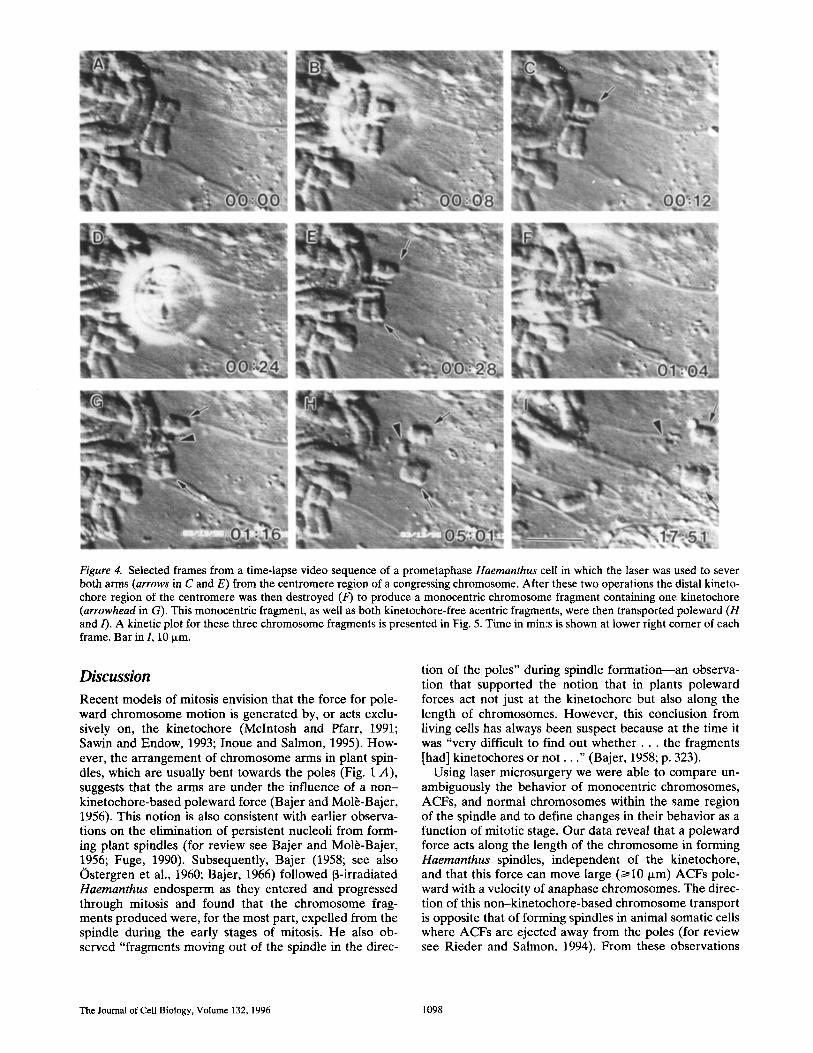
Figure 4. Selected frames from a time-lapse video sequence of a prometaphase Haemanthus cell in which the laser was used to sever both arms (arrows in C and E) from the centromere region of a congressing chromosome. After these two operations the distal kineto- chore region of the centromere was then destroyed (F) to produce a monocentric chromosome fragment containing one kinetochore (arrowhead in G). This monocentric fragment, as well as both kinetochore-free acentric fragments, were then transported poleward (H and/). A kinetic plot for these three chromosome fragments is presented in Fig. 5. Time in min:s is shown at lower right corner of each frame. Bar in I, 10 Ixm.
Discussion
Recent models of mitosis envision that the force for pole- ward chromosome motion is generated by, or acts exclu- sively on, the kinetochore (McIntosh and Pfarr, 1991; Sawin and Endow, 1993; Inoue and Salmon, 1995). How- ever, the arrangement of chromosome arms in plant spin- dles, which are usually bent towards the poles (Fig. 1 A), suggests that the arms are under the influence of a non- kinetochore-based poleward force (Bajer and Mol~-Bajer, 1956). This notion is also consistent with earlier observa- tions on the elimination of persistent nucleoli from form- ing plant spindles (for review see Bajer and Mol~-Bajer, 1956; Fuge, 1990). Subsequently, Bajer (1958; see also Ostergren et al., 1960; Bajer, 1966) followed 13-irradiated Haemanthus endosperm as they entered and progressed through mitosis and found that the chromosome frag- ments produced were, for the most part, expelled from the spindle during the early stages of mitosis. He also ob- served "fragments moving out of the spindle in the direc-
tion of the poles" during spindle formation--an observa- tion that supported the notion that in plants poleward forces act not just at the kinetochore but also along the length of chromosomes. However, this conclusion from living cells has always been suspect because at the time it was "very difficult to find out w h e t h e r . . , the fragments [had] kinetochores or n o t . . . " (Bajer, 1958; p. 323).
Using laser microsurgery we were able to compare un- ambiguously the behavior of monocentric chromosomes, ACFs, and normal chromosomes within the same region of the spindle and to define changes in their behavior as a function of mitotic stage. Our data reveal that a poleward force acts along the length of the chromosome in forming Haemanthus spindles, independent of the kinetochore, and that this force can move large (>~10 p~m) ACFs pole- ward with a velocity of anaphase chromosomes. The direc- tion of this non-kinetochore-based chromosome transport is opposite that of forming spindles in animal somatic cells where ACFs are ejected away from the poles (for review see Rieder and Salmon, 1994). From these observations
The Journal of Cell Biology, Volume 132, 1996 1098

30
E
8 t "
E3
2 5 -
2 0 -
15
10
®
I L I I I L I I I
0 2 4 6 8 10 12 14 16 18 20
Time (min)
Figure 5. A kinetic plot of the monocentric (1) and acentric (2,3) chromosome fragments depicted in Fig. 4. The times shown in Fig. 4 are coincident with points along the time axis of this plot. Note that the velocity of poleward motion is similar for all of the fragments (monocentric = 0.8 ~m/min; acentric = 1.0 ~m/min).
we conclude that there are fundamental differences in the chromosome transport properties between the anastral mitotic spindles of higher plants and the astral mitotic spindles of animals. This raises the possibility that the non-kinetochore-based chromosome transport properties of those anastral spindles formed in animal oocytes (e.g., Xenopus and Drosophila) are more similar to those we de- scribe for plants than for animal somatic cells.
The reason why plant anastral spindles and animal astral spindles differ in their non-kinetochore chromosome trans- port properties is unknown, but it may be related to the different pathways by which the polar regions in these two types of spindles are formed. Unlike for astral spindles, in which the poles are derived from preexisting radial arrays of centrosomal Mts, the polar regions in anastral spindles appear to be organized by the action of the chromosomes and Mt minus-end motors (Verde et al., 1991; Sawin and Endow, 1993; Vernos and Karsenti, 1995; McKim and Hawley, 1995). Thus, it is possible that the poleward trans- port of chromosome arms in Haemanthus spindles, which is mediated by and occurs towards the minus ends of spin- dle Mts (see below), is simply a manifestation of a continu- ous minus-Mt end transport process required to establish and to maintain spindle bipolarity in the absence of well- defined poles.
In Haemanthus, the poleward force acting along the chromosome terminates at the onset of anaphase and is then replaced by an opposite force that now transports ACFs positioned in the spindle to the spindle equator. This away-from-the-pole force is, in turn, replaced during phragmoplast development with another force that trans- ports ACFs poleward. From these observations we con-
clude that the chromosome arms in Haernanthus en- dosperm experience stage-specific forces throughout mitosis independent of the poleward forces acting on the kinetochores.
Does the poleward force acting on chromosome arms in forming Haemanthus spindles also act on other compo- nents within the spindle? According to Bajer and Mol~- Bajer (1956), some small "particles" in Haemanthus metaphase spindles do move poleward with the speed of anaphase chromosomes, but others oscillate irregularly or move to the spindle equator at the same time. Nicklas and Koch (1972) similarly found that 0.7-3.0-p,m diam lipid droplets and mitochondria commonly exhibited poleward motions with the speed of anaphase chromosomes when micromanipulated into metaphase spindles of grasshopper spermatocytes. However, they also emphasized that such particles underwent radial motion out of the spindle as of- ten as poleward motion, and the behavior of ACFs was never addressed. Thus, although limited, the current data suggest that when positioned in the spindle, membrane- bound particles undergo stochastic, bidirectional, "salta- tory" motions along Mts that differ from the exclusively smooth and poleward motion of ACFs.
What roles do the stage-specific non-kinetochore-based chromosome transport forces play during mitosis in Hae- rnanthus? Although we have no direct evidence, we favor the idea that the primary role of the poleward force is to position the chromosomes between the two polar regions during spindle formation, i.e., that it counters the activity of kinetochores (see below). This poleward force then might be terminated at the onset of anaphase so that it does not compete or interfere with the kinetochore-based poleward motion of the chromosomes, which is mandatory for proper chromosome segregation. The away-from-the- pole force that appears in midanaphase may lead to the ac- cumulation of those components needed to construct the cell plate in the region where this structure will ultimately form. Finally, the production of a poleward force during phragmoplast development would transport long chromo- some arms towards the nucleus so that they do not become trapped in the forming cell plate. Such a force might also produce periodic genomic changes by ensuring that natu- rally occurring ACFs ultimately become incorporated into a nucleus.
The Stage-specific Forces that Act along Haemanthus Chromosomes Are Mediated by Spindle Mts
The motion of ACFs is smooth and linear throughout mi- tosis in Haemanthus and, depending on the stage, the ve- locity ranges from 0.5 to 3.4 ixm/min. We also found, as others have (e.g., Bajer, 1958), that ACFs move only when positioned within the spindle (Fig. 8), and that this motion is directed roughly parallel to the long axis of spindle Mts. Structural analyses of fixed cells reveal that ACFs within but not outside the spindle are in contact with numerous Mts (Bajer et al., 1987; Bajer and Vantard, 1988; Fuge, 1990). Finally, all chromosome motion, including the non- kinetochore-based transport that we have described, is in- hibited by drugs that disrupt Mt assembly including colchi- cine (Molr-Bajer, 1958; Bajer, 1966) and chloral hydrate (Mol~-Bajer, 1967). Together these findings strongly sup-
Khodjakov et al. Non-kinetochore-based Chromosome Transport 1099

Figure 6. (A-l) Selected frames from a time-lapse video sequence of a Haemanthus cell in which the laser was used to sever both arms (arrow in B-D) from the centromeric region of a bioriented chromosome just as the cell was entering anaphase. After chromatid dis- junction the two sister chromosome fragments containing the kinetochores (arrowheads in E-G) segregated to opposite polar regions, while the four acentric fragments (arrows in E-H) remained near the spindle equator. These fragments were subsequently transported to the poles during phragmoplast formation (/). A kinetic plot of these chromosome fragments is presented in Fig. 7. (J-L) A series of video frames from another cell illustrating that an acentric chromosome fragment (arrow in J) may become highly stretched towards a pole (arrows in K and L) if it is transiently stuck in the phragmoplast at the time it initiates poleward motion. Time in min:s is at lower right-hand corner of each picture. Bars in I and L, 10 ~m.
por t the content ion that the non-kine tochore chromosome t ransport we observed is med ia ted by Mts.
As in animals, metaphase Haemanthus spindles are or- ganized with the minus ends of their Mts pos i t ioned near the two polar regions, while the plus ends over lap across the spindle equator (Eu teneuer et al., 1982). The force-
producing mechanism for the po leward mot ion of A C F s in Haernanthus is unknown, but there are several possibili- ties. I t could be med ia ted by minus-end--directed Mt mo- tor molecules associated with the chromosome surface. Al ternat ively, nonresolvable globule "part icles and states" are seen to flow constantly poleward, with about the same
The Journal of Cell Biology, Volume 132, 1996 1100

40 ® ®
• !
3O
v
8 20
D 10
50
I I F F I
10 20 30 40 50
Time (rain)
Figure 7. A kinetic plot of the monocentric and acentric chromo- some fragments generated in the anaphase cell shown in Fig. 6. The times shown in Fig. 6 are coincident with points along the time axis of this plot. After disjunction the sister monocentric fragments (curves 1 and 2), each of which contain a single kineto- chore, immediately move towards opposite poles with a velocity of 0.9 ~m/min. By contrast, the two acentric fragments, which dis- join at anaphase onset into four fragments lacking kinetochores (curves 3-6), remain at the spindle equator until the phragmo- plast begins to form (40-min time point). At this time they are then transported towards the polar regions with a velocity of 3.1- 3.3 ~Lm/min. Note that in this example fragments derived from the chromatid segregate to opposite poles (see also Fig. 8).
velocity of anaphase chromosomes, in most if not all plant and animal spindles (Hard and Allen, 1977; Hiramoto and hutsu, 1977; Salmon, E.D., and C.L. Rieder, unpublished observations). A similar "flowing" motion of submicro- scopic material is also seen to occur in association with the Mt arrays of other motility systems including neurons (Allen et al., 1982). Thus, it is possible that the same force- producing mechanism that powers the motion of particles and states also moves ACFs poleward in Haemanthus. A logical candidate for this force producer is Mt flux, which is exhibited even by simple self-organized Mt arrays (Sawin and Mitchison, 1994). Although the flux rate of forming Haemanthus spindles is unknown, it can approach 2.5 Ixm/min in Xenopus spindles assembled in vitro, well within the range of velocities observed for the poleward transport of ACFs in Haemanthus. If flux is involved in the motion of ACFs poleward, then some unidentified chro- mosome-associated protein(s) must tether the chromatin to the fluxing Mt lattice.
The poleward forces acting on ACFs are terminated at anaphase onset. Such an abrupt cessation of poleward transport may be the result of sudden biochemical changes (e.g., destruction of cyclin B and active maturation pro- moting factor) that are known to occur at the metaphase- anaphase transition in animal cells (Minshull et al., 1989; Murray, 1994). The fact that kinetochores continue to
move poleward at ~1.0 ~m/min during anaphase, after the bulk poleward transport of the chromosome ceases, re- veals that the mechanisms responsible for kinetochore and non-kinetochore-based chromosome motility differ.
Bajer and his co-workers reported that ACFs located between the separating centromeres in Haemanthus spin- dles moved "only irregularly and but little" (Ostergren et al., 1960; see also Bajer and Mol~-Bajer, 1956; Bajer, 1958). However, we found that ACFs positioned behind or in front (Fig. 8) of the separating groups of anaphase chro- mosomes were transported near midanaphase to the spin- dle equator at 1.5-2 ixm/min. Because the polarity of spindle Mts does not change between metaphase and midana- phase (Euteneuer et al., 1982), this away-from-the-pole motion cannot be produced by Mt flux. It may be due to the activation of chromosome-associated kinesin-like Mt plus-end motors and/or to the growth of spindle Mt plus ends toward the cell plate (which appears to occur during midanaphase; Fig. 1 B) (Jensen and Bajer, 1973; Bajer and Mol~-Bajer, 1982).
Finally, ACFs positioned on the spindle equator during anaphase suddenly move poleward at 2.5-3.4 ixm/min as the cell plate begins to form. This poleward motion, which is about three times faster than that observed during metaphase, has been previously described in detail by Ba- jer (Bajer, 1958; Bajer and Ostergren, 1963) who thought it was mediated by the same "traction fiber" forces that move kinetochores during anaphase. Although the data on the polarity of Mts within the phragmoplast are incom- plete, they suggest that the plus ends of these Mts are em- bedded in the cell plate and that the minus ends are di- rected poleward (Euteneuer and McIntosh, 1980; Vantard et al., 1990; Liu et al., 1993, 1994). The fact that the ACFs within the phragmoplast often become stretched as they move poleward (Fig. 6) reveals that the force for this mo- tion acts either on the front end of the fragment or along its surface. This makes a "pushing" mechanism based on growing Mt ends unlikely. Alternatively, the poleward motion of ACFs during telophase could be due to Mt mi- nus-end motors associated with the phragmoplast (Asada and Shibaoka, 1994) and/or to Mt flux, the latter of which has been shown to occur in the phragmoplast of glycerin- ated tobacco cells (although at a rate of only 0.1 p,m/min) (Asada et al., 1991).
In summary, the stage-specific motions of ACFs during mitosis in Haemanthus cannot all be mediated by the same force-producing mechanism. Although an Mt pushing force, based on growing Mt ends (Inoue and Salmon, 1995), may be involved in the motion of ACFs to the spin- dle equator during anaphase, it cannot provide the basis for poleward motion during metaphase. Mt flux could be involved in the poleward motion of ACFs during metaphase and telophase, but not in the away-from-the-pole motion in anaphase. Finally, if these motions are all mediated solely by chromatin-associated Mt motors, then at least two different (plus- and minus-directed) motors must be involved.
Does Kinetochore Function Differ during Spindle Formation in Plant and Animal Cells?
During mitosis in vertebrates, individual kinetochores
Khodjakov et al. Non-kinetochore-based Chromosome Transport 1101

Figure 8. (A-I) Selected frames from a time-lapse sequence of a Haemanthus cell progressing from metaphase to telophase. During metaphase the long arm on one chromosome (arrowhead in A) is severed near its centromere with the laser. This arm is then trans- ported into the near pole (white arrow in D-G). After it reaches the polar region, it stops and does not exhibit directional movement (E-G) anymore. In C the left-hand kinetochore on the centromere-containing fragment of this chromosome is destroyed by the laser. This produces a monocentric chromosome fragment, which begins to move towards the right-hand pole (dark arrow/arrowhead in D-F). When anaphase begins (F), this chromosome fragment disjoins into a monocentric fragment (arrowhead in F) and an acentric fragment (arrow in F). The monocentric fragment continues to move poleward as anaphase proceeds (arrowhead in F-H), while the acentric frag- ment (arrow in F-H) is transported to the spindle equator. Later this fragment is transported to the opposite pole during phragmoplast formation (arrow in I). A kinetic plot of these chromosome fragments is presented in Fig. 9. Time in min:s is at the lower right of each picture. Bar in I, 10 i~m.
abruptly switch between states of poleward and away- from-the-pole motion termed "kinetochore directional in- stability" (Skibbens et al., 1993). This switching is thought to be the result of changing tension levels on the kineto- chore, mediated by the motile behavior of the sister kine- tochore and by the magnitude of the polar ejection force in the half-spindle containing the chromosome (Skibbens et al., 1993, 1995; Cassimeris et al., 1994). These findings form the basis of a kinetochore directional instability/po- lar ejection mechanism of chromosome congression that envisions that kinetochores switch into an away-from-the- pole state of motion when they are under sufficiently high tension, and then switch to a state of poleward motion un- der conditions of lower tension. As summarized by Rieder and Salmon (1994), this model explains the constant and
conspicuous oscillatory motions of monooriented and bioriented chromosomes in animal cells, and it is also con- sistent with a wide variety of other chromosome behavior.
Our results indicate that in its explicit form, the kineto- chore directional instability/polar ejection mechansim for chromosome positioning in animal somatic cells is not ap- plicable to higher plants. First, the chromosomes in Hae- manthus do not exhibit the oscillatory motions characteris- tically produced by directionally unstable kinetochores (our data; see also Bajer and Mol6-Bajer, 1971). Second, unlike in vertebrate spindles where the leading kineto- chore on a congressing chromosome is throught to be in a poleward state of motion because it is under low tension, the leading kinetochore on a congressing Haemanthus chromosome is moving towards its pole when it is appar-
The Journal of Cell Biology, Volume 132, 1996 1102

60
5 0 -
4 0 -
(9 ®
~o 3O
i:5 20
10 ®
0 I I I t I I I I
0 10 20 30 40 50 60 70 80
Time (rain)
Figure 9. A kinetic plot of the chromosome fragments shown in Fig. 8. The times shown in Fig. 8 are coincident with points along the time axis of this plot. Curve 1 depicts the 0.7-0.8-~m/min poleward motion of the acentric chromosome arm generated dur- ing m e t a p h a s e (see w h i t e a r r o w in Fig. 8, D - G ) . C urve 2 illus- t r a t es t he behav io r o f t he acent r ic f r a g m e n t (see b l a c k a r r o w in Fig. 8, D - G ) be fo re and af ter its d i s junc t ion f rom the m o n o c e n - tric f r agmen t . No te tha t this f r a g m e n t s tops m o v i n g po l eward at a n a p h a s e onse t and t h e n drif ts for ~ 1 0 min. A f t e r this t ime it is t h e n t r a n s p o r t e d at 1.5 ixm/min back to the sp indle e q u a t o r w h e r e it s tops for a shor t pe r iod o f t ime ( b e t w e e n the 58- and 65- m in t ime poin ts ) be fo re m o v i n g t oward the oppos i t e sp indle pole at 3.0 p.m/min. C u r v e 3 r e p r e s e n t s t he m o n o c e n t r i c c h r o m o s o m e f r a g m e n t (see b l a c k a r r o w h e a d in Fig. 8, D - G ) , which shows a s t eady po leward veloci ty o f 0.75 ~ m / m i n f r o m t he t ime it was cre- a ted in m e t a p h a s e (at the 1-min t ime point ) unt i l t he end o f anaphase .
ently under conditions of maximum high tension--pro- duced by pole-directed forces within the half-spindle that act on the chromosome arms. Although we do not know the magnitude of these poleward forces relative to the poleward force acting on the leading kinetochore, it is likely that they provide significant drag on a chromosome that is moving away from the closest pole. If this is true, then unlike in vertebrate cells, the kinetochores in plant cells may produce or experience a poleward force only when they are under sufficiently high tension. Although speculative, this hypothesis is consistent with the current data on the behavior of congressing Haemanthus chromo- somes and should be testable by additional laser microsur- gery studies.
We thank Drs. M. Koonce, E.D. Salmon, E.A. Smirnova, R. Vale, and W.Z. Cande for critical comments and helpful discussions. We also thank Mrs. C. Hughes for photographic assistance.
The major part of this work was conducted within the Wadsworth Cen- ter's Video-enhanced Light Microscopy Core Facility and was supported by National Institutes of Health grants GM 40198 (to C.L. Rieder), and RR 01219 awarded by the Biomedical Research Technology Program, Na- tional Center for Research Resources (Department of Health and Human
Resources/Public Health Service), to support the Wadsworth Center's Bio- logical Microscopy and Image Reconstruction facility as a National Bio- technological Resource.
Received for publication 28 November 1995 and in revised form 3 January 1996.
References
Afshar, K., N.R. Barton, R.S. Hawley, and L.S.B. Goldstein. 1995. DNA bind- ing and meiotic chromosomal localization of the Drosophila Nod kinesin- like protein. Celt. 81:129-138.
Allen, R.D., J.L. Travis, J.H. Hayden, N.S. Allen, A.C. Breuer, and L.J. Lewis. 1982. Cytoplasmic transport: moving ultrastructural elements common to many cell types revealed by video-enhanced microscopy. Cold Spring Harb. Symp. Quant. Biol. 46:85-87.
Asada, T., and H. Shibaoka. 1994. Isolation of polypeptides with microtubule- translocating activity from phragmoplasts of tobacco BY-2 cells. J. Cell Sci. 107:2249-2257.
Asada, T., S. Sortobe, and H. Shibaoka. 1991. Microtubule translocation in the cytokinetic apparatus of cultured tobacco cells. Nature (Lond.). 350:238-241.
Ault, J.G., and C.L. Rieder. 1994. Centrosome and kinetochore movement dur- ing mitosis. Curr. Opin. Cell Biol. 6:41-49.
Ault, J.G., A.J. DeMarco, E.D. Salmon, and C.L. Rieder. 1991. Studies on the ejection properties of asters: astral microtubule turnover influences the os- cillatory behavior and positioning of mono-oriented chromosomes. J. Cell Sci. 99:701-710.
Bajer, A. 1958. Cine-micrographic studies on chromosome movement in 13-irra- diated cells. Chromosoma (Berl.). 9:319-331.
Bajer, A. 1966. Movement within the mitotic spindle. In Dynamics of Fluids and Plasmas. Academic Press. Inc., New York. 59-80.
Bajer, A., and J. Mol~-Bajer. 1956. Cine-micrographic studies on mitosis in en- dosperm II. Chromosome, cytoplasmic and brownian movements. Chromo- soma (Berl.). 7:558-607.
Bajer, A., and J. Mol~-Bajer. 1971. Architecture and function of the mitotic spindle. Adv. Cell Mol. Biol. 1:213-266.
Bajer, A.S., and J. Mol~-Bajer. 1982. Asters, poles, and transport properties within spindlelike microtubule arrays. Cold Spring Harb. Syrup. Quant. Biol. 46:263-283.
Bajer, A.S., and J. Mol~-Bajer. 1986. Reorganization of microtubules in en- dosperm cells and cell fragments of the higher plant Haemanthus in vivo. Z Cell Biol. 102:263-281.
Bajer, A., and G. Ostergren. 1963. Observation on transverse movements within the phragmoplast. Hereditas (Lund). 50:179-195.
Bajer, A.S., and M. Vantard. 1988. Microtubule dynamics determine chromo- some lagging and transport of acentric fragments. Mutat. Res. 201:271-281.
Bajer, A.S., M. Vantard, and J. Mol~-Bajer. 1987. Multiple mitotic transports expressed by chromosome and particle movement. In Nature and Function of Cytoskeletal Proteins in Motility and Transport. Gustav Fisher Verlag, New York. 171-186.
Berns, M.W., W.H. Wright, and R. Wiegand Steubing. 1991. Laser microbeam as a tool in cell biology, lnt. Rev. Cytol. 129:1~14.
Carpenter, A.T.C. 1991. Distributive segregation: motors in the polar wind? Cell 64:885-890.
Cassimeris, L., C.L. Rieder, and E.D. Salmon. 1994. Microtubule assembly and kinetochore directional instability in vertebrate monopolar spindles: impli- cations for the mechanism of chromosome congression. Z Cell Sci. 107:285- 297.
Cole, R.W., A. Khodjakov, W.H. Wright, and C.L. Rieder. 1995. A differential interference contrast-based light microscopic system for laser microsurgery and optical trapping of selected chromosomes during mitosis in vivo. J. Mi- crosc. Soc. Am. 1:203-215.
De Mey, J., A.M. Lambert, A.S. Bajer, M. Moeremans, and M. De Brabander. 1982. Visualization of microtubules in interphase and mitotic plant cells of Haemanthus endosperm with the immuno-gold staining method. Proc. Natl. Acad. Sci. USA. 79:1898--1902.
Desai, A., and T. J. Mitchison. 1995. A new role for motor proteins as couplers to depolymerizing microtubules. J. Cell Biol. 128:1~,.
Euteneuer, U., and J.R. Mclntosh. 1980. Polarity of midbody and phragmoplast microtubules. J. Cell Biol. 87:509-515.
Euteneuer, U., W.T. Jackson, and J.R. Mclntosh. 1982. Polarity of spindle mi- crotubules in Haemanthus endosperm. J. Cell BioL 94:6444553.
Fuge, H. 1990. Non-kinetochore transport phenomena, microtubule-chromo- some associations, and force transmission in nuclear division. Protoplasma. 158:1-9.
Fuller, M.T. 1995. Riding the polar winds: chromosomes motor down east. Cell. 81:5-8.
Hard, R., and R.D. Allen. 1977. Behavior of kinetochore fibers in Haemanthus katherinae during anaphase movements of chromosomes. J. Cell Sci. 27:47-56.
Hiramoto, Y., and K. Izutsu. 1977. Poleward movement of "markers" existing in mitotic spindles of grasshopper spermatocytes. Cell Struct. Funct. 2:257- 259.
Inoue, S., and E. Salmon. 1995. Force generation by microtubule assembly/dis- assembly in mitosis and related movements. Mol. Biol. Cell. 6:1619-1640.
Khodjakov et al. Non-kinetochore-based Chromosome Transport 1103

Jensen, C., and A. Bajer. 1973. Spindle dynamics and arrangement of microtu- bules. Chromosoma (Berl.). 44:73-89.
Liu, B., J. Marc, H.C. Joshi, and B.A. Palevitz. 1993. A gamma-tubulin-related protein associated with the mierotubule arrays of higher plants in a cell cy- cle-dependent manner. J. Cell Sci. 104:1217-1228.
Liu, B., H.C. Joshi, T.J. Wilson, C.D. Silflow, B.A. Palevitz, and D.P. Snustad. 1994. ~/-Tubulin in Arabidopsis: gene sequence, immunoblot, and immuno- fluorescence studies. Plant Cell. 6:303-314.
Mclntosh, J.R., and G.E. Hering. 1991. Spindle fiber action and chromosome movement. Annu. Rev. Cell Biol. 7:403-426.
Mclntosh, J.R., and C.M. Pfarr. 1991. Mitotic motors. Z Cell Biol. 115:577-585. McKim, K.S., and R.S. Hawley. 1995. Chromosomal control of meiotic cell divi-
sion. Science (Wash. DC). 270:1595-1601. Minshull, J., J. Pines, R. Golsteyn, N. Standart, S. Mackie, A. Colman, J. Blow,
J.V. Ruderman, M. Wu, and T. Hunt. 1989. The role of cyclin synthesis, modification and destruction in the control of cell division. J. Cell Sci. Suppl. 12:77-97.
Mitchison, T.J. 1989. Mitosis: basic concepts. Curr. Opin. Cell Biol. 1:67-74. Mitchison, T.J., and E.D. Salmon. 1992. Poleward kinetochore fiber movement
occurs during both metaphase and anaphase-A in newt lung cell mitosis. J. Cell Biol. 119:569-582.
Mol~:-Bajer, J. 1958. Cine-micrographic analysis of C-mitosis in endosperm. Chromosoma (Berl. ). 9:332-358.
Mol~-Bajer, J. 1967. Chromosome movement in chloral hydrate treated en- dosperm cells in vitro. Chromosoma (BerL). 22:465--480.
Murphy, T.D., and G.H. Karpen. 1995. Interaction between the nod ~ kinesin- like gene and extracentromeric sequences are required for transmission of a Drosophila minichromosome. Cell. 81:139-148.
Murray, A. 1994. Cell cycle checkpoints. Curt. Opin. Cell Biol. 6:8724-876. Nicklas, R.B., and C.A. Koch. 1972. Chromosome micromanipulation. IV. Po-
larized motions within the spindle and models for mitosis. Chromosoma (Berl. ). 39:t-26.
Ostergren, G,, J. Mol~-Bajer, and A. Bajer. 1960. An interpretation of transport phenomena at mitosis. Ann. N. Y. Acad. Sci. 90:381-408.
Rieder, C.L. 1990. Formation of the astral mitotic spindle: ultrastructural basis for the centrosome-kinetochore interaction. Electron Microsc. Rev. 3:269- 300.
Rieder, C.L., and E.D. Salmon. 1994. Motile kinetochores and polar ejection forces dictate chromosome position on the vertebrate mitotic spindle. J. Cell Biol. 124:223-233.
Rieder, C.L., E.A. Davison, L.C. Jensen, L. Cassimeris, and E.D. Salmon. 1986. Oscillatory movements of monooriented chromosomes and their position
relative to the spindle pole result from the ejection properties of the aster and half-spindle. J. Cell Biol. 103:581-591.
Rieder, C.L., J.G. Ault, U. Eichenlaub-Ritter, and G. Sluder. 1993. Morpho- genesis of the mitotic and meiotic spindle: conclusions obtained from one system are not necessarily applicable to the other. In Chromosome Segrega- tion and Aneuploidy. NATO-ASI Series. B.K. Vig, editor. Springer Verlag, Berlin. 72:183-197.
Rieder, C.L., R.W. Cole, A.L. Khodjakov, and G. SludeL 1995. The checkpoint delaying anaphase in response to chromosome monoorientation is mediated by an inhibitory signal produced by unattached kinetochores. J. Cell Biol. 130:941-948.
Salmon, E.D. 1989. Microtubule dynamics and chromosome movement. In Mi- tosis: Molecules and Mechanisms. J.C. Hyams and B.R. Brinkley, editors. Academic Press, London. 119-181.
Sawin, K.E., and S.A. Endow. 1993. Meiosis, mitosis and microtubule motors. Bioessays. 15:399-407.
Sawin, K.E., and T.J. Mitchison. 1994. Microtubule flux in mitosis is indepen- dent of chromosomes, centrosomes and antiparallel microtubules. Mol. Biol. Celt. 5:217-226.
Skibbens, R.V., V.P. Skeen, and E.D. Salmon. 1993. Directional instability of kinetochore motility during chromosome congression and segregation in mi- totic newt lung cells: a push-pull mechanism. J. Cell Biol. 122:859475.
Skibbens, R.V., C.L. Rieder, and E.D. Salmon. 1995. Kinetochore motility after severing between sister centromeres using laser microsurgery: evidence that kinetochore directional instability and position is regulated by tension. J. Cell Sci. 108:2537-2548.
Smirnova, E.A., and A.S. Bajer. 1992. Spindle poles in higher plant mitosis. Cell Motil. Cytoskeleton. 23:1-7.
Vantard, M., N. Levilliers, A.M. Hill, A. Adoutte, and A.M. Lambert. 1990. In- corporation of paramecium axonemal tubulin into higher plant cells reveals functional sites of microtubule assembly. Proc. Natl. Acad. ScL USA. 87: 88254829.
Verde, F., J.M. Berrez, C. Antony, and E. Karsenti. 1991. Taxol-induced micro- tubule asters in mitotic extracts of Xenopus eggs: requirement for phospho- rylated factors and cytoplasmic dynein. J. Cell Biol. 112:1177-1187.
Vernos, I., and E. Karsenti. 1995. Chromosomes take the lead in spindle assem- bly. Trends Cell BioL 5:297-301.
Vernos, I., J. Raats, T. Hirano, J. Heasman, E. Karsenti, and C. Wylie. 1995. Xklpl, a chromosomal Xenopus kinesin-like protein essential for spindle or- ganization and chromosome positioning. Cell. 81:117-127.
Wang, S.-Z., and R. Adler. 1995. Chromokinesin: a DNA-binding, kinesin-like nuclear protein. J. Cell Biol. 128:761-768.
The Journal of Cell Biology, Volume 132, 1996 1104
Copyright © 2022 FDOKUMEN