Chromosome Structure and the C-Value Paradox
12
Chromosome Structure and the C-Value Paradox JOSEPH G . GALL From the time of their discovery in the middle ofthe nineteenth century, chromosomes have held a particular fascination for cell biologists . The past 25 years-the period covered by this volume-has been no exception . During this time a host of new approaches to problems of chromosome and gene orga- nization has been introduced, beginning in the early 1950s with spectrophotometric analysis of nuclear staining and the appli- cation of autoradiography to questions of DNA and RNA synthesis . Many biochemical approaches were worked out, based either on subcellular fractionation or micromethods, and specific molecular probes, such as in situ nucleic acid hybridi- zation and fluorescent antibodies, made it possible to study the sites of specific macromolecules . Most recently, the extraordi- narily powerful combination of molecular cloning, restriction enzyme analysis, and nucleic acid sequencing has opened up an entirely new field of cytogenetic analysis . Despite the enormous advances made during this period in understanding chromosome structure, there remains one dis- concerting feature often called the "C-value paradox ." The paradox is the fact that organisms at the same general level of morphological complexity, which presumably have the same genetic requirements, nevertheless often have genomes whose DNA contents differ by orders of magnitude. Fortunately, many questions about chromosomes can be approached with- out considering this problem. This is true, for instance, of nucleosome structure . Just now, when recombinant DNA methods make it so easy to study sequence organization of individual genes, the C-value paradox is receiving more or less benign neglect . But at some point or another the paradox intrudes upon almost all questions of chromosome organiza- tion, particularly when comparisons are made between orga- nisms with widely varying DNA contents . For instance, the fact that a complex higher eukaryote such as Drosophila has a minute genome makes it impossible to argue that larger amounts of DNA are essential to carry out sequence-specific roles . In an organism with 100 times as much DNA as Dro- sophila, such as a salamander or a bean plant, most of the DNA could be unessential for coding or regulatory functions . This review will focus on a few selected aspects of chromo- some organization, in particular the question of uninemy, the nature of heterochromatin, chromatin compaction, and se- quence organization of repetitive genes. An attempt is made to put findings from the past 25 years into context, but in no sense have I tried to review everything new about chromosomes in JOSEPH G . GALL Department of Biology, Yale University, New Haven, Connecticut THE JOURNAL OF CELL BIOLOGY " VOLUME 91 NO . 3 PT . 2 DECEMBER 1981 3s-14s © The Rockefeller University Press " 0021-9525/81/12/003x/12 $1 .00 this period . The C-value paradox will provide a partial link in the story . Several times during this period it seemed that the paradox could be explained away, but each time the explana- tion was invalidated by new information . The unexpectedly large and variable amount of DNA in eukaryotic genomes remains a major complicating feature in understanding chro- mosome organization . DNA Constancy The staining characteristics of chromosomes, especially their coloration by a variety of synthetic dyes as well as by such natural products as hematoxylin and carmine, originally in- spired the words "chromosome" and "chromatin ." Flemming (1) defined chromatin as "the substance in the cell nucleus which takes up the color during nuclear staining," and in a remarkably accurate conjecture he suggested that chromatin might be the same as the recently discovered "nuclein" of Miescher (2). Even after the existence of DNA in certain nuclei, chiefly those of sperm and thymus, became well accepted, the lack of a specific stain hampered progress at the chromosome level . Fortunately such a stain was found by Feulgen and Rossenbeck in 1924 (3), who modified the familiar Schiff test for aldehydes into a simple and reliable histochemical proce- dure for DNA . The Feulgen reaction was quickly adopted by chromosome cytologists, who were able for the first time to verify the existence of DNA in both plant and animal chro- mosomes and to show that cytochemically demonstrable DNA was, with few exceptions, absent from the cytoplasm. Another quarter century passed, however, before Pollister and Ris (4) demonstrated that the amount of Feulgen stain could be used to estimate the DNA content of a nucleus. Their study and many others that soon followed it, helped convert nuclear cytology from an observational and descriptive subject to an experimental science with a developing theoretical framework . Several generalizations about nuclear DNA emerged from the quantitative Feulgen studies (5) . In particular, it was rec- ognized that each species of animal and plant could be char- acterized by the amount of DNA in its nuclei . In most cases measurements were made on nondividing diploid nuclei, but in some instances the DNA contents of haploid gametes, either sperm or microspore nuclei, were measured and found to be half the diploid amount . From a large number of such mea- surements the idea of DNA constancy was established : the nonreplicating haploid chromosome complement of a species is characterized by a constant amount of DNA, called the C- value for that species . Feulgen dye measurements confirmed the fact, recently discovered by the new technique of autora- 3s Downloaded from http://rupress.org/jcb/article-pdf/91/3/3s/1075489/3s.pdf by guest on 31 May 2022
-
Upload
khangminh22 -
Category
Documents
-
view
6 -
download
0
Transcript of Chromosome Structure and the C-Value Paradox

Chromosome Structure and the C-Value Paradox
JOSEPH G. GALL
From the time oftheir discovery in the middle ofthe nineteenthcentury, chromosomes have held a particular fascination forcell biologists . The past 25 years-the period covered by thisvolume-has been no exception . During this time a host ofnew approaches to problems of chromosome and gene orga-nization has been introduced, beginning in the early 1950s withspectrophotometric analysis of nuclear staining and the appli-cation of autoradiography to questions of DNA and RNAsynthesis . Many biochemical approaches were worked out,based either on subcellular fractionation or micromethods, andspecific molecular probes, such as in situ nucleic acid hybridi-zation and fluorescent antibodies, made it possible to study thesites of specific macromolecules . Most recently, the extraordi-narily powerful combination of molecular cloning, restrictionenzyme analysis, and nucleic acid sequencing has opened upan entirely new field of cytogenetic analysis .Despite the enormous advances made during this period in
understanding chromosome structure, there remains one dis-concerting feature often called the "C-value paradox ." Theparadox is the fact that organisms at the same general level ofmorphological complexity, which presumably have the samegenetic requirements, nevertheless often have genomes whoseDNA contents differ by orders of magnitude. Fortunately,many questions about chromosomes can be approached with-out considering this problem. This is true, for instance, ofnucleosome structure . Just now, when recombinant DNAmethods make it so easy to study sequence organization ofindividual genes, the C-value paradox is receiving more or lessbenign neglect . But at some point or another the paradoxintrudes upon almost all questions of chromosome organiza-tion, particularly when comparisons are made between orga-nisms with widely varying DNA contents . For instance, thefact that a complex higher eukaryote such as Drosophila has aminute genome makes it impossible to argue that largeramounts of DNA are essential to carry out sequence-specificroles . In an organism with 100 times as much DNA as Dro-sophila, such as a salamander or a bean plant, most of theDNA could be unessential for coding or regulatory functions .This review will focus on a few selected aspects of chromo-
some organization, in particular the question of uninemy, thenature of heterochromatin, chromatin compaction, and se-quence organization of repetitive genes. An attempt is made toput findings from the past 25 years into context, but in no sensehave I tried to review everything new about chromosomes in
JOSEPH G . GALL
Department ofBiology, Yale University, New Haven,Connecticut
THE JOURNAL OF CELL BIOLOGY " VOLUME 91 NO . 3 PT . 2 DECEMBER 1981 3s-14s© The Rockefeller University Press " 0021-9525/81/12/003x/12 $1 .00
this period . The C-value paradox will provide a partial link inthe story . Several times during this period it seemed that theparadox could be explained away, but each time the explana-tion was invalidated by new information . The unexpectedlylarge and variable amount of DNA in eukaryotic genomesremains a major complicating feature in understanding chro-mosome organization .
DNA ConstancyThe staining characteristics ofchromosomes, especially their
coloration by a variety of synthetic dyes as well as by suchnatural products as hematoxylin and carmine, originally in-spired the words "chromosome" and "chromatin." Flemming(1) defined chromatin as "the substance in the cell nucleuswhich takes up the color during nuclear staining," and in aremarkably accurate conjecture he suggested that chromatinmight be the same as the recently discovered "nuclein" ofMiescher (2). Even after the existence ofDNA in certain nuclei,chiefly those of sperm and thymus, became well accepted, thelack of a specific stain hampered progress at the chromosomelevel . Fortunately such a stain was found by Feulgen andRossenbeck in 1924 (3), who modified the familiar Schiff testfor aldehydes into a simple and reliable histochemical proce-dure for DNA. The Feulgen reaction was quickly adopted bychromosome cytologists, who were able for the first time toverify the existence of DNA in both plant and animal chro-mosomes and to show that cytochemically demonstrable DNAwas, with few exceptions, absent from the cytoplasm. Anotherquarter century passed, however, before Pollister and Ris (4)demonstrated that the amount of Feulgen stain could be usedto estimate the DNA content of a nucleus. Their study andmany others that soon followed it, helped convert nuclearcytology from an observational and descriptive subject to anexperimental science with a developing theoretical framework .
Several generalizations about nuclear DNA emerged fromthe quantitative Feulgen studies (5) . In particular, it was rec-ognized that each species of animal and plant could be char-acterized by the amount of DNA in its nuclei . In most casesmeasurements were made on nondividing diploid nuclei, butin some instances the DNA contents ofhaploid gametes, eithersperm or microspore nuclei, were measured and found to behalf the diploid amount . From a large number of such mea-surements the idea of DNA constancy was established : thenonreplicating haploid chromosome complement of a speciesis characterized by a constant amount of DNA, called the C-value for that species . Feulgen dye measurements confirmedthe fact, recently discovered by the new technique of autora-
3s
Dow
nloaded from http://rupress.org/jcb/article-pdf/91/3/3s/1075489/3s.pdf by guest on 31 M
ay 2022

diography (6), that DNA replication occurred during inter-phase of the mitotic cycle .A striking feature of the quantitative measurements was the
extreme variation in C-value for different organisms, rangingfrom a low of 0.18 pg in Drosophila melanogaster throughintermediate values of 3-4 pg in various mammals includingman, to highs of 50-100 pg in salamanders and some monocotplants . Even within groups of closely related organisms varia-tions were seen, a factor of two between species in the samegenus being common (7, 8) . The wide range ofC-values at firstdid not trouble chromosome cytologists . They were accustomedto the fact that some organisms had large chromosomes asso-ciated with correspondingly large nuclei and cells, whereasothers had only small ones, and it was not surprising that thosesize differences were reflected in DNA contents . More impor-tantly, the contemporary model of chromosome organizationprovided an explanation for the size differences . According tothis model, chromosomes were multistranded cables consistingof two, four, eight, or more identical subunits (9) . It was notdifficult to suppose that related organisms with different C-values simply had different numbers of subunits in their chro-mosomes. Support for this concept was provided by the factthat related organisms often had similar or identical karyotypesdespite large differences in absolute chromosome size or DNAcontent (10) .
This comfortable picture was called into question by exper-iments that suggested that chromosomes were, in a sense, muchsimpler: they consisted of a single gigantic DNA molecule .This so-called unineme model was slow to take hold, and manyattempts were made to reconcile the new data with a multi-stranded chromosome . By the mid-1960s, however, it was clearthat uninemy was here to stay and that the C-values posed anumber of unresolved problems . If organisms with widelydifferent C-values did not differ in the number of identicalstrands per chromosome, did they contain different numbersof genes? Why was there no clear correlation between morpho-logical complexity and C-value? Why were even the lowest C-values so large? For instance, Drosophila with the lowest C-value outside the fungi had enough DNA to code for well over100,000 proteins, and mammalian genomes were nearly 20times larger . Before considering these questions in more detail,let us look briefly at the evidence for uninemy .
Uninemy
Earlier arguments about chromosome strandedness weresometimes confused by failure to define the problem explicitly.With the clear view of hindsight to guide us, the question iseasy to state. How many DNA molecules are there in onechromatid? Historically the first convincing evidence camefrom the ingenious experiments of Taylor and his colleagues,who followed the distribution of tritium-labeled thymidinethough successive chromosome replications (11) . They showedthat both chromatids ofa chromosome were equally labeled atthe first mitosis after administration of the isotope, but onlyone of the two chromatids was labeled at the second mitosis(or more precisely, because of sister chromatid exchanges, onlyone chromatid was labeled at a given point along the length ofthe chromosome). This distribution of label was called semi-conservative to distinguish it from conservative (one labeledand one unlabeled chromatid at the first division) or dispersive(all chromatids labeled at all divisions). Taylor's demonstrationof semi-conservative distribution of label during chromosome
4s
THE JOURNAL OF CELL BIOLOGY " VOLUME 91, 1981
replication was published at about the same time as Meselsonand Stahl's similar experiment showing the semiconservativedistribution of density label during DNA replication in Esch-erichia coli (12) . Both experiments implied that the unit underconsideration-the chromatid or the DNA molecule-con-sisted of two subunits, that separated but remained intactduring replication. In a second but less well-known set ofexperiments, Taylor demonstrated that the two subunits of thechromatid differed in such a way that rejoining after breakagewas restricted (13) . Because the two strands ofthe DNA doublehelix differed in polarity (5' -+ 3') the simplest interpretationwas that the two subunits of a chromatid corresponded to thosestrands . By incorporating bromodeoxyuridine into chromo-somes and staining with Giemsa (14), it is now possible toreproduce Taylor's results without the need for autoradiogra-phy (Fig. 1) . The staining procedure is particularly valuablefor studying multiple sister chromatid exchanges .
Evidence ofa quite different sort came from observations onlampbrush chromosomes of Amphibian oocytes . Morphologi-cal analysis had shown that the lateral loops of these chromo-somes occurred in pairs corresponding to the two sister chro-matids (15, 16). Although the bulk of each loop consisted of amatrix ofribonucleoprotein, DNase digestion experiments car-ried out by Callan and Macgregor (17) established that thecontinuity of the loops was maintained by DNA. Extendingthese observations Gall (18) demonstrated that loop digestionfollowed two-hit kinetics, which suggested that a loop andhence a chromatid had one DNA molecule as its structural axis(Fig . 2) . In the same experiments the interchromomeric fiber,which presumably corresponded to a pair ofchromatids or twoDNA molecules, followed four-hit kinetics. Shortly afterwardMiller (19, 20) published his extraordinary electron micro-graphs of lampbrush loops showing that the bulk of a loopconsisted oflong fibrils extending laterally from a very delicateaxis . Because these fibrils showed a gradient of lengths andbecause it was known from cytochemical studies that loopswere actively synthesizing RNA, the simplest interpretation
FIGURE 1 Semiconservative replication and sister-chromatid ex-changes visualized in Chinese hamster chromosomes by the BrdU-Giemsa technique . From Wolff and Perry (14) . Using [ 3HlthymidineTaylor et al . (11) were the first to demonstrate semiconservativereplication and provide experimental evidence in favor of a uninememodel of the chromatid . Bar, 10ILm .
Dow
nloaded from http://rupress.org/jcb/article-pdf/91/3/3s/1075489/3s.pdf by guest on 31 M
ay 2022
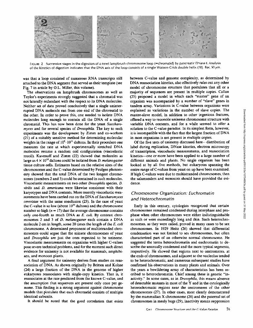
FIGURE 2
Successive stages in the digestion of a newt lampbrush chromosome loop (=chromatid) by pancreatic DNase I . Analysisof the kinetics of digestion indicates that the DNA axis of the loop consists of a single Watson-Crick double helix (18) . Bar, 50ILm .
was that a loop consisted of numerous RNA transcripts stillattached to the DNA segment that served as their template (seeFig . 7 in article by O.L . Miller, this volume) .
The observations on lampbrush chromosomes as well asTaylor's experiments strongly suggested that a chromatid wasnot laterally redundant with the respect to its DNA molecules .Neither set of data proved conclusively that a single uninter-rupted DNA molecule ran from one end of the chromatid tothe other. In order to prove this, one needed to isolate DNAmolecules long enough to contain all the DNA of a singlechromatid. This has now been done for the yeast Saccharo-myces and for several species of Drosophila. The key to suchexperiments was the development by Zimm and co-workers(21) of a suitably sensitive method for determining molecularweights in the range of 108-10 11 daltons . In their procedure onemeasures the rate at which experimentally stretched DNAmolecules resume a random coil configuration (viscoelasticrecoil) . Kavenoff and Zinun (22) showed that molecules aslarge as 4 x 101° daltons could be isolated from D. melanogastertissue culture cells . Estimates based on the relative sizes of thechromosomes and the C-value determined by Feulgen photom-etry showed that the total DNA of the two longest chromo-somes (numbers 2 and 3) could be contained in such molecules.Viscoelastic measurements on two other Drosophila species, D.virdis and D. americana were likewise consistent with theirkaryotypes and DNA contents . More recently viscoelastic mea-surements have been carried out on the DNA ofSaccharomycescerevisiae with the same conclusion (23) . In the case of yeastthe C-value is so low (about 101° daltons) and the chromosomenumber so high (n = 17) that the average chromosome containsonly one-fourth as much DNA as E. coli. By contrast chro-mosomes 2 and 3 of D. melanogaster each contain a DNAmolecule 2 cm in length, some 20 times the length ofthe E. colichromosome. A determined proponent of multistranded chro-mosomes could argue that the minute chromosomes of yeastand Drosophila are just the ones expected to be unineme .Viscoelastic measurements on organisms with higher C-valuespose severe technical problems, and for the moment such directevidence for uninemy is not available for mammals, amphibi-ans, and monocot plants .A final argument for uninemy derives from studies on reas-
sociation of DNA. As shown originally by Britten and Kohne(24) a large fraction of the DNA in the genome of highereukaryotes reassociates with single-copy kinetics . That is, itreassociates at the rate predicted from the known C-value, andthe assumption that sequences are present only once per ge-nome. This fording is a strong argument against chromosomemodels that postulate that each chromatid consists of multipleidentical subunits .
It should be noted that the good correlation that exists
between C-value and genome complexity, as determined byDNA reassociation kinetics, also effectively rules out any othermodel of chromosome structure that postulates that all or amajority of sequences are present in multiple copies . Callan(25) proposed a model in which each "master" gene of anorganism was accompanied by a number of "slave" genes intandem array . Variations in C-value between organisms wereexplained as variations in the number of slave copies . Themaster-slave model, in addition to other ingenious features,offered a way to reconcile unineme chromosome structure withvariable DNA contents, and for a while seemed to offer asolution to the C-value paradox . In its simplest form, however,it is incompatible with the fact that the largest fraction ofDNAin most organisms is not present in multiple copies.Of the five tests of uninemy discussed here-distribution of
label during replication, DNase kinetics, electron microscopyof transcription, viscoelastic measurements, and reassociationkinetics-one or more have been applied to a large number ofdifferent animals and plants . No single organism has beenlooked at by all five methods, but eukaryotes spanning theentire range ofC-values from yeast on up have been examined.If high C-values were due to multistranded chromosomes, thenthe salamanders and monocots should have provided the evi-dence .
Chromosome Organization : Euchromatinand HeterochromatinEarly in this century, cytologists recognized that certain
chromosomes remained condensed during interphase and pro-phase when other chromosomes were either indistinguishableas such or were exceedingly long and thin . Such heterochro-mosomes, as they were called, proved in many cases to be sexchromosomes. In 1929 Heitz (26) showed that differentialcondensation was not limited to sex chromosomes, but oftencharacterized part of an otherwise normal chromosome . Hesuggested the terms heterochromatin and euchromatin to de-scribe the unusually condensed and the more typical segments,respectively. He showed that regions next to centromeres, atthe ends of chromosomes, and adjacent to the nucleolus tendedto be heterochromatic, and numerous subsequent studies haveconfirmed his observations in many plants and animals . Overthe years a bewildering array of characteristics has been as-cribed to heterochromatin. Chief among these is genetic "in-activity ." In some cases, as in Drosophila, this means absenceofdetectable mutants in most of the Y and in the cytologicallyheterochromatic regions near the centromeres of the otherchromosomes (27) . In other cases, most clearly demonstratedby the mammalian X chromosome (28) and the paternal set ofchromosomes in mealy bugs (29), inactivity means suppression
GALL
Chromosome Structure and the C-Value Paradox
5s
Dow
nloaded from http://rupress.org/jcb/article-pdf/91/3/3s/1075489/3s.pdf by guest on 31 M
ay 2022
of function in an otherwise normal chromosome or set ofchromosomes. Other characteristics include late replicationduring the S-period (30), differential replication (31, 32), ab-sence of meiotic recombination (27), effects on euchromaticregions brought into proximity with heterochromatin (33), andeven elimination ofheterochromatin from certain cells (34, 35).Some order was brought into the discussion ofheterochromatinby Brown and Nur (29), who recognized two broad categoriesthat they called facultative and constitutive heterochromatin .They defined these as heterochromatin present in only onehomologue or in both homologues. This rather unusual defi-nition distinguished heterochromatin as a state of an otherwisenormal chromosome (facultative) from heterochromatin as apermanent condition (constitutive) .The distinction between facultative and constitutive hetero-
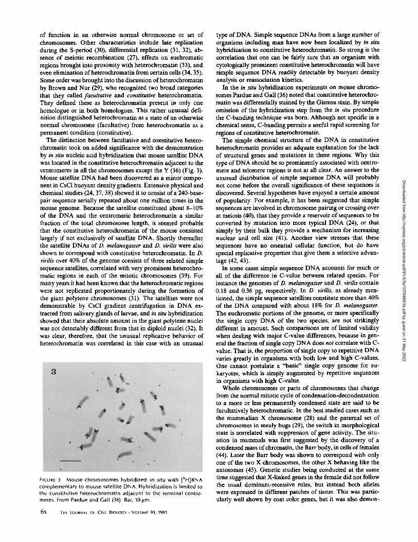
chromatin took on added significance with the demonstrationby in situ nucleic acid hybridization that mouse satellite DNAwas located in the constitutive heterochromatin adjacent to thecentromeres in all the chromosomes except the Y (36) (Fig . 3) .Mouse satellite DNA had been discovered as a minor compo-nent in CsCl buoyant density gradients . Extensive physical andchemical studies (24, 37, 38) showed it to consist of a 240-base-pair sequence serially repeated about one million times in themouse genome . Because the satellite constituted about 8-10%of the DNA and the centromeric heterochromatin a similarfraction of the total chromosome length, it seemed probablethat the constitutive heterochromatin of the mouse consistedlargely if not exclusively of satellite DNA. Shortly thereafterthe satellite DNAs of D . melanogaster and D . virilis were alsoshown to correspond with constitutive heterochromatin. In D.virilis over 40% of the genome consists of three related simplesequence satellites, correlated with very prominent heterochro-matic regions in each of the mitotic chromosomes (39) . Formany years it had been known that the heterochromatic regionswere not replicated proportionately during the formation ofthe giant polytene chromosomes (31) . The satellites were notdemonstrable by CsCl gradient centrifugation in DNA ex-tracted from salivary glands of larvae, and in situ hybridizationshowed that their absolute amount in the giant polytene nucleiwas not detectably different from that in diploid nuclei (32) . Itwas clear, therefore, that the unusual replicative behavior ofheterochromatin was correlated in this case with an unusual
FIGURE 3 Mouse chromosomes hybridized in situ with [3H]RNAcomplementary to mouse satellite DNA. Hybridization is limited tothe constitutive heterochromatin adjacent to the terminal centro-meres . From Pardue and Gall (36) . Bar, 10 gm .
6s
THE JOURNAL Or CELL BIOLOGY " VOLUME 91, 1981
type of DNA. Simple sequence DNAs from a large number oforganisms including man have now been localized by in situhybridization to constitutive heterochromatin . So strong is thecorrelation that one can be fairly sure that an organism withcytologically prominent constitutive heterochromatin will havesimple sequence DNA readily detectable by buoyant densityanalysis or reassociation kinetics.
In the in situ hybridization experiments on mouse chromo-somes Pardue and Gall (36) noted that constitutive heterochro-matin was differentially stained by the Giemsa stain. By simpleomission of the hybridization step from the in situ procedurethe C-banding technique was born. Although not specific in achemical sense, C-banding permits a useful rapid screening forregions of constitutive heterochromatin.The simple chemical structure of the DNA in constitutive
heterochromatin provides an adquate explanation for the lackof structural genes and mutations in these regions . Why thistype of DNA should be so prominently associated with centro-mere and telomere regions is not at all clear. An answer to theunusual distribution of simple sequence DNA will probablynot come before the overall significance of these sequences isdiscovered. Several hypotheses have enjoyed a certain amountof popularity. For example, it has been suggested that simplesequences are involved in chromosome pairing or crossing overat meiosis (40), that they provide a reservoir of sequences to beconverted by mutation into more typical DNA (24), or thatsimply by their bulk they provide a mechanism for increasingnuclear and cell size (41) . Another view stresses that thesesequences have no essential cellular function, but do havespecial replicative properties that give them a selective advan-tage (42, 43) .
In some cases simple sequence DNA accounts for much orall of the difference in C-value between related species. Forinstance the genomes of D . melanogaster and D . virilis contain0.18 and 0.36 pg, respectively . In D. virilis, as already men-tioned, the simple sequence satellites constitute more than 40%of the DNA compared with about 18% for D. melanogaster.The euchromatic portions of the genome, or more specificallythe single copy DNA of the two species, are not strikinglydifferent in amount. Such comparisons are of limited validitywhen dealing with major C-value differences, because in gen-eral the fraction of single copy DNA does not correlate with C-value . That is, the proportion of single copy to repetitive DNAvaries greatly in organisms with both low and high C-values.One cannot postulate a "basic" single copy genome for eu-karyotes, which is simply augmented by repetitive sequencesin organisms with high C-value.Whole chromosomes or parts of chromosomes that change
from the normal mitotic cycle of condensation-decondensationto a more or less permanently condensed state are said to befacultatively heterochromatic . In the best studied cases such asthe mammalian X chromosome (28) and the paternal set ofchromosomes in mealy bugs (29), the switch in morphologicalstate is correlated with suppression of gene activity. The situ-ation in mammals was first suggested by the discovery of acondensed mass ofchromatin, the Barr body, in cells offemales(44) . Later the Barr body was shown to correspond with onlyone of the two X chromosomes, the other X behaving like theautosomes (45). Genetic studies being conducted at the sametime suggested that X-linked genes in the female did not followthe usual dominant-recessive rules, but instead both alleleswere expressed in different patches of tissue . This was partic-ularly well shown by coat color genes, but it was also demon-
Dow
nloaded from http://rupress.org/jcb/article-pdf/91/3/3s/1075489/3s.pdf by guest on 31 M
ay 2022

strated for biochemical markers . The genetic and cytologicalfeatures taken together indicated that inactivation of one Xchromosome occurred early in development in each somaticcell of a normal diploid female, so that the adult soma is amosaic of clones, each clone expressing the genes of only oneX chromosome . The situation in mealy bugs is similar inprinciple, but in this case a whole set of chromosomes isinactivated in male somatic cells. This is usually the paternalset so that males express only genes inherited from theirmother.The mechanism by which functionally active euchromatin is
converted to condensed, inactive heterochromatin is completelyobscure. It has been suggested several times that methylationof DNA might be a primary event in inactivation (46, 47) .Despite the attractive nature of such an hypothesis, includinganalogy to the restiction-modification systems of bacteria, theavailable evidence is scanty . Now that restriction enzymes areavailable whose specificity depends on the state of methylationof nucleotides at the recognition site, it is possible to examinemethylation of particular genes and to test the methylationhypothesis critically (48, 49). Whatever the mechanisms maybe by which regions become heterochromatic, those mecha-nisms may shed light on the process of gene activation duringdevelopment . Nearly all models of embryonic developmentand cell differentiation rely on the concept of differential geneactivation and inactivation. It is possible to imagine that acti-vation or inactivation of individual genes or blocks of genesduring development might proceed by mechanisms similar tothose involved in facultative heterochromatinization .
Nucleosomes and Chromosome Fine StructureThe DNA molecule contained in a chromatid is several
thousand times longer than the chromosome seen at metaphaseofmitosis . For instance, the X chromosome of D. melanogasteris about 1 .8 jim long at metaphase, but contains 1 cm of DNA.How this compaction is achieved, and what happens when thechromatid partially unwinds during interphase or in the for-mation of giant polytene and lampbrush chromosomes arestructural problems yet to be resolved in detail . The first orderof compaction, that which converts the extended DNA mole-cule into a beaded string of nucleosomes, is now well under-stood from a structural standpoint.
Early attempts to examine chromosome structure by electronmicroscopy were notably unsuccessful. Thin sections, whichrevealed exquisite detail in the organization of mitochondria,the endoplasmic reticulum, flagella, and many other cytoplas-mic structures, showed only an indistinct fibrillar and granulararrangement ofthe nuclear contents . Just as light microscopicalstudies proceeded very slowly until squash methods were intro-duced, so electron microscopy of chromosomes had to awaitmethods for unraveling chromosomes for whole mount obser-vations . It was no more possible to deduce the structure of aninterphase nucleus by sectioning it that it would be to do thesame with a ball of string. Even so, the first attempts to spreadchromatin using surface tension forces at an air-water interfacewere not overly informative (50-52) . Such studies did establishthat nuclei and chromosomes of many organisms consisted ofirregular fibers some 200-300 A in diameter, but little internal
FIGURE 4 Electron micrograph of chromatin spread under low ionic conditions on a hydrophilic substrate ("Miller spread") .Nucleosomes are the most prominent feature of such transcriptionally inactive regions . From McKnight and Miller (67) . Bar, 1 [Lm .
Gnu
Chromosome Structure and the C-Value Paradox
7s
Dow
nloaded from http://rupress.org/jcb/article-pdf/91/3/3s/1075489/3s.pdf by guest on 31 M
ay 2022

FIGURE 5 Three stages in packaging of DNA, illustrated by extrachromosomal rDNA molecules from the oocyte of the waterbeetle, Dytiscus . (A) free DNA molecule; (8) DNA and histones in a beaded nucleosome condition, probably lacking histone Fi1;(C) supercoiled state or "200 A fiber." In each case the length of DNA is the same . From Scheer and Zentgraf (135) . Bar, 1 Jam (A);0.5 Jam, (Band C) .
detail was evident. The situation changed dramatically whenMiller (19, 20) introduced the simple expedient of centrifugingchromosomes and chromatin preparations from hypotonic so-lutions onto hydrophilic substrates (Figs. 4 and 5) . Under theseconditions the delicate chromatin fibrils were beautifully dis-played in an extended condition, and it became possible toexamine regions of transcriptional activity, because the nascentribonucleoprotein molecules remained attached . Olins andOlins (53) first calledattention to the regularly beaded structureof chromatin prepared in this fashion. They designated thebeads v-bodies and suggested that they constituted a newstructural feature characteristic of chromatin from many dif-ferent sources. The v-bodies, as described by Olins and Olins,were about 70 A in diameter and were connected by a thinnerfiber of irregular length . Combined biochemical and electronmicroscopical studies by Chambonand his co-workers (54) onadenovirus-2 chromatin demonstrated that each bead was as-sociated with about 200 base-pairs of DNA. Chambon calledthe beads nucleosomes, the name now generally used.At about the same time biochemical experiments also sug-
gested a repeating structure for chromatin. Hewish and Bur-goyne (55) noticed that DNA isolated from rat liver nuclei,
8S
THE JOURNAL Or CELL BIOLOGY " VOLUME 91, 1981
which had been allowed to self-digest, was cut into a series offragments having lengths of about 200, 400, 600, etc . nucleo-tides. The effect was traced to an endogenous nuclease acti-vated by Ca" or Mg". Exactly comparable digestion ofisolated chromatin was obtained with micrococcal nuclease solong as the chromatin was isolated with minimal shearing (56) .The nuclease digestion studies demonstrated that chromatin,as opposed to free DNA, was organized in some manner thatmade the DNA preferentially susceptible to enzymatic attackat regularly repeated intervals.The key to the enzymatic susceptibility clearly had to he in
the association of DNA with histone. Kornberg (57) proposeda model of nucleosome structure in which 200 base-pairs ofDNA were wrapped around a histone octamer consisting oftwo each of the most highly conserved histones, H2A, H2B,H3, and H4 . Kornberg's model made use of the long knownfact that DNA and histone occur in approximately equalamounts by weight and that the four conserved histones arepresent in equimolar amounts. It was also based on his ownstudies, which showed strong binding in solution between thepairs H2A-H2B and H3-H4 (58) . Subsequent studies have donemuch to clarify the specific arrangement of DNA and histone
Dow
nloaded from http://rupress.org/jcb/article-pdf/91/3/3s/1075489/3s.pdf by guest on 31 M
ay 2022
in nucleosomes, but they have left the basic model intact (59-61). Particularly important has been the realization that thehistone octamer is closely associated with about 140 base pairsof DNA to form a "core" nucleosome, the remaining DNAbeing less tightly associated with the octamer and indeedvarying in length from one organism to the next and apparentlyeven between tissues of the same organism . Histone H1 isassociated with this variable linker region between successivenucleosomes .The behavior of nucleosomes during DNA replication has
been studied by Weintraub and co-workers making use ofdensity labeling of the proteins (62) . They have shown that"old" histone octamers remain intact during replication andthat "new" octamers consist entirely of proteins synthesizedduring the time of replication . The exact distribution of oldand new octamers has not yet been determined, although it isknown that successive octamers on a replicated chromatid tendto be all old or all new over short distances. Permanent changesbetween two daughter chromatids could arise if the new octa-mers associated with one of the strands differed from the oldin some respect. In this connection it is of considerable interestthat the histones ofearly and late sea urchin embryos are codedfor by different structural genes (63) .
Transcriptionally active genes differ from bulk chromatinwith respect to their nuclease sensitivity, as originally foundfor the globin gene in erythropoietic tissue . Weintraub andGroudine (64) showed that the a-globin gene in a transcrip-tionally active tissue was more susceptible to nuclease digestionthan bulk chromatin, whereas it was not so in a transcription-ally inactive tissue such as liver. Similar findings have beenreported for the chick ovalbumin gene (65) and several otherhighly active genes . Because these studies involve digestion oftotal chromatin followed by hybridization with specific probes,they are difficult to relate to the behavior of individual nucleo-somes during transcription. Electron microscopic studies ofactive genes (66, 67) often show widely spaced transcriptsbetween which the chromatin appears to have a normal nu-cleosome structure, an exception being the ribosomal RNAgenes that always have closely spaced transcripts . Nevertheless,detailed analysis of specific genes using a combination ofDNase 1 digestion and blot hybridization (68, 69) suggests thatthe whole region of active transcription is altered in a highlyspecific manner and that sites of preferential cutting are ex-posed.The coiling of DNA around the nucleosome core results in
a six- to sevenfold reduction in length relative to fully extendedDNA (200 base-pairs represent 680 A of DNA, whereas nu-cleosomes are approximately 100 A in diam.) . Clearly, there-fore, there must be higher orders of coiling or folding toaccount for the known dimensions of chromosomes. Becausea fiber of approximately 200-300 A diameter occurs as the nextmost complex feature seen by electron microscopy (Fig . 5),several hypotheses have been suggested to account for itsstructure . These models are concerned with the way in whicharrays of nucleosomes may be packed into helical supercoils(70, 71) or superbeads (72) . Until detailed X-ray data becomeavailable it will be hard to choose among these models .Even the 200- to 300-A fiber is considerably longer than a
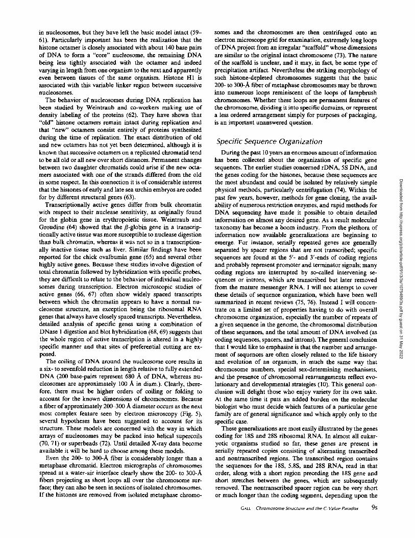
metaphase chromatid . Electron micrographs of chromosomesspread at a water-air interface clearly show the 200- to 300-Afibers projecting as short loops all over the chromosome sur-face; they can also be seen in sections ofisolated chromosomes.If the histones are removed from isolated metaphase chromo-
somes and the chromosomes are then centrifuged onto anelectron microscope grid for examination, extremely long loopsof DNA project from an irregular "scaffold" whose dimensionsare similar to the original intact chromosome (73) . The natureof the scaffold is unclear, and it may, in fact, be some type ofprecipitation artifact . Nevertheless the striking morphology ofsuch histone-depleted chromosomes suggests that the basic200- to 300-A fiber of metaphase chromosomes may be throwninto numerous loops reminiscent of the loops of lampbrushchromosomes. Whether these loops are permanent features ofthe chromosome, dividing it into specific domains, or representa less ordered arrangement simply for purposes of packaging,is an important unanswered question .
Specific Sequence OrganizationDuring the past 10 years an enormous amount ofinformation
has been collected about the organization of specific genesequences. The earlier studies concerned rDNA, 5S DNA, andthe genes coding for the histones, because these sequences arethe most abundant and could be isolated by relatively simplephysical methods, particularly centrifugation (74) . Within thepast few years, however, methods for gene cloning, the avail-ability ofnumerous restriction enzymes, and rapid methods forDNA sequencing have made it possible to obtain detailedinformation on almost any desired gene . As a result moleculartaxonomy has become a boom industry . From the plethora ofinformation now available generalizations are beginning toemerge . For instance, serially repeated genes are generallyseparated by spacer regions that are not transcribed; specificsequences are found at the 5'- and 3'-ends of coding regionsand probably represent promoter and terminator signals; manycoding regions are interrupted by so-called intervening se-quences or introns, which are transcribed but later removedfrom the mature messenger RNA. I will not attempt to coverthese details of sequence organization, which have been wellsummarized in recent reviews (75, 76) . Instead I will concen-trate on a limited set of properties having to do with overallchromosome organization, especially the number of repeats ofa given sequence in the genome, the chromosomal distributionof these sequences, and the total amount of DNA involved (ascoding sequences, spacers, and introns). The general conclusionthat I would like to emphasize is that the number and arrange-ment of sequences are often closely related to the life historyand evolution of an organism, in much the same way thatchromosome numbers, special sex-determining mechanisms,and the presence of chromosomal rearrangements reflect evo-lutionary and developmental strategies (10) . This general con-clusion will delight those who enjoy variety for its own sake .At the same time it puts an added burden on the molecularbiologist who must decide which features of a particular genefamily are of general significance and which apply only to thespecific case.
These generalizations are most easily illustrated by the genescoding for 18S and 28S ribosomal RNA. In almost all eukar-yotic organisms studied so far, these genes are present inserially repeated copies consisting of alternating transcribedand nontranscribed regions . The transcribed region containsthe sequences for the 18S, 5 .8S, and 28S RNA; read in thatorder, along with a short region preceding the 18S gene andshort stretches between the genes, which are subsequentlyremoved. The nontranscribed spacer region can be very shortor much longer than the coding segment, depending upon the
Gnu
Chromosome Structure and the C-Value Paradox
95
Dow
nloaded from http://rupress.org/jcb/article-pdf/91/3/3s/1075489/3s.pdf by guest on 31 M
ay 2022

organism, and often shows variability even within an organism.In at least some cases it has an internal repeating unit so thatit resembles simple sequence DNA (77) .The localization of rDNA at the nucleolus organizer was
fast shown for Drosophila and Xenopus by a combination ofcytogenetic and biochemical data (78, 79). Subsequently theposition of rDNA has frequently been demonstrated by in situhybridization (see review in reference 76) . Many organismshave a single organizer, although multiple sites are not uncom-mon (humans have five, for instance) . Although many orga-nisms have an organizer near the centromere or at an interstitialposition on a chromosome arm, a surprising number have theirnucleolus organizer near the end of the short arm of onechromosome (80) . The significance of this generalization isunknown .Most organisms have from a few dozen to a few hundred
rDNA repeats . For instance, yeast has 140 copies, D. melano-gaster has about 200 copies, and humans between 150 and 200.Very high numbers up to 5,000 or more have been reported forsalamanders and some plants (see review in reference 76) . Inassessing the percent of the genome devoted to rDNA, onemust know the lengths ofthe nontranscribed spacer and inter-vening sequences when they occur . The lengths of spacers arequite variable . They are very short in Bombyx and Sciara, forinstance, so that most of the repeat length is accounted for bythe coding region . At the other extreme very long repeats havebeen described in the mouse and humans (40 kb), the cricketAcheta (35 kb), and the water beetle Dytiscus (29 kb) . Interven-ing sequences have been described in several eukaryotic 28Sgene sequences, where they range in size from 407 bases inTetrahymena pigmentosa (81), about 5 kb in D. melanogaster(82), to 9.8 kb in D. virilis, (83) all organisms with relativelylow C-values . There is no simple relationship between numberofrDNA copies, total rDNA (including spacers and interveningsequences), and C-values . One might have predicted that thenumber or size of rDNA repeats would go up in proportion toC-value. Although it is true that the highest values are foundin high C-value organisms, there is, if anything, a tendency forlow C-value organisms to devote a larger percentage of theirgenome to rDNA (for instance the numbers are 5% for yeast,1% for D. melanogaster, and 0.2% for Xenopus laevis) . The longnontranscribed spacers do not belong to organisms with espe-cially large genomes, and the longest known rDNA intron (D.virdis) is in an organism with a low C-value .One of the most striking features of rDNA is the fact that
copies may exist as free, extrachromosomal molecules in ad-dition to the more typical, chromosomally integrated repeats .The most extreme case of this phenomenon, termed amplifi-cation, is found in oogonia and oocytes of many animals (84,85) . In Xenopus oocytes there are about 2 x 10⁶ rDNA repeatsorganized in approximately 1,000 extrachromosomal nucleolilocated around the periphery ofthe giant oocyte nucleus . Theseamplified genes have been the object of intense investigation(reviewed in reference 86) . It is known that they arise fromchromosomal copies during the earliest oogonal stages (prob-ably as single repeats), that they replicate extrachromosomallyby a rolling circle mechanism primarily during the pachytenestage of meiosis I, and that they engage in intense ribosomalRNA synthesis during vitellogenesis. The overall biologicalsignificance of amplification is reasonably clear . The oocyte isa single cell, which grows to a size many thousand times largerthan a somatic cell and which accumulates ribosomes forprotein synthesis during embryogenesis . The 4C oocyte nucleus
10S
THE JOURNAL OF CELL BIOLOGY " VOLUME 91, 1981
ofXenopus contains only 2,000 integrated rDNA copies, which,if transcribing at maximal rate, would require many years toproduce the 4 hg ofrRNA contained in a mature oocyte . Manygiant cells, which are faced with a similar problem (such as silkgland cells in Bombyx) become polyploid, thereby increasingthe total number of rDNA sequences along with the wholegenome. Such an avenue would not be open to an oocytewithout a complete restructuring of the meiotic phenomena. Ina sense, then, by amplification the oocyte manages to poly-ploidize its rDNA while leaving the rest of the genome intactat the 4C level. This general conclusion was reached in 1942by Painter and Taylor (87) long before the nucleolar DNA ofthe oocyte was recognized as coding for rRNA.rDNA amplification is found in oocytes of many animals,
both vertebrate and invertebrate, but it is not universal. Forinstance, the oocyte nucleus of Drosophila shows no sign ofamplification, yet the oocyte is large and well supplied withribosomal RNA. Here, as in many insects, rRNA comes frompolyploid nurse cells whose cytoplasm is physically continuouswith the oocyte cytoplasm (88) . In still other organisms withsmall oocytes neither amplification nor nurse cells occur, thechromosomal copies of rDNA being adequate for the numberof rRNA molecules needed.rDNA amplification is also found in the macronucleus of
the ciliated protozoan Tetrahymena (89, 90) . Its occurrencehere is correlated with the well-known nuclear dualism ofciliates, which have a transcriptionally inactive diploid micro-nucleus (the germinal nucleus) and a transcriptionally activepolyploid macronucleus (the somatic nucleus) . There is a singlechromosomally integrated rDNA copy in the micronucleus,but several thousand amplified extrachromosomal copies in themacronucleus (91) . The significance of amplification in thiscase seems to be much the same as in oocytes-the large,rapidly growing cell could not synthesize enough rRNA fromthe rDNA copies present in the chromosomes .
Amplification of genes other than rDNA is known in twocases . The first involves cultured cells resistant to the folateanalog methotrexate in which the nomally single copy gene fordihydrofolate reductase may be present in several hundredcopies (92) . The amplified genes are responsible for greatlyincreased production ofdihydrofolate reductase, permitting thecells to function in the presence ofthe drug . The second is therecently discovered amplification of chorion protein genes inthe ovary of Drosophila (93) . This is an unusually interestingcase because it is the first example of a protein-coding genethat amplifies during normal cell differentiation . Cells thatproduce massive amounts of a single protein, for example silkfibroin or egg albumin, ordinarily do so without amplificationof the corresponding gene (94). Large amounts of protein canbe synthesized because the mRNA is stable and because thetissue is active for many hours or days. Spradling and Mahow-ald argue that Drosophila oogenesis proceeds so fast that onlymultiple gene copies can produce the required number ofchorion mRNA molecules (93) . If their argument is correct,one should find other cases of amplification (or multiple chro-mosomal copies) of structural genes in extremely rapidly de-veloping systems .The existence of amplified genes in diverse organisms and
cell types underscores the conclusion that the number ofgenecopies is often understandable only after considering the lifehistory ofthe organism and the specific features of the cell typein which the genes are transcribed . The same general conclu-sion is illustrated in a dramatic fashion by the genes coding for
Dow
nloaded from http://rupress.org/jcb/article-pdf/91/3/3s/1075489/3s.pdf by guest on 31 M
ay 2022

5S RNA. 5S RNA is a small molecule, 120 nucleotides inlength, present as a single copy in the larger ribosomal subunit .In two organisms (yeast and Dictyostelium) the 5S codingsequence is located between the 17S and 25S genes (95, 96),but in all other investigated cases it occurs in tandemly repeatedunits unlinked to the other ribosomal RNA sequences . As withthe 18S and 28S genes, highly conserved SS coding regionsalternate with spacers that may be internally repetitive andvariable in length (97, 98) . The cytological location of the 5Sgenes varies from organism to organism . In Xenopus they occurin clusters nearthe tips ofthe long arms ofall the chromosomes(99) ; in the newt Notophthalmus they are found in the pericen-tromeric heterochromatin of four chromosomes and at oneinterstitial site (100); in Drosophila (101) and maize (102) theyoccur at a single site . Thus there is no obvious pattern to theirlocation.
Their numbers are equally variable . Drosophila has about160 copies (103) whereas X. laevis and X. borealis have about24,000 and 9,000, respectively (104) . As just discussed, thenurse cells of Drosophila make amplification of the genes for18S and 28S rRNA unnecessary. The nurse cells probablyproduce 5S RNA as well. On the other hand, Xenopus lacksnurse cells, and one would suppose that the oocyte wouldamplify the 5S genesjust as it does the 18S and 28S sequences .This is not the case, however (85) ; instead, the large number of5S genes is maintained in the chromosomes primarily for useduring oocyte development. This remarkable conclusion grewout ofthe discovery that oocyte and somatic 5S RNA sequencesdiffer by a few nucleotides (105, 106) . When the 5S genes wereisolated by centrifugation from bulk genomic DNA, they werefound to consist largely of oocyte-type sequences (107) . Onlyafter other minor sets of 5S genes had been isolated andcharacterized were the somatic genes finally discovered in bothX. laevis and X. borealis (108) . They consist ofseveral hundredrepeats with an entirely different spacer from the major oocytespecies. The sequence data make it clear that thousands of 5Sgenes are carried as extra baggage in somatic cells to beexpressed only in oocytes . During oogenesis 5S RNA is syn-thesized at a high rate in previtellogenic oocytes, well beforethe maximal rise in 18S and 28S rRNA synthesis (109). Thuseven though each ribosome will eventually contain one 5Smolecule for each 18S and 28S molecule, the genes are un-linked, and their transcription is temporally uncoordinated .The formation ofribosomes during oogenesis is an important
developmental event requiring synthesis of large amounts of18S, 28S, and 5S ribosomal RNA. As just discussed, it is nowclear that different organisms utilize quite different mecha-nisms to deal with the problem. In some cases genes areamplified extrachromosomally, in others the gene product issupplied by polyploid nurse cells, and in still others a specialset of oocyte genes is maintained in the chromosomes . Surpris-ingly a single organism may utilize two different mechanisms,as in the case of Xenopus, which amplifies the 18S and 28Sgenes, but carries special oocyte 5S genes, even though themature ribosome must contain equimolar quantities of eachRNA. Another such case occurs in the beetle Dytiscus, whichamplifies the 18S and 28S genes in oogonia and oocytes, butwhich also has polyploid nurse cells that supply RNA to theoocyte (110) .The genes coding for histones have been studied extensively
in three species of sea urchin and in Drosophila (reviewed inreference 63). Earlier investigations by ultracentrifugationdemonstrated that the genes were repetitive and probably
clustered (111) . With the advent of molecular cloning it waspossible to obtain a restriction enzyme fragment 7 kb in lengthfrom the sea urchin Strongylocentrotus purpuratus that con-tained one coding region for each of the five histones in theorder H1, H4, H2B, H3, H2A (112) . Each coding region wasseparated from the next adjacent one by a spacer . Altogetherthere are several hundred serial repeats of this five-memberedunit . A similar organization including the same gene order wasdemonstrated in two other species of sea urchin, Lytechinuspictus and Psammechinus miliaris. In Drosophila there are fewergene copies, about 110 in all, but here too there is a repeatingunit containing one each of the five genes (113) . A notabledifference in organization between the three sea urchins andDrosophila is that the coding regions are all in one strand inthe sea urchins, whereas two coding regions are on one strandand three on the other in Drosophila . The Drosophila genes,therefore, cannot be transcribed as a single polycistronic mes-senger . Studies on histones during sea urchin developmenthave shown the remarkable fact that histones produced atdifferent stages may have different primary amino acid se-quences (114) . For instance, histone H1 from cleavage stagesdiffers from its counterpart during gastrulation . The mRNAsfor the two species are different and must be coded for byseparate genes . Even in the case of histone H4, which has thesame amino acid sequence at different stages, the messengerRNAs are distinct . The genes that have been cloned are inevery case those which code for the earliest histone, suggestingbut not proving that the later variants are coded for by rela-tively rare genes. If this turns out to be true, the analogy withthe 5S genes would be close . That is, the organism maymaintain a family of similar, repeated genes for use during acritical stage in its life history when unusually rapid synthesisis necessary. As in the case of the ribosomal RNA genes themechanism used by the sea urchins might not represent aunique solution to the problem . For instance, Xenopus, whichhas much the same need for histones during embryogenesis,has only 20-50 gene copies (115) . Adamson and Woodland(116) suggest that Xenopus synthesizes and stores histones andhistone mRNAs during oogenesis, a protracted period lastingseveral months, and that the increase in histone synthesis incleavage stages is dependent on stored mRNA. It appears thatthe difference in number of histone gene copies between thesea urchins and Xenopus may be correlated with differentsolutions to a developmental problem, although more infor-mation is needed before this conclusion is firm.Genes coding for various proteins have now been cloned by
recombinant DNA methods and their structure examined indetail; the number of new proteins analyzed is increasing at arapid rate, and only a few general comments can be made here .In most cases the genes are ones which code for abundant orsuperabundant proteins, a fact that may have some bearing onthe structures discovered. Although some of the genes may bepresent in only one copy in the genome, most of the examplesstudied consist of a small family of closely related sequences,for instance a- and Q-globin (117-119), actin (120), ovalbumin(121), and vitellogenin (122) . A few, such as the chorion proteingenes in the silk moth, Antheraea consist of a family of se-quences coding for a large number of similar but not identicalpolypeptides (123) . Among the most surprising features is thewidespread occurrence of intervening sequences (or introns)separating the coding sequence into two or more segments(discussed in reference 124) . The number of intervening se-quences per gene varies considerably, there being two in mam-
Gnu
Chromosome Structure and the C-Value Paradox
1 1s
Dow
nloaded from http://rupress.org/jcb/article-pdf/91/3/3s/1075489/3s.pdf by guest on 31 M
ay 2022

malian a-globin, seven in the ovalbumin gene ofthe chick, andan incredible 33 in the vitellogenin gene of Xenopus . There isgood evidence in the case of the hemoglobin genes that theintervening sequences are ancient from an evolutionary stand-point. This is shown by the fact that two intervening sequencesoccur at approximately the same places in the Q-globin genesof several species, as well as in the 8- and -y-variants and in a-globin (119) . Either the intervening sequences were present intheir current locations in the progenitor gene from which theserelated genes were derived or transpositions occur preferen-tially to these sites .
Intervening sequences obviously add to the total DNA con-tent of organisms that possess them. Because of limited data itis not yet possible to relate the C-values of organisms with thenumber and length of their intervening sequences. In yeastvery short intervening sequences have been described in tRNAgenes (125), but until now only one (304 bp) in a protein-coding gene, that which codes for actin (126) . In Dictyostelium,which has a very small genome (C = 0.05 pg), two small intronshave recently been found in a gene coding for an mRNA ofunknown function.' In D. melanogaster (C = 0.18 pg) some ofthe rDNA repeats have introns (82), and an intron has beendescribed in a gene coding for actin (120) . From the limitedinformation available, one gets the impression that organismswith small C-values may have fewer introns than those withlarger ones, but this may be caused in part by spotty sampling .The C-value ofthe chicken (C = 1.2 pg) and Xenopus (C = 3.2pg), which contain such remarkably discontinuous genes, aresmall to moderate by comparison with many other eukaryotes .Organisms with very high C-values have not been examinedfor intervening sequences in protein-coding genes. Notophthal-mus (C = 45 pg) has an average sized rDNA repeat (about 15kb) and a very short 5S repeat (231 bp) with no evidence forintrons . 2 I feel that major differences in C-value will probablynot be directly ascribable to differences in the number andsizes of introns .
Chromomeres, Bands, and LoopsPerhaps the most obvious feature of chromosomes at the
light microscopical level is that they are neither uniform norregularly periodic . Instead they possess aperiodic discontinui-ties represented by chromomeres (especially in meiotic pro-phase), by bands in polytene chromosomes of insects and otherorganisms, and by loops in the lampbrush chromosomes ofoocytes. The number of bands has been counted carefully inthe salivary gland chromosomes of D. melanogaster. The bestestimate, based on the studies of C. B . Bridges and P . N .Bridges is 5059 (127) . The number of chromomeres in a lamp-brush chromosome set varies with age of the oocyte, but thenumber counted during the maximal lampbrush stage is similarto the number ofpolytene bands, e.g., about 5,000 for Triturusand 3,000-6,000 for Plethodon (128, 129) . The number of looppairs is somewhat higher, because there is often more than onepair of loops per chromomere . It is a striking fact that thenumber of bands in Drosophila and the number of chromo-meres in the salamanders are very nearly the same, even thoughthe DNA contents of the two organisms differ by more than100-fold . Does this mean that there are domains of chromo-some structure, whose number remains relatively constant dur-ing chromosome evolution, but whose size varies with C-value?
` Kimmel, A . R., and R . A. Firtel. Personal communication.'Kay, B ., and J . G . Gall. Unpublished observation .
25
THE JOURNAL OF CELL BIOLOGY " VOLUME 91, 1981
This question will be easier to answer once a clearer picture isobtained of a band and interband in Drosophila and otherDiptera. Fortunately that time is not far away . Already fromgenetic analysis of Judd and Young (130) and others, we knowthat a band contains no more than a few complementationgroups, even if the simple correlation of one band = onecomplementation group is an overstatement . The amount ofDNA in a band, i.e., per chromatid, averages 20 kb with arange of perhaps 10-fold between the faintest and most prom-inent bands (the average is obtained by dividing the amount ofeuchromatic DNA, about 10 5 kb, by the number of bands) .The smaller bands simply do not have enough DNA to containmany structural genes along with whatever control regions,spacers, and the like must be present. In the case of the histonegenes it is known from in situ hybridization that the repeatedsequences extend over several bands (131) . Even in the case ofan extraordinarily large puff, the Balbiani ring 2 of Chironomustentans, there may be only one transcription unit (132) . Becauseby hybridization techniques it is possible to select overlappingclones from a clone library of Drosophila ("walking" along thechromosome), there will soon be available several sets of clonesthat extend over more than one band's worth of DNA. Fromthese it should be possible in principle to evaluate the numberofstructural genes and transcription units per band. In the caseof the lampbrush chromosomes, it is reasonably certain frommorphology both at the light microscopical and electron mi-croscopical levels, that a loop often consists of a single tran-scriptional unit. On the other hand, there are clear cases wherethe morphology suggests two or more transcription units (133) .In situ hybridization experiments demonstrate that the RNAtranscripts over a long segment of a loop may hybridize witha specific DNA probe, once again consistent with the notionthat a loop contains one or a small number of transcriptionunits (134) . The missing information in the case of the lamp-brush chromosomes, in order to make a comparison with thebands of the polytene chromosomes, is how many structuralgenes or complementation groups may reasonably be presentin one loop and its associated chromomere.A unified model of eukaryotic chromosome structure might
begin with the postulate that higher organisms have a more orless constant number of chromosome units or domains, roughlyequal to the number of bands in Drosophila polytene chromo-somes or loops in Triturus lampbrush chromosomes. Each ofthese domains would contain one or a small number of struc-tural genes and a correspondingly small number of transcrip-tion units. As the DNA content of the organism went up ordown during evolution, the number of units would remain thesame while the amount of DNA per unit varied enormously.Thus both Drosophila and Triturus would have the same 5,000or so chromosome domains, but the domains in Triturus wouldcontain on average more than 100 times as much DNA asthose in Drosophila . Just how the extra DNA might be orga-nized is open to conjecture . My preference is to suppose thatmuch of it may occur as spacers between the active transcrip-tion units . The extreme form of this model postulates thatactive gene regions are similar in number and organizationthroughout the range of eukaryotic organisms, but that theyare more widely spaced in organisms with high C-values. Acorollary of this model is that changes in DNA content occurmore or less uniformly along the length of the chromosome toaccount for the common observation that related organismswith different C-values may have very similar karyotypes (10) .In order to test these speculations at the molecular level, it will
Dow
nloaded from http://rupress.org/jcb/article-pdf/91/3/3s/1075489/3s.pdf by guest on 31 M
ay 2022

be necessary to compare the organization ofstructural genes inorganisms with a wide range of C-values. Obviously this modelof chromosome structure does not "explain" the C-value par-adox. It does, however, stress that the number of active genesand transcription units need not be correlated with the totalamount of DNA. From a structural standpoint it focusesattention on the organization of the individual chromosomedomains, and it could be critically tested by showing that thespacing of active genes varies more or less linearly with C-value. It has been pointed out several times that DNA contentis positively correlated with nuclear and cell size and inverselywith rate of mitosis and rate of embryonic development (41) .If these correlations are more than fortuitous, it would beuseful to look for ways in which the DNA content of thechromosome domains might regulate the timing ofmitosis andthe rate at which the embryonic program is read .An adequate model of chromosome structure must ulti-
mately encompass not only the organization of individualgenes but also the ways in which these genes are regulatedduring cell function and especially during embryonic devel-opment . In this light the study of chromosome organizationhas only just begun, and we can confidently predict majorchanges in our outlook during the next 25 years.
Many of the topics discussed in this review are treated more exten-sively in the Cold Spring Harbor Symposium, Volume 38 (1974) onChromosome Structure and Function, and Volume 42 (1978) on Chro-matin.
REFERENCES
1 . Flemming, W. 1882 . Zellzubstanz, Kern and Zelltheilung. K. G. Vogel,Leipzig, Germany.
2. Miescher, F. 1871 . Hoppe-Seyler's Med.-chem . Unters. 4:441 .3. Feuigen, R., and H. Rossenbeck . 1924. Hoppe-Seyler's Z. Physiol. Chem.
135:203-248 .4. Pollister, A., and H. Ris. 1948 . Cold Spring Harbor Symp . Quanr. Biol. 12:
147-157.5. Swift, H. 1953 . Int. Rev. Cytol. 2:1-76.6. Howard, A., and S. R. Pelc . 1951 . Exp. Cell Res. 2:178-187 .7. Sparrow, A. H., H. J. Price, and A. G. Underbrink. 1972 . Brookhaven Symp.
Biol. 23 :451-494 .8. Bachmann, K., O. B. Goin, and C. J. Goin. 1972. Brookhaven Symp. Biol.
23 :419-650.9. Kaufmann, B. P., andM. R. McDonald. 1956. Cold Spring Harbor Symp.
Quant. Biol. 21 :233-246 .10. White, M. J. D. 1973 . Animal Cytology and Evolution . 3rd edition . Cam-
bridge University Press, Cambridge, England.11 . Taylor, l. H., P. S. Woods, andW. L. Hughes . 1957 . Proc Nad. Acad. Sci.
U.S.A . 43 :122-128 .12. Meselson, M., and F. W. Stahl. 1958 . Proc. Nad. Acad. Sci. U.&A . 44:671-
682.13. Taylor, J. H. 1958 . Genetics. 43 :515-529 .14. Wolff, S., and P. Perry. 1974 . Chromosoma (Berl.) . 48:341-353 .15 . Guyenot, E., and M. Danon. 1953 . Rev. Suisse Zool. 60 :1-129 .16 . Gall, J. G. 1956 . Brookhaven Symp. Biol. 8:17-32.17 . Callan, H. G., and H. C. Macgregor. 1958 . Nature (Loud.) . 181:1479-1480.18 . Gall, J. G. 1963 . Nature (Land.). 198:36-38 .19. Miller, O. L. 1965 . Nat. Cancer Inst. Monogr. 18:79-99.20. Miller, O. L., and B. Beatty . 1969 . J. Cell Physiol 74(Suppl . 1) : 225-232.21 . Chapman, R. E., L. C. Klotz, D. S. Thompson, and B. H. Zimm . 1969 .
Macromolecules . 2:637-643 .22 . Kavenoff, R., and B. Zimm. 1973 . Chromosoma (Bert) . 41 :1-27.23 . Lauer, G. D., T. M. Roberts, and L. C. Klotz. 1977 . J. Mol Biol. 114:507-
526.24 . Britten, R. J ., and D. E. Kohne. 1968 . Science (Wash . D . C.) . 161:529-540.25 . Callan, H. G. 1967 . J. Cell Sci. 2:1-7 .26 . Heitz, E. 1929. Ber. Dsch . Bot. Ges. 47:274-284 .27 . Lindsley, D. L., and E. H. Grell, 1967 . Genetic Variations of Drosophila
melanogasier . Carnegie Institution of Washington, Washington, D. C.28. Lyon,M. F. 1972. Biol. Rev . Camb. Philos. Soc. 47 :1-35.29. Brown, S. W., and U. Nur. 1964 . Science (Wash. D. C.) . 145:130-136 .30. Lima-de-Faria, A. 1959 . J. Biophys. Biochem . Cyto l 6:457-666.31 . Rudkin, G. T. 1969 . Genetics. 61(Suppl.) :227-238 .
32 . Gall, J. G., E. H. Cohen, and M. L. Polan. 1971 . Chromosoma (Berl.) . 33 :319-344.
33 . Spofford, l . B. 1976 . In The Genetics and Biology of Drosophila . M.Ashbumer andE. Novitski, editors. Academic Press, Inc., New York. Ic:955-1018.
34. Boveri, T. 1887. Anar. Anz. 2:688-693 .35 . Beermann, S. 1977. Chromosome; (Bert) . 60 :297-344 .36 . Pardue, M. L., and J. G. Gall. 1970. Science (Wash . D. C.). 168:1356-1358 .37 . Flamm, W. G., P. M. B. Walker, andM. McCallum. 1969 . J. Mol. Biol. 40:
423-443.38 . Southern, E. 1975 . J. Mot. Biol. 94 :51-69 .39 . Gall, J. G., and D. D. Atherton . 1974. J. Mo l Biol. 85 :633-664.40. Brutlag, D., R. Appets, E. S. Dennis, andW. J . Peacock. 1977 . J. Mol Biol.
112:31-67 .41 . Cavalier-Smith, T. 1978 . J. Cell Sci 34:247-278 .42 . Orgel, L., and F. H. C. Crick. 1980. Nature (Land.). 284:604-607 .43 . Doolittle, W. F., and C. Sapienza. 1980. Nature (Land). 284:601-603 .44 . Barr, M. L., and L. F. Bertram. 1949 . Nature (Land.) . 163:676-677 .45 . Ohno, S., W. D. Kaplan, and R. Kinosita . 1959 . Exp. Cell Res. 18 :282-290.46 . Holliday, R., and J. E. Pugh. 1975 . Science (Wash. D. C.). 187:226-232.47 . Sager, R., and R. Kitchin. 1975 . Science (Wash. D. C) . 189:426-633 .48 . Bird, A. P., and E. Southern . 1978 . J. Mo l Biol. 118:27-47 .49 . Bird, A. P. 1978 . J. Mal. Biol. 118:49-60.50 . Gall, J. G. 1963 . Science (Wash. D. C). 139:120-121 .51 . Gall, l . G. 1966 . Chromosoma (Berl.). 20:221-233.52 . Ris, H., and D. Kubai. 1970. Annu. Rev. Genet. 4:263-294.53 . Olins, A., and D. Olins. 1974 . Science (Wash . D. -C.) . 183:330-332.54 . Oudet, P., M. Gross-Bellard, and.P. Chambon. 1975. Cell. 4:281-300.55 . Hewish, D., and L. Burgoyne . 1973 . Biophys. Biochem . Res. Commun. 52 :
504-510.56 . Noll, H. 1974. Nature (Land.). 251:249-251 .57 . Komberg, R. 1974 . Science (Wash. D . C). 184:868-871 .58 . Komberg, R., and J. Thomas . 1974 . Science (Wash. D. C). 184:865-868 .59 . Finch, J ., L. Lutter, D. Rhodes, R. Brown, B. Ruston, M. Levitt and A.
Klug . 1977 . Nature (Land.). 269:29-36 .60 . Komberg, R. 1977 . Annu. Rev . Biochem . 46 :931-954 .61 . Felsenfeld, G. 1978. Nature (Load.). 271:115-122 .62 . Leffak, M., R. Grainger, and H. Weintraub. 1977 . Cell. 12:837-845 .63 . Kedes, L. H. 1979 . Annu. Rev. Biochem. 48 :837-870.64 . Weintraub, H., and M. Groudine. 1976 . Science (Wash. D. C.). 193:848-
856.65. Garel, A., and R. Axel . 1976 . Proc. Natl. Acad Sc . U. S. A . 73 :3966-3970 .66. Laird, C. D., L. E. Wilkinson, V. E. Foe, andW. Y. Chooi. 1976. Chromo-
soma (Berl.). 58:169-192 .67 . McKnight, S. L., and O. L. Miller . 1976. Cell. 8:305-319.68 . Wu,C., P. M. Bingham, K. J . Livak, R. Holmgren, and S. Elgin. 1979 . Cell.
16 :797-806 .69 . Stalder, J., A. Larsen, J. Engel, M. Dolan, M. Groudine, and H. Weintraub.
1980 . Cell 20:451-660.70 . Finch, J. T., and A. Klug. 1976 . Proc. Nat. Acad. Sci. U S . A . 73 :1897-
1901 .71 . Worcel, A., and C. Benyajati. 1977 . Cell. 12:83-100.72. Hozier, J., M. Renz, and P. Nehls. 1977. Chromosoma (Berl.) . 62 :301-317 .73. Laemmli, U., S. Cheng, K. Adolf, l. Paulson, J. Brown, andW. Baumbach.
1978 . Cold Spring Harbor Symp . Quant. Biol. 42 :351-360 .74 . Brown, D. D., and R. Stem. 1974. Annu. Rev. Biochem. 43 :667-693 .75. Dawid, L, and W. Wahii. 1979 . Dev. Biol. 69 :305-328 .76. Long, E. O., and I. B. Dawid. 1980. Annu. Rev. Biochem. 49:727-764 .77. Fedoroff, N. V. 1979. Cell. 16:697-710 .78. Ritossa, F., and S. Spiegelman. 1965. Proc. Nad. Acad Sci. U. S. A . 53 :
737-745.79. Wallace, H., and M. Bimstiel . 1966. Biochim . Biophys. Acta. 114:296-3 10 .80. Lima-de-Faria, A. 1979. In Specific Eukaryotic Genes. J. Engberg, H.
Klenow, and V. Leick, eds. Alfred Benzon Symp . Munksgaard, Copen-hagen. 13 :25-38.
81 . Wild,M., and J . G. Gall . 1979. Cell. 16 :565-573 .82. Glover, D. M., and D. S. Hogness. 1977 . Cell. 10 :167-176 .83 . Barnett, T., and P. M. M. Rae. 1979. Cell. 16 :763-775 .84. Gall, J. G. 1968 . Proc. Nad. Acad. Sci U. S. A . 60:553-560 .85 . Brown, D., and 1. Dawid. 1968 . Science (Wash. D . C.). 160:272-280.86 . Tobler, H. 1975. In Biochemistry ofAnimal Development, R. Weber, editor.
Academic Press, Inc. New York . 3:91-143 .87 . Painter, T. S., and A. N. Taylor . 1942 . Proc . Natl. A cad. Sci. U. S. A . 28 :
311-317.88. Bier, K., W. Kunz, and D. Ribbert. 1967 . Chromosoma (Berl.). 23:214-254.89. Yao, M.-C., A. Kimmel, and M. Gorovsky . 1974 . Proc. Nad. Acad. Sci. U.
S. A. 71 :3082-3086.90. Gall, J . G. 1974 . Proc. Nall. Acad. Sci. U. S. A . 71 :3078-3081 .91 . Yao, M.-C., and J. G. Gall. 1977 . Cell. 12:121-132.92 . Schimke, R. T., F. W. Alt, R. E. Kellems, R. J . Kaufman, and J . R. Bertino.
1978. Cold Spring Harbor Symp. Quant. Biol. 42:649-657.93 . Spradling, A. C., and A. P. Mahowald . 1980. Proc. Nat. Acad. Sci. U. S. A .
77 :1096-1100.94. Suzuki, Y., L. P. Gage, and D. D. Brown. 1972. J. Mo. Biol. 70:637-
649.95 . Bell, G. L, L. l. DeGennaro, D. H. Gelfand, R. J. Bishop, P. Valenzuela,
Gnu
Chromosome Structure andthe C-Value Paradox
13S
Dow
nloaded from http://rupress.org/jcb/article-pdf/91/3/3s/1075489/3s.pdf by guest on 31 M
ay 2022

andW. J. Rutter . 1977. J. Biot Chem . 252:8118-8125.96 . Maizels, N. 1976. Cell 9:431-438.97 . Brown, D. D., P. C. Wensink, and E. Jordan. 1971 . Proc. Nod. Acad. Sci.
U. S. A. 68:3175-3179.98 . Carrol, D., and D. D. Brown. 1976. Cell. 7:477486.99 . Pardue, M. L., D. D. Brown, andM. L. Birnstiel. 1973 . Chromosoma (Berl.).
42:191-203 .100. Hutchison, N., andM. L. Pardue. 1975 . Chromosoma (Berl.) . 53 :51-69.101. Wimber,D., andD. M. Steffensen. 1970. Scienc e (Wash. D. C) . 170:639-
641.102. Wimber, D., P. Duffey, D. M. Steffensen, andW. Prensky. 1974. Chromo-
soma (Berl.). 47 :353-359 .103. Tartof, K. D., and R. P. Perry. 1970. J. Mol Biol. 51 :171-183 .104. Brown, D. D., andK. Sugimoto. 1973 . J. Mol. Biol. 78 :397-415 .105. Ford, P. J., and E. M. Southern . 1973. Nature (Land.). 241:7-12.106. Wegnez, M., R. Monier, and H. Denis. 1972 . FEBS (Fed. Eur. Biochem.
Soc.) . Lett 25 :13-20 .107. Brownlee, G. G., E. M. Cartwright, and D. D. Brown. 1974 . J. Mol Biol.
89 :703-718 .108. Peterson, R. C., J. L. Doering, andD. D. Brown. 1980 . Cell 20:131-141 .109. Mairy, M., and H. Denis. 1972 . Eur. J. Biochem. 25:535-543.110. Gall, J. G., H. C. Macgregor, andM. E. Kidston. 1968 . Chromosoma (Bert) .
26 :169-187 .III. Kedes, L. H., andM. L. Birnstiel. 1971 . Nature (Land.) . 230:165-169 .112. Kedes, L. H., A. C. Y. Chang, D. Housman, and S. N. Cohen. 1975 . Nature
(Loud.). 255:533-538 .113. Lifton, R. P., M. L. Goldberg, R. W. Karp, and D. S. Hogness. 1978 . Cold
Spring Harbor Symp. Quant. Biol. 42:1047-1051 .114. Newrock, K. M., C. R. Alfageme, R. V. Nardi, and L. H. Cohen. 1978 .
Cold Spring Harbor Symp. Quant. Biol. 42:421-431 .115 . Jacob, E., G. Malacinski, and M. L. Bimstiel. 1976 . Eur. J. Biochem. 69 :45-
54.
1 4S
THE JOURNAL OF CELL BIOLOGY " VOLUME 91, 1981
116. Adamson, E. D., and H. R. Woodland. 1977 . Dev. Biol. 57 :136-149.117. Flavell, R. A., J. M. Kooter, E. DeBoer, P. F. R. Little, and R. Williamson .
1978 . Cell 15 :25-41 .118. Fritsch, E. F., R. M. Lawn, and T. Maniatis. 1980. Cell 19 :959-972.119. Lauer, J., C.-K. J. Shen, and T. Maniatis. 1980. Cell 20:119-130.120. Fyrberg, E. A., K. L. Kindle, N. Davidson, and A. Sodja. 1980 . Cell 19:
365-378 .121 . Breathnack, R., C. Benoist, K. O'Hare, F. Gannon, and P. Chambon. 1978.
Proc. Nod. Acad. Sci. U. S. A. 75 :4853-4857 .121 . Wahli, W., I. B. Dawid, T. Wyler, R. Weber, and G. U. Ryffel . 1980 . Cell.
20 :107-117 .123. Sim, G. K., A. Efstratiadis, C. W. Jones, F. C. Kafatos, M. Koehler, H. M.
Kronenberg, T. Maniatis, J . C. Regier, B. F. Roberts, and N. Rosenthal.1978. Cold Spring Harbor Symp. Quant. Biol. 42:933-945 .
124. Gilbert, W. 1978 . Nature (Loud) . 271:501 .125 . Abelson, J. 1979 . Annu . Rev. Biochem. 48 :1035-1069.126. Gallwitz, D., and I. Sures. 1980. Proc. Nad. Acad. Sci. U. S. A. 77 :2546-
2550 .127. Lefevre, G. 1976 . In The Genetics and Biology of Drosophila la . M.
Ashburner and E. Novitski, editors. Academic Press, Inc., New York. 31-66 .
128 . Vlad, M., and H. C. Macgregor. 1975. Chromosoma (Berl.). 50:327-347 .129. Callan, H. G. 1963 . Int. Rev. Cyto l 15 :1-34.130. Judd, B. H., and M. W. Young. 1974. Cold Spring Harbor Symp. Quant.
Biol. 38 :573-579.131 . Pardue, M. L., L. H. Kedes, E. S. Weinberg, and M. L. Birnstiel. 1977 .
Chromosoma (Berl.) . 63 :135-151 .132. Lamb, M. M. and B. Daneholt. 1979 . Cell. 17:835-848.131 . Scheer, U., W. W. Franke, M. F. Trendelenburg, and H. Spring. 1976. J.
Cell Sci. 22:503-520.134. Old, R. W., H. G. Callan, and K. W. Gross. 1977. J. Cell Sci. 27 :57-79.135. Scheer, U., and H. Zentgraf. 1978. Chromosoma (Berl.) . 69 :243-254 .
Dow
nloaded from http://rupress.org/jcb/article-pdf/91/3/3s/1075489/3s.pdf by guest on 31 M
ay 2022