Array-based sensing with nanoparticles: ‘Chemical noses’ for sensing biomolecules and cell...
13
Array-based sensing with nanoparticles: “Chemical noses” for sensing biomolecules and cell surfaces Oscar R. Miranda, Brian Creran, and Vincent M. Rotello * Department of Chemistry, University of Massachusetts, 710 North Pleasant Street, Amherst, MA 01003, USA Abstract Nanoparticle-based arrays have been used to distinguish a wide range of biomolecular targets through pattern recognition. In this report, we highlight new “chemical nose” methodologies that use nanoparticle systems to provide high sensitivity sensing of biomolecular targets, including fluorescent polymer/gold nanoparticle complexes that can discriminate between different bioanalytes including proteins, bacteria, and mammalian cells as well as dye-based micellar systems for the detection of clinically important metallo- and non-metallo proteins. 1. Introduction Most biological recognition processes occur via specific interactions. However, sensory processes such as taste and smell use “differential” binding where the receptors bind to their analytes through interactions that are selective rather then specific [1,2]. These array based sensing platforms can be trained to generate a response pattern analogous to olfaction, providing versatile detectors [3]. Recently, a variety of array based sensor platforms have been developed for biomacromolecule sensing, including porphyrins [4], oligopeptide functionalized resins [5], and polymers [6,7]. Nanoparticles (NPs) feature sizes commensurate with biomacromolecules, coupled with useful physical and optical properties [8,9]. Modulation of these physicochemical properties can be readily achieved by changing of core and/or ligand structure. In this report, we highlight the recent advances of array based/chemical nose sensors using materials such as gold, dendrimer, and magnetic nanoparticles for the detection and identification of analytes such as proteins, bacteria, and cells. 2. Nanoparticle arrays for sensing proteins Irregular protein concentration levels in biofluids, e.g., serum, urine, and saliva, provide essential information for the early diagnosis of many pathological conditions [1,10,11,12,13,14]. Substantial efforts have been devoted to developing precise and efficient methods for protein sensing [15] including enzyme-labeled immunoassays [16], electrophoresis methods [17], and analytical techniques [18]. Detection and identification of imbalance through of an array-based sensing approach provides a promising alternative to these methods [5]. Array-based sensing approaches are complementary to more traditional [email protected]. Publisher's Disclaimer: This is a PDF file of an unedited manuscript that has been accepted for publication. As a service to our customers we are providing this early version of the manuscript. The manuscript will undergo copyediting, typesetting, and review of the resulting proof before it is published in its final citable form. Please note that during the production process errors may be discovered which could affect the content, and all legal disclaimers that apply to the journal pertain. NIH Public Access Author Manuscript Curr Opin Chem Biol. Author manuscript; available in PMC 2011 December 1. Published in final edited form as: Curr Opin Chem Biol. 2010 December ; 14(6): 728–736. doi:10.1016/j.cbpa.2010.07.021. NIH-PA Author Manuscript NIH-PA Author Manuscript NIH-PA Author Manuscript
-
Upload
independent -
Category
Documents
-
view
2 -
download
0
Transcript of Array-based sensing with nanoparticles: ‘Chemical noses’ for sensing biomolecules and cell...

Array-based sensing with nanoparticles: “Chemical noses” forsensing biomolecules and cell surfaces
Oscar R. Miranda, Brian Creran, and Vincent M. Rotello*Department of Chemistry, University of Massachusetts, 710 North Pleasant Street, Amherst, MA01003, USA
AbstractNanoparticle-based arrays have been used to distinguish a wide range of biomolecular targetsthrough pattern recognition. In this report, we highlight new “chemical nose” methodologies thatuse nanoparticle systems to provide high sensitivity sensing of biomolecular targets, includingfluorescent polymer/gold nanoparticle complexes that can discriminate between differentbioanalytes including proteins, bacteria, and mammalian cells as well as dye-based micellarsystems for the detection of clinically important metallo- and non-metallo proteins.
1. IntroductionMost biological recognition processes occur via specific interactions. However, sensoryprocesses such as taste and smell use “differential” binding where the receptors bind to theiranalytes through interactions that are selective rather then specific [1,2]. These array basedsensing platforms can be trained to generate a response pattern analogous to olfaction,providing versatile detectors [3]. Recently, a variety of array based sensor platforms havebeen developed for biomacromolecule sensing, including porphyrins [4], oligopeptidefunctionalized resins [5], and polymers [6,7].
Nanoparticles (NPs) feature sizes commensurate with biomacromolecules, coupled withuseful physical and optical properties [8,9]. Modulation of these physicochemical propertiescan be readily achieved by changing of core and/or ligand structure. In this report, wehighlight the recent advances of array based/chemical nose sensors using materials such asgold, dendrimer, and magnetic nanoparticles for the detection and identification of analytessuch as proteins, bacteria, and cells.
2. Nanoparticle arrays for sensing proteinsIrregular protein concentration levels in biofluids, e.g., serum, urine, and saliva, provideessential information for the early diagnosis of many pathological conditions[1,10,11,12,13,14]. Substantial efforts have been devoted to developing precise and efficientmethods for protein sensing [15] including enzyme-labeled immunoassays [16],electrophoresis methods [17], and analytical techniques [18]. Detection and identification ofimbalance through of an array-based sensing approach provides a promising alternative tothese methods [5]. Array-based sensing approaches are complementary to more traditional
[email protected]'s Disclaimer: This is a PDF file of an unedited manuscript that has been accepted for publication. As a service to ourcustomers we are providing this early version of the manuscript. The manuscript will undergo copyediting, typesetting, and review ofthe resulting proof before it is published in its final citable form. Please note that during the production process errors may bediscovered which could affect the content, and all legal disclaimers that apply to the journal pertain.
NIH Public AccessAuthor ManuscriptCurr Opin Chem Biol. Author manuscript; available in PMC 2011 December 1.
Published in final edited form as:Curr Opin Chem Biol. 2010 December ; 14(6): 728–736. doi:10.1016/j.cbpa.2010.07.021.
NIH
-PA Author Manuscript
NIH
-PA Author Manuscript
NIH
-PA Author Manuscript

immunosensing strategies (e.g. ELISA), providing versatile systems that can be “trained” torecognize analytes and potentially disease states. In 2007, Rotello et al. fabricated a sensorarray composed of six cationic functionalized gold nanoparticles (AuNPs) and an anionicPPE polymer that can properly identify seven common proteins [19••]. The polymerfluorescence is quenched by gold nanoparticles; the presence of proteins disrupts thenanoparticle–polymer interaction (Figure 1a), producing distinct fluorescence responsepatterns (Figure 1b) based on particle-protein affinity. The effeciency of this system isattributed to both the quenching ability of AuNPs as well as the ‘molecular wire’ effect ofPPE polymer [20]. Since the protein-nanoparticle interactions are determined by theirrespective structural features such as charged, hydrophobic, hydrophilic, and hydrogen-bonding sites [21], the differing affinities lead to a fluorescence response fingerprint patternfor individual proteins (Figure 1b). The raw data responses obtained were subjected to lineardiscriminant analysis (LDA) [22,23] to differentiate the fluorescence patterns of thenanoparticle–PPE systems against the different protein targets. This system showed a limitof detection of 4–215 nM depending on Mw protein and identifed correctly 52 out of 55unknowns samples (94.5% accuracy) [19••].
Polymeric nanoparticles provide a separate class of scaffolds for sensor design.Thayumanavan et al. developed a polymeric micellar nanosystem that responded toelectronic complementarity, allowing the system to be selective for metalloproteins [24].They used eight different fluorescence dye molecules non-covalently bound to the micellarinterior of an amphiphilic homopolymer to generate a pattern that allowed the differentiationof four different metalloproteins with limits of detection of 1–200 μM. In another approach,Thayumanavan et al. reported a micellar disassembly process for transduction [25]. Fivedifferent noncovalently assembled receptors were generated, and the disassembly wasstudied by monitoring the encapsulated dye release in response to five different non-metalloproteins. The disassembly-induced fluorescence change of the guest moleculeproduces protein-specific patterns. The limit of detection in this approach was 8 μM. Morerecently, Thayumanavan et al. introduced a new method where the differential response wasgenerated from a single polymer-surfactant complex with two approaches, i.e. thedisassembly and guest release based pathways and photoinduced charge/energy transferquenching (excited state quenching) (Figure 2a). By varying the transducer using non-metalloprotein and metalloproteins [26•] they were able to generate a limit of detection fornon-metalloproteins of 8 μM (Figure 2b). In the case of the metalloproteins, the limit ofdetection was 80 nM (Figure 2c). Thayumanavan et. al also studied the use of a fluorescentanthracene-core dendrimer system that has carboxylic acid groups on the periphery thataffords a differential response protein pattern though the binding energy transfer process atanalyte concentrations between 1–5 μM [27]. Upon binding, an energy transfer processoccurs with quenching of the fluorescent core. The interchange between quenching andbinding lead to differential responses that allowed discrimination of 3 differentmetalloproteins.
The above studies focused on protein sensing in buffer. The plasma/serum proteome, incontrast, contains more than 20,000 different proteins and an overall protein concentrationof ~1 mM, (71 mg ml−1), providing a complex matrix for sensor design [28,29]. Toconstruct a more effective protein sensing system, Rotello et al. selected green fluorescentprotein (GFP) [30], a stable dimeric biomolecule used as transducer. The use of GFPminimizes aggregation, improving sensor efficiency as compared with the previous methodusing conjugated polymers. In this sensing process, the biocompatible AuNP/GFPconjugated array was able to identify five of the more abundant human serum proteins, i.e.human serum albumin, immunoglobulin G, transferrin, fibrinogen and α–antitrypsin, (Figure3a) in an undiluted solution of human serum (overall protein concentration ~1 mM),obtaining a limit of detection of 500 nM [31•]. Further experiments indicated that mixtures
Miranda et al. Page 2
Curr Opin Chem Biol. Author manuscript; available in PMC 2011 December 1.
NIH
-PA Author Manuscript
NIH
-PA Author Manuscript
NIH
-PA Author Manuscript

of different proteins and the addition of one protein in different concentrations also led to aspecific and reproducible change in the LDA-based patterns (Figure 3b).
The sensitivity of fluorophore displacement strategies is limited by the inherent emissivityof the transducer. To overcome this limitation, Rotello et al. used enzyme-amplified arraysensing (EAAS) to provide a platform with enhanced sensitivity [32••]. Functionalizedcationic AuNPs electrostatically bind to the anionic β-Galactosidase (β-Gal), reversiblyinhibiting the enzyme (Figure 4a) [33]. Displacement of the particle by analyte proteinsrestores β-Gal activity towards a fluorogenic substrate, generating a readout signal that isamplified through enzymatic catalysis. This EAAS system couples the signal amplificationprocess of ELISA with the versatility of the “chemical nose” approach, as it is able to senseand identify a range of biomedically relevant proteins with a limit of detection of 1 nM inboth buffer as well as in desalted urine solution (Figure 4b).
3. Nanoparticle arrays for sensing bacteria and mammalian cellsThe efficient detection of pathogenic microorganisms is of great importance in clinical,forensic, medical, and environmental sciences [34]. In clinical diagnostics, bacterialinfections are identified by plating and culturing, a time-consuming methodology. Whileseveral newer methods including PCR, have been used to detect specific microorganisms[35], a facile wet-chemical method for the timely detection and identification ofmicroorganisms would be of interest in both a clinical setting as well to test for foodspoilage in industrial settings [36].
Rotello et al. created an array-based sensing system based on non-covalent conjugates ofAuNPs and a modified PPE that allows the detection of bacteria within minutes [37•]. Asshown in Figure 5, the anionic PPE polymer is bound initially to the functionalized cationicgold nanoparticle creating a fluorescence-quenched species. When these complexes areexposed to bacteria in solution, there is a competitive equilibrium for the positively chargedAuNPs by both the polymer species and the surface of the bacteria. Depending on theinteraction of bacteria with the functionalized head group, there is a differential polymerrelease from the surface of the AuNP whose fluorescence is restored providing a readableresponse. To test the efficacy of this methodology, they were able to differentiate between12 different bacteria that contain both Gram-positive (e.g. A. azurea, B. subtilis) and Gram-negative (e.g. E. coli, P. putida) species. As shown in Fig. 5c, LDA analysis of thefluorescence responses discerns not only the species, but also betweens strain of E. coli at2×105 cells/mL. In initial studies, they were able to successfully identify 61 out of 64unknowns (95% accuracy) taken from the training set, showing the inherent reliability ofthis system.
Recent research using nanoparticle arrays for cell surface identification has focused on thedetection of cancer. Cancerous cells can be differentiated from non-cancerous ones on thebasis of intracellular and extracellular (cell surface) biomarkers [38]. Cell detection based oncell surface protein biomarkers generally involves the development of specific antibodies[39,40,41]. Intracellular protein biomarkers [42] have been explored by emerging proteomictechniques, e.g. gel electrophoresis (2D-SDS-PAGE) [43] and mass spectrometry [44].Although these lysate-based strategies provide a potential approach for cancer detection,they require both the presence and prior knowledge of intracellular biomarkers [45].
Rotello et al. have employed gold NP–fluorescent conjugated polymer constructs todifferentiate normal cells from their cancerous and metastatic counterparts [46••]. Due totheir cationic surfaces, the functionalized nanoparticles interact with cell surfaces throughboth electrostatic and hydrophobic/hydrophilic interactions. In their study, Three AuNP–PPE constructs were able to differentiate between cell types, but more significantly they are
Miranda et al. Page 3
Curr Opin Chem Biol. Author manuscript; available in PMC 2011 December 1.
NIH
-PA Author Manuscript
NIH
-PA Author Manuscript
NIH
-PA Author Manuscript

able to differentiate between isogenic cell lines are either normal, cancerous, or metastatic,e.g. CDBgeo, TD, and V14 using 20,000 cells. To lower the detection limit of these systems,they were able to develop a differential patterning array biosensor using a complex offunctionalized AuNPs with green fluorescent protein (GFP) [47] that effectively identifiedand differentiated between several types of normal, cancerous, and metastatic isogenicmammalian cancer cells. Changing only the transducer, they have been able to achieve highsensitivity and full differentiation of the mammalian cells at concentrations of ~5000 cells, afour-fold enhancement of sensitivity relative to prior NP-polymer sensors.
Using an alternate approach to transduction, Huang et al. reported a magnetic glyco-nanoparticle (MGNP) based nanosensor array system utilizing carbohydrates as the ligandsto not only detect and differentiate cancer cells but also to quantitatively profile theircarbohydrate binding abilities using magnetic resonance imaging (MRI) signatures (Figure6) [48••]. These glyco-nanoparticles were incubated with the cell suspensions in media, andthen analyzed by MRI for changes in transverse relaxation times (T2). Through this process,they were able to differentiate using LDA 10 different cell types at concentrations as low as105 cells/mL (Figure 6c).
4. ConclusionsBased on the results presented in the review, it is apparent that nanoparticle-based sensorsprovide a powerful platform for analysis of proteins and cell surfaces. Through variation ofsensor design it would appear that almost any system could be differentiated throughappropriate sensor design. As we learn the strategies required for this differentiation,however, we will have to address the complexity of the target systems. Biofluids, such asundiluted human serum, contains a large amount of various proteins, salts, and cells that notonly can inhibit or alter the sensing elements’ ability to detect target analytes, but alsocomplicate pattern generation. Clearly, creation of clinically useful sensor will require co-evolution of chemical and data analysis strategies.
AcknowledgmentsThe research was supported by the National Science Foundation (NSF) Center for Hierarchical Manufacturing atthe University of Massachusetts (NSEC, DMI-0531171), the NIH (GM077173 and AI073425) and the NSF IGERT(DGE-0504485) to B.C.
References and recommended readingPapers of particular interest, published within the annual period of review, have beenhighlighted as:
[•] of special interest
[••] of outstanding interest
1. Bai VU, Kaseb A, Tejwani S, Divine GW, Barrack ER, Menon M, Pardee AB, Reddy GPV.Identification of prostate cancer mRNA markers by averaged differential expression and theirdetection in biopsies, blood, and urine. Proc Natl Acad Sci 2007;104:2343–2348. [PubMed:17283334]
2. Albert KJ, Lewis NS, Schauer CL, Sotzing GA, Stitzel SE, Vaid TP, Walt DR. Cross-reactivechemical sensor arrays. Chem Rev 2000;100:2595–2626. [PubMed: 11749297]
3. Lavigne JJ, Anslyn EV. Sensing a paradigm shift in the field of molecular recognition: Fromselective to differential receptors. Angew Chem Int Ed 2001;40:3119–3130.
Miranda et al. Page 4
Curr Opin Chem Biol. Author manuscript; available in PMC 2011 December 1.
NIH
-PA Author Manuscript
NIH
-PA Author Manuscript
NIH
-PA Author Manuscript

4. Baldini L, Wilson AJ, Hong J, Hamilton AD. Pattern-based detection of different proteins using anarray of fluorescent protein surface receptors. J Am Chem Soc 2004;126:5656–5657. [PubMed:15125644]
5. Wright AT, Anslyn EV. Differential receptor arrays and assays for solution-based molecularrecognition. Chem Soc Rev 2006;35:14–28. [PubMed: 16365639]
6. Miranda OR, You CC, Phillips R, Kim IB, Ghosh PS, Bunz UHF, Rotello VM. Array-based sensingof proteins using conjugated polymers. J Am Chem Soc 2007;129:9856–9857. [PubMed:17658813]
7. Bajaj A, Miranda OR, Phillips R, Kim IB, Jerry DJ, Bunz UHF, Rotello VM. Array-Based Sensingof Normal, Cancerous, and Metastatic Cells Using Conjugated Fluorescent Polymers. J Am ChemSoc 2010;132:1018–1022. [PubMed: 20039629]
8. Daniel MC, Astruc D. Gold nanoparticles: Assembly, supramolecular chemistry, quantum-size-related properties, and applications toward biology, catalysis, and nanotechnology. Chem Rev2004;104:293–346. [PubMed: 14719978]
9. You CC, Verma A, Rotello VM. Engineering the nanoparticle-biomacromolecule interface. SoftMatter 2006;2:190–204.
10. Masson JF, Battaglia TM, Khairallah P, Beaudoin S, Booksh KS. Quantitative measurement ofcardiac markers in undiluted serum. Anal Chem 2007;79:612–619. [PubMed: 17222027]
11. Nielsen SL, Andersen PL, Koch C, Jensenius JC, Thiel S. The level of the serum opsonin, mannan-binding protein in HIV-1 antibody-positive patients. Clin Exp Immunol 1995;100:219–222.[PubMed: 7743658]
12. Andreasson U, Portelius E, Andersson ME, Blennow K, Zetterberg H. Aspects of beta-amyloid asa biomarker for Alzheimer’s disease. Biomarkers Med 2007;1:59–78.
13. Daniels MJ, Wang YM, Lee MY, Venkitaraman AR. Abnormal cytokinesis in cells deficient in thebreast cancer susceptibility protein BRCA2. Science 2004;306:876–879. [PubMed: 15375219]
14. Abdelhafiz AH, Myint MP, Tayek JA, Wheeldon NM. Anemia, Hypoalbuminemia, and RenalImpairment as Predictors of Bleeding Complications in Patients Receiving AnticoagulationTherapy for Nonvalvular Atrial Fibrillation: A Secondary Analysis. Clin Ther 2009;31:1534–1539. [PubMed: 19695402]
15. Acharya G, Chang CL, Doorneweerd DD, Vlashi E, Henne WA, Hartmann LC, Low PS, SavranCA. Immunomagnetic diffractometry for detection of diagnostic serum markers. J Am Chem Soc2007;129:15824–15829. [PubMed: 18047330]
16. Haab BB. Applications of antibody array platforms. Curr Opin Biotechnol 2006;17:415–421.[PubMed: 16837184]
17. McPherson, RA.; Pincus, MR. Henry’s Clinical Diagnosis and Management by LaboratoryMethods. Vol. Chapter 19. Saunders-Elsevier; 2007.
18. Baggerly KA, Morris JS, Coombes KR. Reproducibility of SELDI-TOF protein patterns in serum:comparing datasets from different experiments. Bioinformatics 2004;20:777–U710. [PubMed:14751995]
19••. You CC, Miranda OR, Gider B, Ghosh PS, Kim IB, Erdogan B, Krovi SA, Bunz UHF, RotelloVM. Detection and identification of proteins using nanoparticle-fluorescent polymer ‘chemicalnose’ sensors. Nature Nanotech 2007;2:318–323. The authors used different functional goldnanoparticles and a conjugated polymer to create a series of complex fluorescent “fingerprints”that were able to detect and identify proteins with high accuracy. This is the first time that thisfashionable approach is used to detect biomolecules.
20. Bunz UHF, Rotello VM. Gold Nanoparticle-Fluorophore Complexes: Sensitive and Discerning“Noses” for Biosystems Sensing. Angew Chem Int Ed 2010;49:3268–3279.
21. De M, Miranda OR, Rana S, Rotello VM. Size and geometry dependent protein-nanoparticle self-assembly. Chem Commun 2009:2157–2159.
22. SYSTAT 11.0. Systat Software; Richmond, CA 94804, USA: 2004.23. Jurs PC, Bakken GA, McClelland HE. Computational methods for the analysis of chemical sensor
array data from volatile analytes. Chem Rev 2000;100:2649–2678. [PubMed: 11749299]
Miranda et al. Page 5
Curr Opin Chem Biol. Author manuscript; available in PMC 2011 December 1.
NIH
-PA Author Manuscript
NIH
-PA Author Manuscript
NIH
-PA Author Manuscript

24. Sandanaraj BS, Demont R, Aathimanikandan SV, Savariar EN, Thayumanavan S. Selectivesensing of metalloproteins from nonselective binding using a fluorogenic amphiphilic polymer. JAm Chem Soc 2006;128:10686–10687. [PubMed: 16910656]
25. Savariar EN, Ghosh S, Gonzalez DC, Thayumanavan S. Disassembly of noncovalent amphiphilicpolymers with proteins and utility in pattern sensing. J Am Chem Soc 2008;130:5416–5417.[PubMed: 18384200]
26•. Gonzalez DC, Savariar EN, Thayumanavan S. Fluorescence Patterns from SupramolecularPolymer Assembly and Disassembly for Sensing Metallo- and Nonmetalloproteins. J Am ChemSoc 2009;131:7708–7716. Using fluorescent transducers within an amphiphilic assembly, theauthors demonstrate the ability to differentiate metallo and non-metallo proteins through patternrecognition. [PubMed: 19435315]
27. Jiwpanich S, Sandanaraj BS, Thayumanavan S. Fluorophore-cored dendrimers for patterns inmetalloprotein sensing. Chem Commun 2009:806–808.
28. Anderson NL, Anderson NG. The human plasma proteome - History, character, and diagnosticprospects. Mol Cell Proteomics 2002;1:845–867. [PubMed: 12488461]
29. Anderson NL, Anderson NG. The human plasma proteome: History, character, and diagnosticprospects (vol 1, pg 845, 2002). Mol Cell Proteomics 2003;2:50–50.
30. Tsien RY. The green fluorescent protein. Annu Rev Biochem 1998;67:509–544. [PubMed:9759496]
31•. De M, Rana S, Akpinar H, Miranda OR, Arvizo RR, Bunz UHF, Rotello VM. Sensing of proteinsin human serum using conjugates of nanoparticles and green fluorescent protein. Nature Chem2009;1:461–465. This work not only shows sensing of proteins in a complex biofluid, but also bychanging the fluorophore, one can obtain lower limits of detection for the system. [PubMed:20161380]
32••. Miranda OR, Chen HT, You CC, Mortenson DE, Yang XC, Bunz UHF, Rotello VM. Enzyme-Amplified Array Sensing of Proteins in Solution and in Biofluids. J Am Chem Soc2010;132:5285–5289. The authors explored the use of an enzyme-nanoparticle conjugate as anamplifier to provide an array-based sensor with enhanced sensitivity. In addition, theydemonstrated the robustness of their system using two different matrix systems. [PubMed:20329726]
33. De M, You CC, Srivastava S, Rotello VM. Biomimetic interactions of proteins with functionalizednanoparticies: A thermodynamic study. J Am Chem Soc 2007;129:10747–10753. [PubMed:17672456]
34. Deisingh AK, Thompson M. Detection of infectious and toxigenic bacteria. Analyst2002;127:567–581. [PubMed: 12081030]
35. Oliveira DC, de Lencastre H. Multiplex PCR strategy for rapid identification of structural typesand variants of the mec element in methicillin-resistant Staphylococcus aureus. Antimicrob AgentsChemother 2002;46:2155–2161. [PubMed: 12069968]
36. Maynor MS, Nelson TL, O’Sullivan C, Lavigne JJ. A food freshness sensor using the multistateresponse from analyte-induced aggregation of a cross-reactive poly(thiophene). Org Lett2007;9:3217–3220. [PubMed: 17637024]
37•. Phillips RL, Miranda OR, You CC, Rotello VM, Bunz UHF. Rapid and efficient identification ofbacteria using gold-nanoparticle - Poly(para-phenyleneethynylene) constructs. Angew Chem IntEd 2008;47:2590–2594. Using a polymer-nanoparticle construct array, the authors were able todifferentiate 12 bacteria strains including 3 bacteria of the same species.
38. Tyers M, Mann M. From genomics to proteomics. Nature 2003;422:193–197. [PubMed:12634792]
39. Pantel K, Brakenhoff RH, Brandt B. Detection, clinical relevance and specific biological propertiesof disseminating tumour cells. Nat Rev Cancer 2008;8:329–340. [PubMed: 18404148]
40. Srinivas PR, Kramer BS, Srivastava S. Trends in biomarker research for cancer detection. LancetOncol 2001;2:698–704. [PubMed: 11902541]
41. Petty RD, Nicolson MC, Kerr KM, Collie-Duguid E, Murray GI. Gene expression profiling in non-small cell lung cancer: From molecular mechanisms to clinical application. Clin Cancer Res2004;10:3237–3248. [PubMed: 15161676]
Miranda et al. Page 6
Curr Opin Chem Biol. Author manuscript; available in PMC 2011 December 1.
NIH
-PA Author Manuscript
NIH
-PA Author Manuscript
NIH
-PA Author Manuscript

42. Hanash S. Disease proteomics. Nature 2003;422:226–232. [PubMed: 12634796]43. Shoshan SH, Admon A. Novel technologies for cancer biomarker discovery: humoral proteomics.
Cancer Biomark 2007;3:141–152. [PubMed: 17611305]44. Aebersold R, Mann M. Mass spectrometry-based proteomics. Nature 2003;422:198–207.
[PubMed: 12634793]45. Sanchez-Carbayo M. Antibody arrays: Technical considerations and clinical applications in cancer.
Clin Chem 2006;52:1651–1659. [PubMed: 16809399]46••. Bajaj A, Miranda OR, Kim IB, Phillips RL, Jerry DJ, Bunz UHF, Rotello VM. Detection and
differentiation of normal, cancerous, and metastatic cells using nanoparticle-polymer sensorarrays. Proc Natl Acad Sci 2009;106:10912–10916. and reference therein. Using this strategy, theauthors were able to distinguish different cell types based on the physicochemical nature ofdifferent cell surfaces. [PubMed: 19549846]
47. Bajaj A, Rana S, Miranda OR, Yawe JC, Jerry DJ, Bunz UHF, Rotello VM. Cell surface-baseddifferentiation of cell types and cancer states using a gold nanoparticle-GFP based sensing array.Chem Sci 2010;1:134–138.
48••. El-Boubbou K, Zhu DC, Vasileiou C, Borhan B, Prosperi D, Li W, Huang XF. Magnetic Glyco-Nanoparticles: A Tool To Detect, Differentiate, and Unlock the Glyco-Codes of Cancer viaMagnetic Resonance Imaging. J Am Chem Soc 2010;132:4490–4499. Using magneticnanoparticles bearing carbohydrate ligands, the authors detected and differentiated cancer cellsthrough a magnetic resonance image (MRI) methodology. [PubMed: 20201530]
Miranda et al. Page 7
Curr Opin Chem Biol. Author manuscript; available in PMC 2011 December 1.
NIH
-PA Author Manuscript
NIH
-PA Author Manuscript
NIH
-PA Author Manuscript

Figure 1.Schematic illustration of ‘chemical nose’ sensor array based on AuNP-fluorescent polymerconjugates. a) The competitive binding between protein and quenched polymer-AuNPcomplexes leads to the fluorescence light-up. b) The combination of an array of sensorsgenerates fingerprint response patterns for individual proteins [19].
Miranda et al. Page 8
Curr Opin Chem Biol. Author manuscript; available in PMC 2011 December 1.
NIH
-PA Author Manuscript
NIH
-PA Author Manuscript
NIH
-PA Author Manuscript
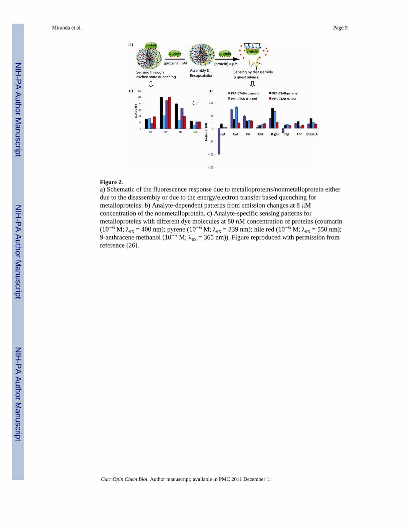
Figure 2.a) Schematic of the fluorescence response due to metalloproteins/nonmetalloprotein eitherdue to the disassembly or due to the energy/electron transfer based quenching formetalloproteins. b) Analyte-dependent patterns from emission changes at 8 μMconcentration of the nonmetalloprotein. c) Analyte-specific sensing patterns formetalloproteins with different dye molecules at 80 nM concentration of proteins (coumarin(10−6 M; λex = 400 nm); pyrene (10−6 M; λex = 339 nm); nile red (10−6 M; λex = 550 nm);9-anthracene methanol (10−5 M; λex = 365 nm)). Figure reproduced with permission fromreference [26].
Miranda et al. Page 9
Curr Opin Chem Biol. Author manuscript; available in PMC 2011 December 1.
NIH
-PA Author Manuscript
NIH
-PA Author Manuscript
NIH
-PA Author Manuscript

Figure 3. GFP-NP sensor arraya) Schematic illustration showing the competitive binding between proteins and quenchedAuNP-GFP complexes whose aggregation leads to the fluorescence “turning on” or furtherquenching using a library of cationic nanoparticles. b) Discrimination of HAS and IgG atdifferent concentrations and mixture of proteins. At the top, canonical score plot for thefluorescence patterns as obtained from LDA for HAS and IgG at different concentrations(500 nM, 1 μM, and 2 μM) with 95% confidence ellipses. At the bottom, HAS and IgG weremixed at 1:1 molar ratio with 250 nM each and 500 nM each, and added to five AuNP-GFPcomplexes. The canonical score plot obtained from LDA analysis were compared with thosefor the 500 nM individual proteins [31].
Miranda et al. Page 10
Curr Opin Chem Biol. Author manuscript; available in PMC 2011 December 1.
NIH
-PA Author Manuscript
NIH
-PA Author Manuscript
NIH
-PA Author Manuscript

Figure 4.A schematic representation of a sensor element in the sensor comprised of β-galactosidase(β-Gal) and cationic AuNPs and differentiation of proteins in 3-D. a) As shown, β-gal isdisplaced from the β-Gal/AuNP complex by protein analytes, restoring the catalytic activityof β-Gal towards the fluorogenic substrate 4-methylumbelliferyl-β-D-galactopyranoside,resulting in an amplified signal for detection. b) Differential protein pattern of the nineproteins at 1 nM. c) Canonical score plot of the first three factors of fluorescence responsepatterns obtained through β-Gal/AuNP sensor array against nine target proteins in 1 nMconcentration [32].
Miranda et al. Page 11
Curr Opin Chem Biol. Author manuscript; available in PMC 2011 December 1.
NIH
-PA Author Manuscript
NIH
-PA Author Manuscript
NIH
-PA Author Manuscript

Figure 5.Array-based sensing of bacteria. a) Schematic diagram of the displacement assay betweenbacteria and the NP-PPE complex. b) Fluorescence response (ΔI) patterns of the NP-PPEsensory array against various classes of bacteria (2×10⁵ bacteria/mL). As shown on the plot,the same strains of bacteria can also be identified. c) Canonical score plot for the first twofactors of simplified fluorescence response patterns obtained with NP-PPE assembly arraysagainst bacteria (95% confidence ellipses shown) [37].
Miranda et al. Page 12
Curr Opin Chem Biol. Author manuscript; available in PMC 2011 December 1.
NIH
-PA Author Manuscript
NIH
-PA Author Manuscript
NIH
-PA Author Manuscript

Figure 6.a) Percentage changes of T2 relaxation time (%ΔT2) obtained upon incubating MGNPs 2–6or the control NP 1 (20 μg/mL) with 10 cell lines (105 cells/mL). The ΔT2 was calculated bydividing the T2 differences between MGNP and MGNP/cancer cell by the correspondinghighest ΔT2 from each MGNP category. b) LDA plots for the first three LDs of ΔT2 patternsobtained with the MGNP array upon binding with the 10 cell lines (105 cells/mL). Fulldifferentiation of the 10 cell lines was achieved. Figure reproduced with permission fromreference [48].
Miranda et al. Page 13
Curr Opin Chem Biol. Author manuscript; available in PMC 2011 December 1.
NIH
-PA Author Manuscript
NIH
-PA Author Manuscript
NIH
-PA Author Manuscript