Understanding productivity through the lens of Finance - ADP ...
Upload
independentCategory
view
1download
0
Accurate Determination of the OxidativePhosphorylation Affinity for ADP in IsolatedMitochondriaGilles Gouspillou1*, Richard Rouland1, Guillaume Calmettes1, Veronique Deschodt-Arsac1, Jean-Michel
Franconi1, Isabelle Bourdel-Marchasson1,2, Philippe Diolez1*
1 Laboratoire de Resonance Magnetique des Systemes Biologiques, UMR 5536 CNRS - Universite Victor Segalen Bordeaux 2, Bordeaux, France, 2 CHU de Bordeaux - Pole
de gerontologie clinique, Hopital Xavier Arnozan, Pessac, France
Abstract
Background: Mitochondrial dysfunctions appear strongly implicated in a wide range of pathologies. Therefore, there is agrowing need in the determination of the normal and pathological integrated response of oxidative phosphorylation tocellular ATP demand. The present study intends to address this issue by providing a method to investigate mitochondrialoxidative phosphorylation affinity for ADP in isolated mitochondria.
Methodology/Principal Findings: The proposed method is based on the simultaneous monitoring of substrate oxidation(determined polarographically) and phosphorylation (determined using the glucose - hexokinase - glucose-6-phosphatedehydrogenase - NADP+ enzymatic system) rates, coupled to the determination of actual ADP and ATP concentrations bybioluminescent assay. This enzymatic system allows the study of oxidative phosphorylation during true steady states in awide range of ADP concentrations. We demonstrate how the application of this method allows an accurate determinationof mitochondrial affinity for ADP from both oxidation (KmVox) and phosphorylation (KmVp) rates. We also demonstrate thatdetermination of KmVox leads to an important overestimation of the mitochondrial affinity for ADP, indicating thatmitochondrial affinity for ADP should be determined using phosphorylation rate. Finally, we show how this method allowsthe direct and precise determination of the mitochondrial coupling efficiency. Data obtained from rat skeletal muscle andliver mitochondria illustrate the discriminating capabilities of this method.
Conclusions/Significance: Because the proposed method allows the accurate determination of mitochondrial oxidativephosphorylation affinity for ADP in isolated mitochondria, it also opens the route to a better understanding of functionalconsequences of mitochondrial adaptations/dysfunctions arising in various physiological/pathophysiological conditions.
Citation: Gouspillou G, Rouland R, Calmettes G, Deschodt-Arsac V, Franconi J-M, et al. (2011) Accurate Determination of the Oxidative Phosphorylation Affinityfor ADP in Isolated Mitochondria. PLoS ONE 6(6): e20709. doi:10.1371/journal.pone.0020709
Editor: Alicia J. Kowaltowski, Instituto de Quımica - Universidade de Sao Paulo, Brazil
Received December 17, 2010; Accepted May 9, 2011; Published June 9, 2011
Copyright: � 2011 Gouspillou et al. This is an open-access article distributed under the terms of the Creative Commons Attribution License, which permitsunrestricted use, distribution, and reproduction in any medium, provided the original author and source are credited.
Funding: Research has been funded by CNRS and Universite Bordeaux Segalen. The funders had no role in study design, data collection and analysis, decision topublish, or preparation of the manuscript.
Competing Interests: The authors have declared that no competing interests exist.
* E-mail: [email protected] (PD); [email protected] (GG)
Introduction
In most tissues, such as skeletal muscle, mitochondria are the
main site of energy production through the oxidative phosphory-
lation pathway. In addition to their crucial role in energy metabo-
lism, mitochondria also take part into other important physiological
functions such as calcium homeostasis, apoptosis signaling and ROS
production. All these roles are in complex and close interaction [1],
making mitochondria a key organelle for cell life and death. It is
therefore not surprising that the scientific interest surrounding
mitochondria is currently growing. Dysfunctions of mitochondrial
oxidative phosphorylation are increasingly investigated to define
and understand the very mechanisms involved in a large number
of pathologies. We [2] and others [3,4,5] have shown that mito-
chondrial dysfunctions are implicated in the aging process and in
age-related pathologies such as Alzeimer’s [6], Parkinson’s and
Hungtington’s diseases and Friedreich’s ataxia [7].
However, the identification of the functional consequences of
mitochondrial dysfunctions on cellular energetics remains a scien-
tific challenge. Indeed, most studies focus on isolated parts of
mitochondrial oxidative phosphorylation, making the link between
mitochondrial dysfunctions and their consequences on cellular
energetics difficult to decipher. It is also well established in the field
of systems biology that the emergence of unexpected properties can
arise from the cooperative interactions between individual compo-
nents of a system [8]. Consequently, the understanding of the
functional consequences of mitochondrial oxidative phosphoryla-
tion dysfunctions on cellular energetics could also benefit from
considering the integrated response of oxidative phosphorylation to
cellular ATP demand. Among useful parameters of mitochondrial
bioenergetics, the mitochondrial coupling efficiency and the
mitochondrial affinity for ADP are in this context of particular
interest. Mitochondrial affinity for ADP is closely related to the
mitochondrial capacity to respond to changes in cellular ATP
PLoS ONE | www.plosone.org 1 June 2011 | Volume 6 | Issue 6 | e20709

demand. This parameter is usually determined in the literature by
the calculation of apparent Km of respiration for ADP using either
isolated mitochondria [9,10,11,12,13] or permeabilized cells
[14,15]. Indeed, for technical reasons, apparent Km for ADP is
most of the time determined from the kinetic response of the
respiratory chain activity (oxygen consumption) to the increase in
ADP concentration. However, this affinity primarily relies on the
response of the ATP production system to this increase in ADP
concentration. As respiratory chain activity is not directly linked to
phosphorylation activity due to the existence of proton leaks
[16,17], well known to vary depending on mitochondrial activity,
the apparent Km for ADP is approximated using these methods and
should therefore be determined using phosphorylation rate. As
compared to isolated mitochondria, the use of permeabilized cells
more closely mimic the physiological conditions, since it allows the
study oxidative phosphorylation without disrupting physical
interactions of mitochondria with their surrounding environment
[18]. However, in permeabilized cells, the phosphorylation rate can
only be indirectly determined under highly specific conditions using
this approach [19], preventing the accurate determination of
mitochondrial phosphorylation affinity for ADP.
The aim of the present paper is to propose a new and easy to use
method allowing a true and accurate determination of oxidative
phosphorylation affinity for ADP using isolated mitochondria. This
method is based on the direct and simultaneous independent
monitoring of oxidation and phosphorylation rates during true
steady states under various ADP concentrations (from very low
(7 mM) to saturating ADP concentrations), the actual ADP and ATP
concentrations being determined by bioluminescent assays. We
demonstrate how this method can provide other useful data about
the energetics of mitochondria in the cell, since its application also
allows the direct determination of mitochondrial coupling efficien-
cy. To achieve this aim, mitochondria isolated from muscle and
liver, two populations known to present significant differences, were
studied in order to validate the method.
Materials and Methods
1. Ethics StatementAll experiments were conducted in agreement with the National
and European Research Council Guide for the care and use of
laboratory animals in accordance with the recommendations of
the Weatherall report, ‘‘The use of non-human primates in
research’’. All animal protocols used were approved by our local
ethics committee named Direction departementale des services
veterinaires de la Gironde, the corresponding license number
being P.D. 3308010 (11/19/2008).
2. Isolation of liver and skeletal muscle mitochondriaIsolation of liver and skeletal muscle mitochondria from 6
month-old male Wistar rats were anesthetized by isofluran inha-
lation and killed by intraperitoneal injection of pentobarbital
(60 mg.kg21). The gastrocnemius muscle and the liver were then
dissected and washed separately in the isolation medium con-
taining 100 mM saccharose, 180 mM KCl, 50 mM Tris, 5 mM
MgCl2, 10 mM EDTA and 0.1% (w/v) BSA (pH 7.2). Before
homogenization, gastrocnemius muscle was minced and exposed
during 5 minutes to protease (2 mg of bacterial proteinase type
XXIV per ml of isolation medium, Sigma-Aldrich: P8038). Mito-
chondria were extracted as described in [2,20].
3. Protein content determinationMitochondrial protein concentration was determined by the
Bradford method [21] using BSA as standard.
4. Simultaneous monitoring of oxidation andphosphorylation rates during steady states
Oxygen consumption and ATP synthesis rate were monitored
simultaneously in a glass vessel (final volume 6 ml, 25uC) in a
medium containing 240 mM Manitol, 100 mM KCl, 1 mM
EGTA, 20 mM MgCl2, 10 mM KH2PO4 and 0.1% (w/v) BSA
(pH 7.2). Glutamate (5 mM)+Malate (1 mM)+succinate (5 mM)
were used as substrates in all experiments.
Oxidation rates were determined polarographically with a Clark
electrode (Rank Brothers). O2 concentration in air-equilibrated
medium was taken as 240 mM [22]. The electrode was connected
to a PowerLab (ADInstrument) for data collection.
Phosphorylation rates were determined using a coupled
enzymatic system composed of Glucose (5 mM) - Hexokinase
(2.5 U.ml21, Sigma-Aldrich, H4502) - Glucose-6-phosphotate
dehydrogenase (2.5 U.ml21, Sigma-Aldrich, G6378) - NADP+
(1.6 mM) [23] (figure 1). Chemicals composing this enzymatic
system were in excess in order to avoid any limitation of
mitochondrial activity. By this system, the ADP phosphorylated
by mitochondria into ATP is regenerated by the phosphorylation
of glucose into glucose-6-phosphate (G6P) catalyzed by Hexoki-
nase (HK) (eq. 1). This first reaction allows the establishment of a
constant ATP turnover ensuring the study of oxidative phosphor-
ylation during steady states. Resulting G6P is then oxidized by
Glucose-6-phosphotate dehydrogenase (G6PDH) to form 6-
phosphogluconate, using NADP+ as electron acceptor (eq. 2). ATP
GlucosezATP
HK
? Glucose-6-phosphatezADP ð1Þ
Glucose-6-phosphatezNADPz
G6PDH
'
6-PhosphogluconatezNADPHzHz
ð2Þ
Consequently, NADPH production is stoichiometrically linked
to mitochondrial ATP synthesis rate, which can therefore be
monitored by spectrometrically measuring the increase in the
absorbance of NADPH at 340 nm. Conversion of NADPH absor-
bance into NADPH concentration was performed using a molar
extinction coefficient of 6.22 mM21.cm21. Changes in rate of
NADPH production were therefore monitored with an optic fiber
connected to a spectrometer placed in the oxygraphic vessel (Cary
50, Varian). Mitochondrial protein concentration in the oxy-
graphic vessel for each recording was 25 mg.ml21 in order to
decrease mitochondrial non-specific absorption at 340 nm.
To avoid any interference by residual adenylate kinase activity,
an excess of P1,P5- Di(adenosine-5)pentaphosphate (AP5A, 20 mM)
was added to the measurement medium in all experiments.
5. Determination of ADP and ATP concentrationsADP and ATP concentrations were determined both at the
onset of steady state and just before the end of each recording
to assess the stability of nucleotides concentrations. The follow-
ing protocol was optimized for concentrations ranging from 5
to 50 mM corresponding to the sensitivity range of the ATP
monitoring reagent used (FLAA-1KT, Sigma-Aldrich) diluted 625
times:
Mitochondrial Phosphorylation Affinity for ADP
PLoS ONE | www.plosone.org 2 June 2011 | Volume 6 | Issue 6 | e20709

Step 1. Baseline assay. 150 ml of the ATP monitoring
reagent were added to a luminometer cuvette. Cuvette was then
placed in the luminometer chamber (Lumac Biocounter M1500,
Lumac) to record the emitted light by the reagent alone. This
value was subtracted to the value obtained during the step 2 for
accurate determination of ATP concentration.
Step 2. Determination of ATP concentration. 10 ml of the
measured medium were taken in the oxygraphic vessel using a
10 ml Hamilton syringe and instantaneously added to the cuvette
prepared during step 1. The amount of emitted light gave ATP
concentration in the sample.
Step 3. Determination of ADP concentration. Phosphoenol
pyruvate (6.5 mM final) and pyruvate kinase (54 U.ml21 final) were
then added to the luminometer cuvette in order to convert all ADP
into ATP. Subtracting the ATP concentration determined during step
2 allowed the determination of the ADP concentration in the assay.
Step 4. Calibration of luminometer response. 2.4 mM of
a standard solution of ATP was systematically added in the cuvette
in order to converter the emitted light by the luciferin-luciferase
reaction into mM of ATP.
After each assay, phosphoenol pyruvate and pyruvate kinase
were added to the ATP monitoring reagent alone to determine the
baseline of light emission under these conditions. This separate
assay was used to correct emitted light obtained during steps 3
and 4.
For experiments where the added ADP concentration at the
onset of the recording was higher than 50 mM, the 10 ml of the
measurement medium taken in the oxygraphic vessel were
diluted in water in order to bring the nucleotides concentration
between 10 and 50 mM. 10 ml of this diluted solution were then
used to determine ADP and ATP concentrations under these
conditions.
Figure 1. Experimental determination of oxidation rate, phosphorylation rate and ADP/ATP concentrations. Our experimental set-upwas composed of an oxygraph, a spectrophotometer and a luminometer. An optic fiber, connected to the spectrophotometer, was inserted in theoxygraphic vessel (picture on the top-left hand corner). Mitochondrial oxidation rate was determined using the Clark electrode of the oxygraph.Phosphorylation rate was assessed, with the help of the optic fiber, by the continuous monitoring of NADPH production in the oxygraphic vessel.Samplings were performed at the onset and the end of the recording to assess both ADP and ATP concentrations using a bioluminescence-basedassay with the help of a luminometer (picture on the top-right hand corner). For clarity, all parameters that were measured during each experimentare highlighted by colored circles. HK: hexokinase, G6PDH: glucose 6 phosphate dehydrogenase, G6P: glucose 6 phosphate, OM: outer membrane,IM: inner membrane.doi:10.1371/journal.pone.0020709.g001
Mitochondrial Phosphorylation Affinity for ADP
PLoS ONE | www.plosone.org 3 June 2011 | Volume 6 | Issue 6 | e20709

6. Determination of the mitochondrial affinity for ADPAll experimental data were treated with a specific homemade
program developed using the Igor Pro software (Wavemetrics). As
both oxidation and phosphorylation rates were simultaneously
determined for each ADP concentration, mitochondrial apparent
Km for ADP could be determined for both oxidation (KmVox) and
phosphorylation (KmVp) rates. KmVox and KmVp for ADP were
determined by fitting respectively oxidation and phosphorylation
rates expressed as a function of the ADP concentration by the
Michaelis-Menten equation (eq. 3):
V (½ADP�)~ Vmax|½ADP�Kmz½ADP� ð3Þ
where [ADP] corresponds to a measured ADP concentration, V to
the corresponding oxidation or phosphorylation rate, Vmax to the
maximal oxidation or phosphorylation rate and apparent Km
corresponds to the Michaelis-Menten constant.
7. StatisticsExperimental values are expressed as mean 6 SD. Comparison
between liver and muscle mitochondria were performed using
unpaired bilateral student’s t-tests. p-values were fixed at 0.05 and
0.01 to consider a significant level of difference between series of
data.
Results and Discussion
Mitochondrial oxidative phosphorylation involves complex
biochemical processes in close interactions. The complete and
accurate study of mitochondrial bioenergetics is therefore a
technical challenge, especially in order to obtain physiologically
relevant information (e.g. under low ADP concentrations). To face
this challenge, we combined and adapted existing methods in an
original way to investigate mitochondrial bioenergetics and its
regulation by ADP. To demonstrate the discriminating capabilities
of the proposed method, we chose to compare mitochondria
isolated from skeletal muscle and liver (classically used as model
mitochondria). Indeed, it has been shown that mitochondrial
oxidative phosphorylation significantly differs in these two tissues
[24] and these differences have been proposed to result from the
distinct physiological roles of these organs [25].
1. Simultaneous determination of oxidation andphosphorylation rates during true steady states
The complete study of mitochondrial oxidative phosphorylation
cannot be restricted to respiratory chain activity. Indeed, due to
proton leak dependency on membrane potential (Dy), the degree
of coupling between oxidation and phosphorylation is known to
largely vary depending on the rate of ATP synthesis and Dy[16,17,26]. This coupling is raised up as the ATP synthesis rate
increases since Dy decreases. Respiratory chain activity is there-
fore not directly representative of phosphorylation activity, espe-
cially under low ADP concentrations, where the rate of ATP
synthesis is low and Dy is high. As a consequence, a better
understanding of oxidative phosphorylation could be obtained
by the concomitant determination of both oxidation and
phosphorylation rates. In addition, the investigation of mitochon-
drial regulation by ADP in isolated mitochondria requires (i) a
precise determination of oxidation and phosphorylation rates,
especially at low ADP concentrations where oxidation and phos-
phorylation rates are low, and (ii) the control and accurate
measurement of ADP concentration accessible to mitochondria
during experiments. These technical difficulties can only be
overcame by studying mitochondrial oxidative phosphorylation
during true steady states, e.g. constant ATP turnover, where
oxidation rate, phosphorylation rate and nucleotides concentra-
tions are stable.
The glucose-hexokinase enzymatic system is widely used as
ADP-regenerating system [27,28] and was therefore chosen to
establish true steady states of oxidative phosphorylation. In
addition, this enzymatic system was coupled to G6PDH-NADP+
in order to determine the phosphorylation rate, since NADPH
produced is stoichiometrically linked to mitochondrial ATP pro-
duction [23] (see materials and methods for details). By placing in
the oxygraphic vessel an optic fiber connected to a spectrometer
in order to monitor changes in NADPH concentration, oxidation
and phosphorylation rates were therefore determined simulta-
neously. Since a constant ATP turnover may be established by
this coupled enzymatic system, various phosphorylation activities
can be easily set-up with decreased experimental errors, and the
study of oxidative phosphorylation under low ADP concentrations
becomes accessible and accurate. By taking samples during each
recording, both ADP and ATP concentrations were determined
using a luciferine-luciferase based assays (see materials and
methods for details).
Figure 2 shows a combined typical recording obtained using
Glutamate+Malate+Succinate substrates in order to reconstitute
the tricarboxylic acid cycle function and consequently to approach
physiological conditions [29]. Mitochondria were added at the
onset of the recording and quickly reached state 4 oxidation rate.
In this example, the addition of 50 mM ADP triggered phos-
phorylation, leading to a corresponding increase in oxidation rate.
Figure 2 shows that both ADP-induced oxidation and phosphor-
ylation rates were constant during the duration of the recording
(approximately 6 min). In order to determine ADP and ATP
concentrations, two samplings were performed at the beginning
and at the end of recording. As it can be seen in figure 2, the
stability of these two concentrations demonstrates the achievement
of true steady states during the time course of the experiment.
2. Measurement of mitochondrial oxidation andphosphorylation affinity for ADP
Mitochondrial affinity for ADP can be investigated by deter-
mining the apparent Km for ADP, a parameter that is related to
the mitochondrial capacity to respond to changes in ATP demand
and primarily reflects the kinetic properties of both the adenine
nucleotide translocator and the F0-F1 ATP-synthase.
In the present study, both oxidation and phosphorylation rates
were therefore determined as described above during steady states
under measured ADP concentrations ranging from 7 to 900 mM.
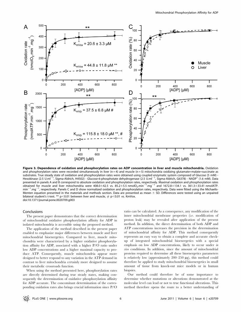
The dependence of oxidation and phosphorylation rates on ADP
concentration for liver and muscle mitochondria are shown in
figure 3. Maximal oxidation and phosphorylation rates were more
than four times higher in muscle as compared to liver mito-
chondria (figure 3 A and B). These results are in accordance with
previously shown higher state III oxidation rate in muscle mito-
chondria [24]. In addition, it is interesting to note that even for
the very low ADP concentrations used in the present study, both
oxidation and phosphorylation rates were significantly much
higher in muscle mitochondria (figure 3A and B). To our know-
ledge, these oxidation and phosphorylation rates kinetics allowed
for the first time the accurate determination of KmVox and KmVp.
As shown in figure 3A and B, mitochondria isolated from skeletal
muscle were characterized by significantly lower KmVox and KmVp
as compared to liver mitochondria, clearly indicating a much
higher affinity for ADP in muscle mitochondria.
Mitochondrial Phosphorylation Affinity for ADP
PLoS ONE | www.plosone.org 4 June 2011 | Volume 6 | Issue 6 | e20709

Interestingly, the KmVp was significantly higher as compared
to KmVox in both tissues (figure 3 A and B). During in vitro
experiments, the onset of phosphorylation caused by ADP
induces a decrease in Dy, which in turn decreases proton leak
and increases respiratory chain activity. The more the phosphor-
ylation rate is high, the more Dy decreases and consequently
the more proton leak decreases [16,17,26]. The contribution of
the oxygen consumption due to non-phosphorylating processes
(e. g. proton leak) in the global activity of the respiratory
chain therefore decreases as phosphorylation rate increases.
Consequently, the determination of KmVox leads to a clear over-
estimation of mitochondrial affinity for ADP. The accurate
determination of mitochondrial oxidative phosphorylation affinity
for ADP therefore requires the determination of the apparent Km
for ADP from phosphorylation rates (KmVp). This can easily and
accurately be done in isolated mitochondria using the proposed
method.
In addition, this method allows the comparison of the kinetics
of oxidation and phosphorylation rates in response to changes in
the ADP concentration between liver and muscle mitochondria
(figure 3 C and D). As shown in figure 3 C, liver and muscle
mitochondria presented only slight differences when compared
to those obtained when considering phosphorylation rate kinetics
(figure 3 D). It can be seen in figure 3 D that phosphorylation
rate is always more responsive in muscle mitochondria as com-
pared to liver for a given ADP concentration. In accordance with
these results, the KmVp to KmVox ratio was significantly higher
in liver mitochondria (2.760.4 vs. 1.860.4, p,0.05). Taken all
together, these results emphasize the interest of the determination
of both KmVox and KmVp, since the information provided by each
of these parameters are complementary in order to characte-
rize the integrated functioning of oxidative phosphorylation. In
addition, major functional differences in the phosphorylation rate
response to an increase in the ADP concentration were revealed
under low ADP concentrations (figure 3 D), which are con-
centrations likely to occur in vivo. Interestingly, the proposed
method ensures a high degree of precision in the determination
of both oxidation and phosphorylation rates for these ADP
concentrations.
3. Determination of mitochondrial coupling efficiencyThe coupling efficiency of oxidative phosphorylation can be
defined as the amount of ATP molecules that mitochondria can
synthesize for each atom of oxygen consumed. This coupling
efficiency is central to the physiology of energy metabolism and
can be assessed by the determination of the well-known P/O ratio
[27,30]. Since both oxidation and phosphorylation rates are
determined simultaneously using the proposed method, this crucial
parameter is directly accessible. P/O ratio was therefore calculated
for every ADP concentration (figure 4). Maximal P/O ratio,
obtained under the highest ADP concentrations (from 200 to
900 mM), was similar between liver and muscle mitochondria.
However and most interestingly, under low ADP concentrations
(from approximately 7 to 150 mM), the P/O ratio was significantly
higher in muscle mitochondria. According to this result, under low
ADP concentrations, muscle mitochondria synthesize a higher
amount of ATP for the same amount of oxygen as compared to
liver mitochondria. In this way muscle mitochondria appear
optimized toward ATP production at a high yield even at low
ADP concentrations when compared to liver mitochondria.
These results demonstrate the effectiveness of the proposed
method in the determination of changes in P/O value in response
to an increase in ADP concentration. Moreover, as compared to
other available methods, the direct determination of both oxi-
dation and phosphorylation rates ensures a more accurate
calculation of P/O value.
Figure 2. Typical recording of oxidation rate, phosphorylation rate and ADP/ATP concentrations. Oxidation and phosphorylation rateswere recorded simultaneously in liver and muscle mitochondria oxidizing glutamate+malate+succinate as substrates. Mitochondrial proteinconcentration in the oxygraphic vessel was 25 mg.ml21. Steady states of oxygen consumption and phosphorylation rates were obtained using thecoupled enzymatic system composed of Glucose (5 mM) - Hexokinase (2.5 U.ml21, Sigma-Aldrich, H4502) - Glucose-6-phosphotate dehydrogenase(2.5 U.ml21, Sigma-Aldrich, G6378) - NADP+ (1.6 mM). Dashed arrows correspond to the sampling of measurement medium taken from theoxygraphic vessel during each recording for determination of ADP and ATP concentrations.doi:10.1371/journal.pone.0020709.g002
Mitochondrial Phosphorylation Affinity for ADP
PLoS ONE | www.plosone.org 5 June 2011 | Volume 6 | Issue 6 | e20709
ConclusionsThe present paper demonstrates that the correct determination
of mitochondrial oxidative phosphorylation affinity for ADP in
isolated mitochondria is accessible using the proposed method.
The application of the method described in the present paper
enabled to emphasize major differences between muscle and liver
mitochondrial bioenergetics. Compared to liver, muscle mito-
chondria were characterized by a higher oxidative phosphoryla-
tion affinity for ADP, associated with a higher P/O ratio under
low ADP concentrations and a higher maximal capacity to pro-
duce ATP. Consequently, muscle mitochondria appear more
designed to better respond to any variation in the ATP demand in
contrast to liver mitochondria certainly more designed to assume
their metabolic crossroads function.
When using the method presented here, phosphorylation rates
are directly determined during true steady states, making con-
sequently the determination of oxidative phosphorylation affinity
for ADP accurate. The concomitant determination of the corres-
ponding oxidation rates also brings crucial information since P/O
ratio can be calculated. As a consequence, any modification of the
inner mitochondrial membrane properties (i.e. modification of
proton leak) may be revealed after application of the present
method. In addition, the direct determination of both ADP and
ATP concentrations increases the precision in the determination
of mitochondrial affinity for ADP. This method consequently
represents an easy way to obtain a complete and accurate check-
up of integrated mitochondrial bioenergetics with a special
emphasis on low ADP concentrations, likely to occur under in
vivo conditions. In addition, since the amount of mitochondrial
proteins required to determine all these bioenergetics parameters
is relatively low (approximately 200–250 mg), this method could
therefore be applied to study mitochondrial bioenergetics in small
amount of tissue from knock-out mice models or in human
biopsies.
Our method could therefore be of some importance to
determine whether mutations or alterations demonstrated at the
molecular level can lead or not to true functional alterations. This
method therefore opens the route to a better understanding of
Figure 3. Dependence of oxidation and phosphorylation rates on ADP concentration in liver and muscle mitochondria. Oxidationand phosphorylation rates were recorded simultaneously in liver (n = 4) and muscle (n = 5) mitochondria oxidizing glutamate+malate+succinate assubstrates. True steady state of oxidation and phosphorylation rates were obtained using coupled enzymatic system composed of Glucose (5 mM) -Hexokinase (2.5 U.ml21, Sigma-Aldrich, H4502) - Glucose-6-phosphotate dehydrogenase (2.5 U.ml21, Sigma-Aldrich, G6378) - NADP+ (1.6 mM). Datapresented in panels A and B correspond to absolute oxidation and phosphorylation rates, respectively. Maximal oxidation and phosphorylation ratesobtained for muscle and liver mitochondria were 408.0642.5 vs. 85.265.5 nmolO2.min21.mg21 and 1672.86134.1 vs. 361.3633.41 nmolATP.min21.mg21, respectively. Panels C and D show normalized oxidation and phosphorylation rates, respectively. Data were fitted using the Michaelis-Menten equation presented in the materials and methods section. Data are presented as mean 6 SD. Differences were tested using an unpairedbilateral student’s t-test. ** p,0.01 between liver and muscle, # p,0.01 vs. KmVox.doi:10.1371/journal.pone.0020709.g003
Mitochondrial Phosphorylation Affinity for ADP
PLoS ONE | www.plosone.org 6 June 2011 | Volume 6 | Issue 6 | e20709

functional consequences of mitochondrial dysfunctions arising in
pathologies.
Acknowledgments
The authors acknowledge Yannick Chatenet for drawing and Dr. Eric
Thiaudiere for helpful discussions.
Author Contributions
Conceived and designed the experiments: GG PD. Performed the
experiments: GG RR. Analyzed the data: GG GC VD-A IB-M PD.
Contributed reagents/materials/analysis tools: GG GC J-MF IB-M PD.
Wrote the paper: GG IB-M PD.
References
1. Brookes PS, Yoon Y, Robotham JL, Anders MW, Sheu SS (2004) Calcium,
ATP, and ROS: a mitochondrial love-hate triangle. Am J Physiol Cell Physiol
287: C817–833.
2. Gouspillou G, Bourdel-Marchasson I, Rouland R, Calmettes G, Franconi JM,
et al. (2010) Alteration of mitochondrial oxidative phosphorylation in aged
skeletal muscle involves modification of adenine nucleotide translocator.
Biochim Biophys Acta 1797: 143–151.
3. Conley KE, Jubrias SA, Esselman PC (2000) Oxidative capacity and ageing in
human muscle. J Physiol 526: 203–210.
4. Dirks AJ, Hofer T, Marzetti E, Pahor M, Leeuwenburgh C (2006)
Mitochondrial DNA mutations, energy metabolism and apoptosis in aging
muscle. Ageing Res Rev 5: 179–195.
5. Figueiredo PA, Mota MP, Appell HJ, Duarte JA (2008) The role of
mitochondria in aging of skeletal muscle. Biogerontology 9: 67–84.
6. Moreira PI, Carvalho C, Zhu X, Smith MA, Perry G (2010) Mitochondrial
dysfunction is a trigger of Alzheimer’s disease pathophysiology. Biochim Biophys
Acta 1802: 2–10.
7. Schapira AH (1999) Mitochondrial involvement in Parkinson’s disease,
Huntington’s disease, hereditary spastic paraplegia and Friedreich’s ataxia.
Biochim Biophys Acta 1410: 159–170.
8. Weiss JN, Yang L, Qu Z (2006) Systems biology approaches to metabolic and
cardiovascular disorders: network perspectives of cardiovascular metabolism.
J Lipid Res 47: 2355–2366.
9. Chance B, Williams GR (1955) Respiratory enzymes in oxidative phosphory-
lation. I. Kinetics of oxygen utilization. J Biol Chem 217: 383–393.
10. Tonkonogi M, Sahlin K (1999) Actively phosphorylating mitochondria are more
resistant to lactic acidosis than inactive mitochondria. Am J Physiol 277:
C288–293.
11. Mogensen M, Sahlin K (2005) Mitochondrial efficiency in rat skeletal muscle:
influence of respiration rate, substrate and muscle type. Acta Physiol Scand 185:
229–236.
12. Affourtit C, Crichton PG, Parker N, Brand MD (2007) Novel uncoupling
proteins. Novartis Found Symp 287: 70–80; discussion 80–91.
13. ter Veld F, Jeneson JA, Nicolay K (2005) Mitochondrial affinity for ADP is
twofold lower in creatine kinase knock-out muscles. Possible role in rescuing
cellular energy homeostasis. FEBS J 272: 956–965.
14. Zoll J, Sanchez H, N’Guessan B, Ribera F, Lampert E, et al. (2002) Physical
activity changes the regulation of mitochondrial respiration in human skeletal
muscle. J Physiol 543: 191–200.
15. Zoll J, Koulmann N, Bahi L, Ventura-Clapier R, Bigard AX (2003) Quantitative
and qualitative adaptation of skeletal muscle mitochondria to increased physical
activity. J Cell Physiol 194: 186–193.
16. Brand MD, Chien LF, Diolez P (1994) Experimental discrimination between
proton leak and redox slip during mitochondrial electron transport. Biochem J
297: 27–29.
17. Brand MD, Harper ME, Taylor HC (1993) Control of the effective P/O ratio of
oxidative phosphorylation in liver mitochondria and hepatocytes. Biochem J
291: 739–748.
18. Kuznetsov AV, Veksler V, Gellerich FN, Saks V, Margreiter R, et al. (2008)
Analysis of mitochondrial function in situ in permeabilized muscle fibers, tissues
and cells. Nat Protoc 3: 965–976.
19. Ouhabi R, Boue-Grabot M, Mazat JP (1998) Mitochondrial ATP synthesis in
permeabilized cells: assessment of the ATP/O values in situ. Anal Biochem 263:
169–175.
20. Cannon B, Lindberg O (1979) Mitochondria from brown adipose tissue:
isolation and properties. Methods Enzymol 55: 65–78.
Figure 4. Changes in the P/O ratio as a function of ADP concentration in liver and muscle mitochondria. P/O ratio was determined bycalculating the phosphorylation to oxidation rates ratio. Data for liver (n = 4) and muscle (n = 5) are presented as mean 6 SD. Differences were testedusing an unpaired bilateral student’s t-test. ** p,0.01 between liver and muscle.doi:10.1371/journal.pone.0020709.g004
Mitochondrial Phosphorylation Affinity for ADP
PLoS ONE | www.plosone.org 7 June 2011 | Volume 6 | Issue 6 | e20709

21. Bradford MM (1976) A rapid and sensitive method for the quantitation of
microgram quantities of protein utilizing the principle of protein-dye binding.Anal Biochem 72: 248–254.
22. Dufour S, Rousse N, Canioni P, Diolez P (1996) Top-down control analysis of
temperature effect on oxidative phosphorylation. Biochem J 314: 743–751.23. Passarella S, Ostuni A, Atlante A, Quagliariello E (1988) Increase in the ADP/
ATP exchange in rat liver mitochondria irradiated in vitro by helium-neon laser.Biochem Biophys Res Commun 156: 978–986.
24. Benard G, Faustin B, Passerieux E, Galinier A, Rocher C, et al. (2006)
Physiological diversity of mitochondrial oxidative phosphorylation. Am J PhysiolCell Physiol 291: C1172–1182.
25. Cairns CB, Walther J, Harken AH, Banerjee A (1998) Mitochondrial oxidativephosphorylation thermodynamic efficiencies reflect physiological organ roles.
Am J Physiol 274: R1376–1383.
26. Amo T, Brand MD (2007) Were inefficient mitochondrial haplogroups selected
during migrations of modern humans? A test using modular kinetic analysis ofcoupling in mitochondria from cybrid cell lines. Biochem J 404: 345–351.
27. Affourtit C, Brand MD (2006) Stronger control of ATP/ADP by proton leak in
pancreatic beta-cells than skeletal muscle mitochondria. Biochem J 393:151–159.
28. Kesseler A, Brand MD (1994) Effects of cadmium on the control and internalregulation of oxidative phosphorylation in potato tuber mitochondria.
Eur J Biochem 225: 907–922.
29. Gnaiger E (2009) Capacity of oxidative phosphorylation in human skeletalmuscle: new perspectives of mitochondrial physiology. Int J Biochem Cell Biol
41: 1837–1845.30. Brand MD (2005) The efficiency and plasticity of mitochondrial energy
transduction. Biochem Soc Trans 33: 897–904.
Mitochondrial Phosphorylation Affinity for ADP
PLoS ONE | www.plosone.org 8 June 2011 | Volume 6 | Issue 6 | e20709
Copyright © 2022 FDOKUMEN