A temporal switch in the insulin-signalling pathway that regulates hepatic IGF-binding protein-1...
11
A temporal switch in the insulin-signalling pathway that regulates hepatic IGF-binding protein-1 gene expression David Finlay, Antonio J Ruiz-Alcaraz, Christopher Lipina, Stephane Perrier and Calum Sutherland Division of Pathology and Neurosciences, University of Dundee, Ninewells Hospital and Medical School, Dundee, Scotland, DD1 9SY, UK (Requests for offprints should be addressed to C Sutherland; Email: [email protected]) Abstract Insulin regulation of hepatic gene transcription is a vital component of glucose homeostasis. Understanding the molecular regulation of this process aids the search for the defect(s) that promotes insulin-resistant states, such as diabetes mellitus. We have previously shown that the insulin regulation of hepatic IGF-binding protein-1 (IGFBP1) expression requires the signalling proteins phosphatidylinositol 3-kinase (PI 3-kinase) and mammalian target of rapamycin (mTOR). In this report, we demonstrate that activation of the mTOR pathway, without activation of its upstream regulator PI 3-kinase, reduces IGFBP1 expression. Therefore, mTOR activation is sufficient to mimic insulin regulation of this gene. However, longer exposure (O3 h) of cells to insulin reduces the importance of this pathway in insulin regulation of the gene, suggesting a temporal switch in signalling mechanisms linking insulin action to the IGFBP1 gene promoter. In contrast, the activation of PI 3-kinase is required for insulin regulation of IGFBP1 under all conditions tested. Therefore, an mTOR-independent, PI 3-kinase- dependent pathway becomes more important in IGFBP1 regulation after long exposure to insulin. This is a novel concept in insulin regulation of gene expression and demonstrates the importance of temporal analysis of signalling processes. Journal of Molecular Endocrinology (2006) 37, 227–237 Introduction Increased serum insulin level following a meal results in complete shutdown of hepatic glucose output (Granner & Pilkis 1990). This is achieved by turning off glycogen breakdown and endogenous gluconeo- genesis (Pilkis & Granner 1992, Nordlie & Foster 1999). The latter process requires transcriptional repression of phosphoenolpyruvate carboxykinase (PEPCK) and glucose-6-phosphatase (G6Pase), rate- limiting steps in gluconeogenesis and loss of this regulation contributes to the hyperglycaemia that characterises type 2 diabetes mellitus (Granner & Pilkis 1990, Granner & O’Brien 1992, Kahn 1994, Hanson & Reshef 1997, Sutherland et al. 2003). Much work is ongoing to understand the molecules and pathways that link the insulin receptor to these gene promoters. Indeed, an insulin-responsive DNA sequence, common to several gene promoters, includ- ing PEPCK and G6Pase, termed the PEPCK-like insulin receptor substrate (IRS) or the thymine-rich insulin-response element (TIRE), has been identified as the likely final target for this insulin-signalling cascade (for review, see O’Brien & Granner 1996, Sutherland et al. 2003). Insulin signalling includes activation of the lipid kinase phosphatidylinositol 3-kinase (PI 3-kinase) to generate the second messen- ger phosphoinositide 3,4,5, trisphosphate (PIP3) (Alessi & Downes 1998, Cantley 2002), the stimulation of the nutrient sensing mammalian target of rapamycin (mTOR) pathway (Raught et al. 2001, Fisher & White 2004), and a relatively weak induction of the Ras-p42/p44 mitogen-activated protein kinase (MAPK) pathway (Denton & Tavare 1995). We and other researchers have demonstrated a requirement for PI 3-kinase in the regulation of most if not all of the TIRE-containing genes (Sutherland et al. 1995, Dickens et al. 1998, Durham et al. 1999). Other molecules that influence the rate of transcription of these genes include the transcription factors, FOXO (Guo et al. 1999, Schmoll et al. 2000, Puigserver et al. 2003), SREBP1c (Becard et al. 2001), PGC1a (Puigserver et al. 2003), CBP (Zhou et al. 2004) and TORC2 (Koo et al. 2005), as well as the kinases protein kinase B (PKB) (Guo et al. 1999, Rena et al. 1999, Schmoll et al. 2000), glycogen synthase kinase-3 (GSK3; Lochhead et al. 2001) and mTOR (Band & Posner 1997, Patel et al. 2002). However, evidence is accumulating that the exact sequence, context and positioning of the TIRE in a gene promoter affects which insulin-signalling pathways will regulate it (Hall & Granner 1999, Patel et al. 2002, 2003, Gan et al. 2005a,b). For example, glucocorticoids and glucagon induce, while insulin represses, PEPCK, G6Pase and IGF-binding protein-1 (IGFBP1) gene transcription, with the effects of insulin dominant over those of the 227 Journal of Molecular Endocrinology (2006) 37, 227–237 DOI: 10.1677/jme.1.02084 0952–5041/06/037–227 q 2006 Society for Endocrinology Printed in Great Britain Online version via http://www.endocrinology-journals.org
Transcript of A temporal switch in the insulin-signalling pathway that regulates hepatic IGF-binding protein-1...
227
A temporal switch in the insuli
n-signalling pathway thatregulates hepatic IGF-binding protein-1 gene expressionDavid Finlay, Antonio J Ruiz-Alcaraz, Christopher Lipina, Stephane Perrier andCalum SutherlandDivision of Pathology and Neurosciences, University of Dundee, Ninewells Hospital and Medical School, Dundee, Scotland, DD1 9SY, UK
(Requests for offprints should be addressed to C Sutherland; Email: [email protected])
Abstract
Insulin regulation of hepatic gene transcription is a vital component of glucose homeostasis. Understanding the molecular
regulationof thisprocessaids thesearch for thedefect(s) thatpromotes insulin-resistant states, suchasdiabetesmellitus.We
havepreviouslyshown that the insulin regulationofhepatic IGF-bindingprotein-1 (IGFBP1)expression requires thesignalling
proteins phosphatidylinositol 3-kinase (PI 3-kinase) and mammalian target of rapamycin (mTOR). In this report, we
demonstrate that activation of themTORpathway, without activation of its upstream regulator PI 3-kinase, reduces IGFBP1
expression. Therefore, mTOR activation is sufficient to mimic insulin regulation of this gene. However, longer exposure
(O3 h)of cells to insulin reduces the importanceof thispathway in insulin regulationof thegene, suggestinga temporal switch
in signalling mechanisms linking insulin action to the IGFBP1 gene promoter. In contrast, the activation of PI 3-kinase is
required for insulin regulation of IGFBP1 under all conditions tested. Therefore, an mTOR-independent, PI 3-kinase-
dependent pathway becomesmore important in IGFBP1 regulation after long exposure to insulin. This is a novel concept in
insulin regulation of gene expression and demonstrates the importance of temporal analysis of signalling processes.
Journal of Molecular Endocrinology (2006) 37, 227–237
Introduction
Increased serum insulin level following a meal resultsin complete shutdown of hepatic glucose output(Granner & Pilkis 1990). This is achieved by turningoff glycogen breakdown and endogenous gluconeo-genesis (Pilkis & Granner 1992, Nordlie & Foster1999). The latter process requires transcriptionalrepression of phosphoenolpyruvate carboxykinase(PEPCK) and glucose-6-phosphatase (G6Pase), rate-limiting steps in gluconeogenesis and loss of thisregulation contributes to the hyperglycaemia thatcharacterises type 2 diabetes mellitus (Granner &Pilkis 1990, Granner & O’Brien 1992, Kahn 1994,Hanson & Reshef 1997, Sutherland et al. 2003). Muchwork is ongoing to understand the molecules andpathways that link the insulin receptor to these genepromoters. Indeed, an insulin-responsive DNAsequence, common to several gene promoters, includ-ing PEPCK and G6Pase, termed the PEPCK-likeinsulin receptor substrate (IRS) or the thymine-richinsulin-response element (TIRE), has been identifiedas the likely final target for this insulin-signallingcascade (for review, see O’Brien & Granner 1996,Sutherland et al. 2003). Insulin signalling includesactivation of the lipid kinase phosphatidylinositol3-kinase (PI 3-kinase) to generate the second messen-ger phosphoinositide 3,4,5, trisphosphate (PIP3)
Journal of Molecular Endocrinology (2006) 37, 227–2370952–5041/06/037–227 q 2006 Society for Endocrinology Printed in Great Britain
(Alessi & Downes 1998, Cantley 2002), the stimulationof the nutrient sensing mammalian target ofrapamycin (mTOR) pathway (Raught et al. 2001,Fisher & White 2004), and a relatively weak inductionof the Ras-p42/p44 mitogen-activated protein kinase(MAPK) pathway (Denton & Tavare 1995). We andother researchers have demonstrated a requirementfor PI 3-kinase in the regulation of most if not all ofthe TIRE-containing genes (Sutherland et al. 1995,Dickens et al. 1998, Durham et al. 1999). Othermolecules that influence the rate of transcription ofthese genes include the transcription factors, FOXO(Guo et al. 1999, Schmoll et al. 2000, Puigserver et al.2003), SREBP1c (Becard et al. 2001), PGC1a(Puigserver et al. 2003), CBP (Zhou et al. 2004) andTORC2 (Koo et al. 2005), as well as the kinases proteinkinase B (PKB) (Guo et al. 1999, Rena et al. 1999,Schmoll et al. 2000), glycogen synthase kinase-3 (GSK3;Lochhead et al. 2001) and mTOR (Band & Posner1997, Patel et al. 2002). However, evidence isaccumulating that the exact sequence, context andpositioning of the TIRE in a gene promoter affectswhich insulin-signalling pathways will regulate it (Hall& Granner 1999, Patel et al. 2002, 2003, Gan et al.2005a,b). For example, glucocorticoids and glucagoninduce, while insulin represses, PEPCK, G6Pase andIGF-binding protein-1 (IGFBP1) gene transcription,with the effects of insulin dominant over those of the
DOI: 10.1677/jme.1.02084Online version via http://www.endocrinology-journals.org

D FINLAY and others $ mTOR and IGFBP1 expression228
other hormones (O’Brien & Granner 1990, Untermanet al. 1991). All three of these gene promoters haverelated TIRE sequences, yet the regulation of IGFBP1gene expression by insulin is the only one of thesegenes depend upon mTOR activation (Band & Posner1997, Patel et al. 2002). Indeed, although PI 3-kinasesignalling appears critical in the regulation of mostmetabolic genes by insulin, only a handful of these(e.g. IGFBP1, HKII and insulin) are sensitive torapamycin (Patel et al. 2002). The activation ofmTOR stimulates phosphorylation and activation ofthe p70 S6 ribosomal protein kinase (S6K; Thomas &Hall 1997, Dufner & Thomas 1999) and eIF-4E BP1(Hara et al. 1998, Beugnet et al. 2003). These processesare well known to modulate insulin regulation ofprotein synthesis (see, for review, Shigemitsu et al.1999, Gingras et al. 2001), but there is less evidencelinking them to transcriptional control. We havepreviously demonstrated that the overexpression ofactive S6K is not sufficient to regulate the rapamycinsensitive IGFBP1 TIRE, and that regulation of IGFBP1gene expression in an S6K1 and S6K2 double knockout (KO) animal appears normal (Patel et al. 2002).This suggests that S6K activation may not link mTORto this gene or is not sufficient to repress it. Thereforein this work, we assess whether activation of thepathway upstream of S6K, without PI 3-kinaseinduction, reduces IGFBP1 expression. In the process,we identify a novel temporal switch from therapamycin-sensitive to a rapamycin-insensitive signal-ling pathway connecting the insulin receptor to theIGFBP1 gene promoter.
Materials and methods
Materials
[a32P]UTP and [g32P]ATP were obtained fromAmersham. Insulin was purchased from Novo Nordisk(Crawley, West Sussex, UK), puromycin from Invitro-gen, cycloheximide and dexamethasone from Sigma-Aldrich, rapamycin and LY294002 from Calbiochem(La Jolla, CA, USA) and the RNase Protection AssayKit II from AMS Biotech/Ambion (Austin, TX, USA).All other chemicals were of the highest gradeavailable.
Cell culture
The rat hepatoma cell line H4IIE was maintained inDulbecco’s modified Eagle’s medium containing1000 mg/l glucose, 5% (v/v) foetal calf serum, asdescribed previously (Forest et al. 1990). Cells were
Journal of Molecular Endocrinology (2006) 37, 227–237
incubated with hormones, at 37 8C, for the times and atthe concentrations indicated in the figures.
RNA isolation and mRNA measurement
H4IIE cells were serum starved overnight and treatedwith hormone/inhibitor for the times and at theconcentrations indicated in the figures. Total cellularRNA was isolated using TriReagent (Sigma) followingthe manufacturer’s instructions. An RNase protectionassay was performed to determine the relative amountsof IGFBP 1 and cyclophilin mRNA in each sample(Patel et al. 2001). Band intensity was quantified on aphosphorimager (Fuji), data calculated as a ratio ofIGFBP 1:cyclophilin mRNA and presented as percen-tage of IGFBP1 expression, where the intensity ofcontrol samples were set at 100%. Alternatively,real-time PCR was used to quantify IGFBP1 andcyclophilin mRNA levels. Briefly, cDNA was synthesisedusing Superscript II Reverse Transciptase Kit (Invi-trogen). PCR analysis was carried out in a model 7700sequence detector (Applied Biosystems, Foster City, CA,USA) with primers and probes as follows: IGFBP15 0-GCTGGATAGCTTCCACCTCATG-3 0 (sense), 5 0-TCCATTTCTTGAGGTCAGTGATCTC-3 0 (antisense) and5 0-CCCCATCCCGTGAGGACCAGC-3 0 (probe); cyclo-philin 5 0-TTACTAGGTCTGGCAGGAAGATTAAAG-3 0
(sense), 5 0-CTGCATCTCTTGTCTCCAATGTG-3 0 (anti-sense) and 5 0-AGAGGACCAAGGCGTTATCGAACTCCTTC-3 0 (probe).
Probes were synthesised with 5 0-FAM (6-carboxyfluor-escein) and 3 0-TAMRA (6-carboxytetramethylrho-damine) modifications. IGFBP1 mRNA abundance ispresented as a ratio to cyclophilin mRNA in the samesample.
Preparation of cell extract
H4IIE cells were incubated in serum-free medium withhormones and inhibitors for the times and at theconcentrations indicated in the figures. Cells were thenscraped into ice-cold lysis buffer (25 mM Tris–HCl (pH7$4), 50 mM NaF, 100 mM NaCl, 1 mM sodiumvanadate, 5 mM EGTA, 1 mM EDTA, 1% (v/v) TritonX-100, 10 mM sodium pyrophosphate, 1 mM benzami-dine, 0$1 mM PMSF, 0$27 M sucrose and 0$1% (v/v)2-mercaptoethanol). Cell debris was removed bycentrifugation at 13 000 r.p.m. for 10 min and theprotein concentration was determined by the methodof Bradford, using BSA as an internal standard.
Immunoprecipitation and assay of p70 S6 kinase
Cell extracts (0$5 mg) were incubated for 1 h on ashaking platform with protein G-Sepharose conjugated
www.endocrinology-journals.org

mTOR and IGFBP1 expression $ D FINLAY and others 229
to 2$5 mg anti-p70 S6K antibody (Upstate, Lake Placid,NY, USA). The immunocomplexes were pelleted andwashed with 1 ml lysis buffer containing 0$5 M NaCl,and twice with 1 ml assay buffer (25 mM MOPS(pH 7$0), 0$4 mM EDTA, 0$1 M NaCl, 0$01% Brij35and 0$1% (v/v) 2-mercaptoethanol). The immuno-precipitated p70 S6K was incubated at 30 8C for 30 min,in a total volume of 50 ml containing 25 mM MOPS (pH7$0), 0$4 mM EDTA, 0$1 M NaCl, 0$01% Brij35, 0$1%(v/v) 2-mercaptoethanol, 10 mM MgCl, 0$1 mM[g32P]ATP (0$5!106 c.p.m./nmol) and 1 mM Cross-tide. A unit of kinase activity is defined as the amountwhich catalyses the phosphorylation of 1 nmol substratein 1 h.
Antibodies for western blot analysis
Antibody to b-actin was purchased from Sigma-Aldrich,total GSK3a/b from Upstate, while the phospho-specific Ser256 FOXO1, Ser9/Ser21 GSK3a/b,Thr308 PKB, Ser473 PKB, Thr202/Tyr204 p42/p44MAPK and Ser235 S6 ribosomal protein antibodieswere purchased from New England Biolabs (Hitchin,Hertfordshire, UK). H4IIE cell lysates were preparedfollowing incubation with hormones as described infigures and analysed by western blot.
Statistical analyses
Student’s t-tests were performed to establish significantdifferences between two groups, and 5% confidencelimits applied.
Results and discussion
Cycloheximide activates the mTOR pathway inde-pendent of PI 3-kinase, and reduces the expression ofIGFBP1
Cycloheximide is a protein synthesis inhibitor (Siegal &Sisler 1963) that is known to activate the hepatic mTOR-signalling pathway in the absence of insulin (Kozma et al.1989, Price et al. 1989). Therefore, we decided to use thisagent to establish whether activation of the mTOR branchof the PI 3-kinase network was sufficient to repress theIGFBP1 gene. First, we characterised the effects ofcycloheximide on the major insulin-signalling pathwaysin the H4IIE cell line (Fig. 1) in order to confirm theselective activation of this pathway. In these cells, mTORsignalling to ribosomal S6 kinase (as measured byphosphorylation of S6 ribosomal protein, Ser235) isactivated by cycloheximide treatment (Fig. 1A). Thisoccurs at concentrations similar to those that block
www.endocrinology-journals.org
protein synthesis (Beugnet et al. 2003), and increases instrength up to 35$5 mM (10 mg/ml). However, exposureof cells to puromycin, a structurally distinct proteinsynthesis inhibitor (Fig. 1A) has no effect on mTORactivity, suggesting that the regulation of mTOR bycycloheximide is not related to general inhibition ofprotein synthesis. The activation of mTOR is sustained forat least 6$5 h (Fig. 1A), while the degree of induction ofthe pathway with 35$5 mM cycloheximide, whethermeasured by S6 phosphorylation (Fig. 1B) or directassay of S6K (Fig. 1C), is similar to that observed afterinsulin treatment. Importantly, cycloheximide inductionof S6 phosphorylation or S6K activity is completely lost inthe presence of 10 nM rapamycin (Fig. 1B and C), therebyconfirming the importance of mTOR to cycloheximide,as well as insulin, regulation of this pathway. Cyclohex-imide (35$5 mM) weakly induces phosphorylation of PKB(Thr308/Ser473) as well as its downstream target GSK3,after 30-min treatment (Fig. 1B). These minor effects areinsensitive to the presence of rapamycin, and are !10%of the stimulation of this branch of the PI 3-kinasepathway by insulin (Fig. 1B). Meanwhile, no increase inphosphorylation of FOXO1, a transcriptional target ofPKB, is observed at any of the time points examined(Fig. 1B) suggesting that the PI 3-kinase–PKB pathway isprobably not stimulated to a degree that initiates many (ifany) downstream effects. Finally, cycloheximide stronglyactivates the p42/44 MAPK pathway to a far greater extentthan obtained by insulin treatment, and this activation isinsensitive to rapamycin (Fig. 1B). Again, this property ofcycloheximide must be independent of protein synthesisinhibition since puromycin does not stimulate MAPK inthe H4IIE cells (Fig. 1B). To our knowledge, this is thefirst demonstration that cycloheximide activates thisgrowth-inducing pathway.
Cycloheximide (35$5 mM) treatment of H4IIE cellsfor 3 h represses IGFBP1 gene expression (Fig. 2).This inhibition is seen on both basal (63$7%, Fig. 2A)and glucocorticoid-induced (70$8%, Fig. 2B) IGFBP1expression. This indicates that activation of mTOR,without PI 3-kinase activation, mimics the effect ofinsulin on IGFBP1 expression. However, this repres-sion is less dramatic than observed with insulin (90%),consistent with the fact that insulin repression ofIGFBP1 expression is not completely blocked byrapamycin (Patel et al. 2002). It should also be notedthat we are measuring mRNA accumulation over agiven time, therefore it is influenced by both the rateof transcription of the gene and the stability of themRNA species. Cycloheximide has been reported tostabilise IGFBP1 mRNA (Ooi et al. 1993), therefore thetrue extent of transcriptional repression in response tomTOR activation by cycloheximide may be O60–70%observed in our experiments. In contrast, insulinrepresses IGFBP1 mRNA predominantly throughinhibition of transcription (Orlowski et al. 1991) and
Journal of Molecular Endocrinology (2006) 37, 227–237
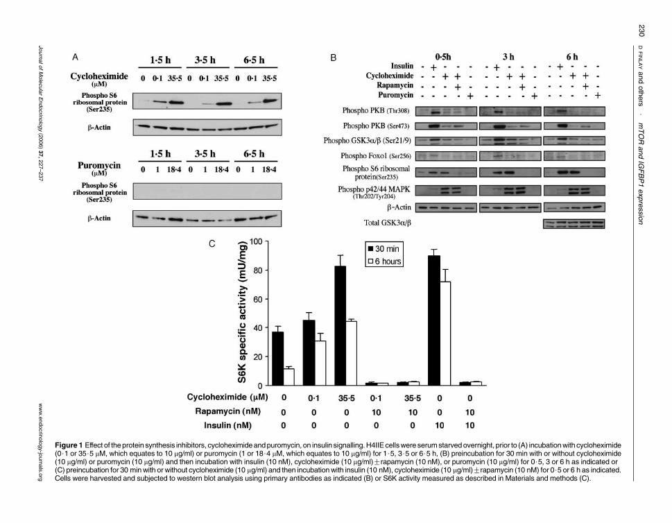
Figure 1 Effect of theprotein synthesis inhibitors, cycloheximideandpuromycin, on insulin signalling.H4IIE cellswere serumstarvedovernight, prior to (A) incubationwith cycloheximide(0$1 or 35$5 mM, which equates to 10 mg/ml) or puromycin (1 or 18$4 mM, which equates to 10 mg/ml) for 1$5, 3$5 or 6$5 h, (B) preincubation for 30 min with or without cycloheximide(10 mg/ml) or puromycin (10 mg/ml) and then incubation with insulin (10 nM), cycloheximide (10 mg/ml)Grapamycin (10 nM), or puromycin (10 mg/ml) for 0$5, 3 or 6 h as indicated or(C) preincubation for 30 minwith or without cycloheximide (10 mg/ml) and then incubationwith insulin (10 nM), cycloheximide (10 mg/ml)Grapamycin (10 nM) for 0$5 or 6 h as indicated.Cells were harvested and subjected to western blot analysis using primary antibodies as indicated (B) or S6K activity measured as described in Materials and methods (C).
DFINLAYandothers
$mTOR
andIG
FBP1expression
230
JournalofMolecularEndocrin
ology(2006)37,227–237
www.endocrin
ology-jo
urnals.org

Figure 2 Cycloheximide treatment inhibits IGFBP1 gene expression. H4IIE cells were serum starved overnight prior to a 30 minpreincubation with or without cycloheximide (10 mg/ml). Cells were then incubatedGcycloheximide (10 mg/ml)Grapamycin(10 nM) in the (A) absence or (B) presence of dexamethasone (500 nM) for the times indicated. (C) Alternatively, cells were serumstarved overnight prior to a 30 min preincubation with or without puromycin (10 mg/ml) followed by incubationGpuromycin(10 mg/ml)Grapamycin (10 nM)Gdexamethasone (500 nM) as indicated for 3 and 6 h. The cells were harvested, total RNAisolated and an RNase protection assay (RPA) performed to determine the levels of IGFBP1mRNA. Results are presented as themeanGS.E.M. of at least four experiments. *P!0$01; †P!0$001; n.s., not significant.
mTOR and IGFBP1 expression $ D FINLAY and others 231
does not affect the stability of mRNA (Unterman et al.1991). Following 6 h incubation with cycloheximidebasal IGFBP1 expression is still reduced compared tocontrol, however, the effect of this agent is lost in theglucocorticoid-stimulated 6 h incubation (Fig. 2B). Itis possible that the effects of cycloheximide on mRNAstability are more apparent in the longer incubationsin the presence of glucocorticoid, thereby masking anygene repression. Equally, the loss of cycloheximiderepression at the longer time points could be due to itseffects on protein synthesis, reducing the level of a keytranscription factor.
In the studies demonstrating stabilisation of IGFBP1mRNA by cycloheximide, it was also noted that thisagent inhibits basal transcription of the IGFBP1 gene(Ooi et al. 1993). This was assumed to be due to
www.endocrinology-journals.org
the inhibition of protein synthesis. However, we findthat an alternative protein synthesis inhibitor, pur-omycin, which does not effect mTOR signalling(Fig. 1B), has a much small repressive effect (32$1%)on basal IGFBP1 gene expression (Fig. 2C). This smalleffect of puromycin is not observed on dexametha-sone-induced IGFBP1 expression levels (Fig. 2C), butis similar to a reported slight decrease in IGFBP1expression with a third protein synthesis inhibitor,anisomycin (Ooi et al. 1993). Therefore, it appears thatthe inhibition of protein synthesis can slightly reducethe rate of basal IGFBP1 transcription, but that anadditional mechanism is invoked in the presence ofcycloheximide. We hypothesised that this secondmechanism requires the activation of the mTORpathway (Fig. 1).
Journal of Molecular Endocrinology (2006) 37, 227–237

Figure 3 Cycloheximide inhibition of IGFBP1 gene expression does not require activation of p42/44 MAPK.H4IIE cells were serum starved overnight prior to incubationGcycloheximide (10 mg/ml)GU0126 (10 mM) inthe presence of dexamethasone (500 nM)Ginsulin (10 nM) for the times indicated. Cells treated withU0126 had also a 30 min preincubation with this inhibitor. The cells were harvested, and total RNA isolatedand Taqman analysis performed to determine the levels of (A) IGFBP1 mRNA or (B) western blot analysisperformed to assess MAPK activation. Results in (A) are presented as the meanGS.E.M. of threeexperiments assayed in triplicate, while (B) is a representative experiment of two separate analysis.
D FINLAY and others $ mTOR and IGFBP1 expression232
The major repressive effect of cycloheximide requires
mTOR activity
The repression of basal or induced IGFBP1 geneexpression by cycloheximide is inhibited by rapamycin,confirming the importance of mTOR activity in thiseffect of cycloheximide (Fig. 2). Also rapamycincompletely blocks the action of cycloheximide, incontrast to its partial effect on insulin repression.Importantly, in a parallel analysis, cycloheximide wasfound to have no repressive effect on G6Pase expression(data not shown). This gene is repressed by insulin,independent of mTOR activation but dependent on PI3-kinase activation (Dickens et al. 1998, Patel et al. 2002).As expected, the small effect of puromycin on IGFBP1expression is not affected by rapamycin (Fig. 2C). Sincecycloheximide also activates p42/p44 MAPK in thesecells (Fig. 1), and this pathway has recently been linkedto the regulation of IGFBP1 expression (Gan et al.2005b), it was important to rule out this pathway in the
Journal of Molecular Endocrinology (2006) 37, 227–237
actions of cycloheximide on the IGFBP1 gene. U0126, arelatively specific inhibitor of p42/44 MAPK activation,has no effect on the ability of cycloheximide to regulateIGFBP1 expression (Fig. 3A), yet completely inhibitscycloheximide induction of the p42/44 MAPK (Fig. 3B).Similarly, a structurally unrelated p42/44 MAPKinhibitor, PD98059, has no effect on cycloheximiderepression of IGFBP1 gene expression (data not shown).Therefore, the cycloheximide regulation of IGFBP1expression requires the mTOR pathway and not p42/44MAPK activation. We have previously found thatsustained activation of S6K1 is not sufficient to repressthe IGFBP1 TIRE (Patel et al. 2002). This suggests that analternative downstream component of mTOR signallinglinks it to the IGFBP1 gene promoter. Yeast contains anmTOR homologue (Tor), but not any S6K activity(Raught et al. 2001). Potential yeast proteins down-stream of Tor, include Msn2p, Msn4p, Gln3p, Tap42p,Mks1p, Ure2p and Gat1p (Beck & Hall 1999; Chan et al.2000, Schmelzle & Hall 2000, Shamji et al. 2000).
www.endocrinology-journals.org

mTOR and IGFBP1 expression $ D FINLAY and others 233
Whether mammalian equivalents of these proteins, orother as yet unidentified mTOR targets, mediate mTORregulation of IGFBP1 remains to be established. Mean-while, it is known that inhibitors of GSK3 repress IGFBP1gene expression (Finlay et al. 2004). There is a slightincrease in GSK3a/b phosphorylation at inhibitoryregulatory sites (Ser21/Ser9 respectively; Sutherlandet al. 1993, Sutherland & Cohen 1994) in response tocycloheximide (Fig. 1B). However, the phosphorylationof GSK3 was not sensitive to rapamycin (Fig. 1B), and therelatively weak phosphorylation (in comparison toinsulin, which inhibits GSK3 approximately 50% inthese cells; Lochhead et al. 2001) dictates that inhibitionof GSK3 is !25%. Taken together with the lack of effectof rapamycin, it seems highly unlikely that GSK3inhibition mediates the effects of cycloheximide onIGFBP1 expression.
Figure 4 Effect of rapamycin on insulin signalling. H4IIE cellswere starved overnight prior to incubationGinsulin (10 nM)Grapamycin (0$5–10 nM) for (A) 0$5 and 3 h or (B) 3, 6 and15 h (10 nM rapamycin) as indicated. Cells were harvestedand subjected to western blot analysis probing with antibodiesas indicated. Similar results were obtained for two suchexperiments.
Time-dependent effects of rapamycin on insulinregulation of IGFBP1 expression
Insulin stimulation of H4IIE cells results in activation ofmTOR and downstream signalling, as judged byincreased phosphorylation at Ser235 of S6 ribosomalprotein (Fig. 4) and S6K activity (Fig. 1C). Tennanomolar of rapamycin is sufficient to completelyblock insulin-induced mTOR signalling between 0$5and 6 h, and actually reduces mTOR activity below basallevels (Figs 1C and 4A). Full induction of mTORsignalling by insulin is maintained (and remainsrapamycin sensitive) for at least 15 h following stimu-lation (Fig. 4B). In contrast, insulin-stimulated phos-phorylation of PKB (Thr308) or GSK3 (Ser21/9) isinsensitive to the presence of 10 nM rapamycin(Fig. 4B). Insulin inhibition of both basal- anddexamethasone-induced IGFBP1 expression is partiallyreduced when cells are incubated with rapamycin for3 h (Fig. 5). Surprisingly, the effect of rapamycin on theregulation of the gene decreases substantially in cellsincubated for longer periods with insulin (Fig. 5). Thisloss of rapamycin sensitivity with time is morepronounced when the gene is first induced usingglucocorticoid (Fig. 5B vs 5A). That is, insulinrepression of IGFBP1 is blocked by w50% if measuredafter 3 h incubation with rapamycin, but w30% after6 h incubation and only w15% after 15 h incubation(Fig. 5B, lower panel). This is despite the inhibition ofmTOR signalling by rapamycin being maintainedacross the full 15 h treatment (Fig. 4). The decreasein the rapamycin block of insulin repression of basalexpression is less pronounced but the trend is similar(Fig. 5A, lower panel).
Insulin repression of IGFBP1 is dependent on PI3-kinase activity (Fig. 6). Since rapamycin sensitivity of theIGFBP1 gene promoter decreases with time (Fig. 5), we
www.endocrinology-journals.org
next examined the requirement for PI 3-kinase activityover the same incubation period. Insulin repression ofbasal (Fig. 6A) and induced (Fig. 6B) IGFBP1 iscompletely blocked by LY294002 (100 mM), a selectiveinhibitor of PI 3-kinase (Vlahos et al. 1994), at all timepoints examined. LY294002 increases IGFBP1 expressionin H4IIE cells, suggesting some basal PI 3-kinase activity inthis immortal cell line. This agent blocks insulin action ofthe mTOR pathway as well as activation of PKB (Alessi &Downes 1998). Therefore, the requirement for PI3-kinase signalling for insulin repression of IGFBP1expression is maintained. This result may explainprevious reports that rapamycin has little effect on insulinrepression of IGFBP1 expression, where mRNA levelswere examined in relatively long incubations (Cichy et al.1998). We have previously found that insulin repressionof the isolated IGFBP1 TIRE is completely blocked in thepresence of rapamycin (Patel et al. 2002). In thoseexperiments, the effect of rapamycin on the isolatedTIRE was measured following transient transfection ofa TIRE-luciferase reporter construct, and the rapamycinsensitivity of this construct is apparent even after 18 hstimulation. This difference between isolated elementand intact promoter suggests that additional insulin-response elements are present in the complete IGFBP1gene promoter. The altered signalling may reflect a
Journal of Molecular Endocrinology (2006) 37, 227–237

Figure 5 Time-dependent mTOR regulation of IGFBP1 gene expression. H4IIE were starved overnight prior toincubationGinsulin (10 nM), Grapamycin (10 nM) in the absence (A) or presence (B) of dexamethasone (500 nM) for 3, 6and 15 h. Cells were harvested, total RNA isolated and an RPA performed to determine IGFBP1 mRNA concentration.Results are presented as the meanGS.E.M. of at least four separate experiments (upper panels). The data are alsopresented as line graphs illustrating the percentage of insulin inhibition of IGFBP1 expressionGrapamycin treatment(lower panels). *P!0$05; †P!0$001; n.s., not significant.
D FINLAY and others $ mTOR and IGFBP1 expression234
temporal switch in the insulin-response elementmediating the repression of IGFBP1. Unterman andcolleagues have recently reported distinct transcrip-tional complexes involved in insulin repression of thispromoter (Gan et al. 2005a), although the identity ofthe novel DNA sequence is not yet elucidated. A time-dependent difference in rapamycin sensitivity of suchcomplexes awaits further characterisation of theseregulatory elements.
Summary
We show for the first time that activation of the mTORpathway is sufficient to reduce IGFBP1 expression,although the effect is not as complete as observed withinsulin. Therefore, this pathway acts in an additivemanner with another PI 3-kinase-dependent pathway tocompletely repress this gene promoter. In addition, we
Journal of Molecular Endocrinology (2006) 37, 227–237
demonstrate that the signalling pathway to the IGFBP1gene promoter alters with the duration of stimulation.That is, the requirement for the mTOR component of theregulation declines with insulin exposure length (Fig. 7).We suggest that this may be a novel paradigm in insulinsignalling and other rapamycin-sensitive actions ofinsulin require further study. Insulin resistance is thoughtto occur through reduced signalling capacity in one ormore signalling pathways (Shulman 2000). Indeed, boththe mTOR and PI 3-kinase pathways are implicated inreduced insulin action in muscle, fat and liver cells(Shulman 2000). The importance of the switch betweenmTOR-dependent and -independent pathways in insulinaction is not clear. In the simplest scenario, loss of mTORsignalling but not PI 3-kinase signalling would delayinsulin repression of IGFBP1 expression, while reducedPI 3-kinase signalling would have a more prolonged effecton this gene. Therefore, IGFBP1 gene expression may bea biomarker that would allow delineation of the signallingdefect in an insulin-resistant subject.
www.endocrinology-journals.org

Figure 6 Insulin inhibition of IGFBP1gene expression is PI 3-kinase dependent. H4IIEwere starved overnight prior to incubationGinsulin (10 nM),GLY294002 (100 mM) in the (A) absence or (B) presence of dexamethasone (500 nM) for 3, 6 and 15 h. Cells wereharvested, total RNA isolated and an RPA performed to determine IGFBP1 mRNA concentration. Results are presented as themeanGS.E.M. of at least four separate experiments (upper panels). The data are also presented as line graphs illustrating thepercentage of insulin inhibition of IGFBP1 expressionGLY294002 treatment (lower panels). †P!0$001; n.s., not significant.
Figure 7 Model of temporal switching of signalling pathwaysmediating insulin inhibition of IGFBP1 gene expression. Therelative contribution of the rapamycin-sensitive and insensitivepathways alters between 3 and 6 h incubation with insulin. Thismay reflect the slow onset of an mTOR-independent signallingpathway or a change in the transcriptional complex mediating therepression in response to insulin.
mTOR and IGFBP1 expression $ D FINLAY and others 235
www.endocrinology-journals.org
Acknowledgements
This work was supported by the Diabetes UK SeniorFellowship (02/0002473). The authors declare thatthere is no conflict of interest that would prejudice theimpartiality of this scientific work.
References
Alessi DR & Downes CP 1998 The role of PI 3-kinase in insulin action.Biochimica et Biophysica Acta 1436 151–164.
Band CJ & Posner BI 1997 PI 3-kinase and p70S6 kinase are requiredfor insulin but not bisperoxovanadium 1,10-phenanthrolineinhibition of IGFBP gene expression. Journal of Biological Chemistry272 138–145.
Becard D, Hainault I, Azzout-Marniche D, Bertry-Coussot L, Ferre P &Foufelle F 2001 Adenovirus-mediated overexpression of sterolregulatory element binding protein-1c mimics insulin effects onhepatic gene expression and glucose homeostasis in diabetic mice.Diabetes 50 2425–2430.
Journal of Molecular Endocrinology (2006) 37, 227–237

D FINLAY and others $ mTOR and IGFBP1 expression236
Beck T & Hall MN 1999 The TOR signalling pathway controls nuclearlocalisation of nutrient-regulated transcription factors. Nature 402689–692.
Beugnet A, Tee AR, Taylor PM & Proud CG 2003 Regulation of targetsof mTOR (mammalian target of rapamycin) signalling byintracellular amino acid availability. Biochemical Journal 372 555–566.
Cantley LC 2002 The PI 3-kinase pathway. Science 296 1655–1657.Chan T-F, Carvalho J, Riles L & Zheng XFS 2000 A chemical genomics
approach toward understanding the global functions of the targetof rapamycin protein (TOR). PNAS 97 13227–13232.
Cichy SB, Uddin S, Danilkovitch A, Guo S, Klippel A & Unterman TG1998 PKB mediates effects of insulin on hepatic IGFBP1 geneexpression through a conserved insulin response sequence.Journal of Biological Chemistry 273 6482–6487.
Denton RM & Tavare JM 1995 Does mitogen-activated-protein kinasehave a role in insulin action? The cases for and against EuropeanJournal of Biochemistry 227 597–611.
Dickens M, Svitek CA, Culbert AA, O’Brien RM & Tavare JM 1998Central role for PI 3-kinase in the repression of glucose-6-phosphatase gene transcription by insulin. Journal of BiologicalChemistry 273 20144–20149.
Dufner A & Thomas G 1999 Ribosomal S6 kinase signaling and controlof translation. Experimental Cell Research 253 100–109.
Durham SK, Suwanichkul A, Scheimann AO, Yee D, Jackson JG, Barr FG& Powell DR 1999 FKHR binds the insulin response element in theinsulin-like growth factor binding protein-1 promoter. Endocrinology140 3140–3146.
Finlay D, Patel S, Dickson LM, Shpiro N, Marquez R, Rhodes CJ &Sutherland C 2004 Glycogen synthase kinase-3 regulates IGFBP-1gene transcription through the thymine-rich insulin responseelement: inhibition is required for full regulation of this promoterelement by insulin. BMC Molecular Biolology 5 15.
Fisher TL & White MF 2004 Signaling pathways: the benefits of goodcommunication. Current Biology 14 R1005–R1007.
Forest CD, O’Brien RM, Lucas PC, Magnuson MA & Granner DK 1990Regulation of PEPCK gene expression by insulin. Use of the stabletransfection approach to locate an insulin responsive sequence.Molecular Endocrinology 4 1302–1310.
Gan L, Han Y, Bastianetto S, Dumont Y, Unterman TG & Quirion R2005a FoxO-dependent and -independent mechanisms mediateSirT1 effects on IGFBP-1 gene expression. Biochemical andBiophysical Research Communications 337 1092–1096.
Gan L, Pan H & Unterman TG 2005b Insulin response sequence-dependent and -independent mechanisms mediate effects ofinsulin on glucocorticoid-stimulated insulin-like growth factorbinding protein-1 promoter activity. Endocrinology 146 4274–4280.
Gingras A-C, Raught B & Sonenberg N 2001 Regulation of translationinitiation by FRAP/mTOR. Genes and Development 15 807–826.
Granner DK & Pilkis S 1990 The genes of hepatic glucose metabolism.Journal of Biological Chemistry 265 10173–10178.
Granner DK & O’Brien RM 1992 Molecular physiology and genetics ofNIDDM. Diabetes Care 15 369–395.
Guo S, Rena G, Cichy S, He X, Cohen P & Unterman TG 1999Phosphorylation of Ser 256 by PKB disrupts transactivation byFKHR and mediates effects of insulin on IGFBP-1 promoter activitythrough a conserved insulin response sequence. Journal of BiologicalChemistry 274 17184–17192.
Hall RK & Granner DG 1999 Insulin regulates expression of metabolicgenes through divergent signaling pathways. Journal of Basic andClinical Physiology and Pharmacology 10 119–133.
Hanson RW & Reshef L 1997 Regulation of PEPCK gene expression.Annual Review of Biochemistry 66 581–611.
Hara K, Yonezawa K, Weng QP, Kozlowski MT, Belham C & Avruch J1998 Amino acid sufficiency and mTOR regulate p70 S6 kinase andeIF-4E BP1 through a common effector mechanism. Journal ofBiological Chemistry 273 14484–14494.
Kahn CR 1994 Insulin action, diabetogenes and the cause of type IIdiabetes. Diabetes 43 1066–1084.
Journal of Molecular Endocrinology (2006) 37, 227–237
Koo SH, Flechner L, Qi L, Zhang X, Screaton RA, Jeffries S, Hedrick S,Xu W, Boussouar F, Brindle P et al. 2005 The CREB coactivatorTORC2 is a key regulator of fasting glucose metabolism. Nature 4371109–1111.
Kozma S, Lane HA, Ferrari S, Luther H, Siegmann M & Thomas G1989 A stimulated S6 kinase from rat liver: identity with the mitogenactivated S6 kinase of 3T3 cells. EMBO Journal 8 412–4132.
Lochhead PA, Coghlan MP, Rice SQJ & Sutherland C 2001 Inhibitionof GSK3 selectively reduces G6Pase and PEPCK gene expression.Diabetes 50 937–947.
Nordlie RC & Foster JD 1999 Regulation of glucose production by theliver. Annual Review of Nutrition 19 379–406.
O’Brien RM & Granner DK 1990 PEPCK gene as a model of inhibitoryeffects of insulin on gene transcription. Diabetes Care 13 327–339.
O’Brien RM & Granner DK 1996 Regulation of gene expression byinsulin. Physiological Reviews 76 1109–1161.
Ooi GK, Brown DR, Suh D-S, Tseng LY-H & Rechler MM 1993Cycloheximide stabilises IGFBP-1 mRNA and inhibits IGFBP-1transcription in H4IIE cells. Journal of Biological Chemistry 26816664–16672.
Orlowski CC, Ooi GT, Brown DR, Yang YW, Tseng LY & Rechler MM1991 Insulin rapidly inhibits insulin-like growth factor-bindingprotein-1 gene expression in H4-II-E rat hepatoma cells. MolecularEndocrinology 5 1180–1187.
Patel S, Lochhead PA, Rena G & Sutherland C 2001 Antagonisticeffects of phorbol esters on insulin regulation of IGFBP-1 but notglucose-6-phosphatase gene expression. Biochemical Journal 359611–619.
Patel S, Lochhead PA, Rena G, Fumagalli S, Pende M, Kozma S,Thomas GM & Sutherland C 2002 Insulin regulation of IGF-bindingprotein-1 gene expression is dependent on mammalian target ofrapamycin (mTOR), but independent of S6K activity. Journal ofBiological Chemistry 277 9889–9895.
Patel S, Lipina C & Sutherland C 2003 Different mechanisms are usedby insulin to repress three genes that contain a homologousthymine-rich insulin response element. FEBS Letters 549 72–76.
Pilkis SJ & Granner DK 1992 Molecular physiology of the regulation ofhepatic gluconeogenesis and glycolysis. Annual Review of Physiology54 885–909.
Price DJ, Nemenoff RA & Avruch J 1989 Purification of a hepatic S6kinase from cycloheximide-treated rats. Journal of Biological Chemistry264 13825–13833.
Puigserver P, Rhee J, Donovan J, Walkey CJ, Yoon JC, Oriente F,Kitamura Y, Altomonte J, Dong H, Accili D et al. 2003 Insulin-regulated hepatic gluconeogenesis through FOXO1-PGC-1alphainteraction. Nature 423 550–555.
Raught B, Gingras A-C & Sonenberg N 2001 The target of rapamycin(TOR) proteins. PNAS 98 7037–7044.
Rena G, Guo S, Cichy SC, Unterman TG & Cohen P 1999Phosphorylation of the transcription factor forkhead family memberFKHR by PKB. Journal of Biological Chemistry 274 17179–17183.
Schmelzle T & Hall MN 2000 TOR, a central controller of cell growth.Cell 103 253–262.
Schmoll D, Walker KS, Alessi DR, Grempler R, Burchell A, Guo S,Walther R & Unterman TG 2000 Regulation of glucose-6-phosphatase gene expression by protein kinase B(alpha) and theforkhead transcription factor FKHR. Journal of Biological Chemistry275 36324–36333.
Shamji AF, Kuruvilla FG & Schreiber SL 2000 Partitioning thetranscriptional program induced by rapamycin among the effectorsof the Tor proteins. Current Biology 10 1574–1581.
Shigemitsu K, Tsujishita Y, Hara K, Nanahoshi M, Avruch J & Yonezawa K1999 Regulation of translational effectors by amino acid andmammalian target of rapamycin signaling pathways. Journal ofBiological Chemistry 274 1058–1065.
Shulman GI 2000 Cellular mechanisms of insulin resistance. Journal ofClinical Investigation 106 171–176.
www.endocrinology-journals.org

mTOR and IGFBP1 expression $ D FINLAY and others 237
Siegal MR & Sisler HD 1963 Inhibition of protein synthesis in vitro bycycloheximide. Nature 200 675–676.
Sutherland C & Cohen P 1994 The a-isoform of glycogen synthasekinase-3 from rabbit skeletal muscle is inactivated by p70 S6 kinase orMAP kinase-activated protein kinase-1 in vitro. FEBSLetters 338 37–42.
Sutherland C, Leighton IA & Cohen P 1993 Inactivation of glycogensynthase kinase-3b by phosphorylation; new kinase connections ininsulin and growth factor signalling. Biochemical Journal 296 15–19.
Sutherland C, O’Brien RM & Granner DK 1995 Phosphatidylinositol3-kinase, but not p70/p85 ribosomal S6 protein kinase, is requiredfor the regulation of Phosphoenolpyruvate Carboxykinase geneexpression by insulin. Journal of Biological Chemistry 270 15501–15506.
Sutherland C, O’Brien RM & Granner DK 2003 Genetic regulation ofglucose metabolism. In Handbook of Physiology, pp 707–734. Eds LSJefferson & AD Cherrington. New York: Oxford University Press.
Thomas G & Hall MN 1997 TOR signalling and control of cell growth.Current Opinion in Cell Biology 9 782–787.
www.endocrinology-journals.org
Unterman TG, Oehler DT, Murphy LJ & Lacson RG 1991 Multi-hormonal regulation of insulin-like growth factor-binding protein-1in rat H4IIE hepatoma cells: the dominant role of insulin.Endocrinology 128 2693–2701.
Vlahos CJ, Matter WF, Hui KY & Brown RF 1994 A specific inhibitor ofphosphatidylinositol 3-kinase, 2-(4-morpholinyl)-8-phenyl-4H-1-benzopyran-4-one (LY294002). Journal of Biological Chemistry 2695241–5248.
Zhou XY, Shibusawa N, Naik K, Porras D, Temple K, Ou H, Kaihara K,Roe MW, Brady MJ & Wondisford FE 2004 Insulin regulation ofhepatic gluconeogenesis through phosphorylation of CREB-bind-ing protein. Nature Medicine 10 633–637.
Received in final form 30 May 2006Accepted 9 June 2006
Journal of Molecular Endocrinology (2006) 37, 227–237