igf-1r inhibition - KI Open Archive Home
83
From the Department of Oncology and Pathology, Division of Cellular and Molecular Tumor Pathology, Cancer Center Karolinska Karolinska Institutet, Stockholm, Sweden IGF-1R INHIBITION: A TOOL FOR FUNCTIONAL STUDIES OF INSULIN- LIKE GROWTH FACTORS FAMILY IN MALIGNANT CELLS. Daiana Vasilcanu, MD Stockholm 2006
-
Upload
khangminh22 -
Category
Documents
-
view
0 -
download
0
Transcript of igf-1r inhibition - KI Open Archive Home

From the Department of Oncology and Pathology,
Division of Cellular and Molecular Tumor Pathology, Cancer Center Karolinska
Karolinska Institutet, Stockholm, Sweden
IGF-1R INHIBITION: A TOOL FOR FUNCTIONAL STUDIES OF INSULIN-LIKE GROWTH FACTORS FAMILY IN
MALIGNANT CELLS.
Daiana Vasilcanu, MD
Stockholm 2006

All previously published papers were reproduced with permission from the publisher. Published and printed by Karolinska University Press Box 200, SE-171 77 Stockholm, Sweden © Daiana Vasilcanu, 2006 ISBN 91-7140-643-3

To my family


ABSTRACT Cancer cells generally posses the capability of overusing normal extracellular signaling for proliferation and/or antiapoptosis to create growth advantage over the normal cells. Major players in extracellular signaling are the growth factor receptors. Among them, an activated IGF-1R is important for the establishment of a malignant cell phenotype. Interestingly, the targeting of IGF-1R can reverse the malignant phenotype in cancer cells and render them sensitive to apoptosis, without seriously affecting the biology of normal cells. For these reasons, IGF-1R seems to be a very promising target in cancer therapy. Recently, we demonstrated that the cyclolignan PPP efficiently inhibited phosphorylation of IGF-1R without interfering with insulin receptor activity. This thesis is centered on: (1) functional studies of IGF-1R using PPP as a tool, with focus on importance for survival and proliferation of malignant cells as well as possible mechanisms of PPP action; (2) possible caveats in clinical applications of PPP (e.g. resistance, side effects secondary to IGF-1R inhibition, effects on glucose uptake). Using a IGF-1R tyrosine kinase consruct, isolated by immunoprecipitation and amplified in insect cells, we found that PPP decreased phosphorylation of tyrosine residue (Y) 1136 in the activation loop of the IGF-1R kinase domain. Studies using dominant-negative constructs of IGF-1R (in which specific tyrosine residues are replaced by phenylalanine) suggest that the inhibition of Y1136 phosphorylation may be important for the inhibition of Akt phosphorylation seen in PPP treated cell cultures. Whether PPP directly or indirectly (e.g. by interfering with IGF-1R associated proteins) inhibits Y1136 phosphorylation is still unknown. It was confirmed that inhibitions of downstream reactions of the phosphatyl inositol-3 kinase/anti-apoptotic pathway (e.g. attenuated Bad phosphorylation, PARP cleavage, caspase activation) were a consequence of the PPP-induced inhibition of IGF-1R. (Paper 1) We demonstrated the presence and growth dependence of IGF-1R in primary cultured craniopharyngioma cells from a subset of affected patients (5 out of 9). Upon treatment with PPP, cells with high IGF-1R expression responded promptly with decreased Akt phosphorylation followed by cell growth inhibition, whereas these responses did not appear in cells with low receptor expression. Our data points to the possibility of using IGF-1R inhibitors (e.g. PPP) as a treatment modality to obtain complete tumor-free conditions before growth hormone substitution. (Paper 2) A general concern with antitumor agents is development of resistance. In light of this problem we aimed to investigate whether malignant cells may develop serious resistance to PPP. After trying to select several malignant cell lines, only two out of 10 survived an 80-week selection. We could observe a temporary and limited increase in IGF-1R expression but there were no rearrangements or amplification of the IGF-1R gene.The resistant cell lines did not exhibit cross-resistance to known cytostatic dugs. In conclusion, no or slight resistance to PPP occurred. (Paper 3) Finally, we confirmed that PPP does not inhibit activity of the highly related insulin receptor and induce diabetogenic effects (like high blood glucose). Instead, in vivo and in vitro studies showed that PPP treatment reduces the blood glucose levels in mice and induces increase of glucose uptake in cells expressing the insulin-dependent glucose-transporter GLUT-4. (Paper 4) Key words: IGF-1R, chemoresistance, IR, glucose uptake, tyrosine kinase inhibitors. ISBN 91-7140-643-3
LIST OF PUBLICATIONS 1 Vasilcanu D*, Girnita A*, Girnita L, Vasilcanu R, Axelson M, Larsson O.
The cyclolignan PPP induces activation loop-specific inhibition of tyrosine phosphorylation of the insulin-like growth factor-1 receptor. Link to the phosphatidyl inositol-3 kinase/Akt apoptotic pathway. Oncogene. 2004 Oct 14; 23(47):7854-62.
2 Ulfarsson E, Karstrom A, Yin S, Girnita A, Vasilcanu D, Thoren M, Kratz G, Hillman J, Axelson M, Larsson O, Girnita L. Expression and growth dependency of the insulin-like growth factor I receptor in craniopharyngioma cells: a novel therapeutic approach. Clin Cancer Res. 2005 Jul 1;11(13):4674-80.
3 Vasilcanu D, Weng W-H, Girnita A, Lui W-O, Vasilcanu R, Axelson M, Larsson O, Larsson C, Girnita L. The insulin-like growth factor-1 receptor inhibitor PPP produces only very limited resistance in tumor cells exposed to long-term selection. Oncogene (In press). 2006
4 Vasilcanu D, Girnita A, Vasilcanu R, Axelson M, Girnita L, Larsson O. The IGF-1 receptor inhibitor PPP induces increased cellular uptake of glucose. Interactions between IGF-1R and insulin receptor? Manuscript, 2006.
* Equal contributions

CONTENTS Introduction..........................................................................................................1
Growth factors in cancer ..............................................................................1 Cancer definition. ...............................................................................1 Growth factors - general considerations. ...........................................1 The IGF family ...................................................................................2 Comparison of IGF-1R and Insulin Receptor structure ....................7 IGF-1R kinase activity .....................................................................10 IR kinase activity ..............................................................................11 Functional domains for signal specificity ........................................12 IGF-1R and IR signaling pathways. Similarities and differences...15
Functions of IGF-1R. Role in malignancy ................................................18 Mitogenic function of IGF-1R .........................................................18 Antiapoptotic function of IGF-1R....................................................19 Role in cell transformation ...............................................................20 Regulation of cell size ......................................................................21
Functions of IR ...........................................................................................23 Insulin induced glucose uptake ........................................................24
IR-IGF-1R cross-talk..................................................................................26 Targeting IGF-1R in cancer .......................................................................28
Blocking of ligand-receptor interaction ...........................................28 Targeting IGF-1R synthesis .............................................................29 Interfering with IGF-1R function.....................................................31 Modulators of IGF-1R internalization and recycling ......................34 Inhibitors of N-linked glycosylation ................................................34
Chemoresistance.........................................................................................35 Craniopharyngioma ....................................................................................37
Aims: ..................................................................................................................39 Materials and methods.......................................................................................40
Reagents......................................................................................................40 Antibodies (Paper 1, 2, 3, 4).......................................................................40 Cell cultures (Paper 1, 3, 4)........................................................................40 Primary cultures of human craniopharyngioma cells (Paper 2)................41 Immunocytochemical stainings..................................................................41 Cell cultures and generation of PPP resistant cells (Paper 3). ..................41 Dominant-negative transfectants (Paper 1). ..............................................42 Immunoprecipitation (Paper 1, 2, 3, 4). .....................................................42 SDS-PAGE and Western blotting (Paper 1, 2, 3, 4)..................................42 Determination of protein content. ..............................................................43 Analysis of IGF-1R kinase peptide (Paper 1)............................................43 RT�PCR for detection of IGF-1R (Paper 1, 3)..........................................43 Assay of cell growth (Paper 2)...................................................................43 Cell viability assay (Paper 1, 2, 3) .............................................................44 Apoptosis assay (Paper 3). .........................................................................44 Assay of caspase-3 activity (Paper 1). .......................................................44 Preparation of DNA and metaphase chromosomes (Paper 3)...................44

Fluorescence in situ hybridization (FISH) (Paper 3). ............................... 45 Assay of 2-deoxy-D-[3H]glucose uptake in cultured cells (Paper 4)....... 45 Fluorescence analysis of glucose uptake (Paper 4)................................... 45 In vivo experiments (Paper 4). ................................................................... 46
RESULTS AND DISCUSSION....................................................................... 47 Paper 1 ........................................................................................................ 47 Paper 2 ........................................................................................................ 49 Paper 3 ........................................................................................................ 49 Paper 4 ........................................................................................................ 50
Major Findings................................................................................................... 53 Acknowledgements ........................................................................................... 54 References.......................................................................................................... 57
LIST OF ABBREVIATIONS 5-FU 5-Fluorouracil 14.3.3 Adaptor scaffolding protein aa Aminoacids ACTH Adrenocorticotropic hormone ABC ATP-binding cassette Akt Protein kinase B A-loop Activation loop of the receptor ALS Acid labile subunit APS Adaptor protein AS Antisense ATP Adenosine triphosphate Bad Bcl associated death promoter Bak Bcl-2 Homologous Antagonist-Killer Protein Bax Bcl-2-Associated X Protein Bcl B-cell leukemia protein BCR-ABL Fusion gene on Philadelphia chromosome BMI Body mass index cAMP ciclicAMP CAP Cbl-associated protein Cbl Cellular product of cbl oncogene c-Crk Adaptor protein in Ras pathway cDNA Complementary DNA (DNA copy of mRNA) DNA Deoxyribonucleic acid EGF Epithelial growth factor ERK1 / 2 Mitogen activated protein kinase 1 / 2 FGF Fibroblast growth factor GH Growth hormone GLUT4 Glucose transporter protein 4 Grb2 Growth factor receptor-bound protein 2 GSK Glycogen synthase kinase hINSR Human insulin receptor gene IGF-1 Insulin-like growth factor 1 IGF-1R Insulin-like growth factor 1 receptor IGF-2 Insulin-like growth factor 2 IGF-2R Insulin-like growth factor 2 receptor IGFBP Insulin-like growth factor binding proteins IR Insulin receptor IR-A/B Insulin receptor isoform A/B IRK Insulin receptor tyrosine kinase IRR Insulin receptor related receptor IRS-1-4 Insulin receptor substrate 1-4 JAK Janus protein tyrosine kinase kb Kilo base kDa Kilo Dalton

Lys Lysine MAPK Mitogen activated protein kinase MDR Multidrug resistance MDR1 Protein product of mdr gene MEK MAP kinase kinase mRNA Messenger ribonucleic acid MRP1 Multidrug resistance-associated protein 1 N Nitrogen NGF Nerve growth factor PARP Poli(ADP-ribose) polymerase PBS Phosphate buffered saline PCR Polymerase chain reaction PDGF Platelet derived growth factor PDK 3-phosphoinositide-dependent kinase PI3K Phosphatidylinositol-3�-kinase PKB Protein kinase B PKC Protein kinase C PTB Phospho-tyrosine binding domain PTEN Phosphatidylinositol phosphatase Raf Protein-serine/threonine kinase (encoded by the raf oncogene) Ras Human homologue of Rat sarcoma RNA Ribonucleic acid rRNA Ribosomal RNA RTK Receptor tyrosine kinase RT-PCR Reverse transcription polymerase chain reaction Ser Serine SH Src homology Shc Src homology and collagen siRNA Small interference RNA Sos Son of the sevenless Src Protein encoded by src proto-oncogene STAT Signal transducer and activator of transcription proteins Thr Threonine TK Tyrosine kinase TSH Thyroid-stimulating hormone TYMS Thymidylate syntase Tyr Tyrosine UBF Upstream binding factor wt Wild type Y Tyrosine

1
INTRODUCTION GROWTH FACTORS IN CANCER
Cancer definition.
Malignant tumor arises from a single cell as a result of a stepwise progression of genetic and epigenetic events (loss of function of tumor suppressor genes, activation of oncogenes, tanslocations resulting in fusion genes, hypermethylation, etc) resulting in a Darwinian process of selection of fittest phenotypes. Selection of tumor cells versus normal cells is attributed to increased proliferation, reduced apoptosis or impaired terminal differentiation. Furthermore, within a neoplasm, mutant clones compete with each other. Those having adequate advantage and aggressiveness will tend to spread and eventually produce a yet more adapted phenotype. The processes of cellular proliferation and progressive acquisition of a specialized phenotype show a high degree of coordination. In particular, these complex signaling networks mediating cell growth, differentiation, migration, and apoptosis are regulated in part by polypeptide growth factors that can act (by autocrine and/or paracrine mechanisms of action) as positive or negative modulators. Because these growth factors are unable to cross the hydrophobic cell membrane, they exert their effects via binding to cell surface receptors, most of which possess intrinsic tyrosine kinase activity. Following interaction of polypeptide growth factors with their specific transmembrane receptors, a cascade of intracellular signals resulting in the activation or repression of various subsets of genes occurs.
Growth factors - general considerations.
Growth factor receptor signals, including those generated from insulin-like growth factor type 1 receptor (IGF-1R), are required for carcinogenesis and tumor progression in many human malignancies.
Many well-known growth factors (e.g., EGF, FGFs, PDGFs and IGFs) bind to receptors with protein tyrosine kinase activity. There are today more than 50 receptor tyrosine kinases (RTKs), structured in at least thirteen different receptor families (Ullrich and Schlessinger 1990). RTKs span the plasma membrane and contain an extracellular portion, which binds the ligand, and an intracellular portion possessing catalytic activity and regulatory sequences. The receptors are often activated by ligand-induced dimerization or oligomerization (Heldin 1995; Heldin and Ostman 1996), depending on covalent organization of the receptors. Most RTKs possess a single polypeptide chain and are monomeric in the absence of ligand. RTKs of the insulin receptor subfamily, comprising the IGF-1R and insulin receptor (IR), are disulfide-linked dimers of two polypeptide chains, forming a α2β2 heterotetramer. Whereas

2
ligand binding to monomeric receptors leads to their dimerization, ligand binding to dimeric receptors induces rearrangement within the their quaternary structure resulting in autophosphorylation of specific tyrosine residues within the kinase domain. (Ullrich and Schlessinger 1990). The catalytic (kinase) domain, a critical component of the receptor, displays the highest level of conservation. Structural motifs being conserved in this region include an ATP-binding site and tyrosine residue(s), which corresponds to the major phosphate acceptor site (Yarden and Ullrich 1988). In the unphosphorylated state, the catalytic activity of dimeric receptors is very low due to the particular inhibitory conformation of a specific activation loop (A-loop) in the kinase region, which interferes with the ATP-binding and phosphotransfer events. The activation of the intrinsic protein kinase activity after ligand binding results in autophosphorylation of specific tyrosine residues whithin the A-loop. Phosphorylation of these tyrosine residues removes the conformational inhibition of the kinase domain. The catalytic activity is enhanced and persists for some time independently of the presence of the ligand.
Concerning monomeric receptors activation, the kinase activity is at a low basal level in the monomeric state, but this activity is sufficient to induce trans-autophosphorylation, once the dimer has been formed. Ligand binding to the extracellular part of the receptor induces juxtaposition of the cytoplasmic parts of the receptors that allows the kinase domains to phosphorylate each other in trans. Autophosphorylation involves two different classes of tyrosine residues and it is the result of the interaction between the intracellular domains of the dimeric receptor which will induce conformational changes leading to an increased kinase activity. One phosphorylation occurs on a conserved tyrosine residue in the A-loop in the kinase domain. The secondary autophosphorylation sites are normally located outside the kinase domains and serve the fundamental function of creating docking sites for downstream signal transduction molecules containing Src homology 2 (SH2) domains or phosphotyrosine binding (PTB) tyrosine domains (Pawson 1995; van der Geer and Pawson 1995).
The tyrosine kinase activity of the growth factor receptors is required for mitogenesis, transformation, and cell differentiation (Ullrich and Schlessinger 1990). Signaling pathways initiated by tyrosine phosphorylation lead to various nuclear events, which eventually elicit dramatically different biological responses. The predominant biological activity of certain receptor tyrosine kinases is to stimulate cell growth and proliferation, while other receptor tyrosine kinases induce growth arrest and promote differentiation. The IGF family
The insulin-like growth factor (IGF) family includes three ligands (IGF-1, IGF-2 and insulin), three cell surface receptors (IGF-1R, IGF-2R and IR), at least six different IGF binding proteins (IGFBP-1 - 6), and multiple IGFBP proteases, which all contribute in regulation and propagation of IGF activity in tissues (figure 1). Additionally, new members of IGF family have been described, like the insulin receptor related receptor (IRR) (Dandekar, Wallach et al. 1998) (Zhang and Roth 1991) and the IGF-1R/IR hybrid receptor (Treadway, Morrison et al. 1989; Frattali, Treadway et al. 1992) but their activation mechanisms and functions are still largely unknown.

3
The IGF signaling system plays critical roles in tissue growth and development. The IGF system is also implicated in various pathophysiological conditions with a particularly prominent role in cancer.
Ligands
IGF-1 and IGF-2 are single-chain polypeptides with 62% sequence homology with proinsulin. Unlike insulin and other peptide hormones being produced and stored in specific endocrine cells and tissues, IGF-1 and IGF-2 are produced by almost any cell in the body. IGF-1
The human IGF-1 gene has been mapped to the long arm of chromosome 12 (Sara and Hall 1990). The gene spans more than 90 kb of chromosomal DNA and consists of at least six exons. IGF-1 is a 70�amino acid peptide with a molecular mass of about 7.5 kDa, organized into four peptide domains: A, B, C, and D. Domains A and
4
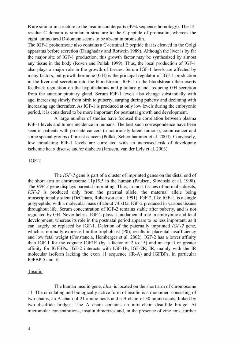
B are similar in structure to the insulin counterparts (49% sequence homology). The 12-residue C domain is similar in structure to the C-peptide of proinsulin, whereas the eight�amino acid D-domain seems to be absent in proinsulin. The IGF-1 prohormone also contains a C-terminal E peptide that is cleaved in the Golgi apparatus before secretion (Daughaday and Rotwein 1989). Although the liver is by far the major site of IGF-1 production, this growth factor may be synthesized by almost any tissue in the body (Rosen and Pollak 1999). Thus, the local production of IGF-1 also plays a major role in the growth of tissues. Serum IGF-1 levels are affected by many factors, but growth hormone (GH) is the principal regulator of IGF-1 production in the liver and secretion into the bloodstream. IGF-1 in the bloodstream then exerts feedback regulation on the hypothalamus and pituitary gland, reducing GH secretion from the anterior pituitary gland. Serum IGF-1 levels also change substantially with age, increasing slowly from birth to puberty, surging during puberty and declining with increasing age thereafter. As IGF-1 is produced at only low levels during the embryonic period, it is considered to be more important for postnatal growth and development.
A large number of studies have focused the correlation between plasma IGF-1 levels and tumor incidence in humans. The best such correspondence have been seen in patients with prostate cancers (a notoriously latent tumour), colon cancer and some special groups of breast cancers (Pollak, Schernhammer et al. 2004). Conversely, low circulating IGF-1 levels are correlated with an increased risk of developing ischemic heart disease and/or diabetes (Janssen, van der Lely et al. 2003). IGF-2
The IGF-2 gene is part of a cluster of imprinted genes on the distal end of
the short arm of chromosome 11p15.5 in the human (Paulsen, Sliwinski et al. 1998). The IGF-2 gene displays parental imprinting. Thus, in most tissues of normal subjects, IGF-2 is produced only from the paternal allele, the maternal allele being transcriptionally silent (DeChiara, Robertson et al. 1991). IGF-2, like IGF-1, is a single polypeptide, with a molecular mass of about 74 kDa. IGF-2 produced in various tissues throughout life. Serum concentration of IGF-2 remains stable after puberty, and is not regulated by GH. Nevertheless, IGF-2 plays a fundamental role in embryonic and fetal development, whereas its role in the postnatal period appears to be less important, as it can largely be replaced by IGF-1. Deletion of the paternally imprinted IGF-2 gene, which is normally expressed in the trophoblast (P0), results in placental insufficiency and low fetal weight (Constancia, Hemberger et al. 2002). IGF-2 has a lower affinity than IGF-1 for the cognate IGF1R (by a factor of 2 to 15) and an equal or greater affinity for IGFBPs. IGF-2 interacts with IGF-1R, IGF-2R, IR, mainly with the IR molecular isoform lacking the exon 11 sequence (IR-A) and IGFBPs, in particular IGFBP-5 and -6. Insulin
The human insulin gene, hIns, is located on the short arm of chromosome
11. The circulating and biologically active form of insulin is a monomer consisting of two chains, an A chain of 21 amino acids and a B chain of 30 amino acids, linked by two disulfide bridges. The A chain contains an intra-chain disulfide bridge. At micromolar concentrations, insulin dimerizes and, in the presence of zinc ions, further
5
associates into hexamers. Insulin is produced by β cells in the islets of Langerhans in the pancreas as proinsulin. Proinsulin is a single polypeptide chain of 86 amino acids that permits correct alignment of three pairs of disulfide bonds. (Figure 1) Insulin secretion by pancreatic β cells can be divided into basal (postabsorptive) and stimulated (postprandial) states. The former prevails during the interprandial phases and plays a major role during the overnight fast; the latter regulates glucose metabolism when carbohydrate is abundant and must be disposed of. Following food ingestion (carbohydrate in particular) pancreatic β cells react through a biphasic physiological response. On stimulation, the β-cell responds with a prompt but short-lived (0�10 min) release of insulin (first phase) followed by a steady and longer-lasting increase in plasma insulin concentration (second phase) concentration. This biphasic physiological insulin secretion is hardly reproducible by exogenous infusion of any insulin-like substances. In type 2 diabetes, loss of the first-phase insulin release, despite the common enhancement of second-phase insulin secretion is an early and quite common defect that may have a pathogenetic role in the development of postprandial hyperglycemia, possibly requiring specific therapeutic intervention (Del Prato, Marchetti et al. 2002) The binding of the hormone to its receptor initiates a series of events within the cells that results in the increased uptake of glucose into the cells, where it is converted into metabolic energy or stored as glycogen and fat. Insulin binding to IR also leads to increased cellular amino acid uptake, increased glycogen synthase activity, increased overall protein synthesis and decreased lipolysis and protein degradation. Most studies suggest that the metabolic functions of insulin are mainly mediated through insulin receptor isoform B (IR-B). This can be explained by hormonal and metabolic regulation of the alternative splicing of insulin receptor. For example, insulin and high glucose concentrations induce the expression of IR-B in some insulin responsive cell lines and tissues (Sell, Reese et al. 1994). However, more recently attention has been directed to the mitogenic effects of insulin and insulin analogs. Convincing evidence indicates that the activation of the isoform A insulin receptor (exon11-) by IGF-2 and insulin results in mitogenic effects and a potentiation of the cancer phenotype.(Giorgino, Belfiore et al. 1991; Frasca, Pandini et al. 1999) IGF binding proteins (IGFBPs) and IGFBP proteases
In the bloodstream and other biological fluids and tissues, IGFs are
usually found associated with one of the six known high-affinity binding proteins (IGFBP-1 to 6) or one of the nine recently described proteins with a low affinity for IGFs (IGFBP-related proteins, IGFBPrPs). IGFBPs play a central role in transporting IGFs in the bloodstream and cerebrospinal fluid and across the capillary barrier to the target cells (Baxter 1994). IGFBPs are the major determinants of IGF bioavailability, as they facilitate the constitution of a slow-release IGF pool in tissues and in the bloodstream. This increases the half-life of IGF in the serum, prevents the overstimulation of cell growth or excessive apoptosis (Rajah, Khare et al. 1999), and regulates both the transport of IGFs between intra and extravascular spaces and their interaction with receptors (Zapf 1995). IGFBP-3 is the predominant IGFBP in serum. Most circulating IGF-1 and IGF-2 are not found in a free form or simply bound to IGFBP- 3, but instead form a ternary complex with IGFBP-3 and a third component, the acid-labile subunit (ALS), in
6
a 1:1:1 molar ratio. More than 99% of circulating IGFs are bound to IGFBPs and at least 75% of the bound IGF is carried as a trimeric complex involving IGFBP-3 and the ALS, which is itself a liver derived GH-regulated glycoprotein (Baxter 1988). ALS is thought to increase the half-life of the IGF/IGFBP- 3 binary complex still further (Boisclair, Hurst et al. 2000).
IGFBP-5 also forms ternary complexes with IGFs and ALS. IGFBP-1 through -4 generally have similar affinities for IGF-1 and IGF-2. By contrast, IGFBP-5 and -6 bind IGF-2 with an affinity respectively, 10 and 100 times greater, than that with which they bind IGF-1. In tissues, IGFBPs interact with extracellular matrix constituents (IGFBP-2 and IGFBP-5) (Arai, Busby et al. 1996), or directly with cell membranes (IGFBP-1 and IGFBP-3) (Delbe, Blat et al. 1991), thereby regulating the interaction between IGFs and IGF1R. There is some evidence that IGFBPs, in addition to regulating IGF bioavailability, also act in an IGF-1 independent manner. IGFPB-3 and IGFBP-5, in particular, have been shown to exhibit effects on proliferation, migration and sensitivity to apoptosis that are independent of their effects on IGF-1R signaling (Baxter 2000).
The other key component of the IGF regulatory system is a group of IGFBP proteases, which cleave intact IGFBPs into small fragments, thereby drastically altering their IGF-binding capacities. Some of these proteases act preferentially within specific tissues, whereas others function in the bloodstream and extracellular space. However, all these enzymes are controlled in an autocrine, paracrine, hormonal fashion. This family of molecules is heterogeneous, including kallikrein- like serine proteases, cathepsins, and matrix metalloproteinases. Receptors
Excepting IGF-2R thought to function as a clearance receptor for IGF-2 (LeRoith, Werner et al. 1995), IGF-1R and IR posses tyrosine kinase activity therefore mediating the biological effect of the three ligands. Thus, IGF-1 functions primarily by activating the IGF-1R, insulin by activating IR (both IR-A and IR-B) whereas IGF-2 can act through either the IGF-1R or through the IR-A isoform. (Figure1)

7
Comparison of IGF-1R and Insulin Receptor structure
Gene structure
The studies on IGF-1R gene structure have established very close homology in
organization and overall size of the IGF-1R and insulin receptor IR genes. However, an exon equivalent to the alternatively spliced exon 11 of the IR is not present in the IGF-1R (Abbott, Bueno et al. 1992). Furthermore, these studies demonstrate that insulin and IGF-1 receptors are the products of distinct genes, located on separate chromosomes, that are controlled by different types of regulatory signals.
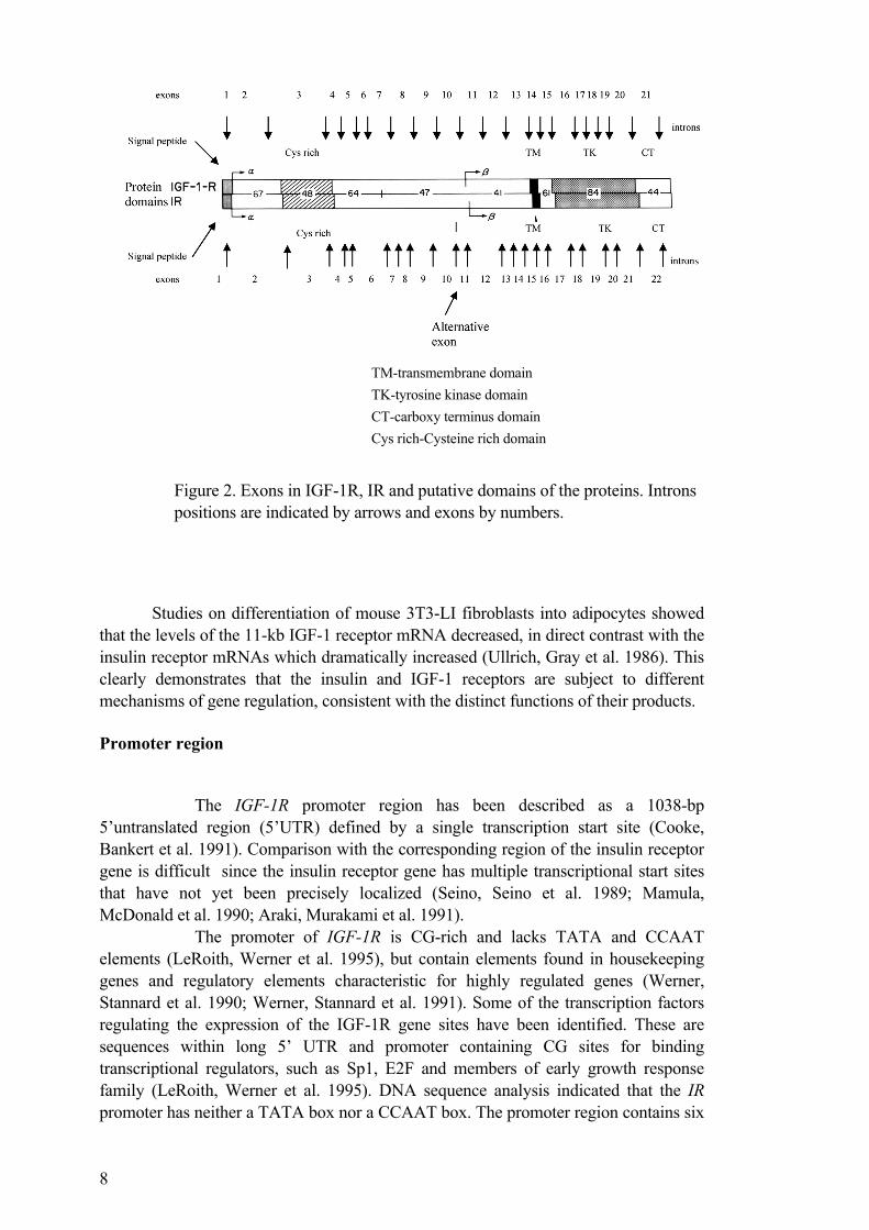
The IGF-1R gene maps to a distinct chromosomal locus 15q25- 26 (Abbott, Bueno et al. 1992) whereas the IR was localized on chromosome 19 band p13.3 - p13.2. Similarly to the IR, IGF-1R consists of 21 exons, 10 for the alpha chain and 11 for the beta chain, spanning over 100kb of the genomic DNA (Abbott, Bueno et al. 1992). The complementary DNA (cDNA) for human IGF-1R consists of 4989 nucleotides and codes for a 1367 amino-acid precursor. The exon / intron organization of the IGR-1R gene, predicted on the basis of cDNA, is quite similar to that of the IR gene, the main difference being that the IR gene contains an alternatively spliced exon 11 not present in the IGF-1R receptor gene (Ebina, Ellis et al. 1985; Ullrich, Bell et al. 1985; Ullrich, Gray et al. 1986). The IGF-1R is organized into functional domains that reflect the exonic arrangement of the gene: exons 1-3 encode the long 5´UTR (~1 kb), the signal peptide, and the N-terminal non-cysteine-rich and the cysteine rich domains of the α-subunit (ligand-binding domain). The rest of the α-subunit is encoded by exons 4-10. Exon 11 encodes the peptide cleavage site that generates the mature α- and β-subunits from the proreceptor. The exon 12-21 encode the β-subunit, with exon 14 encoding the transmembrane and exon 16-20 encoding the tyrosine kinase domain (Ullrich, Gray et al. 1986). (Figure 2)
Nucleotide sequence analysis of IR cDNA revealed a 5181 base-pair-long sequence which coded for 1382 amino acids precursor, including a 27-residue signal peptide. The human insulin receptor gene IR it is comprised of 22 exons. As for human IGF-1R gene, the introns appear to divide the IR gene into segments that encode structural and/or functional elements of the IR protein. Eight mutations in the IR gene that result in expression of structurally abnormal proteins have been described. These mutations are associated with insulin resistance and provide insight into the role of the IR gene in the development of diabetes mellitus (Seino, Seino et al. 1990). The 11 exons encoding the α-subunit of the receptor are dispersed over >90 kbp, whereas the 11 exons encoding the β-subunit are located together in a region of 30 kbp. The analysis of correspondence between exons and structural and functional units or modules of the insulin receptor showed that several of the exons encode well-defined structural units: exon 1, signal peptide; exon 2, putative ligand-binding region; exon 3, cysteine-rich region; exon 11, alternatively spliced miniexon; exon 15, transmembrane domain. The tyrosine kinase domain is encoded by five exons (exons 17-21) together with a small portion of exon 22; the region between the transmembrane and tyrosine kinase domains is encoded by a single exon (exon 16), and the COOH-terminal hydrophilic tail of the insulin receptor by exon 22 (Ebina, Ellis et al. 1985; Ullrich, Bell et al. 1985; Seino, Seino et al. 1989). (Figure 2)
8
TM-transmembrane domain TK-tyrosine kinase domain CT-carboxy terminus domain Cys rich-Cysteine rich domain
Figure 2. Exons in IGF-1R, IR and putative domains of the proteins. Introns positions are indicated by arrows and exons by numbers.
Studies on differentiation of mouse 3T3-LI fibroblasts into adipocytes showed that the levels of the 11-kb IGF-1 receptor mRNA decreased, in direct contrast with the insulin receptor mRNAs which dramatically increased (Ullrich, Gray et al. 1986). This clearly demonstrates that the insulin and IGF-1 receptors are subject to different mechanisms of gene regulation, consistent with the distinct functions of their products. Promoter region
The IGF-1R promoter region has been described as a 1038-bp 5�untranslated region (5�UTR) defined by a single transcription start site (Cooke, Bankert et al. 1991). Comparison with the corresponding region of the insulin receptor gene is difficult since the insulin receptor gene has multiple transcriptional start sites that have not yet been precisely localized (Seino, Seino et al. 1989; Mamula, McDonald et al. 1990; Araki, Murakami et al. 1991).
The promoter of IGF-1R is CG-rich and lacks TATA and CCAAT elements (LeRoith, Werner et al. 1995), but contain elements found in housekeeping genes and regulatory elements characteristic for highly regulated genes (Werner, Stannard et al. 1990; Werner, Stannard et al. 1991). Some of the transcription factors regulating the expression of the IGF-1R gene sites have been identified. These are sequences within long 5� UTR and promoter containing CG sites for binding transcriptional regulators, such as Sp1, E2F and members of early growth response family (LeRoith, Werner et al. 1995). DNA sequence analysis indicated that the IR promoter has neither a TATA box nor a CCAAT box. The promoter region contains six

9
GGGCGG sequences that may be binding sites for the transcription factor Sp1. In addition, there were three TCCC sequences that were putative promoter regulatory regions. Precursors
The receptors are synthesized as single chain proreceptors that are
processed, glycosylated, folded and dimerised to yield the mature α2β2 receptor. Transcription from the IGF-1R gene results in a transcript product of 11kb, often together with a minor band of 7kb (Chernausek, Jacobs et al. 1981; Lowe, Adamo et al. 1989). Both subunits, encoded in the same message, are translated in a precursor protein of 1367 amino acids in length, with the structure: NH2-signal peptide, α subunit and β-subunit-COOH. It is customary to count the amino acid residues of the IGF-1R from the first amino acid of the mature peptide (after removal of the signal peptide), up to 1337 (Ullrich, Gray et al. 1986). Following removal of the signal peptide, the pro-receptor is cleaved after residue 706, to form the α- and β-subunit, linked by disulphide bonds. Transcription from a single complementary DNA clone results in translation of the 1370/1382 amino-acid sequence of the human insulin receptor precursor. This difference in size between the two IR precursors is due to the absence/ presence of a 36-bp segment in the cDNA sequence resulting from alternative splicing of the region encoded by exon 11. The precursor starts with a 27-amino-acid signal sequence, followed by the receptor alpha-subunit, a precursor processing enzyme cleavage site, then the beta-subunit containing a single 23-amino-acid transmembrane sequence. After removal of the precursor signal peptide, the insulin receptor precursor is post-translationally cleaved into two chains (alpha and beta) that are covalently linked. Receptors structure
Both receptors are heterotetrameric glycoprotein composed of two extracellular α subunits and two transmembrane β-subunits connected by disulfide bonds (LeRoith, Werner et al. 1995).
Study of primary structure revealed extensive similarity between the two receptors (70%) (Ullrich, Gray et al. 1986). The α subunit of the IGF-1R containing 706 amino acids and 735 amino-acids for IR, is entirely extracellular and forms a dimer with another α-subunit. A small sequence of 12 amino acids within the α subunit determines the difference between the two isoforms of IR. Thus, IR-A, missing exon 11, is characterized by the absence of 12 amino acid residues at the carboxyl terminus of the IR α-subunit. The α-subunit contains a cysteine-rich domain (aa 148-302), also conserved in the IR (Andersen, Kjeldsen et al. 1990; Gustafson and Rutter 1990; Kjeldsen, Andersen et al. 1991; Schumacher, Mosthaf et al. 1991; Zhang and Roth 1991). The ligand binding pockets of IGF-1 and insulin receptors are formed by the extracellular α subunits and possibly some extracellular portions of the β subunits. Differences in receptor ligand specificities are likely to be dictated by sequence differences within this region, and indeed lower homology was found in the amino-acids sequences of the extracellular cysteine-rich domains (48%), C-terminal of the α subunits (47%) and N-terminal portion of the β subunits (41 %). (Figure 2)
10
These regions are the most hydrophilic sequences of the extracellular domain, and are likely to be exposed on the surface of this domain and function in defining ligand specificity (Ullrich, Gray et al. 1986). In addition to cysteine-rich domains, the locations of most N-linked glycosylation sites are conserved between IGF-1R and insulin receptor α and β subunits.
The β-subunit spans the plasma membrane and contains 627 amino acid residues for IGF-1R and 620 amino-acids for IR. The transmembranous domain is located at position 906-929. The extracellular domain of the β subunit, 196 aa in length, contains all of the 5 potential glycosylation sites. The intracellular part of β-subunit has a similar organization for both receptors consisting of a juxtamembranous, a tyrosine kinase (TK) and a C-terminal domain. The TK domains exhibit the highest homology between the two receptors (84%). The juxtamembranous domains share 61% of homology, whereas the C-terminal domains share only 44% (Ullrich, Gray et al. 1986). (Figure 2) Within the TK domain the ATP binding site represent the highly conserved region of the receptors with 100% homology.
Despite its overall homology with the insulin receptor kinase domain (84%), the IGF-1R tyrosine kinase domain includes three discrete regions of sequence divergence following residues 986, 1072 and 1208. The presence of a highly heterogeneous sequence within otherwise highly conserved tyrosine kinase domains of gene family members appears highly significant and indicates a possible function of this subdomain in definition of specific receptor function. Another low homology level (44%) is represented by the carboxyl terminus. This carboxy-terminal receptor domain may, in conjunction with the nonapeptide sequence at position 1073-1081 and the divergent membrane-proximal region between residues 933 and 955, be responsible for receptor-specific, ligand-induced, intracellular signal generation (Ullrich, Gray et al. 1986).
The IGF-1R, like the IR, undergoes extensive post-translational modification, which include serine and tyrosine phosphorylation, and glycosylation (Ullrich, Gray et al. 1986). The predicted size, based on the protein sequences is 80,423 kDa for the α- and 70,866 kDa for the β-subunit, but due to heavily glycosylation their molecular weights are 135 kDa and 90 kDa, respectively. The α-subunit contains 11 potential glycosylation sites, whereas β-subunit contains only 5. It has been shown that N-linked glycosylation precedes proteolysis of the immature αβ precursor (Jacobs, Kull et al. 1983). IGF-1R kinase activity
As for other RTK, the IGF1R tyrosine kinase domain is essential for the
receptor activity. The catalytic region of IGF-1R contains the ATP binding motif (Gly-
XXX-Gly-XXX-XXX-Gly) at position 976-981, and a catalytic Lys in position 1003, which is critical for the MgATP binding (Hanks, Quinn et al. 1988). Within the TK domain a cluster of three tyrosine residues, located at position 1131, 1135 and 1136, is critical for receptor autophosphorylation (LeRoith, Werner et al. 1995).
Ligand binding to the extracellular α-subunits of the receptor induces phosphorylation of the three tyrosine residues in the A-loop resulting in an increased catalytic activity. The crystal structure of the inactive and phosphorylated kinase

11
domain of the IGF-1R has provided a molecular model of the IGF- 1R catalytic activity (Favelyukis, Till et al. 2001). In the unstimulated state, the activation loop (A-loop), containing the critical tyrosine (Y) residues 1131, 1135 and 1136, behaves as a pseudosubstrate that blocks the active site. Y1135 (being the first tyrosine to be phosphorylated) in the A-loop is bound in cis position in the active site, thus preventing the substrate access and occluding the ATP binding site as well. After ligand binding, the three tyrosines of the A-loop are transphosphorylated by the dimeric subunit partner. Phosphorylation of Y1135 and Y1131 destabilizes the autoinhibitory conformation of the A-loop, whereas phosphorylation of Y1136 stabilizes the catalytically optimized conformation of it (Favelyukis, Till et al. 2001). These changes of the A-loop conformation allow the substrate and ATP access to the kinase active site.
Studies based on peptide mapping showed that the first site of autophosphorylation is Y 1135, followed by 1131 and then by Y 1136 ((Favelyukis, Till et al. 2001). Moreover, the kinetic experiments on autophosphorylation of the purified 0P, 1P, 2P and 3P forms of IGF-1R kinase indicate that each phosphorylation event causes an increase in catalytic efficiency. The overall increase in catalytic efficiency from 0P to 3P is over 120-fold (Favelyukis, Till et al. 2001).
Mutational analysis of the IGF-1R provided information about the structure-function relationship of the IGF-1R. Single substitution of the second tyrosine (1135) has relatively small inhibitory effect on receptor autophosphorylation and, unlike IR, does not result in an increase of basal activity (Stannard, Blakesley et al. 1995). The same effect is obtained by modifying the first tyrosine (1131) (Li, Ferber et al. 1994). In contrast, substitution of the Y 1136 impaired the function of the receptor (Li, Ferber et al. 1994). More interestingly, double substitution of tyrosines 1131/1136 or 1135/1136 reduces autophosphorylation level by 50%, whereas substitution of tyrosines 1131/1135 blocks any detectable autophosphorylation (Hernandez-Sanchez, Blakesley et al. 1995). IR kinase activity
The first studies besides conformational changes within tyrosine kinase
domain were performed by analysis of three-dimensional crystal structure of the insulin receptor tyrosine kinase domain (IRK) in the unphosphorylated (0P), low activity state and in the tris-phosphorylated (3P), high activity state (Hubbard, Wei et al. 1994; Hubbard 1997).
Binding of insulin to the extracellular α-chains results in autophosphorylation of several tyrosine residues in the cytoplasmic portion of the β-subunit.: two in the juxtamembrane region, three in the kinase (catalytic) domain, and two in the C-terminal tail (Tornqvist et al., 1987; Tavare et al., 1988; White et al., 1988; Feener et al., 1993; Kohanski, 1993). Autophosphorylation of Y 1158, Y 1162 and Y 1163 in the A-loop is critical for kinase activity and biological function of the insulin receptor (Rosen, Herrera et al. 1983; Ellis, Clauser et al. 1986).
The crystal structure of the unphosphorylated, low activity form of the insulin receptor kinase domain described by Hubbard (Hubbard, Wei et al. 1994) suggested an autoinhibitory mechanism whereby Y 1162 in the A-loop competes with protein substrates for binding in the active site of the same β-subunit , while residues in

12
the beginning of the A-loop restrict access to ATP-binding site such that cis-autophosphorylation of Y 1162 is prevented. Based on structural studies of the IR kinase domain, it has been suggested that Y 1162 is phosphorylated in trans by the neighboring β-chain (Hubbard, Wei et al. 1994; Hubbard 1997). Prior to autophosphorylation, Y 1162 competes with the other β-chain for the active site (Wei, Hubbard et al. 1995; Hubbard, Mohammadi et al. 1998). The autoinhibitory role for Y 1162 is consistent with the observation that substitution of it with phenylalanine results in an increment of basal kinase activity (Ellis, Clauser et al. 1986; Hubbard, Mohammadi et al. 1998).
Autophosphorylation of Y 1158, Y 1162 and Y 1163 results in dramatic conformation changes of the A-loop. The conformation of the tris-phosphorylated A-loop permits unrestricted access to the binding sites for ATP and protein substrates. The tris-phosphorylated A-loop is stabilized to various extents by the phosphotyrosine (Y) residues, but also by non-tyrosine residues. The bridging interactions of Y 1163 with other A-loop residues imply that Y 1163 is the key Y in stabilizing conformation of the tris-phosphorylated A-loop (Hubbard 1997; Hubbard, Mohammadi et al. 1998).
It has previously been shown that the insulin receptor is also phosphorylated on serine and threonine residues in vivo (Issad, Tavare et al. 1991; Tavare, Zhang et al. 1991), possibly due to direct phosphorylation by adenosine 3�,5�-cyclic monophosphate (cAMP)-dependent protein kinase (Stadtmauer and Rosen 1986), protein kinase C (Jacobs and Cuatrecasas 1986; Lewis, Cao et al. 1990) and casein kinase I-like kinase (Rapuano and Rosen 1991). It has been reported that the insulin receptor itself has serine kinase activity (Baltensperger, Lewis et al. 1992). One of the phosphorylation sites is at Ser-1293/Ser-1294 in the carboxy-terminal (Lewis, Wu et al. 1990), and another is in the juxtamembrane region (Feener, Backer et al. 1993). Because receptor phosphorylated at serine or threonine residues by protein kinase C shows decreased tyrosine kinase activity and phorbol ester-treated cells showed decreased insulin action (Takayama, White et al. 1988), it has been suggested that this phosphorylation opposes the actions of insulin, i.e., serves as a counter regulatory effector. More recent studies of insulin action in cells overexpressing various isoforms of protein kinase C support this hypothesis, and it was suggested that chronically elevated protein kinase C activity may give rise to some of the insulin resistance seen in type II diabetes (Chin, Dickens et al. 1993). Functional domains for signal specificity
The ligand binding characteristics of IGF-1R and IR reflect the potential
of their ligands to regulate the receptor tyrosine kinase activity in intact cells. The chimeric receptor data, in conjunction with IR and IGF-1R point mutants, strongly suggest major contributions of structural determinants in both amino- and carboxyl-terminal IR α-subunit regions for the formation of the insulin-binding pocket, whereas, the residues defining IGF-1 binding are present predominantly in the cysteine-rich domain of the IGF-1R (Schumacher, Mosthaf et al. 1991).
Within the β-subunit of IGF-1R the Y 950 (960 in the IR) is very important for binding to substrates (Shc, IRS-1) and signalling (Tartare-Deckert, Sawka-Verhelle et al. 1995). (Figure 3) Mutation at Y 950 decreases the effectiveness of

13
the receptor, which is, however, still mitogenic in response to IGF-1. The lysine at 1003 is the ATP binding site. Mutation at this site essentially results in a dead receptor.
Mutations at the three Ys of the tyrosine kinase domain result in an almost but not completely, inactive receptor (Baserga 2000). Mutations of the ATP binding site that ablate kinase activity, at the tyrosine cluster in the kinase domain, or at tyrosine 950 (the major binding site for IRS-I) abolish both proliferation and transformation (Gronborg, Wulff et al. 1993; Coppola, Ferber et al. 1994; Li, Ferber et al. 1994), clearly demonstrates that these residues are required for both mitogenic and transformation signaling.
Autophosphorylation of the IGF-1R β-subunit was unaffected by replacement of the C-terminal tyrosine residues (1250, 1251) and the simultaneous mutation of phenyalanine 1310 to tyrosine. The total level of IGF-1R phosphorylation as well as the phosphorylation of IRS-1 and SHC, known substrates of the activated IGF-1R, was unaffected by the C-terminal mutated IGF-1Rs. Furthermore Grb-2 association with phosphorylated IRS-1 and SHC was similar in cells expressing the wild type or the mutated IGF-1Rs. In conclusion the tyrosine residues in the C-terminus of the receptor do not significantly mediate signals that use the MAP kinase or PI 3-kinase pathways (Esposito, Blakesley et al. 1997). However, the C-terminal region of the IGF-1R is required for transformation, and receptors which are truncated to amino acid 1229 fail to transform R2 fibroblasts but retain full mitogenic potential in response to IGF-1 (Surmacz, Sell et al. 1995).
Regarding the antiapoptotic function of IGF-1R, the studies performed by O�Connor et al (O'Connor, Kauffmann-Zeh et al. 1997) suggested that the residues important for protection against apoptosis are distinct from those involved in mitogenesis and that partially overlap with those mediating cell transformation. Thus,

14
point mutation of some residues within C-terminal domain such as Y1250F/Y1251F and H1293F/K1294R ablate antiapoptotic function, whereas IGF-1R C terminal truncation mutants d1229 and d1245 IGF-1Rs retain antiapoptotic activity (O'Connor, Kauffmann-Zeh et al. 1997). Therefore, this describes an alternative antiapoptotic pathway that originates from the serines at positions 1280 to 1283, probably through the intervention of 14.3.3 proteins resulting in translocation of Raf-1 to mitochondria (Peruzzi, Prisco et al. 1999). (Figure 3)
For functional studies of insulin receptor domains, most of the investigation focused the functional role of the phosphotyrosine residues generated on the insulin receptor as a result of autophosphorylation. There are 13 tyrosine residues in the cytoplasmic portion of each insulin receptor half, and the 7 individual autophosphorylation site have been identified and correlated with the exogenous tyrosine kinase activity and biological activity of the receptor. These tyrosine residues occur in three clusters in the primary structure, in the juxtamembrane region (amino acids 960, and 972), the tri-tyrosine region of the catalytic domain (1158, 1162, and 1163), and in the carboxyterminal (1316 and 1322). (figure 3) Substitution of any of these tyrosine residues with phenylalanine affects receptor autophosphorylation and insulin action, although the degree of inhibition is dependent on the site and/or the number of mutations and the cell type (Ellis, Clauser et al. 1986; Yonezawa and Roth 1991; Wilden, Kahn et al. 1992; Wilden, Siddle et al. 1992). When all three tyrosine residues within the kinase domain are changed to phenylalanine or serine, the effects of insulin are totally abolished, including endogenous substrate (IRSl) phosphorylation (Murakami and Rosen 1991; Wilden, Kahn et al. 1992; Wilden, Siddle et al. 1992). Analysis of various single and double mutations in the tris-tyrosine region have given somewhat conflicting data concerning the role of an individual tyrosine within this region (38, 198, 206, 210 (Murakami and Rosen 1991; Yonezawa and Roth 1991; Wilden, Kahn et al. 1992; Wilden, Siddle et al. 1992). Within the juxtamembrane region, mutation to phenylalanine of Y 960 reveal the importance of this residue for IRS-1 phosphorylation and for metabolic and mitogenic responses(White, Livingston et al. 1988). Autophosphorylation and in vitro kinase activity for this mutant was the same as for wild-type receptor. Moreover, substitution and deletions that include Y 960 have established a role for this region in receptor endocytosis (Backer, Kahn et al. 1990; Rajagopalan, Neidigh et al. 1991) via clathrin coated pits (Chen, Goldstein et al. 1990). Substitution of the carboxyl-terminus tyrosines (Y 1316 and Y 1322) of IR has been reported to enhance activation of MAPK and mitogenic signalling, in part due to decreased expression of MAPK phosphatase-1 (Takata, Webster et al. 1991; Takata, Webster et al. 1992; Pang, Milarski et al. 1994). The IR-specific residue Y 1316 has also been shown to facilitate the phosphorylation of pp120, a specific IR substrate in hepatocytes (Soni, Lakkis et al. 2000). The function of this protein is uncertain, but it has been implicated in internalization of IR and downregulation of its mitogenic signaling. In its absence, IR mitogenic responses are enhanced (Soni, Lakkis et al. 2000). Though, the removal of the C-terminal 43 amino acids, or substitution of the distal two tyrosine (Y 1316 and Y 1322) results in loss of metabolic functions (Maegawa, McClain et al. 1988; McClain, Maegawa et al. 1988; Ando, Momomura et al.,1992).

15
IGF-1R and IR signaling pathways. Similarities and differences.
Figure 4. IGF-1R/IR signaling pathways
While the physiological functions mediated by specific ligand activation of IGF-1R and IR are very different, the receptors share the same major substrates, IRS-1, IRS-2 and Shc, and both activates two important signaling pathways MAP kinase and phosphatidylinositol 3� kinase (PI 3-kinase). (Figure 4)
Following activation of the IR or IGF-1R kinase, several substrates are bound to the phosphorylated sites of β-subunits and activated by phosphorylation, such as insulin receptor substrate (IRS-1-4) proteins and the protein, Shc. Once activated, IRS and Shc serve as docking proteins for Src homology 2 (SH2)-domain containing molecules including Grb2, the p85 subunit of phosphatidylinositol- 3-kinase (PI 3-kinase). Grb2 via Sos, a protein which exchanges GTP for GDP on Ras, stimulates the activity of the Ras and mitogen-activated protein (MAP) kinase pathway which involves Raf-1 and extracellular signal related kinase (ERK)-1/2. ERKs are activated via Ras, Raf-1 and MEK, a dual function kinase that phosphorylates ERK on tyrosine and threonine residues leading to ERK activation (Boulton, Nye et al. 1991; Robbins, Cheng et al. 1992)). The extracellular signal regulated MAP kinases, ERK 1 and 2, are key intermediates in the propagation of signals from many growth factor receptors to nuclear events (Seger and Krebs 1995; Lenormand, Brondello et al. 1998). The binding of the p85 and p110 subunit of PI 3-kinase to the IRS family generates phospholipids that participate in activation of 3-phosphoinositide-dependent

16
kinase (PDK1 and PDK2) and promote membrane translocation of Akt. PDK1 and PDK2 phosphorylate Akt at Ser473 and Thr308 resulting in its activation. Downstream targets of Akt include glycogen synthase kinase (GSK) 3, p70S6 kinase, glucose transporter protein (GLUT) 4, Bad and nuclear targets. Akt activation is regulated by several lipid phosphatases such as the protein tyrosine phosphatase, PTEN, or lipid phosphatases.
On the other hand, these two apparently similar receptors can activate different molecules and separate signaling pathways, that may help explain many of the divergent responses resulting from ligand interaction with each receptor.
Studies focused on ERK activation in muscle cells showed that insulin signals through Ras, Raf1, Mek proteins to activate ERK and this pathway is dependent on integrity of actin cytoskeleton. In contrast, IGF-1R may be able to simultaneously activate Ras-dependent and independent pathways to induce ERK activation. The results of this study demonstrated that IGF-1 may be able to activate ERK by a novel pathway, that although involving Mek, may be independent of Ras-, PKC-, and PI 3-kinase and moreover does not depend on an intact cytoskeleton (Tsakiridis, Tsiani et al. 2001).
IR, but not IGF-1R, interacts and phosphorylates pp120. Has been shown that pp120 is a specific substrate of IR and is involved in the internalization of the IR, this effect reducing the mitogenic response to insulin (Najjar, Blakesley et al. 1997). Mutation of the 1316 tyrosine residue in the carboxyl terminus of IR abrogates tyrosine phosphorylation of pp120 and its ability to internalize the IR (Soni, Lakkis et al. 2000).
Further evidence comes from studies by Accili and co-workers who demonstrated that glycogen synthesis is stimulated more effectively by the IR compared with the IGF-1R in hepatocytes and 3T3-L1 adipocytes (Park, Kido et al. 1999). The effect of IGF-1 can be explained entirely by activating the PI 3-kinase/Akt pathways, whereas the insulin effect requires the PI 3-kinase/Akt pathway and a rapamycin-sensitive pathway (Park, Kido et al. 1999).
The IR and the IGF-1R activate the JAK and STAT pathways. The IR binds STAT5B directly and phosphorylates it, whereas the IGF-1R phosphorylates JAK1, JAK2 and then STAT3 (Emanuelli, Peraldi et al. 2000; Zong, Chan et al. 2000).
Analysis of IRS-1 and IRS-2 phosphorylation in response to IR or IGF-1R activation indicates that these receptors are able to catalyse identical phosphorylation patterns in these two proteins (Myers, Grammer et al. 1994; Myers and White 1996). Though, in vivo experiments on mice knock-out for IR suggested that IRS-2 is more tightly coupled to IR and IRS-1 to IGF-1R (Rother, Imai et al. 1998).
14-3-3, a scaffolding protein family involved in regulation of apoptosis and cell cycle progression, has been shown to bind specifically to IGF-1R (Furlanetto, Dey et al. 1997).
c-Crk, an adaptor protein in the Ras pathway, that associates with mSOS and C3G, appears to be an IGF-1R specific substrate (Beitner-Johnson and LeRoith 1995). There are suggestions that IR and IGFIR are activating alternative pathways by acting as G- protein-coupled receptors which engage different G-proteins (Dalle, Ricketts et al. 2001).
Increasing number of studies confirmed previous research suggesting that the carboxyl-terminus of the IR mediates metabolic responses through a preferential PI 3-kinase activation and translocation of glucose transporter protein (GLUT) 4 and stimulation of glycogen synthesis, whereas the carboxyl-terminus of the IGF-1R

17
mediates the mitogenic responses of the cell through an increased Shc phosphorylation, Shc-Grb2 association and activation of MAP kinase (Faria, Blakesley et al. 1994; Kalloo-Hosein, Whitehead et al. 1997; Park, Kido et al. 1999). (Tabel 1) Levels of specificity IGF-1R IR Proximal substrates • IRS1
• IRS1phosphorylation at specific residue (Y895)
• Differentially interaction with Grb10
• IRS2 • IRS1phosphorylation
at Y727 and Y 987 • pp120 • Grb10 associates
preferentially with IR in mouse fibroblasts
• MAD2
Signalling pathways and specific effects
• Mitogenic effects through MAPK pathway
• Phosphorylates JAK1, JAK2, STAT3
• Binds directly and phosphorylates CrkII
• Bind 14.3.3 with role in apoptosis.
• Metabolic effect (glucose uptake, glycogen synthesis) through PI3kinase pathway
• Phosphorylates STAT5B
• Rapid dephosphorylation of FAK kinase
Differential gene regulation
Gene involved in differentiation, and proliferation
Unspecific gene
Different tissue distribution
Absent in liver, low level in adipose tissue
Liver, muscle and fat tissue
Differential time kinetic of ligand-receptor dissociation
Retarded rate of IGF-1-IGF-1R dissociation
Faster insulin-IR dissociation
Structural differences regarding functional specific domains
C-terminal domain of β-subunits
C-terminal domain of β-subunits
Structural differences regarding affinity for ligand binding
Cysteine rich domain of α-subunits for IGF-1 binding
N-and C-terminal domains of α-subunits for insulin>IGF-2>IGF-1 affinity
Differential binding of ligands
Linear binding of IGF-1 Negative cooperativity in insulin binding
Differential features of specific ligands
• Ligand structure • Interaction with
IGFBPs
Ligand structure
Tabel 1. Determinants of IGF-1R/IR specificity

18
In conclusion, the differences in the physiological functions of IGF-1R and IR can be explained by a different tissue distribution or subcellular localization, by structural differences in the ß-subunit, which may result in activation of specific substrates and signal pathways.
Moreover, recent studies that examined the genes responding to insulin and IGF-1 in 3T3 fibroblasts overexpressing the IR or IGF-1R showed that the genes induced by IGF-1 generally were involved in mitogenesis or differentiation, while the genes found to be induced by insulin did not conform to any particular category (Dupont, Dunn et al. 2003). (Tabel 1) FUNCTIONS OF IGF-1R. ROLE IN MALIGNANCY
Several lines of evidence implicate IGF-1 and IGF-1R in malignant transformation (Baserga 1999; Werner and Le Roith 2000; Yu and Rohan 2000). Increased expression of IGF-1, IGF-1R or both has been documented in many human malignancies including carcinomas of the lung, breast, thyroid, gastrointestinal tract and prostate, as well as glioblastoma, neuroblastoma, melanomas rhabdomyosarcoma, and leukemias (Belfiore, Pandini et al. 1999; Hakam, Yeatman et al. 1999; Xie, Skytting et al. 1999; Girnita, Girnita et al. 2000; All-Ericsson, Girnita et al. 2002). Epidemiological prospective studies identified high plasma levels of IGF-1 as potential risk factor for several malignancies (Mantzoros, Tzonou et al. 1997; Hankinson, Willett et al. 1998). In addition, the IGFs are a potent mitogen for a wide range of tumor cell types in vitro (Baserga 1994; Valentinis, Porcu et al. 1994; Werner and LeRoith 1996). Furthermore, several oncogenes have now been shown to affect IGF-1 and IGF-1R expression (Baserga 1994; Werner, Shalita-Chesner et al. 2000). Treatments interfering with IGF-1R expression or function suppressed tumor cell growth (Baserga 1999). IGF-1R is involved not only in the induction of cell transformation but also in the maintenance of the transformed phenotype (LeRoith, Baserga et al. 1995). IGF-1R was identified as a positive regulator of the invasive/metastatic phenotype and IGF-1 as a paracrine growth-promoting factor for liver metastasis (All-Ericsson, Girnita et al. 2002). The transforming function of IGF-1R depends on its mitogenic and antiapoptotic activities. Mitogenic function of IGF-1R
The mitogenic function of IGF-1 was initially proposed based on the
results of experiments using specific anti-IGF-1 antibodies (Russell, Van Wyk et al. 1984). The involvement of the IGF system in the cell cycle progression was demonstrated by the group of Renato Baserga (Baserga and Rubin 1993; Rubin and Baserga 1995). These studies showed that the interaction between IGF-1 and IGF-1R is sufficient for most cells to progress through the cell cycle. IGF-1R expression is the critical determinant that causes cells to switch from a �non-mitogenic� to a �mitogenic� model. In accordance with this hypothesis, Balb/c-3T3 cells stably transfected with an expression vector encoding the IGF-1R can grow in the sole presence of IGF-1. When both the receptor and ligand are expressed, cells are able to grow in the absence of any exogenous growth factor (Pietrzkowski, Lammers et al. 1992). For comparison, growth

19
of parental Balb/c-3T3 cells requires supplementation of the growth media with PDGF and EGF. According to this hypothesis, IGF-1 acts in concert with initiation factors such as EGF and PDGF to induce cell cycle progression (Coppola, Ferber et al. 1994; DeAngelis, Ferber et al. 1995; Baserga, Hongo et al. 1997). Experimental evidence showing that competence factors such as PDGF and FGF increase the expression of the IGF-1R gene by stimulating its promoter activity supports this concept (Rubini, Werner et al. 1994; Hernandez-Sanchez, Werner et al. 1997). Antiapoptotic function of IGF-1R
IGF-1R exhibits a potent antiapoptotic activity. The antiapoptotic
function, in addition to mitogenic one, allows IGF-1R to function as a cell survival agent. Accordingly, the domains of the IGF-1R required for its antiapoptotic function are different from those required for its proliferative role (O'Connor, Kauffmann-Zeh et al. 1997). The capacity of the IGF-1R to protect cells from programmed death has been demonstrated in many different systems, in fibroblasts, neuroderived cells, hematopoietic cells, (Rodriguez-Tarduchy, Collins et al. 1992) and in vivo models (Werner and Le Roith 2000)). These studies proved IGF-1R to be the major single factor determining cell survival. The obvious implication of these findings is that activation of the IGF-1R may rescue cells, tagged for elimination, from apoptosis in the absence of IGFs (Sell, Baserga et al. 1995).
The IGF system of ligands, receptors and binding proteins is undoubtedly a major player in normal cellular growth and differentiation, as well as in aberrant growth seen in neoplastic disorders. Whereas the IGFs and the IGF-1R are not by themselves oncogenes, experimental and epidemiological evidence suggest that they may enhance proliferation of preneoplastic and neoplastic cells (Baserga 1999). Furthermore, down-regulation or functional inactivation of IGF-1R sensitized tumor cells to apoptosis and reversed tumor cell phenotype.
Previous studies suggest that the antiapoptotic function of IGF-1R mediates decreased sensitivity to chemotherapeutic drugs in vitro and in vivo. Moreover, the different strategies of inhibition IGF-1R expression or function resulted in blocking of tumor growth and metastasis and enhanced sensitivity to cytostatic drugs and irradiation (Baserga 1995; Baserga 2000; Yu and Rohan 2000). Targeting IGF-1R results in chemosensitization of sarcomas to conventional cytotoxic drugs, including doxorubicin and vincristine (Scotlandi, Manara et al. 2005) and blocking of IGF-1 signaling conferred sensitivity to the growth-inhibitory actions of trastuzumab in a breast cancer cells model. (Lu, Zi et al. 2004)
The main signaling pathway for IGF-1R-mediated protection from apoptosis has been previously elucidated and consists in the activation of phosphatidylinositol 3-kinase, Akt/protein kinase B, and the phosphorylation and inactivation of Bad, a member of the Bcl-2 family of proteins (Datta, Dudek et al. 1997). In its unphosphorylated state, Bad is localized at the mitochondrial membrane where it interacts with Bcl-2 and prevents Bcl-2 from performing its anti-apoptotic functions. Once phosphorylated by Akt/PKB on Ser 126, Bad associates with the cytosolic protein 14.3.3 and becomes unable to interfere with Bcl-2(Zha, Harada et al. 1996). If Bad is not phosphorylated, proapoptotic proteins (e.g. Bak and Bax) are released from the inhibitory control of Bcl-2, become activated and cause cytochrome c

20
release from mitochondria. This will results in caspase-9 and then caspase-3 activation (Hanahan and Weinberg 2000). Active caspase-3 cleaves and inactivates/activates its specific substrates. For example, the poly(ADP-ribose) polymerase (PARP), which plays an important role in maintenance of the DNA integrity (Bouchard, Rouleau et al. 2003) is inactivated by cleavege. The final result of caspase-3 activity is extensive proteolysis and degradation of DNA, which represent the final steps in the apoptotic process. (Figure 4)
Despite the PI 3-kinase/Akt antiapoptotic pathway being shown to be shared by both IR and IGF-1R for mitogenesis and/or survival, the treatment with inhibitors of PI3K in mouse embryo fibroblasts suggested that whereas IR depends for a survival signal on the IRS-1 pathway, the IGF-1R has other alternative pathways (Prisco, Romano et al. 1999). One alternative pathway is the mitogen activated protein kinase (MAPK) pathway (Parrizas, Gazit et al. 1997; Peruzzi, Prisco et al. 1999), originating at least in part from another major substrate of the IGF-1R, the Shc proteins (Pronk, McGlade et al. 1993; Scheid and Duronio 1998) which bind to the 950 tyrosine residue of IGF-1R, and leading to Ras activation (Ceresa and Pessin 1998). Activation of this pathway results also in Bad phosphorylation at a serine residue, as suggested by the experiments that demonstrated loss of Bad phosphorylation and apoptosis of cells treated with the MEK inhibitor PD98059 (Peruzzi, Prisco et al. 1999).
Finally, a third pathway was proposed by Peruzzi et al. (Peruzzi, Prisco et al. 1999), which depends on the integrity of a serine quartet at residues 1280�1283 of the human IGF-1R (Li, Resnicoff et al. 1996). These serines are known to bind isoforms of the 14.3.3 protein (Craparo, Freund et al. 1997; Furlanetto, Dey et al. 1997), and their presence promotes the mitochondrial translocation of Raf-1 (Peruzzi, Prisco et al. 1999). Interestingly, 14.3.3 binds to IRS-1 even better than it binds to the IGF-1R (Kosaki, Yamada et al. 1998), but it does not bind to the IR (Craparo, Freund et al. 1997; Furlanetto, Dey et al. 1997). 14.3.3 proteins have been implicated in many cellular functions, among which are the stabilization of phosphorylated BAD (Zha, Harada et al. 1996), the enhancement of Raf kinase activity (Freed, Symons et al. 1994; Li, Janosch et al. 1995), and the stabilization of activated Raf-1 (Dent, Jelinek et al. 1995). Targeting of Raf-1 to mitochondria also results in inhibition of apoptosis (Wang, Rapp et al. 1996; Salomoni, Wasik et al. 1998). (Figure 4)
According to the studies of Baserga et al, IGF-1R has at least three pathways for protection of 32D cells from apoptosis induced by IL-3 withdrawal and the combination of any two of these pathways are sufficient for cells survival (Peruzzi, Prisco et al. 1999; Navarro and Baserga 2001). Role in cell transformation
The first compelling evidence that the IGF-1R play an important role in
the transformation of cells came with the observation of Sell et al. (Baserga and Rubin 1993; Sell, Rubini et al. 1993) that R-mouse embryo fibroblast cells with targeted disruption of IGF-1R gene were refractory to transformation by the SV40 large T antigen (Baker, Liu et al. 1993; Liu, Baker et al. 1993). Since mouse embryo fibroblasts, including 3T3 cells, have a strong tendency to transform spontaneously in cultures, the fact that R-cells were resistant to transformation by a viral oncogene c-src and by other different viral and cellular oncogenes (activated Ras- (Sell, Dumenil et al.

21
1994); bovine papilloma virus E5- (Morrione, DeAngelis et al. 1995); human papilloma virus E7 -(Steller, Zou et al. 1996); Ewing sarcoma fusion protein- (Toretsky, Kalebic et al. 1997); activated c-src- (Valentinis, Morrione et al. 1997); overexpressed EGF receptor-(Coppola, Ferber et al. 1994); overexpressed PDGF b receptor- (DeAngelis, Ferber et al. 1995); overexpressed insulin receptor (Miura, Surmacz et al. 1995); overexpressed insulin receptor substrate-1-(D'Ambrosio, Keller et al. 1995), seemed quite remarkable. However, there is an exception, v-src, which is able to induce transformation of R- cells activating directly PI 3-kinase and MAP kinase pathways (Penuel and Martin 1999) and a mutated G alpha 13 (Valentinis and Baserga 2001). In view of that, transfection of R- cells with wild type IGF-1R renders them to susceptible to transformation.
The wild-type IGF-1R, when overexpressed, can transform cells in culture (colony formation in soft agar) (Kaleko, Rutter et al. 1990; Pietrzkowski, Lammers et al. 1992), but then almost anything that is overexpressed is transforming in particular for murine cells.
Mutational experiments on a truncated IGF-1R at C-terminus showed that in this case IGF-1R is still a functional receptor with mitogenic (Surmacz, Sell et al. 1995; Hongo, D'Ambrosio et al. 1996) and anti-apoptotic properties (O'Connor, Kauffmann-Zeh et al. 1997; Prisco, Romano et al. 1999), but with loss of transforming capability on R- cells, even these cells are expressing high level of IRS-1. Moreover, Ras or an overexpressed IRS-1, alone, cannot transform R-cells, although they can transform cells with endogenous IGF-1R (D'Ambrosio, Keller et al. 1995; Surmacz, Sell et al. 1995; Tanaka, Ito et al. 1996).
In conclusions, IGF-1R, with some exceptions, is just a requirement for transformation by other agents. Cells are transformed by other agents and other mechanisms (viral, chemical, genetic), but there is a signal originating from the IGF-1R that facilitates and is quasi-necessary for transformation. The IGF-1R is not an oncogene but that its expression is a requirement for transformation by oncogenes (Baserga 1999). Regulation of cell size
Increase in cell size is an important process required for cell proliferation,
from G1 to G2 phase, a cell must double its DNA amount as well as its size (Fraser and Nurse 1978)), so that the two processes, cell size and cell cycle program, are coordinated. The growth of cell and body size is largely controlled by the activity of RNA polymerase I, which is in turn regulated by a number of proteins at the rDNA promoter, including the upstream binding factor 1 (UBF1) (Grummt 1999). RNA polymerase I activity regulates ribosome biogenesis, the major determinant factor in cell size (Jorgensen, Nishikawa et al. 2002).
The evidence for a role of the IGF axis in the regulation of cell and body size is substantial (Razzini, Ingrosso et al. 2000). Recent reports have shown that IRS-1 can be translocated to the nucleus in cells stimulated with IGF-1 (Prisco, Santini et al. 2002; Tu, Batta et al. 2002; Sun, Tu et al. 2003; Tu, Baffa et al. 2003). All experiments agree that IRS-1 interacts with and co-precipitates the UBF protein, and increases rRNA synthesis (Tu, Batta et al. 2002; Sun, Tu et al. 2003; Wu, Tu et al. 2005). There is evidence that PI3-K can be found in the nucleus (Neri, Borgatti et al. 2002) and
22
nuclear PI3-K has been shown to directly phosphorylate and activate UBF1 (Drakas, Tu et al. 2004). It can therefore be said that IRS-1 activates UBF1 and the rDNA promoter through the indirect phosphorylation (by PI3-K) of specific UBF1 residues and/or by preventing the degradation of UBF1 folowing IGF-1 stimulation (Baserga 2005).
An effect of IGF-1R signaling on cell size was suggested by the experiments of Surmacz et al. (Surmacz, Kaczmarek et al. 1987), who showed that IGF-1 could activate the ribosomal DNA promoter, and further supported by the finding that p70S6K knock-out mice are somewhat smaller than their wild type littermates(Shima, Pende et al. 1998). But the importance of IRS-1 and p70S6K in cell size regulation was demonstrated more rigorously by the recent reports that homologues of both IRS-1 and the S6 kinase regulate cell size in Drosophila (Bohni, Riesgo-Escovar et al. 1999; Montagne, Stewart et al. 1999). The position of Akt in the pathway is less clear, as Akt can also be activated in the absence of IRS-1 (Songyang, Baltimore et al. 1997), but a dependence, at least in part, on PI3-kinase is established through p70S6K inactivation and decrease in cell size as result of rapamycine (an PI3-kinase inhibitor) treatment(Cantley 2002).
However, the role of IGF-1 axis in growth control is well established, the pioneer work of Estratiadis and collaborators has established the responsibility of IGF axis for for ∼ 50 � 60% of normal growth in vivo (Ludwig, Eggenschwiler et al. 1996). The conclusion is that there are other pathways for cell growth but also that IGF-1R signalling is an important one, and that a fraction of it is non-redundant and cannot be replaced by other growth factors. Subsequent experiments, in vivo and in vitro, have shown that the residual growth occurring in the absence of the IGF-1R but in the presence of IGF-2, is due to the activation of the IR by IGF-2 (Louvi, Accili et al. 1997; Morrione, Valentinis et al. 1997), specifically the A isoform of the IR (Sciacca, Costantino et al. 1999).
In addition to the functions described above, several studies described for IGF-1R other properties important in malignancy. IGF-1R has role in cell adhesion and motility, by interacting with integrins (Guilherme and Czech 1998), especially αVβ3 (Vuori and Ruoslahti 1994) and directly with the cell adhesion complex comprised of E-cadherine, β-catenin, and p120-catenin, therefore interfering with its cell adhesion function. Moreover, inhibition of IGF-1R in MCF-7 cells by anti-sense strategies induced a more malignant phenotype, and an decrease, cell-cell adhesion (Pennisi, Barr et al. 2002).
The fact that interference with the function of the IGF-1R results in tumor cell death, inhibition of tumorigenesis and prevention of metastases, may appear to not be especially remarkable since many agents and modalities can do the same. But there is something unique about the IGF-1R. Interference with the functions of the IGF-1R causes (1) causes massive apoptosis of tumor cells in vivo, (2) inhibits tumorigenesis, (3) elicits a host response leading to the eradication of surviving malignant cells but (4) only has a moderate effect on normal cells. Thus, IGF-1R appears to be a promising cancer target.

23
FUNCTIONS OF IR
For IR many studies reported functions in control of metabolism and cell
growth. Previous papers attempted to explain involvement of IR in these two separate functions, describing the active IR as a dual kinase: tyrosine and serine kinase. This hypothesis was based on already accepted theories that in intact cells, the rapid insulin-stimulated phosphorylation of its receptor on tyrosine residues is followed by a slower serine phosphorylation (Kasuga, Zick et al. 1982). Regarding this dual activity of IR, Van Obberghen formulated two theories: one in which both kinases serve separate cellular functions, and another one with sequential activation of the kinases (Van Obberghen, Ballotti et al. 1985)
The first model implies that the tyrosine-specific enzyme activity is involved in insulin's growth-promoting action similar to the tyrosine phosphorylations mediating the cellular responses to growth factors such as EGF, PDGF, and IGF-1. In contrast, the serine kinase would play a role in insulin's metabolic actions. All kinases involved in the control of intermediary metabolism are serine- or threonine-specific (Denton, Brownsey et al. 1981).
In the second model, the two kinases are activated sequentially. Insulin binding to its receptor leads to activation of the constituent tyrosine kinase, which then induces activation of the receptor-associated serine kinase and this last one accounts for the generation of cellular responses to insulin (Gammeltoft and Van Obberghen 1986).
More recently work is based on description of the two splice variants of IR, IR-A (-exon11) and IR-B (+exon11). Limited data are available with regard to functional differences between IR-A and IR-B. IR-A binds insulin with a slight higher affinity, whereas IR-B elicits a slightly stronger kinase activation (Yamaguchi, Flier et al. 1991; Kosaki and Webster 1993). IR-A appears more efficient in mediating receptor endocytosis and insulin degradation (Li Calzi, Choice et al. 1997). Recently, it has been suggested that the two IR isoforms have somewhat different signaling pathway, thus providing a mechanistic basis for selective insulin action (Leibiger, Leibiger et al. 2001). Previous data indicate that different ligands may affect IR-A biological effects. In mouse fibroblasts devoid of IGF-1R and transfected with the IR-A, insulin has a more pronounced metabolic effect, whereas IGF-2 has a more pronounced mitogenic effect (Frasca, Pandini et al. 1999). This predominant mitogenic effect in response to IGF-2 may be relevant to tumor progression, as many malignancies overexpress IR-A and produce IGF-2 (Sciacca, Mineo et al. 2002).
Insulin elicits a diversity of biological responses in a large number of vertebrate tissues. The regulatory effects include activation of transport processes for glucose, amino acids and ions, regulation of enzyme activities involved in carbohydrate, lipid and protein metabolism and modulation of cell growth. These insulin actions show variation in their time of onset and their dose-response relationship ranging from acute effects (seconds to minutes) on transport and enzyme systems at low insulin concentrations to slow promotion (hours) of cell proliferation at higher insulin levels (Denton, Brownsey et al. 1981; Czech 1984). The various effects of insulin on different cell types are mediated by membrane insulin receptors which also show striking tissue and species differences in their subunit size, binding kinetics, and peptide specificity, whereas their tyrosine kinase activity is more conserved (Gammeltoft and Van Obberghen 1986).

24
Figure 5. Insulin receptor signaling for regulation of GLUT-4 translocation
Nevertheless, it seems that in physiological conditions IR (both A and B isoform) elicit metabolic functions, whereas in pathological conditions (e.g. in malignancies) and when administered in supraphysiological concentrations to cells in culture it possesses mitogenic capabilities. One of the most important and characteristic metabolic function of the IR is induction of glucose uptake. Insulin induced glucose uptake
The stimulation of glucose uptake by insulin in muscle and adipose tissue
requires translocation of the GLUT-4 glucose transporter protein from intracellular storage sites to cell surface. Although the cellular dynamics of GLUT-4 vesicle trafficking are well described, the signaling pathways that link the IR to GLUT-4 translocation remain poorly understood. Activation of phosphatidylinositol-3-OH kinase (PI3-kinase) is required for this trafficking event, but is not sufficient to produce GLUT-4 translocation (Pessin and Saltiel 2000). There are several effectors downstream from PI3K that may play a role in insulin-stimulated translocation of GLUT-4. Active serine/threonine kinase Akt, and the atypical protein kinase C isoforms PKC-ζ and PKC-λ are good candidates (Le Good, Ziegler et al. 1998). (figure 5) The activation of PKB and the subsequent stimulation of glucose transport appear to be unique to insulin, even though a number of other ligands and receptors activate PKB.
A second pathway of insulin signaling required for GLUT-4 translocation and glucose uptake is initiated by recruiting the proto-oncogene Cbl to the activated insulin receptor via the adapter protein CAP (Baumann, Ribon et al. 2000). This PI3-

25
kinase-independent pathway is initiated by the binding of APS to the phosphorylated activation loop of the insulin receptor (Liu, Kimura et al. 2002; Ahn, Katsanakis et al. 2004). APS is constitutively bound to CAP and undergoes tyrosine phosphorylation on Tyr-618, allowing it to bind to the variant SH2 domain of c-Cbl . This is followed by the tyrosine phosphorylation of c-Cbl on tyrosines 700 and 774, resulting in the phosphorylated c-Cbl binding to the SH2 domain of Crk (Liu, DeYoung et al. 2003). Following the insulin-stimulated phosphorylation of c-Cbl, the CAP/Cbl complex migrates to the caveolin rich lipid rafts, a movement facilitated by the interaction of the CAP SOHO domain with flotillin, a protein in lipid rafts (Kane, Sano et al. 2002). This allows the Crk/C3G complex to be recruited to this microdomain, where C3G activates the small G protein TC10 (Chang, Adams et al. 2002; Chiang, Hwang et al. 2003). The activation of TC10 occurs independently of PI3-kinase and is crucial for insulin-stimulated GLUT-4 translocation (Saltiel and Pessin 2002). TC10 can influence a number of cellular processes, including changes in the actin cytoskeleton, recruitment of effectors such as the adapter protein CIP4, and assembly of the exocyst complex (Chang, Adams et al. 2002; Inoue, Chang et al. 2003)). The TC10 pathway functions in parallel with PI3-K to stimulate fully GLUT-4 translocation in response to insulin. However, a recent study showed that serine 588 of APS is a newly identified target for protein kinase B in intact cells and in vitro. Furthermore, these data imply that the neoclassical PI3-kinase �independent� pathway involved in glucose uptake receives an input from the classical PI3-kinase pathway, indicating a degree of molecular cross-talk (Katsanakis and Pillay 2005).
The classic Erk1/2 MAPKs do not play a major role in mediating insulin�s metabolic responses, but increased basal MAPK activity seems to contribute to the development of insulin resistance.
On the contrary, the p38 MAPK has been proposed as a positive regulator of insulin action because of its capability to increase, in an as yet unknown manner, the uptake of glucose by the plasma membrane-localized GLUT-4 transporter (Somwar, Koterski et al. 2002).
The insulin responsive glucose transporter GLUT-4 is expressed exclusively in cardiac and skeletal muscle, and adipose tissue (Birnbaum 1989; Charron, Brosius et al. 1989; James, Strube et al. 1989). Insulin stimulates the uptake of glucose in these tissues to maintain blood glucose homeostasis under normal physiological conditions. This process is largely facilitated by the rapid translocation of GLUT-4 from an intracellular compartment to the plasma membrane in response to the binding of insulin to its cell surface receptor, and is associated with a 10-30-fold increase in glucose uptake (Cushman and Wardzala 1980; Suzuki and Kono 1980). Immunogold labeling and EM analysis of rat cardiac myocytes and adipocytes has shown that in the absence of insulin, GLUT-4 is sequestered predominantly within tubulo-vesicular structures clustered either in the trans-Golgi reticulum (TGR) or in the cytoplasm (Slot, Geuze et al. 1991; Slot, Geuze et al. 1991). With insulin treatment, a marked increase in cell surface GLUT-4 labeling (~42%of the total) was observed with a corresponding decrease in labeling within intracellular structures (Slot, Geuze et al. 1991; Slot, Geuze et al. 1991). The intracellular sequestration of GLUT-4 is unique, as other isoforms, such as GLUT-l, have a high cell surface distribution in the absence of insulin (Calderhead, Kitagawa et al. 1990; Haney, Slot et al. 1991; Hudson, Ruiz et al. 1992; Piper, Tai et al. 1993). GLUT-4 is sequestered within intracellular tubulovesicular structures when expressed in heterogonous cell types such as

26
fibroblasts, HepG2 hepatoma and CHO cells (Hudson, Ruiz et al. 1992; Takata, Webster et al. 1992). However, the insulin-dependent movement of GLUT-4 to the cell surface is minimal in these cells (Haney, Slot et al. 1991; Robinson, Pang et al. 1992)2; Verhey, Hausdorff et al. 1993). Since the GLUT-4 glucose transporter travels from intracellular compartments to the plasma membrane, an obvious level of regulation is the cytoskeleton, in particular the actin cytoskeleton. The application of actin modifying drugs like Cytochalasin D which is an actin filament capping protein that inhibits actin filament, inhibit insulin-stimulated glucose uptake and GLUT-4 translocation in isolated rat and 3T3L1 adipocytes. GLUT-4 mRNA was found in various malignant cell lines and tissues (breast cancer (Binder, Binder et al. 1997), gastric tumors (Noguchi, Marat et al. 1999), malignant ovarian epithelia (Shibata, Kajiyama et al. 2005), thyroid carcinoma (Matsuzu, Segade et al. 2004). In contrast to the physiological restriction of GLUT-4 to terminally differentiated muscle and fat cells, malignant cells demonstrate quantitative as well qualitative changes of GLUT expression and activity. Alteration of differentiation-adapted transcription and expression of cell-specific GLUTs may represent a part of the transformation process and contribute to tumor progression (Binder, Binder et al. 1997). IR-IGF-1R CROSS-TALK
The insulin receptor and the IGF-1R are both tyrosine kinase receptor
with tetrameric structure. They are structurally similar and activate almost identical the intracellular signaling events. Yet, the IR and the IGF-1R mediate different effects on metabolism, cell proliferation, apoptosis and differentiation. This discrepancy may be explained partially by the slight structural differences and tissue distribution; however a rational explanation for the divergent biological effects is the interactions with specific substrates. The classical dogma presents the IR as being responsible for the metabolic functions and IGF-1R being in charge with growth, proliferation, protection against apoptosis.
Although a number of studies have reported that IGF-1 can mimic certain metabolic actions of insulin (Frick, Oscarsson et al. 2000), there has been considerable controversy as to whether the metabolic effects of IGF-1 are exerted through IR or IGF-1R or through IR/IGF-1R hybrid receptors (Lammers, Gray et al. 1989; Treadway, Morrison et al. 1991). This issue was readdressed more recently with IR knockout mice. Although IGF-1R cannot fully substitute for the absence of IR, there is now some evidence that certain metabolic effects could be achieved through IGF-1R in IR-/- mice. The lack of a severe insulin resistant phenotype in MIRKO mice with muscle-specific mutations of the insulin receptor can be explained by compensation of defective insulin receptor by alternative IGF-1R signaling (Shefi-Friedman, Wertheimer et al. 2001). In vitro study clearly shows that the IGF-1R can represent an alternative receptor to IR for metabolic signaling in IR-/- myotubes in culture following stimulation with high insulin concentration (Baudry, Bucchini et al. 1999). In conclusion, IGF-1R could represent a substitute to IR for metabolic signaling at least in the skeletal muscle. This is relatively unique, as adipocytes and the liver are not generally considered to be IGF-1-responsive tissues. Yet, it appears from all the recent

27
studies performed using global and/or tissue-specific knockout of genes encoding IR or insulin that the metabolic effects through IGF-1R, in vivo, would be achieved only under certain pathophysiological conditions in which the ligands, IGFs or insulin, become available in excess (Baudry, Lamothe et al. 2001).
Another possible mechanism explaining the effects of IGF-1R on glucose metabolism is the cross-talk between IGF-1R/IR signaling pathways. There are evidence showing that the immortalized brown adipocytes from fetuses of IGF-1R-deficient mice (IGF-1R-/-) showed an increased insulin sensitivity regarding insulin receptor substrate1 (IRS-1) tyrosine phosphorylation. Furthermore, insulin-induced total and IRS-1-associated phosphatidylinositol 3-kinase activities were augmented in IGF-1R-deficient cells compared with wild-type cells. Downstream phosphatidylinositol 3-kinase activation of Akt was elicited at lower doses of insulin in IGF-1R-/-brown adipocytes but this was not reflected in an increase glucose uptake (Mur, Valverde et al. 2002).
Since both receptors are using almost the same signaling pathways and share the same molecules to mediate them, an attractive question would be whether upstream inhibition of a receptor would release the downstream molecules to be more responsive to the activation by the other receptor.
The complexity of IGF signaling is further increased by the formation of hybrid receptors by the dimerization of IGF-1R and IR hemireceptors. Such IGF-1R/IR hybrid receptors have a high affinity for IGF-1, but a very low affinity for insulin. Thus, the presence of a significant number of hybrid receptors may selectively diminish the responsiveness of the cell to insulin, but not to IGF-1. The signaling potential of hybrid receptors is increased by the presence of various IR isoforms. It was recently demonstrated that IGF-1R/IR-A hybrid receptors bind IGF-1, IGF-2, and insulin, whereas IGF-1R/IR-B hybrids bind IGF-1 with high affinity, IGF-2 with low affinity, and do not bind insulin (Pandini, Frasca et al. 2002). Hybrid-Rs are present in cells and tissues coexpressing both IRs and IGF-1Rs and are often the most abundant receptor subtype (Soos, Whittaker et al. 1990; Seely, Reichart et al. 1995). Although the functional studies have consistently shown that Hybrid-Rs behave similarly to homotypic IGF-1Rs rather than to homotypic IRs (Soos, Whittaker et al. 1990; Soos, Field et al. 1993; Belfiore, Pandini et al. 1999), their biological role is still unclear.

28
TARGETING IGF-1R IN CANCER
The high expression of IGF-1R in neoplastic cells and tissues combined
with its crucial roles in cancer cell growth is making this tyrosine receptor an attractive target for anticancer treatment. So far, numerous attempts to directly inhibit IGF-1R functions caused massive apoptosis of tumor cells in vitro and in vivo, inhibition of tumorigenesis (Arteaga 1992; Trojan, Blossey et al. 1992; Kalebic, Tsokos et al. 1994; Resnicoff, Coppola et al. 1994; Resnicoff, Sell et al. 1994; Shapiro, Jones et al. 1994; Reiss, D'Ambrosio et al. 1998) and metastases (Long, Rubin et al. 1995; Dunn, Ehrlich et al. 1998). Overall, strategies leading to downregulation of the receptor, and not only inhibition of its TK activity, have been associated with the strongest antitumour efficacies (Baserga, Peruzzi et al. 2003). This may be due to downregulation of IGF-1R being necessary to produce a complete inhibition of its function (Larsson, Girnita et al. 2005). Targeting of IGF-1R to block its signaling may be obtained by interference with ligand/receptor interactions, receptor synthesis and expression, receptor TK activity, or combinations of these strategies. However, a constant issue in targeting IGF-1R being the structural homology with IR, the finding of a highly specific IGF-1R inhibitor which does not cross-react with IR and induces diabetogenic effects is of great importance. Blocking of ligand-receptor interaction Receptor neutralising antibodies
Strategies using receptor neutralising antibodies aim to block the ligand�receptor interaction (Kalebic, Tsokos et al. 1994). An antibody against the IGF-1R, to be effective, has to inhibit the binding of both IGF-1 and IGF-2, induce the downregulation of the receptor, and have little or no effect on the IR signaling (Baserga 2005).
Most of the antibodies described were effective at downregulating the IGF-1R in cell culture with no, or only modest effect on xenografts and o colony formation in soft agar, such as the antibody described by Hailey et al. (Hailey, Maxwell et al. 2002) the mouse MAb alpha-IR-3 against the alpha domain of IGF-1R (Jacobs, Cook et al. 1986), the EM164 antibody (Maloney, McLaughlin et al. 2003) and the humanized single chain anti-IGF-1R antibody, scFv-Fc antibody described by Sachdev et al. (Li, Liang et al. 2000; Sachdev, Li et al. 2003). Furthermore, it has been reported that alpha-IR-3 may exhibit agonistic abilities towards IGF-1R (De Leon, Wilson et al. 1992; Kato, Faria et al. 1993). Because of their immunogenic properties, murine antibodies are not ideal therapeutics for human patients. To overcome this obstacle, humanized anti-IGF-1R antibodies have been engineered. The h7C10 antibody of Goetsch et al.(Goetsch, Gonzalez et al. 2005) was effective on human cancer xenografts but gave the best results in combination with other forms of therapy, as did the antibody described by Cohen et al. (Cohen, Baker et al. 2005). The most complete published report was probably the one by Burtrum et al. (Burtrum, Zhu et al.

29
2003), using the fully human antibody A12 that was effective in vivo on several types of tumours (of breast, renal, and pancreatic tumors), and in the absence of other therapeutic agents. Downregulation of IGF-1R ligands
Since high levels of IGF-1R ligands have been implicated in the etiology
of many neoplastic diseases, strategies to reduce ligand availability have been developed as possible therapeutics. The strategies to downregulate ligand expression or availability included the use of ribosymes, RNA enzymes that specifically cleave target RNAs (Guo, Ye et al. 2003), triple helix, and antisense RNA (Trojan, Kopinski et al. 2002), and overexpression of IGF-1 binding proteins (Yee 2002). IGF-1 mimetic peptides
A series of small IGF-1 peptide analogues was designed by molecular
modeling of the IGF-1 protein (Pietrzkowski, Lammers et al. 1992; Pietrzkowski, Mulholland et al. 1993) to compete with IGF-1R ligands (Figure 2d). The synthetic peptides were modeled on C and D domains of IGF-1, as these domains contain the least similarity between IGF-1 and insulin. One of the peptides, JB1 (modeled on the D domain) effectively inhibited IGF-1-dependent IGF-1R autophosphorylation and proliferation in several tumor cell lines. The analogues used at nano- or micromolar concentrations exhibited good specificity for IGF-1R, and low toxicity for cells in cell culture (Pietrzkowski, Lammers et al. 1992; Pietrzkowski, Mulholland et al. 1993) However, the efficacy of these compounds against experimental tumors in vivo has never been assessed. Targeting IGF-1R synthesis Antisense strategies
Delivery of antisense oligonucleotides complementary to the region of
the IGF-1R mRNA comprising the translational start site achieved inhibition of IGF-1R at transcriptional level (Resnicoff, Sell et al. 1994). Reduction of IGF-1R expression levels by the use of antisense oligonucelotides targeting IGF-1R specific DNA or mRNA has been demonstrated to interfere with cell proliferation, survival and metastasis in several tumor cell lines, including breast cancer and Ewing�s sarcoma cells (Chernicky, Tan et al. 2002; Scotlandi, Manara et al. 2005). In some cases, the in vivo effect was more pronounced than the in vitro effect (Nakamura, Hongo et al. 2000), in addition to blocking tumor growth, antisense treatment also inhibited metastasis (Chernicky, Yi et al. 2000; Scotlandi, Maini et al. 2002). Resnicoff at al. (Resnicoff, Sell et al. 1994) demonstrated that injection of C6 IGF-1R antisense cells into rats carrying an established wild-type C6 glioblastoma tumor caused complete regression of the tumor. Further investigation of this effect revealed that downregulation of IGF-1R expression by antisense treatment was associated with

30
induction of a systemic immune response leading to the elimination of the untreated tumors (Resnicoff, Sell et al. 1994). Furthermore, down-regulation of IGF-1R expression by antisense molecules caused an increased sensitivity of cells towards irradiation and chemotherapeutic agents. This indicates that IGF-1R inhibitors could become valuable in combination therapy (Scotlandi, Maini et al. 2002).
The limitations of antisense strategies of IGF-1R inhibition consist in moderately decrease of IGF-1R expression levels (Nakamura, Hongo et al. 2000; Scotlandi, Maini et al. 2002) and in possibility of impairment of insulin receptor expression (Bohula, Salisbury et al. 2003). Bohula et al. identified specifically binding antisense oligodeoxynucleotides that hybridized strongly to IGF-1R but not to insulin receptor mRNA and caused IGF-1R downregulation in tumor cells (Bohula, Salisbury et al. 2003). SiRNA interference
Elbashir et al. demonstrated that specific silencing of exogenous or
endogenous genes can be induced in mammalian cells by short duplexes of 21� 23bp (small interfering RNAs, siRNAs) including 19bp of homology to the target gene and 2�3 nucleotides 3� overhangs (Elbashir, Martinez et al. 2001). In many cases, the gene silencing effect is more robust and less variable than that induced by antisense or ribozyme techniques (Harborth, Elbashir et al. 2001; Dudley, Labbe et al. 2002). However only about 50% of siRNAs are effective and the determinants of activity are unclear. Treatment of cells with the siRNA was shown to induce profound IGF-1R gene silencing, blocked IGF-mediated signaling, and enhanced tumor cell radiosensitivity (Bohula, Salisbury et al. 2003). The results with antisense strategies were very promising, both in vitro and in experimental animals, but with any results for delivery in humans. Triple-helix strategy
Oligonucleotide-directed triple helix formation is an approach to block
transcription of specific genes by inhibiting the passage of RNA polymerase along target DNA. The third effector strand (oligoribonucleotide) contains oligopurine sequences potentially capable of forming a triple helix with oligopurine and/or oligopyrimidine sequenced in target DNA. The triple helix strategy has been reported to be effective in downregulation of IGF-1R. Specifically, a plasmid encoding the homopurine RNA sequences designed to form a triplex with a homopurine-homopyrimidine sequence present 3� to the termination codon of the IGF-1R gene, suppressed IGF-1R transcription in rat C6 glioblastoma cells. The triple helix reagent induced dramatic reduction of IGF-1R transcripts and IGF-1R expression and inhibited tumor formation in nude mice (Rininsland, Johnson et al. 1997).

31
Interfering with IGF-1R function Dominant negative mutants and mini-receptors
Another approach to inhibit the IGF-1R signaling is the use of dominant-
negative mutants of the IGF-1R (D'Ambrosio, Ferber et al. 1996; Sachdev, Hartell et al. 2004), method that has been successfully used by several laboratories (Burgaud, Resnicoff et al. 1995; Reiss, D'Ambrosio et al. 1998). Dominant-negative proteins are designed to interfere with the function of wild-type protein, either by direct binding, or competing for binding partners. The dominant negative mutants are effective when introduced into cells in culture, but present the problem common to all plasmids, of an efficient delivery into animals.
Different mutations in the tyrosine kinase domain (ATP binding site, Y 1131, 1135, 1136) were reported to reduce IGF-1R-dependent proliferation in monolayer. In addition, mutations in the C-terminus, especially Y 1250/1251 inhibited anchorage-independent growth or foci formation, but had only limited effects on cell proliferation, (Burgaud, Resnicoff et al. 1995; Blakesley, Koval et al. 1998; Kalebic, Blakesley et al. 1998; Brodt, Fallavollita et al. 2001). In some experimental models, the mutations in the tyrosine kinase domain and in Y 1250/1251 reduced tumor development in animals (Blakesley, Scrimgeour et al. 1996), while in other studies these mutations did not exhibit a dominant-negative effect in vivo (Burgaud, Resnicoff et al. 1995).
More consistent antitumorigenic effects in vivo were obtained with IGF-1R mutants containing large truncations. Two mutants encoding only alpha subunits (468/ STOP and 482/STOP) reduced tumor growth in animals and produced extensive apoptosis in vivo. In addition, the mutants exhibited a bystander effect against the neighboring wild-type cells (D'Ambrosio, Ferber et al. 1996; Reiss, D'Ambrosio et al. 1998; Adachi, Lee et al. 2002; Lee, Park et al. 2003). Furthermore, 468/STOP inhibited experimental metastasis (Dunn, Ehrlich et al. 1998). The mechanism of action of these STOP mutants is not fully understood. On the one hand, they are secreted proteins that are able to bind ligands reducing their bioavailability (D'Ambrosio, Ferber et al. 1996; Lee, Park et al. 2003). On the other hand, 468/STOP has been found in the cytoplasm of the producing cells and reported to inhibit endogenous IGF-1R synthesis (Reiss, Tu et al. 2001). Other two truncated receptors 950/ STOP and 952/STOP that lack most of the beta subunit (including the IGF-1R tyrosine kinase) inhibited tumor growth in animals and IGF-1R signaling in vitro (Prager, Li et al. 1994; Lee, Park et al. 2003). These receptors most likely block IGF-1R activity by dimerization with endogenous wild-type receptors.
An innovative approach to reduce IGF-1R-dependent survival involved mini-receptors expressing C-terminal domains. Since deletion of the entire C-terminus enhanced survival, overexpression of this domain was expected to increase apoptosis. Indeed, mini-receptors containing the last 108 aa of IGF-1R (CF) spliced to the myristylation signal (MyCF) to ensure cell membrane association, induced apoptosis in many cell types, sensitized cells to UV irradiation, and abrogated tumorigenesis in nude mice. Furthermore, mutations of MyCF at residues that were previously found to silence proapoptotic activity of the C-terminus reduced its antitumor potential (Hongo, Yumet et al. 1998).
32
Modulators of IGF-1R tyrosine kinase activity
High-throughput technology combined with computer modeling of the
three dimensional structure of the IGF-1R is currently used to identify low molecular weight compounds blocking the IGF-1R tyrosine kinase. Such chemical inhibitors have the advantage of substrate specificity. Also, they can often be delivered orally with high bioavailability. A functionally active kinase domain has been demonstrated to be essential for all actions of the IGF-1R (Coppola, Ferber et al. 1994)). Therefore, the use of small molecules to inhibit catalytic activity is another possible approach to impair IGF-1R signaling. The development of inhibitors specific for the IGF-1R and inactive toward the insulin receptor poses a significant challenge, as the kinase domains are 84% identical (Ullrich, Gray et al. 1986). ATP antagonists
TYRPHOSTINS
Tyrphostins belong to series of protein tyrosine kinase inhibitors being derived from the benzylidene malononitrile nucleus, resembling the phenolic group of tyrosine, with additional substitutions directed to increase their biological activity. Some of these substitutions resulted in significant discrimination among tyrosine kinases of particular growth factors (NGF, PDGF, EGF). Some of them have shown semi-selective inhibitory effects on the IGF-1R (Parrizas, Gazit et al. 1997). Tyrphostins targeting the substrate of the IGF-1R kinase have also been identified and exhibited a higher selectivity for IGF-1R than earlier compounds (Blum, Gazit et al. 2000). Because these tyrphostins were not stable enough, catechol bioisosteres of them were produced. These tyrphostin derivatives were confirmed to inhibit IGF-1R phosphorylation and blocked formation of tumor cell colonies in soft agar (Blum, Gazit et al. 2003). The compounds inactivated the IGF-1R tyrosine kinase by blocking the substrate binding site; however, crossreactivity with the IR tyrosine kinase was reported (Blum, Gazit et al. 2000). Although two of them (AG1024 and AG1034) showed significant lower IC50 for IGF1R than for insulin receptor phosphorylation. However, these compounds were not tested on the purified IGF1R, so it was not possible to characterize which of the various phospho-forms of IGF1R were targeted. Most importantly, crystallographic studies revealed conformational differences in the phosphorylated forms of IGF-1R and IR kinases, the feature allowing the development ofselective therapeutics (Favelyukis, Till et al. 2001; Pautsch, Zoephel et al. 2001). Several new compounds with enhanced specificity towards IGF-1R and low cross-reactivity with IR entered into preclinical studies.

33
6-5 RING-FUSED COMPOUNDS Li. et al. (Li, Favelyukis et al. 2004) described a group of 6-5 ring-fused compounds that are the first reported inhibitors selective for the unphosphorylated (0P) form of IGF1R. These compounds do not significantly inhibit the fully activated, triply phosphorylated (3P) form. The compounds were also active against IGF1R autophosphorylation in intact Chinese hamster ovary (CHO) cells. The maximum selectivity seen in this study (10-fold, for compound one compound) showed that these inhibitors express a modest degree of selectivity for IGF1R.
PYRROLO-PYRIMIDINES DERIVATIVES
Recently, IGF-1R tyrosine kinase inhibitors based on pyrrolo-pyrimidines derivatives were reported (Garcia-Echeverria, Pearson et al. 2004; Mitsiades, Mitsiades et al. 2004). In cell systems they exhibited a 27-fold selectivity to the IGF-1R compared to the insulin receptor. They inhibited tumor cell growth both in cultures and in multiple myeloma xenografts (Garcia-Echeverria, Pearson et al. 2004; Mitsiades, Mitsiades et al. 2004)). These compounds were equipotent against IGF-1R and IR in biochemical assays (IC50 <200 nM), which are based on the recombinant truncated versions of the receptors encompassing the respective kinase domains, but they showed selectivity for IGF-1R over IR (10�50-fold) in cellular autophosphorylation assays (i.e. when the assay is performed with the native forms of the receptors) (Garcia-Echeverria, Pearson et al. 2004; Mitsiades, Mitsiades et al. 2004). The difficulty in attaining higher selectivity for IGF-1R over the IR arise from the high sequence identity at the kinase domains of IGF-1R and IR (~84%) and, in particular, at the ATP-binding pocket (100%) (Ullrich, Gray et al. 1986), only two residues, which do not have a direct interaction with ATP but are in close proximity to the binding site, are different. Non-ATP antagonists
CYCLOLIGNANS
Recently, we demonstrated that the cyclolignan picropodophyllin (PPP) is an inhibitor of the IGF-1R tyrosine phosphorylation (Girnita, Girnita et al. 2004). It did not inhibit the highly homologous insulin receptor (IR) or tyrosine kinases of other major cancer relevant growth factor receptors (Girnita, Girnita et al. 2004). PPP did not interfere with the IGF-1R tyrosine kinase at the level of ATP binding site (Girnita, Girnita et al. 2004), suggesting other mechanisms of action (e.g. inhibition at the level of receptor substrate). Consistently, treatment with PPP reduced phosphorylation of Akt and ERKs in IGF-1 stimulated cells (Girnita, Girnita et al. 2004). In vivo PPP rapidly caused complete regression of xenografts derived from IGF-1R positive cells and did not affect the tumours derived form IGF-1R negative cells (Girnita, Girnita et al. 2004). The accurate mechanism by which PPP inhibits IGF-1R activity is still unknown.
34
Peptide aptamers
Peptide aptamers are a new class of molecules which are genetically
selected for specific binding to a given protein of interest (Colas, Cohen et al. 1996). Peptide aptamers specifically binding to the catalytic site or the carboxyl terminal domain of the IGF or aptamers to specifically target IGF-1R for degradation were isolated (Bahr and Groner 2005). The effects of these peptides on IGF-1R signaling are under investigation. Modulators of IGF-1R internalization and recycling
Following ligand binding, the IGF-1R/ligand complex is internalized, the
ligand is degraded by endosomal proteinases, and the receptor is recycled to the membrane. One way to reduce IGF-1 effects is to block IGF-1R re-expression on the cell surface. Recent studies suggested that IGF-1R trafficking could be substantially blocked by the inhibition of IGF-1-degrading enzymes, for example, cathepsin. The cathepsin inhibitors, E-64- and CA074-methyl ester, reduced IGF-1R expression on the cell surface and impaired several IGF-1-dependent effects, including DNA synthesis, cell survival, anchorage- independent growth, and synthesis of matrix metalloproteinases in human breast cancer and murine lung carcinoma cells (Brodt, Samani et al. 2000; Navab, Chevet et al. 2001). Herbimycin A, an antibiotic isolated as an agent that reverses morphological transformation induced by v-src, has been shown to induce a profound decrease in the total cellular activity of IGF-1R as well as IR and EGFR by an increased degradation of receptors tyrosine kinase via 20-S proteasome (Sepp-Lorenzino, Ma et al. 1995). Inhibitors of N-linked glycosylation
N-linked glycosylation of IGF-1R is required for its translocation to the
cell surface (Carlberg, Dricu et al. 1996). Using a glycosylation inhibitor, tunicamycin or the 3-hydroxy-3-methylglutaryl coenzyme A (HMG-CoA) reductase inhibitor, lovastatin, Girnita L et al. (Girnita, Wang et al. 2000) showed inhibition of IGF-1R N-linked glycosylation resulted in down-regulation of IGF-1R at the cell surface in two Ewing�s sarcoma cell lines (RD-ES and ES1). The decrease in IGF-1R expression was accompanied by a rapid and substantial decrease in survival of both cell lines.

35
CHEMORESISTANCE
Although the design of cancer chemotherapy has become increasingly aimed at molecular targets, yet there is no cancer treatment that is 100% effective against disseminated cancer. Resistance to treatment with anticancer drugs results from a variety of factors including individual variations in patients and somatic cell genetic differences in tumors, even those from the same tissue of origin. Frequently resistance is intrinsic to the cancer, but as therapy becomes more and more effective, acquired resistance has also become common. The most common reason for acquisition of resistance to a broad range of anticancer drugs is expression of one or more energy-dependent transporters that detect and eject anticancer drugs from cells, but other mechanisms of resistance including insensitivity to drug-induced apoptosis and induction of drug-detoxifying mechanisms probably play an important role in acquired anticancer drug resistance.
Recent data suggest that the anti-apoptotic effect of IGF-1 may mediate decreased sensitivity to chemotherapeutic drugs in vitro and in vivo. Furthermore there are reports about IGF-1R overexpression in in vivo and in vitro models of testicular and ovarian cancer as a way to overtake apoptosis induced by Cisplatin (Dunn, Ehrlich et al. 1998). In these tumors, however, induction of apoptosis by cisplatin is not necessarily dependent on wild-type p53 (Burger, Nooter et al. 1998; Burger, Nooter et al. 1998; Zamble, Jacks et al. 1998) and not correlated with the endogenous expression of Bcl-2 and Bax (Burger, Nooter et al. 1998; Burger, Nooter et al. 1998). Accordingly, in vitro studies of Ewing's sarcoma and primitive peripheral neuroecto- dermal tumors (pPNET) demonstrated that modulation of the IGF-1/IGF-1R/IGFBP system was able to enhance the sensitivity of tumors cells to various cytostatic agents (Hofbauer, Hamilton et al. 1993). Likewise, in a human breast cancer model (HBL100), IGF-1 protected cells against drug-induced apoptosis after treatment with 5-fluorouracil, methotrexate, tamoxifen, or camptothecin without changing the Bax or Bcl-2 levels (Dunn, Hardman et al. 1997). Thus, targeting the IGF-1/IGF-1R system could serve as an approach to overcome clinical drug resistance in certain tumors models (Lowe, Ruley et al. 1993).
For a small number of drugs the mechanisms of aquired chemoresistance consist of alteration by mutation or gene amplification of the specific target of a drug, or loss of a cell surface receptor or transporter for a drug. For instance, in the case of Imatinib (STI571 or CGP57148B) used as an innovative treatment for tumours with a constitutively activated form of c-ABL, c-KIT, or PDGFR ( Philadelphia chromosome- positive (Ph-positive) leukaemias, gastrointestinal stromal tumours, and PDGFR-positive leukaemias), two cellular mechanisms for resistance to imatinib have been identified: amplification of BCR-ABL gene (the target) and mutations in the catalytic domain of the protein (Gambacorti-Passerini, Gunby et al. 2003).
A study on advanced colorecal cancer patients treated with 5-Fluorouracil (5-FU) described amplification of the gene encoding thymidylate synthase (TYMS), a molecular target of 5-FU in the patients with worse outcome. These data suggest that genetic amplification of TYMS is a major mechanism of 5-FU resistance in vivo and have important implications for the management of colorectal cancer patients with recurrent disease (Wang, Diaz et al. 2004).

36
More often, cells express mechanisms of resistance that confer simultaneous resistance to many different structurally and functionally unrelated drugs. This phenomenon, known as multidrug resistance (MDR) (Gottesman, Ambudkar et al. 1994) can result from changes that
• Affect the accumulation of drugs within cells by limiting uptake, enhancing efflux, or affecting membrane lipids such as ceramide (Liu, Han et al. 2001)
• affect the drug target (topoisomerases, c-ABL) (Hasegawa, Abe et al. 1995) • Alter the mechanisms involved in drug metabolism (e.g. glutathione
conjugation). • Enhance DNA repair (Simon and Schindler 1994). • the programmed cell death (apoptosis) that is activated by most anticancer
drugs (amplification of the genes encoding antiapoptotic BCL-2 proteins : BCL2L2, MCL1, and BCL2L10) (Lowe, Ruley et al. 1993)
• alterations in the cell cycle and checkpoints that render cells relatively resistant to the cytotoxic effects of drugs on cancer cells (cyclin E protein levels and cyclin E-associated kinase activity increased in the resistant human colon carcinoma cell line to Flavopiridol).
Many of the most effective drugs, currently in use are associated with MDR occurrence, including anthracyclines, Vinca alkaloids, and epipodophillotoxines. Gene amplification and consequent overexpression of ATP-binding cassette (ABC) transporters ABCB1 (MDR1) or ABCC1 (MRP1) have been observed in a considerable number of drug-resistant cell lines (Kuwano, Uchiumi et al. 2003). Overexpression of P-gp, encoded by the MDR1 gene and one of the 48 known ABC transporters in human, confers resistance to a variety of structurally and functionally unrelated antitumor drugs, such as vinblastine, vincristine, doxorubicin, daunorubicin, etoposide, teniposide, paclitaxel, and many others (Sugimoto and Tsuruo 1987). It is also possible that other members of the ABC transporter family in addition to the MDR, MRP, and MXR family members are involved in clinical cancer drug resistance or in drug transport in the human. Previous comparative genomic hybridization (CGH) studies using cell lines or primary tumors have revealed other regions of chromosomal imbalance associated with acquired resistance (Wasenius, Jekunen et al. 1997; Rao, Houldsworth et al. 1998). Regions involved in amplifications or deletions characteristic of drug-resistant cells are likely to contain additional genes, unidentified, that contribute to chemoresistance.

37
CRANIOPHARYNGIOMA
Craniopharyngiomas are tumorous embryogenic malformations which arise from ectoblastic remnants of Rathke�s pouch. Thus craniopharyngioma can be found anywhere along the path of development of Rathke�s pouch in hypothalamic and pituitary regions, which are of importance in endocrine regulation. Overall there are 0.5�2 new cases of craniopharyngioma per million population occurring each year, 30�50% of which are children and adolescents. Whereas the childhood form of craniopharyngioma mainly presents with an adamantinous histology, the adult type of craniopharyngioma occurs at a peak age of 50�75 years and presents mainly with papillary histology. Although the tumor is of low-grade malignancy and the overall survival rate of patients is high, there is a considerable morbidity even if the tumor can be resected completely (Muller, Bueb et al. 2001; Muller, Heinrich et al. 2003). Postoperative disturbances of the hypothalamic-pituitary axis occur in 85�95% of all cases with craniopharyngioma independent of the degree of tumor resection (De Vile, Grant et al. 1996; DeVile, Grant et al. 1996). Full endocrine recovery of preoperative hormonal disorders has hardly been observed after craniopharyngioma resection (Honegger, Buchfelder et al. 1999). Irreversible diabetes insipidus results in 80�93% after complete surgical resection. Despite growth hormone deficiency, some patients present with normal or slightly enhanced growth rates often accompanied by severe weight gain leading to obesity. The underlying pathogenic mechanisms are still unclear. Current models suggest disorders in prolactine, insulin, or insulin-like growth factor secretion (Geffner 1996).
As treatment, surgical excision is required to establish the diagnosis, to relieve symptoms by decompressing mass effect, and, in some cases, to definitively treat craniopharyngiomas. Craniopharyngiomas frequently encase or are adherent to adjacent structures, which may prohibit efforts for a complete resection. If the tumor is relatively small and limited to the suprasellar/sellar area, complete resection may be feasible via frontotemporal or rarely transsphenoidal approach. When complete excision of craniopharyngioma is not possible, conservative surgical excision should be followed by radiotherapy, except in children under the age of 3, since incompletely resected tumors will regrow without treatment (Weiss, Sutton et al. 1989). Recurrence rates of 5�34% for completely resected tumors have been reported even in the microsurgical excisions (Hoffman, De Silva et al. 1992; Tomita and McLone 1993), and most recurrences have become evident within 5 years (Hoffman, De Silva et al. 1992; Wisoff 1994).
GH replacement therapy is required in the majority of children with GH deficiency after treatment of sellar and suprasellar tumors. Owing to the high cell proliferative ability of human GH (hGH), its influence on tumor recurrence has been debated. Many of its biological effects are indirect and are mediated by its stimulation of IGF-1 expression, acting both locally and in an endocrine manner. It is know also that GH is involved in regulation of IGFBP3 and ALS expression, molecules involved in the binding and bioavability of IGF1. In this way GH can control local binding of IGF-1 to its receptor, IGF-1R. In addition, autocrine or paracrine functions of GH are mediated through locally expressed GHR, IGF-1 and IGF-1R. Moreover, GH itself could have additional IGF-1-independent regulatory roles in gowth and differentiation. For adults with growth hormone deficiency, the long-term follow-up studies reported
38
favorable effects of replacement therapy with hGH on osteopenia, quality of life, cardiac function, lipid profile and on body composition with a decrease in body fat and an increase in lean mass. Regarding the adverse effects of a long-term therapy with hGH, such as increase prevalence of diabetes mellitus, and new neoplasms occurrence or recurrence of the primary brain tumour, the results of the studies are still unclear.
39
AIMS: 1. To investigate mechanisms of the cyclolignan PPP regarding its inhibitory effects on IGF-1R activity and anti-apoptotosis. 2. To investigate IGF-1R expression and dependency as well as possible responsiveness to PPP in primary cultures of craniopharyngioma cells. 3. To investigate whether PPP induces resistance in malignant cells and try to elucidate involved mechanisms. 4. To study molecular mechanism involved in interaction between IGF-1R and IR with special focus on glucose uptake.

40
MATERIALS AND METHODS REAGENTS
PPP was synthesized (Buchardt, Jensen et al. 1986) and following
recrystallization its purity was 99.7%. For experimental purposes, PPP was dissolved in saline (5 mM) or DMSO (0.5mM) before addition to cell cultures. Protein G Sepharose was from Amersham Pharmacia Biotech (Uppsala, Sweden). ApoAlert Caspase-3 Colorimetric Assay Kit was from BD Transduction Laboratories (San Diego, CA, USA). Human recombinant IGF-1, human recombinant insulin, Etoposide, Cytochalasin B, LY294002 and Phosphatase inhibitor cocktail were purchased from Sigma (St Louis, MO, USA). Plasmids containing IGF-1R with ATP binding site and Y1131-35-36F were provided by R Baserga (Thomas Jefferson University, Philadelphia, PA, USA) and the single mutants Y1131F and Y1136F by D LeRoith (NIDDK, National Institutes of Health, Bethesda, MD, USA). The antiIRS-1 antibody was from UBI (Upstate, Lake Placid, NY, USA). ANTIBODIES (PAPER 1, 2, 3, 4).
A monoclonal antibody (mAb) against phosphotyrosine (PY99), polyclonal
antibodies to the β-subunit of IGF-1R (C-20), β-subunit IGF- 1R (H-60), IRS-1, pAkt1 (Ser 473), Akt1, pBad (Ser 136), Bad, PI3K p85 subunit, PARP, to NH2 terminus of GLUT4, monoclonal antibodies to phosphotyrosine (PY99), MDR1 (C-20) and MRP1 (G-1) were from Santa Cruz Biotechnology Inc. (Santa Cruz, CA, USA). Mabs to procaspase-3 and PARP (both included in Apoptosis II Sampler Kit) were from BD Transduction Laboratories (San Diego, CA, USA). A monoclonal antibody to β-actin (clone AC-15) was purchased from Sigma (St Louis, MO, USA). CELL CULTURES (PAPER 1, 3, 4).
BE cells, established from a lymph node metastasis specimen from a patient with
advanced malignant melanoma, as well as melanoma cell lines DFB, were kindly provided by Professor Rolf Kiessling, CCK, R8:01, Karolinska Hospital, Stockholm. The human melanoma cell lines BE and DFB were maintained as previously published (Girnita, Girnita et al. 2003). The cells were cultured in monolayers in standard media RPMI supplemented with 10% fetal bovine serum (FBS) and 1% penicillin-streptomycin. The R- and P6 cell lines obtained from R. Baserga Thomas Jefferson University, Philadelphia, PA. The R- fibroblasts are IGF-1R negative, derived from BALB/3T3 mouse embryo with a targeted disruption of the type 1 receptor for the insulin like growth factors (Rubini, Hongo et al. 1997). The P6 line is a 3T3 derivative overexpressing the human IGF-1R (Rubini, Hongo et al. 1997)). The cells were cultured in monolayers in standard media supplemented with 5% (P6) or 10% fetal bovine serum (FBS). P6 and R- cell lines were cultured in the presence of G-418 (Promega). R- v-src (IGF-1R negative mouse cells transformed with v-src) cell line was

41
maintained as described elsewhere (Girnita, Girnita et al. 2004). The cells were cultured in monolayers in standard media DMEM supplemented with 10% fetal bovine serum (FBS) in the presence of G-418 (Promega). The cells were grown in tissue culture flasks maintained at 95% air/5% CO2 atmosphere at 37°C in a humidified incubator. PRIMARY CULTURES OF HUMAN CRANIOPHARYNGIOMA CELLS (PAPER 2).
Primary cultures of human craniopharyngioma cells were isolated and prepared
from tumor samples in a similar manner as for keratinocytes according to the methods described (30). Briefly, after surgical removal, a portion of the tumors was immediately put in DMEM with 10% FCS and stored at 4jC. Each tumor specimen was cut down to small pieces and washed in PBS + penicillin/streptomycin solution. The tumor cells were dispersed by treatment with trypsin for 30 minutes at 37°C and 5% CO2. The growing cells were then cultured on mitomycin-pretreated 3T3 cells in DMEM/Ham�s F12 medium (3:1; Life Technologies, Gaithersburg, MD) containing insulin (5 Ag/mL), transferrin (5 Ag/mL), T3 (2 X 10⁻⁹ mol/L), hydrocortisone (0.4 Ag/mL), cholera toxin (2 X10⁻¹⁰ mol/L), antibiotics (penicillin 50 Ag/mL and streptomycin 50 units/mL) and 10% FCS (Life Technologies; referred to as complete keratinocyte medium without epidermal growth factor). After 2 days of culture, epidermal growth factor (10 Ag/mL, Sigma, St. Louis, MO) was added to the culture medium. The obtained cultures were used for experiments. IMMUNOCYTOCHEMICAL STAININGS.
Immunostainings of tumor tissues and cell cultures for cytokeratin-7 and IGF-1R
were done using the standard avidin-biotin complex technique as previously described (Ahlen, Wejde et al. 2005). We classified the results of IGF-1R stainings as negative (-) when <10%, weakly positive (+) when 10% to 30%, intermediately positive (++) when 30% to 60%, and strongly positive (+++) when >60% of the epithelial cells were positive. CELL CULTURES AND GENERATION OF PPP RESISTANT CELLS (PAPER 3).
Ten established human cancer cell lines were presented in the study. All lines had
established IGF-1R expression and dependency, and were responsive to PPP treatment with IC50s at less than 0.1 µM. The parental lines are here denoted as Line 1-10. Cells that survived the resistance selection are referred to as Line X Res Y (Y is resistance level in µM). The cells were cultured in monolayers in standard media RPMI supplemented with 10% fetal bovine serum (FBS) and 1% penicillin-streptomycin. In order to establish resistant subclones cells were incubated with increased concentrations of PPP over an 80-week period. As a starting concentration of PPP we used 0.001 µM (1 nM), which is around 50-fold lower than the IC50 values for all these cell lines (Girnita, Girnita et al. 2004). The cells were considered resistant to a given

42
concentration of PPP when we succeeded to culture them two consecutive passages. Up to a PPP concentration of 0.1 µM the concentrations were doubled at each increment (e.g. from 0.01 to 0.02 µM). After reaching a PPP concentration of 0.1 µM the doses were increased by 0.1 µM for each step (like 0.2 to 0.3 µM). DOMINANT-NEGATIVE TRANSFECTANTS (PAPER 1).
The BE melanoma cell line and P6 mouse fibroblasts, plated at subconfluent
density in 6-cm dishes, were transiently transfected with 2 mg/ml DNA using Lipfectamine 2000, respectively, LipofectAMINE Plus (Invitrogen, Carlsbad, CA, USA), essentially as described (Brodt, Fallavollita et al. 2001). After 24 h the transfected cells were splited into six-well plates and cultured for an additional 24 h in the presence of 0.6 mg/ml G418. During the last 12 h, cells were starved and then stimulated for 5 min with 50 ng/ml IGF-1. Protein extracts were prepared for immunoprecipitation or Western blot analyses. IMMUNOPRECIPITATION (PAPER 1, 2, 3, 4).
For determination of phosphorylation status, immunoprecipitation of IGF-1R, IR
or IRS-1 protein was made. The prepared cells compartments were lysed in 500µl of ice-cold PBS-TDS solution (containing PBS, Triton X-100, sodium deoxycholate, and SDS) containing the protease inhibitors and subjected to immunoprecipitation by adding 20 µl of resuspended volume of the sepharose conjugate (Protein G Sepharose) and 1 µg anti-IGF-1R β-subunit antibody (H-60), or antibody to IRS-1 or to IR β-subunit antibody. After overnight incubation at 4°C on a rocker platform, the immunoprecipitates were collected by centrifugation at 6000 rpm for 1 min. The pellet was washed four times with 1 ml of PBD-TDS. Immunoprecipitates were analysed by Western blotting, the material being dissolved in sample buffer for SDS-PAGE. SDS-PAGE AND WESTERN BLOTTING (PAPER 1, 2, 3, 4).
Protein samples were dissolved in a sample buffer containing 0.0625 M Tris-HCl
(pH 6.8), 20% glycerol, 2% SDS, bromophenolblue and 100mM dithiothreitol (DTT). Samples corresponding to 50-100 µg cell protein were analyzed by SDS-PAGE with a 4% stacking gel and 7.5% or 10% separation gel essentially according to the protocol of Laemmli. Molecular weight markers (BioRad, Sweden) were run simultaneously. Following SDS-PAGE the proteins were transferred overnight to nitrocellulose membranes (Hybond, Amersham) and then blocked for 1 h at room temperature in a solution of 5% (w/v) skimmed milk powder and 0.02% (w/v) Tween 20 in PBS, pH 7.5. Incubation with appropriate primary antibody was performed for 1-2 h at room temperature. This was followed by washes with PBS and incubation with a biotinylated secondary antibody (Amersham) for 1 h. After incubation with streptavidin-labeled horse peroxidase, detection was made (Hyperfilm-ECL, Amersham). The films were scanned by Fluor-S (BioRad).

43
DETERMINATION OF PROTEIN CONTENT.
Protein content of cell lysates was determined by a dye-binding assay (Bradford
1976), with a reagent purchased from Bio-Rad. Bovine serum albumin was used as a standard. ANALYSIS OF IGF-1R KINASE PEPTIDE (PAPER 1).
A baculovirus-driven cDNA construct encoding residues 956�1256 of IGF-1R
(IGF-1RK) was obtained from WT Miller (Stony Brook, New York, NY, USA) and S Hubbard (Skirball Institute of Biomolecular Medicine, New York, NY, USA). IGF-1RK was amplified in Sf9 cells as described (Favelyukis, Till et al. 2001), and was isolated and purified by immunoprecipitation using Seize columns (Pierce Biotechnology, Rockford, IL, USA) packed with a goat polyclonal antibody raised against soluble IGF-1R (Ab-2, Oncogene). IGF-1RK autophosphorylation was carried out on ice for different time periods and the reactions were stopped by EDTA. The samples were run on a native gel and silver stained (Cann, Bishop et al. 1998) for detection of the three different IGF-1RK phosphorylation forms (1P, 2P and 3P). RT�PCR FOR DETECTION OF IGF-1R (PAPER 1, 3).
Total RNA was isolated from adherent cells using RNeasy kit (Qiagen, Hilden,
Germany). For the RT�PCR 500 ng total RNA was reverse transcribed to cDNA using random primers (Promega, Madison, WI, USA) in a 20 µl reaction containing 500 mM dNTP (each) (Invitrogen, Carlsbad, CA, USA) and using SuperScript II Reverse Transcriptase (Invitrogen). The primers for IGF-1R were: Forward: 5�-GCC CGA AGG TCT GTG AGG AAG AA-3 Reverse: 5�-GGT ACC GGT GCC AGG TTA TGA-3�; (Girnita, Girnita et al. 2000).Amplification was performed at 94°C for 30 s, 60°C for 30 s and 72°C for 1 min for 29/31 cycles, and a final elongation at 70°C for 7min. Parallel amplification of β-actin was used as an internal control of RNA integrity reaction, as well as a reference standard for semi-quantitative assessment of IGF-1R mRNA expression levels. The primers for b-actin were: Forward: 5�-CAC GGA GTA CTT GCG CTC AGG AGG-3� Reverse: 5�- CAC GGA GTA CTT GCG CTC AGG AGG-3� The PCR products were detected by ethidium bromide staining on a 1% agarose gel and visualized in a Fluor-S MultiImager System (BioRad, Hercules, CA, USA) (Girnita, Girnita et al. 2000). ASSAY OF CELL GROWTH (PAPER 2).
Measurement of de novo DNA replication was done by assessment of
[3H]thymidine incorporation, mainly as described by Kratz et al. (Kratz, Haegerstrand et al. 1991). Craniopharyngioma cells were seeded in 96-well plates (Corning Life Sciences, Schipol-Rijk, Netherlands, 3 x 10³ cells per well) and were allowed to grow for 24 hours before 12 hours of serum starvation. Cells were then incubated for 24

44
hours in DMEM with IGF-1, T3, and growth hormone at indicated concentrations. Controls were incubated with DMEM only. To all the media, [3H]thymidine (Amersham Biosciences, Umea, Sweden, 5 Ci/mmol) was added to a final concentration of 0.5 Amol/L (0.5 ACi/mL). After 24 hours, the cells were rinsed twice with PBS, lysed with 0.1% Triton X-100 in distilled water, and harvested with a Scatron cell harvester. Radioactivity was measured in a scintillation counter (Beckman, Fullerton, CA). Results are expressed as means +/- SD of percentages of increase in quadruplicate cultures. CELL VIABILITY ASSAY (PAPER 1, 2, 3)
Cell viability was assessed in triplicates by the Cell Proliferation kit II (XTT)
(Roche, Mannheim, Germany) which is based on colorimetric change of the yellow 2,3-bis[2-methoxy-4-nitro-5-sulfophenyl]- 2H-tetrazolium-5-carboxanilide inner salt in orange formazan dye by the respiratory chain of viable cells (Roehm, Rodgers et al. 1991). In brief, cells were cultured in 96-wells plates in 100 µl medium. After the incubation periods, 50 µl XTT labeling mixture was added to each well and incubated for additional 1 h. Spectrophotometric absorbance was measured at 492nm using an ELISA reader. APOPTOSIS ASSAY (PAPER 3).
Apoptosis was quantified by FACS after staining with 5µl annexin V (AV)-
fluorescein isothiocyanate (FITC) to detect phosphatidyl serine expression on cells during early apoptotic phases and 10µl propidium iodide (PI) to exclude dead cells. The reagents were obtained from Clontech (Palo Alto, CA). The reading was done with a FACS Calibur (Becton-Dickinson) and data were analyzed with Cell Quest� program. ASSAY OF CASPASE-3 ACTIVITY (PAPER 1).
The enzymatic activity of caspase-3 was determined by a colorimetric assay
provided by BD ApoAlert Caspase-3 Colorimetric Assay Kit, which uses spectrophotometry detection at 405nm of the cromophore p-nitroaniline (p-NA) after its cleavage by caspases from the labeled caspase-specific substrates. Units of protease activity can be quantified using a standard curve established with free p-NA. Comparing the reading of an apoptotic sample with its corresponding noninduced control allows the determination of the fold-increase in protease activity. The experiments were performed, conforming to instructions provided by the manufacturer, in triplicates. As negative controls parallel reactions that did not contain conjugated substrates for active caspase 3 were setup. PREPARATION OF DNA AND METAPHASE CHROMOSOMES (PAPER 3).
Suspensions of parental and resistant Line 2 and Line 3 cells treated with
different concentrations of PPP and consecutively stored at �135 °C were thawed and re-cultured in the presence of PPP at the same concentration as for which the resistance

45
was observed. After culturing for 3-5 days the cells were harvested and used for DNA extraction as well as preparation of metaphase chromosomes using standard methods. FLUORESCENCE IN SITU HYBRIDIZATION (FISH) (PAPER 3).
FISH was performed on metaphase slides of parental Line 2 and Line 2Res (0.04,
0.1 and 0.4) cells, as well as on Line 3 and Line 3Res (0.04, 0.1 and 0.2) cells. Three BAC probes spanning the IGF-1R gene locus in 15q26.3 were used as probes. BAC DNA was isolated using the QIAGEN Plasmid Mini Kit (QIAGEN, Hilden, Germany) according to the manufacturer�s instructions, and labeled by nick translation. The BAC RP11-631H11 was labeled with Texas Red-5-dUTP (NEN Life Science, Boston MA, USA), and RP11-262P8 and RP11-654A16 were labeled with fluorescein-12-dUTP (FITC; NEN Life Science, Boston, MA). The accuracy and specificity of all three BAC clones were confirmed by hybridization onto normal human metaphase chromosomes (Vysis, Inc., Downers Grove, IL). Dual-color FISH was performed using standard methods as previously described (Yang, Lui et al. 2002). In brief, the metaphase slides were denatured in 70% formamide / 2 x SSC (pH 7.0) at 72 ºC for 2 min, followed by dehydration in 70%, 85%, and 100% ethanol for 2 min each. The hybridization mixture, containing 10 ng /µl of each of the labelled probes 631H11, 262P8 and 654A16, was denatured for 7 min at 80 ºC and cohybridized onto metaphase slides of each cell line. The results were analyzed in a Zeiss Axioplan 2 imaging epifluorescence microscope (Carl Zeiss Jena GmbH, Jena, Germany), and documented using the Metasystems Isis imaging system (Metasystems, Altlussheim, Germany). A total of 50 metaphases were scored for each cell line. ASSAY OF 2-DEOXY-D-[3H]GLUCOSE UPTAKE IN CULTURED CELLS (PAPER 4)
Uptake of 2-DG was measured as previously described (Begum 1994)) using 2-
deoxy-D-[3H]glucose. For kinetic analysis, 2-DG uptake was measured in transport buffer containing HEPES buffered saline and 2µM 2-DG for 10 min. Reactions were halted after 10 min by aspirating off the reaction mixture and rapidly rinsing each well 5x with 4°C PBS. Cells were solubilized by addition of 600µl 1M NaOH and incubated for 1 hour. An aliquot (100 µl) of the suspension was removed for protein analysis using the BCA protein assay kit (Pierce Biotechnology, Rockford, IL, USA). After solubilization, 500 µl of the suspension was placed in a scintillation vial, and 5ml scintillation fluid ULTIMA GOLD (Packard BioScience) was added. FLUORESCENCE ANALYSIS OF GLUCOSE UPTAKE (PAPER 4)
DFB cells cultured on chamber slide were serum depleted for 20 hours and then
exposed for 1 hour to 0,5µM PPP, 50 ng/ml insulin, or both. The fluorescent glucose analog 6-[N-(7-nitrobenz-2-oxa-1,3-diazol-4-yl)amino]-6-deoxyglucose (NBDG) from Molecular Probes (Eugene,OR) was added in media at a final concentration 30µM(Aller, Ehmann et al. 1997). DNA was stained with DAPI (Molecular Probes). Slides were mounted with VectaShield (Vector Laboratories) and examined with a Carl

46
Zeiss Axiophot I microscope equipped with an Axiocam color CCD camera (Carl Zeiss). Images were acquired with the AxioVision software (Carl Zeiss) and processed with Photoshop 3.0.5(Adobe Systems). IN VIVO EXPERIMENTS (PAPER 4).
Ten weeks old SCID mice were treated with PPP (20 mg/kg/12 h)
intraperitoneally using DMSO as vehicle. Experimental treatments with PPP (2 mg/kg) were performed by daily intraperitoneal injections of the compound in 10 µl volume of DMSO. Control mice were treated with DMSO only. Three-five mice were treated in each group. After treatment over 7 days mice were sacrificed 4 h after last injections. Blood samples were taken for analysis of concentration of glucose. All experiments were performed according to the ethical guidelines for laboratory animal use and approved by the institutional ethical committee.

47
RESULTS AND DISCUSSION
The IGF-1R has been shown to play a central role in transformation and tumorigenesis. Targeting of IGF-1R signaling by many different approaches has caused a reversal of the transformed phenotype and has induced apoptosis of malignant cells in vitro and in vivo. Moreover, cancer cells are sensitized to conventional chemotherapeutic treatment and irradiation. Therefore, the IGF-1R is a promising target for a specific and selective tumor therapy.
Since the entirely fully active tyrosine kinase of IGF-1R is essential for IGF-1R functions, approaches targeting catalytic activity of the receptor revealed high efficiency in affecting IGF-1R functions. However, the development of small molecule inhibitors specific for IGF-1R, but without effects on the highly homologous insulin receptor, poses a significant challenge. To achieve specificity, subtle differences of sequences within active catalytic domain should be exploited. Therefore, TK inhibitors interfering with the IGF-1R activity at the substrate level would be more selective for IGF-1R inhibition than the ATP-binding inhibitors. In the search for a selective IGF-1R inhibitor one should, however, not only consider the structure of the kinase domain based on crystallography. The endogenous receptor is probably associated to and interacting with various proteins. Such protein-protein interactions may modify the three dimensional structure of the IGF-1R kinase and hence change the targeted site for an inhibitor compared to a �stripped� receptor.
The cyclolignan PPP, developed in our lab, was recently demonstrated to inhibit the activity of IGF-1R, without affecting the highly homologous insulin receptor (Girnita, Girnita et al. 2004), to efficiently induce apoptosis and reduce cell survival in IGF-1R positive intact cells (Girnita, Girnita et al. 2004); in addition PPP caused complete regression of tumor xenografts derived from IGF-1R positive cells but not from those derived form IGF-1R negative cells.
Besides the important therapeutic role of targeting IGF-1R, such approach can also be significant also for basic research with special interest to disclose molecular and functional aspects of the IGF-1R. This thesis has focused on both approaches. PAPER 1
Phosphorylation of tyrosine residue 1136 is important for activation of the PI3K/Akt pathway and for IGF-1R anti-apoptotic function.
The crystal structure of the inactive and phosphorylated kinase domain of the IGF-1R together with mutational studies of IGF-1R has provided a molecular model of the IGF-1R catalytic activation and information about the structure-function relationship of the IGF-1R. Previous studies have shown that phosphorylation of Y1136, which stabilizes the active conformation of the A-loop (Favelyukis, Till et al. 2001), is necessary for autophosphorylation of other tyrosine residues of the β-subunit of IGF-1R. Substitution of Y1136 impaired the function of the receptor (Li, Ferber et

48
al. 1994), whereas substitution of Y1135 or Y1131 has a relatively small inhibitory effect on receptor autophosphorylation (Li, Ferber et al. 1994; Stannard, Blakesley et al. 1995).
In the present study (Paper 1) we could demonstrate that phosphorylation of Y1136 is important for activation of the PI3-K/Akt pathway. First, we sought to assess whether PPP may interfere with IGF-1R autophosphorylation at the level of the kinase A-loop. We used a baculovirus driven recombinant IGF-1R kinase peptide, involving the amino acids 956�1256 (Favelyukis, Till et al. 2001), which was purified by immunoprecipitation after amplification in insect cells. After ATP activation we found that PPP did not allow phosphorylation of Y1136, while sparing the two others (Y1131 and Y1135). This finding provides further support that PPP, in contrast to most other RTK inhibitors, does not interfere with the ATP binding site, because such an inhibitor would not allow the formation of the 1P or 2P peptides from the 0P peptide. Since these experiments were performed on immunoprecipitated kinase constructs we still do not know whether PPP interferes directly with the kinase domain or via some kinase associated proteins. Since phosphorylation of tyrosine 1136 is necessary for autophosphorylation of other tyrosine residues of the β-subunit and full activation of IGF-1R, we raised the question whether specific inhibition of Y1136 phosphorylation is sufficient to reduce phosphorylation of Akt. Using transiently transfected P6 cells (overexpressing IGF-1R) and malignant melanoma cells with different IGF-1R mutants, including Y1136F (tyrosine replaced by phenylalanine), 1131F, Y1131-35-36F (triple mutant) and K1003R (ATP binding site mutant), we analyzed Akt phosphorylation after IGF-1 stimulation in these cells. We could demonstrate that triple and ATP mutant as well as Y1136F completely or strongly blocked phosphorylation of Akt irrespective of time of IGF-1 incubation. Conversely, Akt phosphorylation was weakly affected in the Y1131F transfectant. Taken together, these data suggest that phosphorylation of Y1136 may be important for activation of the PI3K/ Akt pathway.
To evaluate the specificity of PPP we compared its responses on the PI3-K/anti-apoptotic pathway in IGF-1R positive and negative cells (R-)..Inhibition of phosphorylation of IGF-1R and downstream molecules of the PI3K anti-apoptotic pathway (Akt, Bad) was observed at the same range of PPP concentration in both IGF- 1 stimulated and basal conditions. Analysis of kinetics of PPP effect on Akt phosphorylation, caspase 3 activity and PARP cleavage suggested that PPP induces apoptosis by affecting the activity of the key molecules involved in PI3 kinase anti-apoptotic pathway, a consequence of inhibition of the IGF-1R kinase activity. In contrast, corresponding reactions were not affected in cells lacking IGF-1R, implying that PPP does not affect the PI3Kpathway by co-inhibiting other growth factor receptors or by interfering directly with Akt or any of its downstream reactions. Taken together, these results suggest that the preferential inhibitory effect of PPP on the PI3K/Akt pathways may be due to its inhibitory effect on Y1136 phosphorylation. Moreover, it provides further support for PPP specificity for IGF-1R inactivation and for the particular apoptotic effect on malignant cells.

49
PAPER 2 IGF-1R is expressed and functional in craniopharyngioma cells � implications for new therapeutic strategies
Craniopharyngioma is a rare intracranial tumor often occurring in children. Most patients are treated with surgery and postoperatively with radiation. In many cases the patients need substitution therapy with growth hormone.
In our study (Paper 2) we showed for the first time that craniopharyngioma cells express IGF-1R both in cell culture and in paraffin-embedded material from patients and that expression varies greatly between patients. Craniopharyngioma cell cultures from five out of nine patients showed high expression of the IGF-1R and IGF-1 alone promoted growth in four of them, as assessed by [3H] thymidine uptake. The fifth cell line (C5) with high IGF-1R expression was not growth responsive to IGF-1 alone, but well in combination with T3, which in itself had no mitogenic effect. PPP induced inhibition of IGF-1R phosphorylation resulted in marked reduction in proliferation of all five cell lines (also C5) with high expression, suggesting that IGF-1R is also critical for C5 growth, even though this cell line requires permissive factors (e.g., T3) in addition to IGF-1 for proliferation.
According to our present results, one could expect that patients with craniopharyngiomas expressing high level of IGF-1R may have higher incidence of tumor recurrence during growth hormone therapy, comparing with those harboring no/low IGF-1R expressing tumors. The vast majority of published studies regarding cancer risk of GH, however, implicate the IGF-1 system rather than GH per se as having a pivotal role in the development and/or maintenance of cancer (LeRoith and Roberts 2003). Consistently, there are several case-reports study showing that growth hormone therapy might increase the likelihood of tumor recurrence (Wilton and Price 1994).
In conclusion, our study shows that craniopharyngioma cells with high IGF-1R expression are IGF-1-dependent and that attenuation of IGF-1R activity blocks their growth. These observations suggest a functional and therapeutic impact of IGF-1R in a subset of craniopharyngiomas. PAPER 3
The IGF-1R inhibitor produces limited resistance in tumor cells.
We investigated whether cancer cells may develop resistance to inhibitory targeting of IGF-1R in general and to the IGF-1R inhibitor, PPPin particular (Paper 3).
After trying to select several cell lines, with documented IGF-1R expression and apoptotic responsiveness to PPP treatment, only two survived an 80-week selection but could only tolerate modest increases in PPP concentrations. Further increases in doses resulted in massive cell death. This means that the resistant cells had not acquired any tolerance for higher PPP doses than those they had been selected for.
In order to approach the underlying mechanisms, studies of different intracellular parameters were carried out at different resistance levels. For some drugs with specific

50
targets (e.g. imatinib mesylate) the mechanisms of acquired chemoresistance have been attributed to alteration of the specific target by mutations or gene amplifications (e.g. KIT and BCR-ABL). Based on these experiences we assessed IGF-1R gene structure as well as IGF-1R transcription and protein expression. To study IGF-1R dual color FISH was performed on parental and resistant cells of both lines using BAC clones covering the IGF-1R locus. The results demonstrated that IGF-1R was not amplified or structurally rearranged in either of the resistant cell lines.
On the other hand, we found a gradual and temporary increase in both mRNA and protein expression of IGF-1R in both resistant cell lines. Our experiments strongly suggested that the temporary increase in IGF-1R transcripts was due to increased transcription of the IGF-1R gene. Although this increase in IGF-1R expression was transient it might contribute in making the cells competent to further selection. In our particular case, the enhanced IGF-1R transcription might be explained by the release of IGF-1R transcription from negative control by ligand induced IGF-1R activation (Hernandez-Sanchez, Werner et al. 1997). The mechanisms behind the further resistance development are still unknown. Possible mechanisms could, for instance, be that cells acquire specific mutations or specific gene gains resulting in over expression of some signaling molecules. Perhaps, it would be worth giving attention to genes specifically regulated by IGF-1R. Such genes, recently identified by cDNA microarray expression profiling, have been shown to be involved mostly in various cellular functions, including proliferation, differentiation and apoptosis (Dupont, Dunn et al. 2003).
We also investigated the resistant cells for cross-resistance to common cytostatic drugs. None of the resistant cell lines exhibited increased expression of the multidrug resistance proteins MDR1 or MRP1. Consistently, they did not exhibit decreased sensitivity to conventional cytostatic drugs. For several of the drugs (camptothecine, doxorubicin, 5-FU and mitomycin) the sensitivity was substantially increased. It is well known that the IGF-1R makes tumor cells more resistant to different types of cancer drugs and that attenuation of IGF-1R activity increases the efficiency of them (Ye, Liang et al. 2003; Goetsch, Gonzalez et al. 2005). Apparently, this sensitizing effect of IGF-1R inhibition is not lost in PPP resistant cells. It is well known that an increased level of IGF-1R signaling reduce sensitivity to chemotherapeutic drugs in vitro and in vivo. Thus, strategies which target IGF-1R signaling may prevent or delay development of resistance to chemotherapeutic drugs. In conclusion, it is evident that malignant cells produce no, or a remarkably weak resistance to the IGF-1R inhibitor PPP. PAPER 4
Inhibition of IGF-1R by PPP enhances glucose uptake
Despite similarities in structure and activation mechanisms, insulin receptor and IGF-1R serve different physiological functions in vivo. Under physiological conditions the insulin receptor is primarily involved in metabolic functions whereas the IGF-1 receptor mediates growth and differentiation.
As mentioned above, PPP does not co-inhibit the IR in vitro or in vivo (Girnita, Girnita et al. 2004). Neither had it caused any increase in blood glucose concentrations

51
in the anti-tumor efficacy experiments in mice. Rather, blood glucose was lightly to moderately decreased (unpublished results). This response could also be confirmed in an experiment on tumor-free mice (paper 4).
Based on the PPP glucose lowering effects seen in mice we intended to investigate the effects of PPP on glucose uptake, in comparison with insulin, in closer detail in cell systems. To do this we determined the uptake of radioactive (3H-labelled) deoxy D-glucose in melanoma (DFB). Compared to insulin PPP also induced a strong increase in glucose uptake, taking place as early as 10 min but in contrast to insulin this effect was retained even after 3 and 6 h treatments. The same pattern was noticed after combined treatment with insulin and PPP but was slightly higher.
In the IGF-1R negative cell line R-vsrc PPP induced only a very weak increase in glucose uptake whereas insulin was potent and showed a maximal glucose uptake accounting for 250% of the unstimulated control. Furthermore, the addition of PPP to cells treated with insulin had no additive effects on glucose uptake. Taken together, these data suggest that PPP induced glucose uptake requires the presence of the IGF-1R. Indirectly we could show, using the specific GLUT4 translocation inhibitor Cytochalasin B and the PI3 kinase inhibitor LY294002, that PPP induced glucose uptake is mediated by GLUT-4 translocation but was only partially dependent on PI3- kinase activation, which was more important for insulin-induced glucose uptake.
Several clinical studies have demonstrated that lower baseline IGF-1 levels predict subsequent development of impaired glucose tolerance (IGT), type 2 diabetes and cardiovascular diseases. Moreover, administration of recombinant human IGF-1 or combined IGF-1/IGFBP 3 to patients with type 2 diabetes mellitus has been shown to result in an improvement in insulin sensitivity and a reduction in the requirement for exogenously administered insulin to maintain glucose homeostasis. However, the precise role of IGF-1 in maintaining normal glucose homeostasis and insulin sensitivity is not well defined. In these circumstances, one could expect that inhibition of IGF-1R would induce severe insulin-resistance. A patient with an IGF-1 gene deletion was shown to have severe insulin resistance that improved with IGF-1 therapy. Studies conducted in experimental animals have shown that if IGF-1 synthesis by the liver is deleted, the animals become insulin-resistant, and this is improved when IGF-1 is administered. Likewise, deletion of the IGF-1 receptor in muscle in mice induces severe insulin resistance.
In this general context, the hypoglycemic effect of the IGF-1R inhibitor PPP in mice is unique and rather intriguing. Recently, at a conference regarding the role of IGF-1R in cancer, I Goldfine described for the recently characterized IGF-1R inhibitor nordihydroguaiaretic acid (NDGA) (Youngren, Gable et al. 2005) metabolic effects such as lowering of blood glucose levels. The hypoglycemic effect of PPP could involve a direct action on the IGF-1R, a disparity of IGF-1R/IR signaling pathways with hypersensitization of IR signaling, or both. Another mechanism could engage an effect of PPP on hybrid receptors. Although functional studies have consistently shown that Hybrid-Rs behave similarly to homotypic IGF-1Rs rather than to homotypic IRs (Soos, Whittaker et al. 1990; Soos, Field et al. 1993) their biological role is still unclear. The classical dogma presents the IR as being responsible for the metabolic functions and IGF-1R being in charge with growth, proliferation, protection against apoptosis in physiological conditions, although a number of studies have reported that IGF-1 in supraphysiological concentrations can mimic certain metabolic actions of insulin such as enhancing glucose uptake as well as

52
mitogenic capabilities of insulin at micromolar concentrations. An attractive theory that can explain the preference for mitogenic and metabolic functions of IGF-1R and IR, respectively, is that one formulated by De Meyts and co-workers (De Meyts 1994) They postulated that the major determinant for mitogenic activity, as opposed to metabolic activity, is the retarded rate of dissociation of the ligand from the receptor. The prolonged signal preferentially activates the mitogenic pathway. The hypothesis that the rate of internalization of the IR and IGF-1R correlates with the stimulation of metabolic signaling pathways is supported by the finding that the rate of endocytosis of insulin is three times higher than for IGF-1R (Zapf, Hsu et al. 1994). Conversely, the slower rate of internalization should favor ligand-stimulated mitogenic responses.
In conclusion, we could consider that PPP can switch the IGF-1R function from mitogenesis to metabolism. A support for this theory emerging from our study is represented by the absence of the PPP effect on glucose uptake in the IGF-1R negative cell line R-vsrc (Paper 5). This fact is in conformity with PPP specificity for IGF-1R. Altogether, our data suggest that PPP induced glucose uptake requires the presence of the IGF-1R. Since in our experimental model the glucose uptake induced by both insulin and PPP is higher than that induced by insulin, it seems that PPP hypersensitizes IR to insulin action. This would imply an induced disparity between IGF-1R and IR signaling. There are data showing an increased insulin sensitivity reflected in IRS-1 and Akt phosphorylation in immortalized brown adipocytes from fetuses of IGF-1R-deficient mice (IGF-1R-/-) but this was not followed by an increase glucose uptake (Mur, Valverde et al. 2002).
The mechanism of PPP induced glucose uptake has not been completely clarified yet but seems to be GLUT-4 mediated but only partially PI3 kinase dependent.

53
MAJOR FINDINGS
In our studies we have attempted to characterize the mechanisms behind the inhibition of IGF-1R phosphorylation induced by PPP. Our data suggest that PPP interferes with phosphorylation of tyrosine (Y) 1136 in the activation loop of the kinase domain, while sparing the two others (Y1131 and Y1135). Moreover we could conclude that PPP does not interfere with ATP binding site, which furthermore sustain its IGF-1R specificity. The preferential inhibition of phosphorylated Akt, as compared to phosphorylated Erk1/2, after PPP treatment, might be due to specific inhibition of Y1136. PPP was demonstrated to induce apoptosis in malignant cells via inhibition of IGF-1R and therewith affecting Akt and its downstream molecules in the apoptotic pathway. (Paper 1)
The concluding data from the study on primary cultured craniopharyngioma cells suggest a functional impact of IGF-1R in a subset of cases. Our study also points to the possibility in the future of using IGF-1R inhibition as a treatment complement to radiotherapy and in this way reducing the long-term complications for craniopharyngioma patients. (Paper 2)
In the perspective of using the IGF-1R inhibitor PPP, or related compounds in cancer therapy, we found it important to investigate whether serious resistance can be developed in malignant cells and to characterize the acquired resistance. Interestingly, we found that malignant cells produce no or a remarkably weak resistance to PPP. Since drug resistance causes treatment failure in 90% of patients with spread cancer (Longley and Johnston 2005) the property of PPP to not generate serious resistance to itself and cross-resistance to chemotherapeutic drugs, in conjunction with its high efficacy in selectively erasing malignant IGF-1R expressing tumors in vivo, seems important and makes this group of substances especially interesting in the design of new effective anti-cancer agents. (Paper 3)
Overall, our last study demonstrates that IGF-1R inhibition in certain conditions could have anti-diabetic effects. Since for an IGF-1R inhibitor is essential to not inhibit the higly related IR and induce diabetogenic effects, the particular consequence of PPP on glucose uptake imply a special change of accent from mitogenic to metabolic functions of IGF system (Paper 4).
Altogether these data suggest the possibility of using PPP and related compounds in cancer treatment. Moreover, the obtained results may be important for the understanding of the mechanisms involved in pathogenesis of cancer and other IGF-1 and insulin related diseases (atherosclerosis, diabetes mellitus, syndrome X and psoriasis) as well as in the search for new molecular-designed treatments of such human disorder

54
ACKNOWLEDGEMENTS
I am sincerely grateful to all people who have contributed in one way or another to the completion of this thesis work and for all support during the years. Especially I would like to thank: My supervisor, Olle Larsson, for letting me join his research group, for support, precious advice and encouragement in looking for new ways of solving the problems occured in my work, for always being a young and opened mind. Also I would like to thank you for the peaceful discussions about life, cars, dogs� My co-supervisor, Leo Girnita, for guiding my first steps in lab work, for sharing your knowledge in so numerous aspects of science and for your indispensable contribution to all my paper work. Thank you for being my friend. Without you I wouldn�t have managed. Magnus Axelson, our closest collaborator, the most enthusiastic PPP supporter. Catharina Larsson, for critical revision of manuscripts, valuable discussion and suggestions regarding my study. All the others co-authors of the papers for contribution to the present work. The chairmen of the Department of Oncology and Pathology, Karolinska Institute, Tina Dalianis for providing an excellent work environment. Klas Wiman for interesting discussions and excellent scientific guidance and Anders Zetterberg for exceptional cell-cycle lectures. All the members of Olle Larsson�s group for friendship, help and good collaboration, for having so much fun in our escapades, especially at picnics. I would like to thank to Ada, for always being my friend, for helping out with daily routines in Sweden and experimental work, for such a good time we spend together inside and outside the lab; Pädraig, for sharing everything from molecular biology knowledge to reagent and for patience in listening stories about Romanian life style; Bita, for good advices and help with protocols; Shusheng, the student who never give up, for being a great lab compa-nion; Natalie, for endless discussions in cell cultures room of any imaginable topic;Sandra and Linda, for friendship and helps in different ways.I must add other former and present members of the group: Lotta, Mario, Elfar, Eugenio, Garth, Wessen and Yuntao. Rona Stawbridge, for the English revision of the thesis. Ann Britt Spåre and Evi Gustavson Kadaka, for being always of great assistance with all the paper work.

55
My former and present colleagues at the CCK 4th floor, Berta Brodin, the Klas Wiman�s, Anders Zetteberg�s, Gert Auer�s groups, Urologen and Tantolunden,for enjoyable atmosphere; Joe, Sören and Anders Eklöf, for helpful presence. I want to thank some special persons, which always will be close to my heart: Anki Popescu-Greaca, for priceless help in solving problems, support, advice and wonderful time we spent together. All my Romanian friends at CCK, especially Eni, which has succeeded to bring together a lot of Romanians, Daria, Mia, Roxana, Simona, Oana and Mircea. Thank you for always providing laughter or a sympathetic ear. Raluca, special gratitude for nice parties and for introducing me the Swedish-Romanian lifestyle. Andrei, for always lending a helpful hand when need, Marius (Razvan) and Elena, Catalin, Alina and Sergiu Paduraru, Cristina and Mircea Oprica, Anca and Sergiu Catrina, Stefan and Gabriela, Oana, Cristina and Razvan Gal for wonderful time we spent together and for creating a Romanian atmosphere far away from home. Maria Mota, Camelia Panus, Eva Toma, for introducing me to diabetes, Delia Vladu and all the colleagues from Diabetes Department, University Hospital in Craiova and Nicolae Paulescu Institute in Bucharest, for good or stressful moments we shared during clinical work. Professor Valeriu Nestianu, for his support and valuable advice. My husband�s family, Dumitru and Sofia Vasilcanu, for being so supportive, Cristina and Sorin, so far away from us. My two grandmothers, Stefana and Aurica. Sarut mana Maita si Bunica! My younger sister, Sorina and brother-in-law, Horatiu, fresh parents of the lovely baby-girl, Alma, for love and for great time we spent either in Romania or in Sweden. I hope to have that chance to reach together the Cape North. My parents, Luchian and Maria (Mia, for my daughter) Mitran, for your endless love and invaluable support, for always taking care of me, standing by my side when I needed it. Va multumesc pentru tot ajutorul pe catre mi l-ati dat toata viata, si mai ales in timpul perioadei din Suedia. Si datorita voua am reusit sa scriu aceasta teza. Mara, my daughter, for sharing with me all her small universe, for making me laugh or amazing me with unexpected mature thoughts. My husband, Radu, for understanding, for always being by my side, providing me support during the bad moments and sharing the joy of the happy moments. Thank you for taking care of me, Mara and all our domestic practicalities throughout this thesis.

56
This study was supported by grants from the Swedish Cancer Society, the Cancer Society in Stockholm, the Swedish Children Cancer Society, The King Gustav V Jubilee Fund, Biovitrum AB and the Karolinska Institute
57
REFERENCES Abbott, A. M., R. Bueno, et al. (1992). "Insulin-like growth factor I receptor gene
structure." J Biol Chem 267(15): 10759-63. Adachi, Y., C. T. Lee, et al. (2002). "Effects of genetic blockade of the insulin-like
growth factor receptor in human colon cancer cell lines." Gastroenterology 123(4): 1191-204.
Ahlen, J., J. Wejde, et al. (2005). "Insulin-like growth factor type 1 receptor expression correlates to good prognosis in highly malignant soft tissue sarcoma." Clin Cancer Res 11(1): 206-16.
Ahn, M. Y., K. D. Katsanakis, et al. (2004). "Primary and essential role of the adaptor protein APS for recruitment of both c-Cbl and its associated protein CAP in insulin signaling." J Biol Chem 279(20): 21526-32.
Aller, C. B., S. Ehmann, et al. (1997). "Flow cytometric analysis of glucose transport by rat brain cells." Cytometry 27(3): 262-8.
All-Ericsson, C., L. Girnita, et al. (2002). "Insulin-like growth factor-1 receptor in uveal melanoma: a predictor for metastatic disease and a potential therapeutic target." Invest Ophthalmol Vis Sci 43(1): 1-8.
Andersen, A. S., T. Kjeldsen, et al. (1990). "Changing the insulin receptor to possess insulin-like growth factor I ligand specificity." Biochemistry 29(32): 7363-6.
Ando, A., K. Momomura, et al. (1992). "Enhanced insulin-induced mitogenesis and mitogen-activated protein kinase activities in mutant insulin receptors with substitution of two COOH-terminal tyrosine autophosphorylation sites by phenylalanine." J Biol Chem 267(18): 12788-96.
Arai, T., W. Busby, Jr., et al. (1996). "Binding of insulin-like growth factor (IGF) I or II to IGF-binding protein-2 enables it to bind to heparin and extracellular matrix." Endocrinology 137(11): 4571-5.
Araki, E., T. Murakami, et al. (1991). "A cluster of four Sp1 binding sites required for efficient expression of the human insulin receptor gene." J Biol Chem 266(6): 3944-8.
Arteaga, C. L. (1992). "Interference of the IGF system as a strategy to inhibit breast cancer growth." Breast Cancer Res Treat 22(1): 101-6.
Backer, J. M., C. R. Kahn, et al. (1990). "Receptor-mediated internalization of insulin requires a 12-amino acid sequence in the juxtamembrane region of the insulin receptor beta-subunit." J Biol Chem 265(27): 16450-4.
Bahr, C. and B. Groner (2005). "The IGF-1 receptor and its contributions to metastatic tumor growth-novel approaches to the inhibition of IGF-1R function." Growth Factors 23(1): 1-14.
Baker, J., J. P. Liu, et al. (1993). "Role of insulin-like growth factors in embryonic and postnatal growth." Cell 75(1): 73-82.
Baltensperger, K., R. E. Lewis, et al. (1992). "Catalysis of serine and tyrosine autophosphorylation by the human insulin receptor." Proc Natl Acad Sci U S A 89(17): 7885-9.
Baserga, R. (1994). "Oncogenes and the strategy of growth factors." Cell 79(6): 927-30.
Baserga, R. (1995). "The insulin-like growth factor I receptor: a key to tumor growth?" Cancer Res 55(2): 249-52.
Baserga, R. (1999). "The IGF-I receptor in cancer research." Exp Cell Res 253(1): 1-6. Baserga, R. (2000). "The contradictions of the insulin-like growth factor 1 receptor."
Oncogene 19(49): 5574-81. Baserga, R. (2005). "The insulin-like growth factor-I receptor as a target for cancer
therapy." Expert Opin Ther Targets 9(4): 753-68. Baserga, R., A. Hongo, et al. (1997). "The IGF-I receptor in cell growth, transformation
and apoptosis." Biochim Biophys Acta 1332(3): F105-26. Baserga, R., F. Peruzzi, et al. (2003). "The IGF-1 receptor in cancer biology." Int J
Cancer 107(6): 873-7. Baserga, R. and R. Rubin (1993). "Cell cycle and growth control." Crit Rev Eukaryot
Gene Expr 3(1): 47-61.
58
Baudry, A., D. Bucchini, et al. (1999). "[Insulin and its receptor: lessons learned from the disruption of their gene in mice]." Journ Annu Diabetol Hotel Dieu: 105-13.
Baudry, A., B. Lamothe, et al. (2001). "IGF-1 receptor as an alternative receptor for metabolic signaling in insulin receptor-deficient muscle cells." FEBS Lett 488(3): 174-8.
Baumann, C. A., V. Ribon, et al. (2000). "CAP defines a second signalling pathway required for insulin-stimulated glucose transport." Nature 407(6801): 202-7.
Baxter, R. C. (1988). "Characterization of the acid-labile subunit of the growth hormone-dependent insulin-like growth factor binding protein complex." J Clin Endocrinol Metab 67(2): 265-72.
Baxter, R. C. (1994). "Insulin-like growth factor binding proteins in the human circulation: a review." Horm Res 42(4-5): 140-4.
Baxter, R. C. (2000). "Insulin-like growth factor (IGF)-binding proteins: interactions with IGFs and intrinsic bioactivities." Am J Physiol Endocrinol Metab 278(6): E967-76.
Begum, N. (1994). "Phenylarsine oxide inhibits insulin-stimulated protein phosphatase 1 activity and GLUT-4 translocation." Am J Physiol 267(1 Pt 1): E14-23.
Beitner-Johnson, D. and D. LeRoith (1995). "Insulin-like growth factor-I stimulates tyrosine phosphorylation of endogenous c-Crk." J Biol Chem 270(10): 5187-90.
Belfiore, A., G. Pandini, et al. (1999). "Insulin/IGF-I hybrid receptors play a major role in IGF-I signaling in thyroid cancer." Biochimie 81(4): 403-7.
Binder, C., L. Binder, et al. (1997). "Deregulated simultaneous expression of multiple glucose transporter isoforms in malignant cells and tissues." Anticancer Res 17(6D): 4299-304.
Birnbaum, M. J. (1989). "Identification of a novel gene encoding an insulin-responsive glucose transporter protein." Cell 57(2): 305-15.
Blakesley, V. A., A. P. Koval, et al. (1998). "Replacement of tyrosine 1251 in the carboxyl terminus of the insulin-like growth factor-I receptor disrupts the actin cytoskeleton and inhibits proliferation and anchorage-independent growth." J Biol Chem 273(29): 18411-22.
Blakesley, V. A., A. Scrimgeour, et al. (1996). "Signaling via the insulin-like growth factor-I receptor: does it differ from insulin receptor signaling?" Cytokine Growth Factor Rev 7(2): 153-9.
Blum, G., A. Gazit, et al. (2000). "Substrate competitive inhibitors of IGF-1 receptor kinase." Biochemistry 39(51): 15705-12.
Blum, G., A. Gazit, et al. (2003). "Development of new insulin-like growth factor-1 receptor kinase inhibitors using catechol mimics." J Biol Chem 278(42): 40442-54.
Bohni, R., J. Riesgo-Escovar, et al. (1999). "Autonomous control of cell and organ size by CHICO, a Drosophila homolog of vertebrate IRS1-4." Cell 97(7): 865-75.
Bohula, E. A., A. J. Salisbury, et al. (2003). "The efficacy of small interfering RNAs targeted to the type 1 insulin-like growth factor receptor (IGF1R) is influenced by secondary structure in the IGF1R transcript." J Biol Chem 278(18): 15991-7.
Boisclair, Y. R., K. R. Hurst, et al. (2000). "Regulation and role of the acid-labile subunit of the 150-kilodalton insulin-like growth factor complex in the mouse." Pediatr Nephrol 14(7): 562-6.
Bouchard, V. J., M. Rouleau, et al. (2003). "PARP-1, a determinant of cell survival in response to DNA damage." Exp Hematol 31(6): 446-54.
Boulton, T. G., S. H. Nye, et al. (1991). "ERKs: a family of protein-serine/threonine kinases that are activated and tyrosine phosphorylated in response to insulin and NGF." Cell 65(4): 663-75.
Bradford, M. M. (1976). "A rapid and sensitive method for the quantitation of microgram quantities of protein utilizing the principle of protein-dye binding." Anal Biochem 72: 248-54.
Brodt, P., L. Fallavollita, et al. (2001). "Cooperative regulation of the invasive and metastatic phenotypes by different domains of the type I insulin-like growth factor receptor beta subunit." J Biol Chem 276(36): 33608-15.
Brodt, P., A. Samani, et al. (2000). "Inhibition of the type I insulin-like growth factor receptor expression and signaling: novel strategies for antimetastatic therapy." Biochem Pharmacol 60(8): 1101-7.

59
Buchardt, O., R. B. Jensen, et al. (1986). "Thermal chemistry of podophyllotoxin in ethanol and a comparison of the cytostatic activity of the thermolysis products." J Pharm Sci 75(11): 1076-80.
Burgaud, J. L., M. Resnicoff, et al. (1995). "Mutant IGF-I receptors as dominant negatives for growth and transformation." Biochem Biophys Res Commun 214(2): 475-81.
Burger, H., K. Nooter, et al. (1998). "Expression of p53, Bcl-2 and Bax in cisplatin-induced apoptosis in testicular germ cell tumour cell lines." Br J Cancer 77(10): 1562-7.
Burger, H., K. Nooter, et al. (1998). "Expression of p53, p21/WAF/CIP, Bcl-2, Bax, Bcl-x, and Bak in radiation-induced apoptosis in testicular germ cell tumor lines." Int J Radiat Oncol Biol Phys 41(2): 415-24.
Burtrum, D., Z. Zhu, et al. (2003). "A fully human monoclonal antibody to the insulin-like growth factor I receptor blocks ligand-dependent signaling and inhibits human tumor growth in vivo." Cancer Res 63(24): 8912-21.
Calderhead, D. M., K. Kitagawa, et al. (1990). "Insulin regulation of the two glucose transporters in 3T3-L1 adipocytes." J Biol Chem 265(23): 13801-8.
Cann, A. D., S. M. Bishop, et al. (1998). "Partial activation of the insulin receptor kinase domain by juxtamembrane autophosphorylation." Biochemistry 37(32): 11289-300.
Cantley, L. C. (2002). "The phosphoinositide 3-kinase pathway." Science 296(5573): 1655-7.
Carlberg, M., A. Dricu, et al. (1996). "Mevalonic acid is limiting for N-linked glycosylation and translocation of the insulin-like growth factor-1 receptor to the cell surface. Evidence for a new link between 3-hydroxy-3-methylglutaryl-coenzyme a reductase and cell growth." J Biol Chem 271(29): 17453-62.
Ceresa, B. P. and J. E. Pessin (1998). "Insulin regulation of the Ras activation/inactivation cycle." Mol Cell Biochem 182(1-2): 23-9.
Chang, L., R. D. Adams, et al. (2002). "The TC10-interacting protein CIP4/2 is required for insulin-stimulated Glut4 translocation in 3T3L1 adipocytes." Proc Natl Acad Sci U S A 99(20): 12835-40.
Charron, M. J., F. C. Brosius, 3rd, et al. (1989). "A glucose transport protein expressed predominately in insulin-responsive tissues." Proc Natl Acad Sci U S A 86(8): 2535-9.
Chen, W. J., J. L. Goldstein, et al. (1990). "NPXY, a sequence often found in cytoplasmic tails, is required for coated pit-mediated internalization of the low density lipoprotein receptor." J Biol Chem 265(6): 3116-23.
Chernausek, S. D., S. Jacobs, et al. (1981). "Structural similarities between human receptors for somatomedin C and insulin: analysis by affinity labeling." Biochemistry 20(26): 7345-50.
Chernicky, C. L., H. Tan, et al. (2002). "Treatment of murine breast cancer cells with antisense RNA to the type I insulin-like growth factor receptor decreases the level of plasminogen activator transcripts, inhibits cell growth in vitro, and reduces tumorigenesis in vivo." Mol Pathol 55(2): 102-9.
Chernicky, C. L., L. Yi, et al. (2000). "Treatment of human breast cancer cells with antisense RNA to the type I insulin-like growth factor receptor inhibits cell growth, suppresses tumorigenesis, alters the metastatic potential, and prolongs survival in vivo." Cancer Gene Ther 7(3): 384-95.
Chiang, S. H., J. Hwang, et al. (2003). "TCGAP, a multidomain Rho GTPase-activating protein involved in insulin-stimulated glucose transport." Embo J 22(11): 2679-91.
Chin, J. E., M. Dickens, et al. (1993). "Overexpression of protein kinase C isoenzymes alpha, beta I, gamma, and epsilon in cells overexpressing the insulin receptor. Effects on receptor phosphorylation and signaling." J Biol Chem 268(9): 6338-47.
Cohen, B. D., D. A. Baker, et al. (2005). "Combination therapy enhances the inhibition of tumor growth with the fully human anti-type 1 insulin-like growth factor receptor monoclonal antibody CP-751,871." Clin Cancer Res 11(5): 2063-73.
Colas, P., B. Cohen, et al. (1996). "Genetic selection of peptide aptamers that recognize and inhibit cyclin-dependent kinase 2." Nature 380(6574): 548-50.

60
Constancia, M., M. Hemberger, et al. (2002). "Placental-specific IGF-II is a major modulator of placental and fetal growth." Nature 417(6892): 945-8.
Cooke, D. W., L. A. Bankert, et al. (1991). "Analysis of the human type I insulin-like growth factor receptor promoter region." Biochem Biophys Res Commun 177(3): 1113-20.
Coppola, D., A. Ferber, et al. (1994). "A functional insulin-like growth factor I receptor is required for the mitogenic and transforming activities of the epidermal growth factor receptor." Mol Cell Biol 14(7): 4588-95.
Craparo, A., R. Freund, et al. (1997). "14-3-3 (epsilon) interacts with the insulin-like growth factor I receptor and insulin receptor substrate I in a phosphoserine-dependent manner." J Biol Chem 272(17): 11663-9.
Cushman, S. W. and L. J. Wardzala (1980). "Potential mechanism of insulin action on glucose transport in the isolated rat adipose cell. Apparent translocation of intracellular transport systems to the plasma membrane." J Biol Chem 255(10): 4758-62.
Czech, M. P. (1984). "New perspectives on the mechanism of insulin action." Recent Prog Horm Res 40: 347-77.
Dalle, S., W. Ricketts, et al. (2001). "Insulin and insulin-like growth factor I receptors utilize different G protein signaling components." J Biol Chem 276(19): 15688-95.
D'Ambrosio, C., A. Ferber, et al. (1996). "A soluble insulin-like growth factor I receptor that induces apoptosis of tumor cells in vivo and inhibits tumorigenesis." Cancer Res 56(17): 4013-20.
D'Ambrosio, C., S. R. Keller, et al. (1995). "Transforming potential of the insulin receptor substrate 1." Cell Growth Differ 6(5): 557-62.
Dandekar, A. A., B. J. Wallach, et al. (1998). "Comparison of the signaling abilities of the cytoplasmic domains of the insulin receptor and the insulin receptor-related receptor in 3T3-L1 adipocytes." Endocrinology 139(8): 3578-84.
Datta, S. R., H. Dudek, et al. (1997). "Akt phosphorylation of BAD couples survival signals to the cell-intrinsic death machinery." Cell 91(2): 231-41.
Daughaday, W. H. and P. Rotwein (1989). "Insulin-like growth factors I and II. Peptide, messenger ribonucleic acid and gene structures, serum, and tissue concentrations." Endocr Rev 10(1): 68-91.
De Leon, D. D., D. M. Wilson, et al. (1992). "Effects of insulin-like growth factors (IGFs) and IGF receptor antibodies on the proliferation of human breast cancer cells." Growth Factors 6(4): 327-36.
De Meyts, P. (1994). "[Insulin receptors and mechanism of action of insulin and of insulin-like growth factors]." Bull Mem Acad R Med Belg 149(3-4): 181-90; discussion 190-4.
De Vile, C. J., D. B. Grant, et al. (1996). "Management of childhood craniopharyngioma: can the morbidity of radical surgery be predicted?" J Neurosurg 85(1): 73-81.
DeAngelis, T., A. Ferber, et al. (1995). "Insulin-like growth factor I receptor is required for the mitogenic and transforming activities of the platelet-derived growth factor receptor." J Cell Physiol 164(1): 214-21.
DeChiara, T. M., E. J. Robertson, et al. (1991). "Parental imprinting of the mouse insulin-like growth factor II gene." Cell 64(4): 849-59.
Del Prato, S., P. Marchetti, et al. (2002). "Phasic insulin release and metabolic regulation in type 2 diabetes." Diabetes 51 Suppl 1: S109-16.
Delbe, J., C. Blat, et al. (1991). "Presence of IDF45 (mlGFBP-3) binding sites on chick embryo fibroblasts." Biochem Biophys Res Commun 179(1): 495-501.
Dent, P., T. Jelinek, et al. (1995). "Reversal of Raf-1 activation by purified and membrane-associated protein phosphatases." Science 268(5219): 1902-6.
Denton, R. M., R. W. Brownsey, et al. (1981). "A partial view of the mechanism of insulin action." Diabetologia 21(4): 347-62.
DeVile, C. J., D. B. Grant, et al. (1996). "Growth and endocrine sequelae of craniopharyngioma." Arch Dis Child 75(2): 108-14.
Drakas, R., X. Tu, et al. (2004). "Control of cell size through phosphorylation of upstream binding factor 1 by nuclear phosphatidylinositol 3-kinase." Proc Natl Acad Sci U S A 101(25): 9272-6.

61
Dudley, N. R., J. C. Labbe, et al. (2002). "Using RNA interference to identify genes required for RNA interference." Proc Natl Acad Sci U S A 99(7): 4191-6.
Dunn, S. E., M. Ehrlich, et al. (1998). "A dominant negative mutant of the insulin-like growth factor-I receptor inhibits the adhesion, invasion, and metastasis of breast cancer." Cancer Res 58(15): 3353-61.
Dunn, S. E., R. A. Hardman, et al. (1997). "Insulin-like growth factor 1 (IGF-1) alters drug sensitivity of HBL100 human breast cancer cells by inhibition of apoptosis induced by diverse anticancer drugs." Cancer Res 57(13): 2687-93.
Dupont, J., S. E. Dunn, et al. (2003). "Microarray analysis and identification of novel molecules involved in insulin-like growth factor-1 receptor signaling and gene expression." Recent Prog Horm Res 58: 325-42.
Ebina, Y., L. Ellis, et al. (1985). "The human insulin receptor cDNA: the structural basis for hormone-activated transmembrane signalling." Cell 40(4): 747-58.
Elbashir, S. M., J. Martinez, et al. (2001). "Functional anatomy of siRNAs for mediating efficient RNAi in Drosophila melanogaster embryo lysate." Embo J 20(23): 6877-88.
Ellis, L., E. Clauser, et al. (1986). "Replacement of insulin receptor tyrosine residues 1162 and 1163 compromises insulin-stimulated kinase activity and uptake of 2-deoxyglucose." Cell 45(5): 721-32.
Emanuelli, B., P. Peraldi, et al. (2000). "SOCS-3 is an insulin-induced negative regulator of insulin signaling." J Biol Chem 275(21): 15985-91.
Esposito, D. L., V. A. Blakesley, et al. (1997). "Tyrosine residues in the C-terminal domain of the insulin-like growth factor-I receptor mediate mitogenic and tumorigenic signals." Endocrinology 138(7): 2979-88.
Faria, T. N., V. A. Blakesley, et al. (1994). "Role of the carboxyl-terminal domains of the insulin and insulin-like growth factor I receptors in receptor function." J Biol Chem 269(19): 13922-8.
Favelyukis, S., J. H. Till, et al. (2001). "Structure and autoregulation of the insulin-like growth factor 1 receptor kinase." Nat Struct Biol 8(12): 1058-63.
Feener, E. P., J. M. Backer, et al. (1993). "Insulin stimulates serine and tyrosine phosphorylation in the juxtamembrane region of the insulin receptor." J Biol Chem 268(15): 11256-64.
Frasca, F., G. Pandini, et al. (1999). "Insulin receptor isoform A, a newly recognized, high-affinity insulin-like growth factor II receptor in fetal and cancer cells." Mol Cell Biol 19(5): 3278-88.
Fraser, R. S. and P. Nurse (1978). "Novel cell cycle control of RNA synthesis in yeast." Nature 271(5647): 726-30.
Frattali, A. L., J. L. Treadway, et al. (1992). "Insulin/IGF-1 hybrid receptors: implications for the dominant-negative phenotype in syndromes of insulin resistance." J Cell Biochem 48(1): 43-50.
Freed, E., M. Symons, et al. (1994). "Binding of 14-3-3 proteins to the protein kinase Raf and effects on its activation." Science 265(5179): 1713-6.
Frick, F., J. Oscarsson, et al. (2000). "Different effects of IGF-I on insulin-stimulated glucose uptake in adipose tissue and skeletal muscle." Am J Physiol Endocrinol Metab 278(4): E729-37.
Furlanetto, R. W., B. R. Dey, et al. (1997). "14-3-3 proteins interact with the insulin-like growth factor receptor but not the insulin receptor." Biochem J 327 ( Pt 3): 765-71.
Gambacorti-Passerini, C. B., R. H. Gunby, et al. (2003). "Molecular mechanisms of resistance to imatinib in Philadelphia-chromosome-positive leukaemias." Lancet Oncol 4(2): 75-85.
Gammeltoft, S. and E. Van Obberghen (1986). "Protein kinase activity of the insulin receptor." Biochem J 235(1): 1-11.
Garcia-Echeverria, C., M. A. Pearson, et al. (2004). "In vivo antitumor activity of NVP-AEW541-A novel, potent, and selective inhibitor of the IGF-IR kinase." Cancer Cell 5(3): 231-9.
Geffner, M. E. (1996). "The growth without growth hormone syndrome." Endocrinol Metab Clin North Am 25(3): 649-63.

62
Giorgino, F., A. Belfiore, et al. (1991). "Overexpression of insulin receptors in fibroblast and ovary cells induces a ligand-mediated transformed phenotype." Mol Endocrinol 5(3): 452-9.
Girnita, A., L. Girnita, et al. (2004). "Cyclolignans as inhibitors of the insulin-like growth factor-1 receptor and malignant cell growth." Cancer Res 64(1): 236-42.
Girnita, L., A. Girnita, et al. (2000). "Increased expression of insulin-like growth factor I receptor in malignant cells expressing aberrant p53: functional impact." Cancer Res 60(18): 5278-83.
Girnita, L., A. Girnita, et al. (2003). "Mdm2-dependent ubiquitination and degradation of the insulin-like growth factor 1 receptor." Proc Natl Acad Sci U S A 100(14): 8247-52.
Girnita, L., M. Wang, et al. (2000). "Inhibition of N-linked glycosylation down-regulates insulin-like growth factor-1 receptor at the cell surface and kills Ewing's sarcoma cells: therapeutic implications." Anticancer Drug Des 15(1): 67-72.
Goetsch, L., A. Gonzalez, et al. (2005). "A recombinant humanized anti-insulin-like growth factor receptor type I antibody (h7C10) enhances the antitumor activity of vinorelbine and anti-epidermal growth factor receptor therapy against human cancer xenografts." Int J Cancer 113(2): 316-28.
Gottesman, M. M., S. V. Ambudkar, et al. (1994). "Exploiting multidrug resistance to treat cancer." Cold Spring Harb Symp Quant Biol 59: 677-83.
Gronborg, M., B. S. Wulff, et al. (1993). "Structure-function relationship of the insulin-like growth factor-I receptor tyrosine kinase." J Biol Chem 268(31): 23435-40.
Grummt, I. (1999). "Regulation of mammalian ribosomal gene transcription by RNA polymerase I." Prog Nucleic Acid Res Mol Biol 62: 109-54.
Guilherme, A. and M. P. Czech (1998). "Stimulation of IRS-1-associated phosphatidylinositol 3-kinase and Akt/protein kinase B but not glucose transport by beta1-integrin signaling in rat adipocytes." J Biol Chem 273(50): 33119-22.
Guo, N., J. J. Ye, et al. (2003). "The role of insulin-like growth factor-II in cancer growth and progression evidenced by the use of ribozymes and prostate cancer progression models." Growth Horm IGF Res 13(1): 44-53.
Gustafson, T. A. and W. J. Rutter (1990). "The cysteine-rich domains of the insulin and insulin-like growth factor I receptors are primary determinants of hormone binding specificity. Evidence from receptor chimeras." J Biol Chem 265(30): 18663-7.
Hailey, J., E. Maxwell, et al. (2002). "Neutralizing anti-insulin-like growth factor receptor 1 antibodies inhibit receptor function and induce receptor degradation in tumor cells." Mol Cancer Ther 1(14): 1349-53.
Hakam, A., T. J. Yeatman, et al. (1999). "Expression of insulin-like growth factor-1 receptor in human colorectal cancer." Hum Pathol 30(10): 1128-33.
Hanahan, D. and R. A. Weinberg (2000). "The hallmarks of cancer." Cell 100(1): 57-70.
Haney, P. M., J. W. Slot, et al. (1991). "Intracellular targeting of the insulin-regulatable glucose transporter (GLUT4) is isoform specific and independent of cell type." J Cell Biol 114(4): 689-99.
Hankinson, S. E., W. C. Willett, et al. (1998). "Circulating concentrations of insulin-like growth factor-I and risk of breast cancer." Lancet 351(9113): 1393-6.
Hanks, S. K., A. M. Quinn, et al. (1988). "The protein kinase family: conserved features and deduced phylogeny of the catalytic domains." Science 241(4861): 42-52.
Harborth, J., S. M. Elbashir, et al. (2001). "Identification of essential genes in cultured mammalian cells using small interfering RNAs." J Cell Sci 114(Pt 24): 4557-65.
Hasegawa, S., T. Abe, et al. (1995). "Expression of multidrug resistance-associated protein (MRP), MDR1 and DNA topoisomerase II in human multidrug-resistant bladder cancer cell lines." Br J Cancer 71(5): 907-13.
Heldin, C. H. (1995). "Dimerization of cell surface receptors in signal transduction." Cell 80(2): 213-23.

63
Heldin, C. H. and A. Ostman (1996). "Ligand-induced dimerization of growth factor receptors: variations on the theme." Cytokine Growth Factor Rev 7(1): 3-10.
Hernandez-Sanchez, C., V. Blakesley, et al. (1995). "The role of the tyrosine kinase domain of the insulin-like growth factor-I receptor in intracellular signaling, cellular proliferation, and tumorigenesis." J Biol Chem 270(49): 29176-81.
Hernandez-Sanchez, C., H. Werner, et al. (1997). "Differential regulation of insulin-like growth factor-I (IGF-I) receptor gene expression by IGF-I and basic fibroblastic growth factor." J Biol Chem 272(8): 4663-70.
Hofbauer, S., G. Hamilton, et al. (1993). "Insulin-like growth factor-I-dependent growth and in vitro chemosensitivity of Ewing's sarcoma and peripheral primitive neuroectodermal tumour cell lines." Eur J Cancer 29A(2): 241-5.
Hoffman, H. J., M. De Silva, et al. (1992). "Aggressive surgical management of craniopharyngiomas in children." Journal of Neurosurgery 76(1): 47-52.
Honegger, J., M. Buchfelder, et al. (1999). "Surgical treatment of craniopharyngiomas: endocrinological results." J Neurosurg 90(2): 251-7.
Hongo, A., C. D'Ambrosio, et al. (1996). "Mutational analysis of the mitogenic and transforming activities of the insulin-like growth factor I receptor." Oncogene 12(6): 1231-8.
Hongo, A., G. Yumet, et al. (1998). "Inhibition of tumorigenesis and induction of apoptosis in human tumor cells by the stable expression of a myristylated COOH terminus of the insulin-like growth factor I receptor." Cancer Res 58(11): 2477-84.
Hubbard, S. R. (1997). "Crystal structure of the activated insulin receptor tyrosine kinase in complex with peptide substrate and ATP analog." Embo J 16(18): 5572-81.
Hubbard, S. R., M. Mohammadi, et al. (1998). "Autoregulatory mechanisms in protein-tyrosine kinases." J Biol Chem 273(20): 11987-90.
Hubbard, S. R., L. Wei, et al. (1994). "Crystal structure of the tyrosine kinase domain of the human insulin receptor." Nature 372(6508): 746-54.
Hudson, A. W., M. Ruiz, et al. (1992). "Isoform-specific subcellular targeting of glucose transporters in mouse fibroblasts." J Cell Biol 116(3): 785-97.
Inoue, M., L. Chang, et al. (2003). "The exocyst complex is required for targeting of Glut4 to the plasma membrane by insulin." Nature 422(6932): 629-33.
Issad, T., J. M. Tavare, et al. (1991). "Analysis of insulin receptor phosphorylation sites in intact rat liver cells by two-dimensional phosphopeptide mapping. Predominance of the tris-phosphorylated form of the kinase domain after stimulation by insulin." Biochem J 275 ( Pt 1): 15-21.
Jacobs, S., S. Cook, et al. (1986). "Interaction of the monoclonal antibodies alpha IR-1 and alpha IR-3 with insulin and somatomedin-C receptors." Endocrinology 118(1): 223-6.
Jacobs, S. and P. Cuatrecasas (1986). "Phosphorylation of receptors for insulin and insulin-like growth factor I. Effects of hormones and phorbol esters." J Biol Chem 261(2): 934-9.
Jacobs, S., F. C. Kull, Jr., et al. (1983). "Monensin blocks the maturation of receptors for insulin and somatomedin C: identification of receptor precursors." Proc Natl Acad Sci U S A 80(5): 1228-31.
James, D. E., M. Strube, et al. (1989). "Molecular cloning and characterization of an insulin-regulatable glucose transporter." Nature 338(6210): 83-7.
Janssen, J. A., A. J. van der Lely, et al. (2003). "Circulating free insulin-like growth-factor-I (IGF-I) levels should also be measured to estimate the IGF-I bioactivity." J Endocrinol Invest 26(6): 588-94.
Jorgensen, P., J. L. Nishikawa, et al. (2002). "Systematic identification of pathways that couple cell growth and division in yeast." Science 297(5580): 395-400.
Kalebic, T., V. Blakesley, et al. (1998). "Expression of a kinase-deficient IGF-I-R suppresses tumorigenicity of rhabdomyosarcoma cells constitutively expressing a wild type IGF-I-R." Int J Cancer 76(2): 223-7.
Kalebic, T., M. Tsokos, et al. (1994). "In vivo treatment with antibody against IGF-1 receptor suppresses growth of human rhabdomyosarcoma and down-regulates p34cdc2." Cancer Res 54(21): 5531-4.

64
Kaleko, M., W. J. Rutter, et al. (1990). "Overexpression of the human insulinlike growth factor I receptor promotes ligand-dependent neoplastic transformation." Mol Cell Biol 10(2): 464-73.
Kalloo-Hosein, H. E., J. P. Whitehead, et al. (1997). "Differential signaling to glycogen synthesis by the intracellular domain of the insulin versus the insulin-like growth factor-1 receptor. Evidence from studies of TrkC-chimeras." J Biol Chem 272(39): 24325-32.
Kane, S., H. Sano, et al. (2002). "A method to identify serine kinase substrates. Akt phosphorylates a novel adipocyte protein with a Rab GTPase-activating protein (GAP) domain." J Biol Chem 277(25): 22115-8.
Kasuga, M., Y. Zick, et al. (1982). "Insulin stimulation of phosphorylation of the beta subunit of the insulin receptor. Formation of both phosphoserine and phosphotyrosine." J Biol Chem 257(17): 9891-4.
Kato, H., T. N. Faria, et al. (1993). "Role of tyrosine kinase activity in signal transduction by the insulin-like growth factor-I (IGF-I) receptor. Characterization of kinase-deficient IGF-I receptors and the action of an IGF-I-mimetic antibody (alpha IR-3)." J Biol Chem 268(4): 2655-61.
Katsanakis, K. D. and T. S. Pillay (2005). "Cross-talk between the two divergent insulin signaling pathways is revealed by the protein kinase B (Akt)-mediated phosphorylation of adapter protein APS on serine 588." J Biol Chem 280(45): 37827-32.
Kjeldsen, T., A. S. Andersen, et al. (1991). "The ligand specificities of the insulin receptor and the insulin-like growth factor I receptor reside in different regions of a common binding site." Proc Natl Acad Sci U S A 88(10): 4404-8.
Kosaki, A. and N. J. Webster (1993). "Effect of dexamethasone on the alternative splicing of the insulin receptor mRNA and insulin action in HepG2 hepatoma cells." J Biol Chem 268(29): 21990-6.
Kosaki, A., K. Yamada, et al. (1998). "14-3-3beta protein associates with insulin receptor substrate 1 and decreases insulin-stimulated phosphatidylinositol 3'-kinase activity in 3T3L1 adipocytes." J Biol Chem 273(2): 940-4.
Kratz, G., A. Haegerstrand, et al. (1991). "Conditioned medium from cultured human keratinocytes has growth stimulatory properties on different human cell types." J Invest Dermatol 97(6): 1039-43.
Kuwano, M., T. Uchiumi, et al. (2003). "The basic and clinical implications of ABC transporters, Y-box-binding protein-1 (YB-1) and angiogenesis-related factors in human malignancies." Cancer Sci 94(1): 9-14.
Lammers, R., A. Gray, et al. (1989). "Differential signalling potential of insulin- and IGF-1-receptor cytoplasmic domains." Embo J 8(5): 1369-75.
Larsson, O., A. Girnita, et al. (2005). "Role of insulin-like growth factor 1 receptor signalling in cancer." Br J Cancer 92(12): 2097-101.
Le Good, J. A., W. H. Ziegler, et al. (1998). "Protein kinase C isotypes controlled by phosphoinositide 3-kinase through the protein kinase PDK1." Science 281(5385): 2042-5.
Lee, C. T., K. H. Park, et al. (2003). "Recombinant adenoviruses expressing dominant negative insulin-like growth factor-I receptor demonstrate antitumor effects on lung cancer." Cancer Gene Ther 10(1): 57-63.
Leibiger, B., I. B. Leibiger, et al. (2001). "Selective insulin signaling through A and B insulin receptors regulates transcription of insulin and glucokinase genes in pancreatic beta cells." Mol Cell 7(3): 559-70.
Lenormand, P., J. M. Brondello, et al. (1998). "Growth factor-induced p42/p44 MAPK nuclear translocation and retention requires both MAPK activation and neosynthesis of nuclear anchoring proteins." J Cell Biol 142(3): 625-33.
LeRoith, D., R. Baserga, et al. (1995). "Insulin-like growth factors and cancer." Ann Intern Med 122(1): 54-9.
LeRoith, D. and C. T. Roberts, Jr. (2003). "The insulin-like growth factor system and cancer." Cancer Lett 195(2): 127-37.
LeRoith, D., H. Werner, et al. (1995). "Molecular and cellular aspects of the insulin-like growth factor I receptor." Endocr Rev 16(2): 143-63.

65
Lewis, R. E., L. Cao, et al. (1990). "Threonine 1336 of the human insulin receptor is a major target for phosphorylation by protein kinase C." Biochemistry 29(7): 1807-13.
Lewis, R. E., G. P. Wu, et al. (1990). "Insulin-sensitive phosphorylation of serine 1293/1294 on the human insulin receptor by a tightly associated serine kinase." J Biol Chem 265(2): 947-54.
Li Calzi, S., C. V. Choice, et al. (1997). "Differential effect of pp120 on insulin endocytosis by two variant insulin receptor isoforms." Am J Physiol 273(4 Pt 1): E801-8.
Li, S., A. Ferber, et al. (1994). "Mitogenicity and transforming activity of the insulin-like growth factor-I receptor with mutations in the tyrosine kinase domain." J Biol Chem 269(51): 32558-64.
Li, S., P. Janosch, et al. (1995). "Regulation of Raf-1 kinase activity by the 14-3-3 family of proteins." Embo J 14(4): 685-96.
Li, S., M. Resnicoff, et al. (1996). "Effect of mutations at serines 1280-1283 on the mitogenic and transforming activities of the insulin-like growth factor I receptor." J Biol Chem 271(21): 12254-60.
Li, S. L., S. J. Liang, et al. (2000). "Single-chain antibodies against human insulin-like growth factor I receptor: expression, purification, and effect on tumor growth." Cancer Immunol Immunother 49(4-5): 243-52.
Li, W., S. Favelyukis, et al. (2004). "Inhibition of insulin-like growth factor I receptor autophosphorylation by novel 6-5 ring-fused compounds." Biochem Pharmacol 68(1): 145-54.
Liu, J., S. M. DeYoung, et al. (2003). "The roles of Cbl-b and c-Cbl in insulin-stimulated glucose transport." J Biol Chem 278(38): 36754-62.
Liu, J., A. Kimura, et al. (2002). "APS facilitates c-Cbl tyrosine phosphorylation and GLUT4 translocation in response to insulin in 3T3-L1 adipocytes." Mol Cell Biol 22(11): 3599-609.
Liu, J. P., J. Baker, et al. (1993). "Mice carrying null mutations of the genes encoding insulin-like growth factor I (Igf-1) and type 1 IGF receptor (Igf1r)." Cell 75(1): 59-72.
Liu, Y. Y., T. Y. Han, et al. (2001). "Ceramide glycosylation potentiates cellular multidrug resistance." Faseb J 15(3): 719-30.
Long, L., R. Rubin, et al. (1995). "Loss of the metastatic phenotype in murine carcinoma cells expressing an antisense RNA to the insulin-like growth factor receptor." Cancer Res 55(5): 1006-9.
Longley, D. B. and P. G. Johnston (2005). "Molecular mechanisms of drug resistance." J Pathol 205(2): 275-92.
Louvi, A., D. Accili, et al. (1997). "Growth-promoting interaction of IGF-II with the insulin receptor during mouse embryonic development." Dev Biol 189(1): 33-48.
Lowe, S. W., H. E. Ruley, et al. (1993). "p53-dependent apoptosis modulates the cytotoxicity of anticancer agents." Cell 74(6): 957-67.
Lowe, W. L., Jr., M. Adamo, et al. (1989). "Regulation by fasting of rat insulin-like growth factor I and its receptor. Effects on gene expression and binding." J Clin Invest 84(2): 619-26.
Lu, Y., X. Zi, et al. (2004). "Molecular mechanisms underlying IGF-I-induced attenuation of the growth-inhibitory activity of trastuzumab (Herceptin) on SKBR3 breast cancer cells." Int J Cancer 108(3): 334-41.
Ludwig, T., J. Eggenschwiler, et al. (1996). "Mouse mutants lacking the type 2 IGF receptor (IGF2R) are rescued from perinatal lethality in Igf2 and Igf1r null backgrounds." Dev Biol 177(2): 517-35.
Maegawa, H., D. A. McClain, et al. (1988). "Properties of a human insulin receptor with a COOH-terminal truncation. II. Truncated receptors have normal kinase activity but are defective in signaling metabolic effects." J Biol Chem 263(18): 8912-7.
Maloney, E. K., J. L. McLaughlin, et al. (2003). "An anti-insulin-like growth factor I receptor antibody that is a potent inhibitor of cancer cell proliferation." Cancer Res 63(16): 5073-83.

66
Mamula, P. W., A. R. McDonald, et al. (1990). "Regulating insulin-receptor-gene expression by differentiation and hormones." Diabetes Care 13(3): 288-301.
Mantzoros, C. S., A. Tzonou, et al. (1997). "Insulin-like growth factor 1 in relation to prostate cancer and benign prostatic hyperplasia." Br J Cancer 76(9): 1115-8.
Matsuzu, K., F. Segade, et al. (2004). "Differential expression of glucose transporters in normal and pathologic thyroid tissue." Thyroid 14(10): 806-12.
McClain, D. A., H. Maegawa, et al. (1988). "Properties of a human insulin receptor with a COOH-terminal truncation. I. Insulin binding, autophosphorylation, and endocytosis." J Biol Chem 263(18): 8904-11.
Mitsiades, C. S., N. S. Mitsiades, et al. (2004). "Inhibition of the insulin-like growth factor receptor-1 tyrosine kinase activity as a therapeutic strategy for multiple myeloma, other hematologic malignancies, and solid tumors." Cancer Cell 5(3): 221-30.
Miura, M., E. Surmacz, et al. (1995). "Different effects on mitogenesis and transformation of a mutation at tyrosine 1251 of the insulin-like growth factor I receptor." J Biol Chem 270(38): 22639-44.
Montagne, J., M. J. Stewart, et al. (1999). "Drosophila S6 kinase: a regulator of cell size." Science 285(5436): 2126-9.
Morrione, A., T. DeAngelis, et al. (1995). "Failure of the bovine papillomavirus to transform mouse embryo fibroblasts with a targeted disruption of the insulin-like growth factor I receptor genes." J Virol 69(9): 5300-3.
Morrione, A., B. Valentinis, et al. (1997). "Insulin-like growth factor II stimulates cell proliferation through the insulin receptor." Proc Natl Acad Sci U S A 94(8): 3777-82.
Muller, H. L., K. Bueb, et al. (2001). "Obesity after childhood craniopharyngioma--German multicenter study on pre-operative risk factors and quality of life." Klin Padiatr 213(4): 244-9.
Muller, H. L., M. Heinrich, et al. (2003). "Perioperative dexamethasone treatment in childhood craniopharyngioma--influence on short-term and long-term weight gain." Exp Clin Endocrinol Diabetes 111(6): 330-4.
Mur, C., A. M. Valverde, et al. (2002). "Increased insulin sensitivity in IGF-I receptor--deficient brown adipocytes." Diabetes 51(3): 743-54.
Murakami, M. S. and O. M. Rosen (1991). "The role of insulin receptor autophosphorylation in signal transduction." J Biol Chem 266(33): 22653-60.
Myers, M. G., Jr., T. C. Grammer, et al. (1994). "Insulin receptor substrate-1 mediates phosphatidylinositol 3'-kinase and p70S6k signaling during insulin, insulin-like growth factor-1, and interleukin-4 stimulation." J Biol Chem 269(46): 28783-9.
Myers, M. G., Jr. and M. F. White (1996). "Insulin signal transduction and the IRS proteins." Annu Rev Pharmacol Toxicol 36: 615-58.
Najjar, S. M., V. A. Blakesley, et al. (1997). "Differential phosphorylation of pp120 by insulin and insulin-like growth factor-1 receptors: role for the C-terminal domain of the beta-subunit." Biochemistry 36(22): 6827-34.
Nakamura, K., A. Hongo, et al. (2000). "Down-regulation of the insulin-like growth factor I receptor by antisense RNA can reverse the transformed phenotype of human cervical cancer cell lines." Cancer Res 60(3): 760-5.
Navab, R., E. Chevet, et al. (2001). "Inhibition of endosomal insulin-like growth factor-I processing by cysteine proteinase inhibitors blocks receptor-mediated functions." J Biol Chem 276(17): 13644-9.
Navarro, M. and R. Baserga (2001). "Limited redundancy of survival signals from the type 1 insulin-like growth factor receptor." Endocrinology 142(3): 1073-81.
Neri, L. M., P. Borgatti, et al. (2002). "The nuclear phosphoinositide 3-kinase/AKT pathway: a new second messenger system." Biochim Biophys Acta 1584(2-3): 73-80.
Noguchi, Y., D. Marat, et al. (1999). "Expression of facilitative glucose transporters in gastric tumors." Hepatogastroenterology 46(28): 2683-9.
O'Connor, R., A. Kauffmann-Zeh, et al. (1997). "Identification of domains of the insulin-like growth factor I receptor that are required for protection from apoptosis." Mol Cell Biol 17(1): 427-35.

67
Pandini, G., F. Frasca, et al. (2002). "Insulin/insulin-like growth factor I hybrid receptors have different biological characteristics depending on the insulin receptor isoform involved." J Biol Chem 277(42): 39684-95.
Pang, L., K. L. Milarski, et al. (1994). "Mutation of the two carboxyl-terminal tyrosines in the insulin receptor results in enhanced activation of mitogen-activated protein kinase." J Biol Chem 269(14): 10604-8.
Park, B. C., Y. Kido, et al. (1999). "Differential signaling of insulin and IGF-1 receptors to glycogen synthesis in murine hepatocytes." Biochemistry 38(23): 7517-23.
Parrizas, M., A. Gazit, et al. (1997). "Specific inhibition of insulin-like growth factor-1 and insulin receptor tyrosine kinase activity and biological function by tyrphostins." Endocrinology 138(4): 1427-33.
Paulsen, I. T., M. K. Sliwinski, et al. (1998). "Microbial genome analyses: global comparisons of transport capabilities based on phylogenies, bioenergetics and substrate specificities." J Mol Biol 277(3): 573-92.
Pautsch, A., A. Zoephel, et al. (2001). "Crystal structure of bisphosphorylated IGF-1 receptor kinase: insight into domain movements upon kinase activation." Structure (Camb) 9(10): 955-65.
Pawson, T. (1995). "Protein modules and signalling networks." Nature 373(6515): 573-80.
Pennisi, P. A., V. Barr, et al. (2002). "Reduced expression of insulin-like growth factor I receptors in MCF-7 breast cancer cells leads to a more metastatic phenotype." Cancer Res 62(22): 6529-37.
Penuel, E. and G. S. Martin (1999). "Transformation by v-Src: Ras-MAPK and PI3K-mTOR mediate parallel pathways." Mol Biol Cell 10(6): 1693-703.
Peruzzi, F., M. Prisco, et al. (1999). "Multiple signaling pathways of the insulin-like growth factor 1 receptor in protection from apoptosis." Mol Cell Biol 19(10): 7203-15.
Pessin, J. E. and A. R. Saltiel (2000). "Signaling pathways in insulin action: molecular targets of insulin resistance." J Clin Invest 106(2): 165-9.
Pietrzkowski, Z., R. Lammers, et al. (1992). "Constitutive expression of insulin-like growth factor 1 and insulin-like growth factor 1 receptor abrogates all requirements for exogenous growth factors." Cell Growth Differ 3(4): 199-205.
Pietrzkowski, Z., G. Mulholland, et al. (1993). "Inhibition of growth of prostatic cancer cell lines by peptide analogues of insulin-like growth factor 1." Cancer Res 53(5): 1102-6.
Piper, R. C., C. Tai, et al. (1993). "GLUT-4 NH2 terminus contains a phenylalanine-based targeting motif that regulates intracellular sequestration." J Cell Biol 121(6): 1221-32.
Pollak, M. N., E. S. Schernhammer, et al. (2004). "Insulin-like growth factors and neoplasia." Nat Rev Cancer 4(7): 505-18.
Prager, D., H. L. Li, et al. (1994). "Dominant negative inhibition of tumorigenesis in vivo by human insulin-like growth factor I receptor mutant." Proc Natl Acad Sci U S A 91(6): 2181-5.
Prisco, M., G. Romano, et al. (1999). "Insulin and IGF-I receptors signaling in protection from apoptosis." Horm Metab Res 31(2-3): 80-9.
Prisco, M., F. Santini, et al. (2002). "Nuclear translocation of insulin receptor substrate-1 by the simian virus 40 T antigen and the activated type 1 insulin-like growth factor receptor." J Biol Chem 277(35): 32078-85.
Pronk, G. J., J. McGlade, et al. (1993). "Insulin-induced phosphorylation of the 46- and 52-kDa Shc proteins." J Biol Chem 268(8): 5748-53.
Rajagopalan, M., J. L. Neidigh, et al. (1991). "Amino acid sequences Gly-Pro-Leu-Tyr and Asn-Pro-Glu-Tyr in the submembranous domain of the insulin receptor are required for normal endocytosis." J Biol Chem 266(34): 23068-73.
Rajah, R., A. Khare, et al. (1999). "Insulin-like growth factor-binding protein-3 is partially responsible for high-serum-induced apoptosis in PC-3 prostate cancer cells." J Endocrinol 163(3): 487-94.
Rao, P. H., J. Houldsworth, et al. (1998). "Chromosomal amplification is associated with cisplatin resistance of human male germ cell tumors." Cancer Res 58(19): 4260-3.

68
Rapuano, M. and O. M. Rosen (1991). "Phosphorylation of the insulin receptor by a casein kinase I-like enzyme." J Biol Chem 266(20): 12902-7.
Razzini, G., A. Ingrosso, et al. (2000). "Different subcellular localization and phosphoinositides binding of insulin receptor substrate protein pleckstrin homology domains." Mol Endocrinol 14(6): 823-36.
Reiss, K., C. D'Ambrosio, et al. (1998). "Inhibition of tumor growth by a dominant negative mutant of the insulin-like growth factor I receptor with a bystander effect." Clin Cancer Res 4(11): 2647-55.
Reiss, K., X. Tu, et al. (2001). "Intracellular association of a mutant insulin-like growth factor receptor with endogenous receptors." Clin Cancer Res 7(7): 2134-44.
Resnicoff, M., D. Coppola, et al. (1994). "Growth inhibition of human melanoma cells in nude mice by antisense strategies to the type 1 insulin-like growth factor receptor." Cancer Res 54(18): 4848-50.
Resnicoff, M., C. Sell, et al. (1994). "Rat glioblastoma cells expressing an antisense RNA to the insulin-like growth factor-1 (IGF-1) receptor are nontumorigenic and induce regression of wild-type tumors." Cancer Res 54(8): 2218-22.
Rininsland, F., T. R. Johnson, et al. (1997). "Suppression of insulin-like growth factor type I receptor by a triple-helix strategy inhibits IGF-I transcription and tumorigenic potential of rat C6 glioblastoma cells." Proc Natl Acad Sci U S A 94(11): 5854-9.
Robbins, D. J., M. Cheng, et al. (1992). "Evidence for a Ras-dependent extracellular signal-regulated protein kinase (ERK) cascade." Proc Natl Acad Sci U S A 89(15): 6924-8.
Robinson, L. J., S. Pang, et al. (1992). "Translocation of the glucose transporter (GLUT4) to the cell surface in permeabilized 3T3-L1 adipocytes: effects of ATP insulin, and GTP gamma S and localization of GLUT4 to clathrin lattices." J Cell Biol 117(6): 1181-96.
Rodriguez-Tarduchy, G., M. K. Collins, et al. (1992). "Insulin-like growth factor-I inhibits apoptosis in IL-3-dependent hemopoietic cells." J Immunol 149(2): 535-40.
Roehm, N. W., G. H. Rodgers, et al. (1991). "An improved colorimetric assay for cell proliferation and viability utilizing the tetrazolium salt XTT." J Immunol Methods 142(2): 257-65.
Rosen, C. J. and M. Pollak (1999). "Circulating IGF-I: New Perspectives for a New Century." Trends Endocrinol Metab 10(4): 136-141.
Rosen, O. M., R. Herrera, et al. (1983). "Phosphorylation activates the insulin receptor tyrosine protein kinase." Proc Natl Acad Sci U S A 80(11): 3237-40.
Rother, K. I., Y. Imai, et al. (1998). "Evidence that IRS-2 phosphorylation is required for insulin action in hepatocytes." J Biol Chem 273(28): 17491-7.
Rubin, R. and R. Baserga (1995). "Insulin-like growth factor-I receptor. Its role in cell proliferation, apoptosis, and tumorigenicity." Lab Invest 73(3): 311-31.
Rubini, M., A. Hongo, et al. (1997). "The IGF-I receptor in mitogenesis and transformation of mouse embryo cells: role of receptor number." Exp Cell Res 230(2): 284-92.
Rubini, M., H. Werner, et al. (1994). "Platelet-derived growth factor increases the activity of the promoter of the insulin-like growth factor-1 (IGF-1) receptor gene." Exp Cell Res 211(2): 374-9.
Russell, W. E., J. J. Van Wyk, et al. (1984). "Inhibition of the mitogenic effects of plasma by a monoclonal antibody to somatomedin C." Proc Natl Acad Sci U S A 81(8): 2389-92.
Sachdev, D., J. S. Hartell, et al. (2004). "A Dominant Negative Type I Insulin-like Growth Factor Receptor Inhibits Metastasis of Human Cancer Cells." J Biol Chem 279(6): 5017-24.
Sachdev, D., S. L. Li, et al. (2003). "A chimeric humanized single-chain antibody against the type I insulin-like growth factor (IGF) receptor renders breast cancer cells refractory to the mitogenic effects of IGF-I." Cancer Res 63(3): 627-35.
Salomoni, P., M. A. Wasik, et al. (1998). "Expression of constitutively active Raf-1 in the mitochondria restores antiapoptotic and leukemogenic potential of a transformation-deficient BCR/ABL mutant." J Exp Med 187(12): 1995-2007.

69
Saltiel, A. R. and J. E. Pessin (2002). "Insulin signaling pathways in time and space." Trends Cell Biol 12(2): 65-71.
Sara, V. R. and K. Hall (1990). "Insulin-like growth factors and their binding proteins." Physiol Rev 70(3): 591-614.
Scheid, M. P. and V. Duronio (1998). "Dissociation of cytokine-induced phosphorylation of Bad and activation of PKB/akt: involvement of MEK upstream of Bad phosphorylation." Proc Natl Acad Sci U S A 95(13): 7439-44.
Schumacher, R., L. Mosthaf, et al. (1991). "Insulin and insulin-like growth factor-1 binding specificity is determined by distinct regions of their cognate receptors." J Biol Chem 266(29): 19288-95.
Sciacca, L., A. Costantino, et al. (1999). "Insulin receptor activation by IGF-II in breast cancers: evidence for a new autocrine/paracrine mechanism." Oncogene 18(15): 2471-9.
Sciacca, L., R. Mineo, et al. (2002). "In IGF-I receptor-deficient leiomyosarcoma cells autocrine IGF-II induces cell invasion and protection from apoptosis via the insulin receptor isoform A." Oncogene 21(54): 8240-50.
Scotlandi, K., C. Maini, et al. (2002). "Effectiveness of insulin-like growth factor I receptor antisense strategy against Ewing's sarcoma cells." Cancer Gene Ther 9(3): 296-307.
Scotlandi, K., M. C. Manara, et al. (2005). "Antitumor activity of the insulin-like growth factor-I receptor kinase inhibitor NVP-AEW541 in musculoskeletal tumors." Cancer Res 65(9): 3868-76.
Seely, B. L., D. R. Reichart, et al. (1995). "A functional assessment of insulin/insulin-like growth factor-I hybrid receptors." Endocrinology 136(4): 1635-41.
Seger, R. and E. G. Krebs (1995). "The MAPK signaling cascade." Faseb J 9(9): 726-35.
Seino, S., M. Seino, et al. (1990). "Human insulin-receptor gene." Diabetes 39(2): 129-33.
Seino, S., M. Seino, et al. (1989). "Structure of the human insulin receptor gene and characterization of its promoter." Proc Natl Acad Sci U S A 86(1): 114-8.
Sell, C., R. Baserga, et al. (1995). "Insulin-like growth factor I (IGF-I) and the IGF-I receptor prevent etoposide-induced apoptosis." Cancer Res 55(2): 303-6.
Sell, C., G. Dumenil, et al. (1994). "Effect of a null mutation of the insulin-like growth factor I receptor gene on growth and transformation of mouse embryo fibroblasts." Mol Cell Biol 14(6): 3604-12.
Sell, C., M. Rubini, et al. (1993). "Simian virus 40 large tumor antigen is unable to transform mouse embryonic fibroblasts lacking type 1 insulin-like growth factor receptor." Proc Natl Acad Sci U S A 90(23): 11217-21.
Sell, S. M., D. Reese, et al. (1994). "Insulin-inducible changes in insulin receptor mRNA splice variants." J Biol Chem 269(49): 30769-72.
Sepp-Lorenzino, L., Z. Ma, et al. (1995). "Herbimycin A induces the 20 S proteasome- and ubiquitin-dependent degradation of receptor tyrosine kinases." J Biol Chem 270(28): 16580-7.
Shapiro, D. N., B. G. Jones, et al. (1994). "Antisense-mediated reduction in insulin-like growth factor-I receptor expression suppresses the malignant phenotype of a human alveolar rhabdomyosarcoma." J Clin Invest 94(3): 1235-42.
Shefi-Friedman, L., E. Wertheimer, et al. (2001). "Increased IGFR activity and glucose transport in cultured skeletal muscle from insulin receptor null mice." Am J Physiol Endocrinol Metab 281(1): E16-24.
Shibata, K., H. Kajiyama, et al. (2005). "Placental leucine aminopeptidase (P-LAP) and glucose transporter 4 (GLUT4) expression in benign, borderline, and malignant ovarian epithelia." Gynecol Oncol 98(1): 11-8.
Shima, H., M. Pende, et al. (1998). "Disruption of the p70(s6k)/p85(s6k) gene reveals a small mouse phenotype and a new functional S6 kinase." Embo J 17(22): 6649-59.
Simon, S. M. and M. Schindler (1994). "Cell biological mechanisms of multidrug resistance in tumors." Proc Natl Acad Sci U S A 91(9): 3497-504.
Slot, J. W., H. J. Geuze, et al. (1991). "Translocation of the glucose transporter GLUT4 in cardiac myocytes of the rat." Proc Natl Acad Sci U S A 88(17): 7815-9.
70
Slot, J. W., H. J. Geuze, et al. (1991). "Immuno-localization of the insulin regulatable glucose transporter in brown adipose tissue of the rat." J Cell Biol 113(1): 123-35.
Somwar, R., S. Koterski, et al. (2002). "A dominant-negative p38 MAPK mutant and novel selective inhibitors of p38 MAPK reduce insulin-stimulated glucose uptake in 3T3-L1 adipocytes without affecting GLUT4 translocation." J Biol Chem 277(52): 50386-95.
Songyang, Z., D. Baltimore, et al. (1997). "Interleukin 3-dependent survival by the Akt protein kinase." Proc Natl Acad Sci U S A 94(21): 11345-50.
Soni, P., M. Lakkis, et al. (2000). "The differential effects of pp120 (Ceacam 1) on the mitogenic action of insulin and insulin-like growth factor 1 are regulated by the nonconserved tyrosine 1316 in the insulin receptor." Mol Cell Biol 20(11): 3896-905.
Soos, M. A., C. E. Field, et al. (1993). "Purified hybrid insulin/insulin-like growth factor-I receptors bind insulin-like growth factor-I, but not insulin, with high affinity." Biochem J 290 ( Pt 2): 419-26.
Soos, M. A., J. Whittaker, et al. (1990). "Receptors for insulin and insulin-like growth factor-I can form hybrid dimers. Characterisation of hybrid receptors in transfected cells." Biochem J 270(2): 383-90.
Stadtmauer, L. and O. M. Rosen (1986). "Phosphorylation of synthetic insulin receptor peptides by the insulin receptor kinase and evidence that the preferred sequence containing Tyr-1150 is phosphorylated in vivo." J Biol Chem 261(21): 10000-5.
Stannard, B., V. Blakesley, et al. (1995). "Single tyrosine substitution in the insulin-like growth factor I receptor inhibits ligand-induced receptor autophosphorylation and internalization, but not mitogenesis." Endocrinology 136(11): 4918-24.
Steller, M. A., Z. Zou, et al. (1996). "Transformation by human papillomavirus 16 E6 and E7: role of the insulin-like growth factor 1 receptor." Cancer Res 56(21): 5087-91.
Sugimoto, Y. and T. Tsuruo (1987). "DNA-mediated transfer and cloning of a human multidrug-resistant gene of adriamycin-resistant myelogenous leukemia K562." Cancer Res 47(10): 2620-5.
Sun, H., X. Tu, et al. (2003). "Insulin-like growth factor I receptor signaling and nuclear translocation of insulin receptor substrates 1 and 2." Mol Endocrinol 17(3): 472-86.
Surmacz, E., L. Kaczmarek, et al. (1987). "Activation of the ribosomal DNA promoter in cells exposed to insulinlike growth factor I." Mol Cell Biol 7(2): 657-63.
Surmacz, E., C. Sell, et al. (1995). "Dissociation of mitogenesis and transforming activity by C-terminal truncation of the insulin-like growth factor-I receptor." Exp Cell Res 218(1): 370-80.
Suzuki, K. and T. Kono (1980). "Evidence that insulin causes translocation of glucose transport activity to the plasma membrane from an intracellular storage site." Proc Natl Acad Sci U S A 77(5): 2542-5.
Takata, Y., N. J. Webster, et al. (1991). "Mutation of the two carboxyl-terminal tyrosines results in an insulin receptor with normal metabolic signaling but enhanced mitogenic signaling properties." J Biol Chem 266(14): 9135-9.
Takata, Y., N. J. Webster, et al. (1992). "Intracellular signaling by a mutant human insulin receptor lacking the carboxyl-terminal tyrosine autophosphorylation sites." J Biol Chem 267(13): 9065-70.
Takayama, S., M. F. White, et al. (1988). "Phorbol ester-induced serine phosphorylation of the insulin receptor decreases its tyrosine kinase activity." J Biol Chem 263(7): 3440-7.
Tanaka, S., T. Ito, et al. (1996). "Neoplastic transformation induced by insulin receptor substrate-1 overexpression requires an interaction with both Grb2 and Syp signaling molecules." J Biol Chem 271(24): 14610-6.
Tartare-Deckert, S., D. Sawka-Verhelle, et al. (1995). "Evidence for a differential interaction of SHC and the insulin receptor substrate-1 (IRS-1) with the insulin-like growth factor-I (IGF-I) receptor in the yeast two-hybrid system." J Biol Chem 270(40): 23456-60.
Tavare, J. M., B. Zhang, et al. (1991). "Insulin-stimulated serine and threonine phosphorylation of the human insulin receptor. An assessment of the role of

71
serines 1305/1306 and threonine 1348 by their replacement with neutral or negatively charged amino acids." J Biol Chem 266(32): 21804-9.
Tomita, T. and D. G. McLone (1993). "Radical resections of childhood craniopharyngiomas." Pediatric Neurosurgery 19(1): 6-14.
Toretsky, J. A., T. Kalebic, et al. (1997). "The insulin-like growth factor-I receptor is required for EWS/FLI-1 transformation of fibroblasts." J Biol Chem 272(49): 30822-7.
Treadway, J. L., B. D. Morrison, et al. (1989). "Assembly of insulin/insulin-like growth factor-1 hybrid receptors in vitro." J Biol Chem 264(36): 21450-3.
Treadway, J. L., B. D. Morrison, et al. (1991). "Transdominant inhibition of tyrosine kinase activity in mutant insulin/insulin-like growth factor I hybrid receptors." Proc Natl Acad Sci U S A 88(1): 214-8.
Trojan, J., B. K. Blossey, et al. (1992). "Loss of tumorigenicity of rat glioblastoma directed by episome-based antisense cDNA transcription of insulin-like growth factor I." Proc Natl Acad Sci U S A 89(11): 4874-8.
Trojan, L. A., P. Kopinski, et al. (2002). "IGF-I: from diagnostic to triple-helix gene therapy of solid tumors." Acta Biochim Pol 49(4): 979-90.
Tsakiridis, T., E. Tsiani, et al. (2001). "Insulin, insulin-like growth factor-I, and platelet-derived growth factor activate extracellular signal-regulated kinase by distinct pathways in muscle cells." Biochem Biophys Res Commun 288(1): 205-11.
Tu, X., R. Baffa, et al. (2003). "Intracellular redistribution of nuclear and nucleolar proteins during differentiation of 32D murine hemopoietic cells." Exp Cell Res 288(1): 119-30.
Tu, X., P. Batta, et al. (2002). "Nuclear translocation of insulin receptor substrate-1 by oncogenes and Igf-I. Effect on ribosomal RNA synthesis." J Biol Chem 277(46): 44357-65.
Ullrich, A., J. R. Bell, et al. (1985). "Human insulin receptor and its relationship to the tyrosine kinase family of oncogenes." Nature 313(6005): 756-61.
Ullrich, A., A. Gray, et al. (1986). "Insulin-like growth factor I receptor primary structure: comparison with insulin receptor suggests structural determinants that define functional specificity." Embo J 5(10): 2503-12.
Ullrich, A. and J. Schlessinger (1990). "Signal transduction by receptors with tyrosine kinase activity." Cell 61(2): 203-12.
Valentinis, B. and R. Baserga (2001). "IGF-I receptor signalling in transformation and differentiation." Mol Pathol 54(3): 133-7.
Valentinis, B., A. Morrione, et al. (1997). "Insulin-like growth factor I receptor signaling in transformation by src oncogenes." Mol Cell Biol 17(7): 3744-54.
Valentinis, B., P. L. Porcu, et al. (1994). "The role of the insulin-like growth factor I receptor in the transformation by simian virus 40 T antigen." Oncogene 9(3): 825-31.
van der Geer, P. and T. Pawson (1995). "The PTB domain: a new protein module implicated in signal transduction." Trends Biochem Sci 20(7): 277-80.
Van Obberghen, E., R. Ballotti, et al. (1985). "The insulin receptor kinase." Biochimie 67(10-11): 1119-24.
Wang, H. G., U. R. Rapp, et al. (1996). "Bcl-2 targets the protein kinase Raf-1 to mitochondria." Cell 87(4): 629-38.
Wang, T. L., L. A. Diaz, Jr., et al. (2004). "Digital karyotyping identifies thymidylate synthase amplification as a mechanism of resistance to 5-fluorouracil in metastatic colorectal cancer patients." Proc Natl Acad Sci U S A 101(9): 3089-94.
Wasenius, V. M., A. Jekunen, et al. (1997). "Comparative genomic hybridization analysis of chromosomal changes occurring during development of acquired resistance to cisplatin in human ovarian carcinoma cells." Genes Chromosomes Cancer 18(4): 286-91.
Wei, L., S. R. Hubbard, et al. (1995). "Expression, characterization, and crystallization of the catalytic core of the human insulin receptor protein-tyrosine kinase domain." J Biol Chem 270(14): 8122-30.

72
Weiss, M., L. Sutton, et al. (1989). "The role of radiation therapy in the management of childhood craniopharyngioma." International Journal of Radiation Oncology, Biology, Physics 17(6): 1313-21.
Werner, H. and D. Le Roith (2000). "New concepts in regulation and function of the insulin-like growth factors: implications for understanding normal growth and neoplasia." Cell Mol Life Sci 57(6): 932-42.
Werner, H. and D. LeRoith (1996). "The role of the insulin-like growth factor system in human cancer." Adv Cancer Res 68: 183-223.
Werner, H., M. Shalita-Chesner, et al. (2000). "Regulation of the insulin-like growth factor-I receptor gene by oncogenes and antioncogenes: implications in human cancer." Mol Genet Metab 71(1-2): 315-20.
Werner, H., B. Stannard, et al. (1990). "Cloning and characterization of the proximal promoter region of the rat insulin-like growth factor I (IGF-I) receptor gene." Biochem Biophys Res Commun 169(3): 1021-7.
Werner, H., B. Stannard, et al. (1991). "Regulation of insulin-like growth factor I receptor gene expression in normal and pathological states." Adv Exp Med Biol 293: 263-72.
White, M. F., J. N. Livingston, et al. (1988). "Mutation of the insulin receptor at tyrosine 960 inhibits signal transmission but does not affect its tyrosine kinase activity." Cell 54(5): 641-9.
Wilden, P. A., C. R. Kahn, et al. (1992). "Insulin receptor kinase domain autophosphorylation regulates receptor enzymatic function." J Biol Chem 267(23): 16660-8.
Wilden, P. A., K. Siddle, et al. (1992). "The role of insulin receptor kinase domain autophosphorylation in receptor-mediated activities. Analysis with insulin and anti-receptor antibodies." J Biol Chem 267(19): 13719-27.
Wilton, P. and D. A. Price (1994). "Incidence of craniopharyngioma relapses in children treated with growth hormone." Problemy Endokrinologii 40(2): 82-5.
Wisoff, J. H. (1994). "Surgical management of recurrent craniopharyngiomas." Pediatric Neurosurgery 21(Suppl 1): 108-13.
Wu, A., X. Tu, et al. (2005). "Regulation of upstream binding factor 1 activity by insulin-like growth factor I receptor signaling." J Biol Chem 280(4): 2863-72.
Vuori, K. and E. Ruoslahti (1994). "Association of insulin receptor substrate-1 with integrins." Science 266(5190): 1576-8.
Xie, Y., B. Skytting, et al. (1999). "Expression of insulin-like growth factor-1 receptor in synovial sarcoma: association with an aggressive phenotype." Cancer Res 59(15): 3588-91.
Yamaguchi, Y., J. S. Flier, et al. (1991). "Functional properties of two naturally occurring isoforms of the human insulin receptor in Chinese hamster ovary cells." Endocrinology 129(4): 2058-66.
Yang, K., W. O. Lui, et al. (2002). "Co-existence of SYT-SSX1 and SYT-SSX2 fusions in synovial sarcomas." Oncogene 21(26): 4181-90.
Yarden, Y. and A. Ullrich (1988). "Growth factor receptor tyrosine kinases." Annu Rev Biochem 57: 443-78.
Ye, J. J., S. J. Liang, et al. (2003). "Combined effects of tamoxifen and a chimeric humanized single chain antibody against the type I IGF receptor on breast tumor growth in vivo." Horm Metab Res 35(11-12): 836-42.
Yee, D. (2002). "The insulin-like growth factor system as a treatment target in breast cancer." Semin Oncol 29(3 Suppl 11): 86-95.
Yonezawa, K. and R. A. Roth (1991). "Assessment of the in situ tyrosine kinase activity of mutant insulin receptors lacking tyrosine autophosphorylation sites 1162 and 1163." Mol Endocrinol 5(2): 194-200.
Youngren, J. F., K. Gable, et al. (2005). "Nordihydroguaiaretic acid (NDGA) inhibits the IGF-1 and c-erbB2/HER2/neu receptors and suppresses growth in breast cancer cells." Breast Cancer Res Treat 94(1): 37-46.
Yu, H. and T. Rohan (2000). "Role of the insulin-like growth factor family in cancer development and progression." J Natl Cancer Inst 92(18): 1472-89.
Zamble, D. B., T. Jacks, et al. (1998). "p53-Dependent and -independent responses to cisplatin in mouse testicular teratocarcinoma cells." Proc Natl Acad Sci U S A 95(11): 6163-8.

73
Zapf, A., D. Hsu, et al. (1994). "Comparison of the intracellular itineraries of insulin-like growth factor-I and insulin and their receptors in Rat-1 fibroblasts." Endocrinology 134(6): 2445-52.
Zapf, J. (1995). "Physiological role of the insulin-like growth factor binding proteins." Eur J Endocrinol 132(6): 645-54.
Zha, J., H. Harada, et al. (1996). "Serine phosphorylation of death agonist BAD in response to survival factor results in binding to 14-3-3 not BCL-X(L)." Cell 87(4): 619-28.
Zhang, B. and R. A. Roth (1991). "Binding properties of chimeric insulin receptors containing the cysteine-rich domain of either the insulin-like growth factor I receptor or the insulin receptor related receptor." Biochemistry 30(21): 5113-7.
Zong, C. S., J. Chan, et al. (2000). "Mechanism of STAT3 activation by insulin-like growth factor I receptor." J Biol Chem 275(20): 15099-105.