The Effects of the Toxic Cyanobacterium Limnothrix (Strain AC0243) on Bufo marinus Larvae
Upload
michiganstateCategory
view
3download
0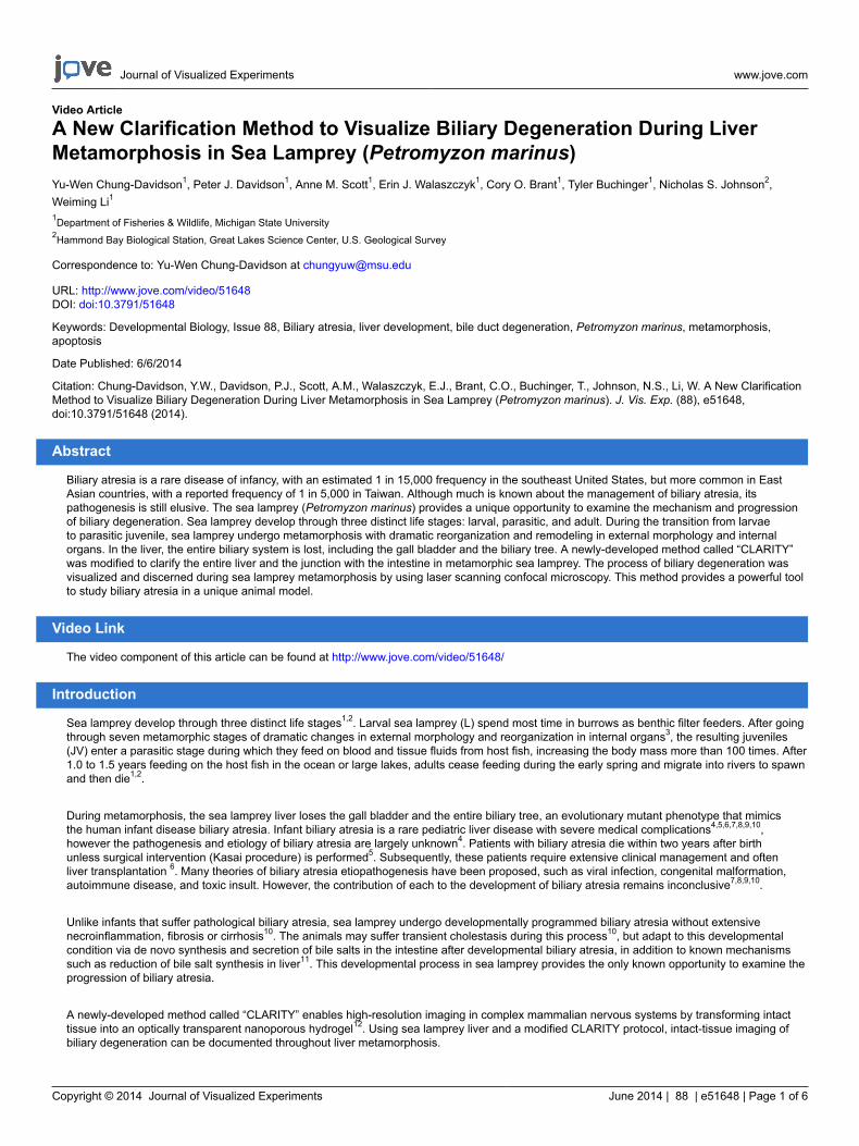
Journal of Visualized Experiments www.jove.com
Copyright © 2014 Journal of Visualized Experiments June 2014 | 88 | e51648 | Page 1 of 6
Video Article
A New Clarification Method to Visualize Biliary Degeneration During LiverMetamorphosis in Sea Lamprey (Petromyzon marinus)Yu-Wen Chung-Davidson1, Peter J. Davidson1, Anne M. Scott1, Erin J. Walaszczyk1, Cory O. Brant1, Tyler Buchinger1, Nicholas S. Johnson2,Weiming Li1
1Department of Fisheries & Wildlife, Michigan State University2Hammond Bay Biological Station, Great Lakes Science Center, U.S. Geological Survey
Correspondence to: Yu-Wen Chung-Davidson at [email protected]
URL: http://www.jove.com/video/51648DOI: doi:10.3791/51648
Keywords: Developmental Biology, Issue 88, Biliary atresia, liver development, bile duct degeneration, Petromyzon marinus, metamorphosis,apoptosis
Date Published: 6/6/2014
Citation: Chung-Davidson, Y.W., Davidson, P.J., Scott, A.M., Walaszczyk, E.J., Brant, C.O., Buchinger, T., Johnson, N.S., Li, W. A New ClarificationMethod to Visualize Biliary Degeneration During Liver Metamorphosis in Sea Lamprey (Petromyzon marinus). J. Vis. Exp. (88), e51648,doi:10.3791/51648 (2014).
Abstract
Biliary atresia is a rare disease of infancy, with an estimated 1 in 15,000 frequency in the southeast United States, but more common in EastAsian countries, with a reported frequency of 1 in 5,000 in Taiwan. Although much is known about the management of biliary atresia, itspathogenesis is still elusive. The sea lamprey (Petromyzon marinus) provides a unique opportunity to examine the mechanism and progressionof biliary degeneration. Sea lamprey develop through three distinct life stages: larval, parasitic, and adult. During the transition from larvaeto parasitic juvenile, sea lamprey undergo metamorphosis with dramatic reorganization and remodeling in external morphology and internalorgans. In the liver, the entire biliary system is lost, including the gall bladder and the biliary tree. A newly-developed method called “CLARITY”was modified to clarify the entire liver and the junction with the intestine in metamorphic sea lamprey. The process of biliary degeneration wasvisualized and discerned during sea lamprey metamorphosis by using laser scanning confocal microscopy. This method provides a powerful toolto study biliary atresia in a unique animal model.
Video Link
The video component of this article can be found at http://www.jove.com/video/51648/
Introduction
Sea lamprey develop through three distinct life stages1,2. Larval sea lamprey (L) spend most time in burrows as benthic filter feeders. After goingthrough seven metamorphic stages of dramatic changes in external morphology and reorganization in internal organs3, the resulting juveniles(JV) enter a parasitic stage during which they feed on blood and tissue fluids from host fish, increasing the body mass more than 100 times. After1.0 to 1.5 years feeding on the host fish in the ocean or large lakes, adults cease feeding during the early spring and migrate into rivers to spawnand then die1,2.
During metamorphosis, the sea lamprey liver loses the gall bladder and the entire biliary tree, an evolutionary mutant phenotype that mimicsthe human infant disease biliary atresia. Infant biliary atresia is a rare pediatric liver disease with severe medical complications4,5,6,7,8,9,10,however the pathogenesis and etiology of biliary atresia are largely unknown4. Patients with biliary atresia die within two years after birthunless surgical intervention (Kasai procedure) is performed5. Subsequently, these patients require extensive clinical management and oftenliver transplantation 6. Many theories of biliary atresia etiopathogenesis have been proposed, such as viral infection, congenital malformation,autoimmune disease, and toxic insult. However, the contribution of each to the development of biliary atresia remains inconclusive7,8,9,10.
Unlike infants that suffer pathological biliary atresia, sea lamprey undergo developmentally programmed biliary atresia without extensivenecroinflammation, fibrosis or cirrhosis10. The animals may suffer transient cholestasis during this process10, but adapt to this developmentalcondition via de novo synthesis and secretion of bile salts in the intestine after developmental biliary atresia, in addition to known mechanismssuch as reduction of bile salt synthesis in liver11. This developmental process in sea lamprey provides the only known opportunity to examine theprogression of biliary atresia.
A newly-developed method called “CLARITY” enables high-resolution imaging in complex mammalian nervous systems by transforming intacttissue into an optically transparent nanoporous hydrogel12. Using sea lamprey liver and a modified CLARITY protocol, intact-tissue imaging ofbiliary degeneration can be documented throughout liver metamorphosis.

Journal of Visualized Experiments www.jove.com
Copyright © 2014 Journal of Visualized Experiments June 2014 | 88 | e51648 | Page 2 of 6
Protocol
1. Solution Preparation
1. Make 1 L 10x 0.1 M phosphate buffer saline (PBS, pH 7.4): Weigh 26.2 g sodium phosphate (monobasic), 115 g sodium phosphate (dibasic),and 87.66 g NaCl. Dissolve in about 800 ml distilled H2O, adjust pH, and bring the volume up to 1 L with distilled H2O.
1. Make 1 L 0.1 M phosphate buffer saline (pH 7.4): Take 100 ml 10x PBS, and add 900 ml distilled H2O.2. Make 1 L 0.1 M phosphate buffer saline (pH 7.4) with 0.1% Triton X-100: Take 100 ml 10x PBS, add 1 ml Triton X-100, and bring the
volume up to 1 L with distilled H2O.
2. Make 1 L 10x 0.1M phosphate buffer (PB, pH 7.4): Weigh 26.2 g sodium phosphate (monobasic) and 115 g sodium phosphate (dibasic).Dissolve in about 800 ml distilled H2O, adjust pH, and bring the volume up to 1 L with distilled H2O.
1. Make 1 L 0.1 M phosphate buffer (pH 7.4): Take 100 ml 10x PB, and add 900 ml distilled H2O.2. Make 1 L 0.1 M phosphate buffer (pH 7.4) with 0.1% Triton X-100: Take 100 ml 10x PB, add 1 ml Triton X-100, and bring the volume up
to 1 L with distilled H2O.
3. Make 400 ml hydrogel monomer solution (4% acrylamide, 0.25% bis-acrylamide, 4% paraformaldehyde, 0.0075% ammonium persulfate,0.0005% saponin in 0.1 M PBS): Weigh 0.3 g ammonium persulfate and 0.2 g saponin. Add 210 ml distilled H2O, 40 ml 40% acrylamide, 10ml 2% bis-acrylamide, 40 ml 10x PBS (pH 7.4), 100 ml 16% paraformaldehyde, and mix well.
4. Make 4 L clearing solution (4% SDS in 0.2 M boric acid): Weigh 49.464 g boric acid and 160 g SDS. Dissolve in about 3.5 L distilled H2O,adjust pH to 8.5 with NaOH, and bring the volume up to 4 L.
5. Make 1 L 80% glycerol solution: Measure 800 ml glycerol, add 200 ml distilled H2O, and mix well.
2. Tissue Preparation
1. Prepare hydrogel monomer solution and aliquot 10 ml to each 15 ml centrifuge tube.2. Dissect the whole tissue and fix in the hydrogel for 2 days at 4 °C. Make sure that the hydrogel solution is at least 10x the volume of the
tissue.3. Bring the hydrogel fixed tissue (in the 15 ml centrifuge tube) to a fume hood.4. Polymerize the hydrogel fixed tissue by adding 5 µl TEMED, mix well, and let sit in the fume hood at room temperature for 2-3 hr.5. Remove the excess gel thoroughly. Note: Extra gel will hinder the clearing process and the antibody penetration during staining.6. Incubate the fixed tissue in 10 ml clearing solution in an incubator shaker set at 70 rpm and 50 °C for several days until the tissue becomes
transparent. Change clearing solution at least once per day and remove the excess gel from the tissue when changing the clearing solution.7. Rinse the tissue in 10 ml 0.1 M PB with 0.1% Triton X-100 at 70 rpm and 37 °C overnight. Repeat this step 3 times and remove the excess
gel from the tissue when changing the buffer solution.8. Incubate the clarified tissue in the primary antibody solution in PB with 0.1% Triton X-100 at 70 rpm and 37 °C for 2 days. Note: Make enough
primary antibody solution to submerge the whole tissue.9. Rinse the tissue in 10 ml PB with 0.1% Triton X-100 at 70 rpm and 37 °C overnight. Repeat this step 3 times and remove the excess gel from
the tissue when changing the buffer solution.10. Perform the following steps in a dark room or under dim light. Cover the samples with foil to prevent photo bleaching.11. Incubate the clarified tissue with the fluorescent secondary antibody solution in PB with 0.1% Triton X-100 and the blocking serum at 70 rpm
and 37 °C for 1 day.12. Rinse the tissue in 10 ml PB with 0.1% Triton X-100 at 70 rpm and 37 °C overnight. Repeat this step 3 times and remove the excess gel from
the tissue when changing the buffer solution.13. Rinse the tissue in 10 ml PBS at 70 rpm and 37 °C overnight. Repeat this step 3 times and remove the excess gel from the tissue when
changing the buffer solution.14. Incubate the clarified tissue in 80% glycerol at 70 rpm and 37 °C overnight.15. Store the fluorescent stained tissue at 4 °C prior to confocal microscopy.
Representative Results
Several important developmental events occur in the hepatobiliary system during sea lamprey metamorphosis. The bile duct and the gall bladderundergo apoptosis and degenerate (Figure 1). Combining the modified clarification method and staining with liver cell marker cytokeratin 19(CK19, present in both cholangiocytes and hepatocytes before and after metamorphosis13) and anti-apoptotic marker Bcl2 using confocalmicroscopy, the entire biliary system was captured along the Z-axis (Figure 2) and the 3-dimensional structure was reconstructed (Figure 3).Laser scanning confocal microscopy was used to perform optical section through the intact liver since most of the biliary system is embeddedinside the liver except the junction where the bile duct enters the intestine. The degenerative process occurs in metamorphic stages 2-4, startingfrom peripheral small bile ductules toward the central large intrahepatic common bile duct (Figure 2), consistent with earlier studies10.

Journal of Visualized Experiments www.jove.com
Copyright © 2014 Journal of Visualized Experiments June 2014 | 88 | e51648 | Page 3 of 6
Figure 1. Gall bladder degeneration during sea lamprey metamorphosis. Sea lamprey lose the gall bladder (black arrows) duringmetamorphosis. The specimens were taken from animals with external morphology between metamorphic stages 2 and 3; i.e. round eye without(stage 2) or with (stage 3) sharp differentiation of iris and pupil, prominent papilla-like projections on the inner dorsal surface of the oral hood(both stages), thickened lips of oral hood (stage 2) forming a rectangular entrance into the oral cavity (stage 3), or "pug-like" snout (stage 3)3.Note that the gall bladder also changed color indicating differences in bile production. I: intestine. L: liver. Please click here to view a largerversion of this figure.
Figure 2. Confocal images showing the biliary tree in sea lamprey liver during metamorphosis. The specimens were taken from animalswith external morphology between metamorphic stages 2 and 3. Top panels are taken from the anterior, and the lower panels from the posteriorend. Images of liver cell marker cytokeratin 19 (1:100 mouse-anti-CK19; stained with 2 µg/ml Alexa Fluor 350-goat-anti-mouse IgG; blue)and anti-apoptotic marker Bcl2 (1:100 rabbit-anti-Bcl2; stained with 2 µg/ml Alexa Fluor 488-donkey-anti-rabbit IgG; green) are superimposedto generate the merged images. White arrows indicate the biliary tree. Black arrows indicate the dark dendritic looking apoptotic cells andsurrounding canaliculi and ductules in the liver. I: intestine. L: liver. Scale bar: 500 µm. Please click here to view a larger version of this figure.
Journal of Visualized Experiments www.jove.com
Copyright © 2014 Journal of Visualized Experiments June 2014 | 88 | e51648 | Page 4 of 6
Figure 3. 3D images reconstructed from merged confocal z-series. 3D images were reconstructed from the confocal z-series of three liversbetween metamorphic stages 2 and 3. Optical sections of merged images of liver cell marker cytokeratin 19 (1:100 mouse-anti-CK19; stainedwith 2 µg/ml Alexa Fluor 350-goat-anti-mouse IgG; blue) and anti-apoptotic marker Bcl2 (1:100 rabbit-anti-Bcl2; stained with 2 µg/ml Alexa Fluor488-donkey-anti-rabbit IgG; green) are reconstructed with the manufacturer’s software to generate the 3D images. Top panels are taken fromthe anterior, and the lower panels from the posterior end. White arrows indicate the biliary tree. Black arrows indicate the dark dendritic lookingapoptotic cells and surrounding canaliculi and ductules in the liver. I: intestine. L: liver. Scale bar: 500 µm. Please click here to view a largerversion of this figure.
Journal of Visualized Experiments www.jove.com
Copyright © 2014 Journal of Visualized Experiments June 2014 | 88 | e51648 | Page 5 of 6
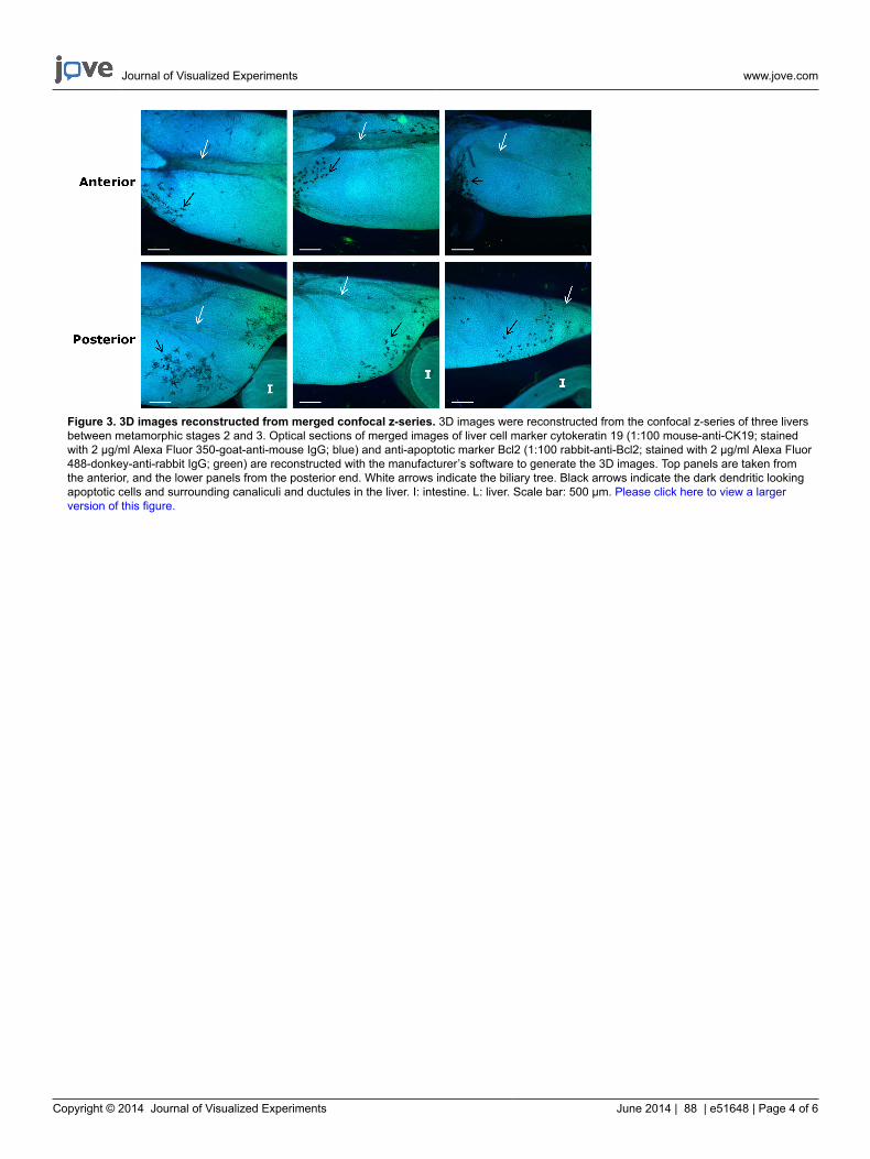
Figure 4. A confocal optical section showing bile ducts with bile droplets in the lumen. Merged image from 3 fluorescent channels; blue:mouse-anti-c-Myc (1:100)/2 µg/ml Alexa Fluor 350-goat-anti-mouse IgG; green: 2 µg/ml Nissl green; Red: rabbit-anti-TGFβRII (1:100)/2 µg/ml Alexa Fluor 594-donkey-anti-rabbit. Scale bar: 500 μm. Bile droplets are indicated by black arrows. The liver specimen was taken betweenmetamorphic stage 2 and 3. Please click here to view a larger version of this figure.
Discussion
This protocol is modified from a new method called “CLARITY”12, which crosslinks intact tissue with polyacrylamide to form a nanoporoushydrogel, and then strips away the plasma membrane of the tissue to achieve optical transparency and macromolecular permeability. “CLARITY”allows intact-tissue imaging of long-range projection and local circuit wiring in the nervous system. This new method can be used to visualizethe entire biliary system in sea lamprey liver during metamorphosis. It is a challenge in biology to obtain high-resolution information and maintainthe global perspective from a complex system such as the entire biliary system or central nervous system using conventional embeddingand sectioning methods. This modified CLARITY method allows a laser beam to penetrate through the intact sea lamprey larval liver up to athickness of 1 to 2 mm using low-power fluorescent objective (4x) in a regular laser scanning confocal microscope. Deeper penetration can beachieved using a more advanced multi-photon confocal microscope. Three-dimensional images can be reconstructed using 3D reconstructionsoftware. Achieving comparable results using conventional methods would be laborious, involving the piecemeal reconstruction of manysections, and would be limited to small tissue volumes. This new method provides valuable information from whole structural analyses of intactsystems12, is less labor-intensive, more efficient and reproducible.
Another advantage of this new method is that the specimen can be reused multiple times for staining with different antibodies12. This makesthe comparison between different molecular markers in the same specimen more reliable and reproducible. The stable framework generatedby hydrogel allows effective removal of antibodies without fine structure damage and maintains the antigenicity for a long time12. The clearingsolution denatures antibodies and disrupts binding, therefore acting as an antibody-stripping solution12. Several rounds of staining have beenperformed in the sea lamprey liver samples and the structure and staining remained optimal.
There are two major modifications in this protocol from the original “CLARITY” protocol12. (1) Free radical generating reagents (ammoniumpersulfate and TEMED) were used to initiate and accelerate the polymerization of the hydrogel instead of VA044. VA044 is photosensitive andnot available in the U.S. (2) No custom-built electrophoresis chamber is used. In the CLARITY method12, due to the size of the tissues (mousebrain vs. lamprey liver), electrophoretic tissue clearing is required to expedite the extraction of the plasma membrane. To avoid the initial cost
Journal of Visualized Experiments www.jove.com
Copyright © 2014 Journal of Visualized Experiments June 2014 | 88 | e51648 | Page 6 of 6
for a custom-built electrophoresis chamber, we first used existing agarose gel electrophoresis devices in our laboratory. This proved to be veryinefficient. However, incubation in the clearing solution with raised temperature (50 °C) is very effective in clearing the plasma membrane.
Although the authors for “CLARITY” cautioned against the use of saponin in the hydrogel, it improves the transparency in the sea lamprey tissuein our procedure. One drawback for confocal imaging is that sea lamprey tissue has strong auto-fluorescence. The choice of fluorophores mustbe tested by comparing the images before and after immunofluorescent staining.
During sea lamprey metamorphosis, the regression of the gall bladder epithelium starts between metamorphic stages 2 and 3. By metamorphicstage 4, the gall bladder disappears entirely from sea lamprey liver14. Bile duct degeneration is most dramatic between metamorphic stages 3and 415. This modified method is a powerful tool to study the cellular and molecular mechanisms of the degeneration of the entire biliary system,which may provide insights for human biliary atresia.
Disclosures
The authors have nothing to disclose.
Acknowledgements
The authors acknowledge the contribution of Hammond Bay Biological Station, Great Lakes Science Center, U.S. Geological Survey. We alsothank Dr. Melinda Frame at the Center for Advanced Microscopy at Michigan State University for her technical support in laser scanning confocalmicroscopy. This study is supported by grants from the Great Lakes Fishery Commission to YWCD and WML.
References
1. Applegate, V.C. Natural history of the sea lamprey (Petromyzon marinus) in Michigan. US Fish and Wildlife Service Special Science Reporton Fishery Service. 55, 237 (1950).
2. Hardisty, M.W., Potter, I.C. The general biology of adult lampreys. In: Hardisty, M.W., Potter, I.C., eds. The biology of lampreys. New York:Academic Press. 1, 127-206 (1971).
3. Youson, J.H., Potter, I.C. A description of the stages in the metamorphosis of the anadromous sea lamprey, Petromyzon marinus L. Can. J.Zool. 57, 1808-1817 (1979).
4. Boomer, L.A., et al. Cholangiocyte apoptosis is an early event during induced metamorphosis in the sea lamprey, Petromyzon marinus L. J.Pediatr. Surg. 45, 114-120 (2010).
5. Kasai, M., Suzuki, H., Ohashi, E., Ohi, R., Chiba, T., Okamoto, A. Technique and results of operative management of biliary atresia. World J.Surg. 2, 571-580 (1978).
6. Suzuki, T., Hashimoto, T., Kondo, S., Sato, Y., Hussein, M.H. Evaluating patients’ outcome post-Kasai operation: a 19-year experience withmodification of the hepatic portoenterostomy and applying a novel steroid therapy regimen. Pediatr. Surg. Int. 26, 825-830 (2010).
7. Hartley, J.L., Davenport, M., Kelly, D.A. Biliary atresia. Lancet. 374, 1704-1713 (2009).8. Morecki, R., Glaser, J.H., Cho, S., Balistreri, W.F., Horwitz, M.S. Biliary atresia and reovirus type 3 infection. New Engl. J. Med. 307, 481-484
(1982).9. Shimadera, S., Iwai, N., Deguchi, E., Kimura, O., Fumino, S., Yokoyama, T. The inv mouse as an experimental model of biliary atresia. J.
Pediatr. Surg. 42, 1555-1560 (2007).10. Sidon, E.W., Youson, J.H. Morphological changes in the liver of the sea lamprey, Petromyzon marinus L., during metamorphosis: I. Atresia of
the bile ducts. J. Morphol. 177, 109-124 (1983).11. Yeh, C.-Y., Chung-Davidson, Y.-W., Wang, H., Li, K., Li, W. Intestinal synthesis and secretion of bile salts as an adaptation to developmental
biliary atresia in the sea lamprey. Proc. Nat. Acad. Sci. U.S.A. 109, 11419-11424 (2012).12. Chung, K., et al. Structural and molecular interrogation of intact biological systems. Nature. 497, 332-337 (2013).13. Alarcón, V.B., Filosa, M.F., Youson, J.H. Cytokeratins in the liver of the sea lamprey (Petromyzon marinus) before and after metamorphosis.
Cell Tissue Res. 287, 365-374 (1997).14. Youson, J.H., Ogilvie, D.R. Ultrastructural features of degeneration of the gallbladder during lamprey biliary atresia. Tissue and Cell. 22,
477-492 (1990).15. Morii, M. et al. Onset of apoptosis in the cystic duct during metamorphosis of a Japanese lamprey, Lethenteron reissneri. Anat. Rec. 293,
1155-1166 (2010).
Copyright © 2022 FDOKUMEN