Epicardial development in lamprey supports an evolutionary origin of the vertebrate epicardium from...
7
Epicardial development in lamprey supports an evolutionary origin of the vertebrate epicardium from an ancestral pronephric external glomerulus Manuel A. Pombal, a Rita Carmona, b Manuel Megı ´as, a Alejandro Ruiz, b Jose ´ Marı ´a Pe ´ rez-Pomares, b and Ramo ´n Mun ˜ oz-Cha ´ puli b, a Neurolam group, Department of Functional Biology and Health Sciences, Faculty of Biology, University of Vigo, 36310 Vigo (Spain) b Department of Animal Biology, Faculty of Sciences, University of Ma ´ laga, 29071 Ma ´ laga (Spain) Author for correspondence (email: [email protected]) SUMMARY The epicardium is the outer layer of the vertebrate heart. Both the embryonic epicardium and its derived mesenchyme are critical to heart development, contributing to the coronary vasculature and modulating the proliferation of the ventricular myocardium. The embryonic epicardium arises from an extracardiac, originally paired progenitor tissue called the proepicardium, a proliferation of coelomic cells found at the limit between the liver and the sinus venosus. Proepicardial cells attach to and spread over the cardiac surface giving rise to the epicardium. Invertebrate hearts always lack of epicardium, and no hypothesis has been proposed about the origin of this tissue and its proepicardial progenitor in vertebrates. We herein describe the epicardial development in a representative of the most basal living lineage of vertebrates, the agnathan Petromyzon marinus (lamprey). The epicardium in lampreys develops by migration of coelomic cells clustered in a paired structure at the roof of the coelomic cavity, between the pronephros and the gut. Later on, these outgrowths differentiate into the pronephric external glomerulus (PEG), a structure composed of capillary networks, mesangial cells, and podocytes. This observation is consistent with the conclusion that the primordia of the most anterior pair of PEG in agnathans have been retained and transformed into the proepicardium in gnathostomes. Glomerular progenitor cells are highly vasculogenic and probably allowed for the vascularization of a cardiac tube primarily devoid of coronary vessels. This new hypothesis accounts for the striking epicardial expression of Wt1 and Pod1, two transcription factors essential for development of the excretory system. INTRODUCTION The heart in vertebrates is constituted of three cell layers, epicardium, myocardium, and endocardium, with a contribu- tion of neural crest cells. Both myocardium and endocardium derive from the precardiac mesoderm. The epicardium is the outer layer of the vertebrate heart, and it gives rise to a mesenchymal cell population which is critical to heart devel- opment, as these epicardially derived cells (EPDC) contribute to the formation of the coronary vasculature and the cardiac interstitium (Vrancken-Peeters et al. 1999; Pe´ rez-Pomares et al. 2002; Guadix et al. 2006). Furthermore, epicardium and EPDC induce the proliferation of the myocardium and it has been reported that their activity is essential for proper devel- opment of the ventricular compact layer (Stuckmann et al. 2003; Lavine et al. 2005; Merki et al. 2005). For this reason, anomalous epicardial development may cause severe heart malformations (Kwee et al. 1995; Yang et al. 1995). The embryonic epicardium arises from an extracardiac and originally paired progenitor tissue called the proepicardium, a proliferation of coelomic cells at the limit beetween the liver and the cardiac sinus venosus in all the vertebrates so far studied (Manner et al. 2001). Both proepicardial primordia develop in fishes (Mun˜oz-Cha´puli et al. 1994) whereas only the right one fully forms in birds (Schulte et al. 2007). In mammals both proepicardial primordia fuse in the midline forming a crescent that covers all the septum transversum area (Schulte et al. 2007). In all cases, proepicardial coelomic cells attach to the cardiac surface and spread on the myocar- dium, giving rise to the epicardium. In contrast to the vertebrate cardiac bauplan, the hearts in non-vertebrate metazoans are usually formed by one or several myoepithelial cell layers but they always lack a epicardium. Despite this main difference, no hypothesis has hitherto been proposed about the evolutionary origin of this tissue and its proepicardial progenitor whose formation is dependent on molecular and cellular mechanisms that are poorly known (Schlueter et al. 2006). We have studied the epicardial development in a repre- sentative of the most basal living lineage of vertebrates, the EVOLUTION & DEVELOPMENT 10:2, 210–216 (2008) & 2008 The Author(s) Journal compilation & 2008 Blackwell Publishing Ltd. 210
Transcript of Epicardial development in lamprey supports an evolutionary origin of the vertebrate epicardium from...
Epicardial development in lamprey supports an evolutionary origin of the
vertebrate epicardium from an ancestral pronephric external glomerulus
Manuel A. Pombal,a Rita Carmona,b Manuel Megıas,a Alejandro Ruiz,b Jose Marıa Perez-Pomares,b andRamon Munoz-Chapulib,�
aNeurolam group, Department of Functional Biology and Health Sciences, Faculty of Biology, University of Vigo, 36310 Vigo
(Spain)bDepartment of Animal Biology, Faculty of Sciences, University of Malaga, 29071 Malaga (Spain)�Author for correspondence (email: [email protected])
SUMMARY The epicardium is the outer layer of thevertebrate heart. Both the embryonic epicardium and itsderived mesenchyme are critical to heart development,contributing to the coronary vasculature and modulating theproliferation of the ventricular myocardium. The embryonicepicardium arises from an extracardiac, originally pairedprogenitor tissue called the proepicardium, a proliferation ofcoelomic cells found at the limit between the liver and the sinusvenosus. Proepicardial cells attach to and spread over thecardiac surface giving rise to the epicardium. Invertebratehearts always lack of epicardium, and no hypothesis has beenproposed about the origin of this tissue and its proepicardialprogenitor in vertebrates. We herein describe the epicardialdevelopment in a representative of the most basal livinglineage of vertebrates, the agnathan Petromyzon marinus
(lamprey). The epicardium in lampreys develops by migrationof coelomic cells clustered in a paired structure at the roof ofthe coelomic cavity, between the pronephros and the gut.Later on, these outgrowths differentiate into the pronephricexternal glomerulus (PEG), a structure composed of capillarynetworks, mesangial cells, and podocytes. This observationis consistent with the conclusion that the primordia of themost anterior pair of PEG in agnathans have been retainedand transformed into the proepicardium in gnathostomes.Glomerular progenitor cells are highly vasculogenic andprobably allowed for the vascularization of a cardiac tubeprimarily devoid of coronary vessels. This new hypothesisaccounts for the striking epicardial expression of Wt1 andPod1, two transcription factors essential for development ofthe excretory system.
INTRODUCTION
The heart in vertebrates is constituted of three cell layers,
epicardium, myocardium, and endocardium, with a contribu-
tion of neural crest cells. Both myocardium and endocardium
derive from the precardiac mesoderm. The epicardium is
the outer layer of the vertebrate heart, and it gives rise to a
mesenchymal cell population which is critical to heart devel-
opment, as these epicardially derived cells (EPDC) contribute
to the formation of the coronary vasculature and the cardiac
interstitium (Vrancken-Peeters et al. 1999; Perez-Pomares
et al. 2002; Guadix et al. 2006). Furthermore, epicardium and
EPDC induce the proliferation of the myocardium and it has
been reported that their activity is essential for proper devel-
opment of the ventricular compact layer (Stuckmann et al.
2003; Lavine et al. 2005; Merki et al. 2005). For this reason,
anomalous epicardial development may cause severe heart
malformations (Kwee et al. 1995; Yang et al. 1995).
The embryonic epicardium arises from an extracardiac and
originally paired progenitor tissue called the proepicardium, a
proliferation of coelomic cells at the limit beetween the liver
and the cardiac sinus venosus in all the vertebrates so far
studied (Manner et al. 2001). Both proepicardial primordia
develop in fishes (Munoz-Chapuli et al. 1994) whereas only
the right one fully forms in birds (Schulte et al. 2007). In
mammals both proepicardial primordia fuse in the midline
forming a crescent that covers all the septum transversum
area (Schulte et al. 2007). In all cases, proepicardial coelomic
cells attach to the cardiac surface and spread on the myocar-
dium, giving rise to the epicardium.
In contrast to the vertebrate cardiac bauplan, the hearts
in non-vertebrate metazoans are usually formed by one or
several myoepithelial cell layers but they always lack a
epicardium. Despite this main difference, no hypothesis has
hitherto been proposed about the evolutionary origin of this
tissue and its proepicardial progenitor whose formation
is dependent on molecular and cellular mechanisms that are
poorly known (Schlueter et al. 2006).
We have studied the epicardial development in a repre-
sentative of the most basal living lineage of vertebrates, the
EVOLUTION & DEVELOPMENT 10:2, 210 –216 (2008)
& 2008 The Author(s)
Journal compilation & 2008 Blackwell Publishing Ltd.
210
agnathan P. marinus (lamprey). Our results have revealed
an unsuspected scenario on the evolutionary origin of the
proepicardium, relating this tissue with the primitive excretory
system of vertebrates. Surprisingly, a relationship between the
epicardium and the kidneys had been previously suspected
due to some common features in gene expression, but an
evolutionary and developmental basis accounting for this re-
lationship was lacking in the literature. The aim of this paper
is to show our findings on the epicardial development in
lampreys and to provide a novel model on the origin of the
epicardium in vertebrates.
MATERIAL AND METHODS
This study was carried out in early prolarvae of the sea lamprey
(P. marinus L.; n522) reared from artificially fertilized eggs.
Gametes were obtained from sexually mature adult lampreys
caught during their upstream migration (from late May to early
July) in the Ulla and Mino Rivers (northwest of Spain). The
methods for maintaining the embryos and prolarvae were essen-
tially the same as those described by Piavis (1971). The fertilized
eggs were transferred to the hatchery incubators and placed into
plastic trays. They were kept with circulating water and under
appropriate conditions of darkness and temperature (181C). Under
these conditions, in our broods hatching occurred at 12–13 days
postfertilization (dpf). Stages of prolarvae are indicated according
to their age in days after fertilization. All experiments were
conducted in accordance with European Community guidelines
on animal care and experimentation to minimize pain and
discomfort.
Prolarvae were fixed in Bouin’s fixative, washed in alcohol,
dehydrated in a graded series of ethanol, and embedded in paraffin.
Transverse and sagittal sections (5–8mm thick) were cut on
a Leica RM 2145 microtome (Leica Microsystems, Barcelona,
Spain) and subsequently stained with hematoxylin–eosin.
Dogfish embryos (Scyliorhinus canicula L.) were used for com-
parative purposes. Fertilized eggs were obtained from adult females
collected in the Bay of Malaga (Western Mediterranean) by
commercial trawl vessels. The eggs were kept in indoor tanks of
well-aerated seawater. Egg capsules were open at intervals and the
embryos were anesthetized in 0.04% tricaine methanesulfonate
(MS-222, Sigma-Aldrich Co., St. Louis, MO, USA) in seawater
and measured. Some embryos were fixed in 1.25% glutaraldehyde
and 1% paraformaldehyde and embedded in Araldite 502 as
described previously (Munoz-Chapuli et al. 1996). Semithin
sections (0.5mm) were obtained with an ultramicrotome and
stained with toluidine blue. Other embryos were fixed in methanol–
acetone–water (2:2:1), paraffin embedded, sectioned, and stained as
described above.
Immunolocalization of the Wilms’ tumor suppressor transcrip-
tion factor (Wt1) in avian embryos was performed as described
previously (Carmona et al. 2001). Briefly, the embryos were excised
and cryoprotected in 10%, 20%, and 30% sucrose solutions,
embedded in OCT and snap frozen in liquid nitrogen-cooled is-
opentane. Cryostat sections were collected on poly-L-lysine-coated
slides and fixed for 10min in 1:1 methanol–acetone at � 201C.
Then, the sections were rehydrated, the endogenous peroxidase
activity was quenched by incubation for 30min with 3% hydrogen
peroxide in Tris-phosphate buffered saline (TPBS). Non-specific
binding sites were saturated for 30min with 16% sheep serum, 1%
bovine serum albumin, and 0.5% Triton X-100 in TPBS (SBT).
Endogenous biotin was blocked with the avidin–biotin blocking kit
(Vector, Burlingame, CA, USA). Sections were subsequently incu-
bated overnight at 41C in polyclonal anti-human Wt1 (Sc-192,
Santa Cruz, Heidelberg, Germany), diluted 1:500 in SBT, washed,
incubated for 1h at room temperature in biotin-conjugated anti-
rabbit goat IgG (Sigma-Aldrich Co.) diluted 1:100 in SBT, washed
again, and incubated for 1h in avidin–peroxidase complex (Sigma-
Aldrich Co.) diluted 1:150 in TPBS. After washing, peroxidase
activity was developed with Sigma Fasts 3,30-diaminobenzidine
(D4168; Sigma-Aldrich Co.) tablets according to the indications of
the supplier.
RESULTS
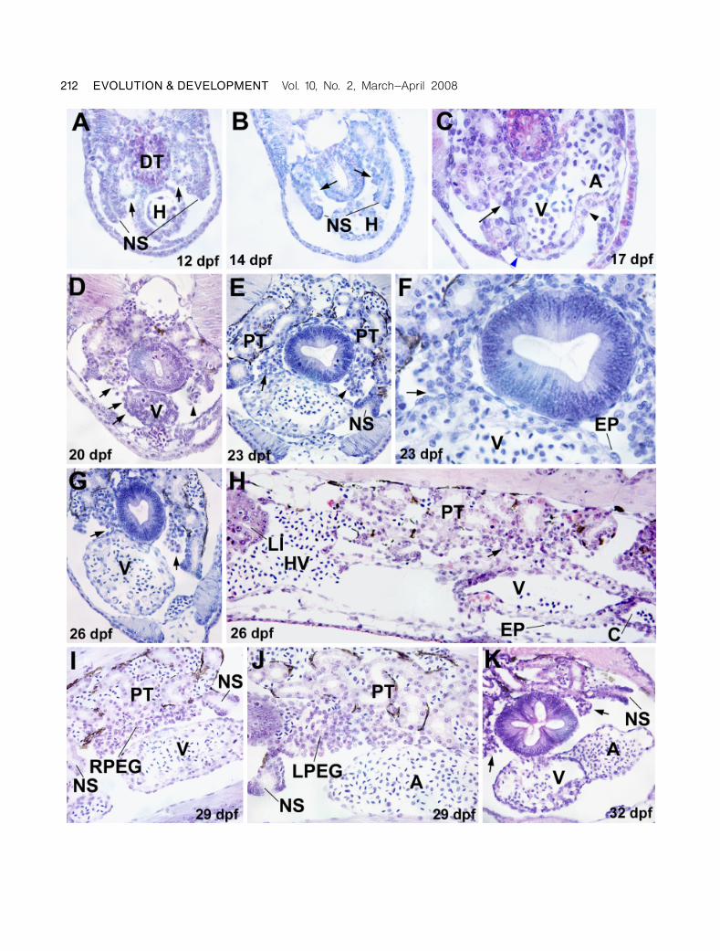
The epicardium in lampreys develops between 17 and 26 dpf,
from a paired outgrowth of coelomic cells in the dorsal
part of the coelomic cavity, between the pronephros and
the gut (Fig. 1). Ciliated nephrostomes were always located
cephalic and caudal to these cell clusters. The coelomic
outgrowths are not yet developed in embryos of 12dpf
(Fig. 1A) while in prolarvae of 14dpf, the earliest evidences
of cell outgrowth in this area are already present (Fig. 1B).
In prolarvae of 17dpf, the right cell cluster attaches to the
dorsal side of the ventricle, close to the atrioventricular
junction (Fig. 1C). From this area, epicardial cells can be
seen apparently spreading over the ventricular surface. How-
ever, the atrium and the ventral side of the atrioventricular
groove are devoid of epicardium. In prolarvae of 20 and
23dpf, the attachment between the right outgrowth of
coelomic cells has spread over the dorsal and right surfaces
of the ventricle forming a flattened, squamous-monostratified
epithelium (Fig. 1, D–F). As a consequence, most of the ven-
tricular and atrial surfaces are lined by epicardial cells. The
contact between the right cell outgrowth and the cardiac sur-
face is disappearing by 26dpf (Fig. 1, G and H). In later
stages, there is no contact between the coelomic cell clusters,
which become the right and left pronephric external
glomerulus (PEG) (Fig. 1, I–K), and the heart surface.
Blood-containing vessels are abundant in these glomeruli
from these stages on.
For comparison purposes, Fig. 2 shows the localization
and structure of the proepicardium in embryos of a basal
gnathostome, the dogfish (S. canicula). In this species the
proepicardium is paired, and it releases free-floating cells into
the pericardial cavity that adhere to the myocardium (Munoz-
Chapuli et al. 1994). However, in earlier stages there is an
attachment between the right proepicardium and the ventric-
The origin of the vertebrate epicardium 211Pombal et al.
212 EVOLUTION & DEVELOPMENT Vol. 10, No. 2, March^April 2008
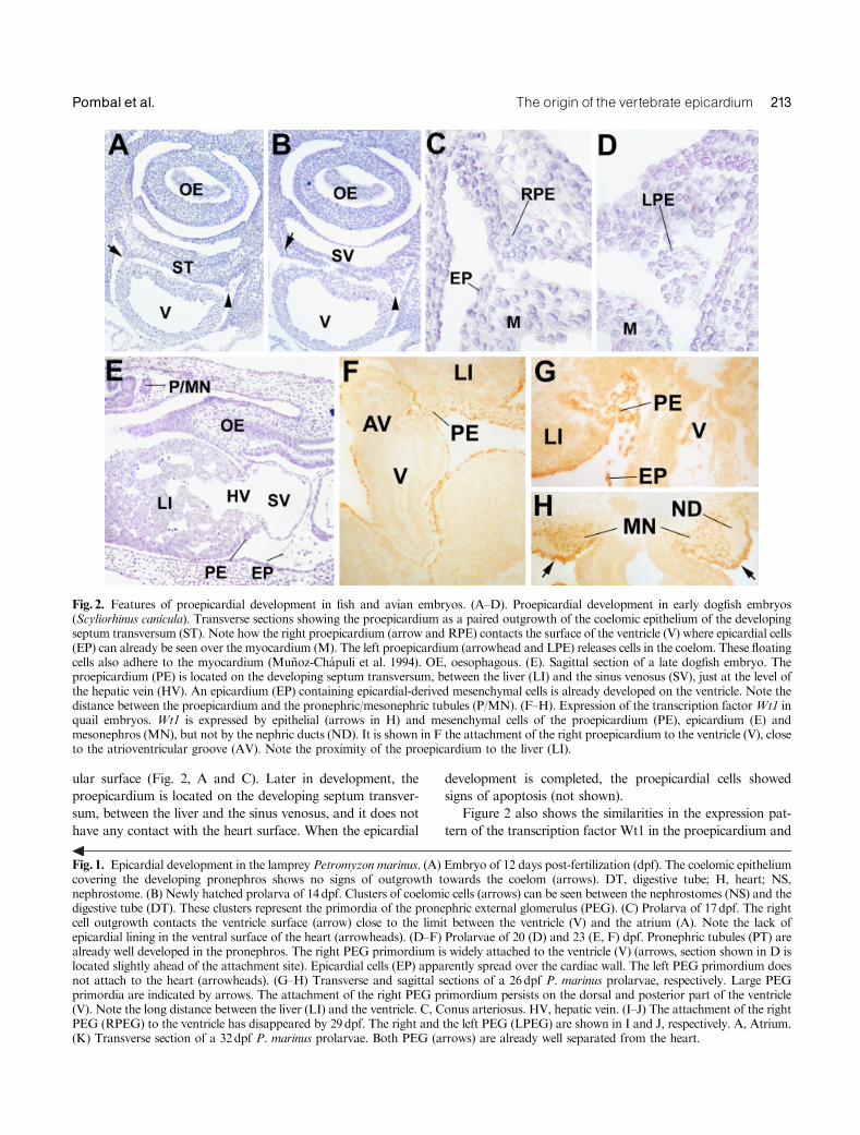
ular surface (Fig. 2, A and C). Later in development, the
proepicardium is located on the developing septum transver-
sum, between the liver and the sinus venosus, and it does not
have any contact with the heart surface. When the epicardial
development is completed, the proepicardial cells showed
signs of apoptosis (not shown).
Figure 2 also shows the similarities in the expression pat-
tern of the transcription factor Wt1 in the proepicardium and
Fig. 2. Features of proepicardial development in fish and avian embryos. (A–D). Proepicardial development in early dogfish embryos(Scyliorhinus canicula). Transverse sections showing the proepicardium as a paired outgrowth of the coelomic epithelium of the developingseptum transversum (ST). Note how the right proepicardium (arrow and RPE) contacts the surface of the ventricle (V) where epicardial cells(EP) can already be seen over the myocardium (M). The left proepicardium (arrowhead and LPE) releases cells in the coelom. These floatingcells also adhere to the myocardium (Munoz-Chapuli et al. 1994). OE, oesophagous. (E). Sagittal section of a late dogfish embryo. Theproepicardium (PE) is located on the developing septum transversum, between the liver (LI) and the sinus venosus (SV), just at the level ofthe hepatic vein (HV). An epicardium (EP) containing epicardial-derived mesenchymal cells is already developed on the ventricle. Note thedistance between the proepicardium and the pronephric/mesonephric tubules (P/MN). (F–H). Expression of the transcription factorWt1 inquail embryos. Wt1 is expressed by epithelial (arrows in H) and mesenchymal cells of the proepicardium (PE), epicardium (E) andmesonephros (MN), but not by the nephric ducts (ND). It is shown in F the attachment of the right proepicardium to the ventricle (V), closeto the atrioventricular groove (AV). Note the proximity of the proepicardium to the liver (LI).
Fig. 1. Epicardial development in the lamprey Petromyzon marinus. (A) Embryo of 12 days post-fertilization (dpf). The coelomic epitheliumcovering the developing pronephros shows no signs of outgrowth towards the coelom (arrows). DT, digestive tube; H, heart; NS,nephrostome. (B) Newly hatched prolarva of 14dpf. Clusters of coelomic cells (arrows) can be seen between the nephrostomes (NS) and thedigestive tube (DT). These clusters represent the primordia of the pronephric external glomerulus (PEG). (C) Prolarva of 17dpf. The rightcell outgrowth contacts the ventricle surface (arrow) close to the limit between the ventricle (V) and the atrium (A). Note the lack ofepicardial lining in the ventral surface of the heart (arrowheads). (D–F) Prolarvae of 20 (D) and 23 (E, F) dpf. Pronephric tubules (PT) arealready well developed in the pronephros. The right PEG primordium is widely attached to the ventricle (V) (arrows, section shown in D islocated slightly ahead of the attachment site). Epicardial cells (EP) apparently spread over the cardiac wall. The left PEG primordium doesnot attach to the heart (arrowheads). (G–H) Transverse and sagittal sections of a 26dpf P. marinus prolarvae, respectively. Large PEGprimordia are indicated by arrows. The attachment of the right PEG primordium persists on the dorsal and posterior part of the ventricle(V). Note the long distance between the liver (LI) and the ventricle. C, Conus arteriosus. HV, hepatic vein. (I–J) The attachment of the rightPEG (RPEG) to the ventricle has disappeared by 29dpf. The right and the left PEG (LPEG) are shown in I and J, respectively. A, Atrium.(K) Transverse section of a 32dpf P. marinus prolarvae. Both PEG (arrows) are already well separated from the heart.
The origin of the vertebrate epicardium 213Pombal et al.

in the mesonephros of quail embryos. In both cases Wt1 is
strongly expressed in the coelomic lining and also, with differ-
ent intensity, in the adjacent mesenchyme. Wt1 expression
apparently decreases as these mesenchymal cells differentiate,
as suggested by the decrease of staining at the inner areas and
also by the lack of expression in the epithelial structures of the
mesonephros.
DISCUSSION
The proepicardium is an originally paired structure present in
all the vertebrate models so far studied. It is originally extra-
cardiac (it does not apparently form from the early primary or
secondary heart fields), but it attaches to the heart surface
and spreads lining the myocardium and giving rise to the
epicardium. No hypothesis had been hitherto made about its
origin and relationships to other embryonic systems. We
herein propose that the proepicardium is the reminiscence of
the PEG progenitor that was conserved through the evolution
essentially due to its role in providing vasculogenic cells to the
heart. The paired PEG primordia gave rise to the bilateral
proepicardia of fishes (Munoz-Chapuli et al. 1994), but the
right PEG primordium/proepicardium seems to have been
favored by the evolution, because it is the only one that at-
taches to the heart in lamprey, dogfish, and birds. Thus, the
attachment of the right PEG primordium/proepicardium to
the heart surface is highly conserved during the evolution of
vertebrates. In mammals, both primordia fuse in the midline
giving rise to a single proepicardium (Schulte et al. 2007), but
attachment to the myocardium also occurs (Nesbitt et al.
2006). The unequal contribution of the PEG primordia to the
epicardium is probably related with the asymmetrical devel-
opment of the heart, because the rightward looping of the
early cardiac tube faces the ventricular surface to the right
primordium. Thus, proepicardial asymmetry seems to be a
consequence of the cardiac asymmetry.
A pair of PEG is present in lamprey and amphibian larvae
(Kluge and Fischer 1990), and they are composed of capillary
networks, mesangial cells, and coelomic-derived podocytes.
Rudimentary PEGs have also been described in the chick
embryo, although their functionality is doubtful (Hiruma and
Nakamura 2003). However, PEGs are functional in both
lamprey and amphibian larvae, thus retaining the original
pattern of the vertebrate excretory system, i.e., a blood-filter-
ing glomerulus which releases the filtrate into the coelomic
cavity, where it is aspirated by the ciliated funnels of the
pronephric ducts. The glomerular filtrate was, in amniote
vertebrates, released into the nephrocele. Consequently, the
PEG disappeared from adult vertebrates.
Proepicardium had never been related with the excretory
system, and in fact it is anatomically associated with the liver
and/or the septum transversum in all the vertebrate models
hitherto studied. Therefore, an open question is how did the
PEG primordium become associated with the liver/septum
domain?We think that the disappearance of the most anterior
part of the pronephros, the progressive enlargement of the
liver, and especially the expansion and looping of the cardiac
inflow tract account for this new localization of the pro-
epicardium (Fig. 3). The enlargement of the cardiac inflow
tract was probably concomitant with a looping of the whole
area, thus accounting for the dorsal location of the atrium
and the new position of the proepicardium caudal to the
ventricle. In fact, in lamprey embryos and prolarvae the liver
is far away from the heart, the atrium is located laterally to
the ventricle and a defined sinus venosus is not present at the
time of epicardial development (Figs. 1 and 3). On the other
hand, the evolutionary expansion of the cardiac inflow tract is
probably recapitulated during the development of amniote
vertebrates. A mesenchymal population which does not ex-
press Nkx2.5 (a marker of the myocardial progenitors) and
expresses instead podoplanin (a podocytic marker), the T-box
gene Tbx18 and Wt1 incorporates to the venous pole of
the embryonic heart (Christoffels et al. 2006; Gittenberger-
de Groot et al. 2007; unpublished observations). It is impor-
tant to remark that Tbx18 is strongly expressed in the pro-
epicardium and genital ridges (Haenig and Kispert 2004). In
addition, the liver growth towards the dorsal and right side,
intermingling with the septum transversum mesenchyme,
Fig. 3. Comparison between the anatomical arrangement of thepronephric external glomerulus (PEG) in agnathans and the pro-epicardium (PE) of gnathostomes. The different localization of theproepicardium in gnathostomes is probably due to the disappear-ance of the most anterior part of the pronephros, the enlargementof the liver, the incorporation of venous areas to the cardiac inflowtract giving rise to the sinus venosus and a further cardiac rotationleaving the atrium dorsal and the proepicardium caudal to theventricle. In this way the primitive PEG primordium became as-sociated to the liver and septum transversum.
214 EVOLUTION & DEVELOPMENT Vol. 10, No. 2, March^April 2008

probably uncoupled the PEG progenitor from the primitive
nephrogenic area. Thus, the anatomical association of the
proepicardium with the liver and the septum transversum
seems to be purely contingent.
A main support of our hypothesis is provided by the
strong epicardial expression of Wt1 and Pod1, two transcrip-
tion factors essential for genitourinary development. (Moore
et al. 1999; Cui et al. 2003). The Wilms’ tumor suppressor
transcription factor Wt1 is essential for development of
kidneys and gonads, two organs which do not develop in
Wt1-deficient mouse embryos (Kreidberg et al. 1993). In these
embryos, the epicardial development is defective, showing
premature and anomalous differentiation (endothelial
differentiation is particularly impaired) as well as reduced
proliferation (Wagner et al. 2005; J. M. Perez-Pomares et al.
unpublished observations). On the other hand, the bHLH
transcription factor epicardin/Pod1 is strongly expressed in
podocytes, epicardial, and epicardial-derived cells. Although
Pod1-null embryos show glomerular defects, spleen agenesis
and hypoplasic lungs and gonads (Quaggin et al. 1999; Lu
et al. 2000; Cui et al. 2003), no epicardial defects have been
reported.
Two signaling systems, mediated by retinoic acid (RA) and
the activin receptor ALK2, respectively, are also essential for
both epicardial/proepicardial development (Lavine et al. 2005;
Merki et al. 2005; Olivey et al. 2006) and PEG differentiation
(Osafune et al. 2002). The myocardial signals that promote
myocardial proliferation are RA-dependent (Lavine et al.
2005), and proepicardial apoptosis has been described in
RXRa-null embryos (Jenkins et al. 2005). On the other hand,
the proepicardial epithelial–mesenchymal transition is in-
duced by TGFb1 and TGFb2 through ALK2 activation
(Olivey et al. 2006). These molecular mechanisms parallel the
role of RA and activin for the induction of the pronephric
glomus and tubules in Xenopus (Osafune et al. 2002).
Finally, a striking feature of the proepicardium can also be
explained by its evolutionary relationship with the prone-
phros. The presence of blood island-like structures in the
epicardium of the mammalian embryos is known since long
time ago (Komiyama et al. 1987; Hirakow 1992). However,
the heart is not considered a true hematopoietic organ.
Recently, some evidence has been provided about hemato-
poietic cells in the heart (basically erythropoietic) derived
from proepicardial-derived progenitors (Kattan et al. 2004;
Tomanek et al. 2006; Wilting et al. 2007). This fact can be
now explained by the pronephric origin of the proepicardium,
since the pronephros is known to be a main hematopoietic site
in fish and amphibians (Carpenter and Turpen 1979; Willett
et al. 1999).
The evolutionary conservation of the cell transfer from the
PEG primordium to the heart illustrates the importance of
these cells for heart evolution. We think that the primary
contribution made by glomerular cells to the cardiac devel-
opment probably was to supply the heart with vascular cells.
The embryonic heart is primarily devoid of blood vessels,
which limits the thickness of the myocardial wall and, con-
sequently, its performance. Glomerular progenitor cells trans-
ferred to the heart and bearing a high vasculogenic potential
might represent the first evolutionary step to accomplish a
complete myocardial vascularization through an epicardial-
derived coronary vascular bed. In this way, the vertebrate
heart could increase its thickness and its performance. Sec-
ondarily the epicardial and epicardial-derived cells probably
acquired a signaling role, producing factors actively inducing
the myocardial proliferation.
We can finally speculate about the relationships between
the proepicardium/heart connection and a striking feature of
the hemichordates, the heart–kidney complex. This structure
is located in the proboscis, above the stomochordFa mouth
diverticle-, and it is constituted by a pulsatile blood sinus
associated to an excretory glomerulus. The apparently dorsal
location of the heart in hemichordates historically ruled out
an evolutionary correspondence with the vertebrate heart.
However, recent studies on gene expression in the hemichor-
date Saccoglossus kowalevskii have shown that the dorsoven-
tral axis of this species is reversed respect to that of vertebrates
(Lowe et al. 2006). This surprising finding opens the possi-
bility that the heart–kidney complex of hemichordates can be
related with the heart–pronephros connection observed in
lamprey embryos and prolarvae. In this case, the pro-
epicardium could be the last vestige of an ancestral link be-
tween the primitive heart and the primitive excretory system
of deuterostomes.
We believe that the recognition of the proepicardium as an
evolutionary derivative of the PEG will provide new avenues
on the molecular mechanisms involved in its development and
the impact of the epicardium on cardiac morphogenesis.
AcknowledgmentsWe thank David Macıas for supplying dogfish sections. This workhas been supported by the European Community’s Sixth FrameworkProgramme contract (‘‘HeartRepair’’) LSHM-CT-2005-018630 andby grants BFU05-00483, SAF2006-26666E, 07V1A12, andBFU2006-14127.
REFERENCES
Carmona, R., Gonzalez-Iriarte, M., Perez-Pomares, J. M., and Munoz-Chapuli, R. 2001. Localization of the Wilm’s tumour protein WT1 inavian embryos. Cell Tissue Res. 303: 173–186.
Carpenter, K. L., and Turpen, J. B. 1979. Experimental studies on hemo-poiesis in the pronephros of Rana pipiens. Differentiation 14: 167–174.
Christoffels, V. M., et al. 2006. Formation of the venous pole of the heartfrom an Nkx2-5-negative precursor population requires Tbx18. Circ.Res. 98: 1555–1563.
Cui, S., Schwartz, L., and Quaggin, S. E. 2003. Pod1 is required in stromalcells for glomerulogenesis. Dev. Dyn. 226: 512–522.
Gittenberger-de Groot, A. C., et al. 2007. Nkx2.5-negative myocardium ofthe posterior heart field and its correlation with podoplanin expression in
The origin of the vertebrate epicardium 215Pombal et al.

cells from the developing cardiac pacemaking and conduction system.Anat. Rec. 290: 115–122.
Guadix, J. A., Carmona, R., Munoz-Chapuli, R., and Perez-Pomares, J. M.2006. In vivo and in vitro analysis of the vasculogenic potential of avianproepicardial and epicardial cells. Dev. Dyn. 235: 1014–1026.
Haenig, B., and Kispert, A. 2004. Analysis of TBX18 expression in chickembryos. Dev. Genes Evol. 214: 407–411.
Hirakow, R. 1992. Epicardial formation in staged human embryos.Kaibogaku Zasshi. 67: 616–622.
Hiruma, T., and Nakamura, H. 2003. Origin and development of thepronephros in the chick embryo. J. Anat. 203: 539–552.
Jenkins, S. J., Hutson, D. R., and Kubalak, S. W. 2005. Analysis of theproepicardium-epicardium transition during the malformation of theRXRa� /� epicardium. Dev. Dyn. 233: 1091–1101.
Kattan, J., Dettman, R. W., and Bristow, J. 2004. Formation and remod-eling of the coronary vascular bed in the embryonic avian heart. Dev.Dyn. 230: 34–43.
Kluge, B., and Fischer, A. 1990. The pronephros of the early ammocoetelarva of lampreys (Cyclostomata, Petromyzontes): fine structure of theexternal glomus. Cell Tissue Res. 260: 249–259.
Komiyama, M., Ito, K., and Shimada, Y. 1987. Origin and development ofthe epicardium in the mouse embryo. Anat. Embryol. 176: 183–189.
Kreidberg, J. A., et al. 1993. WT-1 is required for early kidney development.Cell 74: 679–691.
Kwee, L., et al. 1995. Defective development of the embryonic and extra-embryonic circulatory systems in vascular cell adhesion molecule(VCAM-1) deficient mice. Development 121: 489–503.
Lavine, K. J., et al. 2005. Endocardial and epicardial derived FGF signalsregulate myocardial proliferation and differentiation in vivo. Dev. Cell. 8:85–95.
Lowe, C. J., et al. 2006. Dorsoventral patterning in hemichordates: insightsinto early chordate evolution. PLoS Biol. 4: e291.
Lu, J., Chang, P., Richardson, J. A., Gan, L., Weiler, H., and Olson, E. N.2000. The basic helix-loop-helix transcription factor capsulin controlsspleen organogenesis. Proc. Natl. Acad. Sci. USA 97: 9525–9530.
Manner, J., Perez-Pomares, J. M., Macıas, D., and Munoz-Chapuli, R.2001. The origin, formation and developmental significance of theepicardium: a review. Cell Tissue Organ 169: 89–103.
Merki, E., et al. 2005. Epicardial retinoid X receptor alpha is required formyocardial growth and coronary artery formation. Proc. Natl. Acad.Sci. USA 102: 18455–18460.
Moore, A. W., McInnes, L., Kreidberg, J., Hastie, N. D., and Schedl, A.1999. YAC complementation shows a requirement for Wt1 in the de-velopment of epicardium, adrenal gland and throughout nephrogenesis.Development 126: 1845–1857.
Munoz-Chapuli, R., Macıas, D., Ramos, C., De Andres, V., Gallego, A.,and Navarro, P. 1994. Cardiac development in the dogfish (Scyliorhinuscanicula): a model for the study of vertebrate cardiogenesis. Cardioscience5: 245–253.
Munoz-Chapuli, R., Macıas, D., Ramos, C., Gallego, A., and De Andres,A. V. 1996. Development of the subepicardial mesenchyme and the early
cardiac vessels in the dogfish (Scyliorhinus canicula). J. Exp. Zool. 275:95–111.
Nesbitt, T., Lemley, A., Davis, J., Yost, M. J., Goodwin, R. L., and Potts,J. D. 2006. Epicardial development in the rat: a new perspective.Microsc.Microanal. 12: 390–398.
Olivey, H. E., Mundell, N. A., Austin, A. F., and Barnett, J. V. 2006.Transforming growth factor-b stimulates epithelial-mesenchymal trans-formation in the proepicardium. Dev. Dyn. 235: 50–59.
Osafune, K., Nishinakamura, R., Komazaki, S., and Asashima, M. 2002. Invitro induction of the pronephric duct in Xenopus explants. Dev. GrowthDiffer. 44: 161–167.
Perez-Pomares, J. M., Carmona, R., Gonzalez-Iriarte, M., Atencia, G.,Wessels, A., and Munoz-Chapuli, R. 2002. Origin of coronary endothe-lial cells from epicardial mesothelium in avian embryos. Int. J. Dev. Biol.46: 1005–1013.
Piavis, G. W. 1971. Embryology. In M. W. Hardisty and I. C. Potter(eds.). The Biology of Lampreys. Vol. 1. Academic Press, London, pp.361–400.
Quaggin, S. E., et al. 1999. The basic-helix–loop-helix protein pod1 is crit-ically important for kidney and lung organogenesis. Development 126:5771–5783.
Schlueter, J., Manner, J., and Brand, T. 2006. BMP is an importantregulator of proepicardial identity in the chick embryo. Dev. Biol. 295:546–558.
Schulte, I., Schlueter, J., Abu-Issa, R., Brand, T., and Manner, J. 2007.Morphological and molecular left-right asymmetries in the developmentof the proepicardium: a comparative analysis on mouse and chick em-bryos. Dev. Dyn. 236: 684–695.
Stuckmann, I., Evans, S., and Lassar, A. B. 2003. Erythropoietin andretinoic acid, secreted from the epicardium, are required for cardiacmyocyte proliferation. Dev. Biol. 255: 334–349.
Tomanek, R. J., Hansen, H. K., and Dedkov, E. I. 2006. Vascular pat-terning of the quail coronary system during development. Anat. Rec. ADiscov. Mol. Cell. Evol. Biol. 288: 989–999.
Vrancken-Peeters, M. P., Gittenberger-de Groot, A. C., Mentink, M. M.,and Poelmann, R. E. 1999. Smooth muscle cells and fibroblasts of thecoronary arteries derive from epithelial-mesenchymal transformation ofthe epicardium. Anat. Embryol. 19: 367–378.
Wagner, N., Wagner, K. D., Theres, H., Englert, C., Schedl, A., and Scholz,H. 2005. Coronary vessel development requires activation of the TrkBneurotrophin receptor by the Wilms’ tumor transcription factor Wt1.Genes Dev. 19: 2631–2642.
Willett, C. E., Cortes, A., Zuasti, A., and Zapata, A. G. 1999. Earlyhematopoiesis and developing lymphoid organs in the zebrafish. Dev.Dyn. 214: 323–336.
Wilting, J., Buttler, K., Schulte, I., Papoutsi, M., Schweigerer, L.,and Manner, J. 2007. The proepicardium delivers hemangioblasts butnot lymphangioblasts to the developing heart. Dev. Biol. 305: 451–459.
Yang, J. T., Rayburn, H., and Hynes, R. O. 1995. Cell adhesion eventsmediated by alpha 4 integrins are essential in placental and cardiacdevelopment. Development 121: 549–560.
216 EVOLUTION & DEVELOPMENT Vol. 10, No. 2, March^April 2008