Inhibition of neurotransmitter release in the lamprey reticulospinal synapse by antibody-mediated...
7
EJCB European Journal of Cell Biology 78, 787-793 (1999, November) . © Urban & Fischer Verlag· Jena 787 http://www.urbanfischer.de/journals/ejcb Inhibition of neurotransmiHer release in the lamprey reticulospinal synapse by antibody-mediated disruption of SNAP-25 function Peter Low1)a, Thomas Norlina, Carl Risingera, Dan Larhammar b , Vincent A. Pieribone c , Oleg Shupliakova, Lennart Brodin a : The Nobel Institute for .Neurophy'siology, Department of Neuroscience, Karolinska Institutet, Stockholm/Sweden c Departme.nt of Neuroscience, Umt of Pharmacology, University, Uppsala/Sweden John B. Pierce Laboratory, Cellular and Molecular PhYSIOlogy, Yale University School of Medicine, New Haven, CTIUSA Received July 13, 1999 Accepted August 2, 1999 Exocytosis - syntaxin - synaptobrevin - SNARE - synaptic vesicle The lamprey giant reticulospinal synapse can be used to manipulate the molecular machinery of synaptic vesicle exocy- tosis by presynaptic microinjection. Here we test the effect of disrupting the function of the SNARE protein SNAP-25. Polyclonal SNAP-25 antibodies were shown in an in vitro assay to inhibit the binding between syntaxin and SNAP-25. When microinjected presynaptically, these antibodies pro- duced a potent inhibition of the synaptic response. Ba2+ spikes recorded in the presynaptic axon were not altered, indicating that the effect was not due to a reduced presynaptic Ca2+ entry. Electron microscopic analysis showed that synaptic vesi- cle clusters had a similar organization in synapses of antibody- injected axons as in control axons, and the number of synaptic vesicles in apparent contact with the presynaptic plasma mem- brane was also similar. Clathrin-coated pits, which normally occur at the plasma membrane around stimulated synapses, were not detected after injection of SNAP-25 antibodies, con- sistent with a blockade of vesicle cycling. Thus, SNAP-25 anti- bodies, which disrupt the interaction with syntaxin, inhibit neurotransmitter release without affecting the number of syn- aptic vesicles at the plasma membrane. These results provide further support to the view that the formation of SNARE complexes is critical for membrane fusion, but not for the tar- geting of synaptic vesicles to the presynaptic membrane. Abbreviations. NSF N-ethyl maleimide-sensitive fusion protein. - SNAP-25 Synaptosome-associated protein 25 kD. - SNARE Soluble NSF attachment protein receptor. - VAMP Vesicle-associated mem- brane protein. - TEA -Tetraethyl ammonium. - 4-AP 4-Amino- pyridine. - nx Tetrodotoxin. 1) Dr. Peter tow, Department of Neuroscience, The Nobel Institute for Neurophysiology, Karolinska Institutet, S-l7177 Stockholm/Swe- den, e-mail: [email protected], Fax; + 468325861. Introduction Synaptic transmission depends on the precisely controlled release of neurotransmitter from synaptic vesicles. In the rest- ing nerve terminal, synaptic vesicles are clustered at special- ized release sites where Ca2+ channels are accumulated. When an action potential arrives, the Ca 2 + concentration increases steeply, which induces a rapid exocytosis of one or a few vesi- cles (Llinas et aI., 1995; Borst and Sakmann, 1996; Zucker, 1996). During trains of action potentials, this process is repeated at high rates. The consumed synaptic vesicles are quickly replenished, which involves both mobilization from the clustered pool and recycling via endocytosis (De Camilli and Takei, 1996; Brodin et aI., 1997). To address the molecular basis of these events a variety of approaches have been used, including genetics, in vitro assays and perturbation of living synapses (Augustine et aI., 1996; Robinson and Martin, 1998; Avery et aI., 1999; Fernandez-Chacon and Stidhof, 1999). The giant reticulospinai synapse in the lamprey provides one of the few vertebrate synapses in which the presynaptic machinery can be manipulated. This synapse is glutamatergic, with phar- macological properties similar to those of other excitatory syn- apses in the CNS (Shupliakov et aI., 1992; Krieger et aI., 1996). Its presynaptic axon is unbranched and very large, up to 80 !Jm in diameter. The release sites are located along the axo- nal main trunk which gives good access for manipulation by microinjection (Brodin et aI., 1994). We have previously stud- ied mechanisms involved in the clustering and recycling of synaptic vesicles in the reticulospinal synapse (Pieribone et aI., 1995; Shupliakov et aI., 1997; Gad et aI., 1998). To begin addressing the function of the exocytic machinery, we now focus on the SNARE protein SNAP-25. Thejmportance of this protein for neurotransmitter release has been established in several systems using Botulinum (A and E), which cleave off peptide fragments from the C-terminal end (Dreyer and Schmitt, 1983; Moigo et aI., 1990; Blasi et aI., 1993; Capogna et ai., 1997; Hanson et ai., 1997; Owe-Larsson et ai., 0171-9335/99/78/11-787 $12.00/0
-
Upload
independent -
Category
Documents
-
view
5 -
download
0
Transcript of Inhibition of neurotransmitter release in the lamprey reticulospinal synapse by antibody-mediated...

EJCB European Journal of Cell Biology 78, 787-793 (1999, November) . © Urban & Fischer Verlag· Jena 787 http://www.urbanfischer.de/journals/ejcb
Inhibition of neurotransmiHer release in the lamprey reticulospinal synapse by antibody-mediated disruption of SNAP-25 function
Peter Low1)a, Thomas Norlina, Carl Risingera, Dan Larhammarb, Vincent A. Pieribonec, Oleg Shupliakova,
Lennart Brodina
: The Nobel Institute for .Neurophy'siology, Department of Neuroscience, Karolinska Institutet, Stockholm/Sweden c Departme.nt of Neuroscience, Umt of Pharmacology, U~psala University, Uppsala/Sweden
John B. Pierce Laboratory, Cellular and Molecular PhYSIOlogy, Yale University School of Medicine, New Haven, CTIUSA
Received July 13, 1999 Accepted August 2, 1999
Exocytosis - syntaxin - synaptobrevin - SNARE -synaptic vesicle
The lamprey giant reticulospinal synapse can be used to manipulate the molecular machinery of synaptic vesicle exocytosis by presynaptic microinjection. Here we test the effect of disrupting the function of the SNARE protein SNAP-25. Polyclonal SNAP-25 antibodies were shown in an in vitro assay to inhibit the binding between syntaxin and SNAP-25. When microinjected presynaptically, these antibodies produced a potent inhibition of the synaptic response. Ba2+ spikes recorded in the presynaptic axon were not altered, indicating that the effect was not due to a reduced presynaptic Ca2+ entry. Electron microscopic analysis showed that synaptic vesicle clusters had a similar organization in synapses of antibodyinjected axons as in control axons, and the number of synaptic vesicles in apparent contact with the presynaptic plasma membrane was also similar. Clathrin-coated pits, which normally occur at the plasma membrane around stimulated synapses, were not detected after injection of SNAP-25 antibodies, consistent with a blockade of vesicle cycling. Thus, SNAP-25 antibodies, which disrupt the interaction with syntaxin, inhibit neurotransmitter release without affecting the number of synaptic vesicles at the plasma membrane. These results provide further support to the view that the formation of SNARE complexes is critical for membrane fusion, but not for the targeting of synaptic vesicles to the presynaptic membrane.
Abbreviations. NSF N-ethyl maleimide-sensitive fusion protein. -SNAP-25 Synaptosome-associated protein 25 kD. - SNARE Soluble NSF attachment protein receptor. - VAMP Vesicle-associated membrane protein. - TEA -Tetraethyl ammonium. - 4-AP 4-Aminopyridine. - nx Tetrodotoxin.
1) Dr. Peter tow, Department of Neuroscience, The Nobel Institute for Neurophysiology, Karolinska Institutet, S-l7177 Stockholm/Sweden, e-mail: [email protected], Fax; + 468325861.
Introduction Synaptic transmission depends on the precisely controlled release of neurotransmitter from synaptic vesicles. In the resting nerve terminal, synaptic vesicles are clustered at specialized release sites where Ca2+ channels are accumulated. When an action potential arrives, the Ca2+ concentration increases steeply, which induces a rapid exocytosis of one or a few vesicles (Llinas et aI., 1995; Borst and Sakmann, 1996; Zucker, 1996). During trains of action potentials, this process is repeated at high rates. The consumed synaptic vesicles are quickly replenished, which involves both mobilization from the clustered pool and recycling via endocytosis (De Camilli and Takei, 1996; Brodin et aI., 1997). To address the molecular basis of these events a variety of approaches have been used, including genetics, in vitro assays and perturbation of living synapses (Augustine et aI., 1996; Robinson and Martin, 1998; Avery et aI., 1999; Fernandez-Chacon and Stidhof, 1999). The giant reticulospinai synapse in the lamprey provides one of the few vertebrate synapses in which the presynaptic machinery can be manipulated. This synapse is glutamatergic, with pharmacological properties similar to those of other excitatory synapses in the CNS (Shupliakov et aI., 1992; Krieger et aI., 1996). Its presynaptic axon is unbranched and very large, up to 80 !Jm in diameter. The release sites are located along the axonal main trunk which gives good access for manipulation by microinjection (Brodin et aI., 1994). We have previously studied mechanisms involved in the clustering and recycling of synaptic vesicles in the reticulospinal synapse (Pieribone et aI., 1995; Shupliakov et aI., 1997; Gad et aI., 1998). To begin addressing the function of the exocytic machinery, we now focus on the SNARE protein SNAP-25. Thejmportance of this protein for neurotransmitter release has been established in several systems using Botulinum to~ins (A and E), which cleave off peptide fragments from the C-terminal end (Dreyer and Schmitt, 1983; Moigo et aI., 1990; Blasi et aI., 1993; Capogna et ai., 1997; Hanson et ai., 1997; Owe-Larsson et ai.,
0171-9335/99/78/11-787 $12.00/0
788 P. Low, T. Norlin, C. Risinger et 01.
1997). To effectively disrupt SNAP-25 function in the reticulospinal synapse, microinjection of SNAP-25 antibodies was performed. In vitro experiments showed that these antibodies inhibit the binding between SNAP-25 and syntaxin, an interaction which is critical for the formation of the SNARE complex (Sollner et al., 1993; Sutton et al., 1998). Part of the results have been presented in abstract form (Low et al., Soc. Neurosci. Abstr. 1996).
Materials and methods
Antibody preparation and in vitro protein binding A GST-fusion protein construct corresponding to the entire ORF of chicken SNAP-25 (Catsicas et al., 1991) was made in the plasmid pGEX-KG. The protein was expressed, purified and cleaved with thrombin according to the manufacturers description (Amersham Pharmacia-Biotech, Uppsala, Sweden). Antibodies were raised in rabbit and affinity purified on a column with SNAP-25 linked to NHSactivated Sepharose. For the analysis of protein-protein interactions, incubations were done with glutathione-Sepharose beads (10 !ll). GSTSNAP-25 (0.5 !lg), syntaxin (amino acids 4-266; 0.4 !lg, Kee et al., 1995) and anti-SNAP-25 or control IgG was added (see Fig. 1) and incubated for 2 h at 4°C in PBS containing 0.05 % Tween-20 in a total volume of 50!ll. The beads were then washed extensively and the bound protein was eluted with SDSIPAGE sample buffer. After electrophoresis and transfer to a nitrocellulose membrane, the bound protein was detected with monoclonal mouse antibodies to syntaxin and SNAP-25, respectively. For protein immunoblotting, lamprey spinal cords (from adult Lampetra fluviatilis, 20-30 cm long) were homogenized (20% w/v) in a buffer containing 150mM NaCl, lOmM Hepes (pH 7.4), 1 % Triton X-lOO, 5 mM EDTA, 4mg/ml of each of leupeptin, pepstatin, antipain and aprotinin, 10 mM benzamidine, and 0.4 mM PMSF. Insoluble material was removed by centrifugation. The immunoblotting was performed as described previously (Shupliakov et al., 1997).
In situ hybridization and immunocytochemistry For in situ hybridization, the unfixed lamprey spinal cord and brain were frozen and cut into 14!lm thick sections. A partial cDNA clone encompassing the N-terminal portion of SNAP-25 was isolated from a Lampetra fluviatilis CNS cDNA library (Soderberg et al., 1994) using a Torpedo SNAP-25 cDNA as a probe (Risinger et al., 1993). An oligonucleotide probe (GTCAAGAGAGTAAAGATGCTGGCATCAGGACTTTGGTTATGTTGGAT) complementary to amino acids 36 to 51 was synthesized and end labeled with eSS1-ATP. The sections were incubated and processed using identical protocols as in previous studies (Risinger et al., 1993; SOderberg et al., 1994). After emulsion dipping, sections were exposed for 1 to 4 weeks.
For immunocytochemistry, spinal cords were fixed in 4 % pformaldehyde in 0.1 M phosphate buffer with 0.1 % picric acid. Cryostat sections of 14!lm thickness were mounted on glass slides and incubated overnight with SNAP-25 antibodies diluted 1:5000 in PBS with 0.3 % Triton X-100, followed by FITC-coupled donkey anti-rabbit antibodies (Jackson, West Grove, PA). Control sections were incubated with SNAP-25 antibodies pre-absorbed with SNAP-25 protein. Such preabsorbtion completely eliminated the immunolabeling.
Electrophysiological recording and antibody microinjection The isolated lamprey spinal cord was placed in a recording chamber with Ringer solution maintained at 9°C (Shupliakov et al., 1995). Synaptic responses of single giant reticulospinal axons (resting membrane potential of at least -60mV), evoked via the injection pipette, were recorded from spinal target neurons (resting membrane potential of at least -50 m V) using a second microelectrode filled with 3 M KCl
EJCB
(resistance 50-70 MQ; Brodin et al., 1994). To record Ba2+ spikes, both electrodes were placed in the same axon at a distance of 200-300!lm from each other (Shupliakov et al., 1995). The SNAP-25 and control antibodies were labeled with a monofunctional Cy5 dye (Amersham Pharmacia Biotech) according to the manufacturer's description. The labeled antibodies (in 250mM K acetate and 10mM HEPES, pH 7.4) were introduced in injection micropipettes (resistance 50-70 MQ) and injected into the axons with pressure pulses (5-15 psi) of 200 ms duration (Pieribone et al., 1995). The fluorescence was monitored with a CCD detector cooled to -60°C (Princeton Instruments, Trenton, NJ) and the signal was used to obtain approximately similar concentrations of SNAP-25 and control antibodies in the axon (Pieribone et al., 1995; Shupliakov et al., 1997). The CCD images of the injected axons were also used for the subsequent identification of individual axons in the specimens prepared for electron microscopy.
Electron microscopy Following antibody injection the micro electrode was removed and stimulation (0.2 Hz) was applied via an extracellular electrode (Brodin et al., 1994). The stimulation period was ended after 30 min by replacing the physiological solution with 3 % glutaraldehyde and 0.5 % pformaldehyde in 0.1 M phosphate buffer (pH 7.4). The specimen was post-fixed in OS04, dehydrated in ethanol and embedded in Durcupan ACM (Shupliakov et al., 1995). Ultrathin serial sections from the area of the injection were cut on an LKB ultrotome. After counterstaining with uranyl acetate and lead citrate, the sections were examined in a Philips CM12 electron microscope. Serially sectioned synapses from axons injected with SNAP-25 antibodies, and from adjacent, uninjected axons subjected to the same stimulation were studied. Nine synapses from each group in which the presynaptic membrane was cut transversally were collected (obliquely cut synapses were rejected). The serially sectioned synapses were photographed at a magnification of x45000. Quantitative analysiS of the distribution of synaptic vesicles at active zones was performed in electron micrographs printed at a magnification of x 135000. Only 70nm thick sections (as judged from the interference color) were used. The number of synaptic vesicles in contact with the presynaptic membrane, and the number of vesicles located 0-50nm and 50-100nm from the presynaptic membrane were calculated for each synapse and normalized to the length of the active zone. Only vesicles with distinct membrane borders and a diameter of approximately SOnm were included. Statistical analysis was performed using Excel 5.0 software (Student's paired t-test).
Results
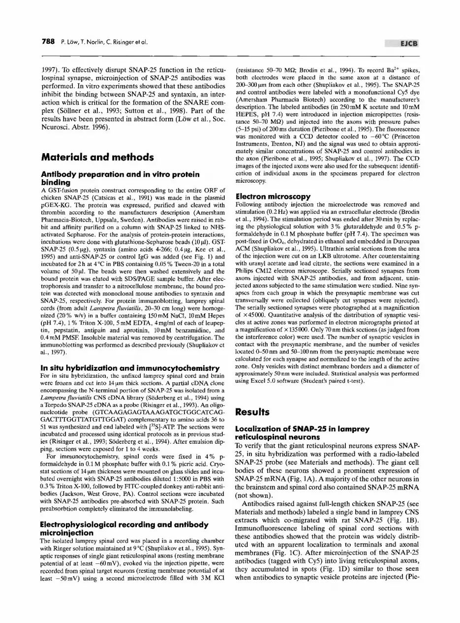
Localization of SNAP-25 in lamprey reticulospinal neurons To verify that the giant reticulospinal neurons express SNAP-25, in situ hybridization was performed with a radio-labeled SNAP-25 probe (see Materials and methods). The giant cell bodies of these neurons showed a prominent expression of SNAP-25 mRNA (Fig .. lA). A majority of the other neurons in the brainstem and spinal cord also contained SNAP-25 mRNA (not shown).
Antibodies raised against full-length chicken SNAP-25 (see Materials and methods) labeled a single band in lamprey CNS extracts which co-migrated with rat SNAP-25 (Fig. lB). Immunofluorescence labeling of spinal cord sections with these antibodies showed that the protein was widely distributed with an apparent localization to terminals and axonal membranes (Fig. Ie). After microinjection of the SNAP-25 antibodies (tagged with Cy5) into living reticulospinal axons, they accumulated in spots (Fig. lD) similar to those seen when antibodies to synaptic vesicle proteins are injected (Pie-

EJCB
o _~_-,- .. ~ ... - .- ~ -
Fig. 1. Localization of SNAP-25 in lamprey neurons. A. In situ hybridization with an oligonucleotide probe complementary to SNAP-25 on a section of the lamprey brainstem at the level of the Vth nerve motor nucleus (V). Labeling is present in two giant cell bodies of bulbar reticulospinal cells (arrows). Other labeled cells include smaller reticular neurons and motor neurons in the Vth nerve motor nucleus (upper left and right). B. Protein imJIlunoblot with SNAP-25 antibodies on extracts from lamprey spinal cord and rat brain. C. Section from the lamprey spinal cord incubated with SNAP-25 antibodies followed by fluorescein-conjugated antibodies. D. CCD image of a living giant reticulospinal axon after injection of Cy5-conjugated SNAP-25 antibodies. Note the accumulation of the labeling in spots, probably corresponding to synaptic release sites within the axon. Scale bars: A, C 100 !Am; D 10 !Am.
ribone et aI., 1995). The accumulation of SNAP-25 antibodies appeared, however, to be less distinct as compared to synapsin and synaptotagmin antibodies (Pieribone et aI., 1995). This is consistent with previous immunocytochemical studies showing that synapsin and synaptotagmin are almost exclusively restricted to synaptic release sites whereas SNAP-25 occurs along the axonal membrane with a relative accumulation at release sites (Boudier et aI., 1996; Duc and Catsicas, 1995; Garcia et aI., 1995).
Effect of SNAP-25 antibodies on protein binding in vitro We next tested whether the SNAP-25 antibodies would inhibit the binding between syntaxin and SNAP-25, which is essential for SNARE complex formation (Sutton et aI., 1998). When GST-SNAP-25 fusion protein was incubated with soluble syntaxin lacking the membrane-anchored C-terminal end (Kee et aI., 1995), an efficient binding between the two proteins
Perturbation of SNAP-25 in the lamprey synapse 789
SYNTAXIN
GST-SNAP25
2.5 0.12 0.25 0.62 1.25 2.5 J..Ig
IgG SNAP251gG
Fig. 2. Effects of antibodies to SNAP-25 in an in vitro protein binding assay. GST-SNAP-25 and syntaxin (amino acids 4-266) were incubated with glutathione-Sepharose and bound protein was analyzed with SDS/PAGE electrophoresis and immunoblotting. Increase in the amount of SNAP-25 antibodies (IgG) led to a decrease in the amount of bound syntaxin. The amount of GST-SNAP-25 bound to the Sepharose beads was not affected by the SNAP-25 antibodies. Bound syntaxin and GST-SNAP-25 were analyzed using monoclonal antibodies.
occurred. The binding was inhibited by addition of increasing concentrations of SNAP-25 antibodies (Fig. 2), whereas control IgGs had no detectable effect. The SNAP-25 antibodies did not affect the binding of GST-SNAP-25 to glutathioneSepharose. , Effect of SNAP-25 antibodies on transmiHer release The effect of the SNAP-25 antibodies on transmitter release was tested by recording excitatory postsynaptic potentials (EPSPs) in a spinal target neuron while stimulating a reticulospinal axon at low frequency (0.2 Hz) (Fig. 3). The reticulospinal EPSP consists of an initial electrotonic component, followed by a chemical component mediated by AMPA and NMDA receptors (Brodin et aI., 1994). Presynaptic injection of SNAP-25 antibodies produced a depression of the chemical EPSP, which coincided with the occurrence of antibody-linked fluorescence in the synaptic area (Fig. 3a, b). Within minutes, no chemical EPSP could be detected (n = 5), while the electrotonic component (Fig. 3b, arrow) was unaffected. To test if the inhibition could be relieved by repetitive stimulation (Dreyer and Schmitt, 1983; Molgo et aI., 1990; Capogna et aI., 1997; Owe-Larsson et aI., 1997) high-frequency impulse trains (20--40 Hz) were applied. No recovery of the chemical EPSP was observed under these conditions (Fig. 3c; n=4). The EPSP' amplitude in reticulospinal synapses can also be enhanced by injecting large depolarizing current pulses via the stimulating electrode, which cause a broadening of the presynaptic spike (Brodin et aI., 1994). Injection of such large current pulses (2-3 times the threshold for eliciting action potentials) did not lead to a detectable recovery of the chemical EPSP (n = 3; not illustrated).
In control experiments rabbit anti-mouse IgGs were injected. These antibodies had no effect on the EPSP amplitude (Fig. 3d, e), and they showed no accumulation in spots within the axons (not shown; for other controls, see Pieribone et aI., 1995; Shupliakov et aI., 1997). To test whether the SNAP-25 antibodies affected presynaptic calcium channels, the reticulospinal axon was impaled with two microelectrodes, one of which contained SNAP-25 antibodies. After bath application of Na+ and K+ channel blockers and a high concentra-

790 P. Low, T. Norlin, C. Risinger et 01.
a 1 -
4 8 time (min)
c
d
4 8 time (min)
2 _ 320
12
12
!' 'iii c CD ~
.5
280 ~ c CD
1il .. o " 240 ;:
340 ,., Cij c .. :;
300 .. u c .. 1il .. o " 260 :;:
b
e
\
o.5mvL 10ms
o.5mvL 10ms
Fig. 3. Inhibition of neurotransmitter release by SNAP-25 antibodies. a, b. Presynaptic microinjection of SNAP-25 antibodies. The plot in a shows the change in peak amplitude of the EPSP. The sweeps in b show averaged EPSPs sampled during the periods marked" 1 " and ,,2" in a. The EPSP was evoked in a spinal neuron by stimulating a giant reticulospinal axon at 0.2 Hz via a micro electrode filled with Cy5-conjugated SNAP-25 antibodies. The protein was injected during the first minute of the recording period. Grey bars in a represent the fluorescence intensity (in arbitrary units) in the area of the presynaptic axon where the release sites mediating the EPSP were located. After the injection of SNAP-25 antibodies, a strong depression of the chemical (1, 2), but not electrotonic (arrow) EPSP occurred. A polysynaptic component (asterisk) remained visible. This component is presumably elicited from synapses located outside the region of the axon containing SNAP-25 antibodies. c. The initial period of a high frequency train applied after the recording period displayed in a. No recovery of the chemical EPSP was visible. d, e. Control experiment in which irrelevant antibodies (rabbit anti-plOuse Ig,Gs) were injected during recording of a reticulospinal EPSP. These antibodies did not depress the chemical EPSP. Designations.as in a, b;
tion of Ba2+, depolarizing steps elicited spikes (Fig. 4), which
reflect Ba2+ entry through presynaptic Ca2+ channels (Mac Vicar and Liinas, 1985; Shupliakov et aI., 1995). These Ba2+
potentials had a similar amplitude and shape after microinjection of SNAP-25 antibodies (Fig. 4; n = 4), indicating that the depression of transmitter release was not due to inhibition of presynaptic calcium entry.
Effects of SNAP-25 antibodies on the synaptic ultrastructure To examine the organization of synaptic vesicles after antibody blockade, axons were injected with SNAP-25 antibodies and thereafter stimulated as above (0.2 Hz) for 30 min. For
EJCB
Control SNAP-25 antibodies
! ;~ J ~ 120 mV
J
J I J I 140 nA
-100 mS
Fig. 4. Lack of effect of SNAP-25 antibodies on presynaptic Ba2+ potentials. In the control recording (left panel) a series of depolarizing current pulses (lower traces) was given via one microelectrode, and the response was measured with a second microelectrode placed 200 f.lm away in the same axon. The largest two pulses evoked depolarizing potentials due to Ba2+ entry through Ca2+ channels (MacVicar and L1inas, 1985). A similar response was evoked after the injection of SNAP-25 antibodies (right panel, 15 min), indicating that presynaptic Ca2+
channels remained functional. The extracellular solution contained 30mM Ba2+, 4mM 4-AP, 15 mM TEA, and 1.5f.lMTTX.
comparison, synapses from adjacent uninjected axons which had received the same stimulation were examined. The general organization of synaptic vesicles did not differ between the two groups of synapses (Fig. 5a, b). Thus, large densely packed vesicle clusters were present at the active zones in both cases. A difference was, however, noted with regard to endocytic intermediates. As in previous studies, clathrin-coated pits were observed at the plasma membrane around the release sites in control axons (arrowhead in Fig. 5a; for quantitative data see Shupliakov et aI., 1997). In antibody-injected axons no coated pits were detected, consistent with an inhibition of synaptic vesicle cycling.
To examine in detail the distribution of synaptic vesicles in the proximity of the active zone membrane, electron micrographs taken at higher magnification (Fig. 6a, b), were subjected to' two types of quantitative analysis. We first counted the number of synaptic vesicles in apparent contact with the presynaptic plasma membrane of active zones ("membrane-contacting vesicles"). Vesicles were included in this group if no gap between the presynaptic membrane and that of the transversely cut vesicle could be distinguished at a magnification of X 135000. The number of such membrane-contacting vesicles did not differ between synapses in control and injected axons (Fig. 6c; p>0.05; t-test, n =9). We then counted the number of vesicles of which a major part (i.e. more than half of the vesicle diameter) fell within a line drawn at a given distance from the plasma membrane. In the region 0-50 nm from the presynaptic membrane the number of synaptic vesicles was sJightlylarger in antibody-injected synapses than in control synapses (Fig. 6c; p<0.005). In the region between 50-100nm, however, the number of vesicles did not differ between the two groups (p>0.05). Thus, inhibitory SNAP-25 antibodies has only subtle effects on the distribution of synaptic vesicles.
Discussion
In the present study polyclonal antibodies were used to disrupt the function of SNAP-25 in the lamprey reticulospinal synapse. The antibodies, which inhibited the in vitro binding between SNAP-25 and syntaxin, suppressed synaptic

EJCB
A a '. -,
" '.
B a
Fig. 5. Electron micrographs of reticulospinal synapses from a control axon (A) and an axon injected with SNAP-25 antibodies (B) from the same spinal cord. Both axons were stimulated at 0.2 Hz for 30 min until the activity was stopped by the fixation . Note the presence of a coated pit (arrowhead) lateral to the synaptic vesicle cluster in the control synapse. a axoplasmic matrix, d dendrite, s synaptic vesicles. Scale bar: A, B: 0.5 f!m.
responses without causing a detectable inhibition of the presynaptic calcium entry. At the ultrastructural level, no major alteration in the synaptic organization was observed, apart from an absence of clathrin-coated endocytic intermediates.
The binding between SNAP-25 and syntaxin is essential for the high affinity interaction of these proteins with VAMP (Chapman et aI., 1994; Pevsner et aI., 1994; Sutton et aI., 1998), which indicates that the SNAP-25 antibodies prevent the formation of the ternary SNARE complex. Botulinum toxins (A and E) which cleave off peptide fragments from the C-terminal of SNAP-25 do not prevent the SNAP-25-syntaxin interaction and the ternary complex can still form, although its stability is reduced (Otto et aI., 1995; Pellegrini et aI., 1995; Chen et aI. , 1999). Thus, the SNAP-25 antibodies used in the present experiments are likely to cause a more complete disruption of SNARE interactions than Botulinum toxins cleaving the C-terminal of SNAP-25. Whether other interactions involving SNAP-25 (Bean et aI., 1997; Kim and Catterall,
C QI c: 0 N
QI
~ 0 ..,
} '" QI
]
'" QI >
~ .0 E ::J c:
20
15
10
5
o
Perturbation of SNAP-25 in the lamprey synapse 791
d -. • SNAP-25 antibody inject ion
o Cont rol
docked
.1
0-50 nmfrom the presynaptic membrane
50-100 nm from the presynaptic membrane
Fig. 6. Organization of synaptic vesicles at -the presynaptic plasma membrane in a control synapse (A) and in a synapse in an axon injected with SNAP-25 antibodies (B) . The electron micrographs show ultrathin sections through the active zone of reticulospinal synapses from the same spinal cord preparation stimulated at 0.2Hz for 30 min until fixation. Arrowheads point to vesicles in contact with the presynaptic membrane (morphologically "docked" vesicles). d postsynaptic dendrite , s synaptic vesicles. Scale bar: A, B: 0.1 f!m. (C) Quantitative distribution of synaptic vesicles at active zones in control synapses and in synapses injected with SNAP-25 antibodies. The histograms represent normalized values for the number of synaptic vesicles in the areas indicated (n = 9 synapses in each group; ± SEM). The numbers of synaptic vesicles differed significantly only in the region 0-50 nm from the presynaptic membrane (* indicates significance at <0.05) .
1997; Schiavo et aI., 1997) were also affected by the antibodies is presently unclear. In previous electrophysiological studies Botulinum toxin A (and in some cases also E) have been shown to produce a "mild" inhibition of synaptic transmission, which can be relieved by various treatments that facilitate release (Dreyer and Schmitt, 1983; Molgo et aI., 1990;

792 P. Low, T. Norlin, C. Risinger et 01.
Capogna et aI., 1997; Owe-Larsson et aI., 1997). After microinjection of SNAP-25 antibodies, no reversal of the inhibition was obtained by high frequency stimulation or injection of depolarizing current pulses. This indicates that the SNAP-25 antibodies cause an effective disruption of synaptic transmission, which appears to resemble that produced by toxins cleaving VAMP and syntaxin (Dreyer and Schmitt, 1983; Molgo et aI., 1990; Capogna et aI., 1997; Broadie et aI., 1995; Marsal et aI., 1997; Owe-Larsson et aI., 1997). The absence of clathrincoated pits in antibody-injected synapses further supports the effectiveness of the antibody inhibition. Our previous studies have shown that clathrin-coated pits are virtually absent in resting axons (maintained in low Ca2+ solution), whereas a significant number is induced by low frequency action potential stimulation (Shupliakov et aI., 1997).
The hypothesis that SNARE interactions mediate the targeting of synaptic vesicles to the plasma membrane (So lIner et aI., 1993) has not been supported by previous studies using invertebrate synapses (Hunt et aI., 1994; Broadie et aI., 1995; Marsal et aI., 1997; O'Connor et aI., 1997; Sugimori et aI., 1998). In the present study, we found no change in the number of vesicles in apparent morphological contact with the plasma membrane ("docked" vesicles) after SNAP-25 antibody injection. When the number of synaptic vesicles located within a narrow region near the plasma membrane was counted, a modest increase was found. In the preceeding studies, relatively large increases in the number of membrane-adjacent vesicles were observed after perturbation of VAMP or syntaxin (Hunt et aI., 1994; Broadie et aI., 1995; Marsal et aI., 1997; Sugimori et aI., 1998; ct. also effects of synaptotagmin antibodies, Mikoshiba et aI., 1995). These findings are probably compatible with the present data, as the density of synaptic vesicles at the squid giant synapse is generally lower than in the lamprey reticulospinal synapse (Shupliakov et aI., 1992). Thus, if fusion but not mobilization of synaptic vesicles is blocked, an accumulation of vesicles may occur in these systems, whereas in the lamprey synapse the vesicles are already densely packed under normal conditions. In one study in squid no accumulation of vesicles near the plasma membrane was observed after perturbation of syntaxin (O'Connor et aI., 1997). The reason for this divergent result is presently unclear (see Discussion in O'Connor et aI., 1997).
A consistent finding in all studies of the effects of perturbing SNAREs in intact synapses is that transmitter release is inhibited whilst the number of vesicles near the plasma membrane is not reduced. This indicates that the function of the SNARE complex is exclusively linked to the exocytic reaction. In vitro studies in different model systems are all consistent with a role of SNAREs in membrane fusion (Hanson et aI., 1997; Robinson and Martin, 1998). However, whereas studies of yeast vacuole fusion indicate an action prior to fusion (Ungermann et aI., 1998), studies using cracked PC12 cells favor a direct role in the Ca2+ -triggered fusion process (Chen et aI., 1999, see also Banerjee et aI., 1996; Avery et aI., 1999).
The mechanisms underlying the targeting of synaptic vesicles to the plasma membrane are still enigmatic. Ultrastructural studies of synaptic release sites have shown that the area in the vicinity of the plasma membrane is occupied by a filamentous network (Landis et aI., 1988; Hirokawa et aI., 1989). At least three proteins, Piccolo, Bassoon and Rim (Cases-Langhoff et aI., 1996; Wang et aI., 1997; Dieck et aI., 1998), appear to be enriched in this area, suggesting possible roles in controlling the rapid and precise movement of synaptic vesicles to the plasma membrane.
EJCB
Acknowledgements. This work was supported by the Swedish Medical Research Council (proj. no. 11287, L. Brodin), the NIH (grant R29NS35941 to V. A. Pieribone) the Natural Sciences Research Council (D. Larhammar), and Petrus and Augusta Hedlunds Stiftelse (L. Brodin). We thank R. H. Scheller, G. Schiavo and J. E. Rothman for providing DNA constructs, and B. Meister for providing monoclonal antibodies. We also thank A. El-Manira and D. Parker for comments on the manuscript, and M. Bredmyr and H. Axegren for technical assistance.
References Augustine, G. J., Burns, M.E., DeBello, W.M., Pettit, D.L.,
Schweitzer, F. E. (1996): Exocytosis: proteins and perturbations. Annu. Rev. Pharmacol. Toxicol. 36, 659-701.
Avery, J., Jahn, R., Edwardson, J. M. (1999): Reconstitution of regulated exocytosis in cell-free systems: A critical appraisal. Annu. Rev. Physiol. 61, 777-807.
Banerjee, A., Kowalchyk, J. A., DasGupta, B. R., Martin, T. F. J. (1996): SNAP-25 is required for a late post docking step in Ca2+ exocytosis. J. BioI. Chern. 271, 20227-20230.
Bean, A. J., Seifert, R., Chen, Y. A., Sacks, R., Scheller, R. H. (1997): Hrs-2 is an ATPase implicated in calcium-regulated secretion. Nature 385,826--829.
Blasi, J., Chapman, E. R., Link, E., Binz, T., Yamasaki, S., De Camilli, P., Siidhof, T. C., Niemann, H., Jahn, R. (1993): Botulinum neurotoxin A selectively cleaves the synaptic protein SNAP-25. Nature 365,160--163.
Borst, J. G. G., Sakmann, B. (1996): Calcium influx and transmitter release in a fast CNS synapse. Nature 383, 431-434.
Boudier, J., Charvin, N., Boudier, J., Fathallah, M., Tagaya, M., Takahashi, M., Seagar, M. J. (1996): Distribution of components of the SNARE complex in relation to transmitter release sites at the frog neuromuscular junction. Eur. J. Neurosci. 8, 545-552.
Broadie, K., Prokop, A., Bellen, H.J., O'Kane, C.J., Schultze, K. L., Sweeney, S. T. (1995): Syntaxin and synaptobrevin function downstream of vesicle docking in Drosophila. Neuron 15, 663-673.
Brodin, L., Low, P., Gad, H., Gustafsson, J., Pieribone, V. A., Shupliakov, O. (1997): Sustained neurotransmitter release: new molecular clues. Eur. J. Neurosci. 9,2503-2511.
Brodin, L., Shupliakov, 0., Hellgren, J., Pieribone, V. A., Hill, R. H. (1994): The reticulospinal glutamate synapse in lamprey: plasticity and presynaptic variability. J. Neurophysiol. 72,592-604.
Capogna, M., McKinney, A., O'Connor, v., Gahwiler, B., Thompson, S. M. (1997): Ca2+ or Sr2+ partially rescues synaptic transmission in hippcampal cultures treated with Botulinum toxin A and C, but not Tetanus toxin. J. Neurosci. 17,7190-7202.
Cases-Langhoff, c., Voss, B., Garner, A.M., Appeltauer, u., Takei, K., Kindler, K., Veh, R.w., DeCamilli, R.w., Gundelfinger, E.D., Garner, C. C. (1996): Piccolo, a novel 420 kDa protein associated with the presynaptic cytomatrix. Eur. J. Cell BioI. 69,214--223.
Catsicas, S., Larhammar, D., Blomqvist, A., Sanna, P. P., Milner, R. J., Wilson, M. C. (1991): Expression of a conserved cell-typespecific protein in nerve terminals coincides with synaptogenesis. Proc. Natl. Acad. Sci. USA 88,785-789.
Chapman, E. R., Seong, A., Barton, N., Jahn, R. (1994): SNAP-25, a t-SNARE which binds to both syntaxin and synaptobrevin via domains that may form coiled coils. J. BioI. Chern. 269, 27427-27432.
Chen, Y. A., Scales, S. J., Patel, S. M., Doung, Y. C., Scheller, R. H. (1999): SNARE complex formation is triggered by Ca2+ and drives membrane fusion. Cell 97, 165-174.
De Camilli, P., Takei, K. (1996): Molecular mechanisms in synaptic vesicle endocytosis and recycling. Neuron 16, 481-486.
Dreyer, F., Schmitt, A. (1983): Transmitter release in tetanus and botulinum A toxin-poisoned mammalian motor endplates and its dependence on nerve stimulation and temperature. Pfliigers Arch. 399, 228--234.
Duc, C., Catsicas, S. (1995): Ultrastructural localization of SNAP-25 within the rat spinal cord and peripheral nervous system. J. Compo Neurol. 356, 152-163.

EJCB
Fernandez-Chacon, R., Siidhof, T. C. (1999): Genetics of synaptic vesicle function: Toward the complete functional anatomy of an organelle. Annu. Rev. Physiol. 61,753-776.
Gad, H., Low, P., Zotova, E., Brodin, L., Shupliakov, O. (1998): Dissociation between Ca2+ -evoked synaptic vesicle exocytosis and clathrin-mediated endocytosis at a central vertebrate synapse. Neuron 21, 607-616.
Garcia, E. P., McPherson, P., Chilcote, T. J., Takei, K., De Camilli, P. (1995): RbSec1a and b colocalize with syntaxin 1 and SNAP-25 throughout the axon, but are not in a stable complex with syntaxin. J. Cell BioI. 129, 105-120.
Hanson, P. I., Heuser, J. E., Jahn, R. (1997): Neurotransmitter release - four years of SNARE complexes. Curro Opin. Neurobiol. 7, 310-315.
Hirokawa, N., Sobue, K., Kanda, K., Harada, A., Yorifuji, H. (1989): The cytoskeletal architecture of the presynaptic terminal and molecular structure of synapsin I. J. Cell BioI. 108, 111-126.
Hunt, J. M., Bommert, K., Chariton, M. P., Kistner, M., Habermann, E., Augustine, G. J., Betz, H. (1994): A post-docking role for synaptobrevin in synaptic vesicle fusion. Neuron 12, 1269-1279.
Kee, Y., Lin, R. S., Hsu, S. C., Scheller, R. H. (1995): Distinct domains of syntaxin are required for synaptic-vesicle fusion complex formation and dissociation. Neuron 14, 991-998.
Kim, D. K., Catterall, W.A. (1997): Caz+-dependent and -independent interactions of the isoforms of the alA subunit of brain Ca2+ channels with presynaptic SNARE proteins. Proc. Natl. Acad. Sci. USA 94, 14782-14786.
Krieger, P., EI-Manira, A., Grillner, S. (1996): Activation of pharmacologically distinct metabotropic glutamate receptors depresses reticulospinal-evoked monosynaptic EPSPs in the lamprey spinal cord. J. Neurophysiol. 76, 3834-3841.
Landis, D. M., Hall, D., Weinstein, A. K., Reese, T. S. (1988): The organization of cytoplasm at the presynaptic active zone of a central nervous system synapse. Neuron 1, 201-209.
Llinas, R., Sugimori, M., Silver, R.B. (1995): Time resolved calcium microdomains and synaptic transmission. J. Physiol. (Paris) 89, 77-81.
MacVicar, B. A., Llinas, R. (1985): Barium action potentials in regenerating axons of the lamprey spinal cord. J. Neurosci. Res. 13, 323-335.
Marsal, J., Ruiz-Montazell, B., Blasi, J., Moreira, J., Contreras, D., Sugimori, M., Llinas, R. (1997): Block of transmitter release by Botulinum Cl.action on syntaxin at the squid giant synapse. Proc. Natl. Acad. Sci. USA 94, 14871-14876.
Mikoshiba, K., Fukuda, M., Moreira, J. E., Lewis, EM., Sugimori, M., Niinobe, M., Llinas, R. (1995): Role of the C2Adomain ofsynaptotagmin in transmitter release as determined by specific antibody injection into the squid giant synapse preterminal. Proc. Natl. Acad. Sci. USA 92,10703-10707.
Molgo, J., Comella, J. X, Angaut-Petit, D., Pecot-Dechavassine, M., Tabti, N., Faille, L., Mallart, A., Thesleff, S. (1990): Presynaptic actions of botulinal neurotoxins at vertebrate neuromuscular junctions. J. Physio!. (Paris) 84,152-166.
O'Connor, V., Heuss, C., De Bello, W. M., Dresbach, T., Charlton, M. P., Hunt, J. H., Pellegrini, L. L., Hodel, A., Burger, M. M., Betz, H., Augustine, G. J., Schafer, T. (1997): Disruption of syntaxin-mediated protein interactions blocks neurotransmitter secretion. Proc. Natl. Acad. Sci. USA 94, 12186-12191.
Otto, H., Hanson, P.I., Chapman, E. R., Blasi, J., Jahn, R. (1995): Poisoning by botulinum neurotoxin A does not inhibit formation or disassembly of the synaptosomal fusion complex. Biochem. Biophys. Res. Commun. 212, 945-952.
Owe-Larsson, B., Kristensson, K., Hill, R. H., Brodin, L. (1997): Distinct effects of Clostridial toxins on activity-dependent modulation of autaptic responses in cultured hippocampal neurons. Eur. J. Neurosci. 9, 1773-1777.
Perturbation of SNAP-25 in the lamprey synapse 793
Pellegrini, L. L., O'Connor, V., Lottspeich, E, Betz, H. (1995): Clostridial neurotoxins compromise the stability of a low energy SNARE complex mediating NSF activation of synaptic vesicle fusion. EMBO J. 14,4705-4713.
Pevsner, J., Hsu, S. C., Braun, J. E., Calakos, N., Ting, A. E., Bennett, M. K., Scheller, R. H. (1994): Specificity and regulation of a synaptic vesicle docking complex. Neuron 13, 353-361.
Pieribone, V. A., Shupliakov, 0., Brodin, L., Hilfiker-Rothenfluh, S., Czernik, A. J., Greengard, P. (1995): Distinct pools of synaptic vesicles in neurotransmitter release. Nature 375, 493-497.
Risinger, c., Blomqvist, A., Lundell, I., Lambertsson, A., Nassel, D., Pieribone, V. A., Brodin, L., Larhammar, D. (1993): Evolutionary conservation of synapse protein SNAP-25 shown by Drosophila and Torpedo cDNA clones. J. BioI. Chern. 268,24408-24414.
Robinson, L. J., Martin, T. E J. (1998): Docking and fusion in neurosecretion. Curro Opin. Cell Bio!. 10,483-492.
Schiavo, G., Stenbeck, G., Rothman, J. E., Sollner, T. H. (1997): Binding of the synaptic vesicle v-SNARE, synaptotagmin, to the plasma membrane t-SNARE, SNAP-25, can explain docked vesicles at neurotoxin-treated synapses. Proc. Nat!. Acad. Sci. USA 94, 997-1001.
ShupIiakov, 0., Brodin, L., Cullheim, S., Ottersen, O. P., StormMathisen, J. (1992): Immunogold quantification of glutamate in two types of excitatory synapse with different firing patterns. J. Neurosci. 12, 3789-3803.
Shupliakov, 0., Pieribone, V. A., Gad, H., Brodin, L. (1995): Synaptic vesicle depletion in reticulospinal axons is reduced by 5-HT: direct evidence for presynaptic modulation of glutamatergic transmission. Eur. J. Neurosci. 7, 1111-1116.
ShupIiakov, 0., Low, P., Grabs, D., Gad, H., Chen, H., David, c., Takei, K., De Camilli, P., Brodin, L. (1997): Synaptic vesicle endocytosis impaired by disruption of dynamin-SH3 domain interactions. Science 276, 259-263.
Soderberg, C., Pieribone, V. A., Dahlstrand, J., Brodin, L., Larhammar, D. (1994): Neuropeptide role of both peptide YY and neuropeptide Y in vertebrates suggested by abundant expression of both mRNAs in a cyclostome brain. J. Neurosci. Res. 37, 633-640.
Sollner, T., Whiteheart, S. W., Brunner, M., Erdjument-Bromage, H., Geromanos, S., Tempst, P., Rothman, J. E. (1993): SNAP receptors implicated in vesicle targeting and fusion. Nature 362, 318-324.
Sugimori, M., Tong, C.-K., Fukuda, M., Moreira, J.E., Kojima, T., Mikoshiba, K., Llinas, R. (1998): Presynaptic injection of syntaxinspecific antibodies blocks transmission in the squid giant synapse. Neuroscience 86, 39-51.
Sutton, R. B., Fasshauer, D., Jahn R., Brunger, A. T. (1998): Crystal structure of a SNARE complex involved in synaptic exocytosis at 2.4 angstrom resolution. Nature 395, 347-353.
Tom Dieck, S., Sanmarti-Vila, L., Langnaese, K., Richter, K., Kindler, S., Soyke, A., Wex, H., Smalla, K. H., Kampf, u., Franzer, J. T., Stumm, M., Garner, C. C., Gundelfinger, E. D. (1998): Bassoon, a novel zinc-finger CAG/glutamine-repeat protein selectively localized at the active zone of presynaptic nerve terminals. J. Cell BioI. 142, 499-509.
Ungermann, C., Sato, K., Wickner, W. (1998): Defining the functions of trans-SNARE pairs. Nature 396, 543-548.
Wang, Y., Okamoto, M., Schmitz, E, Hofmann, K., Siidhof, T. C. (1997): Rim is a putative Rab3 effector in regulating synaptic-vesicle fusion. Nature 388, 593-598.
Zucker, R. (1996): Exocytosis: a molecular and physilogical perspective. Neuron 17, 1049-1055.