TRANSDUKSI SINYAL, SYNAPS DAN NEUROTRANSMITTER
61
TRANSDUKSI SINYAL, SYNAPS DAN NEUROTRANSMITTER Rahmatina B. Herman Fakultas Kedokteran - Unand
-
Upload
kompakbodoh -
Category
Documents
-
view
0 -
download
0
Transcript of TRANSDUKSI SINYAL, SYNAPS DAN NEUROTRANSMITTER

TRANSDUKSI SINYAL, SYNAPS
DAN
NEUROTRANSMITTER
Rahmatina B. Herman Fakultas Kedokteran - Unand

Neuron
Neuron is basic functional unit of nervous system
Central nervous system contains > 100 billion neurons
Neuron is composed of 3 major parts: - Soma : main body of neuron - Axon : a single projection from soma into a
peripheral nerve - Dendrite : great numbers of branching projections,
extend 1 mm into surrounding areas
The output signal travels by way of axon leaving the neuron, then has many separate branches to other parts of nervous system or peripheral body
Incoming signals enter the neuron through dendrites

Structure of a large neuron in brain

Typical anterior motor neuron

Myelinated fiber

Neuron…..
Neuron generally has 4 important functional zones:
1. A receptor or dendritic zone, where multiple local potential changes generated by synaptic connections are integrated
2. A site where propagated action potentials are generated, that is in initial segment of neurons or initial node of Ranvier
3. An axonal process that transmits propagated impulses to nerve endings
4. The nerve endings, where action potentials cause the release of synaptic transmitters

Neuron…..
The axons of many neurons are myelinated, and some are unmyelinated
The myelin sheath envelops the axon except at its ending and at nodes of Ranvier
In central nervous system (CNS) most neurons are myelinated
Lost of myelin (such as in multiple sclerosis) is associated with delayed or blocked conduction in the demyelinated axons

Axonal Transport
Neurons produce and secret transmitter, and secretory zone is at the end of axon
Proteins and polypeptides are synthesized in cell body, and tranported to axonal terminals by axoplasmic flow (orthograde transport)
Synaptic vesicles recycle in membrane, but some used vesicles are carried back to cell body (retrograde transport) and deposited in lysosomes
Some materials taken up at terminal by endocytosis, including nerve growth factor (NGF) are also transported back to cell body

Axonal transport along microtubules by dynein and kinesin

EXCITATION
AND
CONDUCTION

Basic Physics of Membrane Potential
Ion concentration difference on the 2 sides of cell membrane → diffusion → diffusion potential

Basic Physics of Membrane Potential…..
Sodium-Potassium (Na+-K+) pump → active transport

Feedback Control ion Channels
Positive feedback

Feedback Control ion Channels…..
Negative feedback

Nerve Action Potential
Neurons have a low threshold for excitation
Nerve signals are transmitted by action potentials
Action potentials are rapid changes in membrane potential that spread rapidly along nerve fiber membrane
Each action potential begins with a sudden change from normal resting negative membrane potential to positive potential and then ends with almost equally rapid change back to negative potential
To conduct a nerve signal, the action potential moves along the nerve fiber until it comes to the fiber’s end/terminal

Nerve Action Potential…..
The successive stages of action potentials are:
Resting stage (polarization): - Membrane potential before action potential begins
Depolarization stage: - Membrane suddenly becomes very permeable to Na+,
allowing tremendous number of positively charged Na ions to diffuse to interior axon
- May cause overshoot beyond zero level
Repolarization stage: - Soon then Na channels begin to close and K channels
open → rapid diffusion of K+ to exterior → re-establishes normal negative resting membrane potential


The changes in membrane potential (mV) and relative membrane permeability To Na+ and K+ during an action potential
hyperpolarization
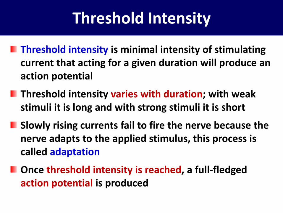
Threshold Intensity
Threshold intensity is minimal intensity of stimulating current that acting for a given duration will produce an action potential
Threshold intensity varies with duration; with weak stimuli it is long and with strong stimuli it is short
Slowly rising currents fail to fire the nerve because the nerve adapts to the applied stimulus, this process is called adaptation
Once threshold intensity is reached, a full-fledged action potential is produced
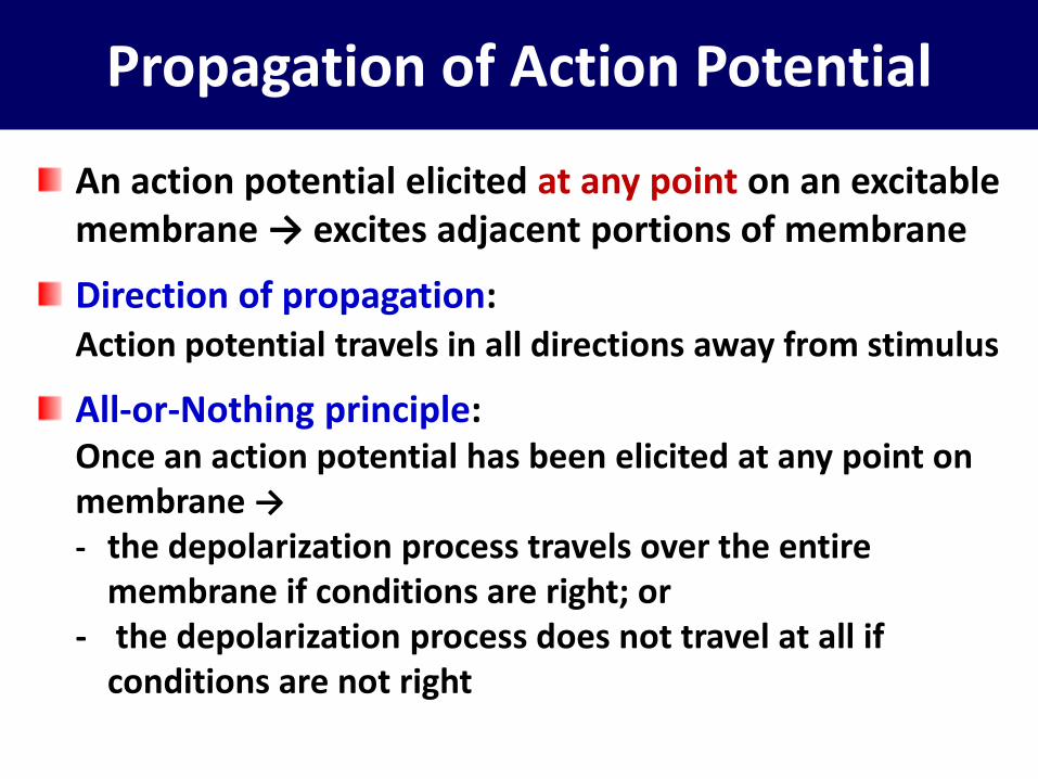
Propagation of Action Potential
An action potential elicited at any point on an excitable membrane → excites adjacent portions of membrane
Direction of propagation: Action potential travels in all directions away from stimulus
All-or-Nothing principle: Once an action potential has been elicited at any point on
membrane →
- the depolarization process travels over the entire membrane if conditions are right; or
- the depolarization process does not travel at all if conditions are not right

Propagation of action potentials in both directions

Electronic Potential and Firing Level
Although subthreshold stimuli do not produce an action potential, but still have effect on membrane potential → localized depolarizing potential change that rises sharply and decays exponentially with time. The magnitude will drops off rapidly as distance between stimulating is increased.
Anodal current produces a hyperpolarizing potential change of similar duration
These potential changes are called electronic potentials
If the firing level reached → an action potential occurs

Electronic potentials and local response

Signal Transmission in Nerve Fiber
Unmyelinated fiber: small fibers
Myelinated fiber: large fibers
Saltatory conduction: conduction from node of Ranvier to node of Ranvier
- No ions can flow through the thick myelin sheaths of myelinated fiber
- Ions can flow with ease through the nodes of Ranvier → action potentials are conducted from node to node

Saltatory conduction

SYNAPSE

Physiologic Anatomy of Synapse
Synapse is the junction point from one neuron to the
next
10,000-20,000 presynaptic terminals (minute synaptic
knob) lie on surfaces of:
- dendrites : 80-95%, and
- soma : 5-20%
Presynaptic terminals are the ends of nerve fibrils that
originate from many other neurons
Incoming signals enter the neuron through synapses
located on the neuronal dendrites and on cell body


Type of Synapses
1. Chemical synapse
- The first neuron secretes at its nerve ending chemical
substance, neurotransmitter, which in turn acts on
receptor proteins in membrane of the next neuron to
excite it; or inhibit it; or modify its sensitivity
- “One-way” conduction at chemical synapses
Signals always be transmitted from presynaptic
neuron that secretes neurotransmitter to
postsynaptic neuron on which neurotransmitter acts
→ signals can be directed toward specific goals

Type of Synapses…..
2. Electrical synapse
- Characterized by direct open fluid channels that conduct electricity from one neuron to the next
- Often transmit signals in either direction
- Most of these consist of small protein tubular structures: gap junctions, that allow free movement of ions from the anterior of one neuron to the next
- By way of gap junctions that action potentials are transmitted from one smooth muscle fiber to the next, and from one cardiac muscle cell to the next

Information Transmission Through Synapse
Information is transmitted in nervous system mainly in the form of nerve action potentials / nerve impulses
Each impulse in its transmission may be:
- facilitated or blocked from one neuron to the next; or
- changed from a single impulse into repetitive impulses; or
- integrated with impulses from other neurons to cause highly intricate patterns of impulses in successive neurons

Information Transmission Through Synapse…..
Synapses determine the directions that the nervous signals will spread through nervous system
Some synapses transmit signals from one neuron to the next with ease, whereas others with difficulty
Facilitatory and inhibitory signals from other areas in nervous system can control synaptic transmission
Sometimes opening for transmission, sometimes closing
Some postsynaptic neurons respond with large numbers of output impulses, others respond with only few

Synaptic Functions in Memory
Each time certain types of sensory signals pass through sequences of synapses, then:
- The synapses become more capable of transmitting the same type of signal the next time (facilitation)
- After the large number of times sensory signals passed through → the synapses become so facilitated
- The synapses can also cause transmission of impulses through the same sequences of synapses even when the sensory input is not excited → a perception of experiencing the original sensations, although the perceptions are only memories of the sensations
Precise mechanisms by which long-term facilitation of synapses occurs in the memory process are not known

Presynaptic Terminals
Presynaptic terminal is separated from postsynaptic
neuronal by synaptic cleft, width of 200-300 angstroms
There are 2 internal structures important to function of
synapse:
- Transmitter vesicles contain neurotransmitter, which
will be released into synaptic cleft and in turn will
either excites or inhibits postsynaptic neuron
- Mitochondria provide ATP which in turn supplies
energy for synthesizing new neurotransmitter

Presynaptic Terminals…..
When an action potential spreads over a presynaptic terminal, depolarization of its membrane causes a small number of vesicles to empty neurotransmitter into the cleft
The released neurotransmitter → immediate change in permeability characteristics of the postsynaptic neuronal membrane → excitation or inhibition of the postsynaptic neuron, depending on neuronal receptor characteristic

Transmitter Releasing Mechanisms
Presynaptic membrane contains large numbers of voltage-gated calcium channels
When action potential depolarizes presynaptic membrane → Ca channels open → large numbers of Ca ions flow into presynaptic terminal
Ca ions then bind with special protein molecules on the release sites of presynaptic membrane → release sites open → neurotransmitter released from vesicles into synaptic cleft

Transmitter Action on Postsynaptic Neuron
Postsynaptic membrane contains large number of receptor proteins which have 2 important components:
1. Binding component
2. Ionophore component:
- ion channel: > cation channel, or > anion channel
- second messenger activator to increase or decrease specific cellular function

Transmitter Action on Postsynaptic Neuron…..
Cation channel:
- Most often allowing Na ions to pass, also K ions and/or Ca ions → excite the neuron
- Transmitter that opens cation channels is excitatory transmitter
Anion channels:
- Allowing mainly Cl ions to pass, and minute quantities of other anion → inhibit the neuron
- Transmitter that opens anion channels is inhibitory transmitter

Physiologic anatomy of synapse

Second Messenger System in Postsynaptic Neuron
In the process of memory, require prolonged changes in neuron for seconds-months after initial transmitter substance is gone
Second messenger causes prolonged effect as follow:
- The ion channels are not suitable for causing prolonged postsynaptic neuronal changes because the channels close within milliseconds after transmitter substance is no longer present
- Prolonged postsynaptic neuronal excitation or inhibition is achieved by activating a second messenger inside postsynaptic neuronal cell itself

Second Messenger System in Postsynaptic Neuron…..
One of most common types of second messenger systems uses a group of proteins called G-proteins
G-protein is attached to the portion of receptor that protrudes into the interior of cell
G-protein consists of 3 components: - alpha (α): activator portion of G-protein - beta (β) and gamma (ϒ) are attached to α and also to
the inside of cell membrane adjacent to receptor protein
On activation by a nerve impulse, α portion separates from β and ϒ → α then is free to move within cytoplasm of cell

Second Messenger System in Postsynaptic Neuron…..
The free α performs one or more multi functions depending on specific characteristic of neuron type
The free α will allow 4 changes to be occurred: 1. Opening specific ion channels through postsynaptic cell
and stay open for a prolonged time 2. Activation of cAMP or cGMP which activate highly
specific metabolic machinery in neuron → long-term changes in cell structure → long-term excitability of neuron
3. Activation of one or more intracellular enzymes → specific chemical functions in cell
4. Activation of gene transcription → formation of new proteins within neuron → changing its metabolic machinery or its structure

Possible effects of G-protein

Mechanism of Excitation in Excitatory Receptors
1. Opening of Na channels → positive electrical charges flow to inside of postsynaptic neuron → potential membrane ↑ → excitatory
2. Depressed conduction through: - Cl channels → diffusion of negatively charged to inside ↓ - K channels → diffusion of positively charged to outside ↓
3. Various changes in internal metabolism of postsynaptic neuron to excite cell activity eg.
- the number of excitatory membrane receptors ↑, or - the number of inhibitory membrane receptors ↓
So, resting membrane potential of postsynaptic ↑ above resting or depolarization, and this membrane potential is called excitatory postsynaptic potential (EPSP)

Mechanism of Inhibition in Inhibitory Receptors
1. Opening of Cl channels → negative electrical charges flow to inside of postsynaptic neuron → negativity inside ↑ → inhibitory
2. Conductance of K ions out of postsynaptic neuron ↑ → diffusion of positive ions to exterior ↑ → negativity inside ↑ → inhibitory
3. Activation of receptor enzymes that inhibit cellular metabolic functions:
- the number of inhibitory membrane receptors ↑, or - the number of excitatory membrane receptors ↓
So, resting membrane potential of postsynaptic ↓, become more negative or hyperpolarization, and this membrane potential is called inhibitory postsynaptic potential (IPSP)


Special Characteristics of Synaptic Transmission
Fatigue of synaptic transmission
- When excitatory synapses are repetitively stimulated at a rapid rate → the number of discharges by postsynaptic neuron is very great at first, but the firing rate becomes progressively less in succeeding milliseconds or seconds
- The development of fatigue is a protective mechanism against excess neuronal activity
- The mechanism of fatigue is: > mainly exhaustion of the stores of transmitter
substance in presynaptic terminals > progressive inactivation of many of postsynaptic
membrane receptors > slow development of abnormal concentrations of ions
inside postsynaptic neuron

Special Characteristics of Synaptic Transmission…..
Synaptic delay Due to during signal transmission from presynaptic to
postsynaptic required certain amount of tome for:
1. Discharge of transmitter by presynaptic terminals
2. Diffusion of transmitter to postsynaptic neuron membrane
3. Action of transmitter on membrane receptor of postsynaptic
4. Action of receptor to increase membrane permeability
5. Inward diffusion of Na ion to raise the excitatory postsynaptic potential to a high enough level to elicit action potential
The minimal period of time required is ± 0.5 millisecond

Special Characteristics of Synaptic Transmission…..
Effect of changes in pH on synaptic transmission: - Alkalosis: ↑↑ neuronal excitability → cerebral epileptic
seizures attack - Acidosis: ↓↓ neuronal activity → can causes coma
Effect of oxygen supply on synaptic transmission: - Hypoxia: for only a few seconds → complete
inexcitability of some neurons → unconscious
Effect of drugs on synaptic transmission: - Caffeine, theophylline, theobromine ↑ neuronal
excitability by ↓ threshold for excitation, such as - Strychnine ↑ neuronal excitability by ↓↓ inhibitory
transmitter substances → severe tonic muscle spasms - Anesthetic drug ↑ neuronal threshold for excitation →
↓ synaptic transmission

NEUROTRANSMITTER

Type of Transmitter
Small molecule
Rapidly acting → acute responses
Transmission of sensory signals → brain → motor signals back
Neuropeptides
More prolonged actions
Long-term changes in numbers of neuronal receptors, long-term opening or closure of certain ion channels, long-term changes in numbers of synapses or size of synapses

Small Molecule
Synthesized in cytosol of presynaptic terminal and are absorbed by active transport into many transmitter vesicles in terminal
If action potential reaches presynaptic terminal → transmitter released into synaptic cleft → act on postsynaptic neuron
The vesicles that store and release small-molecule transmitters are continually recycled and used over and over again
After vesicles fuse with synaptic membrane and open to release transmitter, vesicle membrane becomes part of synaptic membrane
Within seconds-minutes vesicle portion of membrane invaginates back to inside of presynaptic terminal and pinches off to form a new vesicle
The new vesicular membrane still contains appropriate enzyme proteins or transport proteins required for synthesizing and/or concentrating new transmitter inside vesicle


Some of Small-Molecule Transmitter
Acetylcholine
- Secreted by neurons especially: > terminals of large pyramidal cells from motor cortex > neuron in basal ganglia > motor neurons that innervate skeletal muscles > preganglionic neuron of autonomic nervous system > postganglionic of parasympathetic nervous system > some of postganglionic sympathetic nervous system
- Most ACh has excitatory effect, however it has inhibitory effects at some peripheral parasympathetic nerve endings such as inhibition of heart by nervus vagus

Some of Small-Molecule Transmitter…..
Norepinephrine
- Secreted by terminals of many neurons whose cell bodies are located in brain stem and hypothalamus
- Specifically NE secreting neurons located in locus cereleus in pons send nerve fibers to widespread areas of brain to help control overall activity and mood of mind, such as increasing level of wakefulness
- In most of these areas, NE probably activates excitatory receptors, but in a few area it activates inhibitory receptors
- NE is also secreted by most postganglionic neurons of sympathetic nervous system

Some of Small-Molecule Transmitter…..
Dopamine - Secreted by neurons that originate in substantia nigra
- Effect of dopamine is usually inhibition
Glycine - Secreted mainly at synapses in spinal cord
- It is believed to always act as an inhibitory transmitter
GABA (gamma-aminobutyric acid) - Secreted by nerve terminals in spinal cord, cerebellum,
basal ganglia, and many areas of cortex
- It is believed always to cause inhibition

Some of Small-Molecule Transmitter…..
Glutamate - Secreted by presynaptic terminals in many of sensory
pathways entering CNS, in many areas of cerebral cortex
- Probably always causes excitation
Serotonin - Secreted by nuclei that originate in brain stem and project
to many areas in brain and spinal cord, especially to dorsal horns of spinal cord and to hypothalamus
- Acts as an inhibitor of pain pathways in spinal cord
- Inhibitor action in higher regions of nervous system is believed to help control mood, perhaps to cause sleep

Some of Small-Molecule Transmitter…..
Nitric oxide - Secreted by nerve terminals in areas of brain responsible
for long-term behavior and for memory
- Has different mechanism of formation in presynaptic and its actions on postsynaptic with other small-molecule
- It is not performed and stored in vesicles
- It is synthesized almost instantly as needed → diffuses out of presynaptic terminals over a period of seconds → diffuses into postsynaptic neuron nearby
- It postsynaptic neuron, it is usually does not greatly alter membrane potential, but changes intracellular metabolic functions that modify neuronal excitability for seconds, minutes, or perhaps even longer

Neuropeptides
Entirely different class from small-molecule
Synthesized as integral parts of large-protein molecules by ribosomes in neuron and transported in vesicles
After releasing the transmitter in response to action potential, the vesicle is autolyzed and not reused
The amount of transmitter in each releasing much smaller quantities than small-molecule transmitter
Generally ≥ 1000 more times as potent as small-molecule transmitter and much prolonged actions, last for days, months or years