The Effects of the Toxic Cyanobacterium Limnothrix (Strain AC0243) on Bufo marinus Larvae
15
Toxins 2014, 6, 1021-1035; doi:10.3390/toxins6031021 toxins ISSN 2072-6651 www.mdpi.com/journal/toxins Article The Effects of the Toxic Cyanobacterium Limnothrix (Strain AC0243) on Bufo marinus Larvae Olivia Daniels *, Larelle Fabbro and Sandrine Makiela School of Medical and Applied Sciences, Central Queensland University, Rockhampton 4701, Australia; E-Mails: [email protected] (L.F.); [email protected] (S.M.) * Author to whom correspondence should be addressed; E-Mail: [email protected]; Tel.: +61-7-4930-6775; Fax: +61-7-4930-9209. Received: 31 October 2013; in revised form: 17 February 2014 / Accepted: 20 February 2014 / Published: 6 March 2014 Abstract: Limnothrix (strain AC0243) is a cyanobacterium, which has only recently been identified as toxin producing. Under laboratory conditions, Bufo marinus larvae were exposed to 100,000 cells mL −1 of Limnothrix (strain AC0243) live cultures for seven days. Histological examinations were conducted post mortem and revealed damage to the notochord, eyes, brain, liver, kidney, pancreas, gastrointestinal tract, and heart. The histopathological results highlight the toxicological impact of this strain, particularly during developmental stages. Toxicological similarities to β-N-Methylamino-L-alanine are discussed. Keywords: Limnothrix; Geitlerinema; toxic; cyanobacteria; histology; Bufo marinus; notochord; brain; liver; pancreas; BMAA 1. Introduction Limnothrix (strain AC0243) specific research is in its infancy due to its toxicity only recently being discovered. The strain shares intrinsic morphological characteristics with Geitlerinema species with field samples often identified as G. unigranulatum (R.N.Singh) J.Komárek and M.T.P.Azevedo or G. amphibium (C.Agardh ex Gomont) Anagnostidis. However, isolates of this strain have been genetically matched to Limnothrix redekei (Van Goor) Meffert with a 99% similarity, using 16s rRNA gene sequencing analysis [1]. OPEN ACCESS
Transcript of The Effects of the Toxic Cyanobacterium Limnothrix (Strain AC0243) on Bufo marinus Larvae
Toxins 2014, 6, 1021-1035; doi:10.3390/toxins6031021
toxins
ISSN 2072-6651 www.mdpi.com/journal/toxins
Article
The Effects of the Toxic Cyanobacterium Limnothrix (Strain AC0243) on Bufo marinus Larvae
Olivia Daniels *, Larelle Fabbro and Sandrine Makiela
School of Medical and Applied Sciences, Central Queensland University, Rockhampton 4701,
Australia; E-Mails: [email protected] (L.F.); [email protected] (S.M.)
* Author to whom correspondence should be addressed; E-Mail: [email protected];
Tel.: +61-7-4930-6775; Fax: +61-7-4930-9209.
Received: 31 October 2013; in revised form: 17 February 2014 / Accepted: 20 February 2014 /
Published: 6 March 2014
Abstract: Limnothrix (strain AC0243) is a cyanobacterium, which has only recently been
identified as toxin producing. Under laboratory conditions, Bufo marinus larvae were
exposed to 100,000 cells mL−1 of Limnothrix (strain AC0243) live cultures for seven days.
Histological examinations were conducted post mortem and revealed damage to the
notochord, eyes, brain, liver, kidney, pancreas, gastrointestinal tract, and heart. The
histopathological results highlight the toxicological impact of this strain, particularly
during developmental stages. Toxicological similarities to β-N-Methylamino-L-alanine
are discussed.
Keywords: Limnothrix; Geitlerinema; toxic; cyanobacteria; histology; Bufo marinus;
notochord; brain; liver; pancreas; BMAA
1. Introduction
Limnothrix (strain AC0243) specific research is in its infancy due to its toxicity only recently being
discovered. The strain shares intrinsic morphological characteristics with Geitlerinema species with
field samples often identified as G. unigranulatum (R.N.Singh) J.Komárek and M.T.P.Azevedo or
G. amphibium (C.Agardh ex Gomont) Anagnostidis. However, isolates of this strain have been
genetically matched to Limnothrix redekei (Van Goor) Meffert with a 99% similarity, using 16s rRNA
gene sequencing analysis [1].
OPEN ACCESS
Toxins 2014, 6
1022
Limnothrix (strain AC0243) has been shown to produce a water-soluble cytotoxin(s) that inhibits
protein synthesis while independently reducing adenosine triphosphate (ATP) concentrations [1,2]. The
protein synthesis inhibition displayed by this strain showed similarities to cylindrospermopsin (CYN).
However, high performance liquid chromatography photo diode array (HPLC-PDA) and liquid
chromatography mass spectrometry (LC-MS) failed to detect CYN and deoxy-CYN associated with
this material [1]. Extracts of the strain administered to mice via intraperitoneal (IP) injections resulted
in heightened sensitivity and hind limb weakness, suggesting the possibility of neurotoxin exposure [2].
The most common cyanobacterial neurotoxins are anatoxin-a, anatoxin-a(s), and its analogues
homoanatoxin-a, saxitoxin, and β-N-Methylamino-L-alanine (BMAA) [3–7]. Of these, the neurotoxic
amino acid BMAA has been detected in many cyanobacterial species [5,8–10] and has been implicated
in neurological diseases, such as motor neuron disease, amyotrophic lateral sclerosis/Parkinson’s
dementia complex, and Alzeimer’s disease [8,11]. BMAA is a mixed receptor agonist, which impacts
energy and amino acid metabolism in neonates [8,10,12–17]. Of note is the similarity in modification
of energy metabolism produced as a result of exposure of a Vero cell line to Limnothrix (AC0243) and
that produced in neonatal rats following exposure to BMAA, particularly with respect to the decrease
in production of ATP [2,13].
The impacts of toxic cyanobacteria have been well documented in terms of fish and mammalian
species [18–26]. However, the toxigenic effects on anurans are seldom reported. Elements of the
anuran lifestyle are highly reliant upon water as a support medium, source of food, and source of water
for hydration. Although mature anurans generally adapt a terrestrial lifestyle, they rely on water bodies
for cutaneous water absorption as their only means for hydration [27–31]. Cutaneous water absorption
may provide a mode of exposure to cyanotoxins, promoting morbidities or mortalities in exposed
animals [32,33]. The larval stage of most species is spent entirely in the water often grazing on plant
matter and cyanobacteria [34–36]. Larvae, also absorbing water cutaneously, present ideal subjects for
examining the effects of normal routes of exposure to Limnothrix (strain AC0243) that may occur in
their natural habitat.
The benefit of using anuran larvae as test specimens introduces the opportunity to examine the
toxigenic effects of this strain on development stages, particularly stages where at notochord is present.
The notochord is critical for the formation of muscle tissue and skeletal development as well as acting
as a signaling source for the development of other tissues [37–41]. Anuran notochord damage resulting
from cyanotoxin exposure has not been well represented in current literature although development
abnormalities have been noted [42,43]. Such exposure requires further investigation as toxic blooms
are rarely considered in terms of further impacting anuran communities and populations.
1.1. Aim of the Experiments
This study examines the effects of whole cells (live cultures) of toxin producing Limnothrix
(strain AC0243) (100,000 cells mL−1) on anuran larvae, using Bufo marinus as a representative
species. This research was aimed at further understanding the potential impact of exposure on an early
developmental, stage as well as the environmental consequences of Limnothrix (strain AC0243) blooms.
Toxins 2014, 6
1023
1.2. Animal Ethics Committee Approval
Central Queensland University Rockhampton Animal Ethics Committee (AEC) approval (approval
number A10/05-259) was obtained prior to the commencement of the experiments. All experiments
were conducted in accordance with the Australian Code of Practice for the Care and Use of Animals
for Scientific Purposes [44].
2. Results and Discussion
2.1. Water Quality Testing
Results for water quality variables in both the control and treatment groups were similar for
dissolved oxygen, water temperature, and ammonia. No significant differences in dissolved
oxygen (% saturation) were established between the controls and the treatment group (Tamhene’s
Post Hoc test (p = 0.556)). Similarly, water temperatures did not significantly vary between treatments
(p = 0.994). The ammonia concentrations remained stable (<2 ppm) for both groups.
In contrast, the water quality results indicated that pH varied significantly between the controls and
the treatment (100,000 cells mL−1 Limnothrix (strain AC0243) live cultures) (Tamhene’s Post Hoc
test (p < 0.05)). Treatment flasks showed higher pH concentrations than the control flasks (Table 1).
Previous research has shown that growth of cyanobacteria in high concentrations may cause an
increase in the pH [45]. Drops in pH (pH < 6) have been shown to cause mortalities in anuran larvae
either directly, by having a toxic effect on biota or indirectly, by causing an increase in toxicity of
other chemicals or metals [46,47]. The guidelines for ‘normal’ pH range for tropical areas of Australia,
including Rockhampton are between pH 6.00 and 8.00, based on freshwater lakes and reservoirs in the
region [47]. Waters with pH 8 have not been shown to cause significant mortality risks to anuran
larvae [48] and breeding ponds of B. marinus are generally alkaline [49], suggesting that the pH in
these B. marinus experiments fell within a ‘normal’ range and had no direct effect on the animals.
However, it is unclear if the increased pH had any impact on the toxicity of Limnothrix (strain AC0243)
and can not be ruled out as an influencing factor.
2.2. Histology
Mortalities occurred in treatment animals (one on day one, and one on day three) resulting in the
planned fourteen-day experiment being reduced to seven days, with the surviving animals then
euthanized and processed for histology. No deaths occurred in the control group during this period.
Cellular damage was identified in the brain, liver, kidneys, pancreas, gastrointestinal tract, heart, lung
bud, eye, and gill tissues, as a result of exposure to Limnothrix (strain AC0243) live cultures (Table 2).
Toxins 2014, 6
1024
Table 1. Water quality means and standard deviations for controls and treatment flasks
containing 100,000 cells mL−1 of Limnothrix (strain AC0243) live cultures.
Treatment Variable Mean Std. deviation
Control Dissolved oxygen (% saturation) 33.23 8.27 pH 7.51 0.22 Water temperature (°C) 19.75 0.77 Ammonia concentration (ppm) <2 N/A
100,000 cells mL−1 Dissolved oxygen (% saturation) 35.39 6.66 pH 7.89 0.27 Water temperature (°C) 19.98 0.67 Ammonia concentration (ppm) <2 N/A
Table 2. Histopathologies of Bufo marinus larvae exposed to 100,000 cells mL−1 of
Limnothrix (strain AC0243) live cultures.
Body region Pyknotic
cells Eosinophilic
cells Necrotic
cells Karyorrhexis
Inflammatory cells
Tissue vacuolation
Eyes + Brain + + + + + +
Ventricle + Gill filaments + +
Kidneys + Lung bud +
Liver + + + Pancreas + + +
Gastrointestinal tract
+
Notochord
Other pathologies were also seen in the B. marinus sections with the liver, pancreas, brain,
notochord and gastrointestinal tract exhibiting extensive damage (Figures 1–5). These other
pathologies included unidentified cell types and deterioration of hepato-pancreatic region. These
unidentified cell types were also seen on the lens epithelium of the eye. Brain sections showed an
increased thickness of the diencephalon as well as fragmentation and infiltration of cellular debris into
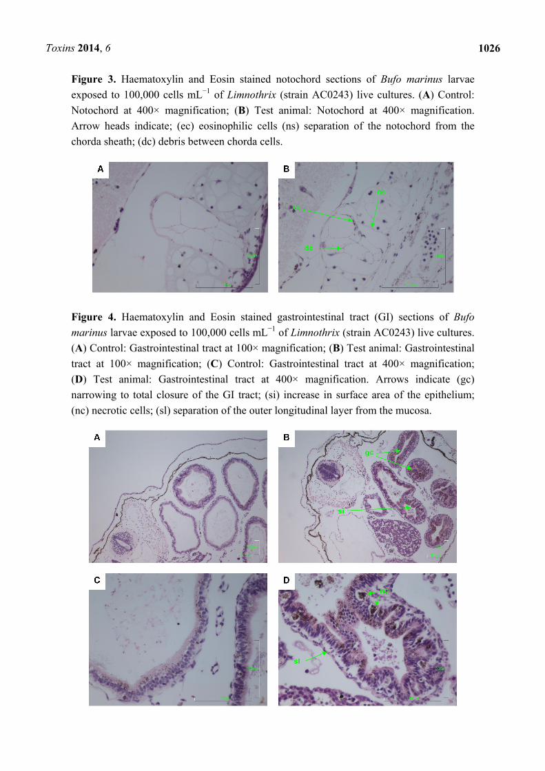
the diocoel. The notochord was affected with separation of the notochord from the chorda sheath and
increased debris between chorda cells shown. The gastrointestinal tract was extensively affected,
illustrated by separation of the outer longitudinal layer from the mucosa in the gastrointestinal tract,
significant increase in surface area of the epithelium and narrowing to total closure of the
gastrointestinal tract.
Other, less extensive pathologies occurred in the heart, kidneys and gill filaments. Heart damage
included vacuolated ventricular connective tissue that appeared more fibrous. The kidney tubules also
appeared more fibrous. The gills showed smoothing (reduction in surface area) of the gill filaments.
Toxins 2014, 6
1025
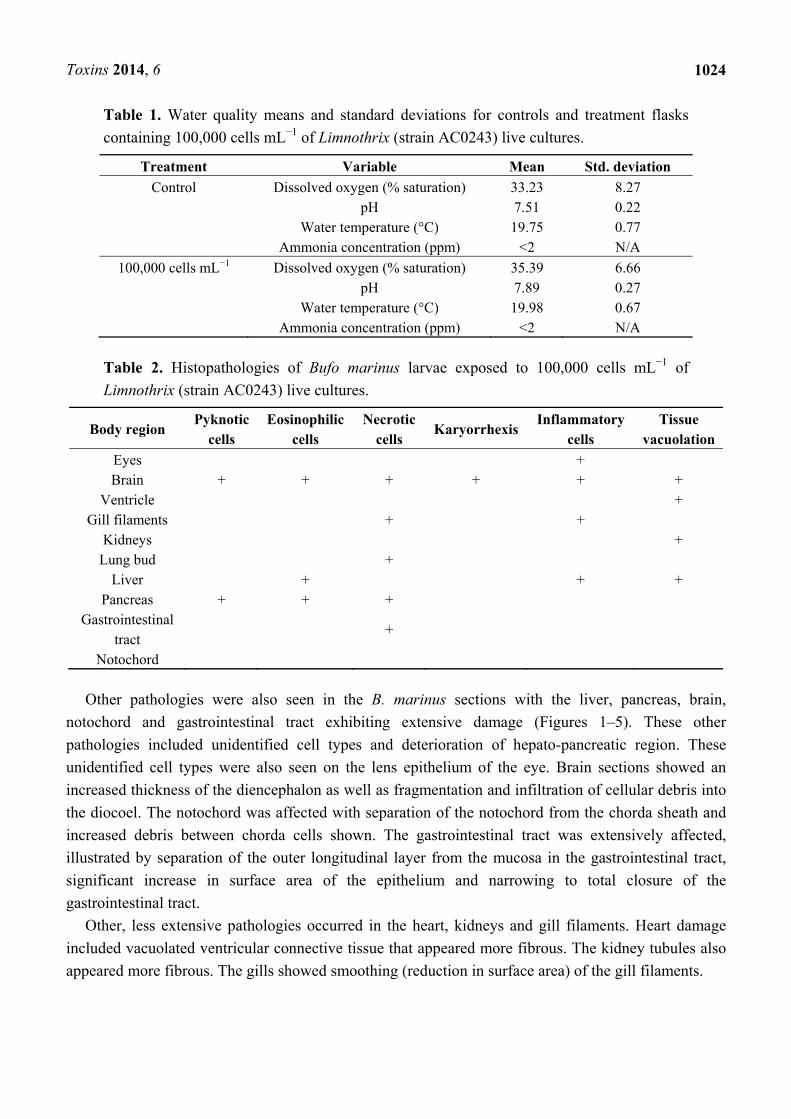
Figure 1. Haematoxylin and Eosin stained brain sections of Bufo marinus larvae exposed
to 100,000 cells mL−1 of Limnothrix (strain AC0243) live cultures. (A) Control: Brain
region at 100× magnification; (B) Test animal: Brain region at 100× magnification;
(C) Control: Brain region at 400× magnification; (D) Test animal: Brain region at 400×
magnification. Arrow indicates (ec) eosinophilic cells; (fd) fragmented grey matter
infiltrating the diocoel.
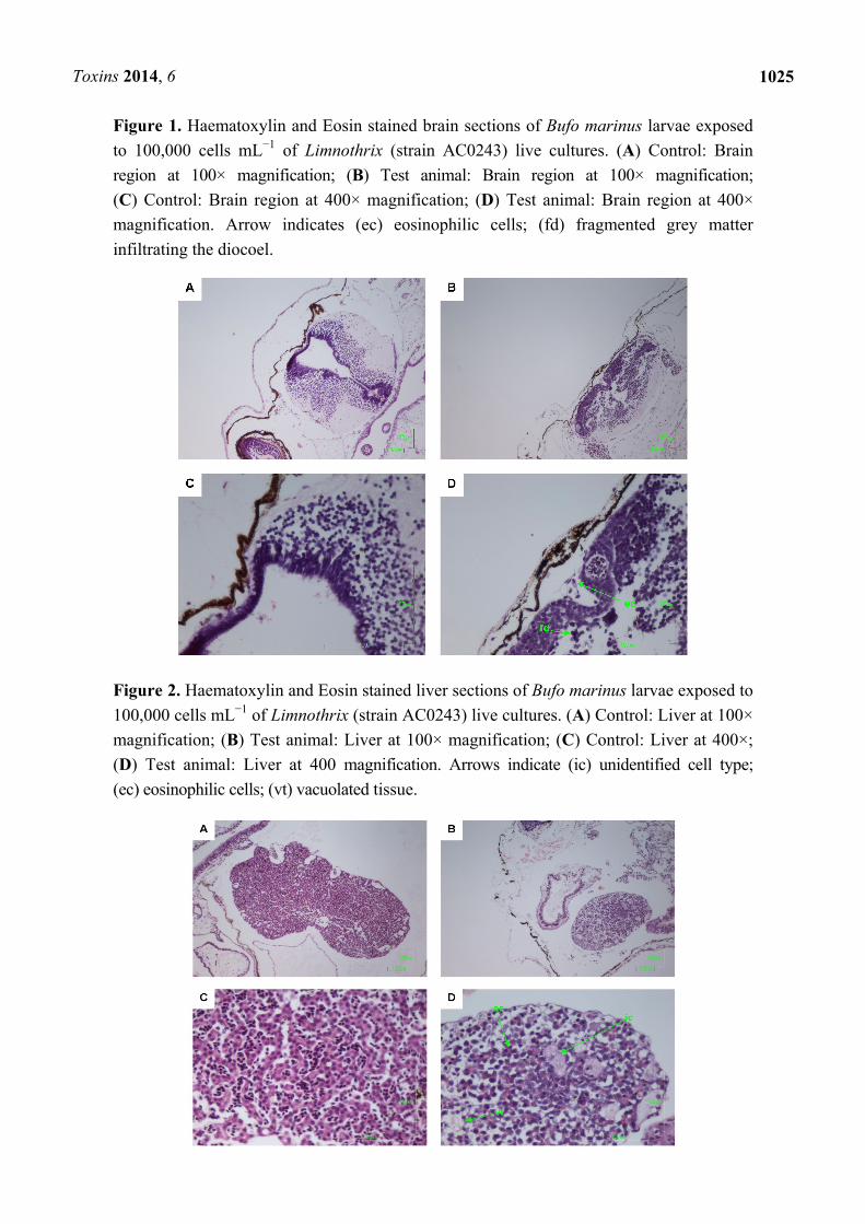
Figure 2. Haematoxylin and Eosin stained liver sections of Bufo marinus larvae exposed to
100,000 cells mL−1 of Limnothrix (strain AC0243) live cultures. (A) Control: Liver at 100×
magnification; (B) Test animal: Liver at 100× magnification; (C) Control: Liver at 400×;
(D) Test animal: Liver at 400 magnification. Arrows indicate (ic) unidentified cell type;
(ec) eosinophilic cells; (vt) vacuolated tissue.
Toxins 2014, 6
1026
Figure 3. Haematoxylin and Eosin stained notochord sections of Bufo marinus larvae
exposed to 100,000 cells mL−1 of Limnothrix (strain AC0243) live cultures. (A) Control:
Notochord at 400× magnification; (B) Test animal: Notochord at 400× magnification.
Arrow heads indicate; (ec) eosinophilic cells (ns) separation of the notochord from the
chorda sheath; (dc) debris between chorda cells.
Figure 4. Haematoxylin and Eosin stained gastrointestinal tract (GI) sections of Bufo
marinus larvae exposed to 100,000 cells mL−1 of Limnothrix (strain AC0243) live cultures.
(A) Control: Gastrointestinal tract at 100× magnification; (B) Test animal: Gastrointestinal
tract at 100× magnification; (C) Control: Gastrointestinal tract at 400× magnification;
(D) Test animal: Gastrointestinal tract at 400× magnification. Arrows indicate (gc)
narrowing to total closure of the GI tract; (si) increase in surface area of the epithelium;
(nc) necrotic cells; (sl) separation of the outer longitudinal layer from the mucosa.
Toxins 2014, 6
1027
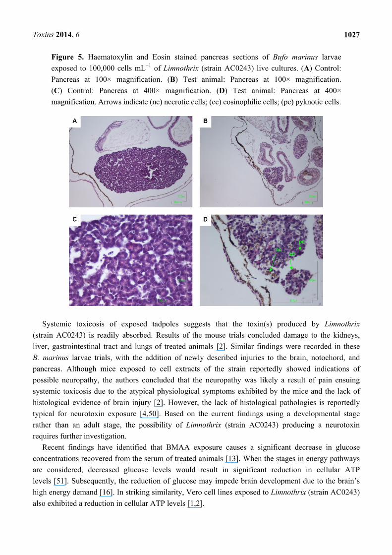
Figure 5. Haematoxylin and Eosin stained pancreas sections of Bufo marinus larvae
exposed to 100,000 cells mL−1 of Limnothrix (strain AC0243) live cultures. (A) Control:
Pancreas at 100× magnification. (B) Test animal: Pancreas at 100× magnification.
(C) Control: Pancreas at 400× magnification. (D) Test animal: Pancreas at 400×
magnification. Arrows indicate (nc) necrotic cells; (ec) eosinophilic cells; (pc) pyknotic cells.
Systemic toxicosis of exposed tadpoles suggests that the toxin(s) produced by Limnothrix
(strain AC0243) is readily absorbed. Results of the mouse trials concluded damage to the kidneys,
liver, gastrointestinal tract and lungs of treated animals [2]. Similar findings were recorded in these
B. marinus larvae trials, with the addition of newly described injuries to the brain, notochord, and
pancreas. Although mice exposed to cell extracts of the strain reportedly showed indications of
possible neuropathy, the authors concluded that the neuropathy was likely a result of pain ensuing
systemic toxicosis due to the atypical physiological symptoms exhibited by the mice and the lack of
histological evidence of brain injury [2]. However, the lack of histological pathologies is reportedly
typical for neurotoxin exposure [4,50]. Based on the current findings using a developmental stage
rather than an adult stage, the possibility of Limnothrix (strain AC0243) producing a neurotoxin
requires further investigation.
Recent findings have identified that BMAA exposure causes a significant decrease in glucose
concentrations recovered from the serum of treated animals [13]. When the stages in energy pathways
are considered, decreased glucose levels would result in significant reduction in cellular ATP
levels [51]. Subsequently, the reduction of glucose may impede brain development due to the brain’s
high energy demand [16]. In striking similarity, Vero cell lines exposed to Limnothrix (strain AC0243)
also exhibited a reduction in cellular ATP levels [1,2].
Toxins 2014, 6
1028
This reduction of ATP concentrations coupled with the pathologies exhibited by the mouse
bioassays and the B. marinus larvae suggest a possible link between Limnothrix (strain AC0243) and
BMAA. The mouse bioassays using Limnothrix (strain AC0243) extracts demonstrated weakness of
the limbs and loss of coordination [2], symptoms typical of BMAA exposure [3,14]. Although
behaviourally, the majority of B. marinus larvae exposed to live cultures of Limnothrix (strain AC0243)
did not show any measurable signs of neuropathy, two animals were witnessed convulsing, also
symptomatic of BMAA exposure [3,14]. Histological examinations showed brain damage in the
treated larvae. Control animals were not impacted.
Differences in histopathologies between the mouse bioassays and the B. marinus larvae may be
explained by the developmental stage of the two animal groups. Tests have shown areas such as the
liver and developing brain of neonatal rats with limited blood brain barrier protection and greater
protein synthesis are more susceptible to BMAA uptake [5,16]. Animals in early development such as
at embryonic and larval stages where the notochord is forming or metamorphosing are reportedly more
susceptible to toxicosis [52,53].
In contrast, adult mice have shown only limited uptake of BMAA in the brain, likely due to greater
blood brain barrier protection, with the majority of uptake occurring in the renal and hepato-pancreatic
regions [54]. Similarly, the mouse bioassays showed histological evidence of extensive liver damage
without obvious brain injuries [2]. Bufo marinus larvae exhibited cellular damage in both regions, also
with damage predominantly occurring in the hepato-pancreatic region. The uptake of BMAA has been
shown to be greater in areas such as the hepato-pancreatic region where protein synthesis is greater [16].
The histopathologies seen in the gastrointestinal tract of the larvae were comparatively different to
those reported in the mouse bioassays. The significant narrowing to complete closure of the GI tract of
the treated larvae could also have been a secondary effect of neuropathy or notochord injury. The
notochord critically contributes to the patterning of the development of the nervous system by
secreting sonic hedgehog (shh) proteins, a primary factor regulating organogenesis [41,55,56].
Notochord abnormalities have been shown contribute to other pathologies such as foregut, sacral, and
anorectal malformations [38,57]. It has also been implicated in the development of sympathoadrenal
progenitors and heart formation [37].
Other possibilities explaining the differences in toxicosis include treatment preparation and the
route of toxin uptake. Previous research has determined the toxin(s) to be water soluble, possibly
further enhancing toxicosis in exposed larvae [1,2]. Aqueous solutions are readily absorbed
cutaneously in anurans, providing an inlet for toxins, as well as maximising the potential for
transdermal uptake [27–31,58]. Grazing habits of the larvae may have also enhanced toxicosis. Oral
ingestion of whole cells has been shown to be significantly more toxic than orally ingested/injected
cell extracts [42,59].
Toxic cyanobacterial blooms have been shown to cause adverse environmental impacts, eliciting
mass mortalities in fauna [60–65]. Despite this, toxic cyanobacteria are seldom considered in terms of
declining anuran populations [43]. The effects of the toxin(s) produced by Limnothrix (strain AC0243)
has shown in this study, on short term exposure to 100,000 cells mL−1, to cause pernicious injuries to
anuran larvae on an individual level, but further imply the likelihood of impacting local communities
and populations. Furthermore, the ramifications of the toxicosis in larvae could also impact predator
populations, as larvae in part contribute as a food source for animals higher in the food chain [66–68].

Toxins 2014, 6
1029
3. Experimental Section
3.1. Limnothrix (Strain AC0243) Culture Collection and Preparation
Single trichome isolates of the original material described in [1] were used in these experiments.
The isolates were added to ASM-1 media made as described in [69] in sterile non-treated Sarstedt
(Nümbrecht, Germany) 75 cm2 tissue culture flasks with ventilated lids. The inoculated flasks were
placed in a climate controlled room set for 27 °C, under a 14/10 h diurnal regime at a light intensity of
10 µE m2 s−1 and subsequently left to grow into dense colonies.
3.2. Larvae Collection and Test Preparation
Methods for the experiment were derived from [32,42]. Bufo marinus larvae at approximately
Porter [70] development stage 33 were collected from a privately owned dam in Cawarral, Central
Queensland. The dam had no known history of cyanobacterial blooms. The larvae were netted and
transferred to a 10 L bucket containing dam water and transported back to Central Queensland
University, Rockhampton. Additional water was collected in BMW Plastics (Dandenong, Victoria,
Australia) 20 L plastic water containers and filtered using Whatmans (Kent, UK) GF/F glass filters in a
Nalgene® (Rochester, New York, NY, USA) vacuum filtration unit as prescribed by [18,32]. Prior to
filtration, five random samples of the water were analysed using PYSER (Kent, UK) 1 mL/1 µL
Sedgewick Rafter counting chambers, to ensure no toxic cyanobacteria were present in the water. The
filtered dam water was used as the control water for the experiments to minimize any stress on the
animals resulting from a change of water conditions.
The collected larvae were placed in labeled 500 mL wide neck Bomex (Beijing, China) Erlenmeyer
flasks (one larva per flask, seven replicates per treatment) in a climate controlled laboratory set at
28 °C, with a 12/12 h artificial diurnal cycle. The flasks were not aerated, to maintain the dissolved
oxygen at approximately 40% saturation, mimicking the larvae’s natural environment. Each flask was
randomly placed in rows. The larvae were fed pre-boiled lettuce and left to acclimatise for 48 h. After
the acclimatisation period and every second day for seven days, two thirds of the flask constituents
were carefully decanted and replaced with control water for the controls or test water consisting of
100,000 cells mL−1 of Limnothrix (strain AC0243) live cultures. This concentration of cells was chosen
as it is the cell concentration considered by the World Health Organisation as the basis for Alert
Level 2 in their monitoring management framework [7]. The correct concentration was achieved by
diluting stock cultures with control water. The concentration was checked prior to water changes using
a PYSER (Kent, UK) 1 mL/1 µL Sedgewick Rafter counting chamber. After each water change for the
duration of the experiment, the flasks in both the control and treatment groups were randomly
repositioned in rows and approximately 5 g of pre-boiled lettuce were added to each flask.
3.3. Water Quality Testing
The decanted flask constituents from section 3.2 were tested for dissolved oxygen (% saturation),
pH and temperature (°C), using a TPS Australia (Springwood, New South Wales, Australia) WP-91
meter immediately after each water change. Ammonia concentrations were also measured using
Toxins 2014, 6
1030
Salifert (Duiven, Holland) ammonia testing kits. Discarded test solution (10 mL) from each flask was
transferred to test tubes with corresponding labels and analysed as per the manufacturer’s instructions.
3.4. Euthanasia of Animals
On completion of the experiment or when animals were deemed moribund, each animal was
anaesthetised by being placed in iced water, followed by submersion in a Sarstedt (Nümbrecht,
Germany) 50 mL Centrifuge tube with 25 mL of 10% neutral buffered formalin for euthanization. The
10% neutral buffered formalin was pre-chilled to maintain anaesthetisation during euthanasia.
3.5. Histology
All histology was carried out by Histology Services, Bundaberg, Australia. The larvae were
manually processed and embedded in paraffin wax. Transverse sections were processed at 4 µm
thicknesses and haematoxylin and eosin (H&E) stained.
After wax mounting, transverse sections were processed at 4 µm thicknesses in ribbons of
4 to 5 sections with 50 µm between ribbons. A LKB (Vienna, Austria) Rotary-ONE microtome was
used with Feather (Osaka, Japan) S35 disposable microtome blades for sectioning. The sections were
placed on Livingstone International (Rosebery, New South Wales, Australia) premium quality glass
slides and deparaffinised. The deparaffinised sections were then stained with Amber Scientific
(Eugene, OR, USA) HH 2.5 L Harris’ haematoxylin and eosin stain.
Coverslips were mounted using Sigma Aldrich (Castle Hill, New South Wales, Australia) D.P.X
mounting resin and left to dry. The sections were analysed using a Nikon (Tokyo, Japan) Eclipse E200
binocular microscope and photographed using a Nikon (Tokyo, Japan) Digital sight DS-L1 camera
under 40×, 100×, and 400× magnifications as specified in each figure.
4. Conclusions
Reports of Limnothrix (strain AC0243) derived poisoning events have not yet been documented as
the toxicity of this strain has only recently been detected. Limnothrix secies are usually found growing
in conjunction with C. raciborskii [51–54]. Consequently, this strain has been overlooked as a causal
agent in poisoning events.
Previous trials have established acute toxicity of Limnothrix (strain AC0243) in mammals [1,2].
The results of this experiment indicate that the toxin(s) produced by Limnothrix (strain AC0243)
causes severe toxicosis to developing vertebrates with injuries particularly noted in the brain,
notochord and pancreas. The damage caused by this strain showed strong similarities to BMAA
intoxication and further investigation is recommended. The systemic toxicosis caused by the toxin(s)
indicates that this strain has the potential to significantly impact upon aquatic populations. Research is
also needed to evaluate the concentration dependent effects of the toxin(s) as well as explore the
long-term consequences of exposure.
Toxins 2014, 6
1031
Acknowledgments
The authors would like to thank Central Queensland University, Rockhampton for scholarship
funding and support. The authors would also like to thank Adam Rose for his assistance.
Conflicts of Interest
The authors declare no conflict of interest.
References
1. Bernard, C.; Froscio, S.; Campbell, R.; Monis, P.; Humpage, A.; Fabbro, L. Novel toxic effects
associated with a tropical Limnothrix /Geitlerinema-like cyanobacterium. Environ. Toxicol. 2011,
26, 260–270.
2. Humpage, A.; Falconer, I.; Bernard, C.; Froscio, S.; Fabbro, L. Toxicity of the cyanobacterium
Limnothrix AC0243 to male balb/C mice. Water Res. 2012, 46, 1576–1583.
3. Aráoz, R.; Molgó, J.; Marsac, N.T.D. Neurotoxic cyanobacterial toxins. Toxicon 2010, 56,
813–828.
4. Carmichael, W. Health effects of toxin-producing cyanobacteria: “The cyanoHABs”.
Hum. Ecol. Risk Assess. 2001, 7, 1393–1407.
5. Karlsson, O.; Lindquist, N.G.; Brittebo, E.B.; Roman, E. Selective brain uptake and behavioral
effects of the cyanobacterial toxin BMAA (β-N-methylamino-L-alanine) following neonatal
administration to rodents. Toxicol. Sci. 2009, 109, 286–295.
6. Watkins, S.; Reich, A.; Fleming, L.; Hammond, R. Neurotoxic shellfish poisoning. Mar. Drugs
2008, 6, 431–455.
7. World Health Organisation (WHO). Toxic Cyanobacteria in Water: A Guide to Their Public
Health Consequences, Monitoring and Management; St Edmundsbury Press: Suffolk, UK, 1999.
8. Chiu, A.S.; Gehringer, M.M.; Braidy, N.; Guillemin, G.J.; Welch, J.H.; Neilan, B.A. Excitotoxic
potential of the cyanotoxin β-methyl-amino-L-alanine (BMAA) in primary human neurons.
Toxicon 2012, 60, 1159–1165.
9. Okle, O.; Rath, L.; Galizia, C.G.; Dietrich, D.R. The cyanobacterial neurotoxin
beta-N-methylamino-L-alanine (BMAA) induces neuronal and behavioral changes in honeybees.
Toxicol. Appl. Pharmacol. 2013, 270, 9–15.
10. Brand, L.E.; Pablo, J.; Compton, A.; Hammerschlag, N.; Mash, D.C. Cyanobacterial blooms and
the occurrence of the neurotoxin, beta-N-methylamino-L-alanine (BMAA), in South Florida
aquatic food webs. Harmful Algae 2010, 9, 620–635.
11. Purdie, E.L.; Samsudin, S.; Eddy, F.B.; Codd, G.A. Effects of the cyanobacterial neurotoxin
β-N-methylamino-L-alanine on the early-life stage development of zebrafish (Danio rerio). Aquat.
Toxicol. 2009, 95, 279–284.
12. De Munck, E.; Muñoz-Sáez, E.; Miguel, B.G.; Solas, M.T.; Ojeda, I.; Martínez, A.; Gil, C.;
Arahuetes, R.M. β-N-Methylamino-L-alanine causes neurological and pathological phenotypes
mimicking Amyotrophic Lateral Sclerosis (ALS): The first step towards an experimental model
for sporadic ALS. Environ. Toxicol. Pharmacol. 2013, 36, 243–255.
Toxins 2014, 6
1032
13. Engskog, M.K.R.; Karlsson, O.; Haglöf, J.; Elmsjö, A.; Brittebo, E.; Arvidsson, T.; Pettersson, C.
The cyanobacterial amino acid β-N-methylamino-L-alanine perturbs the intermediary metabolism
in neonatal rats. Toxicology 2013, 312, 6–11.
14. Karamyan, V.T.; Speth, R.C. Animal models of BMAA neurotoxicity: A critical review.
Life Sci. 2008, 82, 233–246.
15. Karlsson, O.; Berg, A.L.; Lindström, A.K.; Hanrieder, J.; Arnerup, G.; Roman, E.; Bergquist, J.;
Lindquist, N.G.; Brittebo, E.B.; Andersson, M. Neonatal exposure to the cyanobacterial toxin
BMAA induces changes in protein expression, and neurodegeneration in adult hippocampus.
Toxicol. Sci. 2012, 130, 391–404.
16. Karlsson, O.; Jiang, L.; Andersson, M.; Ilag, L.L.; Brittebo, E.B. Protein association of the
neurotoxin and non-protein amino acid BMAA (β-N-methylamino-L-alanine) in the liver and
brain following neonatal administration in rats. Toxicol. Lett. 2014, 226, 1–5.
17. Goto, J.J.; Koenig, J.H.; Ikeda, K. The physiological effect of ingested β-N-methylamino-L-alanine
on a glutamatergic synapse in an in vivo preparation. Comp. Biochem. Physiol. Part C 2012, 156,
171–177.
18. Leeuwangh, P.; Kappers, F.I.; Dekker, M.; Koerselman, W. Toxicity of cyanobacteria in Dutch
lakes and reservoirs. Aquat. Toxicol. 1983, 4, 63–72.
19. Falconer, I.R. Effects on human health of some toxic cyanobacteria (blue-green algae) in
reservoirs, lakes and rivers. Toxic. Assess. 1989, 4, 175–184.
20. Repavich, W.M.; Sonzogni, W.C.; Standridge, J.H.; Wedepohl, R.E.; Meisner, L.F. Cyanobacteria
(blue-green algae) in Wisconsin waters: Acute and chronic toxicity. Water Res. 1990, 24,
225–231.
21. Carmichael, W. Cyanobacteria secondary metabolites-the cyanotoxins. J. Appl. Bacteriol. 1992,
72, 445–459.
22. Negri, A.; Jones, G. Bioaccumulation of Paralytic Shellfish Poisoning (PSP) toxins from the
cyanobacterium Anabaena circinalis by the freshwater mussel Alathyria condola. Toxicon 1995,
33, 667–678.
23. Negri, A.P.; Jones, G.J.; Hindmarsh, M. Sheep mortality associated with paralytic shellfish
poisons from the cyanobacterium Anabaena circinalis. Toxicon 1995, 33, 1321–1329.
24. Falconer, I.R. An overview of problems caused by toxic blue–green algae (cyanobacteria) in
drinking and recreational water. Environ. Toxicol. 1999, 14, 5–12.
25. Ernst, B.; Hoeger, S.J.; O’Brien, E.; Dietrich, D.R. Oral toxicity of the microcystin-containing
cyanobacterium Planktothrix rubescens in European whitefish (Coregonus lavaretus).
Aquat. Toxicol. 2006, 79, 31–40.
26. Brand, L.E.; Campbell, L.; Bresnan, E.K. The biology and ecology of a toxic genus.
Harmful Algae 2012, 14, 156–178.
27. Uchiyama, M.; Konno, N. Hormonal regulation of ion and water transport in anuran amphibians.
Gen. Comp. Endocrinol. 2006, 147, 54–61.
28. Azevedo, R.A.; Carvalho, H.F.; de Brito-Gitirana, L. Hyaluronan in the epidermal and the dermal
extracellular matrix: Its role in cutaneous hydric balance and integrity of anuran integument.
Micron 2007, 38, 607–610.
Toxins 2014, 6
1033
29. Maejima, S.; Yamada, T.; Hamada, T.; Matsuda, K.; Uchiyama, M. Effects of hypertonic stimuli
and arginine vasotocin (AVT) on water absorption response in Japanese treefrog,
Hyla japonica. Gen. Comp. Endocrinol. 2008, 157, 196–202.
30. Suzuki, M.; Tanaka, S. Molecular and cellular regulation of water homeostasis in anuran
amphibians by aquaporins. Comp. Biochem. Physiol. Part A 2009, 153, 231–241.
31. Maejima, S.; Konno, N.; Matsuda, K.; Uchiyama, M. Central angiotensin II stimulates cutaneous
water intake behavior via an angiotensin II type-1 receptor pathway in the Japanese tree frog Hyla
japonica. Horm. Behav. 2010, 58, 457–464.
32. White, S.H.; Duivenvoorden, L.J.; Fabbro, L.D.; Eaglesham, G.K. Mortality and toxin
bioaccumulation in Bufo marinus following exposure to Cylindrospermopsis raciborskii Cell
extracts and live cultures. Environ. Pollut. 2007, 147, 158–167.
33. Junges, C.M.; Peltzer, P.M.; Lajmanovich, R.C.; Attademo, A.M.; Cabagna Zenklusen, M.C.;
Basso, A. Toxicity of the fungicide Trifloxystrobin on tadpoles and its effect on fish-tadpole
interaction. Chemosphere 2012, 87, 1348–1354.
34. Gillespie, G.R. Impacts of sediment loads, tadpole density, and food type on the growth and
development of tadpoles of the spotted tree frog Litoria spenceri: An in-stream Experiment. Biol.
Conserv. 2002, 106, 141–150.
35. Lamberti, G.A.; Feminella, J.W.; Pringle, C.M. Methods in Stream Ecology, 2nd ed.; Academic
Press: New York, NY, USA, 2007; pp. 537–559.
36. Tyler, M. Frogs and Toads as Experimental Animals; The University of Adelaide: Adelade,
Australia, 2009.
37. Stemple, D.; Solnica-Krezel, L.; Zwartkruis, F.; Neuhauss, S.C.; Schier, A.F.; Malicki, J.; Stainier,
D.Y.; Abdelilah, S.; Rangini, Z.; Mountcastle-Shah, E.; et al. Mutations affecting development of
the notochord in zebrafish. Development 1996, 123, 117–128.
38. Qi, B.Q.; Beasley, S.W.; Frizelle, F.A. Evidence that the notochord may be pivotal in the
development of sacral and anorectal malformations. J. Pediatr. Surg. 2003, 38, 1310–1316.
39. Platz, F. Structural and experimental investigations of the functional anatomy and the turgor of
the notochord in the larval tail of anuran tadpoles. Ann. Anat. 2006, 188, 289–302.
40. Pagnon-Minot, A.; Malbouyres, M.; Haftek-Terreau, Z.; Kim, H.R.; Sasaki, T.; Thisse, C.; Thisse, B.;
Ingham, P.W.; Ruggiero, F.; Le Guellec, D.; et al. A novel factor in zebrafish notochord
differentiation and muscle development. Dev. Biol. 2008, 316, 21–35.
41. Ribes, V.; Balaskas, N.; Sasai, N.; Cruz, C.; Dessaud, E.; Cayuso, J.; Tozer, S.; Yang, L.L.;
Novitch, B.; Marti, E.; et al. Distinct sonic hedgehog signaling dynamics specify floor plate and
ventral neuronal progenitors in the vertevrate neural tube. Genes Dev. 2010, 24, 1186–1200.
42. Kinnear, S. Cylindrospermopsin in whole cell extracts and live cultures of Cylindrospermopsis
raciborskii: Ecotoxicity, bioaccumulation and management. PhD thesis, School of Biological and
Environmental Sciences, Central Queensland University, Rockhampton, QLD, Australia, July
2006.
43. Ziková, A.; Lorenz, C.; Lutz, I.; Pflugmacher, S.; Kloas, W. Physiological responses of Xenopus
laevis tadpoles exposed to cyanobacterial biomass containing microcystin-LR. Aquat. Toxicol.
2013, 128–129, 25–33.
Toxins 2014, 6
1034
44. National Health and Medical Research Council. Australian Code of Practice for the Care and Use
of Animals for Scientific Purposes; Australian Government: Canberra, Australia, 2004.
45. Gao, Y.; Cornwell J.C.; Stoecker D.K.; Owens, M.S. Effects of cyanobacterial-driven pH
increases on sediment nutrient fluxes and coupled nitrification-denitrification in a shallow fresh
water estuary. Biogeosciences 2012, 9, 2697–2710.
46. Marques, S.M.; Chaves, S.; Gonçalves, F.; Pereira, R. Evaluation of growth, biochemical and
bioaccumulation parameters in Pelophylax perezi tadpoles, following an in-situ acute exposure to
three different effluent ponds from a uranium mine. Sci. Total Environ. 2013,
445–446, 321–328.
47. ANZECC. Australian and New Zealand Guidelines for Fresh and Marine Water Quality;
Australian and New Zealand Environment and Conservation Council: Canberra, Australia, 2000.
48. Seigel, R.; Dismore, A.; Richter, S. Using well water to increase hydroperiod as a management
option for pond-breeding amphibians. Wildl. Soc. Bull. 2006, 34, 1022–1027.
49. Economic Development and Inovation Department of Employment; Markula, A., Csurhes, S.,
Hannan-Jones, M., Eds.; Biosecurity Queensland: Brisbane, Australia, 2010.
50. Faassen, E.J.; Harkema, L.; Begeman, L.; Lurling, M. First report of (Homo)Anatoxin-a and dog
neurotoxicosis after ingestion of benthic cyanobacteria in the Netherlands. Toxicon 2012, 60,
378–384.
51. Ioudina, M.; Uemura, E.; Greenlee, H.W. Glucose insufficiency alters neuronal viability and
increases susceptibility to glutamate toxicity. Brain Res. 2004, 1004, 188–192.
52. Berry, J.P.; Gantar, M.; Gibbs, P.D.L.; Schmale, M.C. The zebrafish (Danio rerio) embryo as a
model system for identification and characterization of developmental toxins from marine and
freshwater microalgae. Comp. Biochem. Physiol. Part C 2007, 145, 61–72.
53. Stewart, I. Environmental risk factors for temporal lobe epilepsy—Is prenatal exposure to the
marine algal neurotoxin domoic acid a potentially preventable cause? Med. Hypotheses 2010, 74,
466–481.
54. Karlsson, O.; Berg, C.; Brittebo, E.B.; Lindquist, N.G. Retention of the cyanobacterial neurotoxin
β-N-Methylamino-L-Alanine in melanin and neuromelanin-containing cells—A possible link
between Parkinson-dementia complex and pigmentary retinopathy. Pigment Cell Melanoma Res.
2009, 22, 120–130.
55. Müller, F.; Albert, S.; Blader, P.; Fischer, N.; Hallonet, M.; Strähle, U. Direct actin of the
nodal-related signal cyclops in induction of sonic hedgehog in the ventral midline of the CNS.
Development 2000, 127, 3889–3897.
56. Lek, M.; Dias, J.M.; Marklund, U.; Uhde, C.W.; Kurdija, S.; Lei, Q.; Sussel, L.; Rubenstein, J.L.;
Matise, M.P.; Arnold, H.H.; et al. A homeodomain feedback circuit underlies step-function
interpretation of a Shh morphogen gradient during ventral neural patterning. Development 2010,
137, 4051–4060.
57. Qi, B.Q.; Beasley, S.W. Relationship of the notochord to foregut development in the fetal rat
model of esophageal atresia. J. Pediatr. Surg. 1999, 34, 1593–1598.
58. McDairmid, R.; Altig, R. Tadpoles: The Biology of Anuran Larvae; The University of Chicago
Press: Chicago, IL, USA, 1999; p. 436.
Toxins 2014, 6
1035
59. Kankaanpää, H.T.; Holliday, J.; Schröder, H.; Goddard, T.J.; von Fister, R.; Carmichael, W.W.
Cyanobacteria and prawn farming in Northern New South Wales, Australia—A case study on
cyanobacteria diversity and hepatotoxin bioaccumulation. Toxicol. Appl. Pharmacol. 2005, 203,
243–256.
60. Quiblier, C.; Susanna, W.; Isidora, E.S.; Mark, H.; Aurélie, V.; Jean-François, H. A review of
current knowledge on toxic benthic freshwater cyanobacteria—Ecology, toxin production and risk
management. Water Res. 2013, 47, 5464–5479.
61. Handeland, K.; Østensvik, Ø. Microcystin poisoning in roe deer (Capreolus capreolus). Toxicon
2010, 56, 1076–1078.
62. Krienitz, L.; Ballot, A.; Kotut, K.; Wiegand, C.; Pütz, S.; Metcalf, J.S.; Codd, G.A.;
Pflugmacher, S. Contribution of hot spring cyanobacteria to the mysterious deaths of lesser
flamingos at Lake Bogoria, Kenya. FEMS Microbiol. Ecol. 2003, 43, 141–148.
63. Lopez Rodas, V.; Costas, E. Preference of mice to consume Microcystis aeruginosa
(toxin-producing cyanobacteria): A possible explanation for numerous fatalities of livestock and
wildlife. Res. Vet. Sci. 1999, 67, 107–110.
64. Soll, M.; Williams, M. Mortality of white rhinoceros (Ceratotherium simum) Suspected to be
associated with the blue-green alga (Microcystis aeruginosa). J. S. Afr. Vet. Assoc. 1985, 56,
49–51.
65. Stewart, I.; Seawright, A.A.; Shaw, G.R. Advances in Medicine and Biology; Hudnell, H., Ed.;
Springer Science + Business Media, LLC: New York, NY, USA, 2008; Volume 619, Chapter 28,
pp. 613–637.
66. Jara, F.G.; Perotti, M.G. Risk of predation and behavioural response in three anuran species:
Influence of tadpole size and predator type. Hydrobiologia 2010, 644, 313–324.
67. Kiesecker, J.; Chivers, D.P.; Marco, A.; Quilchano, C.; Anderson, M.T.; Blaustein, A.R.
Identification of a disturbance signal in larval red-legged frogs, Rana aurora. Anim. Behav. 1999,
57, 1295–1300.
68. Mirza, R.S.; Ferrari, M.C.O.; Kiesecker, J.M.; Chivers, D.P. Responses of american toad tadpoles
to predation cues: Behavioural response thresholds, threat-sensitivity and acquired predation
recognition. Behaviour 2006, 143, 877–889.
69. Gorham, P.; Lachlan, J.M.; Hammer, U.; Kim, W. Isolation and culture of toxic strains of
Anabaena flos-aquae (Lyngb) De Breb. Verh. Int. Ver. fuer Theor. und Angew. Limnol. 1964, 15,
796–804.
70. Porter, K. Herpetology; W.B. Saunders Company: Philadelphia, PA, USA, 1972.
© 2014 by the authors; licensee MDPI, Basel, Switzerland. This article is an open access article
distributed under the terms and conditions of the Creative Commons Attribution license
(http://creativecommons.org/licenses/by/3.0/).