Toxicological Profile for Bromomethane - Agency for Toxic ...
Upload
independentCategory
view
1download
0
This article appeared in a journal published by Elsevier. The attachedcopy is furnished to the author for internal non-commercial researchand education use, including for instruction at the authors institution
and sharing with colleagues.
Other uses, including reproduction and distribution, or selling orlicensing copies, or posting to personal, institutional or third party
websites are prohibited.
In most cases authors are permitted to post their version of thearticle (e.g. in Word or Tex form) to their personal website orinstitutional repository. Authors requiring further information
regarding Elsevier’s archiving and manuscript policies areencouraged to visit:
http://www.elsevier.com/copyright

Author's personal copy
Inflammatory effects of the toxic cyanobacterium Geitlerinemaamphibium
Camila Ranzatto Dogo a,*, Fernanda Miriane Bruni b, Fabiana Elias c, Marisa Rangel d,Patricia Araujo Pantoja e, Célia Leite Sant’Anna a, Carla Lima b, Monica Lopes-Ferreira b,Luciana Retz de Carvalho a,1
aCenter of Phycological Research, Institute of Botany, Av. Miguel Estéfano, 3687, Água Funda, 04301-902 São Paulo, SP, BrazilbCenter of Applied Toxicology, Butantan Institute, Av. Vital Brasil, 1500 05503-900 São Paulo, SP, BrazilcDepartment of Pathology, School of Veterinary Medicine and Animal Science, University of São Paulo, Av. Prof. Dr. Orlando Marques de Paiva, 87,Cidade Universitária, 05508-270 São Paulo, SP, Brazild Laboratory of Immunopathology, Butantan Institute, Av. Vital Brasil, 1500 05503-900 São Paulo, SP, BrazileChemical Engineering Department, Polytechnic School of the University of São Paulo, Av. Prof. Luciano Gualberto, 380, trav 3, Cidade Universitária,05508-900 São Paulo, SP, Brazil
a r t i c l e i n f o
Article history:Received 22 February 2011Received in revised form 12 July 2011Accepted 18 July 2011Available online 16 August 2011
Keywords:CyanobacteriaGeitlerinema amphibiumInflammatory activity
a b s t r a c t
Toxic cyanobacteria in public water reservoirs may cause severe health issues for live-stock and human beings. Geitlerinema amphibium, which is frequently found in SãoPaulo City’s drinking water supplies, showed toxicity in the standard mouse bioassay,while displaying signs of intoxication and post-mortem findings different from thoseshowed by animals intoxicated by known cyanotoxins. We report here the alterationscaused by G. amphibium methanolic extract on mouse microcirculatory network, as seenby in vivo intravital microscopy, besides observations on leukocyte migration, cytokinequantitation, and results of toxicological essays. Our data showed that G. amphibiummethanolic extract displayed time- and dose-dependent pro-inflammatory activity, andthat at lower doses [125 and 250 mg/kg body weight (b.w.)] increased the leukocyterolling, caused partial venular stasis, as well as induced an increase in leukocyte countsin the peripheral blood and peritoneal washings. At higher doses (500 and 1000 mg/kgb.w.), the extract caused ischemic injury leading to animal death. As confirmed by massspectrometric studies and polymyxin B test, the G. amphibium methanolic extract didnot contain lipopolysaccharides.
� 2011 Elsevier Ltd. All rights reserved.
1. Introduction
Cyanobacteria are photosynthetic prokaryotes thatinhabit environments, in which moisture and light are
present, including land, water, and extreme environments.Currently, they are a public health issue due to their abilityto produce potent toxins known as cyanotoxins (Sant’Annaand Azevedo, 2006).
The cyanotoxins are classified as hepatotoxins, neuro-toxins, cytotoxins, and dermatotoxins according to theireffects on mammals. The hepatotoxin group encompassesthe microcystins and nodularins; the neurotoxin groupcontains the saxitoxin and anatoxin alkaloids named andthe amino acid b-methylaminoalanine; the cytotoxins arerepresented by the cylindrospermopsins, and the derma-totoxins are composed of the lipopolysaccharides (Carvalhoet al., 2008).
* Corresponding author. Tel.: þ55 1150736300x290; fax: þ551150733678.
E-mail addresses: [email protected] (C.R. Dogo), [email protected] (F.M. Bruni), [email protected] (F. Elias),[email protected] (M. Rangel), [email protected] (P.A. Pantoja),[email protected] (C.L. Sant’Anna), [email protected](C. Lima), [email protected] (M. Lopes-Ferreira), [email protected] (L.R. de Carvalho).
1 Tel.: þ55 1150736300x290; fax: þ55 1150733678.
Contents lists available at ScienceDirect
Toxicon
journal homepage: www.elsevier .com/locate/ toxicon
0041-0101/$ – see front matter � 2011 Elsevier Ltd. All rights reserved.doi:10.1016/j.toxicon.2011.07.010
Toxicon 58 (2011) 464–470
Author's personal copy
Cyanobacteria also produce unique secondary metabo-lites that possess various types of bioactivity, such as anti-fungal (Skulberg, 2000), anti-enzymatic (Yamaki et al.,2005), anti-tumoral (Tan, 2010), antimicrobial (Chlipalaet al., 2009) and anti-viral (Kanekiyo et al., 2005) activity;some cyanobacteria synthesize bioactive and toxiccompounds simultaneously (Kuiper-Goodman et al., 1999).
In our ongoing study of Brazilian cyanobacterial toxicity,we found that Geitlerinema amphibium [¼Geitlerinemaunigranulatum (Bittencourt-Oliveira et al., 2009)] demon-strated toxicity in a standard cyanotoxin bioassay(Sant’Anna et al., 2008) while displaying signs of intoxica-tion and post-mortem findings different from those shownin animals intoxicated by known cyanotoxins (Carvalhoet al., 2008). This suggested the existence of a new cyano-toxin. G. amphibium is a freshwater species that does notform blooms (Torgan and De Paula, 1994; Tucci et al., 2006)but is found in public water supply reservoirs of the SãoPaulo, Brazil, metropolitan region with a frequency greaterthan 50% (Carvalho et al., 2007; Lopes and Sawatani,Unpublished results, 2009; Sant’Anna et al., 2007).
The standard bioassay for assessing cyanobacterialtoxicity consists of administering, to Swiss mice (19–21 g),through the intra-peritoneal route, a dried cell lysate orextract (20 g dried cells/kg�1 b.w.) dissolved in 0.9 percentsaline solution (0.1–1 ml). In addition to this assay, post-mortem and histological analyses are used (Harada et al.,1999). However, this procedure does not provide informa-tion about the in vivo intoxication mechanism.
The intravital microscopy technique permits the obser-vation of important events that occur in the microcircula-tion and bloodstream and in vivo observation of themicrocirculatory network and its components (arterioles,capillaries, and post-capillary venules); leukocyte rolling,adhesion and transmigration; and changes caused byinfectious/toxic agents. This technique is frequentlyemployed to observe the effects of snake, fish, andamphibian toxins on mammals. To our knowledge, the onlycyanotoxin study employing this technique was one byKujbida et al. (2009) that used intravital microscopy toestimate the increase in leukocyte rolling caused bymicrocystin-LR.
The frequency with which G. amphibium is found inpublic reservoirs, along with its toxicity, makes it a pop-ulation health risk and requires studies on its effects onmammals. In addition to the data from classical mouseassays, these studies should include information about thebiological processes occurring during intoxication.
To fulfill these requirements, this paper presents theobserved intoxication signs, histological data, and thein vivo evaluation of the effects of a G. amphibium CCIBt 920strain methanolic extract on mouse microcirculation usingintravital microscopy.
2. Material and methods
2.1. The organism
G. amphibium CCIBt 920 strain was collected on August15, 2002, from Guarapiranga Reservoir, one of the fivereservoirs that make up the basin of the Upper Tietê, São
Paulo, Brazil. It has since been kept in the CyanobacteriaCulture Collection of the São Paulo Institute of Botany,under the following conditions: temperature 23 � 1 �C,irradiance 40–50 mmol/m�2 s�1 and 14:10 h light:dark inASM-1 medium (Azevedo and Sant’Anna, 2003).
2.2. Biomass production
Biomass was produced by cultivating the unialgal non-axenic strain in 9 l of ASM-1 under the same conditions ofillumination and temperature described above, withconstant stirring (70 rpm) or aeration until the exponentialgrowth phase. Cells were harvested by centrifugation andfrozen at �20 �C.
2.3. Sample preparation
The frozen biomass was lyophilized and subjected toultrasound-assisted extraction with MeOH/H2O 75:25 v/v(5�, 30 s, 100 W); the extracts were combined and centri-fuged (1750�g, 50min), andthesupernatantwasevaporatedto dryness under reduced pressure (Fastner et al., 1998).
2.4. Animals
The present study was conducted according to thepolicies of the Brazilian College of Animal Experimentationand authorized by the Ethics Committee for AnimalResearch of the Butantan Institute. All bioassays werecarried out on Swiss male mice provided by the ButantanInstitute Animal Facility and housed at 23 � 1 �C undera 12:12 h light:dark cycle. The animals weighed between 18and 22 g andwere kept in groups of five per cage, with foodand water ad libitum.
2.5. Standard bioassay
To evaluate theG. amphibiumminimal lethal dose (MLD),doses corresponding to 1000, 750, 500 and 250 mg/kg b.w.of dried methanolic extract were diluted in 500 ml of salinesolution (0.9% w/v) and administered intra-peritoneally.Clinical signs of intoxication were observed and afterdeath or sacrifice, post-mortem examinationwas carried out(Harada et al., 1999). In all tests, n ¼ 3.
2.6. Histological analysis
Liver, lung, and kidney samples from the standardbioassays were fixed in buffered formalin, processed byconventional histological techniques, and stained withhematoxylin/eosin.
2.7. Microcirculatory alterations
Microcirculatory changes in the mouse cremastermuscle were observed and recorded by intravital micros-copy after topical and intra-peritoneal administration ofdifferent concentrations of G. amphibium methanolicextract. The concentrations of topically administered doseswere 20, 40 and 120 mg per 20 ml of sterile saline and forintra-peritoneally injected doses were 6, 125, 250, 500 and
C.R. Dogo et al. / Toxicon 58 (2011) 464–470 465

Author's personal copy
1000mg/kg b.w. per 500 ml of sterile saline. Control animalsreceived an equivalent volume of sterile saline solution. Themice were anesthetized with pentobarbital sodium (Hyp-nol� Cristália, 50 mg/kg), and kept on a heating pad tomaintain body temperature at 37 �C.
The cremaster muscle was prepared for intravitalmicroscopy by transluminescence as described by Lomonteet al. (1994). Preparationswere keptmoist by irrigationwithphosphate-buffered saline (PBS) 0.15 M. Data on arteriolarand venular diameter, thrombus formation, leukocytesrolling, adherence, and transmigration were recorded atbaseline and every 5min until 30min after the beginning ofthe experiment (n ¼ 3) (Granger and Kubes, 1994).
Intravital microscopy was performed using an opticalmicroscope (Axiolab, Carl-Zeiss, Oberkochen, DE) attachedto an AxioCam ICcI camera.
2.8. Total and differential leukocyte counts in blood
Blood samples were collected from the ophthalmicvenous plexus immediately before and 30 min after theintra-peritoneal administration of 125 mg/kg b.w. ofG. amphibium methanolic extract. For total counting, 10 mlblood samples collected in sodium citrate tubes were pro-cessed according to Ferreira et al. (2007). Giemsa-stainedblood smears that were obtained from whole bloodsamples collected pre- and post-drug administration weredifferentially counted. Counts were performed under anoptical microscope (Axiolab).
2.9. Total and differential leukocyte count in the peritoneum
Peritoneal exudate cells were collected 0.5, 2, 6, and 24 hafter intra-peritoneal injection of 125 mg/kg b.w. of G.amphibium methanolic extract. Peritoneal cavities werewashed out with 5 ml PBS þ EDTA 50 mM, and the cellularsuspensions were centrifuged for 10min at 400�g and 4 �C.The supernatants were stored at �20 �C until furtheranalysis for cytokines and chemokines, and cell pelletswere resuspended in PBS/EDTA. Aliquots of 10 ml of cellsuspension were diluted with 190 ml of Turk’s solution andused to determine total counts. Differential cell countswere performed on Giemsa-stained Cytospin preparations,using 100 ml of cell suspension. The control group ofanimals received intra-peritoneal injections of sterile salineunder identical experimental conditions.
2.10. Peritoneal fluid cytokine and chemokine quantitation
Measurements of IL-1, IL-6, TNF-a, and the chemokinesKC and MCP-1 in the supernatants of the peritoneal fluidwere made by ELISA, using the OpEIA kit (BD-Phamingen,San Diego, CA, USA). The detection limits were 15.62, 31.25,1.9, 3.9, and 7.81 pg ml�1 for IL-1b, IL-6, TNF-a, KC, andMCP-1, respectively.
2.11. Polymyxin B (POL.B) test
Four groups of animals were employed (three animalsper group). Group I (920 þ POL.B) received 500 mg/kg b.w.of G. amphibium methanolic extract plus 1 mg/kg b.w. of
polymyxin B; group II (POL.B) was treated with 1 mg/kgb.w. of polymyxin B; group III (CCIBt 920) was treated with500 mg/kg b.w. of G. amphibium methanolic extract; andthe negative control, group IV (Group Co), was inoculatedwith sterile saline/diluents. The administration was con-ducted via the intra-peritoneal route (n ¼ 3).
2.12. MALDI-MS
To evaluate the presence of lipopolysaccharides, themethanolic extract of G. amphibium was subjected to massspectral analysis in Shimadzu Biotech equipment withLaunchpad software (version 2.8.2.). Analyses were per-formed in reflectron mode, mass range 10–50,000 D, witha synaptic acid (SIGMA) matrix. Internal calibration was per-formed with bradykinin (fragment), M ¼ 756.3997 D; ACTH(fragment)M ¼ 2465.19189 D and P14R,M ¼ 1532.8582 D.
2.13. Statistical analysis
The statistical analysis was carried out with GraphPadPrism, version 5.0. Student’s t-testwas used to compare twogroups and ANOVA followed by Dunnet’s post hoc test wasused formore than twogroups,with the level of significanceset at P < 0.05. Data are expressed as mean � S.D.
3. Results
3.1. Minimal lethal dose (MLD), intoxication signs andhistological changes
The intra-peritoneal MLD of G. amphibium methanolicextract was 500 mg/kg b.w. The first clinical intoxicationsigns appeared 15 min after extract administration. Theanimals presented signs of strong dyspnea, weakness andprostration, and death occurred within 34 min to 36 h afterinoculation. No obvious gross pathological change otherthan congestion of the examined organs was observed onpost-mortem examination. Lung tissue histological analysisin the group of animals that received 125 mg/kg b.w. ofmethanolic extract revealed diffuse, slight to moderatecongestion and type II pneumocyte hyperplasia, as well asthe presence of microthrombi in small-caliber vessels andmultifocal emphysema (Fig. 1b). For the group that hadreceived 500 mg/kg b.w., lung tissue histological exami-nation showed diffuse, severe congestion, emphysema,edema, multifocal hemorrhage and type II pneumocytehyperplasia (Fig. 1c and d).
Histological observations of the liver sections of thegroups treated with 125 and 500 mg/kg b.w. of methanolicextract showed diffuse, slight to moderate congestion andmarked, diffuse congestion, respectively; in both groups,kidneys, spleen, and heart exhibited diffuse, mild tomoderate congestion (data not shown).
3.2. Alterations of the cremaster microcirculatory networkinduced by administration of G. amphibium methanolicextract
Topically administered doses of 20 and 40 mg of meth-anolic extract did not cause alterations of the
C.R. Dogo et al. / Toxicon 58 (2011) 464–470466

Author's personal copy
microcirculation; however, the 120 mg dose caused transi-tory venular stasis (Fig. 2).
All intra-peritoneal doses produced venular stasis andincrease in the number of rolling leukocytes (Fig. 3a and b)and higher doses (500 and 1000 mg/kg b.w.) resulted incomplete venular stasis and ischemic process (Fig. 3c andd).
3.3. Alterations in the kinetics of peripheral blood andperitoneal fluid leukocytes, as well as induction of cytokinesproduction
There was an increase in blood leukocyte values(**P < 0.01) (Fig. 4) and in peritoneal fluid after 24 h
(***P < 0.001) (Fig. 5). In blood, the increase occurred30 min after administration of the methanolic extract, butin peritoneal fluid, the augmentation of leukocyte numberoccurred 6 h after injection, and peak values were reachedonly after 24 h.
In almost all analyses, neutrophils were the majorcomponent of the cell population, except for the samplecollected 24 h after toxin administration, in which themacrophage number was augmented as well.
Cytokine analysis in peritoneal fluid showed that theIL-1b concentrations increased 30 min after the toxinadministration (***P < 0.0001) and returned to basallevels within 24 h; TNF-a also increased after 30 min to
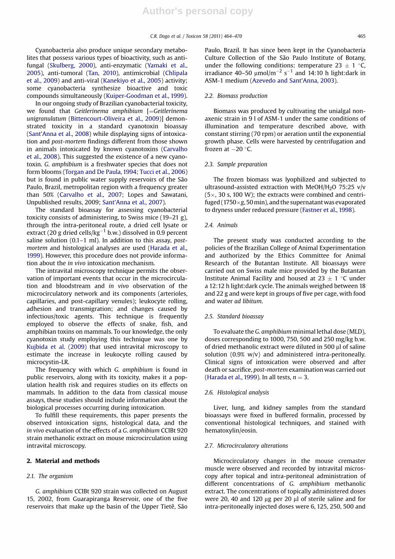
Fig. 1. Effects caused by G. amphibium CCIBt 920 methanolic extract in mouse lung. a) Control tissue, b) mild to moderate diffuse congestion (arrow) andmicrothrombus (*) after intra-peritoneal administration of a 125 mg/kg dose; c) and d) marked diffuse congestion (arrows) after intra-peritoneal administrationof a 500 mg/kg dose. HE 10� (a and b); 40� (c and d).
Fig. 2. Transient venular stasis after topical application of 120 mg of the methanolic extract. a) Venule before extract administration, b) the same venule, after5 min of extract administration and c) after 15 min of extract administration, with restored blood flow.
C.R. Dogo et al. / Toxicon 58 (2011) 464–470 467

Author's personal copy
a level which was maintained until the end of theexperiment, although this increase was not statisticallysignificant. In contrast, IL-6 increased 2 h after extractinoculation (*P < 0.05), returning to baseline after 24 h(Fig. 6).
The chemokines KC and MCP-1 both reached their peaklevels at 2 h post-inoculation (*P < 0.05) and started todecrease after the 6 h time point. KC concentrationsreturned to basal levels within 24 h, whereas MCP-1concentrations remained moderately elevated during thistime (Fig. 6).
3.4. Investigation of the presence of lipopolysaccharides inG. amphibium methanolic extract – polymixin B test andmass spectrometry (MALDI/MS)
Polymixin B did not suppress the lethal effects of G.amphibium methanolic extract when administeredtogether, as can be seen from Table 1. All animals inocu-lated with the G. amphibium methanolic extract died, asthe mice which received both extract and POL.B. Massspectrometric analyses showed that G. amphibiummethanolic extract was composed of substances withmolecular weights less than 3000 D (data not shown).Together, these results are consistent with the absence oflipopolysaccharides in the G. amphibium methanolicextract.
4. Discussion
The intoxication signs, time until death, post-mortemmacroscopic alterations and histological changes observed
Fig. 3. Effects induced by different doses of G. amphibium CCIBt 920 methanolic extract in microcirculation. a) Leukocyte rolling, after an intra-peritoneal dose of250 mg/kg b.w., b) partial venular stasis and fibrin deposit after an intra-peritoneal dose of 250 mg/kg b.w., c) venule and arteriole after an intra-peritoneal doseof 500 mg/kg b.w., and d) after an intra-peritoneal dose of 1000 mg/kg b.w. Continue arrow: venule; dashed arrow: arteriole.
Fig. 4. Total and differential leukocyte count in blood 30 min after the intra-peritoneal administration of a 125 mg/kg dose. Data are mean � S.D. of n � 3animals. *P < 0.05; **P < 0.01 compared with the respective control groups.
C.R. Dogo et al. / Toxicon 58 (2011) 464–470468

Author's personal copy
in intoxicated animals ruled out the possibility that G.amphibium toxins might be known as cyanotoxins.
MLD datum showed that G. amphibium extract can beclassified as presenting “medium toxicity”. As it contains anunknown toxin, there is a considerable potential forcausing damage or bioaccumulating in aquatic animals andeliciting injury to human beings resulting from a long termexposure.
Initial studies on the microcirculatory network viatopical administration of G. amphibium methanolic extracthave shown that only the highest dose applied (120 mg) wasable to induce changes, which were mainly transient ven-ular stasis.
The same doses and conditions that are typically used instandard mouse bioassays were employed to evaluate themicrocirculatory changes caused by G. amphibium meth-anolic extract. In cremaster muscle, lower doses (125 and250 mg/kg b.w.) and short incubation periods induced anincrease in leukocyte number, formation of fibrin deposits
Fig. 5. Total and differential leukocyte count in peritoneal cavity after intra-peritoneal administration of a 125 mg/kg dose at different incubation times.Data are mean � S.D. of n � 3 animals. ***P < 0.0001 compared with therespective control group.
3000
4000
* * 400
500
**
0
1000
2000
3000
IL
6 (p
g/m
l)
0
100
200
300***
IL
1 (
pg/m
l)
100
150 #
3
4
5
0
50
-1 (p
g/m
l)M
CP-
0
1
2-(p
g/m
l)T
NF-
80
100
# #Control0.5 hour
20
40
60
80 0.5 ou
6 hours24 hours
2 hours
KC
(pg
/ml)
0
a IL-6
MCP-1
MCP-1
TNF-α
IL1βb
c d
e
Fig. 6. Cytokines and chemokines concentrations after 125 mg/kg of G. amphibium CCIBt 920 methanolic extract administration at different incubation times (0.5,2, 6 and 24 h). a) IL-6 concentrations: *P < 0.05 compared to control, 0.5 and 24 h groups; b) IL-1b concentrations: **P < 0.0001 compared to Control, 6 and 24 hgroups and ***P < 0.05 compared to Control and 6 h groups; c) MCP-1 concentrations: #P < 0.05 compared to Control and 0.5 h groups; d) no statisticaldifferences and e) KC concentrations: *P < 0.05 compared to the other groups.
C.R. Dogo et al. / Toxicon 58 (2011) 464–470 469

Author's personal copy
and stasis of some venules (Fig. 3a and b), but they did notcause arteriolar damage. Increasing doses (500 and1000 mg/kg b.w.) and incubation periods resulted in anenhanced inflammatory response that progressed to anischemic process (Fig. 3c and d), leading to animal death.These data (Fig. 3) demonstrate that there was a dose-dependent response of inflammatory effects to cyano-toxin concentration.
Cell recruitment data from blood and the peritonealwash indicated an association between microcirculatoryalterations and a systemic inflammatory response to cya-nobacterial toxins: in both blood and cremaster musclemicrocirculation, neutrophils increased about 30 min aftercyanobacterial toxin administration; in the peritoneum theneutrophil increase occurred 6 h after administration,reaching its maximum value 24 h after inoculation whenmacrophages were also numerous (Fig. 4).
This cellmigrationpattern is analogous to those observedin acute inflammation processes: during the first 24 h, thePMN are the predominant cell type, after which the MN arethemostnumerous cells. The same inflammatory response isinduced by lipopolysaccharides from the Gram-negativebacterial cell wall (Melnicoff et al., 1989; Ryan and Majno,1977), which contains substances ranging in molecularweight from 8 to 20 kDa (Westphal and Jann, 1965) and thetoxicity of which may be suppressed by polymyxin B.
Studies on the actions of microcystin, anatoxin-a, andnodularin on the mouse immune system have shown thatthese cyanotoxins display immunosuppressive activityeffects (Rymuszka and Sieroslawska, 2009), which areantagonistic to those exerted by the toxin produced byG. amphibium. As a matter of fact, such cyanobacteriumwasnot included among the ones studied by the authors.
Acknowledgments
This work was supported by the Conselho Nacional dePesquisas CNPq (Proc. 410731/2006-4) and by a post-graduate scholarship from Coordenação de Aperfeiçoa-mento de Pessoal de Nível Superior – CAPES.
Conflict of interest
None.
References
Azevedo, M.T.P., Sant’Anna, C.L., 2003. Sphaerocavum brasiliense, a newplanktic genus and species of cyanobacteria from reservoirs of SaoPaulo State, Brazil. Algol. Stud. 109, 79–92.
Bittencourt-Oliveira, M.C., Moura, A.N., Oliveira, M.C., Massola Jr., N.S.,2009. Geitlerinema species (Oscillatoriales, Cyanobacteria) revealed
by cellular morphology, ultrastructure and DNA sequencing. J. Phycol.45, 716–725.
Carvalho, L.R., Sant’Anna, C.L., Gemelgo, M.C.P., Azevedo, M.T.P., 2007.Cyanobacterial occurrence and detection of microcystin by planarchromatography in surface water of Billings and GuarapirangaReservoirs, SP, Brazil. Rev. Bras. Bot. 30, 141–148.
Carvalho, L.R., Haraguchi, M., Górniak, S.L., 2008. Intoxicação produzidapor algas de água doce. In: Spinosa, H.S., Górniak, S.L., Palermo-Neto, J. (Eds.), Toxicologia Aplicada à Medicina Veterinária. EditoraManole, Barueri, pp. 621–640.
Chlipala, G., Mo, S., Carcache de Blanco, E.J., Ito, A., Bazarek, S., Orjala, J.,2009. Investigation of antimicrobial and protease-inhibitory activityfrom cultured cyanobacteria. Pharm. Biol. 47 (1), 53–60.
Fastner, J., Flieger, I., Neumann, V., 1998. Optimized extraction of micro-cystins from field samples: a comparison of different solvents andprocedures. Water Res. 32, 3177–3181.
Ferreira, C.K.O., Prestes, J., Donatto, F.F., Vieira, W.H.B., Palanch, A.C.,Cavaglieri, C.R., 2007. Efeitos agudos no exercício de curta duraçãosobre capacidade fagocitária de macrófagos peritoneais em ratossedentários. Rev. Bras. Fis. 11 (3), 191–197.
Granger, D.N., Kubes, P., 1994. The microcirculation and inflammation:modulation of leukocyte-endothelial cell adhesion. J. Leukoc. Biol. 55,662–675.
Harada, K., Kondo, F., Lawton, L., 1999. Laboratory analysis of cyanotoxins.In: Chorus, I., Bartram, J. (Eds.), Toxic Cyanobacteria in Water. A Guideto their Public Health Consequences, Monitoring and Management. E& FN SPON, New York, pp. 369–399.
Kanekiyo, K., Lee, J.-B., Hayashi, K., Takemaka, H., Hayakawa, Y., Endo, S.,Hayashi, T., 2005. Isolation of an antiviral polysaccharide, nostoflan,from a terrestrial cyanobacterium, Nostoc flagelliforme. J. Nat. Prod. 68(7), 1037–1041.
Kuiper-Goodman, T., Falconer, I., Fitzgerald, J., 1999. Human healthaspects. In: Chorus, Bartram, J. (Eds.), Toxic Cyanobacteria in Water: AGuide to their Public Health Consequences, Monitoring andManagement. E & FN Spon, New York, pp. 113–153.
Kujbida, P., Hatanaka, E., Vinolo, M.A.R., Waisman, K., Cavalcanti, D.M.H.,Curi, R., Farsky, S.H.P., Pinto, E., 2009.Microcystins -LA, -YRand -LRactionon neutrophil migration. Biochem. Biophys. Res. Commun. 382, 9–14.
Lomonte, B., Lungren, J., Johansson, B., Bagge, U.,1994. The dynamics of localtissue damage induced by Bothrops asper venom andmyotoxin II on themouse cremaster muscle; an intravital study. Toxicon 32, 41–55.
Melnicoff, M.J., Horan, P.K., Morahan, P.S., 1989. Kinectics of changes inperitoneal cell populations following acute inflammation. Cell.Immunol. 118, 178–191.
Ryan, G.B., Majno, G., 1977. Acute inflammation: a review. Am. J. Pathol.86, 185–276.
Rymuszka, A., Sieroslawska, A., 2009. The immunotoxic and nephrotoxicinfluence of cyanotoxins to vertebrates. Centr. Eur. J. Immunol. 34 (2),129–136.
Sant’Anna, C.L., Azevedo, M.T.P., 2006. Identificação e ilustração dosprincipais gêneros de cianobactérias. In: Sant’Anna, C.L., Azevedo, M.T.P., Agujaro, L.F., Carvalho, M.C., Carvalho, L.R., Souza, R.R.C. (Eds.),Manual ilustrado para identificação e contagem de cianobactériasplanctônicas de águas continentas brasileiras, first ed. Interciência,Rio de Janeiro, pp. 35–53.
Sant’Anna, C.L., Melcher, S.S., Carvalho, M.C., Gemelgo, M.P., Azevedo, M.T.P., 2007. Planktic cyanobacteria from Alto Tietê basin, SP, Brazil. Rev.Bras. Bot. 30, 1–17.
Sant’Anna, C.L., Azevedo, M.T.P., Werner, V.R., Dogo, C.R., Rios, F.R.,Carvalho, L.R., 2008. Review of toxic species of cyanobacteria in Brazil.Algol. Stud. 126, 251–265.
Skulberg, O.M., 2000. Microalgae as a source of bioactive molecules –
experience from cyanophyte research. J. Appl. Phycol. 12, 341–348.Tan, L.T., 2010. Filamentous tropical marine cyanobacteria: a rich source
of natural products for anticancer drug discovery. J. Appl. Phycol.. doi:10.1007/s10811-010-9506-x.
Torgan, L.C., De Paula, M.C.E., 1994. Geitlerinem unigranulatum (Ag. ExGom. Anagn.) (Cyanophyta-Pseudanabaenaceae) em um lago do suldo Brasil. Iheringia (Sér. Bot.) 45, 75–87.
Tucci, A., Sant’Anna, C.L., Gentil, R.C., Azevedo, M.T.P., 2006. Fitoplânctondo Lago das Garças, Sao Paulo, Brasil: um reservatório urbanoeutrófico. Hoehnea 33 (2), 147–175.
Westphal, O., Jann, K., 1965. In: Whistler, R.L., Wolfan, M.L. (Eds.), BacterialLipopolysaccharides: Extraction with Phenolwater and FurtherApplications of the Procedure. Methods in Carbohydrate Chemistry,vol. 5. Academic Press Inc., New York, pp. 83–91.
Yamaki, H., Sitachitta, N., Sano, T., Kaya, K., 2005. Two new chymotrypsininhibitors isolated from the cyanobacterium Microcystis aeruginosaNIES-88. J. Nat. Prod. 68, 14–18.
Table 1Results presented by the tests with G. amphibium CCIBt 920 meth-anolic extract and polymyxin B.
Group Survivors/tested animals
Co 3/3CCIBT 920 0/3POL.B 3/3920 þ POL.B 0/3
C.R. Dogo et al. / Toxicon 58 (2011) 464–470470
Copyright © 2022 FDOKUMEN