Binding characteristics of copper and cadmium by cyanobacterium Spirulina platensis
Upload
independentCategory
view
1download
0
ELSEVIER Plant Science 136 (1998) 109- 120
Purification and characterization of phycocyanin from marine cyanobacterium Synechococcus sp. 109201
the
Julio Abalde”**, Liliana BetancourPb, Enrique Torres”, Angeles Cid”, Clive Barwell’
” Lahoratorio de Microhiologia. Facultad de Ciencias, Unioersidad de A Coruria, Campus da Zapateira s:n, 15071 A Coruria. Spain
b Instituto de Oceanologia. La Hahana, Cuba
’ S~~hool qf’ Pharmacy and Biomedical Science. Unirersity of Portsmouth, Portsmouth, L’R
Received 26 February 1998: received in form 3 June 1998: accepted 8 June 1998
Abstract
This paper describes a suitable method for the optimum extraction and isolation of phycocyanin from the cyanobacterium SJlnechococcus sp. 109201 isolated from Caribbean waters. Phycocyanin from this microorganism
was purified to homogeneity and some of its properties were investigated. The purification steps consisted of extraction, hydrophobic interaction chromatography and ion exchange chromatography. Freezing at - Zl”C-thaw- ing at 4°C using an alkaline buffer was the best method for extracting phycocyanin from Synechococcus sp. 109201. The best extraction was obtained using butyl-sepharose resin for hydrophobic interaction chromatography and 0.05 M Tris-HCl (pH = 7) containing 10% ethanol for phycocyanin elution. Finally, phycocyanin was further purified by ion exchange chromatography using Q-sepharose and eluted with a complex isocratic system. The estimated molecular weight of the phycocyanin purified from Synechococcus sp. 109201 was 102 000 daltons by gel filtration
and the isoelectric point was 4.6. When analyzed by SDS-PAGE, Synechococcus sp. 109201 phycocyanin migrated as two bands having an apparent molecular weight of 21360 and 18 980 Da. The first band corresponds to /j phycocyanin subunits, whereas the second corresponds to c( phycocyanin subunits. So. this phycocyanin was characterized as (s(~~~/),~~~)). 6 1998 Elsevier Science Ireland Ltd. All rights reserved.
Phycocyanin; Protein purification; Cyanobacteria; Synechococcus sp.
1. Introduction
* Corresponding author. E-mail: [email protected]
The phycobiliproteins are pigments present in cyanobacteria, red algae and cryptomonads. They
0168-9452/98/$19.00 ‘(:s 1998 Elsevier Science Ireland Ltd. All rights reserved.
PII: SOl68-9452(98)001 13-7
are in association with the outer surface of the photosynthetic lamellae, being constituents of the photosystem II light-harvesting apparatus. The phycobiliproteins are organized into supramolecu- lar complexes, the phycobilisomes, which are found as regular arrays on the surface of thy- lakoid membranes [1,2]. Phycobiliproteins are a brilliantly colored family of water-soluble proteins bearing covalently attached, open-chain te- trapyrroles known as phycobilins. On the basis of their visible absorption properties, the phyco- biliproteins have been assigned to four spectro- scopic classes: phycoerythrocyanin WC, /i Amax = 575 nm). phycoerythrins (PE, &_ = 565575 nm), phycocyanins (PC. AArnax = 615- 640 nml and allophycocyanin (APC, 1 Amax = 650-655 nm) [3]. Phycobilisomes consist of allophycocyanin cores surrounded by phyco- cyanin and (when present) phycoerythrin on the periphery. Several colorless polypeptides serve to link phycobiliproteins in the phycobilisome and to attach them to the thylakoid membrane [4]. The fundamental unit of all phycobiliproteins consists of the trimeric or hexameric aggregation (olfi),, (ap), of two dissimilar polypeptides, SI and jj’ subunits present in an equimolecular stoichiome- try [5-71. Phycobiliproteins are easily isolated as pigment-protein complexes, soluble in water and very fluorescent [8]. Thus, the purification proce- dure is facilitated because the phycobiliproteins are very soluble in water, allowing to separate these molecules from other pigments that are not. These molecules have been extracted from algae and cyanobacteria by different methods [9], but appropriate control of ionic strength and pH dur- ing the extraction procedure is crucial for complex stability. Purification methods usually rely on gel, adsorption and ion exchange chromatography [6,10, Ill. Present purification procedure consists of three steps: extraction, hydrophobic and ion exchange chromatography.
Some characteristics of phycobiliproteins make them well-suited for fluorescence analyses as fluorescent tags with numerous applications in flow cytometry, histochemistry, immunoassay and detection of reactive oxygen species [12]. They can also be used as natural dyes [ 131 and antitumoral agents [ 141.
This paper describes a suitable method for the optimum extraction and isolation of phycocyanin from the cyanobacterium S~nrchococcz~s sp. 109201. Synechococcus spp. are important com- ponents of the marine microbial food web. These organisms are widely distributed, occurring in both oligotrophic waters as well as in temperate coastal and oceanic waters [15,16].
2. Materials and Methods
2.1. Organism and culture conditions
The marine cyanobacterium Synechococcus sp. 109201 was obtained from the Culture Collection of Instituto de Oceanologia of Cuba. This organ- ism was cultured in ALGAL-l medium [17] at 18 + 1°C with constant bubbling of air at a flow rate of 2 1 min ‘. Cultures were illuminated with a light intensity of 60 nE m ’ s - ‘, provided by cool-white fluorescent tubes and with a dark:light cycle of 12:12 h.
2.2. Prepuration of cell-free extracts
Cells were harvested by centrifugation at 5000 x g for 5 min and resuspended in distilled water. Three extraction methods were assayed:
l sonication at 4°C: resuspended cells were disrupted by sonication for 5 min at lo4 Kcy- cles - ‘. The extract was clarified by centrifuga- tion at 10 000 g for 15 min.
l freezing in liquid N,-thawing at 4°C: resus- pended cells were frozen in liquid Nz and thawed at 4°C three times. The extract was clarified by centrifugation at 10000 x g for 15 min.
0 freezing at - 21”C-thawing at 4°C: resus- pended cells were successively frozen at - 2 1°C and thawed at 4°C three times. The extract was clarified by centrifugation at 10000 x g for 15 min. Different extraction solu- tions were assayed with this method: distilled water, 10 mM phosphate buffer (pH = 7), 0.15 M sodium chloride. 0.05 M Tris-HCl (pH = 7) calcium chloride 10 g 1~ ’ and an alkaline buffer composed of 0.5 g l_ ’ NaHCO, and 0.5

J. Abake et al. /Plant Science 136 (1998) IOY- 120
0.05 /’
O-1 550 600 625
Fig. 1. Absorption spectra of purified C-phycocyanin from S~~c~/~cocctrs sp. 109101
g 1 --’ CaCO, (pH = 10.5). All solutions con- tained 0.05% sodium azide to avoid microbial growth. The phycocyanin concentration and purity in
the supernatants were measured and calculated as described in Section 2.3.
-7.3. Analyticul procedures
Ph?-cocyunin concentration: the phycocyanin (PC) concentration in the supernatants was calcu- lated by spectrophotometric absorption at 652 and 615 nm using following equation [18]:
PC (mg ml-‘) = (AhI5 - 0.474 (&))/5.34
Pur$ication jirctor: the purity of phycocyanin
extract was monitored spectrophotometrically by
the A,&AzXO ratio.
-7.4. Ewluation qf the puriJication method
Hydrophobic interaction chromatography and ion exchange chromatography were used and evaluated as a two-step procedure for purifying the extracted phycocyanin obtained from the cyanobacterium Synechococcus sp. 109201.
2.4.1. Hydrophobic interuction chromutogruph~~ Resin evaluation. Different resins were assayed
for the phycocyanin purification: butyl-sep- harose4-fast flow, octyl-sepharoseCL-4B and phenyl-sepharose6-fast flow (Pharmacia). 0.5 ml of each resin was placed in a tube and equili- brated with 0.05 M Tris-HCl (pH = 7) containing 0.5 M (NH&SO,. After equilibration. the resins were centrifuged at 700 x g for 2 min and the supernatant was discarded. The resins were loaded with 2 ml of the Synechococcus sp. 109201 phycocyanin extract in the presence of solid (NH,),SO, at a final concentration of 0.5 M. The
Table I Comparison of different methods for the extraction of phyco-
cyanin from S~ne&c~occ~us sp. 109201 using distilled water as
solvent
Extraction method Concentration Purity (,d,,,,, AZnO)
(pg ml-‘)
Sonication at 4°C 7.44 + 0.0’ (I. I Freezing at -3l”G 13.42 + 0.02 0.17
thawing at 4°C
Freezing in liquid 9.41 * 0.01 0.18
N, thawing at 4°C _____

Table 2
Concentration and purity of the phycocyanin extracted from
S~~~c/~ocuc~c~s sp. 109201 by the freezing at -Zl’C-thawing
at 4°C method with different extraction solutions
Extraction solution Concentration Purity (,4dA2d (p&! I-‘)
Distilled water
Alkaline phase
Tris-HCI 0.05 M
(pH = 7)
13.42 i I .46 0.27
27. II + 3.6 0.43
11.91 * I.91 0.75
Calcium chloride
Phosphate buffer IO
mM (pH = 7)
9.76+ I.16 0.25
9.47 + I .33 0.19
Sodium chloride 9.42 & 0.37 0.18
mixture was gently shaken for 10 min. For pig- ment release, different solutions were assayed and added to each tube: decreasing concentrations of (NH&SO, (0.5, 0.25, 0.125 and 0 M) prepared in 0.05 M Tris-HCl (pH = 7) and 30% ethanol in 0.05 M Tris-HCl (pH = 7). After 10 min, the resin with each solution was centrifuged at 700 x g for 2 min, and the supernatants were monitored spectrophotometrically.
Purification on butyl-sepharose4-fast flow. A phycocyanin extract was chromatographed on a butyl-sepharose column (Pharmacia XK26) (2 x 2.6 cm) at a flow rate of 2 ml min ~ ’ and at room
temperature. The elution was monitored at 620 nm. Different elution conditions were assayed:
gradient elution with 0.5 to 0 M (NH,),SO, in
0.05 M Tris-HCl (pH = 7). gradient elution with 0.5 to 0 M (NHJ2SOJ in 0.05 M Tris--HCl (pH = 7) containing 50%
ethanol. isocratic elution with 0.05 M Tris-HCl (pH = 7) containing 35% ethanol. isocratic elution with 0.05 M Tris-HCl (pH =
7) containing 10% ethanol. isocratic elution with only 0.05 M Tris-HCI (pH = 7) solution. In all cases. 20 ml of the pigment-protein ex-
tract containing 0.5 M (NH,),SO, were applied to the column and they were eluted with 50 ml of each mobile phase. The fractions obtained were characterized spectrophotometrically.
2.4.1. Ion t3&lnge c.hr.or?lutogr.uph!,
The phycocyanin fraction from the hydropho- bic interaction chromatography eluted with 0.05 M Tris-HCl (pH = 7) containing 10% ethanol was chromatographed on an ion exchange column (Pharmacia XK16) (2 x 1.6 cm). The stationary phase used was 5 ml of Q-sepharose-fast flow resin (Pharmacia). Different elution conditions
were assayed:
0.25
ethanol 20% 0.000 0.125 0.250 0.500
Amonium sulfate concentration
Fig. 3. Evaluation of different hydrophobic interaction resins for the purification of S~,)~(.(./~O(.O(.(.U.) sp. 109201 phycocyanin

Table 3 Evaluation of elution systems and mobile phases used in hydrophobic interaction chromatography (butyl-sepharose) for the purification of S?,nrc,hoc,occ~ts sp. 109201 phycocyanin
-
Elution system Mobile phase Purity (A,,,,, Recover\ (“,;N)
.~?,,,I - .___ __~
Gradient 0.5- 0 M (NH&SO, in 0.05 M Tris--HCl (pH = 7) 3.8 43.1
grndicnt O.S- II M (NH,),SO, in 0.05 M Tris--tiCI (pH = 7) containing 50”;1 2.9 hO.7
ethanol
Iaocrntic 0.05 M Tris- HCI (pH = 7) containing 25’!;, ethanol 2.x 6.2
laowrtic 0.05 M Tris HCI (pH = 7) containing lO”,L ethanol 3.1 12.2
Isocratic 0.05 M Tris -HCI (pH = 7)
gradient elution with 0 to 4 M NaCl in 0.05 M containing 10% ethanol. The chromatography was Tris-HCl (pH = 7). recorded at 380 and 610 nm.
isocratic elution with 0.05 M Tris-HCl (pH = 7) containing 0.3 M NaCl. isocratic elution, the phycocyanin fraction was previously dialyzed against 50 mM acetate
buffer (pH = 5). The column was equilibrated with 50 mM acetate buffer (pH = 5); the sample was loaded, and the column was washed with
this acetate buffer and with 0.05 M Tris-HCl (pH = 7); finally, the sample was eluted with 0.05 M Tris HCl (pH = 7) containing 0.2 M NaCI.
In all cases, the sample was eluted with 50 ml of
The fractions corresponding to phycocyanin were pooled and dialyzed against 50 mM acetate buffer pH = 5.
The dialyzed sample was applied to an ion exchange column containing Q-sepharose. The column was equilibrated with 50 mM acetate buffer pH = 5 and after the sample was loaded, the column was washed with the same buffer. Later, the column was washed with 0.05 M Tris-HCl (pH = 7) and the sample was eluted with 0.05 M Tris- HCl (pH = 7) containing 0.2 M NaCl. The chromatog- raphy was recorded at 380 and 670 nm. each mobile phase at a flow rate of 3 ml min ‘. The
elution was monitored at 620 nm and the fractions
obtained were characterized spectrophoto- metrically.
On the basis of the results obtained in the
previous sections, phycocyanin from S~~ecI~ococ~- (‘IIS sp. 109201 was purified using the most suitable
method. A clarified crude extract obtained by the freezing at - 21”C-thawing at 4°C method and with the alkaline buffer was diluted with 0.05 M Tris-- HCl (pH = 7) containing 0.5 M (NH,),SO, to obtain an absorbance of 1.1 at 620 nm. A total of 100 ml of this extract was applied to an hydropho- bic interaction column containing 20 ml of butyl- scpharose. The column was previously equilibrated with 0.05 M Tris-HCl (pH = 7) and 0.5 M (NH,),SO,. After the sample was loaded the column was eluted with 0.05 M Tris-HCl (pH = 7)
Molecular weight estimation of the native Sow- t~hocotws sp. 109301 phycocyanin was performed under nondenaturing conditions by gel filtration chromatography. The sample was injected on a Superdex 300 (98 x I cm. Pharmacia) HPLC column and eluted with 0.05 M Tris HCI (pH = 7) buffer containing 0.1 M NaCl at a flow rate of 0.5 ml min ‘. The column was previously equilibrated with the same buffer. The chromatography was monitored at 380 nm.
The column was calibrated with the following standard proteins: apoferritin (443 OOO), /j -amylase (300 000). lactate dehydrogenase ( 140 000), Bovine serum albumin (66 000). carbonic anhydrase (39 000) and cytochrome c ( 12 400).

114 J. Abalclr et al. , Plam Science 136 (1998) 10% l-70
2.52. Isoelectric jbcusing 3. Results The isoelectric point of the purified phyco-
cyanin was estimated by isoelectric focusing. An horizontal electrophoresis chamber (Multiphor II, Pharmacia Biotech) with a polyacrylamide gel (Ampholine PAG plate, Pharmacia Biotech) was used. The carrier electrolytes were of wide pH range (3.5-9.5). The separation was run at a constant power of 30 W and a controlled temperature of 10°C. The anode solution was 1 M H,PO, and the cathode solution was 1 M NaOH.
3.1. Evaluation of dlyferent methods for the estraction of phycocyanin jiom Synechococcus sp. I09201
The different methods used for the extraction of phycocyanin showed the presence of this molecule in the Synechococcus sp. 109201. The absorption spectra of the extracts from this cyanobacterium showed a single peak at 620 nm, corresponding to C-phycocyanin (Fig. 1) [20].
The isoelectric point of phycocyanin was deter- mined using protein markers of known isoelec- tric points: trypsinogen (PZ = 9.30), lentil lectil basic (8.65), lentil lectil middle (8.45), lentil lectil acidic (8.15), myoglobin basic (7.35), myoglobin acidic (6.85), carbonic anhydrase (human) (6.55). carbonic anhydrase (cow) (5.85), a-lactoglobulin A (5.20), trypsin inhibitor (4.55), amyloglucosi- dase (3.50). Proteins were visualized with a stain- ing solution PhastGel Blue R (Pharmacia Biotech).
2.5.3. Determination of the molecular weight by SDS-PAGE
SDS-PAGE was performed with an Excel SDS Homogeneous 12.5 gel (Pharmacia Biotech) as described in [19]. Proteins were detected by the same procedure used for isoelectric focusing. The standard proteins were: phosphorilase b (94 OOO), bovine serum albumin (67 OOO), ovalbumin (43 000), carbonic anhydrase (30 000), soya-bean trypsin inhibitor (20 100) and sc-lactalbumin (14000).
Three extraction methods were assayed and their efficiency was determined by means of the phycocyanin concentration and purity (expressed as A,2,/A2,, ratio). The highest phycocyanin con- centration (13.42 pg ml - ‘) and the higher purity (0.27) were obtained with the freezing at - 21”C- thawing at 4°C method (Table l).Once the freez- ing at - Zl”C-thawing at 4°C method was selected for the extraction of phycocyanin from Synechococcus sp. 109201, different extraction so- lutions were assayed to obtain higher concentra- tions and purity than with distilled water. The results are showed in Table 2. The highest concen- tration (27.11 pg ml - ‘) and purity (0.43) were obtained with the alkaline solution containing 0.5 g 1~~ ’ NaHCO, and 0.5 g 1~ ’ CaCO, (pH = 10.5). The remaining extraction solutions yielded worse results than those obtained with distilled water.
3.1.1. Evuluution oj’ the pur$ication method
Hydrophobic interaction chromatography: hy- drophobic interaction chromatography was used for the purification of phycocyanin from Syne-
Table 4
Evaluation of elution systems and mobile phases used in ion exchange chromatography (Q-sepharose) for the purification of
.S~wecl~~occus sp. 109201 phycocyanin
Elution system Mobile phase Purity (A,Z,,: Recovery (%)
AXl,)
Gradient
Isocratic
Isocratic
O-4 M NaCl in 0.05 M Tris+HCI (pH = 7)
0.05 M Tris-HCI (pH = 7) containing 0.2 M NaCl
Loading in 50 mM (pH = 5) acetate buffer and eluting with 0.05 M
Tris-HCI (pH = 7) containing 0.2 M NaCl
4.1 34.7
4.2 45.8
4.5 46.2

J. Ahulir et ul. ‘Plnnr Science 136 (1998) 109. l-70
0.00 50.00 100.00 150.00 200.00
Fig. 3. Chromatogram of phycocyanin crude extract obtained by
recording the absorbance at 380 and 620 nm.
Lhococ~us sp. 109201, but previously different resins and different eluents were evaluated. The results are showed in Fig. 2. The data obtained indicate that the phenyl- and octyl-sepharose are not suitable for the purification of this phyco- cyanin because the protein is adsorbed too strongly to the resin and the recovery is very low (19 and 8.9”%, respectively). The highest re- covery was achieved with the butyl-sepharose resin.
Once the resin was selected, the purification of phycocyanin was performed by means of hydro- phobic interaction chromatography using this
resin. Different elution conditions were assayed. Table 3 shows that the best purity (3.8) was obtained with the gradient system using 0.5 M to 0 M (NH&SO, in 0.05 M Tris-HCl (pH =
7) although recovery was low (43.1%) in com- parison with isocratic system using 0.05 M Tris--HCl (pH = 7) containing 10% ethanol (72.20%). Since the purity values obtained with the different elution systems were very similar, the isocratic system with 0.05 M Tris-HCl (pH = 7) containing 10% ethanol was selected
min
hydrophobic interaction chromatography on butyl-sepharose
because with this system the recovery was the highest.
Ion exchange chromatography: the fraction containing the phycocyanin was applied to a Q- sepharose column and different elution systems were assayed. The results are showed in Table 4. The best purity (4.5) and recovery (46.2%) were obtained with an isocratic system in which the
extract was loaded on the column in 50 mM acetate buffer (pH = 5) and eluted with 0.05 M Tris-HCI (pH = 7) containing 0.2 M NaCl.
3.1.2. Pur$ication oJ’ph~voc~-anin ,fiot71 Synechococcus sp. 109201
With the obtained results, S~wechococcus sp. 109201 phycocyanin was purified by hydropho- bic interaction chromatography and ion ex- change chromatography using the suitable methods. Fig. 3 shows the chromatogrdm ob- tained from hydrophobic interaction chromatog- raphy on butyl-sepharose recording the absorbance at 280 and 620 nm. The sharp peak detected at 620 and 280 nm is the phycocyanin and the wide peak detected only at 280 nm cor-

116 J. Abalde et al. /Plant Science 136 (1998) 109- 120
mV
3500.00
2250.00
15oo.oa
75o.oc
?
. l .
#
:
: I r
I I I I 0.00 50.00 100.00 150.00 200.00
min
Fig. 4. Q-sepharose chromatography of the hydrophobic interaction fraction containing the phycocyanin. The absorbance was
recorded at 280 and 620 nm.
responds to contaminating molecules present in the extract. After hydrophobic interaction chro- matography, the recovery was 83.4% with a purification factor of 3.1.
Fig. 4 shows the chromatogram obtained from ion exchange chromatography on Q-sepharose recording the absorbance at 280 and 620 nm. Only one peak was obtained, both at 280 and 620 nm, corresponding to phycocyanin. The purity factor was 4.85 and the recovery was 76.56% in relation to the crude extract.
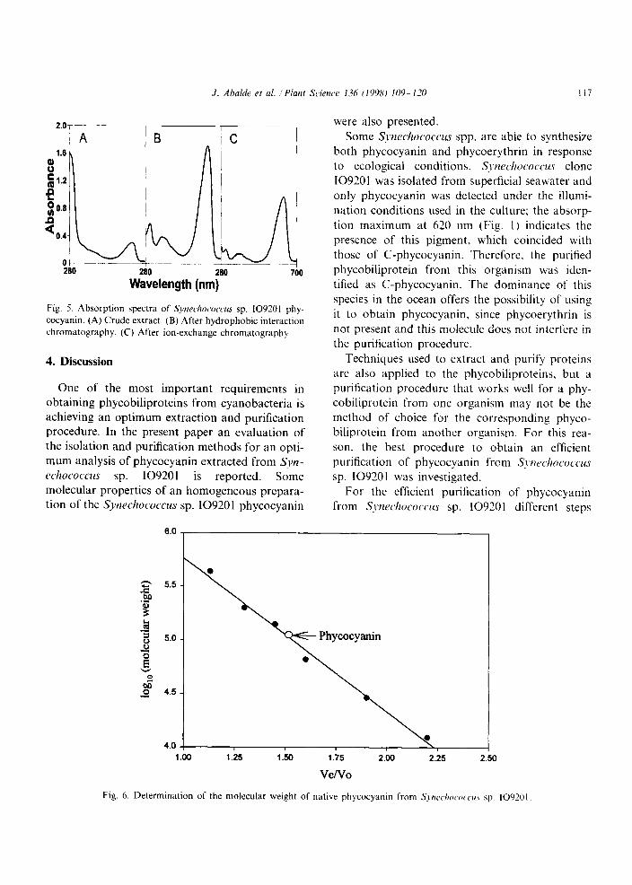
The results from the purification procedure are shown in Table 5. The spectrophotometric charac- terization of the different phycocyanin fractions eluted in each purification step confirms the re- sults obtained (Fig. 5).
3.1.3. Determination of the molecular weight The molecular weight of the native puri-
fied phycocyanin was determined by gel filtra- tion on a Superdex 200 column. The molecu- lar weight obtained was 102 960 + 670 daltons (Fig. 6).
3.1.4. Determination of the isoelectric point The isoelectric point of the native phycocyanin
purified was determined by isoelectric focusing. The phycocyanin fraction was detected in a single band with an isoelectric point of 4.6 (Fig. 7).
3.1.5. Molecular weight by SDS-PAGE Two bands (A and B) were observed on the gels
and their molecular weights were calculated to be 21 360 + 980 and 18 980 + 870 Da, respectively (Fig. 8).
Table 5
Summary of the purification of S~~cl~ococcus sp. 109201
phycocyanin
Purification
step
Purification factor
(A&AA
Recovery (“A)
Crude extract 0.43 100 Butyl-sepharose 3.2 83.4 Q-sepharose 4.85 76.56
J. Ahal& et al. ‘Piam Srienw 136 (1998) lO9- l-70 117
-7----- ,
_ ic I
Wavelength (nm)
Fig. 5. Absorption spectra of .S~~~echococcus sp. IO9201 phy-
cocyanin. (A) Crude extract. (B) After hydrophobic interaction
chromatography. (C) After ion-exchange chromatography.
4. Discussion
One of the most important requirements in obtaining phycobiliproteins from cyanobacteria is
achieving an optimum extraction and purification
procedure. In the present paper an evaluation of
the isolation and purification methods for an opti-
mum analysis of phycocyanin extracted from S_VZ- ~chococcus sp. 109201 is reported. Some
molecular properties of an homogeneous prepara-
tion of the S~~rchococcus sp. 109201 phycocyanin
were also presented.
Some S~nechococcus spp. are able to synthesize
both phycocyanin and phycoerythrin in response
to ecological conditions. S~~echococcus clone
109201 was isolated from superficial seawater and
only phycocyanin was detected under the illumi-
nation conditions used in the culture; the absorp-
tion maximum at 620 nm (Fig. I) indicates the
presence of this pigment, which coincided with
those of C-phycocyanin. Therefore, the purified
phycobiliprotein from this organism was iden-
tified as C-phycocyanin. The dominance of this
species in the ocean offers the possibility of using
it to obtain phycocyanin, since phycoerythrin is
not present and this molecule does not interfere in
the purification procedure.
Techniques used to extract and purify proteins
are also applied to the phycobiliproteins, but a
purification procedure that works well for a phy-
cobiliprotein from one organism may not be the
method of choice for the corresponding phyco-
biliprotein from another organism. For this rea-
son, the best procedure to obtain an efficient
purification of phycocyanin from S_t~rcl~ococcus
sp. 109201 was investigated.
For the efficient purification of phycocyanin
from S~~~zeclzococ~~ sp. 109201 different steps
1.00 1.25 1.50 1.75 2.00 2.25 2.50
Vefvo
Fig. 6. Determination of the molecular weight of native phycocyanin from S?,nc,c./~oc,~,c,t,~r.c sp. 109201,

PH (PII + ANODE
3.5 ~__
4.6 --
5.2 -~
5.9 ~--
8.2 --
;*; XI .
9.3 _-.__ - CATHODE
.-
A B
Fig. 7. Separation of phycocyanin from S~nec/rococc~~ sp.
109201 by isoelectric focusing. (A) Protein markers of known
isoelectric point: trypsinogen (pl= 9.30). lentil lectil basic
(8.65). lentil lectil middle (8.45). lentil lectil acidic (8.15).
myoglobin basic (7.35) myoglobin acidic (6.85). carbonic an-
hydrase (human) (6.55) carbonic anhydrase (cow) (5.85). /I-
lactoglobulin A (5.20). trypsin inhibitor (4.55).
amyloglucosidase (3.50). (B) Phycocyanin purified from S.txe-
~~/IO(.ONU.S sp. 10920 I
were used. In each step, different systems were assayed. First, different extraction methods were assayed. The extraction procedure and extraction buffers used can greatly affect the pigment recov- ery. Highest concentrations and purity were ob- tained when S~~rechococcus cells were broken by freezing at - Zl”C-thawing at 4°C (Table 1). This method is usually used to extract pigments from cryptomonads [21,22] and besides of this, it is an economical method. Other method used to extract biliproteins from cryptomonads was by freezing in liquid nitrogen-thawing at 4°C [23], this method was used with Svnechococcus sp. 109201 cells, but it gave worse results than freezing at - 21”C- thawing at 4°C (Table 1). Cyanobacteria are usu- ally ruptured with a French press or by ultrasonication [24,25], but ultrasonication did not
give good results with Synedzococcus sp. 109201 cells (Table 1).
The extraction buffer composed of 0.5 g 1~ ’ NaHCO, and 0.5 g l- ’ CaCO, (pH = 10.5) was the most suitable for extracting phycocyanin from
S?,tzc~~ho~occlls sp. 109201 (Table 2). Phosphate buffer and distilled water were used to extract phycocyanin from Anubwnu sp. and cryptomonads [5,23] but these buffers gave worse results with S_t~echococlus sp. 10920 1. Calcium chloride was also used to extract phycocyanin from Spirulinu
rnusitm with good results [l 11, but the extraction of phycocyanin from S~~cckococcus sp. 109201 using this solution was poor.
Once the suitable extraction method was deter- mined, hydrophobic interaction chromatography and ion-exchange chromatography were used for the purification steps.
The pigments extracted are usually salted out
with ammonium sulphate and after dialyzed overnight. To avoid the dialyzed step, hydrophobic interaction chromatography was used. Using this chromatographic technique instead of dialysis al- lows to do other purification step. which improves the purification. Different resins were evaluated for
hydrophobic interaction chromatography since choosing the right hydrophobic ligand is of consid- erable importance in protein recovery. Synechococ- ms sp. 109201 phycocyanin was absorbed too
94000
67000
43000
30000
1 2
Fig. 8. SDS-PAGE of purified phycocyanin from SJWC/WWC-
cus sp. 109201, Lane I, phycocyanin from S_t~rc/zococctrs sp.
109201: lane 2. marker proteins: phosphorilase b (94000).
bovine serum albumin (67 000). ovalbumin (43 000). carbonic
anhydrase (30000). soya-bean trypsin inhibitor (20 100) and
%-lactalbumin (14000).

.I. Ah& et al. Phi Shncr 116 (1998) 109-120 I IU
strongly to octyl-sepharose and therefore, very
strong eluting conditions would be required (Fig.
2). In contrast, butyl-sepharose was more suitable
for phycocyanin recovery. Since a variety of conditions can be used for
elution from hydrophobic resins, it was necessary
to select the suitable elution method for the opti-
mum Qnechococcza sp. 109201 phycocyanin sep-
aration. In this case, after studying different
elution conditions an isocratic system using 0.05
M Tris-HCl (pH = 7) containing 10% ethanol
was shown to be the best for eluting this protein
and for obtaining an optimum Synechococczu sp.
109201 phycocyanin separation (Table 3).
The next purification step was ion exchange
chromatography. This procedure is the most com-
monly used chromatographic method for protein
purification, including phycobiliproteins
[5,10,25,26]. In the case of S~wechococcus sp.
109201 phycocyanin ion exchange chromatogra-
phy using Q-sepharose and an isocratic system
with a complex mobile phase (Table 4) yielded the best results.
C-phycocyanin is considered pure (especially
for applications as a fluorescent label) when the
absorption ratio of visible maximum to 280 nm is
greater than 4.0. With the purification protocol
used in this work, the purification factor obtained was 4.85 (Table 5); in other works the purity
factor obtained for phycobiliproteins was 3.91,
6.13 or 2.10 [lO,l 1,271. This purification proce-
dure resulted in a homogeneous preparation as
seen by the single protein band obtained by non-
denaturing electrophoresis. This same result was
obtained in the isoelectric focusing gel (Fig. 8).
The purified phycocyanin from S~nrcfm~occus
sp. 109201 had a molecular weight of 102 960 i_
670 Da on gel permeation and a p/= 4.6. The phycocyanin was separated by SDS-PAGE to de-
termine its subunit composition. The elec-
trophoresis revealed two bands with molecular
weights of 18 980 + 870 and 21 360 + 980. In ac-
cordance with the XP structure, the first band
corresponds to a subunit and the second one corresponds to b subunit. The 102 960 molecular weight of Synechcoccus sp. 109201 phycocyanin
when divided by the monomers molecular weight
suggests that it is a grouping of trimers (a/~‘)?
(18980+21360)*3=121020.
In conclusion, the present work describes a
complete method for the purification of C-phyco- cyanin from the marine cyanobacterium SJ~FW-
chocotws sp. 109201. Cells were broken by freezing at - 21”C-thawing at 4°C and the phy-
cocyanin was extracted by using an alkaline
buffer. Hydrophobic interaction chromatography
and ion exchange chromatography were used to
purify the phycocyanin. With this procedure. phy-
cocyanin was purified with an Ah2,,/A1X,, of 4.85.
Obtained data for phycocyanin S~~~zechococcus
sp. 109201 are in accordance with a trimeric structure of this molecule.
References
[II
PI
PI
PI
151
tfJ1
[71
[81
E. Gantt, Phycobilisomes, Ann. Rcl. Plant Physiol. 32
(1981) 327 -347.
C. Gcimez-Lojero. B. P&z-G6mer. G. Prado-Flores,
D.W. Krogmann. A. C6rabez-Trejo. ,A. Peiia-Diaz. The
phycobilisomes of the cyanobacterium ..1rfhro.v~iru (Spir-
dimr) ml.virmr. Int. J. Biochem. Cell Biol. 29 (19Y7)
II91 1205.
W.A. Sidler. Phycobilisome and phycobiliprotein struc-
tures. in: D.A. Bryant (Ed. ). The Molecular Biology ol
Cyanobacteria. vol. I. Kluwer Academic. Dordrecht. The
Netherlands. 1994. pp. 140 -316.
G. Cohen-Bazire. D. Bryant. Phycobilisomcs: composi-
tion and structure. in: N. Carr, B. Whitton (Eds.). The
Biology of the Cyanobacteria. Blackmcll. NCM’ York.
1982. pp. I& 191.
.4. Ducret. W. Sidler. E. Wehrli. G. I-rank. H. Zuber.
Isolation. characterization and electron microscopy analy-
sis of a hemidiscoidal phycobilisome type from the
cyanobacterium .~r~crhtrozu sp. PCC 7 120. Eur. J.
Biochem. 236 (1996) 1010~ 1024.
N.R. Hayashi. K. Terazono. N. Hasegawa. T. Kodama.
I’. Igarashi. Identification and characterization of phyco-
biliprotein from II thermophilic cyanobacterium.
(%rooc,oc,c,idit~p~i,s sp. strain TS-X2 I. J. Ferment. Biocng.
84 (1997) 475 477.
J. Marquardt. H. Senger. H. Miqashita. S. Miyachi. E.
M(irschel, Isolation and characterizatmn of biliprotein
aggregates from .-1~trr~~o~~lf/o~i.v rmrrinu. a PI.r,~./r/~,vorl-like
prokariote containing- mainly chlorophyll d. FEBS Lett.
410 (1997) 428 -332.
A.N. Glazer, Photosynthetic accesory proteins with bilin
prosthetic groups. in: E.E. Corm. P.K. Stumpf (Eds.). The
Biochemistry of Plants. \ol. 8. Academic Press. New
York. 1981. pp. 51-96.

I20
[91
[lOI
1111
SY
[I31
[I41
[I51
iI61
u71
J. Ahalde et al. Plarzt Scirrwe 136 (1998) IOY- l-70
K.S. Rowan. Photosynthetic Pigments of Algae, Cam-
bridge University Press, Cambridge, 1989. p. 334.
M. Duerring. G.B. Schmidt. R. Huber, Isolation.
cryastallization. crystal structure analysis and refinement
of constitutive C-phycocyanin from the chromatically
adapting cyanobacterium Frmye/h diphsiphon at I .66 A
resolution, J. Mol. Biol. 217 (1991) 5777592.
A. Herrera. S. Boussiba, V. Napoleone. A. Hohlberg.
Recovery of c-phycocyanin from the cyanobacterium
.Spirzr/irzu r~uzimu. J. Appl. Phycol. I ( 1989) 335 331.
A.N. Glazer, Phycobiliproteins-a family of valuable,
widely used fluorophores, J. Appl. Phycol. 6 (1994) lO5-
112.
2. Cohen, Products of microalgae, in: A. Richmond
(Ed.), Handbook of Microalgae Mass Culture, CRC
Press, Boca Raton. FL. 1986. pp. 421-454.
N. Iijima, I. Fujii. H. Shimamatsu, S. Katoh (1982)
Patent No.: PI l50-7%A82679.
J.G. Stockner, N.J. Antia, Algal picoplankton from
marine and freshwater ecosystems: A multidisciplinary
perspective. Can. J. Fish. Aquat. Sci. 43 (1986) 2472
3503.
J.B. Waterbury, S.W. Watson, R.R.L. Guillard. L.E.
Brand, Widespread occurrence of a unicellular. marine.
planctonic cyanobacterium, Nature 277 (1979) 2933294.
J. Fabregas. J. Abalde. C. Herrero, B. Cabezas, M. Veiga.
Growth of the marine microalga Trtrusrlmis surcica in
batch cultures with different salinities and nutrient con-
[‘81
[I91
PI
PII
[22]
[23]
P41
P51
WI
P71
centrations, Aquaculture 42 ( 1984) 207- 2 15.
A. Bennett, L. Bogorad. Complimentary chromatic adap-
tation in a filamentous blue-green alga. J. Cell Biol. 58
(1973) 419~ 435.
U.K. Laemmli. Reagent and gel preparation for SDS-
PAGE slab gel. Nature 727 (1970) 680.
G. Yamanaka. A.N. Glazer, Dynamic aspects of phyco-
bilisome structure, Arch. Microbial. 124 (1980) 39947.
E. Morschel, W. Wehrmeyer. Cryptomonad biliprotein:
phycocyanin-645 from a C%roomontrs species, Arch. Mi-
crobiol. 105 (1975) 153 158.
A.N. Glazer, G. Cohen-Bazire. A comparision of crypto-
phytan phycocyanins, Arch. Microbial. 104 (1975) 29 32.
D.R.A. Hill. K.S. Rowan, The biliproteins of the Crypto-
phyceae. Phycologia 28 ( 1989) 455 -463.
D.A. Bryant, Phycoerythrin and phycocyanin: properties
and occurrence in cyanobacteria, J. Cm. Microbial. 128
(1982) 835~m844.
M.P. Padgett. D.W. Krogmann. Large scale preparation
of pure phycobiliproteins. Photosynth. Res. II (1987)
2% 235.
A.N. Glazer, The phycobilisomes. in: L. Packer. A.N.
Glazer (Eds.), Methods of Enzymology. vol. 167. Aca-
demic Press. California. 1988, pp. 304 --312.
Q, Kaixian. M. Franklin. M.A. Borowitzka, The study
for isolation and purification of R-phycoerythrin from a
red algae. Appl. Biochem. Biotech. 43 (1993) I33 139.
Copyright © 2022 FDOKUMEN