Synthesis, assembly and degradation of thylakoid membrane proteins

Photosynthesis Research 78: 143–152, 2003.© 2003 Kluwer Academic Publishers. Printed in the Netherlands.
143
Regular paper
Phase transition of thylakoid membranes modulates photoinhibitionin the cyanobacterium Anabaena siamensis
Ricardo M. Chaloub∗, Luiz Mauro T. Silva, Marcoaurelio A. Rodrigues & Cesar P. Dos SantosDepartamento de Bioquımica, Instituto de Quımica, Universidade Federal do Rio de Janeiro, Ilha do Fundão,21910-240, Rio de Janeiro, RJ, Brazil; ∗Author for correspondence (e-mail: [email protected]; fax: +55-21-25627266)
Received 22 October 2002; accepted in revised form 8 July 2003
Key words: cyanobacteria, fatty acids composition, phase transition temperature, photoinhibition and recoveryfrom photoinhibition
Abstract
A shift of the growth temperature from 40 ◦C to 18 ◦C promoted an increase in the degree of fatty acids unsaturationand a decrease, from 26 ◦C to 0 ◦C, of the phase transition temperature of thylakoid membranes in Anabaenasiamensis. The pattern of photoinhibition of photosynthesis at distinct temperatures varied as a function of thephase transition temperature. In the absence of streptomycin, a pronounced photoinhibition at temperatures near thephase transition (26 ◦C) was observed in cells grown at 40 ◦C, while protection from photodamage was observedat chilling temperatures (15 ◦C to 5 ◦C). In this same range of temperature, such a protection was not verifiedif cells were grown at 18 ◦C. In both types of cells, however, the rate of photoinactivation in the presence ofstreptomycin was progressively decreased by lowering the temperature of photoinhibition. When recovery fromphotoinhibition was followed at the respective temperature in which cells were grown, the restoration profile of thephotosynthetic O2 evolution to initial levels was essentially the same in both types of cells. The protective effectof low temperatures against photoinhibition was attributed to a decreased solubility and diffusion of oxygen inthe thylakoid membranes due to an increase of the membrane viscosity that would avoid the photogeneration ofreactive oxygen species around PS II.
Abbreviations: Chl – chlorophyll; DCMU – 3-(3′,4′-dichlorophenyl)-1,1-dimethylurea; DGDG – digalactosyldiacylglycerol; MGDG – monogalactosyl diacylglycerol; O−
2• – superoxide radical; PG – phosphatidylglycerol;
PS II – Photosystem II; P680 – reaction center primary electron donor in PS II; ROS – reactive oxygen species;SQDG – sulfoquinovosyl diacylglycerol
Introduction
Exposure of oxygenic photosynthetic organisms tostrong light may result in a reduction in both light-limited and light-saturated photosynthesis. The pri-mary target of this process is the reaction-center D1protein of PS II (Mattoo et al. 1981; Nanba and Satoh1987). The absorbed light energy that cannot be pho-tochemically dissipated may promote the generationof long-lived excited states of chlorophyll which trig-ger damage to the D1 protein through the formation
of ROS (Vass et al. 1992; Hideg et al. 1994, 2002;Telfer et al. 1994; Okada et al. 1996). After photoin-activation of the electron transport through PS II, thisactivity can be recovered through a repair cycle thatinvolves selective proteolysis and removal of the in-active D1, and integration of a newly synthesized D1polypeptide in the PS II holocomplex (Zhang and Aro2002). This protein is synthesized as a precursor form(pD1), which is processed after its insertion into thethylakoid membranes. Unlike plants, the presence oftwo isoforms of the D1 protein is well established in

144
many cyanobacteria (Golden 1995), and a transientswitch from form I (D1:1) to form II (D1:2) has beenobserved upon exposure either to high light (Schaeferand Golden 1989; Clarke et al. 1993) or to low tem-peratures (Campbell et al. 1995; Sippola et al. 1998).When the repair process cannot keep up with the rateof photodamage, inactive PS II accumulate in thethylakoids and chronic photoinhibition is observed.
At low temperatures, the increase in photoinhibi-tion of photosynthesis has been ascribed to a decreasein the utilization of energy through the photosyn-thetic carbon metabolism as well as in the rate ofD1 turnover (Krause 1994). A downward shift in thetemperature promotes a rapid decrease in the spe-cific rate constant of PS II repair in higher plants andcyanobacteria (Greer and Laing 1988; Wünschmannand Brand 1992). Genetic manipulation of acyl-lipiddesaturase genes in cyanobacteria has revealed thatunsaturation of membrane lipid fatty acids protects PSII complex from photoinhibition at low temperaturesby enhancing the recovery from photoinhibitory dam-age (Tasaka et al. 1996; Gombos et al. 1997; Kanervoet al. 1997). It has been suggested that a higher fluid-ity of thylakoid membranes promoted by increasingthe unsaturation level of membrane lipids lead to anenhanced rate of D1 turnover (Kanervo et al. 1997;Sarcina et al. 2001).
As occurs in other organisms, changes in the de-gree of unsaturation of fatty acids with growth tem-perature have been observed in cyanobacteria (Satoand Murata 1980; Wada and Murata 1990). A down-ward shift in growth temperature generally increasesthe degree of unsaturation of membrane lipids, andthe membrane fluidity is increased because the ex-tent of unsaturation of membrane lipids is the majorfactor that influences the phase transition temperatureof these membrane (Cossins 1994).
Given that most of the studies of tolerance to lowtemperature photoinhibition has been performed withunicellular cyanobacteria cells, we decided to studythe influence of the physical state of thylakoid mem-branes on photoinhibition and recovery of photosyn-thesis under nitrogen-fixing conditions. We have usedthe filamentous N2-fixing cyanobacterium Anabaenasiamensis, which was isolated from experimental ricefields of the University of Bangkoc. The microor-ganism presents high heterocysts frequency, rangingabout 22% of total cells, and showed essentially thesame doubling time when grown on molecular nitro-gen, ammonium or nitrate (Antarikanonda 1985). Inthe present work, we have used cells that had been
grown at two different temperatures (40 ◦C and 18 ◦C)to investigate the influence of the membrane fluidityon both photoinhibition and restoration of the photo-synthesis in A. siamensis. Both types of cells werephotoinhibited in the presence or absence of strepto-mycin at temperatures ranging from 40 ◦C to 5 ◦C, andloss of PS II activity was determined by measuringphotosynthetic O2 evolution. Recovery from photoin-hibition at distinct temperatures was followed underdim light at the respective growth temperatures (40 ◦Cor 18 ◦C) as well as at an intermediate temperature(30 ◦C). Our results suggest that PS II photodamage aswell as PS II repair could be modulated by the physicalstate of lipids in the thylakoid membranes.
Materials and methods
Organism and culture conditions
The filamentous nitrogen-fixing cyanobacterium A.siamensis is deposited under the signature Ana-baena sp. (SAG B 11.82) in the Sammlung vonAlgenkulturen, University of Göttingen (Germany),and was kindly provided by Dr A. Vonshak(Ben-Gurion University, Israel). Cells were pho-toautotrophically batch-cultured under continuouslight (50 µmol photons m−2 s−1) in the ASM-1 culturemedium (Gorham et al. 1964). In all growth con-ditions NaNO3 was replaced by NaCl, so that cellsgrew only under nitrogen-fixing conditions. Cultureswere gassed with filtered air and flasks were main-tained in an incubator (Forma Scientific Inc., Marietta,Ohio) at temperature calibrated either at 40 ± 1 (40C-cells) or at 18 ± 1 ◦C (18C-cells). Acclimation to lowtemperature was obtained by a gradual decreasing ofgrowth temperature from 40 ◦C to 18 ◦C (2 ◦C at each3 days of culturing). Subsequently, cells were culturedat this temperature for 30 days prior to measurements.The cultures used for experiments were always inthe late exponential growth phase. Cellular growthwas followed by measuring the apparent turbidity at750 nm.
Analysis of lipids and fatty acids
Total lipids were extracted with a mixture of CHCl3and methanol (1:2; v/v), and both lipid classes andfatty acids were analyzed by the procedure describedby Sato and Murata (1988). Extracted lipids weresubjected to methanolysis in the presence of a mix-ture of HCl and methanol (2.5:97.5; w/w) at 85 ◦C

145
for 150 min. The esterified fatty acids were ana-lyzed with a gas–liquid chromatograph (GC-7A,Shimadzu, Kyoto, Japan) equipped with a hydrogenflame-ionization detector adjusted at 280 ◦C and with acolumn Omegavax 320 (30 m × 0.32 mm ID, 0.25 µmfilm). The column temperature was programmed torun at 160 ◦C for 3 min, then to increase to 180 ◦C at arate of 5 ◦C min−1 and finally to increase up to 230 ◦Cat a rate of 2 ◦C min−1. The flow rate of He (carriergas) was 1.7 ml min−1. Fatty acid methyl esters wereidentified by comparing the retention time with knownstandards. The lipid class composition was determinedby adding a known amount of pentadecanoic acid(15:0) to the separated fractions of lipid classes as aninternal standard.
Although the lipid analysis was performed forwhole cells, we presume that the lipids were mainlyoriginated from thylakoid membranes as verified inother cyanobacteria (Murata et al. 1981; Sarcina et al.2001). In addition, fatty acids and lipids compositionin thylakoid membranes isolated from Synechococcuscells (PCC 7942) were essentially the same as in totallipids (Gombos et al. 1997).
Photoinhibitory treatments and reactivation
Cells were resuspended (25 µg Chl ml−1, final con-centration) in fresh growth medium supplementedwith 10 mM NaHCO3 (pH 7.2). The suspensionwas maintained at distinct temperatures and exposedto 1.5 mmol photons m−2 s−1 as previously described(Silva et al. 2001). Samples were withdrawn at dif-ferent times, and photoinhibition was followed bymeasurements of net O2 exchange. Reactivation ofphotosynthesis after partial photoinhibition was doneunder dim light (10 µmol photons m−2 s−1) in thepresence of 10 mM NaHCO3 at the indicated temper-ature. Chlorophyll was quantified spectrophotometri-cally in methanollic extracts by using the extinctioncoefficient given by MacKinney (1941). Oxygen evol-ution by whole cells (4 µg Chl ml−1) was measured at25 ◦C in a Clark-type electrode according to Silva et al.(2001).
Fluorescence measurements
Both phase transition of the thylakoid membranes andenergy transfer between phycobilisomes and PS IIunits were followed using Chl-a as a native fluores-cence probe. Cells were resuspended (2 µg Chl ml−1,final concentration) in fresh growth medium and dark-adapted for 5 min in the presence of 15 µM DCMU
in order to eliminate the influence of the photochem-ical reactions of photosynthesis on fluorescence yield.The temperature of phase transition was obtained fromcurves of fluorescence yield versus temperature by ex-citing Chl-a at 430 nm and detecting the fluorescenceat 684 nm (Murata and Fork 1975). The efficiency ofexcitation energy transfer from phycobilisomes to PSII units was followed by exciting phycobiliproteinsat 565 nm and monitoring fluorescence in the spec-tral region from 600 to 750 nm (Foguel and Chaloub1992). In both cases, fluorescence was measured atdistinct temperatures in a fluorometer equipped witha thermostattable cuvette holder as well as with auto-matic correction of both excitation and emission lights(PTI Inc., Lawrenceville, New Jersey). Temperaturewas lowered from either 40 ◦C or 18 ◦C to 0 ◦C, andcuvette surfaces were gassed with nitrogen to avoidwater condensation. Spectra were obtained after 3–5min in order to achieve temperature equilibration.
Light intensity measurements
Photosynthetically active radiation (PAR) was mea-sured with a LI-COR integrating quantum/radiometer/photometer equipped with a LI-COR 185-B quantumsensor cell.
Results presented here are the average values ofthree to five independent experiments for separatelygrown cultures, and error bars represent standarddeviation.
Results
Membrane lipids, fatty acid composition,and fluidity of thylakoid membranes
Analysis of the total lipids by thin-layer chroma-tography showed similar lipid class compositionwhether cells were grown at either 40 ◦C or 18 ◦C(Table 1). In both cases, MGDG accounted for about
Table 1. Lipid class composition in cells of A. siamensis that hadbeen grown at 18 ◦C and 40 ◦Ca
Growth Lipid class (mol%)
temperatureMGDG DGDG SQDG PG
18 ◦C 49.6 ± 2.2 24.7 ± 1.6 12.3 ± 1.1 13.4 ± 1.0
40 ◦C 52.6 ± 1.5 23.1 ± 1.5 11.0 ± 0.9 13.3 ± 1.1
a The values are the averages from three independent experiments.

146
Table 2. Fatty acid composition of total lipids from A. siamensis that had been grown at 18 ◦C and40 ◦Ca
Lipid Growth Fatty acid (mol%)class temperature (◦C)
14:0 16:0 16:1 18:0 18:1 18:2 18:3 18:4
MGDG 18 0.6 23.4 9.1 6.3 6.5 4.4 18.2 31.5
DGDG 18 1.1 18.9 8.6 7.4 12.5 7.6 18.1 25.8
SQDG 18 2.0 20.8 9.1 1.6 21.7 9.5 23.3 –
PG 18 1.4 26.0 9.1 9.9 14.3 11.7 27.6 –
MGDG 40 0.7 33.8 5.4 3.9 12.6 16.2 14.8 12.6
DGDG 40 1.4 30.4 5.5 8.9 15.6 11.2 14.9 12.1
SQDG 40 2.2 30.3 6.4 13.5 21.0 14.4 12.5 –
PG 40 1.8 30.4 6.2 12.4 20.8 14.2 14.2 –
a The values are the averages from three independent experiments, and standard deviations werewithin 8%.
50% of the total lipids. However, remarkable changeswere observed in the fatty acids composition of thefour major lipids in A. siamensis when the growth tem-perature was changed from 40 ◦C to 18 ◦C (Table 2).In 18C-cells the content of palmitic acid decreasedabout 30% in all lipid classes, while the level of palmi-toleic acid increased by 1.5 times. Taking in accountthe percentual contribution of each lipid class for themembrane composition (Table 1), acclimation fromgrowing at 40 ◦C to 18 ◦C promoted a decrease ofabout 20% the C14 and C16 fatty acids content andan increase by the same amount the content of C18.The acclimation also resulted in 30% decrease of sat-urated fatty acids and 50% decrease of the linoleicacid (18:2), mainly in the MGDG class. In con-trast, a remarkable increase of polyunsaturated fattyacids was found. The most unsaturated ones (18:4),which were only present in the galactolipids (MGDGand DGDG), increased about 2.5 times. In addition,the 18:3 fatty acid content rose twice mainly in theacidic lipids (SQDG and PG). No significant changeof monounsaturated fatty acids (16:1 plus 18:1) wasobserved.
To examine the influence of membrane lipid un-saturation on the fluidity of the thylakoid membranes,DCMU-treated cells were irradiated with 430 nmlight. In this condition, the chlorophyll fluorescenceyield is expected to be the maximum near the phasetransition temperatures. Figure 1 shows the influenceof temperature on the fluorescence yield for 40C-and 18C-cells, and a maximum of Chl fluorescencewas observed at about 26 ◦C and about 0 ◦C to 1 ◦C,respectively.
Figure 1. Typical temperature dependence of Chl a fluorescence ofA. siamensis that had been grown at 18 ◦C (•) and 40 ◦C (�). Cells(2 µg Chl ml−1) were excited at 435 nm, and Chl fluorescence in thepresence of 15 µM DCMU was detected at 684 nm.
Temperature dependence of photoinhibitionin the presence or absence of D1 repair
The effect of temperature on photoinhibition in bothtypes of cells was compared. A. siamensis was ex-posed to 1.5 mmol photons m−2 s−1 at temperaturesvarying from 40 ◦C to 5 ◦C, and the time courseof photoinhibition was followed in the absence orpresence of the translation inhibitor streptomycin(250 µg ml−1). In the absence of streptomycin, 40C-cells presented an initial rapid decrease followed by aquasi-steady-state-level of the photosynthetic rate un-der photoinhibitory treatment at 40 ◦C (Figure 2A).At lower temperatures the photosynthetic activity

147
Figure 2. Effect of temperature on photoinhibition in A. siamensis that had been grown at 40 ◦C (A and B) or 18 ◦C (C and D). Cells(25 µg Chl ml−1) were exposed to 1.5 mmol photons m−2 s−1 in the absence (A and C) or in the presence (B and D) of 250 µg ml−1 strepto-mycin at different temperatures: 5 ◦C (•), 10 ◦C (�), 15 ◦C (�), 20 ◦C (�), 25 ◦C (�), 30 ◦C (�), and 40 ◦C (◦). The 100% relative activitiescorrespond to 169.9 ± 10 and 64.9 ± 5.3 µmol O2 mg−1 Chl h−1 in cells grown at 40 ◦C and 18 ◦C, respectively.
declined steadily, and an extensive photoinhibitionwas observed at 30 ◦C and at 25 ◦C after 20 minof photoinhibitory treatment. Surprisingly, a gradualincreasing protection from photoinhibition was veri-fied by progressive lowering of the photoinhibitorytemperature below to the phase transition tempera-ture of the thylakoid membranes (Figure 2A). In theabsence of D1 synthesis (plus streptomycin), pho-toinhibition was much more pronounced at 40 ◦C anda complete inhibition of photosynthesis was attainedwithin 25 min (Figure 2B). However, a continuousincreasing protection from PS II photodamage waspromoted by progressive lowering of the temperatureof photoinhibition.
When 18C-cells were photoinhibited in theabsence of streptomycin, the photoinhibition wasincreased by a downward shift of temperature(Figure 2C). At 40 ◦C or 30 ◦C, the photosyntheticactivity decreased in the first 15 min to a quasi-steady-state value of about 50% to 60% of the initial value.Lower levels of photosynthetic O2 evolution werereached after 20 min of photoinhibitory treatment at
25 ◦C. At temperatures lower than 25 ◦C no suchplateau was observed, rather the oxygen evolutionactivity declined progressively with the time-course ofphotoinhibition. During photoinhibitory treatment inthe presence of streptomycin, the photosynthetic activ-ity declined steadily and a progressive protection fromphotodamage was verified again by lowering the tem-perature of photoinhibition (Figure 2D). Subtractionof the photoinhibition curves obtained in the absenceand in the presence of streptomycin correspondedto the recovery during the photoinhibitory treatment.This occurred only at 40 ◦C in 40C-cells (Figures 2Aand B), whereas recovery was observed in the rangefrom 40 ◦C to 25 ◦C in 18C-cells (Figures 2C andD).
Temperature dependence of energy transferfrom phycobilisomes
Given the protection from photoinhibition at lowtemperatures could be promoted by an energetic

148
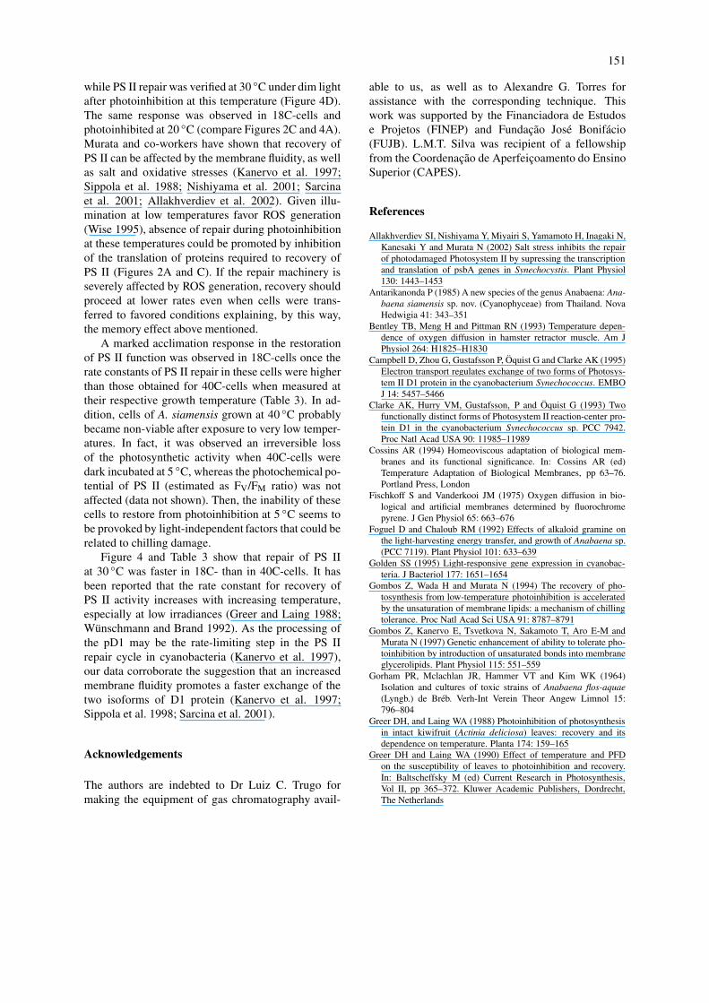
Figure 3. Typical temperature dependence of fluorescence spectrain A. siamensis that had been grown at 18 ◦C (A) and 40 ◦C (B).Cells (2 µg Chl ml−1) were excited at 565 nm, and the fluorescencespectra were normalized at 662 nm.
uncoupling between phycobilisomes and PS II units,the influence of temperature on the energy trans-fer between these antenna systems was investigated.Then, 40C- and 18C-cells were excited with greenlight (565 nm), which is absorbed only by phycobil-isomes, and 15 µM DCMU was added to maintain thePS II reaction center in a closed state. Under theseconditions a prominent emission appeared at 684 nm,which is attributed to PS II-Chl antenna-sensitizingfluorescence (Figure 3). Irrespective of the growthtemperature, the 684 nm emission was significantlydepressed only at temperatures below 7 ◦C in bothtypes of cells.
Temperature dependence of recoveryfrom photoinhibition
After photoinhibition of both types of cells atdistinct temperatures to about 50% of their photo-synthetic capacity, recovery of PS II activity was
followed subsequently the reduction of irradiance to10 µmol photons m−2 s−1. As shown in Figure 4, re-covery of photosynthesis was affected by temperaturesduring recovery, photoinhibition and growth. When18C-cells recovered from photoinhibition at 18 ◦C,restoration of photosynthetic activity was completein 60 or 120 min when photoinhibition occurred ateither 30 ◦C or 20 ◦C and 10 ◦C, respectively, but itwas incomplete if cells were photoinhibited at 5 ◦C(Figure 4A). Essentially the same results were ob-served in 40C-cells when the recovery occurred at40 ◦C, except that no reactivation of oxygen evolu-tion activity was verified after photoinhibition at 5 ◦C(Figure 4B). Figures 4C and D show that restora-tion of the photosynthetic O2 evolution at 30 ◦C wasfaster in 18C-cells (Figure 4C) than in 40C-cells(Figure 4D). In these cells, recovery was incom-plete when photoinhibition occurred at temperatureslower than 30 ◦C. In contrast to 18C-cells, no re-covery from photoinhibition at 5 ◦C was observed in40C-cells.
Table 3 shows the rate constants for recovery ofphotosynthesis of data presented in Figure 4, assum-ing that the kinetics can be described adequately by apseudo-first-order process. In all the conditions stud-ied the rate constants enhanced with the increase of thephotoinhibitory temperature. Furthermore, the higherthe temperature of recovery was, the higher the rateconstant of recovery observed in both types of cells.Despite the recovery temperature, however, 18C-cellspresented higher rate constant values than cells grownat 40 ◦C.
Table 3. Post-photoinhibitory rate constants for recovery of photo-synthesis (h−1) in cells of A. siammensis that had been grown at18 ◦C and 40 ◦Ca
Photoinhibition Recovery temperaturetemperature
(◦C) Cells grown at 18 ◦C Cells grown at 40 ◦C
18 ◦C 30 ◦C 40 ◦C 30 ◦C
5 0.8 1.7 0.0 0.0
10 1.1 1.7 1.1 0.3
20 1.8 2.4 2.2 1.3
30 4.2 5.1 3.0 2.5
a Rate constants were estimated from data presented in Figure 4making the assumption that the kinetics can be described by apseudo-first-order process.

149
Figure 4. Recovery of photosynthesis at growth temperature (A and B) or at 30 ◦C (C and D) after partial photoinhibition (about 50%) of A.siamensis that had been grown at 18 ◦C (A and C) and 40 ◦C (B and D). Cells (25 µg Chl ml−1) were first exposed to 1.5 mmol photons m−2 s−1
at 5 ◦C (�), 10 ◦C (�), 20 ◦C (�), and 30 ◦C (•), and subsequently allowed to recover at 10 µmol photons m−2 s−1 at 18 ◦C (A), 40 ◦C (B) or30 ◦C (C and D). The 100% relative activities correspond to 159.1 ± 5.9 and to 60.6 ± 2.5 µmol O2 mg−1 Chl h−1 in cells grown at 18 ◦C and40 ◦C, respectively.
Discussion
Thylakoid membrane phase transitionand tolerance to low-temperaturephotoinhibition
In order to study the influence of the physical stateof thylakoid membranes on photoinhibition and re-covery of photosynthesis under N2-fixing conditions,cells of A. siamensis were grown at 40 ◦C and at18 ◦C in the absence of combined nitrogen source. Adownward shift in growth temperature did not changethe lipid molecular species in A. siamensis (Table 1).Similar results were obtained in another filamentousN2-fixing cyanobacterium, Anabaena variabilis (Satoet al. 1979), but lipid composition varied with growthtemperature in unicellular species such as Anacystis
nidulans and Synechococcus 7942 (Sato et al. 1979;Porankiewicz et al. 1998). On the other hand, re-sponses of cyanobacterial cells to lower temperaturescomprise desaturation of fatty acids in membrane lip-ids. We also found a cold-induced desaturation ofthe fatty acid composition in A. siamensis (Table 2)such that phase transition of thylakoid membranes de-creased from about 26 ◦C to about 0 ◦C to 1 ◦C in40C-cells and in 18C-cells, respectively (Figure 1).
As reported previously in other cyanobacteria(Kirilovsky et al. 1990; Wünschmann and Brand1992), we found that photoinactivation of PS II inthe absence of D1 synthesis declined by lowering thephotoinhibitory temperature in both 40C- and 18C-cells (Figures 2B and D). However, we found a breakof the increasing protection between 15 ◦C and 5 ◦Cin both types of cells whereas such a break was only

150
verified in Synechocystis 6714 cells when the photoin-hibitory treatment was done at temperatures below thephase transition (Kirilovsky et al. 1990). Manipulationof acyl-lipid desaturase genes has shown that PS IIinactivation and D1-protein degradation either at thegrowth temperature or at lower temperatures proceedindependently of the level of membrane lipid unsat-uration (Gombos et al. 1994, 1997; Kanervo et al.1995, 1997; Sippola et al. 1998). Thus, it seems thatadditional factors play a role on PS II photodamage be-cause 40C- and 18C-cells presented distinct rates andextent of photoinactivation when the photoinhibitorytreatment was done at 15 ◦C to 5 ◦C (Figures 2B andD).
Protection of PS II from photodamage by lower-ing temperature could be promoted by a cold-induceddecoupling of PBS from thylakoids, which has beenobserved in several strains of cyanobacteria (Murataand Fork 1975; Schreiber et al. 1979; Manodori andMelis 1985; Li et al. 2001). In line with the con-clusions made by Kirilovsky et al. (1990), energeticuncoupling between PBS and PS II in A. siamensisdid not correlate with the protection of PS II fromphotodamage observed at low temperatures (compareFigures 2B and 3A, as well as Figures 2D and 3B).In fact, decoupling of PBS from thylakoids seemsto occur independently of the physical state of themembrane lipids since it was observed at the sametemperature in both types of cells (Figure 3).
In the absence of streptomycin, photodamage ofPS II units seems to be correlated with the physicalstate of the membrane lipids. At temperatures rangingfrom 15 ◦C to 5 ◦C, comparison of Figures 2A and Cshows that protection from photodamage was higherwhen the membrane lipids were in the phase-separated(40C-cells) than in the liquid-crystalline states (18C-cells).
It is noticeable that 40C-cells were less photoin-hibited at chilling temperatures (15 ◦C to 5 ◦C) thanat the growth temperature (Figure 2A). This was pro-moted by a marked increase in the protection of PSII from photoinactivation once no PS II repair wasverified at temperatures ranging from 30 ◦C to 5 ◦Cduring photoinhibitory treatment (compare Figures 2Aand B). Since singlet oxygen, formed from triplet-excited P680, is probably the reactive oxygen speciesthat triggers the D1 protein for degradation (Vass et al.1992; Telfer et al. 1994; Hideg et al. 1994, 2002;Okada et al. 1996), a plausible explanation for theresults described above could be related to oxygen dif-fusion in thylakoid membranes. In fact, O2 diffusion
rises sharply in a temperature range associated withphase transition in both artificial phospholipids mem-branes and membranes of muscle retractor of hamster(Fischkoff and Vanderkooi 1975; Bentley et al. 1993).The oxygen diffusion-concentration product in thecenter of spinach thylakoid membranes at 20 ◦C wasabout two times higher than in water (Ligeza et al.1998). Then, chemical reactions such as formation ofROS and lipid peroxidation can be expected to proceedmore readily in the membrane center than in the regionclose to the membrane surface. Furthermore, the coef-ficient of O2 partition between membrane and waterincreases significantly above the phase transition tem-perature (Smotkin et al. 1991). Therefore, the prom-inent photoinhibition at temperatures around 26 ◦Cin 40C-cells could be due to an increased solubilityand diffusion of oxygen in thylakoid membranes. In-versely, an increasing membrane viscosity promotedat temperatures below the phase transition of lipids inthe membranes would avoid photogeneration of ROSaround PS II.
Treatments of cyanobacterial cells that increasedintracellular concentrations of ROS, such as O−
2• and
H2O2, did not promote any effect on the rate of D1photodamage (Nishiyama et al. 2001). In this case, itseems that both externally added and cytosolic gen-erated ROS are unable to penetrate the hydrophobicenvironment, such as the microenvironment in the vi-cinity of the special pair of chlorophyll, in order toreact with P680 and damage the D1 protein.
Influence of growth, photoinhibitionand recovery temperatures on the recoveryfrom photoinhibition
Photoinhibition is a function of both photodamage andrepair. As occurs in higher plants (Greer and Laing1988, 1990; Sundby et al. 1993), the rate of PS IIrepair in cyanobacteria depends on many factors, suchas growth conditions as well as irradiance and temper-ature during the recovery process (Samuelsson et al.1987; Wünschmann and Brand 1992; Gombos et al.1997). Of all factors that influence the recovery pro-cess, however, Figure 4, as well as Table 3, show thatthe photoinhibitory temperature presented a markedinfluence as if a sort of memory of the photoinhibit-ory condition existed. Furthermore, PS II repair dur-ing photoinhibition was influenced by distinct factorsfrom those that affect post-photoinhibition recovery.No recovery was indeed observed in 40C-cells duringphotoinhibition at 30 ◦C (compare Figures 2A and B)
151
while PS II repair was verified at 30 ◦C under dim lightafter photoinhibition at this temperature (Figure 4D).The same response was observed in 18C-cells andphotoinhibited at 20 ◦C (compare Figures 2C and 4A).Murata and co-workers have shown that recovery ofPS II can be affected by the membrane fluidity, as wellas salt and oxidative stresses (Kanervo et al. 1997;Sippola et al. 1988; Nishiyama et al. 2001; Sarcinaet al. 2001; Allakhverdiev et al. 2002). Given illu-mination at low temperatures favor ROS generation(Wise 1995), absence of repair during photoinhibitionat these temperatures could be promoted by inhibitionof the translation of proteins required to recovery ofPS II (Figures 2A and C). If the repair machinery isseverely affected by ROS generation, recovery shouldproceed at lower rates even when cells were trans-ferred to favored conditions explaining, by this way,the memory effect above mentioned.
A marked acclimation response in the restorationof PS II function was observed in 18C-cells once therate constants of PS II repair in these cells were higherthan those obtained for 40C-cells when measured attheir respective growth temperature (Table 3). In ad-dition, cells of A. siamensis grown at 40 ◦C probablybecame non-viable after exposure to very low temper-atures. In fact, it was observed an irreversible lossof the photosynthetic activity when 40C-cells weredark incubated at 5 ◦C, whereas the photochemical po-tential of PS II (estimated as FV/FM ratio) was notaffected (data not shown). Then, the inability of thesecells to restore from photoinhibition at 5 ◦C seems tobe provoked by light-independent factors that could berelated to chilling damage.
Figure 4 and Table 3 show that repair of PS IIat 30 ◦C was faster in 18C- than in 40C-cells. It hasbeen reported that the rate constant for recovery ofPS II activity increases with increasing temperature,especially at low irradiances (Greer and Laing 1988;Wünschmann and Brand 1992). As the processing ofthe pD1 may be the rate-limiting step in the PS IIrepair cycle in cyanobacteria (Kanervo et al. 1997),our data corroborate the suggestion that an increasedmembrane fluidity promotes a faster exchange of thetwo isoforms of D1 protein (Kanervo et al. 1997;Sippola et al. 1998; Sarcina et al. 2001).
Acknowledgements
The authors are indebted to Dr Luiz C. Trugo formaking the equipment of gas chromatography avail-
able to us, as well as to Alexandre G. Torres forassistance with the corresponding technique. Thiswork was supported by the Financiadora de Estudose Projetos (FINEP) and Fundação José Bonifácio(FUJB). L.M.T. Silva was recipient of a fellowshipfrom the Coordenação de Aperfeiçoamento do EnsinoSuperior (CAPES).
References
Allakhverdiev SI, Nishiyama Y, Miyairi S, Yamamoto H, Inagaki N,Kanesaki Y and Murata N (2002) Salt stress inhibits the repairof photodamaged Photosystem II by supressing the transcriptionand translation of psbA genes in Synechocystis. Plant Physiol130: 1443–1453
Antarikanonda P (1985) A new species of the genus Anabaena: Ana-baena siamensis sp. nov. (Cyanophyceae) from Thailand. NovaHedwigia 41: 343–351
Bentley TB, Meng H and Pittman RN (1993) Temperature depen-dence of oxygen diffusion in hamster retractor muscle. Am JPhysiol 264: H1825–H1830
Campbell D, Zhou G, Gustafsson P, Öquist G and Clarke AK (1995)Electron transport regulates exchange of two forms of Photosys-tem II D1 protein in the cyanobacterium Synechococcus. EMBOJ 14: 5457–5466
Clarke AK, Hurry VM, Gustafsson, P and Öquist G (1993) Twofunctionally distinct forms of Photosystem II reaction-center pro-tein D1 in the cyanobacterium Synechococcus sp. PCC 7942.Proc Natl Acad USA 90: 11985–11989
Cossins AR (1994) Homeoviscous adaptation of biological mem-branes and its functional significance. In: Cossins AR (ed)Temperature Adaptation of Biological Membranes, pp 63–76.Portland Press, London
Fischkoff S and Vanderkooi JM (1975) Oxygen diffusion in bio-logical and artificial membranes determined by fluorochromepyrene. J Gen Physiol 65: 663–676
Foguel D and Chaloub RM (1992) Effects of alkaloid gramine onthe light-harvesting energy transfer, and growth of Anabaena sp.(PCC 7119). Plant Physiol 101: 633–639
Golden SS (1995) Light-responsive gene expression in cyanobac-teria. J Bacteriol 177: 1651–1654
Gombos Z, Wada H and Murata N (1994) The recovery of pho-tosynthesis from low-temperature photoinhibition is acceleratedby the unsaturation of membrane lipids: a mechanism of chillingtolerance. Proc Natl Acad Sci USA 91: 8787–8791
Gombos Z, Kanervo E, Tsvetkova N, Sakamoto T, Aro E-M andMurata N (1997) Genetic enhancement of ability to tolerate pho-toinhibition by introduction of unsaturated bonds into membraneglycerolipids. Plant Physiol 115: 551–559
Gorham PR, Mclachlan JR, Hammer VT and Kim WK (1964)Isolation and cultures of toxic strains of Anabaena flos-aquae(Lyngb.) de Bréb. Verh-Int Verein Theor Angew Limnol 15:796–804
Greer DH, and Laing WA (1988) Photoinhibition of photosynthesisin intact kiwifruit (Actinia deliciosa) leaves: recovery and itsdependence on temperature. Planta 174: 159–165
Greer DH and Laing WA (1990) Effect of temperature and PFDon the susceptibility of leaves to photoinhibition and recovery.In: Baltscheffsky M (ed) Current Research in Photosynthesis,Vol II, pp 365–372. Kluwer Academic Publishers, Dordrecht,The Netherlands

152
Hideg E, Spetea C and Vass I (1994) Singlet oxygen and free radicalproduction during acceptor- and donor-side-induced photoinhib-ition. Studies with spin trapping EPR spectroscopy. BiochimBiophys Acta 1186: 143–152
Hideg E, Barta C, Kalai T, Vass I and Asada K (2002) Detectionof singlet oxygen and superoxide with fluorescent sensors inleaves under stress by photoinhibition or UV radiation. Plant CellPhysiol 43: 1154–1164
Kanervo E, Aro E-M and Murata N (1995) Low unsaturation levelof thylakoid membrane lipids limits turnover of the D1 proteinof Photosystem II at high irradiance. FEBS Lett 364: 239–242
Kanervo E, Tasaka Y, Murata N and Aro E-M (1997) Membranelipid unsaturation modulates processing of the Photosystem IIreaction-center protein D1 at low temperature. Plant Physiol 114:841–849
Kirilovsky DL, Vernotte C and Etienne A-L (1990) Protection fromphotoinhibition by low temperature in Synechocystis 6714 and inChlamydomonas reinhardtii: detection of an intermediary state.Biochemistry 29: 8100–8106
Krause GH (1994) Photoinhibition induced by low temperatures.In: Baker NR and Bowyer JR (eds) Photoinhibition of Photo-synthesis: from Molecular Mechanism to the Field, pp 331–363.Bios Scientific Publishers, Oxford
Ligeza A, Tikhonov AN, Hyde JS and Subczinsky WK (1998)Oxygen permeability of thylakoid membranes: electron para-magnetic resonance spin labeling study. Biochim Biophys Acta1365: 453–463
Li Y, Zhang J, Xie J, Zhao J and Jiang L (2001) Temperature-induced decoupling of phycobilisomes from reaction centers.Biochim Biophys Acta 1504: 229–234
MacKinney G (1941) Absorption of light by chlorophyll solutions.J Biol Chem 140: 315–322
Manodori A and Melis A (1985) Phycobilisome-Photosystem II as-sociation in Synechococcus 6301 (Cyanophyceae). FEBS Lett181: 791–796
Mattoo AK, Pick U, Hoffman-Falk H and Edelman M (1981) Therapidly metabolized 32,000 dalton polypeptide is the protein-aceous shield regulating Photosystem II electron transfer andmediating diuron herbicide sensitivity in chloroplasts. Proc NatlAcad Sci USA 78: 1572–1576
Murata N and Fork D (1975) Temperature dependence of chloro-phyll a fluorescence in relation to the physical phase of mem-brane lipids in algae and higher plants. Plant Physiol 56: 791–796
Murata N, Sato N, Omata T and Kuwabara T (1981) Separation andcharacterization of thylakoid and cell envelope of the blue-greenalga (cyanobacterium) Anacystis nidulans. Plant Cell Physiol 22:855–866
Nanba O and Satoh K (1987) Isolation of Photosystem II reactioncenter consisting of D-1 and D-2 polypeptides and cytochromeb-559. Proc Natl Acad Sci USA 89: 7929–7933
Nishiyama Y, Yamamoto H, Allakhverdiev SI, Inaba M, YokotaA and Murata N (2001) Oxidative stress inhibits the repair ofphotodamage to the photosynthetic machinery. EMBO J 20:5587–5594
Okada K, Ikeuchi M, Yamamoto N, Ono T and Miyao M (1996)Selective and specific cleavage of the D1 and D2 proteins of Pho-tosystem II by exposure to singlet oxygen: factors responsible forthe susceptibility to cleavage of the proteins. Biochim BiophysActa 1274: 73–79
Porankiewicz J, Selstam E, Campbell D and Oqüist G (1998) Mem-brane lipid composition and restoration of photosynthesis duringlow temperature acclimation in Synechococcus sp. strain PCC7942. Physiol Planta 104: 405–412
Samuelsson G, Lönneborg A, Gustafsson P and Öquist G (1987)The susceptibility of photosynthesis to photoinhibition and the
capacity of recovery in high and low light grown cyanobacteriaAnacystis nidulans. Plant Physiol 83: 438–441
Sarcina M, Murata N, Tobin MJ and Mullineaux CW (2001)Thylakoid membrane fluidity and its crucial importance in pho-toinhibition. S8-013, In: CD-ROM Version of the Proceedingsof the 12th. International Congress on Photosynthesis, Brisbane,Australia. CSIRO Publishing, Collingwood, Australia
Sato N and Murata N (1980) Temperature-shift induced responsesin lipids in the blue-green alga Anabaena variabilis. The cent-ral role of diacylmonogalactosylglycerol in thermo-adaptation.Biochim Biophys Acta 619: 353–366
Sato N and Murata N (1988) Membrane lipids. Meth Enzymol 167:251–259
Sato N, Murata N, Miura Y and Ueta N (1979) Effect of growth tem-perature on lipid and fatty acid compositions in the blue-greenalgae, Anabaena variabilis and Anacystis nidulans. BiochimBiophys Acta 572: 19–28
Schaefer MR and Golden SS (1989) Differential expression of mem-bers of a cyanobacterial psbA gene family in response to light.J Bacteriol 171: 3973–3981
Schreiber U, Rijgersberg CP and Amesz J (1979) Temperature-dependent reversible changes in phycobilisome thylakoid mem-brane attachment in Anacystis nidulans. FEBS Lett 104: 327–331
Silva LMT, Dos Santos CP and Chaloub RM (2001) Effect of therespiratory activity on photoinhibition of the cyanobacteriumSynechocystis sp. Photosynth Res 68: 61–69
Sippola K, Kanervo E, Murata N and Aro E-M (1988) A geneticallyengineered increase in fatty acid unsaturation in Synechococ-cus sp. PCC 7942 allows exchange of D1 proteins forms andsustenance of Photosystem II activity at low temperature. Eur JBiochem 251: 641–648
Smotkin ES, Moy T and Plachy WZ (1991) Dioxygen solubilityin aqueous phosphatidylcholine dispersions. J Membr Biol 114:97–112
Sundby C, McCaffery S and Anderson JM (1993) Turnover of Pho-tosystem II D1 protein in higher plants under photoinhibitory andnon-photoinhibitory irradiances. J Biol Chem 268: 25476–25482
Tasaka Y, Gombos Z, Nishiyama Y, Mohanty P, Ohbba T, OhkiK and Murata N (1996) Target mutagenesis of acyl-lipid de-saturases in Synechocystis: evidence for the important rolesof polyunsaturated membrane lipids in growth, respiration andphotosynthesis. EMBO J 15: 6416–6425
Telfer A, Bishop SM, Phillips D and Barber J (1994) The isolatedphotosynthetic reaction center of PS II as a sensitiser for theformation of singlet oxygen; detection and quantum yield de-termination using a chemical trapping technique. J Biol Chem269: 13244–13253
Vass I, Styring S, Hundal T, Koivuniemi A, Aro E-M and AnderssonB (1992) The reversible and irreversible intermediates duringphotoinhibition of Photosystem II – stable reduced QA speciespromote chlorophyll triplet formation. Proc Natl Acad Sci USA89: 1408–1412
Wada H and Murata N (1990) Temperature-induced changes in thefatty acid composition of the cyanobacterium Synechocystis PCC6803. Plant Physiol 92: 1062–1069
Wise RR (1995) Chilling-enhanced photo-oxidation: the produc-tion, action and study of reactive oxygen species produced duringchilling in the light. Photosynth Res 45: 79–97
Wünschmann G and Brand JJ (1992) Rapid turnover of a componentrequired for photosynthesis explains temperature dependenceand kinetics of photoinhibition in a cyanobacterium Synechococ-cus 6301. Planta 186: 426–433
Zhang L and Aro E-M (2002) Synthesis, membrane insertion and as-sembly of the chloroplast-encoded D1 protein into PhotosystemII. FEBS Lett 512: 13–18
Copyright © 2022 FDOKUMEN