Synthesis, assembly and degradation of thylakoid membrane proteins
20
Synthesis, assembly and degradation of thylakoid membrane proteins Yves Choquet, Olivier Vallon * CNRS/UPR1261, Institut de Biologie Physico-Chimique, 13, rue Pierre-et-Marie-Curie, 75005 Paris, France (Received 15 March 2000; accepted 17 April 2000) Abstract — The thylakoid membrane of chloroplasts contains four major protein complexes, involved in the photosynthetic electron transfer chain and in ATP synthesis. These complexes are built from a large number of polypeptide subunits encoded either in the nuclear or in the plastid genome. In this review, we are considering the mechanism that couples assembly (association of the polypeptides with each other and with their cofactors) with the upstream and downstream steps of the biogenetic pathway, translation and proteolytic degradation. We present the contrasting images of assembly that have emerged from a variety of approaches (studies of photosynthesis mutants, developmental studies and direct biochemical analysis of the kinetics of assembly). We develop the concept of control by epistasy of synthesis, through which the translation of certain subunits is controlled by the state of assembly of the complex and address the question of its mechanisms. We describe additional factors that assist in the integration and assembly of thylakoid membrane proteins. © 2000 Société française de biochimie et biologie moléculaire / Éditions scientifiques et médicales Elsevier SAS thylakoid membrane / chloroplast / photosynthesis / membrane protein assembly / translation / proteolysis 1. Introduction The light reactions of oxygenic photosynthesis are carried out by four large enzymatic complexes located in the thylakoid membrane (figure 1). All of them are oligo- meric proteins, made of between 7 and more than 20 polypeptides, and they bind a variety of cofactors and pigments in a well-defined structural arrangement. In recent years, our understanding of the system has received tremendous benefit from the two most rapidly expanding fields of biology: genomics and 3D-structure analysis. A number of chloroplast genomes have been fully se- quenced, as well as the complete genome of the cyano- bacterium Synechocystis sp. PCC6803, while the sequence of the Arabidopsis nuclear genome will soon be com- pleted. With the help of reverse genetics, this will put in our hands the complete set of polypeptides from which this complex system is built up (structural genes, regula- tors and assembly catalysts). The 3D-structures of photo- system I (PSI) and light harvesting complex II will soon reach atomic resolution, while those of the other com- plexes, the photosystem II (PSII) reaction center, cyto- chrome b 6 f and the H + -ATP synthase, either are underway or can be largely inferred from those of their bacterial or mitochondrial homologues. Still, one of the most fascinating aspects of this system remains poorly understood. We largely ignore how the interactions that bind these polypeptides together are established and destroyed throughout the biogenesis of the membrane. This is an arduous question not only because of the large number of polypeptides involved, but also because of their dual genetic origin. Within a same complex, subunits encoded by the chloroplast genome are associated in a defined stoichiometry with subunits that are encoded by the nuclear genome, translated on cytoso- lic ribosomes and imported into the chloroplast. Thus, specific mechanisms should operate to allow the stoichio- metric accumulation of the various subunits encoded by the two genetic compartments in the amount required for their assembly. At the level of translation, some degree of coordination appears necessary, not only to avoid wasteful production of subunits in excess of their assembly part- ners, but also because some of these polypeptides bind chlorophyll, whose triplet state would lead to the forma- tion of deleterious singlet oxygen when photochemistry is defective. The two genetic compartments, which use widely different systems for producing proteins, must exchange specific signals to coordinate gene expression. In the chloroplast, polycistronic transcription units may combine genes for different complexes and may be trimmed to monocistronic mRNAs. Consequently, the level of expression of each protein has to be regulated individually. As described elsewhere in this issue (Barkan and Goldschmidt-Clermont, Monde et al., Zerges), a large part of the control of chloroplast gene expression has moved to the post-transcriptional level, with numerous nucleus-encoded factors governing mRNA processing, stability and translation for a specific chloroplast gene product. * Correspondence and reprints: [email protected] Abbreviations: CES, control by epistasy of synthesis; LHCP, light harvesting chlorophyll a/b protein; PSI, photosystem I; PSII, photosystem II; SRP, signal recognition particle Biochimie 82 (2000) 615-634 © 2000 Société française de biochimie et biologie moléculaire / Éditions scientifiques et médicales Elsevier SAS. All rights reserved. S030090840000609X/FLA
Transcript of Synthesis, assembly and degradation of thylakoid membrane proteins
Synthesis, assembly and degradation of thylakoid membrane proteins
Yves Choquet, Olivier Vallon*
CNRS/UPR1261, Institut de Biologie Physico-Chimique, 13, rue Pierre-et-Marie-Curie, 75005 Paris, France
(Received 15 March 2000; accepted 17 April 2000)
Abstract — The thylakoid membrane of chloroplasts contains four major protein complexes, involved in the photosynthetic electrontransfer chain and in ATP synthesis. These complexes are built from a large number of polypeptide subunits encoded either in thenuclear or in the plastid genome. In this review, we are considering the mechanism that couples assembly (association of thepolypeptides with each other and with their cofactors) with the upstream and downstream steps of the biogenetic pathway, translationand proteolytic degradation. We present the contrasting images of assembly that have emerged from a variety of approaches (studiesof photosynthesis mutants, developmental studies and direct biochemical analysis of the kinetics of assembly). We develop the conceptof control by epistasy of synthesis, through which the translation of certain subunits is controlled by the state of assembly of thecomplex and address the question of its mechanisms. We describe additional factors that assist in the integration and assembly ofthylakoid membrane proteins. © 2000 Société française de biochimie et biologie moléculaire / Éditions scientifiques et médicalesElsevier SAS
thylakoid membrane / chloroplast / photosynthesis / membrane protein assembly / translation / proteolysis
1. Introduction
The light reactions of oxygenic photosynthesis arecarried out by four large enzymatic complexes located inthe thylakoid membrane(figure 1). All of them are oligo-meric proteins, made of between 7 and more than 20polypeptides, and they bind a variety of cofactors andpigments in a well-defined structural arrangement. Inrecent years, our understanding of the system has receivedtremendous benefit from the two most rapidly expandingfields of biology: genomics and 3D-structure analysis. Anumber of chloroplast genomes have been fully se-quenced, as well as the complete genome of the cyano-bacteriumSynechocystis sp. PCC6803, while the sequenceof the Arabidopsis nuclear genome will soon be com-pleted. With the help of reverse genetics, this will put inour hands the complete set of polypeptides from whichthis complex system is built up (structural genes, regula-tors and assembly catalysts). The 3D-structures of photo-system I (PSI) and light harvesting complex II will soonreach atomic resolution, while those of the other com-plexes, the photosystem II (PSII) reaction center, cyto-chromeb6f and the H+-ATP synthase, either are underwayor can be largely inferred from those of their bacterial ormitochondrial homologues.
Still, one of the most fascinating aspects of this systemremains poorly understood. We largely ignore how the
interactions that bind these polypeptides together areestablished and destroyed throughout the biogenesis of themembrane. This is an arduous question not only becauseof the large number of polypeptides involved, but alsobecause of their dual genetic origin. Within a samecomplex, subunits encoded by the chloroplast genome areassociated in a defined stoichiometry with subunits thatare encoded by the nuclear genome, translated on cytoso-lic ribosomes and imported into the chloroplast. Thus,specific mechanisms should operate to allow the stoichio-metric accumulation of the various subunits encoded bythe two genetic compartments in the amount required fortheir assembly. At the level of translation, some degree ofcoordination appears necessary, not only to avoid wastefulproduction of subunits in excess of their assembly part-ners, but also because some of these polypeptides bindchlorophyll, whose triplet state would lead to the forma-tion of deleterious singlet oxygen when photochemistry isdefective. The two genetic compartments, which usewidely different systems for producing proteins, mustexchange specific signals to coordinate gene expression.In the chloroplast, polycistronic transcription units maycombine genes for different complexes and may betrimmed to monocistronic mRNAs. Consequently, thelevel of expression of each protein has to be regulatedindividually. As described elsewhere in this issue (Barkanand Goldschmidt-Clermont, Monde et al., Zerges), a largepart of the control of chloroplast gene expression hasmoved to the post-transcriptional level, with numerousnucleus-encoded factors governing mRNA processing,stability and translation for a specific chloroplast geneproduct.
* Correspondence and reprints: [email protected]: CES, control by epistasy of synthesis; LHCP,light harvesting chlorophylla/b protein; PSI, photosystem I;PSII, photosystem II; SRP, signal recognition particle
Biochimie 82 (2000) 615−634© 2000 Société française de biochimie et biologie moléculaire / Éditions scientifiques et médicales Elsevier SAS. All rights reserved.S030090840000609X/FLA

Photosynthesis research has now put in our hands alarge panel of genetic, biochemical and biophysical tools,with which we can address this important issue in asystematical manner, with the hope to shed some light onthe general principle that governs assembly of complexmembrane proteins. The biogenesis of thylakoid mem-brane proteins has recently been reviewed in detail [1]. Inthe present paper, we will concentrate on the mechanismsthat couple the assembly process to two essential momentsof the life of a polypeptide, translation and proteolysis.
2. Cytochrome b6f: a case study.
In this section we will describe the assembly of thecytochrome b6f complex, to serve as an illustration of the
common themes encountered in the study of the otherthylakoid membrane proteins.
Cytochrome b6f is the simplest complex of the thyla-koid membrane in terms of polypeptide composition. Itcatalyses the reduction of plastocyanin at the expense ofreduced plastoquinol and contributes to the formation ofthe proton gradient used to synthesize ATP. It is structur-ally and functionally homologous to the cytochrome bc1complex found in mitochondria and bacteria, whosestructure has been determined by X-ray crystallography[2, 3]. The isolated complex [4, 5] contains four majorsubunits: cytochrome f binds one c-type heme, cyto-chrome b6 carries two b-type hemes, subunit IV is devoidof prosthetic groups but contributes to the formation of thequinol binding site, and the Rieske protein binds the[2Fe-2S] cluster. The first three subunits are encoded by
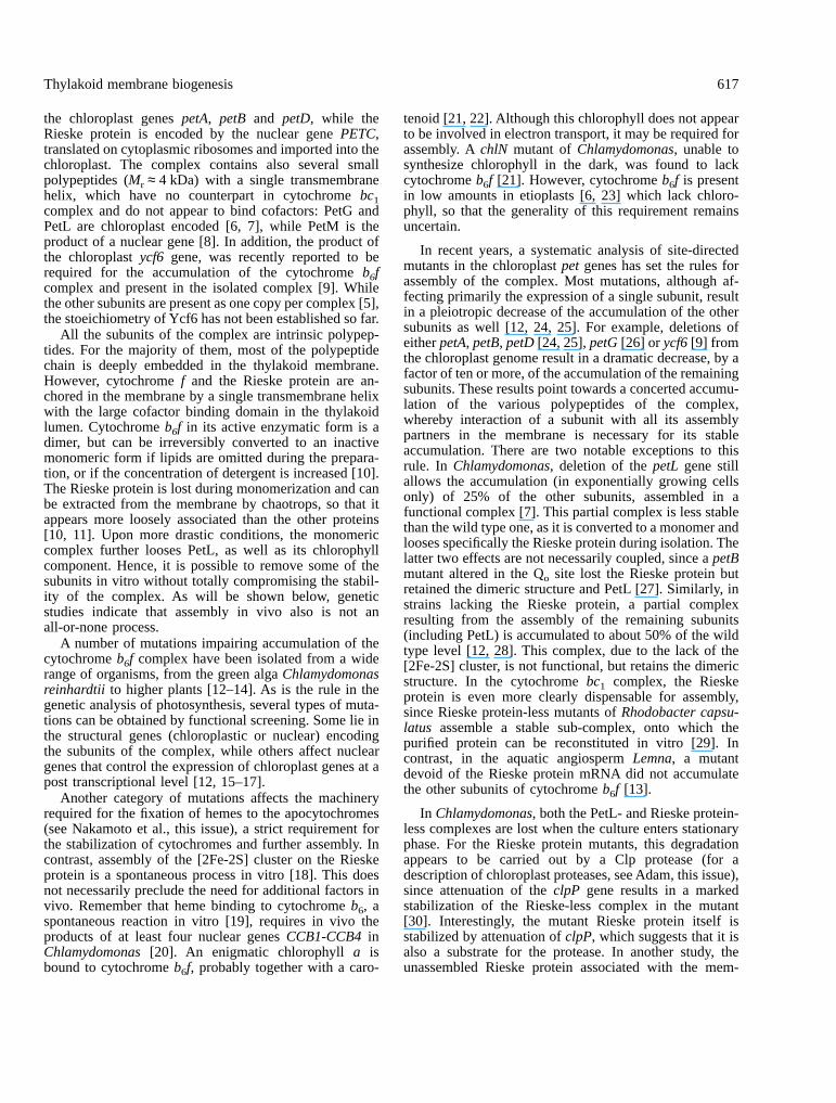
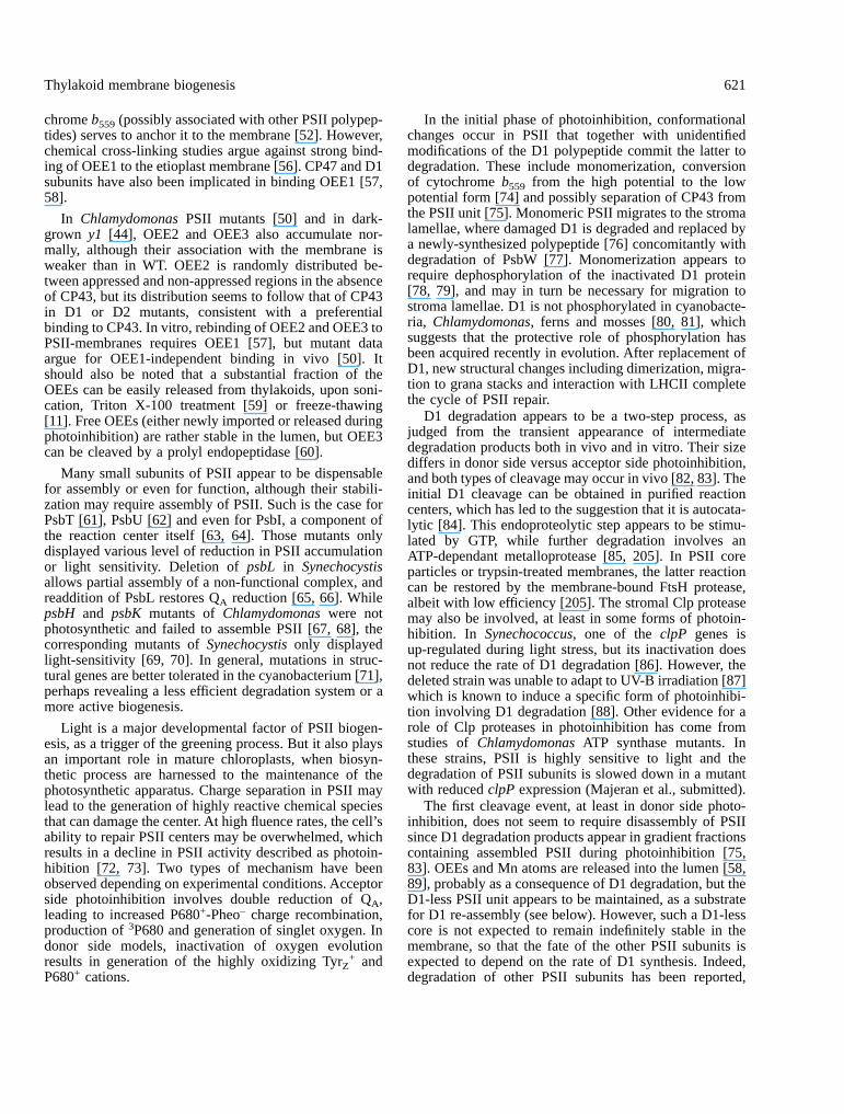
Figure 1. Supramolecular organization of the thylakoid membrane. Chloroplast-encoded subunits are indicated in light colors andnucleus-encoded subunits in dark colors. Each subunit is indicated by is common name, or by the letter of the gene (psb, pet, psa)that encodes it. Light harvested by the photosystems I and II (PSI, PSII) and their associated light harvesting complexes (LHCI,LHCII) triggers oxidation of a photochemical chlorophyll a dimer (P680, P700, respectively). In PSII, P680+ is reduced by electronsabstracted from water through a Mn cluster and a reactive tyrosine YZ. On the acceptor side, the intra-protein electron transfer chaincomprises a pheophytin, a non-heme iron and a tightly bound quinone QΑ. At the QB site, plastoquinone (PQ) is reduced toplastoquinol, which will be oxidized at the Qo site of cytochrome b6f. One electron travels through the Rieske protein and cytochromef to the soluble electron carrier plastocyanin (PC). The other electron, via cytochrome b6 hemes, reduces another PQ at the Qi site,resulting in H+ pumping by the so-called Q-cycle. On the lumenal face of PSI, PC reduces oxidized P700. Two parallel electrontransfer chains in PSI converge towards FX, a cluster at the interface of PsaA and PsaB. After reducing the FA and FB clusters andferredoxine (Fd), the electron is used by ferredoxine-NADP+-reductase (FNR) to generate NADPH. Operation of the electron transferchain generates a ∆µH+ which is used by the CF0-CF1 ATP synthase to generate ATP.
616 Choquet and Vallon
the chloroplast genes petA, petB and petD, while theRieske protein is encoded by the nuclear gene PETC,translated on cytoplasmic ribosomes and imported into thechloroplast. The complex contains also several smallpolypeptides (Mr ≈ 4 kDa) with a single transmembranehelix, which have no counterpart in cytochrome bc1complex and do not appear to bind cofactors: PetG andPetL are chloroplast encoded [6, 7], while PetM is theproduct of a nuclear gene [8]. In addition, the product ofthe chloroplast ycf6 gene, was recently reported to berequired for the accumulation of the cytochrome b6fcomplex and present in the isolated complex [9]. Whilethe other subunits are present as one copy per complex [5],the stoeichiometry of Ycf6 has not been established so far.
All the subunits of the complex are intrinsic polypep-tides. For the majority of them, most of the polypeptidechain is deeply embedded in the thylakoid membrane.However, cytochrome f and the Rieske protein are an-chored in the membrane by a single transmembrane helixwith the large cofactor binding domain in the thylakoidlumen. Cytochrome b6f in its active enzymatic form is adimer, but can be irreversibly converted to an inactivemonomeric form if lipids are omitted during the prepara-tion, or if the concentration of detergent is increased [10].The Rieske protein is lost during monomerization and canbe extracted from the membrane by chaotrops, so that itappears more loosely associated than the other proteins[10, 11]. Upon more drastic conditions, the monomericcomplex further looses PetL, as well as its chlorophyllcomponent. Hence, it is possible to remove some of thesubunits in vitro without totally compromising the stabil-ity of the complex. As will be shown below, geneticstudies indicate that assembly in vivo also is not anall-or-none process.
A number of mutations impairing accumulation of thecytochrome b6f complex have been isolated from a widerange of organisms, from the green alga Chlamydomonasreinhardtii to higher plants [12–14]. As is the rule in thegenetic analysis of photosynthesis, several types of muta-tions can be obtained by functional screening. Some lie inthe structural genes (chloroplastic or nuclear) encodingthe subunits of the complex, while others affect nucleargenes that control the expression of chloroplast genes at apost transcriptional level [12, 15–17].
Another category of mutations affects the machineryrequired for the fixation of hemes to the apocytochromes(see Nakamoto et al., this issue), a strict requirement forthe stabilization of cytochromes and further assembly. Incontrast, assembly of the [2Fe-2S] cluster on the Rieskeprotein is a spontaneous process in vitro [18]. This doesnot necessarily preclude the need for additional factors invivo. Remember that heme binding to cytochrome b6, aspontaneous reaction in vitro [19], requires in vivo theproducts of at least four nuclear genes CCB1-CCB4 inChlamydomonas [20]. An enigmatic chlorophyll a isbound to cytochrome b6f, probably together with a caro-
tenoid [21, 22]. Although this chlorophyll does not appearto be involved in electron transport, it may be required forassembly. A chlN mutant of Chlamydomonas, unable tosynthesize chlorophyll in the dark, was found to lackcytochrome b6f [21]. However, cytochrome b6f is presentin low amounts in etioplasts [6, 23] which lack chloro-phyll, so that the generality of this requirement remainsuncertain.
In recent years, a systematic analysis of site-directedmutants in the chloroplast pet genes has set the rules forassembly of the complex. Most mutations, although af-fecting primarily the expression of a single subunit, resultin a pleiotropic decrease of the accumulation of the othersubunits as well [12, 24, 25]. For example, deletions ofeither petA, petB, petD [24, 25], petG [26] or ycf6 [9] fromthe chloroplast genome result in a dramatic decrease, by afactor of ten or more, of the accumulation of the remainingsubunits. These results point towards a concerted accumu-lation of the various polypeptides of the complex,whereby interaction of a subunit with all its assemblypartners in the membrane is necessary for its stableaccumulation. There are two notable exceptions to thisrule. In Chlamydomonas, deletion of the petL gene stillallows the accumulation (in exponentially growing cellsonly) of 25% of the other subunits, assembled in afunctional complex [7]. This partial complex is less stablethan the wild type one, as it is converted to a monomer andlooses specifically the Rieske protein during isolation. Thelatter two effects are not necessarily coupled, since a petBmutant altered in the Qo site lost the Rieske protein butretained the dimeric structure and PetL [27]. Similarly, instrains lacking the Rieske protein, a partial complexresulting from the assembly of the remaining subunits(including PetL) is accumulated to about 50% of the wildtype level [12, 28]. This complex, due to the lack of the[2Fe-2S] cluster, is not functional, but retains the dimericstructure. In the cytochrome bc1 complex, the Rieskeprotein is even more clearly dispensable for assembly,since Rieske protein-less mutants of Rhodobacter capsu-latus assemble a stable sub-complex, onto which thepurified protein can be reconstituted in vitro [29]. Incontrast, in the aquatic angiosperm Lemna, a mutantdevoid of the Rieske protein mRNA did not accumulatethe other subunits of cytochrome b6f [13].
In Chlamydomonas, both the PetL- and Rieske protein-less complexes are lost when the culture enters stationaryphase. For the Rieske protein mutants, this degradationappears to be carried out by a Clp protease (for adescription of chloroplast proteases, see Adam, this issue),since attenuation of the clpP gene results in a markedstabilization of the Rieske-less complex in the mutant[30]. Interestingly, the mutant Rieske protein itself isstabilized by attenuation of clpP, which suggests that it isalso a substrate for the protease. In another study, theunassembled Rieske protein associated with the mem-
Thylakoid membrane biogenesis 617
brane during in organello import appeared to be degradedby the FtsH protease [31].
Two types of mechanisms appear responsible for theconcerted accumulation of cytochrome b6f subunits: deg-radation of unassembled polypeptides, and regulation oftranslation by assembly. Both mechanisms have beenstudied in detail in Chlamydomonas, by pulse chaseexperiments performed on strains deleted for one of thepet genes. These experiments showed that the half life ofsubunit IV and cytochrome b6, but not their initialsynthesis rate, was highly decreased upon deletion ofanother subunit of the complex. For example the half lifeof subunit IV drops from almost 2 h in the wild type to45 min and 15 min in strains lacking cytochrome f orcytochrome b6, respectively [24]. This indicates that theunassembled subunit IV is rapidly degraded when itcannot assemble, i.e., that assembly converts it from ahighly unstable form to a form that is no longer suscep-tible to proteolytic attack. This could be brought aboutsimply by shielding those motifs that can be recognized byproteases. The protease(s) responsible for this type ofdegradation have not been identified yet. ClpP does notseem to be involved, since attenuation of the gene did notlead to enhanced accumulation of the fully unassembledsubunits in this type of mutants [30].
The behavior of cytochrome f appears completelydifferent from that of the other subunits. In the absence ofits assembly partners, cytochrome b6 or subunit IV, itssynthesis rate drops to about 10% of that observed in thewild type [24]. Cytochrome f synthesized in those condi-tions is inserted in the membrane and is as stable as in thewild type, even though it is not assembled. Thus, there isa hierarchical organization of the expression of the cyto-chrome b6f subunits, which has been described as acontrol by epistasy of synthesis (CES) [32, 33]. Cyto-chrome b6 and subunit IV are dominant over cytochromef, whereas cytochrome f, which requires the presence of itsdominant assembly partners to be synthesized at wild typerate, is called a CES protein.
At the molecular level, this assembly-mediated controlof cytochrome f translation is an autoregulation of trans-lation. The signal responsible for this regulation is carriedby the C-terminal domain of the cytochrome f, i.e., thetransmembrane helix and the 15 amino acids stromalextension. Indeed, strains lacking accumulation of thisC-terminal domain (either because the whole cytochromef is unstable and fails to accumulate, or because this regionwas specifically deleted or mutated) escape autoregula-tion: they show a three-fold oversynthesis of cytochromef, whether the dominant subunits are synthesized or not[33, 34]. Thus, cytochrome f synthesis is already repressedto some extent in wild type, in order to adjust theproduction of cytochrome f to the synthesis rate of theother subunits. Site directed mutagenesis experiments onthe C-terminal domain of cytochrome f have narroweddown the regulatory motif to an eight amino acid stretch
located immediately after the end of the transmembranehelix (Choquet and Wollman, unpublished).
2.1. Membrane-KKKQFEKV
Mutations at positions 2, 3, 6 and 7 did not affectautoregulation, but a Lys to Met substitution at position 1or a Phe to Ser substitution at position 5 yielded acompletely unregulated cytochrome f, with a three-foldincreased translation rate. The importance of the otherresidues, as well of the possible implication of residuesfrom the transmembrane helix, is under study.
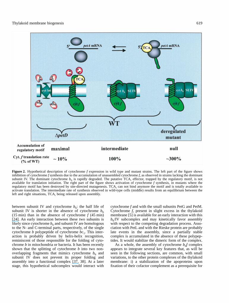
The target for this autoregulation resides within thepetA 5’UTR, which governs translation initiation. Strainswhere cytochrome f is translated under the control ofanother unrelated 5’UTR no longer exhibit the assembly-dependent control of cytochrome f synthesis. Furthermore,the petA mRNA 5’UTR is able per se to confer the CESbehavior to a reporter gene translated under its control[33]. It was proposed that the regulatory motif carried byunassembled cytochrome f is able to interact directly orindirectly with the 5’UTR of the petA mRNA. Uponassembly, it would be shielded by the other subunits orundergo a conformational change impairing this interac-tion. As this motif seems too short to promote a sequencespecific protein-RNA interaction, the interaction is likelyindirect and should therefore rely on a ternary effector.This factor would be trapped by the regulatory motif ofunassembled cytochrome f; it would be released uponassembly and become available for translation, as illus-trated in figure 2. A possible candidate is the nucleus-encoded TCA1 factor, the only factor identified to datethat is required for the translation initiation of the petAmRNA [35] (Wostrikoff et al., unpublished observations.).However, the participation of TCA1 in the CES processstill awaits demonstration.
Whether the epistatic control of cytochrome f synthesisalso operates in the chloroplast of higher plants remains tobe elucidated. Tobacco mutants with a deletion in petD orpetB failed to accumulate the complex [25], but synthesisof cytochrome f has not been measured directly. Theobserved reduction in the size of petA-associated poly-somes is difficult to interpret, since the tobacco petA isassociated with other reading frames in a complex tran-scription unit. The crp1 mutant of maize lacks translatablepetD messenger due to a failure to process the petDmRNA. Interestingly, this mutant also shows a reducedsynthesis of cytochrome f [17]. But a close examination ofthe phenotype of the mutant and of the sequence of thecloned crp1 gene led the authors to conclude that thedecreased cytochrome f synthesis was a primary effect ofthe mutation rather than an epistatic consequence of thedecreased accumulation of subunit IV [36].
Together, the data presented above suggest a pathway,largely hypothetical, for the assembly of the cytochromeb6f complex. Genetic data point to an early interaction
618 Choquet and Vallon
between subunit IV and cytochrome b6: the half life ofsubunit IV is shorter in the absence of cytochrome b6(15 min) than in the absence of cytochrome f (45 min)[24]. An early interaction between these two subunits islikely since cytochrome b6 and subunit IV are homologousto the N- and C-terminal parts, respectively, of the singlecytochrome b polypeptide of cytochrome bc1. This inter-action is probably driven by helix-helix recognition,reminiscent of those responsible for the folding of cyto-chrome b in mitochondria or bacteria. It has been recentlyshown that the splitting of cytochrome b into two non-overlapping fragments that mimics cytochrome b6 andsubunit IV does not prevent its proper folding andassembly into a functional complex [37, 38]. At a laterstage, this hypothetical subcomplex would interact with
cytochrome f and with the small subunits PetG and PetM.Cytochrome f, present in slight excess in the thylakoidmembrane [5] is available for an early interaction with thisb6/IV subcomplex and may kinetically favor assemblywith respect to the competing degradation process. Asso-ciation with PetL and with the Rieske protein are probablylate events in the assembly, since a partially stablecomplex is accumulated in the absence of these polypep-tides. It would stabilize the dimeric form of the complex.
As a whole, the assembly of cytochrome b6f complexappears to integrate several key features that, as will beseen in the following sections, are common, with smallvariations, to the other protein complexes of the thylakoidmembrane: i) a stabilization of the apoproteins uponfixation of their cofactor complement as a prerequisite for
Figure 2. Hypothetical description of cytochrome f expression in wild type and mutant strains. The left part of the figure showsinhibition of cytochrome f synthesis due to the accumulation of unassembled cytochrome f, as observed in strains lacking the dominantsubunit IV. The dominant cytochrome b6 is rapidly degraded. The putative TCAi effector, trapped by the regulatory motif, is notavailable for translation initiation. The right part of the figure shows activation of cytochrome f synthesis, in mutants where theregulatory motif has been destroyed by site-directed mutagenesis. TCAi can not bind anymore the motif and is totally available toactivate translation. The intermediate rate of synthesis observed in wild-type cells (middle) results from an equilibrium between theleft and right situations, TCAi being released upon assembly.
Thylakoid membrane biogenesis 619
assembly; ii) a concerted accumulation of the subunit ofthe complex resulting from: 1) a rapid proteolytic degra-dation of most unassembled subunits; and 2) an assembly-controlled regulation of the synthesis of one subunit; andiii) a sequential assembly pathway, with a progressiveincrease in the stability of the complex as more subunitsare incorporated.
3. Complexes and sub-complexes: hints to theassembly pathway
In this section, we will summarize our current under-standing of the mechanisms of assembly of the othercomplexes. As very few real kinetic data are available, wewill rely mostly on in vitro studies of the biochemicalstability of the interactions between subunits and onmutant studies suggesting the occurrence of intermediatestages of assembly.
3.1. Photosystem II
PSII is by far the photosynthetic complex for whichbiochemical and genetic dissection has been analyzed ingreatest detail. The smallest preparation able to performthe basic reaction of photosystem II contains six polypep-tides: D1 and D2 subunits (homologous to the L and Msubunits of the bacterial reaction center), the α and �subunits of cytochrome b559, and two polypeptides with asingle membrane-spanning helix, PsbI and PsbW. Largercomplexes have been isolated which contain in additionthe CP47 chlorophyll a-binding protein and PsbT, plusPsbL and PsbK [39]. The latter preparation is a dimer, asis probably the case for PSII in situ. The classical PSIIcore preparations contain additional integral subunits,among which the CP43 antenna protein. The nuclear-encoded extrinsic polypeptides (OEE1, OEE2 and OEE3)bind to the lumenal surface where they concur to thestabilization of the oxygen-evolution site. Higher orderstructures have been isolated from higher plants, wherePSII arranged as a dimer is surrounded by the nucleus-encoded chlorophyll a/b-binding proteins, forming vari-ous types of functional units [40, 41].
The picture emerging from developmental, genetic andtime-resolved biochemical studies of PSII assembly ap-pears somewhat more complex: clearly, biological assem-bly is not the exact reverse of biochemical disassembly.Two types of processes must be considered, although theymay overlap at times: the mechanisms through which aPSII unit is built up in a developing chloroplast and thosethat insure its maintenance in the mature organelle.
PSII activity is absent in etioplasts and appears duringgreening, a process that has been extensively studied inplants [42, 43] and in the y1 mutant of Chlamydomonasthat does not synthesize chlorophyll in the dark [44]. ThemRNAs for D1, D2, CP47 and CP43 (i.e., the chlorophyll-
binding polypeptides) are present in etioplasts, but theirtranslation products are not detected. Light triggers accu-mulation of these polypeptides, maybe by a direct effecton translation [45], or most probably by allowing chloro-phyll synthesis required for stabilization of the apopro-teins [46, 47]. But many PSII proteins do not share thisstrict requirement for light. Cytochrome b559, PsbW, PsbHand PsbS are present in etioplasts, as are the extrinsicOEE1 and OEE2 polypeptides [42]. Their relativeamounts increase during greening, significantly faster thanthat of the chlorophyll a-binding proteins [42], so that thelatter will meet a large pool of their assembly partners asthey enter the membrane. This raises the question whetherthese components are assembled in etioplasts and whetherthey can serve as a nucleus for the assembly of thechlorophyll a-binding proteins. Recently, cytochrome b559has been detected in 90–200 kDa complexes in barleyetioplast membrane [48].
Genetic studies may indirectly reveal intermediatesteps in the assembly of PSII. As a rule, mutations ingenes coding for the chlorophyll-binding subunits affectthe stability or synthesis of most of their assemblypartners. Mutants devoid of D1 are severely depleted inD2 and CP47, and vice versa [49]. CP43 seems to besomewhat more independent, since CP43-less mutantsstill can accumulate sizeable amounts of D1, D2 andCP47, assembled in a detergent-stable complex, andconversely CP43 can accumulate to some extent in mu-tants lacking D1, D2 or CP47 [50]. But by and large, thefour chlorophyll a-binding subunits seem to depend oneach other for their accumulation in the membrane.
In contrast, cytochrome b559, although it is part of thereaction center complex, accumulates to normal levels inthe absence of the chlorophyll-binding subunits. This hasbeen observed in D1-less mutants of Chlamydomonas[51], barley [52, 53] and Synechocystis [54]. The converseis not true, since both the α and � subunits of cytochromeb559 are necessary for PSII biogenesis [51, 55]. Similarly,the nuclear-encoded OEE1, OEE2 and OEE3 polypeptidesaccumulate to normal levels in mutants lacking polypep-tides D1, D2, CP47 or CP43 [50], consistent with theirpresence in etioplasts and in dark-grown y1 cells, whilemutants lacking the OEE proteins still stably accumulatethe rest of the PSII in the membrane. Interestingly, OEE1appears to depend on cytochrome b559 for its stability, asthe cytochrome b559 mutant of Chlamydomonas com-pletely lacks OEE1 [51]. This places cytochrome b559 at across-road of PSII assembly. In vir115, cytochrome b559and OEE1 segregate to the appressed regions of thethylakoid membrane, in contrast to the traces of CP47 andCP43 which are randomly distributed in the two domains[52]. Preferential localization of OEE1 to the appressedregions has also been reported for Chlamydomonas mu-tants lacking proteins D1, D2 or CP47 [50]. Since thelumenal OEE1 cannot by itself sense the appression stateon the stromal surface, it has been proposed that cyto-
620 Choquet and Vallon
chrome b559 (possibly associated with other PSII polypep-tides) serves to anchor it to the membrane [52]. However,chemical cross-linking studies argue against strong bind-ing of OEE1 to the etioplast membrane [56]. CP47 and D1subunits have also been implicated in binding OEE1 [57,58].
In Chlamydomonas PSII mutants [50] and in dark-grown y1 [44], OEE2 and OEE3 also accumulate nor-mally, although their association with the membrane isweaker than in WT. OEE2 is randomly distributed be-tween appressed and non-appressed regions in the absenceof CP43, but its distribution seems to follow that of CP43in D1 or D2 mutants, consistent with a preferentialbinding to CP43. In vitro, rebinding of OEE2 and OEE3 toPSII-membranes requires OEE1 [57], but mutant dataargue for OEE1-independent binding in vivo [50]. Itshould also be noted that a substantial fraction of theOEEs can be easily released from thylakoids, upon soni-cation, Triton X-100 treatment [59] or freeze-thawing[11]. Free OEEs (either newly imported or released duringphotoinhibition) are rather stable in the lumen, but OEE3can be cleaved by a prolyl endopeptidase [60].
Many small subunits of PSII appear to be dispensablefor assembly or even for function, although their stabili-zation may require assembly of PSII. Such is the case forPsbT [61], PsbU [62] and even for PsbI, a component ofthe reaction center itself [63, 64]. Those mutants onlydisplayed various level of reduction in PSII accumulationor light sensitivity. Deletion of psbL in Synechocystisallows partial assembly of a non-functional complex, andreaddition of PsbL restores QA reduction [65, 66]. WhilepsbH and psbK mutants of Chlamydomonas were notphotosynthetic and failed to assemble PSII [67, 68], thecorresponding mutants of Synechocystis only displayedlight-sensitivity [69, 70]. In general, mutations in struc-tural genes are better tolerated in the cyanobacterium [71],perhaps revealing a less efficient degradation system or amore active biogenesis.
Light is a major developmental factor of PSII biogen-esis, as a trigger of the greening process. But it also playsan important role in mature chloroplasts, when biosyn-thetic process are harnessed to the maintenance of thephotosynthetic apparatus. Charge separation in PSII maylead to the generation of highly reactive chemical speciesthat can damage the center. At high fluence rates, the cell’sability to repair PSII centers may be overwhelmed, whichresults in a decline in PSII activity described as photoin-hibition [72, 73]. Two types of mechanism have beenobserved depending on experimental conditions. Acceptorside photoinhibition involves double reduction of QA,leading to increased P680+-Pheo– charge recombination,production of 3P680 and generation of singlet oxygen. Indonor side models, inactivation of oxygen evolutionresults in generation of the highly oxidizing TyrZ
+ andP680+ cations.
In the initial phase of photoinhibition, conformationalchanges occur in PSII that together with unidentifiedmodifications of the D1 polypeptide commit the latter todegradation. These include monomerization, conversionof cytochrome b559 from the high potential to the lowpotential form [74] and possibly separation of CP43 fromthe PSII unit [75]. Monomeric PSII migrates to the stromalamellae, where damaged D1 is degraded and replaced bya newly-synthesized polypeptide [76] concomitantly withdegradation of PsbW [77]. Monomerization appears torequire dephosphorylation of the inactivated D1 protein[78, 79], and may in turn be necessary for migration tostroma lamellae. D1 is not phosphorylated in cyanobacte-ria, Chlamydomonas, ferns and mosses [80, 81], whichsuggests that the protective role of phosphorylation hasbeen acquired recently in evolution. After replacement ofD1, new structural changes including dimerization, migra-tion to grana stacks and interaction with LHCII completethe cycle of PSII repair.
D1 degradation appears to be a two-step process, asjudged from the transient appearance of intermediatedegradation products both in vivo and in vitro. Their sizediffers in donor side versus acceptor side photoinhibition,and both types of cleavage may occur in vivo [82, 83]. Theinitial D1 cleavage can be obtained in purified reactioncenters, which has led to the suggestion that it is autocata-lytic [84]. This endoproteolytic step appears to be stimu-lated by GTP, while further degradation involves anATP-dependant metalloprotease [85, 205]. In PSII coreparticles or trypsin-treated membranes, the latter reactioncan be restored by the membrane-bound FtsH protease,albeit with low efficiency [205]. The stromal Clp proteasemay also be involved, at least in some forms of photoin-hibition. In Synechococcus, one of the clpP genes isup-regulated during light stress, but its inactivation doesnot reduce the rate of D1 degradation [86]. However, thedeleted strain was unable to adapt to UV-B irradiation [87]which is known to induce a specific form of photoinhibi-tion involving D1 degradation [88]. Other evidence for arole of Clp proteases in photoinhibition has come fromstudies of Chlamydomonas ATP synthase mutants. Inthese strains, PSII is highly sensitive to light and thedegradation of PSII subunits is slowed down in a mutantwith reduced clpP expression (Majeran et al., submitted).
The first cleavage event, at least in donor side photo-inhibition, does not seem to require disassembly of PSIIsince D1 degradation products appear in gradient fractionscontaining assembled PSII during photoinhibition [75,83]. OEEs and Mn atoms are released into the lumen [58,89], probably as a consequence of D1 degradation, but theD1-less PSII unit appears to be maintained, as a substratefor D1 re-assembly (see below). However, such a D1-lesscore is not expected to remain indefinitely stable in themembrane, so that the fate of the other PSII subunits isexpected to depend on the rate of D1 synthesis. Indeed,degradation of other PSII subunits has been reported,
Thylakoid membrane biogenesis 621
mostly in in vivo studies involving strong or prolongedillumination. After D1 and PsbW, D2 appear as the mostsensitive [90], but CP47 and CP43 and even cytochromeb559 can be degraded as well [91–93]. Clearly, the abilityto restore a functional PSII unit after photoinhibition willdepend partly on the ability to keep PSII in a reactivableform, either through reversal of the inactivation process orthrough de novo D1 synthesis. Interaction with molecularchaperones may participate in the photoprotection of PSII.Aggregates have been observed between D1 and otherPSII subunits and they can be resolved by addition ofstromal fractions which may include proteases and chap-erones [94]. In Chlamydomonas, overexpression of thechloroplastic Hsp70 has been shown to protect fromphotoinhibition and to enhance recovery, while anti-senseconstructs that prevented the light-induced increase inHsp70 had the opposite effect [92].
During photoinhibition, synthesis of D1 is enhanced,resulting from an increase in psbA transcripts and intranslation initiation [95]. Increased D1 synthesis seems tobe a consequence of D1 degradation, rather than of thelight treatment itself. In cytochrome b6f mutants, PSIIinactivation is retarded and D1 is not degraded as long asthe plastoquinone pool is maintained reduced [96]. Inthose conditions, no increase of D1 synthesis is observed.Apparently, a D1-less complex is a required partner forproductive D1 translation.
Even at light intensities below saturation for photosyn-thesis, D1 turns over substantially faster than the otherPSII subunits [97], so that studies of the biogenesis of D1in low light are also relevant to PSII repair duringphotoinhibition (see section 6). The preferential labelingof D1 has been exploited to study the time course of itsmembrane integration and association with its assemblypartners. D1 is synthesized on membrane-bound ribo-somes [98] and inserted cotranslationally in the mem-brane. D1 elongation is stimulated by a light-driven ∆H+
[99]. Newly synthesized D1 appears initially in stromalamellae [97], where its proteolytic processing occurs (i.e.,removal of its C-terminal tail by the lumenal CtpAprotease, allowing liganding of the manganese cluster andoxygen evolution [100]). During the elongation of D1,ribosomes pause at specific sites, leading to the formationof well-defined translation intermediates. The most promi-nent ones (17 and 24 kDa) correspond to the exit from theribosome of the second and fourth transmembrane helices,respectively [101, 102]. Ribosome pausing has beensuggested to be required either for the binding of pigmentsto apo-D1 or for the proper insertion of the nascent chainin the thylakoid membrane. Indeed, D1 has to bindchlorophyll a and � carotene to become fully functionaland failure to bind these pigments leads to an earlydegradation of the protein. In pulse experiments per-formed on etioplasts, no full length translation product canbe detected, but rather degradation products of 23 kDa,suggesting a competition between assembly and degrada-
tion processes during D1 synthesis [103]. Supplementa-tion of etioplasts with in vitro synthesized chlorophyll a orZn-Pheophytin restores the stable accumulation of D1 [46,104], but presence or absence of chlorophyll has noconsequences on the pattern of ribosome pausing [47].Thus, ribosome pausing is more likely required to facili-tate the folding of the nascent polypeptide, its insertion inthe membrane and the various interactions that would leadto its assembly into a PSII complex. Ribosome pausing isdevelopmentally regulated, increasing in intensity as theleaf ages, which may indicate a specific link to PSII-repair, as opposed to de novo synthesis of PSII units.
Early biochemical studies [105, 106] suggested that D1can associate directly with a pre-formed PSII core com-plex. Recently, by refining their solubilization conditions,van Wijk and collaborators have demonstrated co-translational assembly of D1 within the PSII unit [107].After a 2.5 min labeling pulse, D1 appears into fractionscorresponding to the reaction center complex and to largermonomeric complexes with CP47 and CP43 attached.Even the precursor form is never found free in themembrane, confirming that assembly precedes C-terminalprocessing [108]. Perhaps the most spectacular finding ofvan Wijk and collaborators is that D2 and CP47 interactwith D1 nascent chains, i.e., during translation elongation[107]. In ribosome nascent chain complexes, anti-D2 andanti-CP47 antibodies can immunoprecipitate D1 and its17 kDa and 25 kDa translation intermediates. The reasonfor the slow rate of translation elongation and for ribo-some pausing may be to allow escape of pairs of trans-membrane helices from the putative translocon channeland their interaction with D2. Hence, the picture thatemerges is that of a tight coordination of D1 translationwith its integration into the preexisting core complex. Thiscomplex would contain at least D2 and CP47, probablyassociated with PsbI and cytochrome b559 which had beenshown earlier to interact rapidly with D1 [108]. The vir115
gene product may be necessary for this interaction tooccur in mature chloroplasts, while a different pathwaywould prevail during greening, when pauses are lessabundant [101] and the vir115 gene product is dispensablefor productive D1 translation [109]. Recently, traceamounts of D2 have been found in etioplast membranes,migrating as a high Mr complex [48]. In that study, newlysynthesized D1 appeared in 45–90 kDa fractions, suggest-ing that D1 incorporates directly into some kind ofcomplex. D1 assembly in etioplasts may be co-translational as well, but the recipient complex clearlydiffers from that in mature chloroplasts.
3.2. Photosystem I
The structure of PSI, as described by biochemical [110]and X-ray crystallographic studies [111] resembles some-what that of PSII, probably indicative of a common origin[112]. The core of PSI is composed of chloroplast-
622 Choquet and Vallon
encoded subunits, the large intrinsic homologous PsaAand B subunits that bind chlorophyll a and most of thecofactors of electron transport, plus PsaC which carriestwo Fe-S clusters. The isolated center contains alsoseveral small intrinsic chloroplast- or nucleus-encodedsubunits (PsaI, J, M and PsaF, G, H, K, L, respectively). Inaddition, three nucleus-encoded extrinsic polypeptides arefound, either on the stromal surface (PsaD and E, servingin binding ferredoxin and ferredoxin-NADP-reductase) oron the lumenal surface (PsaN, absent in cyanobacteria). Inthe crystal structure at 4 Å resolution, 34 transmembraneand nine surface helices can be identified, some of whichascribable to a particular subunit. The cofactors of elec-tron transfer are gathered around a central symmetry axisrelating PsaA and B and PsaD and E. The PsaC polypep-tide sits in the middle, on the stromal surface. In themembrane intrinsic part, antenna chlorophylls are sepa-rated form the central domain by the N-terminal helices ofPsaA and B, while the smaller intrinsic subunits constitutean outer shell, with some contribution to chlorophyllbinding [111, 113].
This concentric organization is reflected in biochemicaland genetic studies of the assembly of the complex. Thelarge PSI unit isolated from plants [114] can be separatedinto a peripheral chlorophyll a/b antenna and a corecomplex carrying out the photochemical reactions. Thelatter can be progressively trimmed by further detergenttreatment down to a CPI preparation capable of P700photooxydation, containing only PsaA and PsaB. In par-ticular, PsaF could be removed by Triton X-100 concomi-tantly with loss of plastocyanin photooxidation, whileSDS caused loss of PsaE together with NADP+ photore-duction [115]. The extrinsic PsaC, D and E subunits canbe reassembled onto salt-washed reaction centers or mem-branes from deletion mutants. This has allowed detailedstudies on their requirements for binding [116–119],showing that binding is sequential but cooperative in mostcases. PsaC is required for the binding of PsaD and E[120], but the presence of PsaD stabilizes PsaC [121] andPsaE binds better in the presence of PsaD [122]. Devel-opmental studies in greening plastids confirm the centralrole of PsaA/B as a nucleus around which other subunitsassemble: the psaD and E genes are expressed only afterthe onset of accumulation of PsaA/B, consistent with asequential assembly process [110].
From the study of deletion mutants in Synechocystis, itappears that in vivo assembly of the PSI center is a robustprocess, even more so than that of PSII. Synechocystismutants in psaE, F, L, I and J are still capable ofphotoautotrophic growth, even though specific defectshave been described, respectively in linear and cyclicelectron transport [123, 124], Mg2+ requirement [125],trimerization and PsaD binding [126, 127], stability ofPsaL [128] and of PsaF [129]. However, photosynthesiswas severely reduced in mutants of psaD [130], andcompletely impaired in the psaA, B and C mutants [120,
131, 132]. The psaC mutant can still assemble a PsaA/Bheterodimer and PsaF, but both PsaA and PsaB arerequired for the stable membrane association of any PSIpolypeptide. Little information is available for the role ofPsaG, H and K which are nucleus-encoded in plants, butthey may be expected to be largely dispensable for PSIassembly since they are absent in cyanobacteria. InChlamydomonas, deletion and site-directed mutants areavailable for PsaA, B, C, F and J [133–138], and theirphenotypes are largely congruent with those of the cyano-bacterial mutants. As already noted for PSII, these muta-tions prove generally more destabilizing to the wholecomplex than in Synechocystis, for example the psaCmutant completely lacks PsaA/B [136].
3.3. ATP synthase
H+-driven ATP synthases utilize the H+ gradient gener-ated by an electron transfer chain to synthesize ATP at theexpense of ADP and Pi [139]. The enzyme is composed oftwo sub-complexes, called F1 and F0 in bacteria andmitochondria, CF1 and CF0 in chloroplasts. The intrinsicCF0 and extrinsic CF1 can be extracted and purified eithertogether or separately and reconstituted in vitro [140,141]. The 3-D structure of the mitochondrial F1, alone orcomplexed with part of F0, has been elucidated at atomicresolution [142, 143], shedding light on the mechanism ofthe enzyme. It also allows a better understanding of howit is assembled in vivo or reconstituted in vitro.
The CF0 moiety can be described as combining a stator(subunit IV and the two homologous subunits I and II) anda rotor composed of a ring of the highly hydrophobicsubunit III, the ‘proteolipid’ . The stoichiometry has beendescribed as 1:1:1:12, but the recent crystal structure ofthe mitochondrial enzyme contains only 10 proteolipidsubunits [143]. The proteolipid oligomer is held by stronginteractions: it can be preserved through gel electrophore-sis even after boiling in SDS (Vallon, unpublished).Purified CF1 is composed of five subunits, α, �, γ, δ andε, in the stoichiometry 3:3:1:1:1. In vitro, the α and �subunits can be reconstituted together as (α�) dimers or(α�)3 hexamers with the help of chloroplast chaperones.This core is further stabilized when the γ subunit binds,which also allows interaction with the inhibitory ε subunit[144, 145]. In CF1-stripped thylakoids, the γ, δ and εsubunits together can ‘plug’ the H+ leak in open CF0complexes [146]. During catalysis, protons from thelumenal side are thought to reach a critical Asp residue insubunit III, then be expelled to the stromal side via aproton channel in subunit IV. This would cause rotation ofthe ring of subunits III and of the associated γ/ε stalk.Rotation of the γ subunit would trigger conformationalchanges in the (α�)3 core permitting ATP synthesis.
In vivo assembly studies of chloroplast ATP synthasehave been led mostly in Chlamydomonas. In all themutants available, lack of one subunit results in complete
Thylakoid membrane biogenesis 623
impairment of function [147–149]. However, the exacteffect on the accumulation of the other subunits varieswith the mutation, reflecting differences in the stability ofthe assembly intermediates.
In general, mutants defective in CF0 subunits allowsome accumulation of relatively large amounts of at leastsome CF1 subunits. For example, strain ac46 is devoid ofCF0 subunit III because it lacks the atpH transcript(Drapier, personal communication). It is also unable tosynthesize subunit IV, whereas subunit I is integrated intothe membrane at a normal rate [147]. In this strain, theCF1 subunits are synthesized normally but they do notbind to the membrane. A large part is rapidly degraded, butthe rest appears to assemble into a partial CF1 complex:the cell accumulates 35–40% of the normal content of α,� and γ in the stroma, while δ is almost undetectable.These levels do not decrease when the cell enters station-ary phase, suggesting a stable assembly of (α�)3γ (unpub-lished results). In FUD18 which lacks subunit I of CF0,CF1 also does not bind to the membrane. But FUD23, astrain lacking synthesis of subunit IV, binds sizeableamounts of α, �, γ and ε of CF1 on its membrane [147]and one may wonder whether subunit III whose synthesisis normal in this mutant may by itself serve as a CF1binding site.
In contrast, the stability of CF0 appears to dependheavily on the presence of CF1. All the ATP synthasemutants lacking a CF1 subunit appear to lack all CF0subunits and have a low H+ conductance [147, 150]. CF0subunits are synthesized normally in a 5’ pulse, but theyfail to accumulate, indicative of rapid degradation. This istrue also for some mutants that are still able to assemble apartial CF1 in the stroma. A mutant lacking γ [151]accumulates 15–25% α and � (Drapier, personal commu-nication), probably in an assembled form. The FUD17mutant, with a frameshift in the atpE gene encoding the εsubunit [152], also accumulates high levels of α and � (γwas not investigated) [147]. In this case, a small fractionappears to associate with the membrane. In contrast,mutants lacking � fail to accumulate any of the other CF1polypeptides [147–150], while a mutant that does nottranslate α can accumulate � (but no other CF1 subunit) ina soluble form [153]. An early interaction between α and� is demonstrated by the study of the FUD16 mutant [147,154]. This mutant carries two point mutations in the atpAgene for α, causing its over-accumulation in an aggregatedstate. These chloroplast inclusion bodies contain also largeamounts of �, but no subunit γ. A low level of ATPaseactivity can be detected on the membrane, probably as aresult of escape from the aggregation pathway. Interest-ingly, accumulation of α and � was totally prevented whenan additional mutation was introduced reducing the rate oftranslation of α. Clearly, aggregation of the mutated formof α requires a high rate of translation. When overex-pressed in E. coli, CF1-α generally aggregates, while �remains in a soluble form [155, 156]. Since refolding of α
after urea denaturation can be promoted by Mg-ATPbinding [156], it can be speculated that the FUD16mutation causes aggregation by impairing the ATP bindingsite nearby.
In summary, assembly of the ATP synthase appears toinvolve a variety of inter-subunit interactions within CF0or CF1 and between these sub-complexes. The partialcomplexes that can be accumulated in ATP synthasemutants include isolated � (but none of the other sub-units), α� complexes with or without γ and possibly amembrane-bound CF1-subunit III complex. In addition, αand � can aggregate when folding of α is perturbed. Allthese objects are subject to proteolysis, sometimes inkinetic competition with assembly. The proteases involvedare not identified, but the ClpP protease appears to controlthe level of accumulation of � in the α-less mutant(Vallon, unpublished). As is true for the other complexes,all these sub-complexes do not necessarily represent trueintermediate stages of assembly. More work is needed tocharacterize precisely the sub-complexes formed in WTand mutant chloroplasts, and to identify those involved inregulating translation of atp genes (see section 4).
4. Regulation of translation by assembly: control byepistasy of synthesis
As outlined in the previous sections, assembly controlsnot only the stability of newly synthesized polypeptides,but also for some of them their translation rate, a phenom-enon described as control by epistasy of synthesis (CES)[32, 33]. Thus, there is a hierarchical organization in theexpression of the subunits of a complex. The CESsubunits are those whose rate of synthesis is dependentupon the presence of some assembly partners, calleddominant proteins.
Mutant studies in Chlamydomonas show that in eachcomplex of the thylakoid membrane, at least one subunitis under epistatic control. As detailed above, mutantslacking cytochrome b6 or subunit IV exhibit a reduced rateof synthesis of cytochrome f [24, 33]. Similarly, the PsaAprotein is not synthesized in mutants lacking PsaB orexpressing an unstable mutated version of it, whereasmutants lacking PsaA in contrast show normal translationof PsaB, followed by rapid degradation [133, 157]. Thecore subunits of PSII are also under translational control,since D1 mutants show a reduced synthesis of CP47 [50,158, 159], and D2 mutants of D1 and CP47 [50, 160]. Thiscan be described as an epistatic cascade, where D2appears necessary for D1 synthesis, which in turn isrequired for that of CP47. In the ATP synthase, thesynthesis rate of the α subunit is considerably decreased inmutants lacking the � subunit, while mutants with reducedor abolished translation of subunit α present a stimulationof � synthesis [147, 153, 154]. The FUD16 mutantproducing an aggregation-prone CF1-α presents oversyn-
624 Choquet and Vallon
thesis of both α and � [154]. Rubisco also is underepistatic control, since a mutant lacking the small subunitshows a reduction in translation of the chloroplast-encoded large subunit [161].
Evidence for such a control of translation by assemblyis not limited to Chlamydomonas. In the vir115 mutant ofbarley, primarily impaired in the expression of D1, syn-thesis of CP47 is also reduced [53, 109]. Although onecannot exclude a dual effect of the mutation on theexpression of both polypeptides, this observation is easilyunderstood if CP47 is a CES protein in barley as it is inChlamydomonas. In tobacco, antisense plants with re-duced expression of the small subunit of Rubisco alsoshow a decreased synthesis of the large subunit [162].Epistatic mechanisms may even contribute to the biogen-esis of mitochondrial complexes: in the yeast Saccharo-myces cerevisiae, the rate of synthesis of themitochondrion-encoded COXI, but not its half-life, isreduced when the cytochrome oxidase complex does notassemble due to the absence of other COX subunits [163,164]. Mutants deficient for the synthesis of subunit 9 ofthe mitochondrial ATP synthase show reduced synthesis ofAtp6 and Atp8 [165, 166].
In all cases, the dominant/CES couples are pairs ofpolypeptides that are in close contact in the final complex.So it is tempting to propose that the control is mediated bya protein motif carried by the CES subunit, as has beendemonstrated for cytochrome f. This motif, shielded uponassembly, would exert a negative feed-back on the syn-thesis of the CES protein. Since it should interact with thetranslation machinery, the motif is rather to be found onthe stromal face of the membrane. A prerequisite forautoregulation is that the CES protein be at least margin-ally stable in the non-assembled state. Indeed, the lowamounts of the CES proteins translated in situations ofregulation (cytochrome f, D1, CP47, CF1-α) have beenfound fairly stable in pulse chase experiments [24, 50,153]. Other types of mechanisms, e.g., a positive feed-back exerted by a motif present on the dominant subunit,can also be envisioned. The exact mechanisms by whichPsaB, CF1-�, D2 or D1 control psaA, atpA, psbA and psbBtranslation, respectively, remain to be elucidated. Clearly,only site-directed mutagenesis can unambiguously iden-tify the regulatory motifs in epistatic processes and test thegenerality of the model. This was conveniently achievedfor cytochrome f by deleting the C-terminal stroma-exposed domain, the most likely candidate [33, 34].
In the case of the ATP synthase CF1, the situation israther complex: not only is α under epistatic control of �,but synthesis of � is stimulated in the absence of α. Thelatter observation is unique in the biogenesis of thethylakoid membrane and suggests that α negatively con-trols atpB translation [153]. Although epistasy in CF1 canbe modeled with free unassembled subunits, a role canalso be envisioned for the α� dimers. They could forexample be responsible for the negative control of atpB: in
this way, the downstream steps of assembly could controlproduction of the building blocks (Rimbault et al., per-sonal communication).
In the case of petA, the CES process is exerted at thelevel of translation initiation: the 5’UTR of the mRNA isable to confer epistatic control to a reporter gene. Al-though other mechanisms can be envisioned, this seems tobe the general strategy of epistatic systems. In the ATPsynthase of Chlamydomonas, epistatic control of α by �and overexpression of � in the absence of α are mediatedat the level of translation initiation: cytochrome f synthesisdriven by atpA or atpB 5’UTRs is respectively repressedin the absence of � or stimulated in the absence of α(Rimbault et al., personal communication). The sameseems true for Rubisco: in tobacco antisens plants with areduced expression of the small subunit, the amount ofrbcL mRNA, encoding the large subunit is unaffected butits binding to polysomes is reduced suggesting a specificdecrease in translation initiation [162]. It has been recentlyreported that translation initiation of D1 is decreased in aChlamydomonas mutant lacking D2 [167]. The situationwith PsaA is less clear: expression of a reporter genetranslated under control of the psaA 5’UTR was founddependent upon the presence of PsaB in one case, but notin an other, depending on the reporter gene used [157](Wostrikoff et al., unpublished results). It seems unlikelythat all those CES proteins that are regulated at the levelof translation initiation have evolved a specific RNA-binding motif able to bind their own messenger 5’UTR.The interaction is more likely indirect and will rely onternary effectors able to modulate translation efficiency,depending on their fixation to the regulatory motif. Thevarious nuclear-encoded translation factors, whose regu-latory function is still pending, could turn out to play animportant role in the assembly of complexes throughtranslational autoregulation of CES polypeptides.
It should be stressed that the CES process, best char-acterized in mutants deficient in the assembly of acomplex, also operates in the wild type. It participates inthe fine tuning of subunit production, as shown by thethree-fold overexpression of cytochrome f in mutantslacking the regulatory motif. Since it concerns a limitedsubset of chloroplast encoded proteins, its energy-savingrole in preventing excess production of useless subunits isonly limited. Rather, the unique properties of the CESproteins offers a mean to facilitate sequential multistepassembly. Dominant proteins are in general quite unstable.To be efficient, assembly must be kinetically favored overthe competing degradation process. The dominantpolypeptides will meet as they enter the membrane lowbut stable amounts of unassembled CES subunits, avail-able for a rapid interaction that will protect them fromdegradation. Conversely, the stability of the CES proteinsmakes it almost mandatory to control their synthesis,otherwise they may accumulate in excess. A particularlyinteresting case is that of the epistatic cascade of PSII
Thylakoid membrane biogenesis 625
which parallels the structural organization of the PSII unit.Epistasy would ensure that D1 is only synthesized whenD2, with which it interacts cotranslationally, is present.Then, the D1-D2 reaction center will allow the synthesisof CP47, the next subunit in the hypothetical assemblypathway. Note that partial assembly does not alwaysstabilize a protein: D1, being a CES protein, is poorlytranslated in the absence of D2, but is stable in themembrane. In contrast, it is synthesized at wild type rateand rapidly degraded in mutants deficient for the synthesisof CP43. Ironically, while unassembled D1 is not a targetfor proteases, it becomes one upon partial assembly withD2 and CP47 [50].
5. Catalysts of membrane integration and assembly
As may appear from the above description of assembly,many of the important decisions in the life of a polypep-tide are taken at the time of its integration into themembrane. All the chloroplast-encoded thylakoid mem-brane proteins (and many soluble proteins as well) aretranslated on membrane-bound ribosomes [168, 169].Those that are imported from the cytoplasm are targeted tothe membrane by signals found in the mature protein or inan N-terminal signal peptide that is cleaved after translo-cation to the lumenal space. Understanding how transla-tional regulation is achieved, how a polypeptide enters theassembly or the degradation pathway, how cofactors findtheir way to their site in the protein etc, requires a betterknowledge of the state of the polypeptide as it enters orcrosses the membrane and of the proteins with which itmay interact at this stage. In vitro, some integral proteinscan insert into the thylakoid membrane spontaneously,i.e., with no apparent requirement for a proteinaceousinsertion apparatus or energy source [43, 170–172]. Inmost cases, however, integration or translocation of theprotein is mediated by one of three specific pathways thatthe chloroplast has inherited form its eubacterial ancestor[173].
The Tat pathway is involved in the translocation ofOEE2, OEE3, PsbT, PsaN and the Rieske protein. As aresult, the maize hcf106 and tha4 mutants which lack thehomologues of the bacterial TatA and TatB proteins,respectively [174, 175], have pleiotropic deficiencies inmost of the complexes of the membrane. The name Tatstands for twin arginine translocation, because its sub-strates generally have two arginines at the N-terminalboundary of the hydrophobic part of their signal peptide[176, 177]. Like its bacterial homologue, it is strictlydependent on the presence of a transmembrane ∆µH+. TheTat system appears devoted to the translocation of tightlyfolded proteins, presumably after their cofactors havebeen attached in the cytoplasmic/stromal compartment. Ithas even been suggested that folding is a prerequisite fortranslocation and that the system has a proofreading
function, avoiding export of cofactor-less or misfoldedproteins. Most of the Rieske protein imported in organelloassociates with the stromal surface of the membrane in aprotease-sensitive form [31], which may be due to ineffi-cient association of the iron-sulfur cluster. However,mutations in the Rieske protein impairing binding of theFe-S cluster do not prevent its translocation and assemblywith the complex [178], so that this may not be true in allcases. The Sec-dependent pathway also directs transloca-tion of lumenal (plastocyanin, OEE1) and membraneanchored (cytochrome f, PsaF) polypeptides [179]. Trans-location requires ATP, is blocked by azide and facilitatedby a transmembrane ∆µH+. SecA interacts with thesubstrate on the stromal surface, while translocation isoperated through a transmembrane complex (translocon)involving at least the SecY and SecE homologues [180].
In contrast, the signal recognition particle (SRP) path-way appears specialized in the insertion of polytopicintegral proteins. Chloroplastic SRP is composed of twopolypeptides of 54 kDa and 43 kDa, but differs from itseubacterial homologue in lacking an RNA component[181]. One role of SRP in vivo seems to be the mainte-nance of newly imported light harvesting chlorophyll a/bproteins (LHCPs) in a soluble form, and together with thechloroplast equivalent of FtsY, to promote their integra-tion into the membrane in a GTP-dependent reaction[182–185]. LHCPs can associate spontaneously to thethylakoid, due to their hydrophobic characteristics. How-ever, this associated form is not protected against proteasedigestion, in contrast to the form produced by SRP-dependent translocation [172, 186]. Integration is thoughtto occur on stroma lamellae. The following events ofchlorophyll binding, trimerization, association with thereaction centers and migration to grana stacks probablyare spontaneous events that can be observed in vitro [187,188]. Although the severity of thylakoid biogenesis de-fects in secY mutants [189] had led to the hypothesis thatthe SRP pathway also uses SecY in the translocation step,in vitro studies rather indicate that SecY functions only inthe SecA-dependent pathway [180, 190]. The recentlyidentified Oxa1p translocase appears instead as respon-sible for integration of LHCP [191].
In addition to LHCPs, SRP54 may also be implicated inthe biogenesis of chloroplast-encoded polytopic mem-brane proteins. In vitro, an interaction has been observedbetween cpSRP54 and a specific subset of thylakoidprotein precursors, those with particularly hydrophobicsignal sequence, including the chloroplast encoded cyto-chrome f [192]. Furthermore, cross-linking studies haveshown a close association of SRP54 (but not SRP43) withD1 during the early stage of its translation [193]. Mutantslacking cpSRP43 show reduced LHCP content but normalaccumulation of reaction center polypeptides [194, 195],while mutants of SRP54 have yellow first true leaves,show delayed chloroplast differentiation and present areduction in the accumulation of seven (out of 11) LHCP
626 Choquet and Vallon
proteins and of the reaction center proteins D1, D2 andPsaA/B [194, 196]. A reduction in LHCP and D1 contenthas also been observed in a cytochrome f signal sequencemutant [206], suggesting that the translocation machiner-ies for these proteins partly overlap.
The main function of the systems described above is intranslocation, rather than in assembly per se. Whetherspecific assembly catalysts operate in the thylakoid mem-brane is still a matter of debate. An assembly catalystwould allow specific steps of the sequential assembly of acomplex, without being itself part of the final protein.Such could be the case for the Ycf3 and Ycf4 proteinswhich are required for PSI accumulation (whereas manyPSI subunits are not), but are not found associated with thepurified complex [197, 207]. Ycf4 in addition occurs inthe WT in sub-stoichiometric amount compared with PSIsubunits. Synthesis of the chloroplast encoded PSI sub-units is not affected in the absence of either protein [197,207], and their function in PSI biogenesis is still unknown.Mutation of ycf4 in Synechocystis does not completelyprevent PSI assembly [198]. Rather than being trueassembly catalysts, they could be involved in cofactorbinding or in the biogenesis of a nucleus-encoded subunit,or play some other indirect role in assembly or stability.Similarly, the exact function of the btpA gene whosemutation in Synechocystis results in an 85% decrease ofPSI accumulation [199], remains to be elucidated. Anothercandidate assembly catalyst is the Arabidopsis Hcf136factor, whose absence prevents accumulation of both thecore PSII subunits and the OEEs [200].
6. Repair versus ab initio assembly
In the above, assembly of thylakoid membrane proteincomplexes is largely described in terms of successivesteps, whereby the individual polypeptides are addedsequentially to a ‘nucleus’ to form the final complex.Some of these intermediates can be observed with bio-chemical techniques, but most of them are only deducedfrom genetic or in vitro dissociation or reconstitutionstudies. Although this sequential view (‘ab initio’ assem-bly) may prove correct in many cases, especially duringgreening, assembly of a newly synthesized subunit canalso proceed by direct integration into a preexistingcomplex.
As outlined above, a replacement mechanism has nowbeen demonstrated for the D1 protein of PSII in maturechloroplasts, as was suggested early on by its higherturnover rate. Similarly, the three-times higher rate ofsynthesis of the α subunit of CF1 as compared to the �subunit in Chlamydomonas [153] may indicate either thata large part of α is degraded in the early steps of assembly,or that the assembled CF1 undergoes cycles of degradationand replacement of α. The latter explanation is supported
by the finding that the ratio of labeling is similar in 5-minand 45-min pulses, and that it is also observed in themembrane-bound polypeptides after 5 min of labeling[147]. By analogy with PSII, the membrane-bound ATPsynthase could shed its α subunits faster than its �, so thata large part of newly synthesized α would integratedirectly in the ATP synthase. The complex regulation ofthe translational efficiencies of the atpA and atpB mRNAswould have evolved to maintain the levels of free α and �to a minimum.
How can we identify cases of assembly by replace-ment? In practice, the fact that a newly synthesizedpolypeptide can rapidly integrate into a full-size complexin organello or when incubated with isolated thylakoids isa strong argument. The extrinsic subunits of PSI, whentranslated in vitro, can be imported into chloroplasts orinsert into intact thylakoids, and they co-purify with thenative complex [122, 201]. Integration into thylakoidsdoes not require ATP or stromal factors and purified PSIcenters can also integrate PsaD [202]. The precursor formof PsaD assembles even better with PSI than the matureform, although removal of the transit peptide appearsnecessary for full integration [203]. In those experimentswith tracer molecules, the intactness of the receptormembrane or isolated center is critical, otherwise one mayjust be witnessing in vitro reconstitution, as opposed toreplacement. But the fact that membranes from a psaEmutant integrates only 2–3 times more PsaE than WT[122] suggests that PsaE replacement is a very efficientprocess in WT membranes.
Several studies using intact chloroplasts also speak infavor of a replacement mechanism. In a very elegantstudy, Shinohara et al. [169] showed that isolated peachloroplasts could assemble a complete ATP synthasecomplex, even though the essential nucleus-encoded γ, δand CF0-II subunits were not provided. In addition, uponimport of labeled translation products from poly(A)+
RNA, radioactive γ, δ and CF0-II were found in theCF0-CF1 fractions, indicating that assembly had pro-ceeded to completion. No care was taken to inhibitchloroplast translation in the latter experiment, so thepossibility remains that assembly had occurred with newlysynthesized chloroplast polypeptides. However, this studyalso showed that isolated thylakoids could direct transla-tion of α and � and that they ended up in part in a full sizeATP synthase complex. A possibility remains that pools ofunassembled partner subunits were present or generatedduring the assay by dissociation of pre-existing com-plexes, as is also the case in other studies showing inorganello assembly of � into CF1 [204], or of the Rieskeprotein into cytochrome b6f [178]. Clearly, the distinctionbetween ab initio assembly and replacement can be madeonly in time-resolved studies, as demonstrated by vanWijk and collaborators.
Thylakoid membrane biogenesis 627

7. Summary and conclusion
Assembly of thylakoid membrane complexes is anextremely complex process whose mis-functioning couldresult in wasteful or even deleterious accumulation ofincomplete, non-functional proteins in the membrane.This commands that assembly feeds back on the upstreamand downstream processes, i.e., translation and degrada-tion. Numerous mechanisms have evolved to permitefficient and properly timed delivery of assembly-competent subunits. Developmental and light-regulatedcontrols on transcription and translation insure thatchlorophyll-proteins are produced only when pigments areavailable. The initiation of translation of certain subunitsis controlled by the availability of their assembly partnerswhich in turn depend on them for rapid stabilization.Membrane insertion of chloroplast-encoded intrinsicpolypeptides occurs co-translationally, in some cases con-comitantly with assembly. Failure to meet the properassembly partner or cofactor usually results in rapiddegradation of the non-assembled or misfolded subunits.However, incomplete complexes, sometimes of a rela-tively high stability, can accumulate in certain mutants,thus revealing probable intermediate steps of assembly.Not all subunits contribute equally to the stability of thecomplex. In particular, numerous small subunits with asingle membrane span appear dispensable for assembly. Ingeneral, nucleus-encoded polypeptides are less essential tostability than the large core subunits synthesized in thechloroplast. Once fully assembled, a protein complex stillcan be subject to proteolysis, in some cases restricted tocertain subunits of higher turnover rate. A vast array ofproteases is probably involved in quality control andregulation at all stages of the biogenetic process.
Acknowledgments
We thank F.A. Wollman and all the members of the Wollmanlaboratory for sharing unpublished data and for stimulatingdiscussion. Work in our laboratory is supported by CNRS(UPR1261).
References
[1] Wollman F.A., Minai L., Nechushtai R., The biogenesis andassembly of photosynthetic proteins in thylakoid membranes,Biochim. Biophys. Acta 1411 (1999) 21–85.
[2] Xia D., Yu C.A., Kim H., Xia J.Z., Kachurin A.M., Zhang L.,Yu L., et al., Crystal structure of the cytochrome bc1 complexfrom bovine heart mitochondria, Science 277 (1997) 60–66.
[3] Zhang Z., Huang L., Shulmeister V.M., Chi Y.I., Kim K.K.,Hung L.W., Crofts A.R., et al., Electron transfer by domainmovement in cytochrome bc1, Nature 392 (1998) 677–684.
[4] Hurt E., Hauska G., a cytochrome f/b6 complex of five polypep-tides with plastoquinol-plastocyanin-oxidoreductase activityfrom spinach chloroplasts, Eur. J. Biochem. 117 (1981) 591–599.
[5] Pierre Y., Breyton C., Kramer D., Popot J.-L., Purification andcharacterisation of the cytochrome b6f complex of Chlamydomo-nas reinhardtii, J. Biol. Chem. 49 (1995) 29342–29349.
[6] Haley J., Bogorad L., A 4-kDa maize chloroplast polypeptideassociated with the cytochrome b6-f complex: subunit 5, encodedby the chloroplast petE gene, Proc. Natl. Acad. Sci. USA 86(1989) 1534–1538.
[7] Takahashi Y., Rahire M., Breyton C., Popot J.-L., Joliot P.,Rochaix J.-D., The chloroplast ycf7 (petL) open reading frame ofChlamydomonas reinhardtii encodes a small functionally impor-tant subunit of the cytochrome b6f complex, EMBO J. 15 (1996)3498–3506.
[8] de Vitry C., Breyton C., Pierre Y., Popot J.L., The 4-kDanuclear-encoded PetM polypeptide of the chloroplast cytochromeb6f complex. Nucleic acid and protein sequences, targetingsignals, transmembrane topology, J. Biol. Chem. 271 (1996)10667–10671.
[9] Hager M., Biehler K., Illerhaus J., Ruf S., Bock R., Targetedinactivation of the smallest plastid genome-encoded open readingframe reveals a novel and essential subunit of the cytochrome b6fcomplex, EMBO J. 18 (1999) 5834–5842.
[10] Breyton C., Tribet C., Olive J., Dubacq J.P., Popot J.L., Dimer tomonomer conversion of the cytochrome b6f complex. Causes andconsequences, J. Biol. Chem. 272 (1997) 21892–21900.
[11] Breyton C., de Vitry C., Popot J.L., Membrane association ofcytochrome b6f subunits; the Rieske protein of Chlamydomonasreinhardtii is an extrinsic protein, J. Biol. Chem. 269 (1994)7597–7602.
[12] Lemaire C., Girard-Bascou J., Wollman F.A., Bennoun P., Studieson the cytochrome b6f complex. I. Characterization of thecomplex subunits in Chlamydomonas reinhardtii, Biochim. Bio-phys. Acta 851 (1986) 229–238.
[13] Bruce B.D., Malkin R., Biosynthesis of the chloroplast cyto-chrome b6f complex: studies in a photosynthetic mutant ofLemna, Plant Cell 3 (1991) 203–212.
[14] Voelker R., Barkan A., Nuclear genes required for post-translational steps in the biogenesis of the chloroplast cyto-chrome b6f complex in maize, Mol. Gen. Genet. 249 (1995)507–514.
[15] Bendall D.S., Sanguansermsri M., Girard-Bascou J., Bennoun P.,Mutations in Chlamydomonas reinhardtii affecting the cyto-chrome bf complex, FEBS Lett. 203 (1986) 31–35.
[16] Gumpel N.J., Ralley L., Girard-Bascou J., Wollman F.A., Nu-gent J.H., Purton S., Nuclear mutants of Chlamydomonas rein-hardtii defective in the biogenesis of the cytochrome b6f com-plex, Plant Mol. Biol. 29 (1995) 921–932.
[17] Barkan A., Walker M., Nolasco M., Johnson D., A nuclearmutation in maize blocks the processing and translation ofseveral chloroplast mRNAs and provides evidence for the differ-ential translation of alternative mRNA forms, EMBO J. 13 (1994)3170–3181.
[18] Wu X., Tsapin A., Shin Cho Y., Lee T.X., Metzger S.,Genzlinger L., Holton B., et al., Reconstitution of the 2Fe-2Scenter into overproduced Rieske protein and mutational analysisof inhibitor specificity in cytochrome b6 in cyanobacteria, in:Mathis P. (Ed.), Photosynthesis: from light to biosphere, KluwerAcademic Publishers, Dordrecht, 1995, pp. 769–772.
[19] Kalsbeck W.A., Robertson D.E., Pandey R.K., Smith K.M.,Dutton P.L., Bocian D.F., Structural and electronic properties ofthe heme cofactors in a multi-heme synthetic cytochrome,Biochemistry 35 (1996) 3429–3438.
[20] Kuras R., de Vitry C., Choquet Y., Girard-Bascou J., Culler D.,Buschlen S., Merchant S., et al., Molecular genetic identificationof a pathway for heme binding to cytochrome b6, J. Biol. Chem.272 (1997) 32427–32435.
[21] Pierre Y., Breyton C., Lemoine Y., Robert B., Vernotte C.,Popot J.L., On the presence and role of a molecule of chlorophylla in the cytochrome b6f complex, J. Biol. Chem. 272 (1997)21901–21908.
628 Choquet and Vallon
[22] Zhang H., Huang D., Cramer W.A., Stoichiometrically boundbeta-carotene in the cytochrome b6f complex of oxygenic pho-tosynthesis protects against oxygen damage, J. Biol. Chem. 274(1999) 1581–1587.
[23] De Heij H.T., Jochemsen A.G., Willemsen P.T., Groot G.S.,Protein synthesis during chloroplast development in Spirodelaoligorhiza. Coordinated synthesis of chloroplast-encoded andnuclear- encoded subunits of ATPase and ribulose-1,5-bisphosphate carboxylase, Eur. J. Biochem. 138 (1984)161–168.
[24] Kuras R., Wollman F.A., The assembly of cytochrome b6fcomplexes: an approach using genetic transformation of thegreen alga Chlamydomonas reinhardtii, EMBO J. 13 (1994)1019–1027.
[25] Monde R.A., Zito F., Olive J., Wollman F.A., Stern D.B.,Post-translational defects in tobacco chloroplast mutants lackingthe cytochrome b6f complex, Plant J. 21 (2000) 61–72.
[26] Berthold D.A., Schmidt C.L., Malkin R., The deletion of petG inChlamydomonas reinhardtii disrupts the cytochrome bf complex,J. Biol. Chem. 270 (1995) 29293–29298.
[27] Finazzi G., Buschlen S., de Vitry C., Rappaport F., Joliot P.,Wollman F.-A., Function-directed mutagenesis of the cytochromeb6f complex in Chlamydomonas reinhardtii: involvement of thecd loop of cytochrome b6 in quinol binding to the Qo site,Biochemistry 39 (1997) 2867–2874.
[28] de Vitry C., Finazzi G., Baymann F., Kallas T., Analysis of thenucleus-encoded and chloroplast-targeted rieske protein by clas-sic and site-directed mutagenesis of Chlamydomonas, Plant Cell11 (1999) 2031–2044.
[29] Valkova-Valchanova M.B., Saribas A.S., Gibney B.R., Dut-ton P.L., Daldal F., Isolation and characterization of a two-subunitcytochrome b-c1 subcomplex from Rhodobacter capsulatus andreconstitution of its ubihydroquinone oxidation (Qo) site withpurified Fe-S protein subunit, Biochemistry 37 (1998)16242–16251.
[30] Majeran W., Wollman F.A., Vallon O., Evidence for a role ofClpP in the degradation of the chloroplast cytochrome b6fcomplex, Plant Cell 12 (2000) 137–150.
[31] Ostersetzer O., Adam Z., Light-stimulated degradation of anunassembled Rieske FeS protein by a thylakoid-bound protease:the possible role of the FtsH protease, Plant Cell 9 (1997)957–965.
[32] Wollman F.A., Kuras R., Choquet Y., Epistatic effects in thyla-koid protein synthesis: the example of cytochrome f, in: Mathis P.(Ed.), Photosynthesis: from light to biosphere, Kluwer AcademicPublishers, Dordrecht, 1995, pp. 737–742.
[33] Choquet Y., Stern D.B., Wostrikoff K., Kuras R., Girard-Bas-cou J., Wollman F.A., Translation of cytochrome f is autoregu-lated through the 5’untranslated region of petA mRNA inChlamydomonas chloroplasts, Proc. Natl. Acad. Sci. USA 95(1998) 4380–4385.
[34] Kuras R., Wollman F.A., Joliot P., Conversion of cytochrome f toa soluble form in vivo in Chlamydomonas reinhardtii, Biochem-istry 34 (1995) 7468–7475.
[35] Girard-Bascou J., Choquet Y., Gumpel N.J., Culler D., Purton S.,Merchant S., Laquerriere F.E., et al., Nuclear control of theexpression of the chloroplast pet genes in Chlamydomonasreinhardtii, in: Mathis P. (Ed.), Photosynthesis: from light tobiosphere, Kluwer Academic Publishers, Dordrecht, 1995,pp. 683–686.
[36] Fisk D.G., Walker M.B., Barkan A., Molecular cloning of themaize gene crp1 reveals similarity between regulators of mito-chondrial and chloroplast gene expression, EMBO J. 18 (1999)2621–2630.
[37] Kuras R., Guergova-Kuras M., Crofts A.R., Steps toward con-structing a cytochrome b6 f complex in the purple bacteriumRhodobacter sphaeroides: an example of the structural plasticityof a membrane cytochrome, Biochemistry 37 (1998)16280–16288.
[38] Saribas A.S., Mandaci S., Daldal F., An engineered cytochromeb6c1 complex with a split cytochrome b is able to supportphotosynthetic growth of Rhodobacter capsulatus, J. Bacteriol.181 (1999) 5365–5372.
[39] Zheleva D., Sharma J., Panico M., Morris H.R., Barber J.,Isolation and characterization of monomeric and dimeric CP47-reaction center photosystem II complexes, J. Biol. Chem. 273(1998) 16122–16127.
[40] Boekema E.J., Van Roon H., Calkoen F., Bassi R., Dekker J.P.,Multiple types of association of photosystem II and its light-harvesting antenna in partially solubilized photosystem II mem-branes, Biochemistry 38 (1999) 2233–2239.
[41] Hankamer B., Barber J., Boekema E.J., Structure and membraneorganization of Photosystem II in green plants, Annu. Rev. PlantPhysiol. Mol. Biol. 48 (1997) 641–671.
[42] Funk C., Adamska I., Green B.R., Andersson B., Renger G., Thenuclear-encoded chlorophyll-binding photosystem II-S protein isstable in the absence of pigments, J. Biol. Chem. 270 (1995)30141–30147.
[43] Lorkovic Z.J., Schroder W.P., Pakrasi H.B., Irrgang K.D., Her-rmann R.G., Oelmuller R., Molecular characterization of PsbW,a nuclear-encoded component of the photosystem II reactioncenter complex in spinach, Proc. Natl. Acad. Sci. USA 92 (1995)8930–8934.
[44] Malnoe P., Mayfield S.P., Rochaix J.D., Comparative analysis ofthe biogenesis of photosystem II in the wild- type and Y-1 mutantof Chlamydomonas reinhardtii, J. Cell Biol. 106 (1988) 609–616.
[45] Edhofer I., Muhlbauer S.K., Eichacker L.A., Light regulates therate of translation elongation of chloroplast reaction centerprotein D1, Eur. J. Biochem. 257 (1998) 78–84.
[46] Eichacker L.A., Soll J., Lauterbach P., Rudiger W., Klein R.R.,Mullet J.E., In vitro synthesis of chlorophyll a in the dark triggersaccumulation of chlorophyll a apoproteins in barley etioplasts,J. Biol. Chem. 265 (1990) 13566–13571.
[47] Kim J., Eichacker L.A., Rudiger W., Mullet J.E., Chlorophyllregulates accumulation of the plastid-encoded chlorophyll pro-teins P700 and D1 by increasing apoprotein stability, PlantPhysiol. 104 (1994) 907–916.
[48] Muller B., Eichacker L.A., Assembly of the D1 precursor inmonomeric photosystem II reaction center precomplexes pre-cedes chlorophyll a-triggered accumulation of reaction center IIin barley etioplasts, Plant Cell 11 (1999) 2365–2378.
[49] Erickson J.M., Assembly of Photosystem II, in: Rochaix J.-D.,Goldschmidt-Clermont M., Merchant S. (Eds.), The molecularbiology of chloroplasts and mitochondria in Chlamydomonas,Kluwer Academic Publishers, Dordrecht, 1998, pp. 225–285.
[50] de Vitry C., Olive J., Drapier D., Recouvreur M., Wollman F.A.,Posttranslational events leading to the assembly of photosystemII protein complex: a study using photosynthesis mutants fromChlamydomonas reinhardtii, J. Cell Biol. 109 (1989) 991–1006.
[51] Morais F., Barber J., Nixon P.J., The chloroplast-encoded alphasubunit of cytochrome b-559 is required for assembly of thephotosystem two complex in both the light and the dark inChlamydomonas reinhardtii, J. Biol. Chem. 273 (1998)29315–29320.
[52] Simpson D.J., Vallon O., Von Wettstein D., Freeze-fracturestudies on barley plastid membranes. VIII: In viridis-115, a mutantcompletley lacking Photosystem II, oxygen evolution enhancer 1(OEE1) and α-subunit of cytochrome b-559 accumulate inappressed thylakoids, Biochim. Biophys. Acta 975 (1989)164–174.
[53] Gamble P.E., Mullet J.E., Translation and stability of proteinsencoded by the plastid psbA and psbB genes are regulated by anuclear gene during light-induced chloroplast development inbarley, J. Biol. Chem. 264 (1989) 7236–7243.
[54] Nilsson F., Andersson B., Jansson C., Photosystem II character-istics of a constructed Synechocystis 6803 mutant lacking syn-thesis of the D1 polypeptide, Plant Mol. Biol. 14 (1990)1051–1054.
Thylakoid membrane biogenesis 629
[55] Pakrasi H.B., de Ciechi P., Whitmarsh J., Site directed mutagen-esis of the heme axial ligands of cytochrome b559 affects thestability of the photosystem II complex, EMBO J. 10 (1991)1619–1627.
[56] Hashimoto A., Akasaka T., Yamamoto Y., Characteristics of theassembly of the 33 kDa oxygen-evolving complex protein in theetioplasts and the developping chloroplasts of barley seedlings,Biochim. Biophys. Acta 1183 (1996) 397–407.
[57] Bricker T.M., Ghanotakis D.F., Introduction to oxygen evolutionand the oxygen-evolving complex, in: Ort D.R., Yocum C.F.(Eds.), Oxygenic Photosynthesis: The Light Reactions. SeriesAdvances in Photosynthesis, Kluwer Academic Publishers, Dor-drecht, 1996, pp. 113–136.
[58] Eisenberg-Domovich Y., Oelmuller R., Herrmann R.G., Ohad I.,Role of the RCII-D1 protein in the reversible association of theoxygen- evolving complex proteins with the lumenal side ofphotosystem II, J. Biol. Chem. 270 (1995) 30181–30186.
[59] Hashimoto A., Yamamoto Y., Theg S.M., Unassembled subunitsof the photosynthetic oxygen-evolving complex present in thethylakoid lumen are long-lived and assembly-competent, FEBSLett. 391 (1996) 29–34.
[60] Kuwabara T., Suzuki K., A prolyl endoproteinase that actsspecifically on the extrinsic 18-kDa protein of photosystem II:purification and further characterization, Plant Cell Physiol. 35(1994) 665–675.
[61] Monod C., Takahashi Y., Goldschmidt-Clermont M., Roch-aix J.D., The chloroplast ycf8 open reading frame encodes aphotosystem II polypeptide which maintains photosynthetic ac-tivity under adverse growth conditions, EMBO J. 13 (1994)2747–2754.
[62] Shen J.R., Ikeuchi M., Inoue Y., Analysis of the psbU geneencoding the 12-kDa extrinsic protein of photosystem II andstudies on its role by deletion mutagenesis in Synechocystis sp.PCC 6803, J. Biol. Chem. 272 (1997) 17821–17826.
[63] Ikeuchi M., Shukla V.K., Pakrasi H.B., Inoue Y., Directedinactivation of the psbI gene does not affect photosystem II in thecyanobacterium Synechocystis sp. PCC 6803, Mol. Gen. Genet.249 (1995) 622–628.
[64] Kunstner P., Guardiola A., Takahashi Y., Rochaix J.D., A mutantstrain of Chlamydomonas reinhardtii lacking the chloroplastphotosystem II PsbI gene grows photoautotrophically, J. Biol.Chem. 270 (1995) 9651–9654.
[65] Anbudurai P.R., Pakrasi H.B., Mutational analysis of the PsbLprotein of photosystem II in the cyanobacterium Synechocystissp. PCC 6803, Z. Naturforsch. 48 (1993) 267–274.
[66] Kitamura K., Ozawa S., Shiina T., Toyoshima Y., L protein,encoded by psbL, restores normal functioning of the primaryquinone acceptor, QA, in isolated D1/D2/CP47/Cytb-559/I pho-tosystem II reaction center core complex, FEBS Lett. 354 (1994)113–116.
[67] Summer E.J., Schmid V.H., Bruns B.U., Schmidt G.W., Require-ment for the H phosphoprotein in photosystem II of Chlamy-domonas reinhardtii, Plant Physiol. 113 (1997) 1359–1368.
[68] Takahashi Y., Matsumoto H., Goldschmidt-Clermont M., Roch-aix J.D., Directed disruption of the Chlamydomonas chloroplastpsbK gene destabilizes the photosystem II reaction center com-plex, Plant Mol. Biol. 24 (1994) 779–788.
[69] Mayers S.R., Dubbs J.M., Vass I., Hideg E., Nagy L., Barber J.,Further characterization of the psbH locus of Synechocystis sp.PCC 6803: inactivation of psbH impairs QA to QB electrontransport in photosystem 2, Biochemistry 32 (1993) 1454–1465.
[70] Ikeuchi M., Eggers B., Shen G.Z., Webber A., Yu J.J., Hirano A.,Inoue Y., et al., Cloning of the psbK gene from Synechocystis sp.PCC 6803 and characterization of photosystem II in mutantslacking PSII-K, J. Biol. Chem. 266 (1991) 11111–11115.
[71] Vermaas W.F.J., Ikeuchi M., Inoue Y., Protein composition of thephotosystem II core complex in genetically engineered mutantsof the cyanobacterium Synechocystis sp. PCC 6803, Photosynth.Res. 17 (1988) 97–113.
[72] Kyle D.J., Ohad I., Arntzen C.J., Membrane protein damage andrepair: Selective loss of a quinone-protein function in chloroplastmembranes, Proc. Natl. Acad. Sci. USA 81 (1984) 4070–4074.
[73] Ohad I., Keren N., Zer H., Gong H., Mor T.S., Gal A., Tal S., etal., Light-induced degradation of the photosystem II reactioncentre D1 protein in vivo: an integrative approach, in: Baker N.(Ed.), Photoinhibition from the molecule to the field, BiosScientific Publishers, Oxford, UK, 1994, pp. 161–177.
[74] Mor T.S., Hundal T., Ohad I., Andersson B., The fate ofcytochrome b559 during anaerobic photoinhibition and its recov-ery process, Photosynth. Res. 53 (1997) 205–213.
[75] Barbato R., Friso G., Rigoni F., Vecchia F.D., Giacometti G.M.,Structural changes and lateral redistribution of photosystem IIduring donor side photoinhibition of thylakoids, J. Cell Biol. 119(1992) 325–335.
[76] Aro E.M., Virgin I., Andersson B., Photoinhibition of Photosys-tem II. Inactivation, protein damage and turnover, Biochim.Biophys. Acta 1143 (1993) 113–134.
[77] Hagman A., Shi L.X., Rintamaki E., Andersson B., Schro-der W.P., The nuclear-encoded PsbW protein subunit of photo-system II undergoes light-induced proteolysis, Biochemistry 36(1997) 12666–12671.
[78] Kruse O., Zheleva D., Barber J., Stabilization of photosystem twodimers by phosphorylation: implication for the regulation of theturnover of D1 protein, FEBS Lett. 408 (1997) 276–280.
[79] Rintamaki E., Kettunen R., Aro E.M., Differential D1 dephos-phorylation in functional and photodamaged photosystem IIcenters. Dephosphorylation is a prerequisite for degradation ofdamaged D1, J. Biol. Chem. 271 (1996) 14870–14875.
[80] de Vitry C., Diner B.A., Popot J.L., Photosystem II particles fromChlamydomonas reinhardtii. Purification, molecular weight,small subunit composition, and protein phosphorylation, J. Biol.Chem. 266 (1991) 16614–16621.
[81] Pursiheimo S., Rintamaki E., Baena-Gonzalez E., Aro E.M.,Thylakoid protein phosphorylation in evolutionally divergentspecies with oxygenic photosynthesis, FEBS Lett. 423 (1998)178–182.
[82] De Las Rivas J., Andersson B., Barber J., Two sites of primarydegradation of the D1-protein induced by acceptor or donor sidephoto-inhibition in photosystem II core complexes, FEBS Lett.301 (1992) 246–252.
[83] Shipton C.A., Barber J., In vivo and in vitro photoinhibitionreactions generate similar degradation fragments of D1 and D2photosystem-II reaction-centre proteins, Eur. J. Biochem. 220(1994) 801–808.
[84] Shipton C.A., Barber J., Photoinduced degradation of the D1polypeptide in isolated reaction centers of photosystem II:evidence for an autoproteolytic process triggered by the oxidizingside of the photosystem, Proc. Natl. Acad. Sci. USA 88 (1991)6691–6695.
[85] Spetea C., Hundal T., Lohmann F., Andersson B., GTP bound tochloroplast thylakoid membranes is required for light- induced,multienzyme degradation of the photosystem II D1 protein, Proc.Natl. Acad. Sci. USA 96 (1999) 6547–6552.
[86] Clarke A.K., Schelin J., Porankiewicz J., Inactivation of theclpP1 gene for the proteolytic subunit of the ATP- dependent Clpprotease in the cyanobacterium Synechococcus limits growth andlight acclimation, Plant Mol. Biol. 37 (1998) 791–801.
[87] Porankiewicz J., Schelin J., Clarke A.K., The ATP-dependent Clpprotease is essential for acclimation to UV-B and low temperaturein the cyanobacterium Synechococcus, Mol. Microbiol. 29 (1998)275–283.
[88] Barbato R., Frizzo A., Friso G., Rigoni F., Giacometti G.M.,Degradation of the D1 protein of photosystem-II reaction centreby ultraviolet-B radiation requires the presence of functionalmanganese on the donor side, Eur. J. Biochem. 227 (1995)723–729.
630 Choquet and Vallon
[89] Hundal T., Virgin I., Styring S., Andersson B., Changes in theorganization of photosystem II following light-induced D1-protein degradation, Biochim. Biophys. Acta 1017 (1990)235–241.
[90] Schuster G., Timberg R., Ohad I., Turnover of thylakoid photo-system II proteins during photoinhibition of Chlamydomonasreinhardtii, Eur. J. Biochem. 177 (1988) 403–410.
[91] Zer H., Ohad I., Photoinactivation of photosystem II induceschanges in the photochemical reaction center II abolishing theregulatory role of the QB site in the D1 protein degradation, Eur.J. Biochem. 231 (1995) 448–453.
[92] Schroda M., Vallon O., Wollman F.A., Beck C.F., A chloroplast-targeted heat shock protein 70 (HSP70) contributes to thephotoprotection and repair of photosystem II during and afterphotoinhibition, Plant Cell 11 (1999) 1165–1178.
[93] Ortega J.M., Roncel M., Losada M., Light-induced degradationof cytochrome b559 during photoinhibition of the photosystem IIreaction center, FEBS Lett. 458 (1999) 87–92.
[94] Ishikawa Y., Nakatani E., Henmi T., Ferjani A., Harada Y.,Tamura N., Yamamoto Y., Turnover of the aggregates andcross-linked products of the D1 protein generated by acceptor-side photoinhibition of photosystem II, Biochim. Biophys. Acta1413 (1999) 147–158.
[95] Kettunen R., Pursiheimo S., Rintamaki E., Van Wijk K.J.,Aro E.M., Transcriptional and translational adjustments of psbAgene expression in mature chloroplasts during photoinhibitionand subsequent repair of photosystem II, Eur. J. Biochem. 247(1997) 441–448.
[96] Zer H., Prasil O., Ohad I., Role of plastoquinol oxidoreduction inregulation of photochemical reaction center II D1 protein turn-over in vivo, J. Biol. Chem. 269 (1994) 17670–17676.
[97] Mattoo A.K., Edelman M., Intramembrane translocation andposttranslational palmitoylation of the chloroplast 32-kDaherbicide-binding protein, Proc. Natl. Acad. Sci. USA 84 (1987)1497–1501.
[98] Klein R.R., Mason H.S., Mullet J.E., Light-regulated translationof chloroplast proteins. I. Transcripts of psaA-psaB, psbA, andrbcL are associated with polysomes in dark-grown and illumi-nated barley seedlings, J. Cell Biol. 106 (1988) 289–301.
[99] Muhlbauer S.K., Eichacker L.A., Light-dependent formation ofthe photosynthetic proton gradient regulates translation elonga-tion in chloroplasts, J. Biol. Chem. 273 (1998) 20935–20940.
[100] Anbudurai P.R., Mor T.S., Ohad I., Shestakov S.V., Pakrasi H.B.,The ctpA gene encodes the C-terminal processing protease for theD1 protein of the photosystem II reaction center complex, Proc.Natl. Acad. Sci. USA 91 (1994) 8082–8086.
[101] Kim J., Klein P.G., Mullet J.E., Ribosomes pause at specific sitesduring synthesis of membrane-bound chloroplast reaction centerprotein D1, J. Biol. Chem. 266 (1991) 14931–14938.
[102] Kim J., Klein P.G., Mullet J.E., Synthesis and turnover ofphotosystem II reaction center protein D1. Ribosome pausingincreases during chloroplast development, J. Biol. Chem. 269(1994) 17918–17923.
[103] Mullet J.E., Klein P.G., Klein R.R., Chlorophyll regulates accu-mulation of the plastid-encoded chlorophyll apoproteins CP43and D1 by increasing apoprotein stability, Proc. Natl. Acad. Sci.USA 87 (1990) 4038–4042.
[104] Eichacker L.A., Helfrich M., Rudiger W., Muller B., Stabilizationof chlorophyll a-binding apoproteins P700, CP47, CP43, D2, andD1 by chlorophyll a or Zn-pheophytin a, J. Biol. Chem. 271(1996) 32174–32179.
[105] Adir N., Shochat S., Ohad I., Light-dependent D1 proteinsynthesis and translocation is regulated by reaction center II.Reaction center II serves as an acceptor for the D1 precursor,J. Biol. Chem. 265 (1990) 12563–12568.
[106] Van Wijk K.J., Andersson B., Aro E.M., Kinetic resolution of theincorporation of the D1 protein into photosystem II and localiza-tion of assembly intermediates in thylakoid membranes ofspinach chloroplasts, J. Biol. Chem. 271 (1996) 9627–9636.
[107] Zhang L., Paakkarinen V., van Wijk K.J., Aro E.M., Co-translational assembly of the D1 protein into photosystem II,J. Biol. Chem. 274 (1999) 16062–16067.
[108] Van Wijk K.J., Roobol-Boza M., Kettunen R., Andersson B.,Aro E.M., Synthesis and assembly of the D1 protein intophotosystem II: processing of the C-terminus and identificationof the initial assembly partners and complexes during photosys-tem II repair, Biochemistry 36 (1997) 6178–6186.
[109] Kim J., Klein P.G., Mullet J.E., Vir-115 gene product is requiredto stabilize D1 translation intermediates in chloroplasts, PlantMol. Biol. 25 (1994) 459–467.
[110] Nechushtai R., Eden A., Cohen Y., Klein J., Introduction toPhotosystem I: Reaction Center Function, Composition andStructure, in: Ort D.R., Yocum C.F. (Eds.), Oxygenic Photosyn-thesis: The Light Reactions. Series Advances in Photosynthesis,Kluwer Academic Publishers, Dordrecht, 1995, pp. 289–311.
[111] Rodermel S.R., Bogorad L., Molecular evolution and nucleotidesequences of the maize plastid genes for the alpha subunit of CF1(atpA) and the proteolipid subunit of CF0 (atpH), Genetics 116(1987) 127–139.
[112] Nitschke W., Rutherford A.W., Photosynthetic reaction centres:variations on a common structural theme? Trends Biochem. Sci.16 (1991) 241–245.
[113] Soukoulis V., Savikhin S., Xu W., Chitnis P.R., Struve W.S.,Electronic spectra of PS I mutants: the peripheral subunits do notbind red chlorophylls in Synechocystis sp. PCC 6803, Biophys. J.76 (1999) 2711–2715.
[114] Mullet J.E., Burke J.J., Arntzen C.J., Chlorophyll proteins ofphotosystem I, Plant Physiol. 65 (1980) 814–822.
[115] Bengis C., Nelson N., Subunit structure of chloroplast photosys-tem I reaction center, J. Biol. Chem. 252 (1977) 4564–4568.
[116] Li N., Warren P.V., Golbeck J.H., Frank G., Zuber H., Bry-ant D.A., Polypeptide composition of the Photosystem I complexand the Photosystem I core protein from Synechococcus sp. PCC6301, Biochim. Biophys. Acta 1059 (1991) 215–225.
[117] Naver H., Scott M.P., Golbeck J.H., Olsen C.E., Scheller H.V.,The eight-amino acid internal loop of PSI-C mediates associationof low molecular mass iron-sulfur proteins with the P700-FX corein photosystem I, J. Biol. Chem. 273 (1998) 18778–18783.
[118] Zhao J.D., Warren P.V., Li N., Bryant D.A., Golbeck J.H.,Reconstitution of electron transport in photosystem I with PsaCand PsaD proteins expressed in Escherichia coli, FEBS Lett. 276(1990) 175–180.
[119] Jin P., Sun J., Chitnis P.R., Structural features and assembly of thesoluble overexpressed PsaD subunit of photosystem I, Biochim.Biophys. Acta 1410 (1999) 7–18.
[120] Yu J., Smart L.B., Jung Y.S., Golbeck J., Mc Intosh L., Absenceof PsaC subunit allows assembly of photosystem I core butprevents the binding of PsaD and PsaE in Synechocystis sp.PCC6803, Plant Mol. Biol. 29 (1995) 331–342.
[121] Li N., Zhao J.D., Warren P.V., Warden J.T., Bryant D.A.,Golbeck J.H., PsaD is required for the stable binding of PsaC tothe photosystem I core protein of Synechococcus sp. PCC 6301,Biochemistry 30 (1991) 7863–7872.
[122] Chitnis P.R., Nelson N., Assembly of 2 subunits of the cyano-bacterial photosystem I on the N-side of thylakoid membranes,Plant Physiol. 99 (1992) 239–246.
[123] Xu Q., Jung Y.S., Chitnis V.P., Guikema J.A., Golbeck J.H.,Chitnis P.R., Mutational analysis of photosystem I polypeptidesin Synechocystis sp. PCC 6803. Subunit requirements for reduc-tion of NADP+ mediated by ferredoxin and flavodoxin, J. Biol.Chem. 269 (1994) 21512–21518.
[124] Yu L., Zhao J.D., Muhlenhoff U., Bryant D.A., Golbeck J.H.,PsaE is required for in vivo cyclic electron flow around Photo-system I in the cyanobacterium Synechococcus sp. PCC7002,Plant Physiol. 103 (1993) 171–180.
[125] Chitnis P.R., Purvis D., Nelson N., Molecular cloning andtargeted mutagenesis of the gene psaF encoding subunit III ofphotosystem I from the cyanobacterium Synechocystis sp. PCC6803, J. Biol. Chem. 266 (1991) 20146–20151.
Thylakoid membrane biogenesis 631
[126] Chitnis V.P., Chitnis P.R., PsaL subunit is required for theformation of photosystem I trimers in the cyanobacterium Syn-echocystis sp. PCC 6803, FEBS Lett. 336 (1993) 330–334.
[127] Xu Q., Armbrust T.S., Guikema J.A., Chitnis P.R., Organizationof photosystem I polypeptides. a structural interaction betweenthe psaD and psaL subunits, Plant Physiol. 106 (1994)1057–1063.
[128] Xu Q., Hoppe D., Chitnis V.P., Odom W.R., Guikema J.A.,Chitnis P.R., Mutational analysis of photosystem I polypeptidesin the cyanobacterium Synechocystis sp. PCC 6803. Targetedinactivation of psaI reveals the function of PsaI in the structuralorganization of PsaL, J. Biol. Chem. 270 (1995) 16243–16250.
[129] Xu Q., Yu L., Chitnis V.P., Chitnis P.R., Function and organiza-tion of photosystem I in a cyanobacterial mutant strain that lacksPsaF and PsaJ subunits, J. Biol. Chem. 269 (1994) 3205–3211.
[130] Chitnis P.R., Reilly P.A., Nelson N., Insertional inactivation ofthe gene encoding subunit II of photosystem I from the cyano-bacterium Synechocystis sp. PCC 6803, J. Biol. Chem. 264(1989) 18381–18385.
[131] Smart L.B., Anderson S.L., Mc Intosh L., Targeted geneticinactivation of the photosystem I reaction center in the cyano-bacterium Synechocystis sp. PCC 6803, EMBO J. 10 (1991)3289–3296.
[132] Smart L.B., McIntosh L., Genetic inactivation of the psaB genein Synechocystis sp. PCC 6803 disrupts assembly of photosystemI, Plant Mol. Biol. 21 (1993) 177–180.
[133] Girard-Bascou J., Choquet Y., Schneider M., Delosme M.,Dron M., Characterization of a chloroplast mutation in the psaA2gene of Chlamydomonas reinhardtii, Curr. Genet. 12 (1987)489–495.
[134] Redding K., Macmillan F., Leibl W., Brettel K., Hanley J.,Rutherford A.W., Breton J., et al., A systematic survey ofconserved histidines in the core subunits of Photosystem I bysite-directed mutagenesis reveals the likely axial ligands of P700,EMBO J. 17 (1998) 50–60.
[135] Webber A.N., Gibbs P.B., Ward J.B., Bingham S.E., Site-directedmutagenesis of the photosystem I reaction center in chloroplasts- the proline-cysteine motif, J. Biol. Chem. 268 (1993)12990–12995.
[136] Takahashi Y., Goldschmidt-Clermont M., Soen S.Y., Fran-zen L.G., Rochaix J.D., Directed chloroplast transformation inChlamydomonas reinhardtii: insertional inactivation of the psaCgene encoding the iron sulfur protein destabilizes photosystem I,EMBO J. 10 (1991) 2033–2040.
[137] Farah J., Rappaport F., Choquet Y., Joliot P., Rochaix J.D.,Isolation of a psaF-deficient mutant of Chlamydomonas rein-hardtii: efficient interaction of plastocyanin with the photosystemI reaction center is mediated by the PsaF subunit, EMBO J. 14(1995) 4976–4984.
[138] Fischer N., Boudreau E., Hippler M., Drepper F., Haehnel W.,Rochaix J.D., A large fraction of PsaF is nonfunctional inphotosystem I complexes lacking the PsaJ subunit, Biochemistry38 (1999) 5546–5552.
[139] Boyer P.D., The ATP synthase--a splendid molecular machine,Annu. Rev. Biochem. 66 (1997) 717–749.
[140] Pick U., Racker E., Purification and reconstitution of the N,N’ -dicyclohexylcarbodiimide- sensitive ATPase complex fromspinach chloroplasts, J. Biol. Chem. 254 (1979) 2793–2799.
[141] Younis H.M., Winget G.D., Racker E., Requirement of the deltasubunit of chloroplast coupling factor 1 for photophosphoryla-tion, J. Biol. Chem. 252 (1977) 1814–1818.
[142] Abrahams J.P., Leslie A.G., Lutter R., Walker J.E., Structure at2.8 Å resolution of F1-ATPase from bovine heart mitochondria,Nature 370 (1994) 621–628.
[143] Stock D., Leslie A.G., Walker J.E., Molecular architecture of therotary motor in ATP synthase, Science 286 (1999) 1700–1705.
[144] Chen G.G., Jagendorf A.T., Chloroplast molecular chaperone-assisted refolding and reconstitution of an active multisubunitcoupling factor CF1 core, Proc. Natl. Acad. Sci. USA 91 (1994)11497–11501.
[145] Gao F., Lipscomb B., Wu I., Richter M.L., In vitro assembly ofthe core catalytic complex of the chloroplast ATP synthase,J. Biol. Chem. 270 (1995) 9763–9769.
[146] Engelbrecht S., Junge W., Added subunit � of CF1 as well asγ/δ/ε restore photophosphorylation in partly CF1-depleted thyla-koids, Biochim. Biophys. Acta 1140 (1992) 157–162.
[147] Lemaire C., Wollman F.A., The chloroplast ATP synthase inChlamydomonas reinhardtii. II. Biochemical studies on its bio-genesis using mutants defective in photophosphorylation, J. Biol.Chem. 264 (1989) 10235–10242.
[148] Piccioni R.G., Bennoun P., Chua N.H., A nuclear mutant ofChlamydomonas reinhardtii defective in photosynthetic photo-phosphorylation. Characterization of the algal coupling factorATPase, Eur. J. Biochem. 117 (1981) 93–102.
[149] Fiedler H.R., Schmid R., Leu S., Shavit N., Strotmann H.,Isolation of CF0CF1 from Chlamydomonas reinhardtii cw15 andthe N- terminal amino acid sequences of the CF0CF1 subunits,FEBS Lett. 377 (1995) 163–166.
[150] Lemaire C., Wollman F.A., The chloroplast ATP synthase inChlamydomonas reinhardtii. I. Characterization of its nine con-stitutive subunits, J. Biol. Chem. 264 (1989) 10228–10234.
[151] Smart E.J., Selman B.R., Complementation of a Chlamydomonasreinhardtii mutant defective in the nuclear gene encoding thechloroplast coupling factor 1 (CF1) gamma-subunit (atpC),J. Bioenerg. Biomembr. 25 (1993) 275–284.
[152] Robertson D., Boynton J.E., Gillham N.W., Cotranscription ofthe wild-type chloroplast atpE gene encoding the CF1/CF0epsilon subunit with the 3’half of the rps7 gene in Chlamydomo-nas reinhardtii and characterization of frameshift mutations inatpE, Mol. Gen. Genet. 221 (1990) 155–163.
[153] Drapier D., Girard-Bascou J., Wollman F.-A., Evidence fornuclear control of the expression of the atpA and atpB chloroplastgenes in Chlamydomonas, Plant Cell 4 (1992) 283–295.
[154] Ketchner S.L., Drapier D., Olive J., Gaudriault S., Girard-Bascou J., Wollman F.A., Chloroplasts can accommodate inclu-sion bodies. Evidence from a mutant of Chlamydomonas rein-hardtii defective in the assembly of the chloroplast ATP synthase,J. Biol. Chem. 270 (1995) 15299–15306.
[155] Steinemann D., Engelbrecht S., Lill H., Reassembly of Syn-echocystis sp. PCC 6803 F1-ATPase from its over-expressedsubunits, FEBS Lett. 362 (1995) 171–174.
[156] Du Z., Gromet-Elhanan Z., Refolding of recombinant alpha andbeta subunits of the Rhodospirillum rubrum FoF1 ATP synthaseinto functional monomers that reconstitute an active alpha(1)beta(1)-dimer, Eur. J. Biochem. 263 (1999) 430–437.
[157] Stampacchia O., Girard-Bascou J., Zanasco J.L., Zerges W.,Bennoun P., Rochaix J.D., A nuclear-encoded function essentialfor translation of the chloroplast psaB mRNA in Chlamydomo-nas, Plant Cell 9 (1997) 773–782.
[158] Bennoun P., Spierer-Herz M., Erickson J., Girard-Bascou J.,Pierre Y., Delosme M., Rochaix J.D., Characterization of photo-system II mutants of Chlamydomonas reinhardii lacking the psbAgene, Plant Mol. Biol. 6 (1986) 151–160.
[159] Jensen K.H., Herrin D.L., Plumley F.G., Schmidt G.W., Biogen-esis of photosystem II complexes: transcriptional, translational,and posttranslational regulation, J. Cell Biol. 103 (1986)1315–1325.
[160] Erickson J.M., Rahire M., Malnoe P., Girard-Bascou J., Pierre Y.,Bennoun P., Rochaix J.D., Lack of the D2 protein in a Chlamy-domonas reinhardtii psbD mutant affects photosystem II stabilityand D1 expression, EMBO J. 5 (1986) 1745–1754.
[161] Khrebtukova I., Spreitzer R.J., Elimination of the Chlamydomo-nas gene family that encodes the small subunit of ribulose-1,5-bisphosphate carboxylase/oxygenase, Proc. Natl. Acad. Sci. USA93 (1996) 13689–13693.
[162] Rodermel S., Haley J., Jiang C.Z., Tsai C.H., Bogorad L., Amechanism for intergenomic integration: abundance of ribulosebisphosphate carboxylase small-subunit protein influences thetranslation of the large-subunit mRNA, Proc. Natl. Acad. Sci.USA 93 (1996) 3881–3885.
632 Choquet and Vallon
[163] Calder K.M., McEwen J.E., Deletion of the COX7 gene inSaccharomyces cerevisiae reveals a role for cytochrome c oxi-dase subunit VII in assembly of remaining subunits, Mol.Microbiol. 5 (1991) 1769–1777.
[164] Nakai T., Yasuhara T., Fujiki Y., Ohashi A., Multiple genes,including a member of the AAA family, are essential fordegradation of unassembled subunit 2 of cytochrome c oxidase inyeast mitochondria, Mol. Cell Biol. 15 (1995) 4441–4452.
[165] Ooi B.G., Lukins H.B., Linnane A.W., Nagley P., Biogenesis ofmitochondria: a mutation in the 5’ -untranslated region of yeastmitochondrial oli1 mRNA leading to impairment in translation ofsubunit 9 of the mitochondrial ATPase complex, Nucleic AcidsRes. 15 (1987) 1965–1977.
[166] Payne M.J., Schweizer E., Lukins H.B., Properties of two nuclearpet mutants affecting expression of the mitochondrial oli1 gene ofSaccharomyces cerevisiae, Curr. Genet. 19 (1991) 343–351.
[167] Bruick R.K., Mayfield S.P., Light-activated translation of chlo-roplast mRNAs, Trends in Plant, Science 4 (1999) 190–195.
[168] Muhlbauer S.K., Eichacker L.A., The stromal protein largesubunit of ribulose-1, 5-bisphosphate carboxylase is translated bymembrane-bound ribosomes, Eur. J. Biochem. 261 (1999)784–788.
[169] Shinohara K., Minami E., Watanabe A., Synthesis and assemblyof H+-ATPase complex by isolated ‘ rough’ thylakoids, Arch.Biochem. Biophys. 260 (1988) 452–460.
[170] Michl D., Robinson C., Shackleton J.B., Herrmann R.G., Klos-gen R.B., Targeting of proteins to the thylakoids by bipartitepresequences: CF0II is imported by a novel, third pathway,EMBO J. 13 (1994) 1310–1317.
[171] Kim S.J., Robinson D., Robinson C., An Arabidopsis thalianacDNA encoding PS II-X, a 4.1 kDa component of photosystemII: a bipartite presequence mediates SecA/delta pH- independenttargeting into thylakoids, FEBS Lett. 390 (1996) 175–178.
[172] Kim S.J., Jansson S., Hoffman N.E., Robinson C., Mant A.,Distinct ‘assisted’ and ‘spontaneous’ mechanisms for the inser-tion of polytopic chlorophyll-binding proteins into the thylakoidmembrane, J. Biol. Chem. 274 (1999) 4715–4721.
[173] Dalbey R.E., Robinson C., Protein translocation into and acrossthe bacterial plasma membrane and the plant thylakoid mem-brane, Trends. Biochem. Sci. 24 (1999) 17–22.
[174] Walker M.B., Roy L.M., Coleman E., Voelker R., Barkan A., Themaize tha4 gene functions in sec-independent protein transport inchloroplasts and is related to hcf106, tatA, and tatB, J. Cell Biol.147 (1999) 267–276.
[175] Settles A.M., Yonetani A., Baron A., Bush D.R., Cline K.,Martienssen R., Sec-independent protein translocation by themaize Hcf106 protein, Science 278 (1997) 1467–1470.
[176] Berks B.C., A common export pathway for proteins bindingcomplex redox cofactors? Mol. Microbiol. 22 (1996) 393–404.
[177] Brink S., Bogsch E.G., Edwards W.R., Hynds P.J., Robinson C.,Targeting of thylakoid proteins by the delta pH-driven twin-arginine translocation pathway requires a specific signal in thehydrophobic domain in conjunction with the twin-arginine motif,FEBS Lett. 434 (1998) 425–430.
[178] Kapazoglou A., Mould R.M., Gray J.C., Assembly of the rieskeiron-sulphur protein into the cytochrome bf complex in thylakoidmembranes of isolated pea chloroplasts, Eur. J. Biochem. 267(2000) 352–360.
[179] Voelker R., Mendel-Hartvig J., Barkan A., Transposon-disruptionof a maize nuclear gene, tha1, encoding a chloroplast SecAhomologue: in vivo role of cp-SecA in thylakoid protein target-ing, Genetics 145 (1997) 467–478.
[180] Schuenemann D., Amin P., Hartmann E., Hoffman N.E., Chlo-roplast SecY is complexed to SecE and involved in the translo-cation of the 33-kDa but not the 23-kDa subunit of the oxygen-evolving complex, J. Biol. Chem. 274 (1999) 12177–12182.
[181] Lippuner V., Chou I.T., Scott S.V., Ettinger W.F., Theg S.M.,Gasser C.S., Cloning and characterization of chloroplast andcytosolic forms of cyclophilin from Arabidopsis thaliana, J. Biol.Chem. 269 (1994) 7863–7868.
[182] Li X., Henry R., Yuan J., Cline K., Hoffman N.E., A chloroplasthomologue of the signal recognition particle subunit SRP54 isinvolved in the posttranslational integration of a protein intothylakoid membranes, Proc. Natl. Acad. Sci. USA 92 (1995)3789–3793.
[183] Schuenemann D., Gupta S., Persello-Cartieaux F., Klimyuk V.I.,Jones J.D.G., Nussaume L., Hoffman N.E., A novel signalrecognition particle targets light-harvesting proteins to the thyla-koid membranes, Proc. Natl. Acad. Sci. USA 95 (1998)10312–10316.
[184] Tu C.J., Schuenemann D., Hoffman N.E., Chloroplast FtsY,chloroplast signal recognition particle, and GTP are required toreconstitute the soluble phase of light-harvesting chlorophyllprotein transport into thylakoid membranes, J. Biol. Chem. 274(1999) 27219–27224.
[185] Schuenemann D., Amin P., Hoffman N.E., Functional divergenceof the plastid and cytosolic forms of the 54-kDa subunit of signalrecognition particle, Biochem. Biophys. Res. Commun. 254(1999) 253–258.
[186] Huang L.Q., Adam Z., Hoffman N.E., Deletion mutants ofchlorophyll a/b binding proteins are efficiently imported intochloroplasts but do not integrate into thylakoid membranes, PlantPhysiol. 99 (1992) 247–255.
[187] Paulsen H., Finkenzeller B., Kuhlein N., Pigments induce foldingof light-harvesting chlorophyll a/b-binding protein, Eur. J. Bio-chem. 215 (1993) 809–816.
[188] Adam Z., Hoffman N.E., Biogenesis of a photosystem-1 light-harvesting complex -Evidence for a membrane intermediate,Plant Physiol. 102 (1993) 35–43.
[189] Roy L.M., Barkan A., A SecY homologue is required for theelaboration of the chloroplast thylakoid membrane and fornormal chloroplast gene expression, J. Cell Biol. 141 (1998)385–395.
[190] Mori H., Summer E.J., Ma X., Cline K., Component specificityfor the thylakoidal Sec and Delta pH-dependent protein transportpathways, J. Cell Biol. 146 (1999) 45–56.
[191] Moore M., Harrison M.S., Peterson E.C., Henry R., Chloroplastoxa1p homolog albino3 is required for post-translational integra-tion of the light harvesting chlorophyll-binding protein intothylakoid membranes, J. Biol. Chem. 275 (2000) 1529–1532.
[192] High S., Henry R., Mould R.M., Valent Q., Meacock S., Cline K.,Gray J.C., et al., Chloroplast SRP54 interacts with a specificsubset of thylakoid precursor proteins, J. Biol. Chem. 272 (1997)11622–11628.
[193] Nilsson R., Brunner J., Hoffman N.E., Van Wijk K.J., Interactionsof ribosome nascent chain complexes of the chloroplast- encodedD1 thylakoid membrane protein with cpSRP54, EMBO J. 18(1999) 733–742.
[194] Amin P., Sy D.A., Pilgrim M.L., Parry D.H., Nussaume L.,Hoffman N.E., Arabidopsis mutants lacking the 43- and 54-kilodalton subunits of the chloroplast signal recognition particlehave distinct phenotypes, Plant Physiol. 121 (1999) 61–70.
[195] Klimyuk V.I., Persello-Cartieaux F., Havaux M., Contard-David P., Schuenemann D., Meiherhoff K., Gouet P., et al., Achromodomain protein encoded by the Arabidopsis CAO gene isa plant-specific component of the chloroplast signal recognitionparticle pathway that is involved in LHCP targeting, Plant Cell 11(1999) 87–100.
[196] Pilgrim M.L., Van Wijk K.J., Parry D.H., Sy D.A., Hoffman N.E.,Expression of a dominant negative form of cpSRP54 inhibitschloroplast biogenesis in Arabidopsis, Plant J. 13 (1998)177–186.
[197] Boudreau E., Takahashi Y., Lemieux C., Turmel M., Roch-aix J.D., The chloroplast ycf3 and ycf4 open reading frames ofChlamydomonas reinhardtii are required for the accumulation ofthe photosystem I complex, EMBO J. 16 (1997) 6095–6104.
Thylakoid membrane biogenesis 633

[198] Wilde A., Hartel H., Hubschmann T., Hoffmann P., Shesta-kov S.V., Borner T., Inactivation of a Synechocystis sp strain PCC6803 gene with homology to conserved chloroplast open readingframe 184 increases the photosystem II-to-photosystem I ratio,Plant Cell 7 (1995) 649–658.
[199] Bartsevich V.V., Pakrasi H.B., Molecular identification of a novelprotein that regulates biogenesis of photosystem I, a membraneprotein complex, J. Biol. Chem. 272 (1997) 6382–6387.
[200] Meurer J., Plucken H., Kowallik K.V., Westhoff P., A nuclear-encoded protein of prokaryotic origin is essential for the stabilityof photosystem II in Arabidopsis thaliana, EMBO J. 17 (1998)5286–5297.
[201] Cohen Y., Steppuhn J., Herrmann R.G., Yalovsky S., Nechush-tai R., Insertion and assembly of the precursor of subunit II intothe photosystem I complex may precede its processing, EMBO J.11 (1992) 79–85.
[202] Cohen Y., Nechushtai R., Assembly and processing of subunit II(PsaD) precursor in the isolated photosystem-I complex, FEBSLett. 302 (1992) 15–17.
[203] Minai L., Cohen Y., Chitnis P.R., Nechushtai R., The precursor ofPsaD assembles into the photosystem I complex in two steps,Proc. Natl. Acad. Sci. USA 93 (1996) 6338–6342.
[204] Chen G.G., Jagendorf A.T., Import and assembly of the beta-subunit of chloroplast coupling factor 1 (CF1) into isolated intactchloroplasts, J. Biol. Chem. 268 (1993) 2363–2367.
[205] Lindahl M., Spetea C., Hundal T., Oppenheim A.B., Adam Z.,Andersson B., The thylakoid FtsH protease plays a role in thelight-induced turnover of the photosystem II D1 protein, PlantCell (2000) 419–432.
[206] Smith T.A., Kohorn B.D., Mutations in a signal sequence for thethylakoid membrane identify multiple protein transport pathwaysand nuclear suppressors, J. Cell Biol. 126 (1994) 365–374.
[207] Ruf S., Kossel H., Bock R., Targeted inactivation of a tobaccointron-containing open reading frame reveals a novel chloroplast-encoded photosystem I-related gene, J. Cell Biol. 139 (1997)95–102.
634 Choquet and Vallon