
Presence of glutamate, glycine, and γ‐aminobutyric acid in the retina of the larval sea lamprey:...
18
Presence of Glutamate, Glycine, and - Aminobutyric Acid in the Retina of the Larval Sea Lamprey: Comparative Immunohistochemical Study of Classical Neurotransmitters in Larval and Postmetamorphic Retinas VERONA VILLAR-CERVIN ˜ O, 1 XESU ´ S M. ABALO, 1 BEGON ˜ A VILLAR-CHEDA, 1 MIGUEL MELE ´ NDEZ-FERRO, 1 EMMA PE ´ REZ-COSTAS, 1 GAY R. HOLSTEIN, 2 GIORGIO P. MARTINELLI, 2 MARI ´ A CELINA RODICIO, 1 AND RAMO ´ N ANADO ´ N 1 * 1 Departamento de Biologı ´a Celular y Ecologı ´a, Facultad de Biologı ´a, Universidad de Santiago de Compostela, Santiago de Compostela 15782, Spain 2 Department of Neurology, Mount Sinai School of Medicine, New York, New York 10029 ABSTRACT The neurochemistry of the retina of the larval and postmetamorphic sea lamprey was studied via immunocytochemistry using antibodies directed against the major candidate neuro- transmitters [glutamate, glycine, -aminobutyric acid (GABA), aspartate, dopamine, serotonin] and the neurotransmitter-synthesizing enzyme tyrosine hydroxylase. Immunoreactivity to rod opsin and calretinin was also used to distinguish some retinal cells. Two retinal regions are present in larvae: the central retina, with opsin-immunoreactive photoreceptors, and the lateral retina, which lacks photoreceptors and is mainly neuroblastic. We observed calretinin- immunostained ganglion cells in both retinal regions; immunolabeled bipolar cells were detected in the central retina only. Glutamate immunoreactivity was present in photoreceptors, ganglion cells, and bipolar cells. Faint to moderate glycine immunostaining was observed in photorecep- tors and some cells of the ganglion cell/inner plexiform layer. No GABA-immunolabeled perikarya were observed. GABA-immunoreactive centrifugal fibers were present in the central and lateral retina. These centrifugal fibers contacted glutamate-immunostained ganglion cells. No aspartate, serotonin, dopamine, or TH immunoreactivity was observed in larvae, whereas these molecules, as well as GABA, glycine, and glutamate, were detected in neurons of the retina of recently transformed lamprey. Immunoreactivity to GABA was observed in outer horizontal cells, some bipolar cells, and numerous amacrine cells, whereas immunoreactivity to glycine was found in amacrine cells and interplexiform cells. Dopamine and serotonin immunoreactivity was found in scattered amacrine cells. Amacrine and horizontal cells did not express classical neu- rotransmitters (with the possible exception of glycine) during larval life, so transmitter- expressing cells of the larval retina appear to participate only in the vertical processing pathway. J. Comp. Neurol. 499:810 – 827, 2006. © 2006 Wiley-Liss, Inc. Indexing terms: glutamate; GABA; immunocytochemistry; confocal laser scanning microscopy; ganglion cell; development; retina; lamprey Grant sponsor: Spanish Ministerio de Educacio ´n y Ciencia; Grant number: BFU2004-01080; Grant sponsor: Xunta de Galicia (Spain); Grant number: PGIDIT04PXIB20003PR; Grant number: PGIDIT05PXIC20004PN; Grant sponsor: National Institute of Deafness and Other Communication Disorders; Grant number: R01 DC006677 (to G.R.H.). *Correspondence to: Ramo ´n Anado ´n, Departamento de Biologı ´a Celular y Ecologı ´a, Facultad de Biologı ´a, Universidad de Santiago de Compostela, Santiago de Compostela 15782, Spain. E-mail: [email protected] Received 24 May 2005; Revised 1 March 2006; Accepted 30 June 2006 DOI 10.1002/cne.21136 Published online in Wiley InterScience (www.interscience.wiley.com). THE JOURNAL OF COMPARATIVE NEUROLOGY 499:810 – 827 (2006) © 2006 WILEY-LISS, INC.
Transcript of Presence of glutamate, glycine, and γ‐aminobutyric acid in the retina of the larval sea lamprey:...

Presence of Glutamate, Glycine, and �-Aminobutyric Acid in the Retina of the
Larval Sea Lamprey: ComparativeImmunohistochemical Study of Classical
Neurotransmitters in Larval andPostmetamorphic Retinas
VERONA VILLAR-CERVINO,1 XESUS M. ABALO,1 BEGONA VILLAR-CHEDA,1
MIGUEL MELENDEZ-FERRO,1 EMMA PEREZ-COSTAS,1 GAY R. HOLSTEIN,2
GIORGIO P. MARTINELLI,2 MARIA CELINA RODICIO,1AND RAMON ANADON1*
1Departamento de Biologıa Celular y Ecologıa, Facultad de Biologıa, Universidad deSantiago de Compostela, Santiago de Compostela 15782, Spain
2Department of Neurology, Mount Sinai School of Medicine, New York, New York 10029
ABSTRACTThe neurochemistry of the retina of the larval and postmetamorphic sea lamprey was
studied via immunocytochemistry using antibodies directed against the major candidate neuro-transmitters [glutamate, glycine, �-aminobutyric acid (GABA), aspartate, dopamine, serotonin]and the neurotransmitter-synthesizing enzyme tyrosine hydroxylase. Immunoreactivity to rodopsin and calretinin was also used to distinguish some retinal cells. Two retinal regions arepresent in larvae: the central retina, with opsin-immunoreactive photoreceptors, and the lateralretina, which lacks photoreceptors and is mainly neuroblastic. We observed calretinin-immunostained ganglion cells in both retinal regions; immunolabeled bipolar cells were detectedin the central retina only. Glutamate immunoreactivity was present in photoreceptors, ganglioncells, and bipolar cells. Faint to moderate glycine immunostaining was observed in photorecep-tors and some cells of the ganglion cell/inner plexiform layer. No GABA-immunolabeledperikarya were observed. GABA-immunoreactive centrifugal fibers were present in the centraland lateral retina. These centrifugal fibers contacted glutamate-immunostained ganglion cells.No aspartate, serotonin, dopamine, or TH immunoreactivity was observed in larvae, whereasthese molecules, as well as GABA, glycine, and glutamate, were detected in neurons of the retinaof recently transformed lamprey. Immunoreactivity to GABA was observed in outer horizontalcells, some bipolar cells, and numerous amacrine cells, whereas immunoreactivity to glycine wasfound in amacrine cells and interplexiform cells. Dopamine and serotonin immunoreactivity wasfound in scattered amacrine cells. Amacrine and horizontal cells did not express classical neu-rotransmitters (with the possible exception of glycine) during larval life, so transmitter-expressing cells of the larval retina appear to participate only in the vertical processing pathway.J. Comp. Neurol. 499:810–827, 2006. © 2006 Wiley-Liss, Inc.
Indexing terms: glutamate; GABA; immunocytochemistry; confocal laser scanning microscopy;
ganglion cell; development; retina; lamprey
Grant sponsor: Spanish Ministerio de Educacion y Ciencia; Grant number:BFU2004-01080; Grant sponsor: Xunta de Galicia (Spain); Grant number:PGIDIT04PXIB20003PR; Grant number: PGIDIT05PXIC20004PN; Grantsponsor: National Institute of Deafness and Other Communication Disorders;Grant number: R01 DC006677 (to G.R.H.).
*Correspondence to: Ramon Anadon, Departamento de Biologıa Celulary Ecologıa, Facultad de Biologıa, Universidad de Santiago de Compostela,Santiago de Compostela 15782, Spain. E-mail: [email protected]
Received 24 May 2005; Revised 1 March 2006; Accepted 30 June 2006DOI 10.1002/cne.21136Published online in Wiley InterScience (www.interscience.wiley.com).
THE JOURNAL OF COMPARATIVE NEUROLOGY 499:810–827 (2006)
© 2006 WILEY-LISS, INC.
The development of the lamprey retina and eye is ex-ceptional among vertebrates. After an initial embryonicperiod, when a small retina with ganglion cells and asingle type of photoreceptor appears (Kleerekoper, 1972;Dickson and Collard, 1979; De Miguel and Anadon, 1987;Melendez-Ferro et al., 2002a), retinal growth slows untilthe midlarval stage (starting in sea lamprey larvae ofabout 60 mm in body length), when the margins of theretina are transformed into a highly proliferating neuro-epithelium that produces by progressive lateral expansiona large undifferentiated retina lacking photoreceptors.During the second half of the larval period, between 60mm and transformation at 120 mm length in sea lamprey,the retina comprises a small early-differentiated centralregion and an extensive lateral undifferentiated zone. Thelayering and differentiation of the lateral retina and theappearance of the two types of adult photoreceptors occurduring transformation (De Miguel and Anadon, 1987).Previous studies have shown that the expression ofopsin in the central retina occurs early in prolarvae,before differentiation of outer photoreceptor segments(Melendez-Ferro et al., 2002a). Tract-tracing studies andGABA immunocytochemistry have revealed the earlyappearance of ganglion cells and retinofugal projections(De Miguel et al., 1989, 1990) as well as retinopetal�-aminobutyric acid (GABA)-immunoreactive (-ir) fibers(Rodicio et al., 1995; Anadon et al., 1998; Melendez-Ferroet al., 2002a). Both ganglion cells and GABAergic retino-petal fibers are present in the lateral retina during thesecond larval period (Anadon et al., 1998). However, de-spite Golgi impregnation studies indicating that the majorclasses of retinal neurons appear during the late larvalperiod (Rubinson and Cain, 1989), to the best of ourknowledge the presence of classical neurotransmitters hasnot been reported in any of these cells, either in the cen-tral or in the lateral larval retina. In fact, we recentlyreported that cholinergic amacrine cells do not appear inthe lamprey retina until early metamorphosis (Pombal etal., 2003). Previous studies thus suggest that, aside fromthe GABAergic fibers of central origin, all neurotransmit-ters are absent from cells of the larval retina. Accordingly,it is not known whether the early differentiated (opsin-expressing) central retina has functional neural circuitry.
In the present study, the putative neurochemical differ-entiation of neural circuitry in the lamprey retina wasanalyzed during the larval period by using a set of anti-bodies directed against several classical neurotransmit-ters (glutamate, GABA, glycine, aspartate, serotonin, anddopamine) and a neurotransmitter-synthesizing enzyme(tyrosine hydroxylase). For comparison, the retina of re-cently transformed young lampreys was also analyzed.The results provide the first demonstration of glutamateimmunoreactivity in neurons of the central and lateralretina of larvae and reveal further details of the centrifu-gal innervation by GABA-ir fibers. The significance ofthese findings is discussed in the context of lamprey biol-ogy and its remarkable retinal development.
MATERIALS AND METHODS
Animals
Larval sea lampreys, Petromyzon marinus L., rangingfrom 30 to 156 mm in length, were collected from the RiverUlla (Galicia, Northwest Spain) and maintained in aer-
ated aquaria before processing. Four recently metamor-phosed young sea lamprey, provided by the Estacion Bi-oloxica de Ximonde, were also used for comparison. Allprocedures conformed to European Community guidelineson animal care and experimentation. Animals were deeplyanesthetized with benzocaine (0.05%; Sigma, St. Louis,MO) prior to fixation. For glycine (GLY; n � 12), GABA(n � 12), glutamate (GLU; n � 8), aspartate (n � 5), anddopamine (DA; n � 10) immunohistochemistry, heads of40 larvae and eyes of four young postmetamorphic lam-preys were fixed by immersion in 5% glutaraldehyde/1%sodium metabisulfite in 0.05 M Tris buffer (pH 7.4). Forserotonin (5HT; n � 10), calretinin (CR; n � 5), andtyrosine hydroxylase (TH; n � 6) immunohistochemistry,larval heads and two recently metamorphosed lampreyswere fixed by immersion in cold 4% paraformaldehyde in0.1 M phosphate buffer (PB) at pH 7.4. To visualize theseantisera, samples were embedded in Tissue Tek (Sakura,Torrance, CA) and cut on a cryostat (16 �m thick). Foropsin immunocytochemistry (n � 3), larval heads werefixed in Bouin’s fluid, embedded in paraffin, and sectionedon a rotary microtome (10 �m thick).
Two additional larvae were fixed with 2% glutaralde-hyde and 2% paraformaldehyde in Tris buffer containing1% metabisulfite and embedded in Epon 812 (ElectronMicroscopy Sciences, Fort Washington, PA). Semithin sec-tions were obtained with glass knives on an ultramic-rotome and mounted on glass slides.
Single-label immunohistochemistry
For brightfield light microscopy, specimens were pro-cessed according to the peroxidase-antiperoxidase (PAP)technique. Sections were incubated in one of the followingprimary antisera raised in rabbits: 5HT (Incstar, Stillwa-ter, MN; dilution 1:5,000), DA (H.W.M. Steinbusch, Maas-tricht, The Netherlands; 1:900), TH (Chemicon, Temecula,CA; 1:1,000), GABA (Affiniti, Mamhead, United Kingdom;1:1,000), GLU (Sigma; 1:2,000), GLY (Chemicon; 1:200),aspartate (Chemicon; 1:200), opsin (CERN-922 anti-bovine rod opsin provided by Prof. W. DeGrip; 1:1,000),and calretinin (SWant, Bellinzona, Switzerland; 1:1,500;see Table 1). The tissue was subsequently incubated withgoat anti-rabbit immunoglobulin (Sigma; 1:100) and rab-bit PAP complex (Sigma; 1:400). Other sections were se-quentially incubated in mouse anti-GABA (GABA93; Mar-tinelli; 1:50), rabbit anti-mouse IgG (Dako, Glostrup,Denmark; 1:600), and mouse PAP complex (Sigma; 1:600).The immunocomplexes were developed by immersion in3,3�-diaminobenzidine (Sigma; 0.6 mg/ml) with 0.003%H2O2. Photomicrographs were obtained with an OlympusDP 12 color digital camera (Olympus, Tokyo, Japan). Im-ages were converted to gray scale and adjusted for bright-ness and contrast in Corel Photo-Paint (Corel, Ottawa,Ontario, Canada). Prior to GLU, GABA, and GLY immu-nostaining, semithin plastic sections were deplasticizedwith ethanol-NaOH, rinsed four times with PB, and thenimmunostained following a protocol similar to that usedfor cryostat sections.
Double immunofluorescence and confocallaser scanning microscopy
For double immunofluorescence, sections were pre-treated with 0.2% NaBH4 in water for 45 minutes at roomtemperature. Alternate series of sections were sequen-tially incubated for 3 days at 4°C with a mixture of 1)
The Journal of Comparative Neurology. DOI 10.1002/cne
811NEUROTRANSMITTERS IN THE LARVAL LAMPREY RETINA
rabbit polyclonal anti-GLY antiserum (Chemicon; 1:100)and mouse monoclonal anti-GABA (GABA93; 1:50; Hol-stein et al., 2004), or 2) rabbit polyclonal anti-GLU anti-serum (Chemicon; 1:1,000) and mouse monoclonal anti-GABA (GABA93; 1:50), or rabbit polyclonal antiaspartate(Chemicon; 1:100) and mouse monoclonal anti-GABA(GABA93; 1:50). All sections were subsequently incubatedwith Cy3-conjugated goat anti-rabbit immunoglobulin(Chemicon; 1:200) and fluorescein-conjugated goat anti-mouse IgG (Chemicon; 1:50). Sections were observed andphotographed with a spectral confocal microscope (LeicaTCS-SP2). For presentation of some figures, single-channel stack projections were inverted and then adjustedfor brightness and contrast in Adobe Photoshop (Adobe,San Jose, CA). All antibodies for both single and doubleimmunolabeling were diluted in Tris-buffered saline con-taining 0.2% Triton X-100 and 3% normal goat serum.
Specificity controls
Control sections were processed as described above, ex-cept for the omission of primary antisera. No staining wasobserved in these controls. Moreover, staining of antiseraagainst neurotransmitter-BSA (glycine, aspartate, GABA,serotonin, dopamine) or neurotransmitter-KLH (gluta-mate) conjugates was not modified by preabsorption of theprimary antiserum with BSA (Sigma) or KLH (Sigma),respectively. As positive controls for cases of negativeimmunostaining of the larval retina, we used the adjacentbrain tissue in the same head sections and the brain andthe retina of metamorphosed lampreys.
The specificity of all primary antibodies has been well-characterized by the suppliers (Table 1). According to thesupplier, the antiglutamate antiserum recognizesL-glutamic acid immobilized on an affinity membrane,and no cross-reaction is observed with L-aspartic acid,L-glutamine, L-asparagine, and L-alanine. Weak cross-reactivity is observed with Gly-Asp, GABA, �-alanine, gly-cine, and 5-aminovaleric acid (amino acid concentration5–10 mM). The antiaspartate antiserum recognizesL-aspartic acid immobilized on affinity membrane. Nocross-reaction of this antiserum is observed withL-glutamic acid, L-glutamine, or L-alanine. Weak cross-reactivity is observed with L-asparagine, GABA,�-alanine, glycine, and 5-aminovaleric acid (amino acidconcentration 5–10 mM). The cross-reactivity of the gly-cine antiserum determined by ELISA or RIA assays indi-cates that it only weakly or very weakly cross-reacts withalanine (1/100)-, GABA (1/500)-, taurine (1/1,000)-, aspar-
tate (1/20,000)-, or glutamate (�1/20,000)-BSA conjugateswith regard to the glycine-BSA conjugate. The aspartateantiserum is highly specific for the L-aspartate-BSA con-jugate; the cross-reactivities determined using an ELISAtest by competition experiments with GLU-BSA andGABA-BSA conjugates are much lower (1/30,000 and �1/100,000, respectively) than with L-aspartate-BSA conju-gate. The mab93 monoclonal GABA antibody was testedby ELISA against BSA conjugates of GABA, 16 aminoacids, histamine, serotonin, adrenaline, noradrenaline,and histamine; it showed high specificity for GABA con-jugate and negligible levels of cross-reactivity with theother conjugates (Holstein et al., 2004). The specificity of
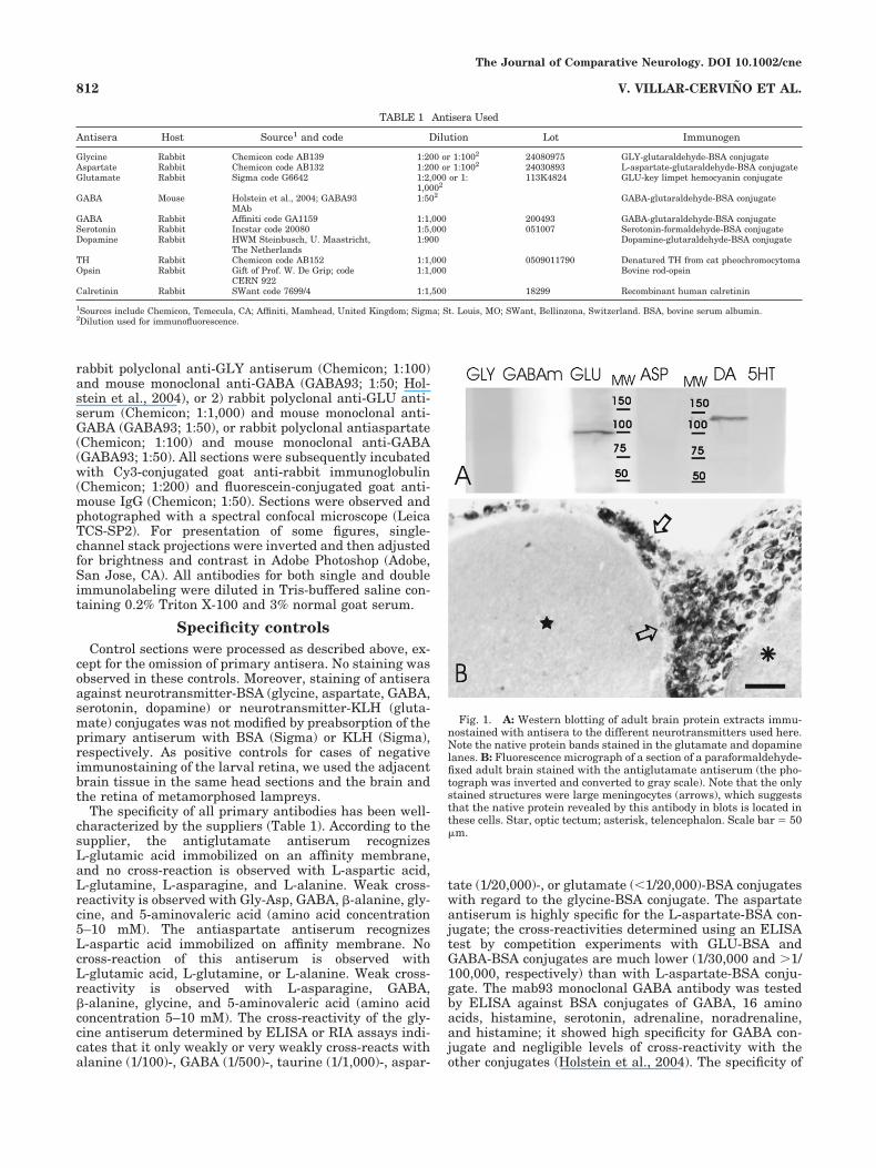
Fig. 1. A: Western blotting of adult brain protein extracts immu-nostained with antisera to the different neurotransmitters used here.Note the native protein bands stained in the glutamate and dopaminelanes. B: Fluorescence micrograph of a section of a paraformaldehyde-fixed adult brain stained with the antiglutamate antiserum (the pho-tograph was inverted and converted to gray scale). Note that the onlystained structures were large meningocytes (arrows), which suggeststhat the native protein revealed by this antibody in blots is located inthese cells. Star, optic tectum; asterisk, telencephalon. Scale bar � 50�m.
TABLE 1 Antisera Used
Antisera Host Source1 and code Dilution Lot Immunogen
Glycine Rabbit Chemicon code AB139 1:200 or 1:1002 24080975 GLY-glutaraldehyde-BSA conjugateAspartate Rabbit Chemicon code AB132 1:200 or 1:1002 24030893 L-aspartate-glutaraldehyde-BSA conjugateGlutamate Rabbit Sigma code G6642 1:2,000 or 1:
1,0002113K4824 GLU-key limpet hemocyanin conjugate
GABA Mouse Holstein et al., 2004; GABA93MAb
1:502 GABA-glutaraldehyde-BSA conjugate
GABA Rabbit Affiniti code GA1159 1:1,000 200493 GABA-glutaraldehyde-BSA conjugateSerotonin Rabbit Incstar code 20080 1:5,000 051007 Serotonin-formaldehyde-BSA conjugateDopamine Rabbit HWM Steinbusch, U. Maastricht,
The Netherlands1:900 Dopamine-glutaraldehyde-BSA conjugate
TH Rabbit Chemicon code AB152 1:1,000 0509011790 Denatured TH from cat pheochromocytomaOpsin Rabbit Gift of Prof. W. De Grip; code
CERN 9221:1,000 Bovine rod-opsin
Calretinin Rabbit SWant code 7699/4 1:1,500 18299 Recombinant human calretinin
1Sources include Chemicon, Temecula, CA; Affiniti, Mamhead, United Kingdom; Sigma; St. Louis, MO; SWant, Bellinzona, Switzerland. BSA, bovine serum albumin.2Dilution used for immunofluorescence.
The Journal of Comparative Neurology. DOI 10.1002/cne
812 V. VILLAR-CERVINO ET AL.
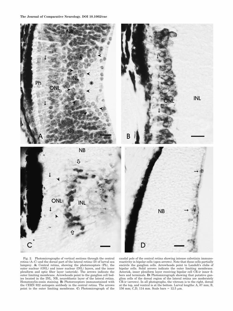
Fig. 2. Photomicrographs of vertical sections through the centralretina (A–C) and the dorsal part of the lateral retina (D) of larval sealamprey. A: Central retina, showing the photoreceptors (Ph), theouter nuclear (ONL) and inner nuclear (INL) layers, and the innerplexiform and optic fiber layer (asterisk). The arrows indicate theouter limiting membrane. Arrowheads point to the ganglion cell bod-ies located in the INL. NB, neuroblastic layer of the lateral retina.Hematoxylin-eosin staining. B: Photoreceptors immunostained withthe CERN 922 antiopsin antibody in the central retina. The arrowspoint to the outer limiting membrane. C: Photomicrograph of the
caudal pole of the central retina showing intense calretinin immuno-reactivity in bipolar cells (open arrows). Note that these cells partiallyencircle the ganglion cells. Arrowheads point to Landolt’s clubs ofbipolar cells. Solid arrows indicate the outer limiting membrane.Asterisk, inner plexiform layer receiving bipolar cell CR-ir inner fi-bers and terminals. D: Photomicrograph showing that putative gan-glion cells of the dorsal region of the lateral retina are moderatelyCR-ir (arrows). In all photographs, the vitreum is to the right, dorsalat the top, and ventral is at the bottom. Larval lengths: A, 87 mm; B,156 mm; C,D, 114 mm. Scale bars � 12.5 �m.
The Journal of Comparative Neurology. DOI 10.1002/cne

Figure 3
The Journal of Comparative Neurology. DOI 10.1002/cne
814 V. VILLAR-CERVINO ET AL.
the rabbit anti-GABA antiserum has been extensivelycharacterized by ELISA and immunohistochemical tech-niques and was adsorbed against BSA-glutaraldehyde.The dopamine antiserum does cross-react with noradren-aline for less that 10% and with other monoamines for lessthan 1%. The antiserotonin antibody does not cross-reactwith 5-hydroxytryptophan, 5-hydroxyindole-3-acetic acid,or dopamine. The calretinin antiserum does not cross-react with calbindin D-28k or other known calcium-binding proteins, as determined by its distribution in thebrain as well as by immunoblots. This antiserum wasalready tested by Western blotting in lamprey brain ex-tracts, showing a single stained band of the appropriatemolecular weight (Villar-Cheda et al., 2006). The antiop-sin antiserum was shown to stain specifically photorecep-tors in lampreys (Garcıa-Fernandez et al., 1997;Melendez-Ferro et al., 2002a) and teleosts (Candal et al.,2005). This immunoreaction was intended only for dem-onstrating photoreceptors, so characterization of opsintype specificity was not done.
As a further specificity control, antisera to glutamate,GABA, glycine, aspartate, and serotonin were analyzed byWestern blotting with protein extracts of adult sea lam-prey brain, as detailed previously (Villar-Cheda et al.,2006). Briefly, brains (including meninges) were homoge-nized at 4°C in 6 volumes of modified RIPA buffer (50 mMTris-HCl, 1 mM EDTA, 150 mM NaCl, 1 mM phenylmeth-ylsulfonylfluoride, 1% Triton X-100, 0.1% sodium dodecylsulfate, 5 �g/ml aprotinin, pH 7.4). The homogenate wascentrifuged at 20,000g, and the supernatant was precipi-tated with 6 volumes of 100% methanol. The precipitatewas resuspended in modified RIPA buffer, and 25 �g oftotal protein per lane was loaded onto 12% acrylamidegels, resolved by SDS-PAGE, and then electroblotted in aMini-Protean 3 cell (Bio-Rad, Hercules, CA) onto 0.2-�mpolyvinylidene difluoride membranes (Bio-Rad). Nonspe-cific binding sites on the membrane were blocked by incu-bating in 5% powdered nonfat milk dissolved in Tris-buffered saline containing 0.05% Tween 20 (TBST) for 1hour. After blocking, lanes in membranes were cut off,
rinsed in TBST, and each incubated overnight with one ofthe neurotransmitter primary antibodies. The membraneswere then rinsed in TBST, incubated with either goatanti-rabbit or goat anti-mouse HRP-conjugated antibodies(Bio-Rad; 1:15,000), rinsed again in TBST, and incubatedwith enhanced chemiluminescent reagent (Bio-RadImmun-Star HRP kit). Precision Plus protein standards(Bio-Rad) were used as molecular weight (MW) markers.
In Western blots, no protein band was stained with theglycine, GABA, serotonin, or aspartate antibodies (Fig.1A), strongly suggesting that these antisera do not cross-react with native proteins of the lamprey CNS. The anti-glutamate antibody stained a protein band of about 100kDa in blots (Fig. 1A). To investigate the distribution ofthis glutamate-like immunoreactivity, cryostat sections oflamprey brain fixed in buffered 4% paraformaldehydewere submitted to the immunofluorescence procedure de-scribed above. In these sections, strong staining was ob-served in large, ovoid meningeal cells, whereas neuronsand nerve fibers of the adjacent nervous tissue were notstained (Fig. 1B). Likewise, in sections fixed in buffered5% glutaraldehyde, these large meningocytes were alsostained. Because neurons and fibers were not stained inthese paraformaldehyde-fixed controls, together theseresults support the idea that the native protein thatcross-reacts in Western blots with the antiglutamateantibody is located in meningocytes and, accordingly,does not interfere with immunocytochemical analysis ofglutaraldehyde-coupled glutamate in the nervous tissue.A protein band of about 135 kDa was stained in blots withthe dopamine antiserum (Fig. 1A). However, demonstra-tion of DA immunoreactivity in the brain and retina wasstrictly dependent on fixation with glutaraldehyde andaddition of metabisulfite to incubation solutions. That dis-tribution of dopamine immunoreactivity revealed withthis antiserum in brain and retina matched with that oftyrosine hydroxylase (Pombal et al., 1997; Abalo et al.,2005; present results) strongly suggests that the sub-stance demonstrated in lamprey is DA.
Additional material
Series of larval lamprey heads from our collectionstained with hematoxylin-eosin were used for topographiclandmarks.
RESULTS
General organization of the larval eyeand retina
With hematoxylin-eosin staining, the eyes of larval lam-preys between 60 and 156 mm in length showed the tworetinal regions characteristic of the second half of larvallife (De Miguel and Anadon, 1987): a central retina withdifferentiated photoreceptor and outer nuclear and innernuclear/ganglion cell layers (Fig. 2A) and a lateral regionconsisting primarily of a thick neuroepithelium in whichonly a thin optic fiber/inner plexiform layer (IPL) is dis-tinguishable in the innermost region. This latter layerenlarges considerably near the optic nerve head. The cen-tral retina is located mostly dorsal to the optic nerve,which has an asymmetrical location in the eye.
Opsin and calretinin immunoreactivities
The CERN-922 antiopsin antibody revealed the pres-ence of opsin-ir photoreceptor cells in the central retina of
Fig. 3. Projections of stacks of 0.5-�m-thick confocal microscopeoptical sections of double-labeled retinae of larva (A–D). A: Verticalsection at the level of the optic nerve entrance (asterisk). Most GLU-irstructures are in the central retina, whereas the adjacent neuroblasticlayer of the lateral retina lacks significant immunoreactivity. Thethick solid arrow indicates the photoreceptor layer, the thin arrow thephotoreceptor perikarya, the curved arrow the layer of bipolar cells,and the open arrow ganglion cells. B: Detail of the GLU-ir ganglioncells at the level of the optic nerve entrance. Note the rich GABA-irinnervation of the IPL (star) and numerous GABA-ir boutons outlin-ing ganglion cell perikarya. All puncta are uniquely stained in singleconfocal image planes; the yellow color in some boutons is due tosuperposition of other structures in the image stacks. C: Detail of asingle confocal 0.5-�m-thick section of the region containing ganglioncells showing the distinct glutamate (in red in C), GABA (in greenchannel in C�), and double GLU/GABA immunofluorescence (in C��).D: Section just caudal to the optic nerve entrance showing GLY-irphotoreceptors (thick arrows) and perikarya in the ONL (thin arrows)and some GLY-ir cells (arrowheads) in the inner part of the IPL thatextend in the lateral retina. Note the position of GLY-ir cells withregard to the plexus of GABA-ir retinopetal fibers. Open arrows indi-cate the region containing ganglion cells in the differentiated retina.Curved arrow points to the pigmented retinal epithelium. In all pan-els, the vitreum is to the right (asterisk in D), dorsal is at the top, andventral is at the bottom. Scale bars � 20 �m in A,B,D; 10 �m in C.
The Journal of Comparative Neurology. DOI 10.1002/cne
815NEUROTRANSMITTERS IN THE LARVAL LAMPREY RETINA
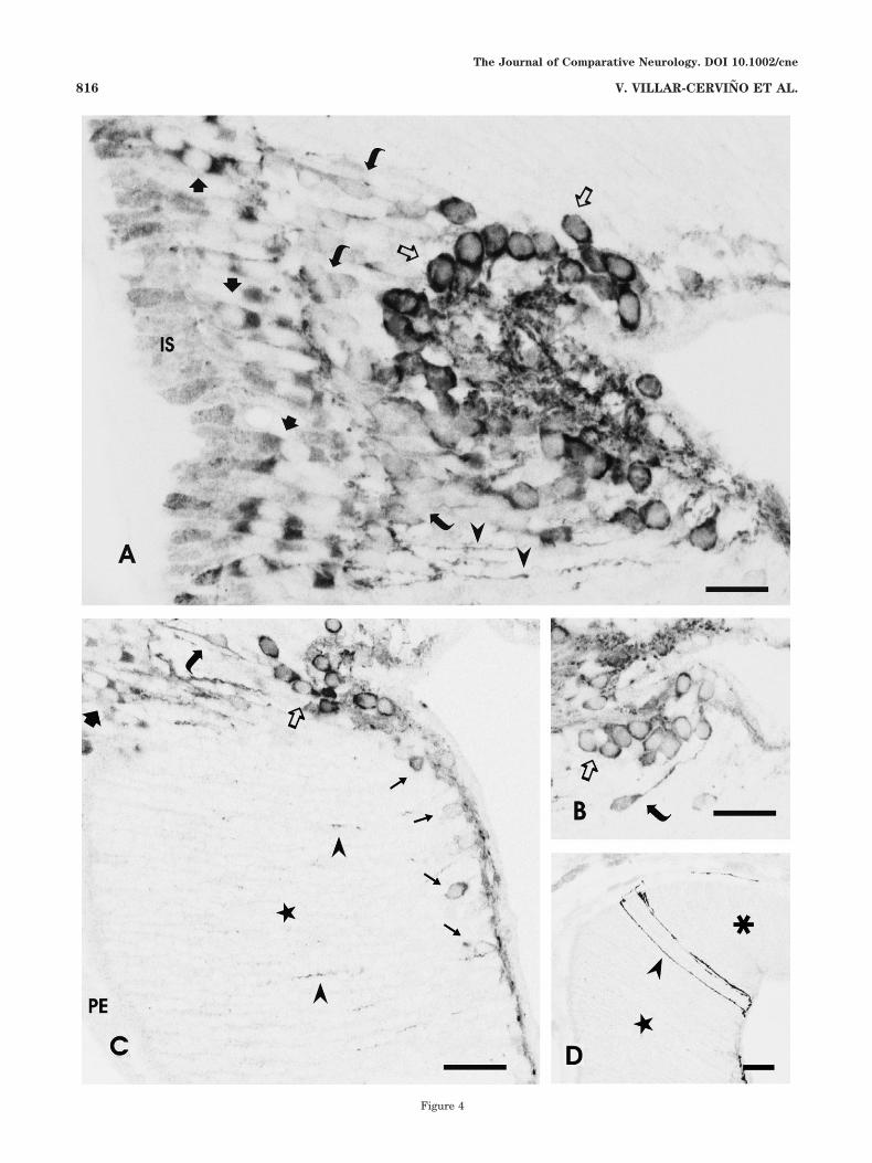
Figure 4
The Journal of Comparative Neurology. DOI 10.1002/cne
816 V. VILLAR-CERVINO ET AL.
the larvae. The antibody stained both segments of thephotoreceptors as well as their perikarya, thereby allow-ing the unequivocal localization of the cells to the centralretina. Photoreceptor perikarya formed two rows of cellsbelow the outer limiting membrane in larvae ranging from38 to 156 mm in length. The cell bodies were oval andlacked any conspicuous inner processes (Fig. 2B), which isconsistent with previous electron microscopic observa-tions (De Miguel et al., 1989).
In the retina of adult sea lamprey, the calretinin (CR)antibody intensely stained two types of bipolar cell as wellas some other cells in the inner nuclear layer (INL; innerhorizontal cells, a few amacrine cells, and some ganglioncells; Villar-Cheda et al., 2006), which is similar to thestaining reported for the retina of river lamprey (Dalil-Thiney et al., 1994). In the larval retina, intense CR im-munoreactivity was observed in bipolar cells of the centralretina (Fig. 2C), and faint to moderate staining was ob-tained in the putative ganglion cells of both the centraland the lateral retinal areas (Fig. 2C,D). [The location ofganglion cells in larval sea lamprey has been demon-strated in previous tract-tracing experiments (De Miguelet al., 1989; Anadon et al., 1998).] The CR-ir bipolar cellsexhibit outer and inner radial processes (Fig. 2C); theinner processes branch in the region of the inner plexiformlayer. The perikarya of CR-ir bipolar cells are more nu-merous caudal to the optic nerve exit and are located in acentral band that is well-separated from the rows of pho-toreceptors (Fig. 2C).
Neurotransmitter immunoreactivity in thelarval retina and brain
Among the six classical neurotransmitters (glutamate,GABA, glycine, aspartate, serotonin, and dopamine) andthe catecholamine-synthesizing enzyme (TH) investigatedin the larval retina, we observed immunoreactivity forglutamate, glycine, and GABA only. The negative resultsfor aspartate, serotonin, dopamine, and TH were not dueto false negative results, as indicated by the positive stain-ing of cells and fibers in the brain present in the samehead sections of the larvae. Each of the antibodies yielded
intense immunostaining of specific neuronal populationsand of numerous fibers throughout the brain. The distri-bution of these GABA-, serotonin-, dopamine-, and TH-irsystems is consistent with that reported in previous stud-ies in sea lamprey larvae (Melendez-Ferro et al., 2002b,2003; Abalo et al., 2005; Rodicio et al., 2005) and/or withthe distributions of serotonin and dopamine in adult riverlamprey (Pierre et al., 1992, 1997). A comprehensive studyof glutamate and glycine systems in the larval lampreybrain is currently in progress, but further description isbeyond the scope of the present report. As a positive con-trol in the retina, we used retinas of recently metamor-phosed lampreys (see below).
Glutamate immunoreactivity in thelarval retina
Central retina. Glutamate immunoreactivity was ob-served in both the central and the lateral regions of thelarval retina, although with distinctly different stainingpatterns (Figs. 3A,B, 4A–D). Several types of GLU-ir cellswere observed in the larval central retina, including pho-toreceptors, ganglion cells, and putative bipolar cells(Figs. 3A,B, 4A–C). These neurons did not show immuno-reactivity to GABA (Fig. 3A,B), precluding the possibilitythat the GLU staining reflected its role as an intermediarymetabolite in the synthesis of GABA. Faint GLU immu-noreactivity was observed in the inner segments of thephotoreceptors, whereas the apical and basal poles of theoval photoreceptor perikarya showed intense GLU-ir, andthe cell nuclei were immunonegative (Figs. 3A,B, 4A,C).
The position of horizontal cells in the INL of the centralretina of the larval sea lamprey is known from Golgiimpregnation studies (Rubinson and Cain, 1989). Thesecells can be appreciated as a loose cell region just belowthe outer nuclear layer in hematoxylin-eosin-stained ret-inae (Fig. 2A). These horizontal cells are not GLU immu-nostained in our material. In the middle of the INL thereare small, loosely scattered, faintly GLU-ir cells with elon-gated perikarya (Figs. 3A,B, 4A,C). On the basis of theirshape and position in the outer part of the INL, correlatedwith cells of similar position and appearance in Golgistains (Rubinson and Cain, 1989), the cells were identifiedas bipolar cells. They were located in the same position asthe bipolar cells revealed by CR immunocytochemistry.
Intensely GLU-ir neurons with rather large sphericalperikarya were observed in the inner part of the INL, theIPL, and the ganglion cell layer (Figs. 3A–C, 4A–C). Basedon their morphology and position in comparison with ourprevious tract-tracing results (De Miguel et al., 1989; Ana-don et al., 1998), these cells were interpreted as ganglioncells. Some of these cells exhibited characteristic ascend-ing processes that reached the outer plexiform layer (OPL;Fig. 4A) and occasionally branched. Ganglion cells withprocesses ascending to the OPL correspond to the biplexi-form ganglion cells described by De Miguel et al. (1989). Ithas yet to be determined whether all GLU-ir ganglion cellsof the central retina possess such ascending processes.GLU-ir axons of ganglion cells of the central and lateralretina course in the IPL/optic fiber layer to the optic nerve.
Lateral retina. In the lateral retina, GLU immunola-beling was observed only in cells located in the innermostretinal layer. Immunostaining was most prominent to-ward the central retina and very scarce, if at all present,laterally (Fig. 4C,D). GLU-ir cells were smaller than theganglion cells of the central retina but by their size and
Fig. 4. Photomicrographs showing the morphology of GLU-ir cellsand processes of the larval retina. All figures are single-channelprojections of a few confocal 0.5-�m sections (inverted and convertedto gray scale). A: Section of the central retina showing glutamateimmunoreactivity in the inner segment (IS) and perikarya of photo-receptors (solid arrows), bipolar cells (curved arrows), and ganglioncells (open arrows). Note the differences in immunoreactivity amongthe three neuronal types. The arrowheads point to dendritic processesof biplexiform ganglion cells ascending to the OPL. B: Detail of athree-section projection showing ganglion cells (open arrow) and abipolar cell (curved arrow). C: Section of the lateral retina showingglutamate-ir neurons (thin arrows) close to the primordial IPL, thethin layer of cells and processes close to the vitreum (below). Some ofthe processes of putative biplexiform ganglion cells ascend throughthe very thick neuroblastic layer (star). Note at the upper left theborder of the differentiated central retina showing GLU-ir photore-ceptors (thick arrow), bipolar cells (curved arrow), and ganglion cells(open arrow). D: Section through the lateral retina of a large larvashowing a few conspicuous glutamate-ir processes ascending to theouter limiting membrane between the lateral (star) and the marginal(asterisk) regions. In all figures, the vitreum is to the right, dorsal isat the top, and ventral is at the bottom. Scale bars � 20 �m in A; 30�m in B–D.
The Journal of Comparative Neurology. DOI 10.1002/cne
817NEUROTRANSMITTERS IN THE LARVAL LAMPREY RETINA
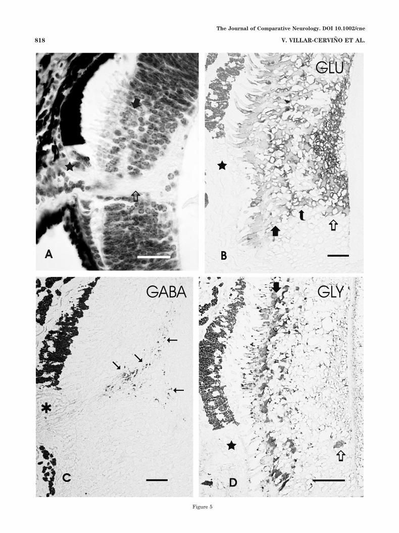
Figure 5
The Journal of Comparative Neurology. DOI 10.1002/cne
818 V. VILLAR-CERVINO ET AL.
distribution appeared to correspond to the ganglion cellsdemonstrated in previous tract-tracing studies (De Miguelet al., 1989; Anadon et al., 1998). Optic fibers were alsoGLU immunopositive, providing additional support forthe interpretation of the GLU-ir cells as ganglion cells.Near the central retina, some ganglion cells issued longradial processes directed toward the outer regions of theretina (Fig. 4C), similar to the processes of some ganglioncells of the central region. The cells of the very thickneuroblastic layer of the lateral retina did not show GLUimmunoreactivity above background. In the lateral retina,occasional GLU-ir processes were observed to ascendthrough the neuroblastic layer, contacting the outer lim-iting membrane (Fig. 4D). These processes appeared atthe transition between the lateral region, which containedGLU-ir structures, and the marginal region, which is ex-clusively neuroblastic.
Colocalization of GLU and GABA
Labeling experiments using our monoclonal anti-GABAantibody resulted in specific immunostaining of fibers inthe optic nerve and retina, in close agreement with previ-ous results from our laboratory (Anadon et al., 1998).Given the absence of any GABA-immunolabeled perikaryain the larval retina, these processes are interpreted asretinopetal fibers. Additional details of the retinopetalGABA-ir system in relation to retinal ganglion cells wereobtained through double-label studies with polyclonalanti-GLU and monoclonal anti-GABA antibodies andspectral laser confocal microscopy.
The immunolabeling with the green fluorescent second-ary antibody used for GABA demonstration may be con-fused with yellowish autofluorescent bodies, locatedmainly in the central retina near the outer limiting mem-brane but also occasionally scattered through the INL, asseen in control sections. Spectral confocal microscopy al-lowed us to differentiate these autofluorescent bodies fromthe GABA-ir fibers and boutons by means of image cap-ture using two closely spaced wavelength ranges, one spe-cific for the green fluorophore emission spectrum (490–524 nm) and the other covering a slightly wider range ofemission (482–539 nm). With this approach, the retinope-tal fibers were seen to be distributed throughout the lat-eral and central retina, although the density of such fibersin the central part far exceeded that in the lateral region(Fig. 3A–C). In the central retina, the GABA-ir fibers are
found in both the IPL and the adjacent INL (Fig. 3B,C).The highly specific labeling of the monoclonal GABA an-tibody together with the high spatial resolution of theconfocal microscopy revealed that GABA-ir boutons areassociated with ganglion cells (Fig. 3B,C). This associationis a distinctive feature of retinal ganglion cells. Analysis of0.5-�m-thick confocal sections did not reveal clear labelingof glutamate in GABA-ir boutons, suggesting that theseboutons do not accumulate glutamate above basal levels.
GLY immunoreactivity
GLY immunoreactivity was observed in differentiatedcells of both the central and the lateral regions of theretina (Fig. 3D), whereas the thick neuroblastic layer ofthe lateral retina did not show GLY immunoreactivityabove background. In the central retina, the perikarya ofphotoreceptors were moderately or strongly GLY-ir,whereas the inner photoreceptor segments showed fainterimmunoreactivity (Fig. 3D). In addition, moderate to highintensity GLY immunoreactivity was observed in scat-tered cells of the outer and inner sides of the IPL charac-terized by its GABA-ir fiber plexus, i.e., a region alsocontaining putative ganglion cells (Fig. 3D). However, thenumber of GLY-ir cells was clearly minor in comparisonwith that of GLU-ir cells. In addition, comparison of pho-tographs of double immunostaining GABA/GLY (Fig. 3D)with those of GABA/GLU (Fig. 3A–C) clearly indicatesthat the GLY-ir cells did not form a cap over the IPL aswas observed with glutamate immunostaining, the largeganglion cells of this region being only very faintly GLY-ir(Fig. 3D). Moreover, no ascending GLY-ir processes wereobserved, in contrast to the results obtained with gluta-mate immunocytochemistry. In the lateral retina, somecells close to the primordial IPL showed faint to moderateGLY immunoreactivity (Fig. 3D). As in the central retina,these cells appeared in the region containing ganglioncells. Whereas double GABA/GLY immunostaining (Fig.3D) suggests that GABA-ir retinopetal fibers might con-tact GLY-ir cells processes, no specific relationship ofthese fibers with glycinergic perikarya was observed.
GABA, GLY, and GLU immunostaining incontrol semithin plastic sections
To confirm the results obtained by confocal microscopy,some semithin plastic sections were stained with antiseraagainst GLU, GABA, and GLY. To facilitate identificationof structures of these sections, a similar hematoxylin-eosin-stained section is shown (Fig. 5A). In plastic sec-tions, GLU, GABA, and GLY immunoreactivities wereobserved in similar locations of the larval retina to thoseobserved with confocal microscopy (Fig. 5B–D). GLU-ircells were present in the regions of the photoreceptor cells,bipolar cells, and ganglion cells (Fig. 5B). In semithinsections, the monoclonal GABA antibody stained only fi-bers in the IPL (Fig. 5C), which were clearly identifiableas centrifugal fibers by the absence of any stained retinalperikaryon. Photoreceptors showed GLY immunoreactiv-ity in semithin plastic sections, and a few GLY-ir cellswere also observed in the innermost central retina (Fig.5D).
Neurotransmitter immunoreactivity in thepostmetamorphic retina
For comparison with the larvae and to serve as positivecontrols, we examined retinas from recently metamor-
Fig. 5. Hematoxylin-eosin-stained section (A) and postembeddingstained, semithin plastic vertical sections of the larval retina showingimmunoreactivity to glutamate (B), GABA (C), and glycine (D).A: Photomicrograph showing a section of the retina at a level similarto that in C. Star, ventral unpigmented part of pigment epithelium;open arrow, optic nerve entrance; solid arrow, layer of photoreceptorperikarya. B: Section showing glutamate immunoreactivity in cells ofthe vertical pathways. The thick arrow points to the GLU-ir photore-ceptor perikarya, the curved arrow designates the layer of bipolarcells, and the open arrow indicates ganglion cells. The star marks theunpigmented ventral region of the pigment epithelium. C: Sectionthrough the level of the optic nerve (asterisk) showing that GABAimmunoreactivity is restricted to centrifugal fibers (arrows). D: Sec-tion showing glycine immunoreactivity in photoreceptors and inscarce cells (open arrow) of the inner part of the retina. In all figures,the vitreum is to the right, dorsal is at the top, and ventral is at thebottom. Brightfield microscopy. Scale bars � 50 �m in A; 20 �m inB–D.
The Journal of Comparative Neurology. DOI 10.1002/cne
819NEUROTRANSMITTERS IN THE LARVAL LAMPREY RETINA
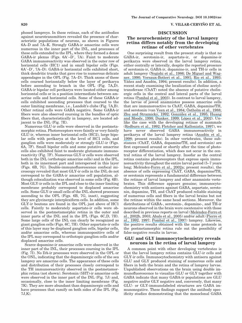
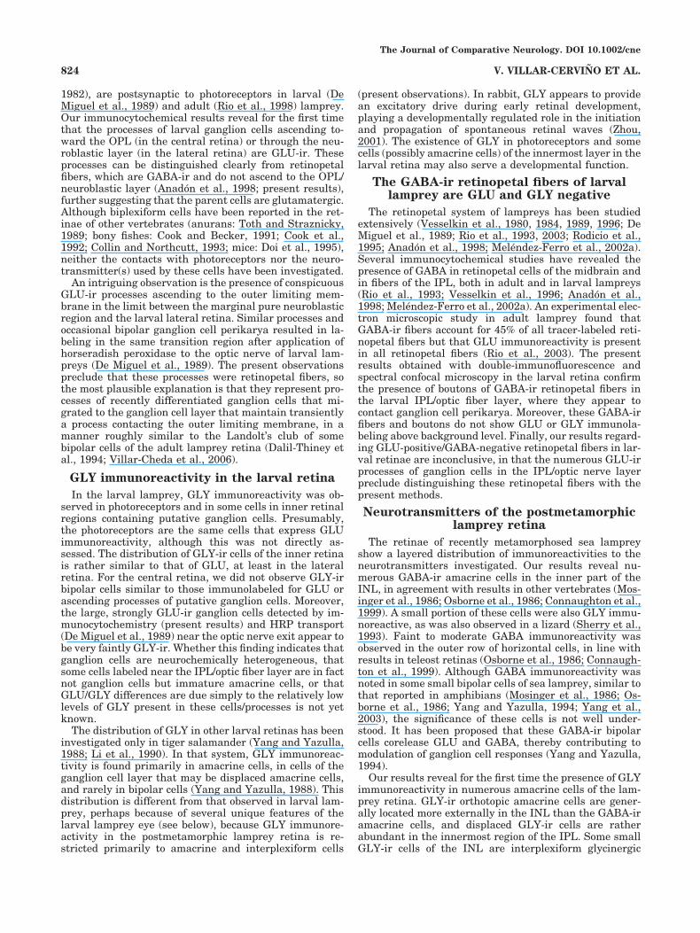
phosed lampreys. In these retinas, each of the antibodiesagainst neurotransmitters revealed the presence of char-acteristic populations of neurons, as shown in Figures6A–D and 7A–K. Strongly GABA-ir amacrine cells werenumerous in the inner part of the INL, and processes ofthese cells extended to the IPL, where they formed a denseGABA-ir plexus (Figs. 6A�–D�, 7A). Faint to moderateGABA immunoreactivity was observed in the outer row ofhorizontal cells (HC1) and in small bipolar cells (Figs.6A�–D�, 7A–D). GABA-ir horizontal cells exhibited short,thick dendritic trunks that gave rise to numerous delicateappendages in the OPL (Fig. 7A–D). Thick axons of thesecells coursed horizontally below the layer of perikaryabefore ascending to branch in the OPL (Fig. 7A,D).GABA-ir bipolar cell perikarya were located either amonghorizontal cells or in a position intermediate between am-acrine cells and horizontal cells. Some of these GABA-ircells exhibited ascending processes that coursed to theouter limiting membrane, i.e., Landolt’s clubs (Fig. 7A,B).Other retinal cells were GABA negative. Some GABA-irfibers were also observed coursing in the bundles of opticfibers that, characteristically in lamprey, are located ad-jacent to the INL (Fig. 7E).
Numerous GLU-ir cells were observed in the postmeta-morphic retina. Photoreceptors were faintly or very faintlyGLU-ir, whereas inner horizontal cells (HC2), large bipo-lar cells with perikarya at the level of HC1 cells, andganglion cells were moderately or strongly GLU-ir (Figs.6A, 7F). Small bipolar cells and some putative amacrinecells also exhibited faint to moderate glutamate immuno-reactivity (Fig. 7F). Numerous GLY-ir cells were observedboth in the INL (orthotopic amacrine cells) and in the IPL,both in its innermost part and interspersed in this layer(Figs. 6B, 7G). Double-immunofluorescence confocal mi-croscopy revealed that most GLY-ir cells in the INL do notcorrespond to the GABA-ir amacrine cell population, al-though colocalization was observed in some cells (Fig. 6B).Most GLY-ir cells in the IPL and close to the inner limitingmembrane probably correspond to displaced amacrinecells. Some GLY-ir small cells of the INL showed processesascending to the OPL (Figs. 6B, 7G, inset), suggestingthey are glycinergic interplexiform cells. In addition, someGLY-ir boutons are found in the OPL just above of HC1cells. Faintly to moderately aspartate-ir cells were ob-served in the postmetamorphic retina in the outer andinner parts of the INL and in the IPL (Figs. 6C,D, 7H).Some large cells of the INL can clearly be recognized asgiant ganglion cells (Figs. 6C, 7H). Other aspartate-ir cellsof this layer may be displaced ganglion cells, bipolar cells,and/or amacrine cells, whereas immunopositive cells ofthe IPL may correspond to orthotopic ganglion cells and/ordisplaced amacrine cells.
Scarce dopamine-ir amacrine cells were observed in theinner part of the INL, their processes coursing in the IPL(Fig. 7I). No DA-ir processes were observed in the OPL orthe ONL, indicating that the dopaminergic cells of the sealamprey are amacrine cells. The appearance of these cellsand distribution of their processes closely correspond tothe TH immunoreactivity observed in the postmetamor-phic retina (not shown). Serotonin (5HT)-ir amacrine cellswere observed in the inner part of the INL (Fig. 7J) and,occasionally, close to the inner limiting membrane (Fig.7K). They are more abundant than dopaminergic cells andhave processes that ramify on both sides of the IPL (Fig.7J,K).
DISCUSSION
The neurochemistry of the larval lampreyretina differs notably from the developing
retinae of other vertebrates
One surprising result from the present study is that noGABA-ir, serotonin-ir, aspartate-ir, or dopamine-irperikarya were observed in the larval lamprey retina,either centrally or laterally, despite the reported presenceof serotonin-ir, GABA-ir, dopamine-ir, and TH-ir cells inadult lamprey (Negishi et al., 1986; De Miguel and Wag-ner, 1990; Versaux-Botteri et al., 1991; Rio et al., 1993;Yanez and Anadon, 1994; present results). In addition, arecent study examining the localization of choline acetyl-transferase (ChAT) noted the absence of putative cholin-ergic cells in the central and lateral parts of the larvalretina (Pombal et al., 2003). In contrast to larval lamprey,the larvae of jawed anamniotes possess amacrine cellsthat are immunoreactive to ChAT, GABA, dopamine/TH,and serotonin (van Veen et al., 1984; Ostholm et al., 1988;Zhu and Straznicky, 1992; Gonzalez et al., 1995; Huangand Moody, 1998; Dunker, 1999; Lopez et al., 2002). Un-like the case with the developing retinae of mammals(Redburn et al., 1992; Fletcher and Kalloniatis, 1997), wehave never observed GABA immunoreactivity inperikarya of the larval lamprey retina (Anadon et al.,1998; present results). In other vertebrates, these sub-stances (ChAT, GABA, dopamine/TH, and serotonin) arefirst expressed around or shortly after the time of photo-receptor differentiation, which does not occur in the cen-tral retina of the larval lamprey. Insofar as the centralretina contains photoreceptors that express opsin immu-noreactivity throughout the entire larval period (5–7 yearslong; Melendez-Ferro et al., 2002a; present results), theabsence of cells expressing ChAT, GABA, dopamine/TH,or serotonin represents a fundamental difference betweenthe retinae of larval lamprey and other anamniote verte-brates. This difference appears genuine; immunocyto-chemistry with antisera against GABA, aspartate, seroto-nin, dopamine, TH, and ChAT produced reliable stainingof numerous cells and fibers in the larval brains but notthe retinae within the same head sections. Moreover, thedistributions of GABA-, serotonin-, dopamine-, and TH-irneurons observed in the brain were coextensive with thosedescribed in previous reports on larval (Melendez-Ferro etal., 2002b, 2003; Abalo et al., 2005) and/or adult (Pierre etal., 1992, 1997; Pombal et al., 1997) lamprey. Likewise,the positive results obtained with the same protocols inthe postmetamorphic retina rule out the possibility offalse-negative results in larvae.
GLU and GLY immunocytochemistry revealsneurons in the retina of larval lamprey
A common point with other developing vertebrates isthat the larval lamprey retina contains GLU-ir cells andGLY-ir cells. Immunocytochemistry with antisera againstGLU and GLY produced staining of numerous cells andfibers in both the brain and the retina of lamprey larvae.Unpublished observations on the brain using double im-munofluorescence to visualize GLU or GLY together withGABA indicate that many GABA-ir populations are GLUnegative and/or GLY negative and, conversely, that manyGLU- or GLY-immunolabeled structures are GABA im-munonegative. These findings support the antibody spec-ificity studies demonstrating that the monoclonal GABA
The Journal of Comparative Neurology. DOI 10.1002/cne
820 V. VILLAR-CERVINO ET AL.

Fig. 6. Projections of stacks of 0.5-�m-thick confocal microscopeoptical sections of double-labeled retinae of postmetamorphic sea lam-prey showing in the left panels glutamate (A), glycine (B), and aspar-tate (C,D) immunofluorescence (in the red channel), in the centralpanels immunoreactivity to GABA (in green), and in the right panels,double immunolabeling. A–A��: Section showing abundant glutamateimmunoreactivity in horizontal cells and putative ganglion cells (ar-rowhead). Colocalization of GABA and glutamate is occasionallyfound in cells of the INL and IPL (thin arrows). The curved arrowpoints to a bundle of optic fibers close to the INL. Small arrows pointto clearly double-labeled cells. B–B��: Sections showing glycine immu-
noreactivity in amacrine cells of the INL and the IPL. Colocalizationwith GABA is observed in some amacrine cells (thin arrow). The openarrows point to glycinergic interplexiform cells. Arrowheads, inter-plexiform cell processes in the outer plexiform layer. C–C��,D–D��:Sections showing abundant aspartate immunoreactivity in some hor-izontal cells, in ganglion cells (arrowheads), and in some cells of theIPL. Thin arrows point to putative amacrine cells showing colocaliza-tion of aspartate and GABA immunoreactivity. In A, thick whitearrows point to the outer limiting membrane. In all photographs,sclera is toward the top, and the vitreum at the bottom. Scale bars �20 �m.
The Journal of Comparative Neurology. DOI 10.1002/cne

Figure 7
The Journal of Comparative Neurology. DOI 10.1002/cne
822 V. VILLAR-CERVINO ET AL.

antibody does not recognize either of the other two aminoacids (Holstein et al., 2004). In line with this evidence inthe brain, no correspondence was observed between theGLU- and the GABA-ir and between the GLY- and theGABA-ir structures in double-labeling experiments in thelarval retina.
GLU-ir cells of the larval lamprey retina:comparison with the retinae of other
vertebrates
Developmental studies of GLU-ir cells in the vertebrateretina are very scarce: as far as we are aware, this hasbeen addressed exclusively in mammals (Redburn et al.,1992; Pow et al., 1994; Fletcher and Kalloniatis, 1997). Inthe rabbit retina, GLU immunoreactivity first appears byembryonic day 20 (E20). Ganglion cells and photorecep-tors are GLU-ir at birth (Redburn et al., 1992), and bipolarcells and some amacrine cells also become GLU-ir at aboutpostnatal day 10 (P10; Pow et al., 1994). Some horizontalcells are reported to be GLU-ir at birth by Redburn et al.(1992) although not by Pow et al. (1994).
The unique retina of larval lampreys affords the oppor-tunity to explore simultaneously the immunoreactivity toGLU and GABA during three distinct developmentalphases in the same larvae. Whereas the central retina hasa layered organization with photoreceptors, horizontalcells, bipolar cells, and ganglion cells, the lateral retinaconsists mainly of a thick neuroblastic layer as well asdifferentiating ganglion cells and retinopetal fibers in theinnermost region, and the marginal retina is a purelyneuroblastic region lacking identifiable ganglion cells or
retinopetal fibers (De Miguel and Anadon, 1987; DeMiguel et al., 1989; Anadon et al., 1998; Melendez-Ferro etal., 2002; present results). The distribution of GLU in thecentral retina of the larval sea lamprey is reminiscent ofthat reported before eye opening in the vertical system ofrabbit, immunoreactivity being observed in photorecep-tors, some bipolar cells, and ganglion cells. These GLU-ircells of the larval lamprey retina are not GABA-ir in thepostmetamorphic lamprey, with the exception of GABAexpression in a bipolar cell subtype (present results), andno GLU immunoreactivity was observed in the region offuture GABAergic amacrine cells. These findings are con-sistent with our interpretation that GLU is the neuro-transmitter utilized by neurons of the vertical pathwaysin the larval retina.
For the lateral and marginal retina, our results demon-strate the absence of GLU immunoreactivity above back-ground in cells of the neuroblastic layers. In fact, thepresence of GLU-ir cells in the innermost layers providesa biological marker to distinguish the lateral from themarginal regions. One interesting observation concernsthe long GLU-ir processes of ganglion cells ascendingthrough the neuroblastic layer. These processes may beviewed as either transient trailing processes of recentlymigrated cells, or processes of biplexiform cells awaitingthe differentiation of photoreceptors and the OPL. On theother hand, the absence of GLU-ir and GABA-ir fibers andcells in the marginal region reinforces the exclusivelyproliferative nature of this zone.
With regard to the retinae of adult vertebrates, most stud-ies indicate the presence of GLU in most photoreceptors,bipolar cells, and ganglion cells (teleosts: Kageyama andMeyer, 1989; Van Haesendonck and Missotten, 1990; Marcet al., 1990, 1995; Connaughton et al., 1999; amphibians:Yang and Yazulla, 1994; reptiles: Schutte, 1995; birds: Kal-loniatis and Fletcher, 1993; mammals: Davanger et al.,1991; Crooks and Kolb, 1992; Jojich and Pourcho, 1996;Kalloniatis et al., 1996). GLU has also been reported in aGABA-negative horizontal cell type in goldfish (Marc et al.,1995) and in some horizontal cells of the tiger salamander(Yang and Yazulla, 1994). The colocalization of GLU withGABA or GLY has been observed in amacrine cells of tigersalamander (Yang and Yazulla, 1994), amacrine and hori-zontal cells of chicken (Kalloniatis and Fletcher, 1993), andamacrine cells of some mammals (Jojich and Pourcho, 1996).The current results indicate the presence of immunoreactiv-ity to GLU in some cell types of the retina of postmetamor-phic sea lamprey, including some horizontal and amacrinecells. As reported for goldfish, the GLU-ir horizontal cells oflamprey (inner row of horizontal cells; HC2 cells) do notcorrespond to the faint GABA-ir horizontal cells (outer row;HC1 cells). In postmetamorphic lamprey retina, photorecep-tors and small bipolar cells exhibit only faint GLU immuno-reactivity.
Biplexiform ganglion cells of larval lampreyare demonstrated via GLU
immunocytochemistry
The presence of biplexiform ganglion cells has beenreported from studies using retrograde tract-tracing inlarval (De Miguel et al., 1989; Anadon et al., 1998) andadult (Fritzsch and Collin, 1990; Rio et al., 1998) lampreyretinae. Ultrastructural observations indicate that theseganglion cells, first described for the primate (Mariani,
Fig. 7. Photomicrographs of vertical sections of the postmetamor-phic retina showing the immunoreactivity to GABA (A–E), glutamate(F), glycine (G), aspartate (H), dopamine (I), and serotonin (J,K).A: Section showing strong GABA immunoreactivity in some amacrinecells and moderate immunoreactivity in type 1 horizontal cells and insome bipolar cells (open arrows). The arrowheads point to appendagesof horizontal cells in the OPL. Note Landolt’s clubs of bipolar cellscoursing to the outer limiting membrane (solid arrows). B: Detail ofthe Landolt’s club (arrow) of a GABA-ir bipolar cell with perikaryonlocated among horizontal cells. C: Detail of dendritic appendages ofGABA-ir horizontal cells. D: Detail of axons of GABA-ir horizontalcells coursing below the perikarya (thick arrows) and ascending to theOPL (thin arrow). E: Detail of several putative retinopetal GABA-irfibers coursing in a fascicle of optic fibers close to amacrine cells of theINL. F: Section showing abundant glutamate immunoreactivity inHC2 horizontal cells (open arrow) and in other neurons of the INL andthe IPL. Thin arrows, small bipolar cells; arrowheads, large bipolarcells with perikarya just below the outer plexiform layer. G: Sectionshowing the distribution of glycine-ir amacrine cells in the INL andIPL. The arrowhead points to the outer plexiform layer showing a fewGLY-ir boutons. Inset: Detail of a glycinergic interplexiform cell(open arrow) showing an ascending process (thin arrow) coursing tothe OPL (arrowhead). H: Section showing a large aspartate immuno-reactive ganglion cell (double arrow) and other cells showing abun-dant immunoreactivity in the INL and IPL (single arrows). I: Detail ofa dopamine-ir amacrine cell of the INL. Note that DA-ir processescourse in outer parts of the IPL. J: Serotonin-ir amacrine cells locatedin the INL. Note that 5HT-ir fibers mainly course in outer and innersublaminae of the IPL. K: Section showing a serotoninergic displacedamacrine cell (arrow) that is close to the inner limiting membrane.A–H: Single channel projections of confocal stacks (inverted and con-verted to gray scale). I–K: Brightfield microscopy. In these photo-graphs of the adult retina, the sclera is at the top, and the vitreum atthe bottom. Scale bars � 20 �m in A,F–H; 20 �m in E (applies to B–E);25 �m I–K.
The Journal of Comparative Neurology. DOI 10.1002/cne
823NEUROTRANSMITTERS IN THE LARVAL LAMPREY RETINA
1982), are postsynaptic to photoreceptors in larval (DeMiguel et al., 1989) and adult (Rio et al., 1998) lamprey.Our immunocytochemical results reveal for the first timethat the processes of larval ganglion cells ascending to-ward the OPL (in the central retina) or through the neu-roblastic layer (in the lateral retina) are GLU-ir. Theseprocesses can be distinguished clearly from retinopetalfibers, which are GABA-ir and do not ascend to the OPL/neuroblastic layer (Anadon et al., 1998; present results),further suggesting that the parent cells are glutamatergic.Although biplexiform cells have been reported in the ret-inae of other vertebrates (anurans: Toth and Straznicky,1989; bony fishes: Cook and Becker, 1991; Cook et al.,1992; Collin and Northcutt, 1993; mice: Doi et al., 1995),neither the contacts with photoreceptors nor the neuro-transmitter(s) used by these cells have been investigated.
An intriguing observation is the presence of conspicuousGLU-ir processes ascending to the outer limiting mem-brane in the limit between the marginal pure neuroblasticregion and the larval lateral retina. Similar processes andoccasional bipolar ganglion cell perikarya resulted in la-beling in the same transition region after application ofhorseradish peroxidase to the optic nerve of larval lam-preys (De Miguel et al., 1989). The present observationspreclude that these processes were retinopetal fibers, sothe most plausible explanation is that they represent pro-cesses of recently differentiated ganglion cells that mi-grated to the ganglion cell layer that maintain transientlya process contacting the outer limiting membrane, in amanner roughly similar to the Landolt’s club of somebipolar cells of the adult lamprey retina (Dalil-Thiney etal., 1994; Villar-Cheda et al., 2006).
GLY immunoreactivity in the larval retina
In the larval lamprey, GLY immunoreactivity was ob-served in photoreceptors and in some cells in inner retinalregions containing putative ganglion cells. Presumably,the photoreceptors are the same cells that express GLUimmunoreactivity, although this was not directly as-sessed. The distribution of GLY-ir cells of the inner retinais rather similar to that of GLU, at least in the lateralretina. For the central retina, we did not observe GLY-irbipolar cells similar to those immunolabeled for GLU orascending processes of putative ganglion cells. Moreover,the large, strongly GLU-ir ganglion cells detected by im-munocytochemistry (present results) and HRP transport(De Miguel et al., 1989) near the optic nerve exit appear tobe very faintly GLY-ir. Whether this finding indicates thatganglion cells are neurochemically heterogeneous, thatsome cells labeled near the IPL/optic fiber layer are in factnot ganglion cells but immature amacrine cells, or thatGLU/GLY differences are due simply to the relatively lowlevels of GLY present in these cells/processes is not yetknown.
The distribution of GLY in other larval retinas has beeninvestigated only in tiger salamander (Yang and Yazulla,1988; Li et al., 1990). In that system, GLY immunoreac-tivity is found primarily in amacrine cells, in cells of theganglion cell layer that may be displaced amacrine cells,and rarely in bipolar cells (Yang and Yazulla, 1988). Thisdistribution is different from that observed in larval lam-prey, perhaps because of several unique features of thelarval lamprey eye (see below), because GLY immunore-activity in the postmetamorphic lamprey retina is re-stricted primarily to amacrine and interplexiform cells
(present observations). In rabbit, GLY appears to providean excitatory drive during early retinal development,playing a developmentally regulated role in the initiationand propagation of spontaneous retinal waves (Zhou,2001). The existence of GLY in photoreceptors and somecells (possibly amacrine cells) of the innermost layer in thelarval retina may also serve a developmental function.
The GABA-ir retinopetal fibers of larvallamprey are GLU and GLY negative
The retinopetal system of lampreys has been studiedextensively (Vesselkin et al., 1980, 1984, 1989, 1996; DeMiguel et al., 1989; Rio et al., 1993, 2003; Rodicio et al.,1995; Anadon et al., 1998; Melendez-Ferro et al., 2002a).Several immunocytochemical studies have revealed thepresence of GABA in retinopetal cells of the midbrain andin fibers of the IPL, both in adult and in larval lampreys(Rio et al., 1993; Vesselkin et al., 1996; Anadon et al.,1998; Melendez-Ferro et al., 2002a). An experimental elec-tron microscopic study in adult lamprey found thatGABA-ir fibers account for 45% of all tracer-labeled reti-nopetal fibers but that GLU immunoreactivity is presentin all retinopetal fibers (Rio et al., 2003). The presentresults obtained with double-immunofluorescence andspectral confocal microscopy in the larval retina confirmthe presence of boutons of GABA-ir retinopetal fibers inthe larval IPL/optic fiber layer, where they appear tocontact ganglion cell perikarya. Moreover, these GABA-irfibers and boutons do not show GLU or GLY immunola-beling above background level. Finally, our results regard-ing GLU-positive/GABA-negative retinopetal fibers in lar-val retinae are inconclusive, in that the numerous GLU-irprocesses of ganglion cells in the IPL/optic nerve layerpreclude distinguishing these retinopetal fibers with thepresent methods.
Neurotransmitters of the postmetamorphiclamprey retina
The retinae of recently metamorphosed sea lampreyshow a layered distribution of immunoreactivities to theneurotransmitters investigated. Our results reveal nu-merous GABA-ir amacrine cells in the inner part of theINL, in agreement with results in other vertebrates (Mos-inger et al., 1986; Osborne et al., 1986; Connaughton et al.,1999). A small portion of these cells were also GLY immu-noreactive, as was also observed in a lizard (Sherry et al.,1993). Faint to moderate GABA immunoreactivity wasobserved in the outer row of horizontal cells, in line withresults in teleost retinas (Osborne et al., 1986; Connaugh-ton et al., 1999). Although GABA immunoreactivity wasnoted in some small bipolar cells of sea lamprey, similar tothat reported in amphibians (Mosinger et al., 1986; Os-borne et al., 1986; Yang and Yazulla, 1994; Yang et al.,2003), the significance of these cells is not well under-stood. It has been proposed that these GABA-ir bipolarcells corelease GLU and GABA, thereby contributing tomodulation of ganglion cell responses (Yang and Yazulla,1994).
Our results reveal for the first time the presence of GLYimmunoreactivity in numerous amacrine cells of the lam-prey retina. GLY-ir orthotopic amacrine cells are gener-ally located more externally in the INL than the GABA-iramacrine cells, and displaced GLY-ir cells are ratherabundant in the innermost region of the IPL. Some smallGLY-ir cells of the INL are interplexiform glycinergic
The Journal of Comparative Neurology. DOI 10.1002/cne
824 V. VILLAR-CERVINO ET AL.
cells, as indicated by the presence of processes coursing tothe OPL. The presence of GLY-ir interplexiform cells hasalso been observed in teleosts (Marc and Lam, 1981; Kal-loniatis and Marc, 1990; Yazulla and Studholme, 1990;Connaughton et al., 1999), amphibians (Yang and Ya-zulla, 1988; Vitanova et al., 2004), reptiles (Eldred andCheung, 1989; Sherry et al., 1993), and chicks (Kalloniatisand Fletcher, 1993). This GLY distribution is roughlysimilar to that reported in teleosts (Yazulla and Stud-holme, 1990; Connaughton et al., 1999). Our results alsoindicate that GLU immunoreactivity is abundant in cellsand processes of the INL and IPL, with high levels ininner horizontal cells and in other INL cells (ganglioncells, large bipolar cells, putative amacrine cells). Colocal-ization of GABA and GLU was observed in some amacrinecells. The presence of GLU immunoreactivity in someGABA- or GLY-ir amacrine cells has been also reported inthe retina of tiger salamander (Yang, 1996). Aspartateimmunoreactivity is abundant in putative ganglion cells,including giant ganglion cells, as well as in some amacrinecells, and moderate levels were also observed in innerhorizontal cells, suggesting colocalization of aspartate andGLU in some cell populations.
Our results with antibodies against both dopamine andTH do not reveal immunoreactive processes in the OPL ofthe postmetamorphic sea lamprey retina, confirming theidentify of the dopaminergic cells as amacrine cells (Yanezand Anadon, 1994). Although the presence of dopaminer-gic interplexiform cells in the sea lamprey retina has beenreported with an antibody against TH (De Miguel andWagner, 1990), this staining might be due to cross-reaction with other substances. Unlike the case in sealamprey, many teleost retinas show a well-developed do-paminergic interplexiform cell system (Osborne et al.,1984; Yazulla and Zucker, 1988; Kalloniatis and Marc,1990; Wagner and Behrens, 1993; Frohlich et al., 1995),although, in pure-rod teleost retinae, dopaminergic cellsare amacrine cells (Frohlich et al., 1995). The serotonin-iramacrine cells of the postmetamorphic sea lamprey retinaare mostly orthotopic, and their processes branch in outerand/or inner sublaminae of the IPL. These amacrine cellsare similar to those described for river lamprey (Versaux-Botteri et al., 1991).
Functional considerations
The eye of the larval lamprey is covered by a thick,nontransparent skin and has an immature lens, indicat-ing that it is not an image-forming eye (Kleerekoper,1972). Thus, larval lampreys must wait several years untilmetamorphosis to acquire a truly functional, camera-typeeye. However, the larval central retina shows numeroustraits suggesting functional maturation beginning in earlyposthatching stages, including the presence of opsin-expressing photoreceptors with well-developed outer seg-ments (Melendez-Ferro et al., 2003), the presence of brain-projecting ganglion cells and of centrifugal fibers (DeMiguel et al., 1989; Anadon et al., 1998), and the expres-sion of GLU in cells of the vertical pathway (presentresults). Together, these traits strongly suggest that atleast some parts of the larval retina are functional, butperhaps as a nondirectional or broadly directional photo-receptive organ like a simple ocellus. In this regard, theabsence of neurotransmitter-ir amacrine cells throughoutthe entire larval period may be interpreted in terms ofimmaturity of the image-forming circuitry, which requires
opposing influences to extract the different qualities of theimage (movement vs. static background, light/dark con-trast, etc). In some way, the larval lamprey eye is compa-rable to the lamprey pineal organ, which also lacks image-forming circuitry.
It is likely that the functional roles of GLU and GABA inthe retina of larval lamprey depend on the specific neuro-nal types and retinal regions. Although GLU and GABAare viewed as the major excitatory and inhibitory neuro-transmitters in the vertebrate brain, their various actionsdepend on developmental stage and on the nature andsubcellular distribution of pre- and postsynaptic iono-tropic and metabotropic receptor subtypes(s) (Ben-Ari etal., 1990; Cherubini et al., 1991, 1998). It is well docu-mented that GABA acts as an excitatory neurotransmitterduring retinal development, because of the high mem-brane potential of developing neurons and/or actionthrough elevation of Ca2� levels (Redburn-Johnson,1998).
In addition to its functions in the adult vertebrate ret-ina, GLU appears to participate in several processes thatoccur during development. In the developing cat retina,stratification of ON and OFF ganglion cell dendrites de-pends on activity mediated by metabotropic GLU recep-tors (Bodnarenko and Chalupa, 1993; Bodnarenko et al.,1995). In addition, responses of early fetal ganglion cells toionotropic GLU receptor agonists before synaptogenesis inthe INL appear remarkably similar to those of postnatalcells: GLU and AMPA produce fast desensitizing currents,kainate yields large steady-state currents, and the appli-cation of N-methyl-D-aspartate results in multiple chan-nel openings (Liets and Chalupa, 2001). Ganglion cells inthe developing chick retina (E5–E6 stages) begin to ex-press ionotropic GLU receptor subunits before any distinc-tion of an IPL has occurred (Silveira dos Santos Bredarioland Hamassaki-Britto, 2001). Insofar as the appearanceof the chick embryo retina at these stages is remarkablysimilar to that observed in the lateral retina of larvallamprey, it is conceivable that the GLU-ir ganglion cellsobserved there are also GLU receptive. Although it may beassumed that larval lamprey photoreceptors, as withthose of other vertebrates, are hyperpolarized by light andrelease GLU in the dark, the actual functions of theseneurotransmitters in the retina cannot be deduced fromtheir distributions. Moreover, the precise actions of GLUand GABA may be different in the central and in thelateral regions of the larval lamprey retina. In the lateralretina, GABA released by retinopetal fibers is probablythe most influential neurotransmitter: perhaps GABA ex-erts some excitation on differentiating GLU-ir ganglioncells that are deprived of sensory vertical input long term.In addition, a role for GLU released by the lateral retinalganglion cells, or by glutamatergic retinopetal fibers ifthey are present, on the maintenance or differentiation ofthis part of the retina cannot be ruled out. In the centralretina, however, the glutamatergic vertical neuronal sys-tem appears functional, and it is more probable thatGABA acts as an inhibitory neurotransmitter.
ACKNOWLEDGMENTS
We thank the staff of the Ximonde Biological Station forproviding lampreys used in this study and the Servicio deMicroscopıa Electronica (University of Santiago de Com-
The Journal of Comparative Neurology. DOI 10.1002/cne
825NEUROTRANSMITTERS IN THE LARVAL LAMPREY RETINA
postela) for confocal microscope facilities. V.V.-C. was sup-ported by a Xunta de Galicia grant.
LITERATURE CITED
Abalo XM, Villar-Cheda B, Anadon R, Rodicio MC. 2005. Development ofthe dopamine-immunoreactive system in the central nervous system ofthe sea lamprey. Brain Res Bull 66:560–564.
Anadon R, Melendez-Ferro M, Perez-Costas E, Pombal MA, Rodicio MC.1998. Centrifugal fibers are the only GABAergic structures of theretina of the larval sea lamprey: an immunocytochemical study. BrainRes 782:297–302.
Ben-Ari Y, Rovira C, Gaiarsa JL, Corradetti R, Robain O, Cherubini E.1990. GABAergic mechanisms in the CA3 hippocampal region duringearly postnatal life. Prog Brain Res 83:313–321.
Bodnarenko SR, Chalupa LM. 1993. Stratification of ON and OFF ganglioncell dendrites depends on glutamate-mediated afferent activity in thedeveloping retina. Nature 364:144–146.
Bodnarenko SR, Jeyarasasingam G, Chalupa LM. 1995. Development andregulation of dendritic stratification in retinal ganglion cells byglutamate-mediated afferent activity. J Neurosci 15:7037–7045.
Candal E, Anadon R, DeGrip WJ, Rodrıguez-Moldes I. 2005. Patterns ofcell proliferation and cell death in the developing retina and optictectum of the brown trout. Brain Res Dev Brain Res 154:101–119.
Cherubini E, Gaiarsa JL, Ben-Ari Y. 1991. GABA: an excitatory transmit-ter in early postnatal life. Trends Neurosci 14:515–519.
Cherubini E, Martina M, Sciancalepore M, Strata F. 1998. GABA excitesimmature CA3 pyramidal cells through bicuculline-sensitive and-insensitive chloride-dependent receptors. Perspect Dev Neurobiol5:289–304.
Collin SP, Northcutt RG. 1993. The visual system of the Florida garfish,Lepisosteus platyrhincus (Ginglymodi). III. Retinal ganglion cells.Brain Behav Evol 42:295–320.
Connaughton VP, Behar TN, Liu WL, Massey SC. 1999. Immunocytochem-ical localization of excitatory and inhibitory neurotransmitters in thezebrafish retina. Vis Neurosci 16:483–490.
Cook JE, Becker DL. 1991. Regular mosaics of large displaced and non-displaced ganglion cells in the retina of a cichlid fish. J Comp Neurol306:668–684.
Cook JE, Becker DL, Kapila R. 1992. Independent mosaics of large inner-and outer-stratified ganglion cells in the goldfish retina. J Comp Neurol318:355–366.
Crooks J, Kolb H. 1992. Localization of GABA, glycine, glutamate andtyrosine hydroxylase in the human retina. J Comp Neurol 315:287–302.
Dalil-Thiney N, Pochet R, Versaux-Botteri C, Vesselkin N, Reperant J,Nguyen-Legros J. 1994. Immunohistochemical localization ofcalbindin-D28K and calretinin in the lamprey retina. J Comp Neurol340:140–147.
Davanger S, Ottersen OP, Storm-Mathisen J. 1991. Glutamate, GABA,and glycine in the human retina: an immunocytochemical investiga-tion. J Comp Neurol 311:483–494.
De Miguel E, Anadon R. 1987. The development of retina and the optictectum of Petromyzon marinus, L. A light microscopic study. J Hirn-forsch 28:445–456.
De Miguel E, Wagner HJ. 1990. Tyrosine hydroxylase immunoreactiveinterplexiform cells in the lamprey retina. Neurosci Lett 113:151–155.
De Miguel E, Rodicio MC, Anadon R. 1989. Ganglion cells and retinopetalfibers of the larval lamprey retina: an HRP ultrastructural study.Neurosci Lett 106:1–6.
De Miguel E, Rodicio MC, Anadon R. 1990. Organization of the visualsystem in larval lampreys: an HRP study. J Comp Neurol 302:529–542.
Dickson DH, Collard TR. 1979. Retinal development in the lamprey (Petro-myzon marinus L.): premetamorphic ammocoete eye. Am J Anat 154:321–336.
Doi M, Uji Y, Yamamura H. 1995. Morphological classification of retinalganglion cells in mice. J Comp Neurol 356:368–386.
Dunker N. 1999. Serotonergic neurons and processes in the adult anddeveloping retina of Ichthyophis kohtaoensis (Amphibia; Gym-nophiona). Anat Embryol 199:35–43.
Eldred WD, Cheung K. 1989. Immunocytochemical localization of glycinein the retina of the turtle (Pseudemys scripta). Vis Neurosci 2:331–338.
Fletcher EL, Kalloniatis M. 1997. Localisation of amino acid neurotrans-
mitters during postnatal development of the rat retina. J Comp Neurol380:449–471.
Fritzsch B, Collin SP. 1990. Dendritic distribution of two populations ofganglion cells and the retinopetal fibers in the retina of the silverlamprey (Ichthyomyzon unicuspis). Vis Neurosci 4:533–545.
Frohlich E, Negishi K, Wagner HJ. 1995. The occurrence of dopaminergicinterplexiform cells correlates with the presence of cones in the retinaeof fish. Vis Neurosci 12:359–369.
Garcıa-Fernandez JM, Jimenez AJ, Gonzalez B, Pombal MA, Foster RG.1997. An immunocytochemical study of encephalic photoreceptors inthree species of lamprey. Cell Tissue Res 288:267–278.
Gonzalez A, Marın O, Smeets WJ. 1995. Development of catecholaminesystems in the central nervous system of the newt Pleurodeles waltliias revealed by tyrosine hydroxylase immunohistochemistry. J CompNeurol 360:33–48.
Holstein GR, Martinelli GP, Henderson SC, Friedrich VL Jr, Rabbitt RD,Highstein SM. 2004. Gamma-aminobutyric acid is present in a spa-tially discrete subpopulation of hair cells in the crista ampullaris of thetoadfish Opsanus tau. J Comp Neurol 471:1–10.
Huang S, Moody SA. 1998. Dual expression of GABA or serotonin anddopamine in Xenopus amacrine cells is transient and may be regulatedby laminar cues. Vis Neurosci 15:969–977.
Jojich L, Pourcho RG. 1996. Glutamate immunoreactivity in the cat retina:a quantitative study. Vis Neurosci 13:117–133.
Kageyama GH, Meyer RL. 1989. Glutamate-immunoreactivity in the ret-ina and optic tectum of goldfish. Brain Res 503:118–127.
Kalloniatis M, Fletcher EL. 1993. Immunocytochemical localization of theamino acid neurotransmitters in the chicken retina. J Comp Neurol336:174–193.
Kalloniatis M, Marc RE. 1990. Interplexiform cells of the goldfish retina.J Comp Neurol 297:340–358.
Kalloniatis M, Marc RE, Murry RF. 1996. Amino acid signatures in theprimate retina. J Neurosci 16:6807–6829.
Kleerekoper H. 1972. The sense organs. In: MW Hardisty MW, Potter IC,editors. The biology of lampreys, vol 2. London: Academic Press. p373–404.
Li T, Wu SM, Lam DM, Watt CB. 1990. Localization of classical neuro-transmitters in interneurons of the larval tiger salamander. InvestOphthalmol Vis Sci 31:262–271.
Liets LC, Chalupa LM. 2001. Glutamate-mediated responses in developingretinal ganglion cells. Prog Brain Res 134:1–16.
Lopez JM, Moreno N, Gonzalez A. 2002. Localization of choline acetyl-transferase in the developing and adult retina of Xenopus laevis. Neu-rosci Lett 330:61–64.
Marc RE, Lam DM. 1981. Glycinergic pathways in the goldfish retina.J Neurosci 1:152–165.
Marc RE, Liu WL, Kalloniatis M, Raiguel SF, van Haesendonck E. 1990.Patterns of glutamate immunoreactivity in the goldfish retina. J Neu-rosci 10:4006–4034.
Marc RE, Murry RF, Basinger SF. 1995. Pattern recognition of amino acidsignatures in retinal neurons. J Neurosci 15:5106–5129.
Mariani AP. 1982. Biplexiform cells: ganglion cells of the primate retinathat contact photoreceptors. Science 216:1134–1136.
Melendez-Ferro M, Villar-Cheda B, Abalo XM, Perez-Costas E, Rodrıguez-Munoz R, DeGrip WJ, Yanez J, Rodicio MC, Anadon R. 2002a. Earlydevelopment of the retina and pineal complex in the sea lamprey:comparative immunocytochemical study. J Comp Neurol 442:250–265.
Melendez-Ferro M, Perez-Costas E, Villar-Cheda B, Abalo XM, Rodrıguez-Munoz R, Rodicio MC, Anadon R. 2002b. Ontogeny of gamma-aminobutyric acid-immunoreactive neuronal populations in the fore-brain and midbrain of the sea lamprey. J Comp Neurol 446:360–376.
Melendez-Ferro M, Perez-Costas E, Villar-Cheda B, Rodrıguez-Munoz R,Anadon R, Rodicio MC. 2003. Ontogeny of gamma-aminobutyric acid-immunoreactive neurons in the rhombencephalon and spinal cord ofthe sea lamprey. J Comp Neurol 464:17–35.
Mosinger JL, Yazulla S, Studholme KM. 1986. GABA-like immunoreactiv-ity in the vertebrate retina: a species comparison. Exp Eye Res 42:631–644.
Negishi K, Kiyama H, Kato S, Teranishi T, Hatakenaka S, Katayama Y,Miki N, Tohyama M. 1986. An immunohistochemical study on the riverlamprey retina. Brain Res 362:389–393.
Osborne NN, Patel S, Vigny A. 1984. Dopaminergic neurones in variousretinas and the postnatal development of tyrosine-hydroxylase immu-noreactivity in the rabbit retina. Histochemistry 80:389–393.
The Journal of Comparative Neurology. DOI 10.1002/cne
826 V. VILLAR-CERVINO ET AL.
Osborne NN, Patel S, Beaton DW, Neuhoff V. 1986. GABA neurones inretinas of different species and their postnatal development in situ andin culture in the rabbit retina. Cell Tissue Res 243:117–123.
Ostholm T, Ekstrom P, Bruun A, van Veen T. 1988. Temporal disparity inpineal and retinal ontogeny. Brain Res 470:1–13.
Pierre J, Reperant J, Ward R, Vesselkin NP, Rio JP, Miceli D, Kratskin I.1992. The serotoninergic system of the brain of the lamprey, Lampetrafluviatilis: an evolutionary perspective. J Chem Neuroanat 5:195–219.
Pierre J, Mahouche M, Suderevskaya EI, Reperant J, Ward R. 1997.Immunocytochemical localization of dopamine and its synthetic en-zymes in the central nervous system of the lamprey Lampetra fluvia-tilis. J Comp Neurol 380:119–135.
Pombal MA, El Manira A, Grillner S. 1997. Afferents of the lampreystriatum with special reference to the dopaminergic system: a com-bined tracing and immunohistochemical study. J Comp Neurol 386:71–91.
Pombal MA, Abalo XM, Rodicio MC, Anadon R, Gonzalez A. 2003. Cholineacetyltransferase-immunoreactive neurons in the retina of adult anddeveloping lampreys. Brain Res 993:154–163.
Pow DV, Crook DK, Wong RO. 1994. Early appearance and transientexpression of putative amino acid neurotransmitters and related mol-ecules in the developing rabbit retina: an immunocytochemical study.Vis Neurosci 11:1115–1134.
Redburn DA, Agarwal SH, Messersmith EK, Mitchell CK. 1992. Develop-ment of the glutamate system in rabbit retina. Neurochem Res 17:61–66.
Redburn-Johnson D. 1998. GABA as a developmental neurotransmitter inthe outer plexiform layer of the vertebrate retina. Perspect Dev Neu-robiol 5:261–267.
Rio JP, Vesselkin NP, Kirpitchnikova E, Kenigfest NB, Versaux-Botteri C,Reperant J. 1993. Presumptive GABAergic centrifugal input to thelamprey retina: a double-labeling study with axonal tracing and GABAimmunocytochemistry. Brain Res 600:9–19.
Rio JP, Vesselkin NP, Reperant J, Kenigfest NB, Versaux-Botteri C. 1998.Lamprey ganglion cells contact photoreceptor cells. Neurosci Lett 250:103–106.
Rio JP, Reperant J, Vesselkin NP, Kenigfest-Rio NB, Miceli D. 2003. Dualinnervation of the lamprey retina by GABAergic and glutamatergicretinopetal fibers. A quantitative EM immunogold study. Brain Res959:336–342.
Rodicio MC, Pombal MA, Anadon R. 1995. Early development and organi-zation of the retinopetal system in the larval sea lamprey, Petromyzonmarinus L. An HRP study. Anat Embryol 192:517–526.
Rodicio MC, Villar-Cervino V, Abalo XM, Villar-Cheda B, Melendez-FerroM, Perez-Costas E, Anadon R. 2005. Chemoarchitecture of the dorsalcolumn nucleus of the larval sea lamprey. Brain Res Bull 66:536–540.
Rubinson K, Cain H. 1989. Neural differentiation in the retina of the larvalsea lamprey (Petromyzon marinus). Vis Neurosci 3:241–248.
Schutte M. 1995. Effects of kainic acid and piperidine dicarboxylic acid ondisplaced bipolar cells in the turtle retina. J Neurocytol 24:361–369.
Sherry DM, Micich A, Yazulla S. 1993. Glycine in the lizard retina: com-parison to the GABA system. Vis Neurosci 10:693–702.
Silveira dos Santos Bredariol A, Hamassaki-Britto DE. 2001. Ionotropicglutamate receptors during the development of the chick retina.J Comp Neurol 441:58–70.
Toth P, Straznicky C. 1989. Biplexiform ganglion cells in the retina ofXenopus laevis. Brain Res 499:378–382.
Van Haesendonck E, Missotten L. 1990. Glutamate-like immunoreactivity
in the retina of a marine teleost, the dragonet. Neurosci Lett 111:281–286.
van Veen T, Ekstrom P, Nyberg L, Borg B, Vigh-Teichmann I, Vigh B.1984. Serotonin and opsin immunoreactivities in the developing pinealorgan of the three-spined stickleback, Gasterosteus aculeatus L. CellTissue Res 237:559–564.
Versaux-Botteri C, Dalil N, Kenigfest N, Reperant J, Vesselkin N, Nguyen-Legros J. 1991. Immunohistochemical localization of retinal serotonincells in the lamprey (Lampetra fluviatilis). Vis Neurosci 7:171–177.
Vesselkin NP, Ermakova TV, Reperant J, Kosareva AA, Kenigfest NB.1980. The retinofugal and retinopetal systems in Lampetra fluviatilis.An experimental study using radioautographic and HRP methods.Brain Res 195:453–460.
Vesselkin NP, Reperant J, Kenigfest NB, Miceli D, Ermakova TV, Rio JP.1984. An anatomical and electrophysiological study of the centrifugalvisual system in the lamprey (Lampetra fluviatilis). Brain Res 292:41–56.
Vesselkin NP, Reperant J, Kenigfest NB, Rio JP, Miceli D, Shupliakov OV.1989. Centrifugal innervation of the lamprey retina. Light- and elec-tron microscopic and electrophysiological investigations. Brain Res493:51–65.
Vesselkin NP, Rio JP, Reperant J, Kenigfest NB, Adanina VO. 1996.Retinopetal projections in lampreys. Brain Behav Evol 48:277–286.
Villar-Cheda B, Abalo XM, Anadon R, Rodicio MC. 2006. Calbindin andcalretinin immunoreactivity in the retina of adult and larval sea lam-prey. Brain Res 1068:118–130.
Vitanova L, Haverkamp S, Wassle H. 2004. Immunocytochemical localiza-tion of glycine and glycine receptors in the retina of the frog Ranaridibunda. Cell Tissue Res 317:227–235.
Wagner HJ, Behrens UD. 1993. Microanatomy of the dopaminergic systemin the rainbow trout retina. Vis Res 33:1345–1358.
Yang CY. 1996. Glutamate immunoreactivity in the tiger salamanderretina differentiates between GABA-immunoreactive and glycine-immunoreactive amacrine cells. J Neurocytol 25:391–403.
Yang CY, Yazulla S. 1988. Light microscopic localization of putative gly-cinergic neurons in the larval tiger salamander retina by immunocy-tochemical and autoradiographical methods. J Comp Neurol 272:343–357.
Yang CY, Yazulla S. 1994. Glutamate-, GABA-, and GAD-immunoreactivities co-localize in bipolar cells of tiger salamander ret-ina. Vis Neurosci 11:1193–1203.
Yanez J, Anadon R. 1994. Are the dopaminergic cells of the lamprey retinainterplexiform cells? A dopamine, tyrosine hydroxylase and dopaminebeta-hydroxylase immunocytochemical study. Neurosci Lett 165:63–66.
Yang CY, Zhang J, Yazulla S. 2003. Differential synaptic organization ofGABAergic bipolar cells and non-GABAergic (glutamatergic) bipolarcells in the tiger salamander retina. J Comp Neurol 455:187–197.
Yazulla S, Studholme KM. 1990. Multiple subtypes of glycine-immunoreactive neurons in the goldfish retina: single- and double-labelstudies. Vis Neurosci 4:299–309.
Yazulla S, Zucker CL. 1988. Synaptic organization of dopaminergic inter-plexiform cells in the goldfish retina. Vis Neurosci 1:13–29.
Zhou ZJ. 2001. A critical role of the strychnine-sensitive glycinergic systemin spontaneous retinal waves of the developing rabbit. J Neurosci21:5158–5168.
Zhu B, Straznicky C. 1992. Large serotonin-like immunoreactive amacrinecells in the retina of developing Xenopus laevis. Brain Res Dev BrainRes 69:109–116.
The Journal of Comparative Neurology. DOI 10.1002/cne
827NEUROTRANSMITTERS IN THE LARVAL LAMPREY RETINA