Larval rearing and production of spat of pearl oyster Pinctada fucata (Gould
15
Aquaculture, 34 (1983) 287-301 Elsevier Science Publishers B.V., Amsterdam - Printed in The Netherlands 287 LARVAL REARING AND PRODUCTION OF SPAT OF PEARL OYSTER PINCTADA FUCATA (GOULD) K. ALAGARSWAMI*, S. DHARMARAJ, T.S. VELAYUDHAN, A. CHELLAM, A.C.C. VICTOR and A.D. GANDHI Central Marine Fisheries Research Institute, Tuticorin Research Centre, Tu ticorin 628001 (India) *Present address: Central Marine Fisheries Research Institute, P.B. No: 1912, Cochin 682018 (India) (Accepted 27 October 1982) ABSTRACT Alagarswami, K., Dharmaraj, S., Velayudhan, T.S., Chellam, A., Victor, A.C.C. and Gandhi, A.D., 1983. Larval rearing and production of spat of pearl oyster Pinctada fucata (Gould). Aquaculture, 34: 287-301. The Indian pearl oyster Pinctada fucata was spawned in the laboratory and the larvae were successfully reared to spat setting under tropical conditions. The larve grows through the straight-hinge, umbo, eye spot and pediveliger stages in the pelagic phase before metamorphosing to plantigrade and setting on a substratum as spat, and these stages are described. Large differences were noticed in larval growth within and between four rearing experiments. Zsochrysis galbana was used as standard food throughout larval rearing at a cell concentration range 86--350/rl. Spatfall occurred on day 24-32 on a variety of substrata. The highest density of 4.71/cm1 was observed on fihreglass tank bottom. Growth of P. fucata larvae appears to be a step function, and that of spat up to 13 weeks describes a curvilinear form. INTRODUCTION Following the development of techniques for farming of pearl oyster Pinctadtr ficata and production of cultured pearls in India (Alagarswami, 1974), it became important to breed the species artificially as the popula- tions in the natural beds display drastic fluctuations (Alagarswami and Qasim, 1973). Spat collection in inshore areas and bays yields only small quantities which comprise several species of pearl oysters with a low per- centage of P. fucata (Alagarswami, 1977). Methods for controlled spawn- ing of pearl oyster were developed earlier (Alagarswami et al., 1980a) and larval rearing could be carried out only up to the straight-hinge stage (Ala- garswami et al., 1980b). Subsequently success has been achieved in produc- tion of pearl oyster spat in the experiments described in this paper. The few works on breeding of the Japanese pearl oyster (Kobayashi, 0044-8486/83/$03.00 0 1983 Elsevier Science Publishers B.V.
-
Upload
independent -
Category
Documents
-
view
0 -
download
0
Transcript of Larval rearing and production of spat of pearl oyster Pinctada fucata (Gould

Aquaculture, 34 (1983) 287-301 Elsevier Science Publishers B.V., Amsterdam - Printed in The Netherlands
287
LARVAL REARING AND PRODUCTION OF SPAT OF PEARL OYSTER PINCTADA FUCATA (GOULD)
K. ALAGARSWAMI*, S. DHARMARAJ, T.S. VELAYUDHAN, A. CHELLAM, A.C.C. VICTOR and A.D. GANDHI
Central Marine Fisheries Research Institute, Tuticorin Research Centre, Tu ticorin 628001
(India) *Present address: Central Marine Fisheries Research Institute, P.B. No: 1912, Cochin 682018 (India)
(Accepted 27 October 1982)
ABSTRACT
Alagarswami, K., Dharmaraj, S., Velayudhan, T.S., Chellam, A., Victor, A.C.C. and Gandhi, A.D., 1983. Larval rearing and production of spat of pearl oyster Pinctada fucata (Gould). Aquaculture, 34: 287-301.
The Indian pearl oyster Pinctada fucata was spawned in the laboratory and the larvae were successfully reared to spat setting under tropical conditions. The larve grows through the straight-hinge, umbo, eye spot and pediveliger stages in the pelagic phase before metamorphosing to plantigrade and setting on a substratum as spat, and these stages are described. Large differences were noticed in larval growth within and between four rearing experiments. Zsochrysis galbana was used as standard food throughout larval rearing at a cell concentration range 86--350/rl. Spatfall occurred on day 24-32 on a variety of substrata. The highest density of 4.71/cm1 was observed on fihreglass tank bottom. Growth of P. fucata larvae appears to be a step function, and that of spat up to 13 weeks describes a curvilinear form.
INTRODUCTION
Following the development of techniques for farming of pearl oyster Pinctadtr ficata and production of cultured pearls in India (Alagarswami, 1974), it became important to breed the species artificially as the popula- tions in the natural beds display drastic fluctuations (Alagarswami and Qasim, 1973). Spat collection in inshore areas and bays yields only small quantities which comprise several species of pearl oysters with a low per- centage of P. fucata (Alagarswami, 1977). Methods for controlled spawn- ing of pearl oyster were developed earlier (Alagarswami et al., 1980a) and larval rearing could be carried out only up to the straight-hinge stage (Ala- garswami et al., 1980b). Subsequently success has been achieved in produc- tion of pearl oyster spat in the experiments described in this paper.
The few works on breeding of the Japanese pearl oyster (Kobayashi,
0044-8486/83/$03.00 0 1983 Elsevier Science Publishers B.V.

288
1948; Wada, 1961; Wada, 1973) do not provide complete and sequential data up to spat settlement. Minaur (1969) failed to obtain successful meta- morphosis of the pediveligers in the Australian pearl oyster. The present work was undertaken primarily to develop the basic techniques of artificial breeding of the Indian pearl oyster and also to elucidate the principles and problems of rearing tropical bivalve larvae in general for detailed investiga- tions in the future.
MATERIAL AND METHODS
Spawning
Four sets of larval rearing experiments were conducted during the period August 1981-January 1982, taking advantage of natural spawning of the pearl oyster Pinctada fucata in the laboratory at Tuticorin. Ripe pearl oysters were spawned by placing 50 in a glass trough or 200 in a fibreglass tank containing 6 1 and 90 1 of seawater, respectively. On spawning, the water in the vessels turned ‘milky’ and, after ensuring that both eggs and sperm were present in good concentration, the parent oysters were removed. Fertilisation took place in the same medium and fertilised eggs settled at the bottom. After decanting the supernatant water containing sperm, un- fertilised eggs and debris, fresh seawater was added and decanted four to five times until only the fertilised eggs remained in the vessels.
Larval rearing
When the embryos reached morula stage (about 4 h from fertilisation), they began to swim in the water column and they were siphoned out, using a polythene tube, into the larval rearing vessels. Ten-litre glass jars containing 9 1 seawater and rectangular fibreglass tanks of 75 X 50 X 40 cm containing 90 1 seawater were used as larval rearing vessels. Seawater was changed daily and, on alternate days, the vessels themselves were changed. Towards and during spat setting, water only was changed.
Seawater used in larval rearing was drawn from a seawater well dug in the bay, passed through sedimentation tanks and a sandfilter bed, and stored in a concrete sump. The sump water was pumped to the laboratory and the delivery end was plugged with surgical cotton. The water was neither further filtered nor treated with ultraviolet germicidal light. No antibiotics were used in the experiments. The larval rearing tanks were not aerated until the spat had set.
The larvae were measured on two axes, the longer anteroposterior axis (APM) which is the shell length and the shorter dorsoventral axis (DVM), the shell height. Since the growth axis of the adult pearl oyster is along the dorsoventral, the growth of the larva was deduced from the DVM. The mean values of larval and spat size were derived from 50 measure- ments each.

Environmental parameters
Four sets of larval rearing were done at ambient temperature and water temperature range was 27.k29.6% during rearing 1 (August-September 1981), 28.2-29.8”C during rearing 2 (SeptemberOctober 1981), 24.6- 27.3”C during rearing 3 (November-December 1981) and 24.3-27.2% during rearing 4 (December 1981-January 1982). The salinity range was 33.51-35.12°/oo in rearing 1, 32.98-34.00°/oo in rearing 2, 28.00-30.77 o/oo in rearing 3 and 27.50-28.25°/oo in rearing 4. Throughout the experi- ments the pH range was narrow, 7.38-7.90.
Algal culture
289
The chrysophycean yellow-brown flagellate Isochrysis galbana, mea- suring 7-8 pm, was used as standard food for the larvae, and was cultured
Fig. 1. Eggs of Pinctada fucata spawned in the laboratory. The germinal vesicle is seen in most of the eggs, size 47.5 pm.

290
in Walne’s enrichment medium (Walne, 1974) at 24°C. Fluorescent lights were used to provide illumination between 06.00 and 18.00 h daily. In- tense blooms were obtained on day 3 from inoculation of stock culture into 20-l glass carboys. Spat were fed with a mixed bloom of phytoplank- ton, also containing I. g&bane, obtained from outdoor fibreglass tanks.
RESULTS
Larval rearing
Straight-hinge stage Alagarswami et al. (1980b)
from eggs (Fig. l), spawned straight-hinge veliger (Fig. 2). conch I) is transparent with
have described the growth of Z? fucata larva and fertilized in the laboratory, up to the The D-shaped shell of the veliger (prodisso- conspicuous granules. The velum and other
Fig. 2. Straight-hinge larvae, size 72.5 X 57.5 pm.
291
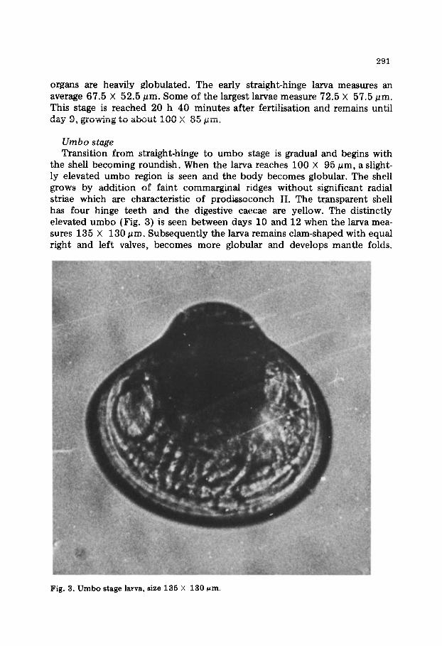
organs are heavily globulated. The early straight-hinge larva measures an average 67.5 X 52.5 (urn. Some of the largest larvae measure 72.5 X 57.5 pm. This stage is reached 20 h 40 minutes after fertilisation and remains until day 9, growing to about 100 X 85 pm.
Umbo stage Transition from straight-hinge to umbo stage is gradual and begins with
the shell becoming roundish. When the larva reaches 100 X 95 (urn, a slight- ly elevated umbo region is seen and the body becomes globular. The shell grows by addition of faint commarginal ridges without significant radial striae which are characteristic of prodissoconch II. The tr,ansparent shell has four hinge teeth and the digestive caecae are yellow. The distinctly elevated umbo (Fig. 3) is seen between days 10 and 12 when the larva mea- sures 135 X 130 pm. Subsequently the larva remains clam-shaped with equal right and left valves, becomes more globular and develops mantle folds.
Fig. 3. Umbo stage larva, size 135 X 130 pm.

292
Eye spot stage On day 15 the larva measures 210 X 190 pm and develops an eye spot
which becomes deeply pigmented. It persists even after metamorphosis and is visible in a spat of 3.9 mm. The larva develops a foot primordium and subsequently the ctenidial ridge.
Pediveliger stage The development of a functional foot is seen on day 20 when the larva
is 230 X 200 pm. The velum is pushed to the anterior side and the velar crown gradually begins to shrink in size. The larva acquires two modes of locomotion, swimming with the ciliary motion of velum and crawling with the foot. This facility moves the larva in its ‘exploratory wandering’ during settlement. Gill filaments develop during this stage and, according to the size of the larva, 2-4 filaments are seen on either side.
Plan tigrade The pediveliger, on finally settling on a substratum at the end of its
wandering phase, metamorphoses into a plantigrade (Bayne, 1976), which begins to lead a sessile life on day 22 when it measures 250 X 240 pm. The velum disintegrates and disappears. Labial palps appear, and additional gill filaments develop. The shell growth is not by concentric growth lines but by the formation of a very thin, transparent, uniform conchiolin film around the clam-shaped globular shell except in the vertex of the umbo region. The new shell is strongly sculptured with irregularly rectangular or oval markings. This is the beginning of the formation of the adult shell or the dissoconch (Fig. 4). The animal maintains a vertical position while in movement. While at rest, it adheres to the substratum with the tip of the foot and cannot easily be dislodged.
Spat The plantigrade develops the characteristic adult shell and transforms to
a young spat. The hinge line, anterior and posterior auricles and the byssal notch assume specific shape. With further deposition of shell material, which is thin and flat, the globular shape is lost. The left valve is slightly more concave than the right valve. The spat attaches itself to the substratum with byssal threads and lies on one side. The typical Pinctada fucata spat is recognised at size 330 X 300 pm on day 24 (Fig. 5).
The sizes and days given for the different larval and postlarval stages above are those at which they appeared in fairly good concentrations in rearing 2 for which continuous observations were made and which gave the best results of spat production among the four rearings. However, the ranges were wide. The umbo stage was reached between days 11 and 15 and the eye spot stage between days 15 and 24 in different rearings. The slowest growth was recorded in experiment 4.

293
Fig. 4. Plantigrade larva, size 250 x 240 pm.
Larval density The larval density in the rearing vessels varied in different experiments.
Before introducing the straight-hinge stage in the vessels, the larvae were sampled and their number per ml of water was determined in experiments 1 and 4. In experiment 1, the larval density was 8/ml in glass jars and 3/ml in fibreglass tanks, and in experiment 4, it was lo/ml and 28/ml, respec- tively. Subsequent densities could not be worked out.
Feeding Beginning day 1 (24 h after fertilisation), Isochrysis galbana was sup-
plied to the larvae daily at 15.00 h. The cell count was determined using a haemocytometer. In batch culture in 20-l glass carboys, the cell density of algal bloom ranged from 3.3 X lo6 to 7.1 X lo6 cells/ml. Sufficient quantites (250 ml-2 1) were added to the rearing vessels containing larvae to give a particular cell concentration in the rearing vessels. In experiment 1,

294
Fig. 5. Young spat of Pinctada fucata, size 330 x 300 pm.
the food concentration was 100 cells/p1 in glass jars, providing 12 000 cells/larva day-‘, and 80 cells//d in fibreglass tanks providing 25 000 cells/ larva day-‘. In experiment 2, the concentration was 120 cells/p1 in fibreglass tanks; in experiment 3 it was 350 cells//A in glass jars; and in experiment 4 it was 200 cells/p1 in glass jars. In these experiments the actual consump- tion of algal cells was not determined.
Spat setting
Fibreglass, frosted and plain glass plates were used as spat collectors, as well as fibrillated synthetic ropes, split bamboo and coconut leaf stem. Dense spat setting occurred on the fibreglass tank bottom, and the mean setting rate in the five tanks in experiment 1 was 4.71 spat/cm2. The top corners of the tank also showed heavy spatfall but the sides had only sparse

295
setting. Spatfall was poor on the bottom of perspex tank and glass jars with 0.75 and 0.50 spat/cm2, respectively. Among the plates, fibreglass gave the highest setting rate (0.24/cm2), closely followed by blue glass (0.23/cm2). In horizontal suspensions, the upper surface of the plates had a slightly higher spat density than the lower surface. Vertical surfaces also showed good spat setting. In the case of split bamboo, the density was greater on the lower concave surface than on the upper convex surface. The coconut leaf stem showed heavy settlement (Fig. 6). The synthetic ropes used for suspending the plates also gave satisfactory results.
Fig. 6. Dense settlement of spat on a piece of coconut leaf stem in the rearing tank.
Larval growth
The relationship between the larval shell length (APM) and shell height (DVM) is linear and is described by the equation
y = 1.924 + 1.07 x, with r = 0.976
where y represents APM and x the DVM in pm. Fig, 7 describes the larval growth to day 16. From D-shell (day 1) to umbo stage (day 12), the larvae grew from a mean size of 52.5 pm to 126.0 pm, an average growth of 6.7 pm/ day. On day 16, the mean size of the total larvae was 178.9 pm and that of the eyed larva 208.3 pm, a growth of 13.2 pm/day and 20.6 pm/day, re- spectively, between days 12 and 16. Pediveligers at 230 X 200 I.trn appeared on day 20, plantigrades at 250 X 240 pm on day 22 and young spat at 330 X 300 pm on day 24. There was differential growth of larvae in each batch. For instance, on day 24, there were eyed larvae (mean DVM 200.0 urn), pediveligers (217.5 gm), plantigrades (289.5 pm) and spat in the same tank.

t t
I I I I I I I I I I I I I 4 5 6 7 8 9 IO II 12 13 14 15 16
DAY FROM FERTILISATION
Fig. 7. Growth of Pinctada ficata larvae up to day 16 from fertilisation. The bar rep- resents the range of DVM and the dot the mean size. Data are from experiment 2 of the series.
12 .
l Observed Values P
El0 c o Expected Values
.
i - I” (n4- s % -
2-
d-c 1 I I I I I I,,,,, I 2 3 4 5 6 7 8 9 IO II 12 13
WEEKS
Fig. 8. Growth of spat of Pinctada fucata in the laboratory for 13 weeks from 10 October 1981.

297
Growth of spat
The growth curve is shown in Fig. 8. Mean shell heights have been plotted for 13 weeks starting from size 0.37 mm on 10 October 1981. The plots showed a curvilinear form. After logarithmic transformation, the linear rela- tionship was found to be
log, y = log, a + b log, x
where y represents shell height in mm and x represents weeks. This linear relationship has a high correlation with r = 0.987. Transforming this to the original form, the resultant equation was found to be
y = 0.26995 x1.4752.
The spat about 3 months old with extensive shell growth processes and typical pigmentation are shown in Fig. 9.
Fig. 9. Young pearl oysters, 3 months old, produced by artificial rearing.

298
DISCUSSION
Larvae of many temperate and subtropical species of bivalves have been successfully reared in the laboratory, e.g. Loosanoff and Davis (1963), Nishikawa (1971), Walne (1974) and Bayne (1976). In marked contrast, there have been only a few attempts to rear pearl oysters. Herdman (1903) studied the early life-history of Pinctada vulguris (= P. fucatu) up to day 3 and bridged the stage with further larval stages obtained in the plankton. Cahn (1949) provided abstract information on the larval development of P. martensii and P. maxima. The Japanese works on breeding of pearl oyster have been sketchy and largely relate to spawning induction, artificial fertili- sation and early development (Kobayashi, 1948; Wada, 1953; Wada, 1961). Rearing of P. maxima was attempted by Minaur (1969). The present study, together with the work of Alagarswami et al. (1980a, b), provide a detailed account of the success in artificial rearing of the pearl oyster P. fucata in the tropics.
The Japanese workers divided the larval stages of P. fucata as early D- shaped (72 X 60 pm), D-shaped (76 X 65 pm), early umbo stage (84 X 73pm), umbo stage (96 X 87 pm) and full-grown stage (209 X 195 pm) and these stages are reached, respectively, at 20 h, on day 2, 5, 8 and 25 (Shinju Yoshoku Zensho Henshu Iinkai Henshu, 1965). Ota (1957) observed the pigment (eye) spot at the late umbo stage when the shell length was 170-200 pm. In the present study, the larval and postlarval stages of P. fucatu have been divided as follows: straight-hinge (67.5 X 52.5 pm), umbo (135 X 130 pm), eye spot (210 X 190 pm), pediveliger (230 X 200 pm), plantigrade (250 X 240 pm) and spat (330 X 300 pm). These stages are reached, respectively, at 20 h 40 minutes, on day 12, 15, 20, 22 and 24. Ota (1957) found attachment of spat at shell length range 200-230 pm with a mean of 214.52 f 1.09 pm. The earliest spat observed in the present study measures 330 X 300 pm. Taking an overview of larval development under the Japanese subtropical and Indian tropical conditions, the pelagic larval phase of the pearl oyster appears to be similar, lasting about 20 days, with setting of spat taking place between days 20 and 25.
According to Minaur (1969), the early straight-hinge larva of P. maxima measured approximately 75 X 70 pm, 5-day old larva 96 X 87 pm and 12 day old (umbo) 125 X 112 pm. The pediveliger stage was reached after 14- 20 days at approximate size of 130 X 110 pm. He did not notice any eye spot in P. maxima, although its presence has been reported for P. margariti- feru (Crossland, 1957). Although P. maxima larvae were reared beyond 30 days with various setting substrates, the pediveligers (180 X 160 pm) did not complete metamorphosis (Minaur, 1969).
Usually, seawater is filtered specially to remove particles as small as 3.2 pm and treated with ultraviolet light and/or antibiotics to kill bacteria (Loosanoff and Davis, 1963; Minaur, 1969; Walne, 1974). In the present study water drawn from a seawater well and filtered through sandbed and

299
cotton wool was used without any further treatment. Daily changes of seawater, cleaning of rearing vessels and careful screening of larvae have proved beneficial in larval rearing.
Individual investigators have used different bivalve larval densities in rearing experiments. Nishikawa (1971) used a density of 0.3-0.5/ml and Walne (1974) 1.3/ml. In pearl oysters, much higher larval densities have been used. Minaur (1969) reared P. maxima larvae lO-15/ml and Wada (1973) used 20/ml concentration for P. fucata. In the present study, dif- ferent larval concentrations ranging from 3 to 28/ml were used, approxi- mately similar to the range for bivalve larvae.
Minaur (1969) fed the larvae of Pinctada maxima with different algal species at a density of 100 cells/pi. P. fucata larvae were fed with Mono- chrysis lutheri, Chaetoceros calcitrans and Chlorella sp. in approximately equal numbers of each species at a combined concentration of 10 cells/p1 on days l-12 and at 20 cells/p1 on days 13-25/26 (Wada, 1973). In the present work, I. gulbana was supplied to the straight-hinge larvae at a con- centration of SO-120 cells/p1 although higher concentration was used on certain days. This is almost equal to the food concentrations used by Minaur (1969) and Walne (1974), but much higher than that used by Wada (1973). However, the “critical cell concentration” remains to be worked out for different stages, as also the use of mixed species of algae.
Delayed metamorphosis of bivalve larvae has been reported by several workers and it is usually associated with crowding, ration and tempera- ture (Loosanoff and Davis, 1963; Bayne, 1965). Spat settlement took place, respectively, on days 25, 24, 30 and 32 in experiments l-4. The length of the larval phase appears inversely related to the ambient temperature and salinity conditions of rearing. The setting on day 24 is associated with temperature 28.2-298°C and salinity 32.98-34.00°~o. The setting on day 32 is related to temperature 24.3-27.2% and salinity 27.50-28.25°/,0. It is suggested that, besides other variables, lower temperature and salinity may prolong the larval phase of P. ficata and cause delay in setting.
The growth curve of lamellibranch larvae is generally sigmoidal, although a linear fit to some data in the middle size ranges is not unreasonable (Bayne, 1976). Walne’s (1974) figures on the growth of 0. edulis larvae for the first 10 days appear to give a linear relationship. Minaur’s (1969) data for P. maxima up to about 20 days show a linear fit but the larvae did not grow further. Growth of P. fucatu larvae, according to the present data (Fig. 7), appears to be a step function in which different growth rates pertain to different stages. Wada’s (1973) data on growth of P. fucata larvae also show this pattern. Growth of spat up to 13 weeks (Fig. 8) shows a curvilinear form.
The present work is the first detailed study on the larval rearing of pearl oyster in India and has given the basic technology for the production of spat. Further investigations are required to determine optimum larval dens- ity, critical cell concentration, use of local tropical species of microalgae

and diatoms as food, and factors influencing spat setting in order to improve and standardise the procedures for hatchery level production.
ACKNOWLEDGEMENTS
The authors are grateful to Dr. E.G. Silas, Director, Central Marine Fisheries Research Institute, for his keen interest in this work and encourage- ment given. They would like to thank Shri K. Nagappan Nayar for the facilities and discussions, Dr. K. Alagaraja for statistical assistance and Shri C.P. Gopinathan for I. gulbana stock culture.
REFERENCES
Alagarswami, K., 1974. Development of cultured pearls in India. Curr. Sci., 43: 205- 207.
Alagarswami, K., 1977. Larval transport and settlement of pearl oysters (genus Pinctada) in the Gulf of Mannar. Proc. Symp. Warm. Wat. Zoopl., National Institute of Ocean- ography, Goa, Special Publication, pp. 678--686.
Alagarswami, K. and Qasim, S.Z., 1973. Pearl culture - its potential and implications in India. Indian J. Fish., 20: 533-550.
Alagarswami, K., Dharmaraj, S., Velayudhan, T.S., Chellam, A. and Victor, A.C.C., 1980a. On controlled spawning in the pearl oyster Pinctada fucata (Gould). Symp. Coast. Aquacult., Marine Biological Association of India, Cochin, Abstr. 64.
Alagarswami, K., Dharmaraj, S., Velayudhan, T.S., Chellam, A. and Victor, A.C.C., 1980b. Larval development of the pearl oyster Pinctada fucata (Gould). Symp. Coast. Aquacult., Marine Biological Association of India, Cochin, Abstr. 181.
Bayne, B.L., 1965. Growth and the delay of metamorphosis of the larvae of Mytilus edulis (L.). Ophelia, 2: l-47.
Bayne, B.L., 1976. The biology of mussel larvae. In: B.L. Bayne (Editor), Marine Mussels: Their Ecology and Physiology. Cambridge University Press, Cambridge, pp. 81-120.
Cahn, A.R., 1949. Pearl culture in Japan. Fish. Leafl., U.S. Fish Wildl. Serv., 357: 1-91. Crossland, C., 1957. The cultivation of the mother-of-pearl oyster in the Red Sea. Aust.
J. Mar. Freshwat. Res., 8: 111-130. Herdman, W.A., 1903. Observations and experiments on the life-history and habits of
the pearl oyster. In: W.A. Herdman (Editor), Report Pearl Oyster Fisheries, Gulf of Mannar. Roy. Sot., London, pp. 125-146.
Kobayashi, S., 1948. On the study of pearl culture. 1. On development of Pinctada martensii in tanks. Bull. Jpn. Sot. Sci. Fish., 17: 65-72.
Loosanoff, V.L. and Davis, H.C., 1963. Rearing of bivalve mollusks. Adv. Mar. Biol., 1: l-136.
Minaur, J., 1969. Experiments on the artificial rearing of the larvae of Pinctada maxima (Jameson) (Lamellibranchia). Aust. J. Mar. Freshwat. Res., 20: 175-187.
Nishikawa, N., 1971. The rearing of larvae and seedlings of bivalves. In: T. Imai (Edi- tor), Aquaculture in Shallow Seas. Oxford and IBH Publ. Co., New Delhi, pp. 579- 600 (translated from Japanese).
Ota, S., 1957. Notes on the identification of free swimming larva of pearl oyster (Pincta- da martensii). Bull. Natl. Pearl Res. Lab., 2: 128-l 32 (in Japanese).
Shinju Yoshoku Zensho Henshu Iinkai Henshu, 1965. Shinju Yoshoku Zensho (Treatise on pearl culture). Zenkoku Shinju Yoshoku Gyogyo Kyodo Kumiai Rengokai, 702 pp. (in Japanese).

301
Wada, K.T., 1973. Growth of Japanese pearl oyster larvae fed with three species of microalgae. Bull. Natl. Pearl Res. Lab., 17: 2075-2083 (in Japanese, with English Summary).
Wada, S., 1953. Biology of the silver-lip pearl oyster Pinctada maxima (Jameson): artifi- cial fertilisation and development. Margarita, 1: 3-15.
Wada, S.K., 1961. Fertilisation of Crassostrea and Pinctada eggs as related to germinal vesicle breakdown. Mem. Fat. Fish. Kagoshima Univ., 10: l-8.
Walne, P.R., 1974. Culture of Bivalve Molluscs. Fishing News (Books) Ltd., Surrey, Great Britain, 173 pp.