Influence of rearing conditions and respiratory disease on haptoglobin levels in the pig at...
8
Influence of rearing conditions and respiratory disease on haptoglobin levels in the pig at slaughter J.R. Amory a, * , A.M. Mackenzie a , P.D. Eckersall b , M.J. Stear b , G.P. Pearce a,1 a Animal Science Research Centre, Harper Adams University College, Newport, Shropshire TF10 8NB, UK b Division of Animal Production and Public Health, Institute of Comparative Medicine, Faculty of Veterinary Medicine, University of Glasgow, Bearsden Road, Glasgow G61 1QH, UK Accepted 30 January 2007 Abstract Associations between serum concentrations of haptoglobin, pathological lung lesions indicative of Mycoplasma hyopneumoniae (EP) or Actinobacillus pleuropneumoniae (PL) infection at slaughter and previous rearing environment were investigated in 510 pigs (90–100 kg live weight) from 17 farms in England. Haptoglobin concentrations were significantly higher in pigs showing pathological signs of EP infection compared to those without signs of this disease (EP positive median 0.43 mg ml 1 vs. EP negative median 0.26 mg ml 1 , p < 0.01). However, there were no significant associations between serum haptoglobin concentrations and pathological signs of PL. The presence of solid partitions compared with barred or similar open partitions was associated with a decrease of 0.44 mg ml 1 farm mean haptoglobin concentration, whilst an increase in pen size of 10 m 2 was associated with a decrease of 0.08 mg ml 1 farm mean hap- toglobin concentration. The findings indicate that pathological signs of EP were associated with increased serum haptoglobin at slaugh- ter, which in turn was influenced by components of the farm environment. Ó 2007 Elsevier Ltd. All rights reserved. Keywords: Pigs; Respiratory disease; Haptoglobin; Environment 1. Introduction The response of a pig to its environment may be moni- tored by using physiological markers, or ‘‘biomarkers’’ (Eckersall, 2000). Acute phase proteins (APPs) represent an important group of biomarkers with potential use as indicators of tissue injury associated with changes in health status in commercially reared pigs (Gymnich and Petersen, 2004). APPs are produced by the liver in response to cyto- kines associated with inflammation, infection or tissue injury and function to restore homeostasis in the body fol- lowing injury or infection (Eckersall, 2000; Colditz, 2002; Petersen et al., 2004) and the acute phase protein Hapto- globin, has been identified as a sensitive indicator of respi- ratory infection and its concentration in blood serum has been suggested as a means of non-specific surveillance of pig health status (Heegaard et al., 1998). There is an increasing number of scientific publications describing the acute phase response in the pig and the effects of infection (Heegaard et al., 1998), inflammatory response to an injection of turpentine (Eckersall et al., 1996), specific husbandry practices (Francisco et al., 1996a,b), transport (Saco et al., 2003), clinical signs of disease (Petersen et al., 2002a), histopathological signs of disease (Chen et al., 2003), and presence of clinical dis- eases such as Postweaning Multisystemic Wasting Syn- drome (Segales et al., 2004) and Porcine Intestinal Adenomatosis (Geers et al., 2003). Haptoglobin levels have been reported to be higher in pigs from organic com- pared with non-organic pig herds (Millet et al., 2005), in indoor compared with outdoor herds (Franek and Bilkei, 0034-5288/$ - see front matter Ó 2007 Elsevier Ltd. All rights reserved. doi:10.1016/j.rvsc.2007.01.012 * Corresponding author. Present Address: Department of Science, Agriculture and Technology, Writtle College, Chelmsford, Essex CM1 E-mail address: [email protected] (J.R. Amory). 1 Present address: Department of Veterinary Medicine, University of Cambridge, Madingley Road, Cambridge CB3 0ES, UK. www.elsevier.com/locate/rvsc Research in Veterinary Science 83 (2007) 428–435
-
Upload
hertie-school -
Category
Documents
-
view
5 -
download
0
Transcript of Influence of rearing conditions and respiratory disease on haptoglobin levels in the pig at...
www.elsevier.com/locate/rvsc
Research in Veterinary Science 83 (2007) 428–435
Influence of rearing conditions and respiratory disease onhaptoglobin levels in the pig at slaughter
J.R. Amory a,*, A.M. Mackenzie a, P.D. Eckersall b, M.J. Stear b, G.P. Pearce a,1
a Animal Science Research Centre, Harper Adams University College, Newport, Shropshire TF10 8NB, UKb Division of Animal Production and Public Health, Institute of Comparative Medicine, Faculty of Veterinary Medicine, University of Glasgow,
Bearsden Road, Glasgow G61 1QH, UK
Accepted 30 January 2007
Abstract
Associations between serum concentrations of haptoglobin, pathological lung lesions indicative of Mycoplasma hyopneumoniae (EP)or Actinobacillus pleuropneumoniae (PL) infection at slaughter and previous rearing environment were investigated in 510 pigs (90–100 kglive weight) from 17 farms in England. Haptoglobin concentrations were significantly higher in pigs showing pathological signs of EPinfection compared to those without signs of this disease (EP positive median 0.43 mg ml�1 vs. EP negative median 0.26 mg ml�1,p < 0.01). However, there were no significant associations between serum haptoglobin concentrations and pathological signs of PL.The presence of solid partitions compared with barred or similar open partitions was associated with a decrease of 0.44 mg ml�1 farmmean haptoglobin concentration, whilst an increase in pen size of 10 m2 was associated with a decrease of 0.08 mg ml�1 farm mean hap-toglobin concentration. The findings indicate that pathological signs of EP were associated with increased serum haptoglobin at slaugh-ter, which in turn was influenced by components of the farm environment.� 2007 Elsevier Ltd. All rights reserved.
Keywords: Pigs; Respiratory disease; Haptoglobin; Environment
1. Introduction
The response of a pig to its environment may be moni-tored by using physiological markers, or ‘‘biomarkers’’(Eckersall, 2000). Acute phase proteins (APPs) representan important group of biomarkers with potential use asindicators of tissue injury associated with changes in healthstatus in commercially reared pigs (Gymnich and Petersen,2004). APPs are produced by the liver in response to cyto-kines associated with inflammation, infection or tissueinjury and function to restore homeostasis in the body fol-lowing injury or infection (Eckersall, 2000; Colditz, 2002;
0034-5288/$ - see front matter � 2007 Elsevier Ltd. All rights reserved.
doi:10.1016/j.rvsc.2007.01.012
* Corresponding author. Present Address: Department of Science,Agriculture and Technology, Writtle College, Chelmsford, Essex CM1
E-mail address: [email protected] (J.R. Amory).1 Present address: Department of Veterinary Medicine, University of
Cambridge, Madingley Road, Cambridge CB3 0ES, UK.
Petersen et al., 2004) and the acute phase protein Hapto-globin, has been identified as a sensitive indicator of respi-ratory infection and its concentration in blood serum hasbeen suggested as a means of non-specific surveillance ofpig health status (Heegaard et al., 1998).
There is an increasing number of scientific publicationsdescribing the acute phase response in the pig and theeffects of infection (Heegaard et al., 1998), inflammatoryresponse to an injection of turpentine (Eckersall et al.,1996), specific husbandry practices (Francisco et al.,1996a,b), transport (Saco et al., 2003), clinical signs ofdisease (Petersen et al., 2002a), histopathological signsof disease (Chen et al., 2003), and presence of clinical dis-eases such as Postweaning Multisystemic Wasting Syn-drome (Segales et al., 2004) and Porcine IntestinalAdenomatosis (Geers et al., 2003). Haptoglobin levelshave been reported to be higher in pigs from organic com-pared with non-organic pig herds (Millet et al., 2005), inindoor compared with outdoor herds (Franek and Bilkei,

J.R. Amory et al. / Research in Veterinary Science 83 (2007) 428–435 429
2004) and in herds with poor levels of hygiene (Gymnichand Petersen, 2004). However, there is little informationdescribing the effects of specific aspects of the rearingenvironment on mediating the variability of acute phaseproteins in the slaughter pig. Knowledge of such variationmay be important in identifying factors within husbandrysystems associated with increased acute phase responseand also in the development of their use in ante- andpost-mortem inspection (Saini and Webert, 1991) andproduction chain orientated health management systems(Gymnich and Petersen, 2004).
This study was designed to investigate the relationshipbetween pathological signs of two major respiratory dis-eases of pigs, enzootic pneumonia and pleuropneumonia,and serum haptoglobin concentrations at slaughter. Thestudy also aimed to identify husbandry and managementprocedures important in influencing the acute phaseresponse, as determined by levels of serum haptoglobin inslaughtered pigs reared under commercial conditions.
2. Materials and methods
Seventeen farms were involved in the study that deliv-ered pigs to one of three participating abattoirs in the mid-lands region of England. Each farmer was given aquestionnaire detailing various aspects of the farm environ-ment, husbandry system and pig health based on a previousstudy investigating risk factors for respiratory diseases inNew Zealand (Stark et al., 1998). For the purpose of thisstudy grower pigs were classified as 20–45 kg and finishers45 kg – slaughter weight.
Blood samples and lungs were collected from 30 ran-domly selected pigs from a single delivery. Following stun-ning, blood samples were collected in 30 ml universal tubesfrom the throat as they were exsanguinated. The collectedblood was allowed to clot overnight at 4 �C and serumwas then obtained by centrifugation for 15 min at 1000g
and stored at �20 �C until subsequent analysis. The serumhaptoglobin concentration was determined according tothe haemoglobin binding activity method of Eckersall(2000) that allows measurement of haptoglobin from allspecies. A bovine standard of known haptoglobin concen-tration was used as standard (Horadagoda et al., 1994).
Each farm was assessed for their pneumonia status bycalculating the prevalence of affected lungs at slaughterand their degree of consolidation. In order to determine tis-sue damage due to enzootic pneumonia (EP score) thedegree of consolidation was assessed by estimating the per-centage of the surface of each lobe of the lung showingsigns of consolidation. This percentage was multiplied bya weighting factor for each lobe based on their relative vol-umes (0.25 for the caudal lobes, 0.1 for the cranial andaccessory lobes) and totalled to give a score out of 100for each pig based on the methodology of Straw et al.(1986a). In order to determine tissue damage due to pleuro-pneumonia (PL score) the percentage of each lobe esti-mated to be missing due to adhesion of pleural
membranes was scored pneumonias a measure of grade 2pleurisy based on the methodology of Straw et al.(1986b). Although pleuritis can be attributed to a numberof causal organisms, infection with Actinobacillus pleuro-
pneumoniae is by far the most common in the UK (Done,1999).
Continuous variables were quantified by calculatingmedian, minimum and maximum figures. Differences inserum haptoglobin concentration between pigs identifiedas having or not having pathological signs of enzooticpneumonia were calculated using the Kruskal–Wallis test(Petrie and Watson, 1999, p. 146). This was repeated forthose identified as having or not having pathological signsof pleuropneumonia. In addition, both types of pneumoniawere considered together by creating four groups one neg-ative for both types, one positive for EP only, one positivefor PL only and one positive for both types.. Regressionequations were calculated to determine whether either EPor PL score could be predicted by the actual serum acutephase protein concentration in individual pigs. In orderto determine which terms were to be considered for theconstruction of a multiple regression model, univariableanalyses of variance were carried out between individualfarm factors and the dependent variables. Those terms giv-ing an F-probability p < 0.25 were put forward for the mul-tiple regression model as suggested by Hosmer andLemeshow (1989). Similarly for the continuous indepen-dent variables, those giving correlations of r > 0.23 (equiv-alent to p < 0.25 with 16 degrees of freedom based on 17farms) were also put forward for the multiple regressionmodel. This was carried out using forwards and backwardsstepwise selection procedures to ascertain the best model.Finally all original terms were added individually to themodel to check significance. Descriptive statistics, analysisof variance, Pearson correlation coefficients and the step-wise regression procedure were performed using S-Plus(version 6.2) for Windows statistics package. Non-para-metric statistics were performed using Minitab (version11) for Windows statistics package.
3. Results
Of the 17 farms that took part in this study, eight werebreeder-finisher units and 9 were finishing-only units, allslaughtering finished pigs between 90 and 100 kg liveweight. Nine farms provided bedding for their grower pigs,while eight provided bedding for the finishers. Nine farmsused a slatted method of slurry disposal for their growers,while 10 used slatted floors for their finishers. Five farmsvaccinated against enzootic pneumonia, one farm vacci-nated for pleuropneumonia, four farms injected individualgrower pigs for treatment of respiratory disease and onefarm injected finisher pigs for treatment of respiratory dis-ease. However, the effects of vaccination and medicationstatus were not investigated in detail in this study. Tenfarms gave in-feed/water medication to their growers andfive farms gave in-feed/water medication to their finishers.

Table 3Descriptive statistics regarding serum haptoglobin
Individual pig serumhaptoglobin (mg ml�1,n = 455)
Farm mean serumhaptoglobin (mg ml�1,n = 17)
Mean 0.64 0.64Minimum 0.00 0.27First
quartile0.04 0.43
Median 0.33 0.51Third
quartile0.96 0.82
Maximum 8.55 1.26
430 J.R. Amory et al. / Research in Veterinary Science 83 (2007) 428–435
Median and range figures for the continuous variables areshown in Table 1.
Signs of enzootic pneumonia were the most prevalentlesion type in this study with 147 pigs out of the 510 exam-ined showing these signs (varying from 0% to 26% of totallung volume affected). Only one of the 17 farms did notshow gross lesions associated with enzootic pneumonia.Pathological lesions associated with pleuropneumonia werefound in 34 of the 510 pigs examined (signs of pleurisyvarying from 0% to 48% of total lung volume affected inall pigs). Four out of the 17 farms did not show the lungtissue damage attributed to pleuropneumonia in this study.Distributions of lesion scores are presented in Table 2. Ele-ven out of the 510 pigs showed signs of both diseases.
Mean individual serum haptoglobin was 0.64 mg ml�1
(range 0–8.55 mg ml�1), whilst the farm mean haptoglobinconcentration varied from 0.27 to 1.26 mg ml�1 (Table 3).There was a significant increase in haptoglobin concentra-
Table 1Median, minimum and maximum figures for continuous managementvariables from the respiratory disease and acute phase protein study
Median Minimum Maximum
No. of sows on unit 78 0 (finishersonly)
491
Distance to nearest pig farm(miles)
2 0.25 5
Weaning age (days) 24 21 28No. of stages after weaning 2 1 4Grower pen size (m2) 12.5 5 100No. of grower pigs per pen 25 14 200Grower space allowance (m2/
pig)0.5 0.36 1.25
No. of grower pigs per room 140 20 290Finisher pen size (m2) 13.3 6.75 100No. of finisher pigs per pen 20 10 200Finisher space allowance (m2/
pig)0.68 0.47 0.93
No. of finisher pigs per room 149 100 220
Table 2Distribution of the severity of lesion scores for enzootic pneumonia andpleuropneumonia
Pneumoniascorea (%)
Enzootic pneumonia(% of pigs, n = 510)
Pleuropneumonia(% of pigs, n = 510)
0 71.2 93.3>0–1 5.9 0.0>1–2 7.1 0.8>2–5 9.0 1.6>5–10 3.3 1.8>10–20 2.9 1.8>20–30 0.6 0.2>30–40 0 0.4>40–50 0 0.2>50 0 0
a Score was ascribed by estimating the percentage of the surface of eachlobe of the lung showing signs of consolidation multiplied by a weightingfactor for each lobe based on their relative volumes (0.25 for the caudallobes, 0.1 for the cranial and accessory lobes) and totalled to give a scoreout of 100 for each pig.
tion in those pigs showing pathological signs of EP com-pared to those without pathological signs of this disease(EP positive median 0.43 vs. EP negative median 0.26,p < 0.01). However, there were no significant differencesin serum haptoglobin concentration between pigs negativeor positive for pathological signs of pleuropneumonia. His-tograms showing the population profiles for animals iden-tified as having pathological signs of EP or PL arepresented in Figs. 1 and 2, respectively. When consideringboth types of pneumonia together, analysis showed signif-icant differences for haptoglobin (p < 0.05, see Fig. 3).
Nine factors were found to be associated (p < 0.25) withan increase in farm mean haptoglobin concentration (cate-gorical variables are shown in Table 4). There was a highlevel of correlation between these factors (Table 5). Froma relatively large number of input variables only tworemained in the final multiple regression model for serumhaptoglobin concentration based on the farm mean values.Solid pen divisions in the finisher accommodation and fin-isher pen were associated with a decrease of 0.44 mg ml�1
mean serum haptoglobin concentration compared with alack of solid partitions. An increase in finisher pen size of10 m2 was associated with a decrease of 0.08 mg ml�1 meanserum haptoglobin concentration. Table 6 shows the pre-dictive model for farm mean serum haptoglobinconcentration.
4. Discussion and conclusions
In addition to being used for monitoring infectious dis-ease progression it has been suggested that acute phase pro-tein measurements can be used for the prognosis anddiagnosis of disease and the evaluation of general healthstatus (Heegaard et al., 1998; Eckersall, 2000; Petersenet al., 2002a). In the present study, haptoglobin concentra-tions were significantly higher (0.26–0.43 mg ml�1,p < 0.01) in pigs that showed pathological signs of enzooticpneumonia compared to those that did not. Althoughreported normal levels of serum haptoglobin concentra-tions vary (probably due to the lack of a recognised refer-ence preparation for harmonised calibration of assays)concentrations are generally quoted as being less than0.5 mg ml�1 (Heegaard et al., 1998). In the animals in thisstudy a number in all groups (EP and PL positive and

0
5
10
15
20
25
30
35
40
45
0
0.2
0.4
0.6
0.8 1
1.2
1.4
1.6
1.8 2
2.2
2.4
2.6
2.8 3
3.2
3.4
3.6
3.8 4 10
Serum Haptoglobin concentration (mg/ml)
Prop
ortio
n of
pop
ulat
ion
EP free (341 pigs) EP positive (147 pigs)
>4
Fig. 1. Distribution of serum haptoglobin concentration for pigs with or without pathological signs of enzootic pneumonia (EP).
0
10
20
30
40
50
60
0
0.2
0.4
0.6
0.8 1
1.2
1.4
1.6
1.8 2
2.2
2.4
2.6
2.8 3
3.2
3.4
3.6
3.8 4 10
Serum Haptoglobin concentration (mg/ml)
Prop
ortio
n of
pop
ulat
ion
PL free (455 pigs) PL positive (33 pigs)
>4
Fig. 2. Distribution of serum haptoglobin concentration for pigs with or without pathological signs of pleuropneumonia (PL).
J.R. Amory et al. / Research in Veterinary Science 83 (2007) 428–435 431
negative) had haptoglobin concentrations greater than3 mg ml�1 and without the possibility of identifying thecause of these elevated levels these outliers were retainedin the analysis. In a study of a conventional herd, (Eurellet al., 1992) found levels varying between 0.1 and0.6 mg ml�1compared to the range of 0–8.55 mg ml�1
found in the current study, however the 3rd quartile of0.96 mg ml�1 is a more equivalent comparator. Othershave reported normal levels as low as 0.06 mg ml�1, a meanof 0.19 mg ml�1 in a herd chronically infected with pleuro-pneumonia and a mean of 0.24 mg ml�1 in a herd undergo-ing acute Actinobacillus infection (Hall et al., 1992). The
higher mean levels found in the current study may be dueto the standard used for calibration (bovine haptoglobin)instead of the human haptoglobin used by Hall et al.(1992) as previously suggested by Heegaard et al. (1998).
Respiratory lesions in pigs at slaughter are common inthe UK. Done (1999) reported that the prevalence of enzo-otic pneumonia in the UK can be as high as 90%, althoughthe average remains about 50% (Done, 1999). This averagefigure is higher than the 29% prevalence of enzootic pneu-monia found in the current study, which was carried outduring the months of April and May and the prevalenceof pneumonia is known to be reduced during the summer

Fig. 3. Serum haptoglobin concentrations of pigs identified as negative forEP and PL, having either just EP or just PL or both conditions.
Table 4Farm factors associated with differences in serum haptoglobinconcentration
Farm factor No.farms
Mean Pooled standarddeviation
p-Value
Solid partitions between finisher pensYes 8 0.45 0.250 0.012No 9 0.80
Fan-assisted ventilation for finishersYes 5 0.90 0.253 0.014No 12 0.53
Solid partitions between grower pensYes 10 0.54 0.286 0.115No 7 0.78
Fan-assisted ventilation for growersYes 4 0.87 0.279 0.076No 13 0.57
Finishers kept on an all-in all-out policyYes 7 0.51 0.290 0.146No 10 0.73
Finishers treated by injection for respectively diseaseYes 1 1.12 0.284 0.103No 16 0.61
Growers’ effluentRemoved daily 6 0.46 0.278 0.070Removed less thandaily
11 0.74
Coughing observed in finishersYes 12 0.70 0.296 0.228No 5 0.50
Pigs > 12 weeks old in room with pigs 5+ weeks youngerYes 3 0.85 0.294 0.195No 14 0.59
Type of unitBreeder-finisher 8 0.73 0.297 0.242Finishers only 9 0.56
432 J.R. Amory et al. / Research in Veterinary Science 83 (2007) 428–435
months (Stark et al., 1998). Prevalence figures are similarfor other pig producing countries with climates similar tothe UK (52% in New Zealand (Stark et al., 1998); 84% inSweden (Wallgren et al., 1994a); 70% in Norway (Liumand Falk, 1991); 75% in Ont., Canada (Wilson et al.,1986)).
There is little information as to actual prevalence ofActinobacillus pleuropneumoniae infection in the UK, moreoften the presence of pleuritis (the formation of pleuralmembranes) is recorded and has been reported as being lessthan 20% in the UK (Done, 1999). This is similar to theseven per cent prevalence for pigs showing pleuritis in thecurrent study, pleuritis also being expected to be reducedin the summer months (Stark et al., 1998). Prevalence fig-ures are rare for other countries for actual pleuropneumo-nia (2.7% in New Zealand (Stark et al., 1998)) but are morecommon for pleuritis, which was measured in the currentstudy (19.1% in New Zealand (Stark et al., 1998); 6% inSweden (Wallgren et al., 1994a); 29% in Norway (Liumand Falk, 1991); 11% in Ont., Canada (Wilson et al.,1986)). It is recommended that to develop this research,future studies would utilise histological and serologicaltechniques to confirm the causes of infection.
Table 5Correlations between dependent variables related to farm mean haptoglobin c
Dependent variables 1
1 Solid partitions between finisher pens –2 Fan-assisted ventilation for finishers3 Solid partitions between grower pens 0.554 Fan-assisted ventilation for growers5 Finishers kept on an all-in all-out policy 0.416 Finishers treated by injection for respectively disease7 Growers’ effluent removed daily �0.548 Coughing observed in finishers �0.439 Pigs >12 weeks old in room with pigs 5+ weeks younger10 Breeder/finisher unit (vs. finisher only)
(N.B., Pearson’s coefficient ‘r’ for p = 0.1 and p = 0.05 are 0.40 and 0.47, resp
This is the first report finding a relationship betweenpathological lesions indicative of M. hyopneumonia infec-tion and serum haptoglobin concentrations. This is consis-tent with report of Franek and Bilkei (2004), where serumhaptoglobin concentration was highly correlated to serumantibody titres to Mycoplasma hyopneumonia in outdoor
oncentration
2 3 4 5 6 7 8 9
––
0.86 –0.46 –
0.45 –�0.63 –
––
0.45 0.49
ectively. For clarity only relationships of p < 0.1 are shown).
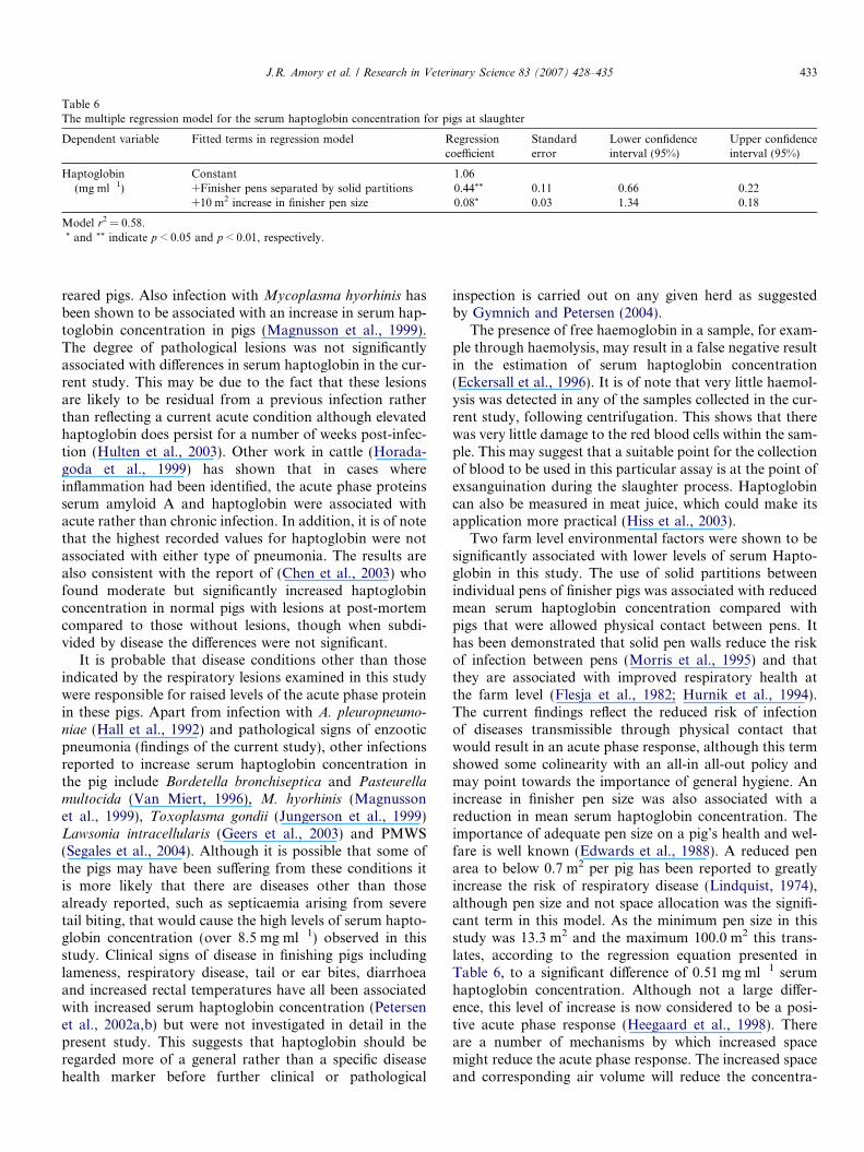
Table 6The multiple regression model for the serum haptoglobin concentration for pigs at slaughter
Dependent variable Fitted terms in regression model Regressioncoefficient
Standarderror
Lower confidenceinterval (95%)
Upper confidenceinterval (95%)
Haptoglobin(mg ml�1)
Constant 1.06+Finisher pens separated by solid partitions �0.44** 0.11 �0.66 �0.22+10 m2 increase in finisher pen size �0.08* 0.03 �1.34 �0.18
Model r2 = 0.58.* and ** indicate p < 0.05 and p < 0.01, respectively.
J.R. Amory et al. / Research in Veterinary Science 83 (2007) 428–435 433
reared pigs. Also infection with Mycoplasma hyorhinis hasbeen shown to be associated with an increase in serum hap-toglobin concentration in pigs (Magnusson et al., 1999).The degree of pathological lesions was not significantlyassociated with differences in serum haptoglobin in the cur-rent study. This may be due to the fact that these lesionsare likely to be residual from a previous infection ratherthan reflecting a current acute condition although elevatedhaptoglobin does persist for a number of weeks post-infec-tion (Hulten et al., 2003). Other work in cattle (Horada-goda et al., 1999) has shown that in cases whereinflammation had been identified, the acute phase proteinsserum amyloid A and haptoglobin were associated withacute rather than chronic infection. In addition, it is of notethat the highest recorded values for haptoglobin were notassociated with either type of pneumonia. The results arealso consistent with the report of (Chen et al., 2003) whofound moderate but significantly increased haptoglobinconcentration in normal pigs with lesions at post-mortemcompared to those without lesions, though when subdi-vided by disease the differences were not significant.
It is probable that disease conditions other than thoseindicated by the respiratory lesions examined in this studywere responsible for raised levels of the acute phase proteinin these pigs. Apart from infection with A. pleuropneumo-
niae (Hall et al., 1992) and pathological signs of enzooticpneumonia (findings of the current study), other infectionsreported to increase serum haptoglobin concentration inthe pig include Bordetella bronchiseptica and Pasteurellamultocida (Van Miert, 1996), M. hyorhinis (Magnussonet al., 1999), Toxoplasma gondii (Jungerson et al., 1999)Lawsonia intracellularis (Geers et al., 2003) and PMWS(Segales et al., 2004). Although it is possible that some ofthe pigs may have been suffering from these conditions itis more likely that there are diseases other than thosealready reported, such as septicaemia arising from severetail biting, that would cause the high levels of serum hapto-globin concentration (over 8.5 mg ml�1) observed in thisstudy. Clinical signs of disease in finishing pigs includinglameness, respiratory disease, tail or ear bites, diarrhoeaand increased rectal temperatures have all been associatedwith increased serum haptoglobin concentration (Petersenet al., 2002a,b) but were not investigated in detail in thepresent study. This suggests that haptoglobin should beregarded more of a general rather than a specific diseasehealth marker before further clinical or pathological
inspection is carried out on any given herd as suggestedby Gymnich and Petersen (2004).
The presence of free haemoglobin in a sample, for exam-ple through haemolysis, may result in a false negative resultin the estimation of serum haptoglobin concentration(Eckersall et al., 1996). It is of note that very little haemol-ysis was detected in any of the samples collected in the cur-rent study, following centrifugation. This shows that therewas very little damage to the red blood cells within the sam-ple. This may suggest that a suitable point for the collectionof blood to be used in this particular assay is at the point ofexsanguination during the slaughter process. Haptoglobincan also be measured in meat juice, which could make itsapplication more practical (Hiss et al., 2003).
Two farm level environmental factors were shown to besignificantly associated with lower levels of serum Hapto-globin in this study. The use of solid partitions betweenindividual pens of finisher pigs was associated with reducedmean serum haptoglobin concentration compared withpigs that were allowed physical contact between pens. Ithas been demonstrated that solid pen walls reduce the riskof infection between pens (Morris et al., 1995) and thatthey are associated with improved respiratory health atthe farm level (Flesja et al., 1982; Hurnik et al., 1994).The current findings reflect the reduced risk of infectionof diseases transmissible through physical contact thatwould result in an acute phase response, although this termshowed some colinearity with an all-in all-out policy andmay point towards the importance of general hygiene. Anincrease in finisher pen size was also associated with areduction in mean serum haptoglobin concentration. Theimportance of adequate pen size on a pig’s health and wel-fare is well known (Edwards et al., 1988). A reduced penarea to below 0.7 m2 per pig has been reported to greatlyincrease the risk of respiratory disease (Lindquist, 1974),although pen size and not space allocation was the signifi-cant term in this model. As the minimum pen size in thisstudy was 13.3 m2 and the maximum 100.0 m2 this trans-lates, according to the regression equation presented inTable 6, to a significant difference of 0.51 mg ml�1 serumhaptoglobin concentration. Although not a large differ-ence, this level of increase is now considered to be a posi-tive acute phase response (Heegaard et al., 1998). Thereare a number of mechanisms by which increased spacemight reduce the acute phase response. The increased spaceand corresponding air volume will reduce the concentra-

434 J.R. Amory et al. / Research in Veterinary Science 83 (2007) 428–435
tion of infectious agents found as airborne or non-airborneparticles (Stark, 2000). The associated reduction in pig con-tact may also reduce cross-infection and possible stress dueto social interaction. Stress resulting in the production ofcortisol that has been demonstrated to influence the abilityof a pig to cope with pathogenic infection (Johnson et al.,1994; Wallgren et al., 1994b) affecting the risk of an acutephase response occurring and has also been shown to becorrelated to the haptoglobin response to exposure torespiratory pathogens such as M. hyopneumoniae (Franekand Bilkei, 2004). Notable were the absence of other fac-tors, such as medication and vaccination against respira-tory disease. However, the records of this were variableand may have reflected actual practice meaning that varia-tion was too high to find a significant effect.
In conclusion, this study has demonstrated that the pres-ence of pathological signs of enzootic pneumonia was asso-ciated with variation in levels of serum haptoglobinconcentration measured at slaughter, which in turn wasinfluenced by components of the farm environment.Although there has not been a previous study assessingthe levels of acute phase proteins in commercial pig herdsin the UK, the levels measured in the present study are con-sistent with those previously reported in controlled trialswith A. pleuropneumoniae infection in this species (Heeg-aard et al., 1998). It is suggested that although haptoglobinis a useful indicator of enzootic pneumonia (infection withM. hyopneumoniae), the level of variation, presumablyfrom other inflammatory conditions, suggests that thisacute phase protein may be more useful as a general indi-cator of pig health status rather than specific respiratoryinfection. This is in agreement with the findings of otherauthors (Petersen et al., 2002b).
References
Chen, H.H., Lin, J.H., Fung, H.P., Ho, L.L., Yang, P.C., Lee, W.C., Lee,Y.P., Chu, R.M., 2003. Serum acute phase proteins and swine healthstatus. Can. J. Vet. Res. 67, 283–290.
Colditz, I.G., 2002. Effects of the immune system on metabolism:implications for production and disease resistance in livestock.Livestock Product. Sci. 75, 257–268.
Done, S.H., 1999. Respiratory infections. Pig Int. 29 (5), 35.Eckersall, P.D., 2000. Acute phase proteins as markers of infection and
inflammation: monitoring animal health, animal welfare and foodsafety. Irish Vet. J. 53, 307–311.
Eckersall, P.D., Saini, P.K., McComb, C., 1996. The acute phase responseof acid soluble glycoprotein, a-1-acid glycoprotein, ceruloplasmin,haptoglobin and C-reactive protein, in the pig. Vet. Immunol.Immunopathol. 51, 377–385.
Edwards, S.A., Armsby, A.W., Large, J.W., 1988. Effects of floor areaallowance on performance of growing pigs kept on fully slatted floors.Animal Product. 46, 453–459.
Eurell, T.E., Bane, D.P., Hall, W.F., Schaeffer, D.J., 1992. Serumhaptoglobin concentration as an indicator of weight gain in pigs.Can. J. Vet. Res. 56, 6–9.
Flesja, K.I., Forus, I.B., Solberg, I., 1982. Pathological lesions in swine atslaughter. V. Pathological lesions in relation to some environmentalfactors in the herds. Acta Vet. Scand. 23, 169–183.
Francisco, C.J., Bane, D.P., Unverzagt, J., 1996a. The effects ofenrofloxacin and tiamulin on serum haptoglobin and a-1-acid glyco-
protein concentrations in modified medicated-early-weaned pigs.Swine Health Prod. 4, 113–117.
Francisco, C.J., Bane, D.P., Wiegel, R.M., Unverzagt, J., 1996b. Theinfluence of pen density, weaning age, and feeder space on serumhaptoglobin concentration in young growing swine. Swine HealthProd. 4, 67–71.
Franek, S.P., Bilkei, G., 2004. Influence of non-confinement rearing underhigh infectious pressure from Mycoplasma hyopneumoniae: pig perfor-mance, acute phase proteins and cortisol assessment. Acta VeterinariaBrno 73, 335–340.
Geers, R., Petersen, B., Huysmans, K., Knura-Deszczka, S., De Becker,M., Gymnich, S., Henot, D., Hiss, S., Sauerwein, H., 2003. On-farmmonitoring of pig welfare by assessment of housing, management,health records and plasma haptoglobin. Animal Welfare 12, 643–647.
Gymnich, S., Petersen, B., 2004. Haptoglobin as a screening parameter inhealth management systems in piglet rearing. Pig News Info. 15, 111–118.
Hall, W.F., Thomas, E.E., Hansen, R.D., Herr, L.G., 1992. Serumhaptoglobin concentration in swine naturally or experimentally infectedwith Actinobacillus pleuropneumoniae. JAVMA 201, 1730–1733.
Heegaard, P.M., Klausen, J., Nielsen, J.P., Gonzalez-Ramon, N., Pineiro,M., Lampreave, F., Alava, M.A., 1998. The porcine acute phaseresponse to infection with Actinobacillus pleuropneumoniae. Haptoglo-bin, C-reactive protein, major acute phase protein and serum amyloidA protein are sensitive indicators of infection. Comp. Biochem.Physiol. B Biochem. Mol. Biol. 119, 365–373.
Hiss, S., Knura-Deszcka, S., Regula, G., Hennies, M., Gymnich, S.,Petersen, B., Sauerwein, H., 2003. Development of an enzyme immunoassay for the determination of porcine haptoglobin in various bodyfluids: testing the significance of meat juice measurements for qualitymonitoring programs. Vet. Immunol. Immunopathol. 96, 73–82.
Horadagoda, A., Eckersall, P.D., Hodgson, J.C., Gibbs, H.A., Moon,G.M., 1994. Immediate responses in serum TNF-alpha and acute-phase protein concentrations to infection with Pasteurella haemolytica
A1 in calves. Res. Vet. Sci. 57, 129–132.Horadagoda, N.U., Knox, K.M.G., Gibbs, H.A., Reid, S.W.J., Horada-
goda, A., Edwards, S.E.R., Eckersall, P.D., 1999. Acute phase proteinsin cattle: discrimination between acute and chronic inflammation. Vet.Rec. 144, 437–441.
Hosmer, D.W., Lemeshow, S., 1989. Model-building strategies andmethods for logistic regression. Appl. Logistic Reg.. Wiley, NewYork, pp. 82–134.
Hulten, C., Johansson, E., Fossum, C., Wallgren, P., 2003. Interleukin 6,serum amyloid A and haptoglobin as markers of treatment efficacy inpigs experimentally infected with Actinobacillus pleuropneumoniae. Vet.Microbiol. 95, 75–89.
Hurnik, D., Dohoo, I.R., Bate, L.A., 1994. Types of farm management asrisk factors for swine respiratory disease. Prev. Vet. Med. 20, 147–157.
Johnson, R.W., von Borell, E.H., Anderson, L.L., Kojic, L.D., Cunnick,J.E., 1994. Intracerebroventricular injection of corticotrophin releasinghormone in the pig: acute effects on behaviour, adrenocorticotrophicsecretion and immune suppression. Endocrinology 135, 642–648.
Jungerson, G., Jensen, L., Riber, U., Heegaard, P.M.H., Petersen, E.,Poulsen, J.S.D., Billehansen, V., Lind, P., 1999. Pathogenicity ofselected Toxoplasma gondii isolates in young pigs. Int. J. Parasitol. 29,1307–1319.
Lindquist, J.O., 1974. Animal health and environment in the productionof fattening pigs. Acta Veter. Scand. 51 (Suppl.), 1–78.
Lium, B.M., Falk, K., 1991. An abattoir survey of pneumonia andpleuritis in slaughter weight swine from 9 selected herds. Acta Veter.Scand. 32, 55–65.
Magnusson, U., Wilkie, B., Artursson, K., Mallard, B., 1999. Interferon-alpha and haptoglobin in pigs selectively bred for high and lowimmune response and infected with Mycoplasma hyorhinis. Vet.Immunol. Immunopathol. 68, 131–137.
Millet, S., Cox, E., Buyse, J., Goddeeris, B.M., Janssens, G.P.J., 2005.Immunocompetence of fattening pigs fed organic versus conventionaldiets in organic versus conventional housing. Vet. J. 169, 293–299.
J.R. Amory et al. / Research in Veterinary Science 83 (2007) 428–435 435
Morris, C.R., Gardner, I.A., Hietala, S.K., Carpenter, T.E., Anderson,R.J., Parker, K.M., 1995. Seroepidemiologic study of natural trans-mission of Mycoplasma hyopneumoniae in a swine herd. Prev. Vet.Med. 21, 323–337.
Petersen, H.H., Dideriksen, D., Christiansen, B.M., Nielsen, J.P., 2002a.Serum haptoglobin concentration as a marker of clinical signs infinishing pigs. Vet. Rec. 151, 85–89.
Petersen, H.H., Ersboll, A.K., Jensen, C.S., Nielsen, J.P., 2002b. Serum-haptoglobin concentration in Danish slaughter pigs of different healthstatus. Prev. Vet. Med. 54, 325–335.
Petersen, H.H., Nielsen, J.P., Heegaard, P.M.H., 2004. Application ofacute phase protein measurement in veterinary clinical chemistry. Vet.Res. 35, 163–187.
Petrie, A., Watson, P., 1999. Statistics for Veterinary and Animal Science.Blackwell Science.
Saco, Y., Docampo, M.J., Fabrega, E., Manteca, X., Diestre, A.,Lampreave, F., Bassols, A., 2003. Effect of transport stress on serumhaptoglobin and pig-MAP in pigs. Animal Welfare 12, 403–409.
Saini, P.K., Webert, D.W., 1991. Application of acute phase reactantsduring antemortem and postmortem meat inspection. J. Am. Vet.Assoc. 198, 1898–1901.
Segales, J., Pineiro, C., Lampreave, F., Nofrarias, M., Mateu, E.,Calsamiglia, M., Andres, M., Morales, J., Pineiro, M., Domingo,M., 2004. Haptoglobin and pig-major acute protein are increased inpigs with postweaning multisystemic wasting syndrome (PMWS). Vet.Res. 35, 275–282.
Stark, K.D.C., 2000. Epidemiological investigation of the influence ofenvironmental risk factors on respiratory diseases in swine – aliterature review. Vet. J. 159, 37–56.
Stark, K.D.C., Pfeiffer, D.U., Morris, R.S., 1998. Risk factors forrespiratory diseases in New Zealand pig herds. New Zeal. Vet. J. 46, 3–10.
Straw, B.E., Backstrom, L., Leman, A.D., 1986a. Examination of swine atslaughter. Part II. Findings at slaughter and their significance. Comp.Cont. Edu. Pract. Veter. 8, S106–S112.
Straw, B.E., Backstrom, L., Leman, A.D., 1986b. Evaluation of swine atslaughter. Part I. The mechanics of examination, and epidemiologicconsiderations. Comp. Cont. Edu. Pract. Veter. 8, S41–S48.
Van Miert, A.S., 1996. Serum haptoglobin concentration in growing swineafter intrenasal challenge with Bordetella bronchiseptica and toxigenicPasteurella multocida type D. Can. J. Vet. Res. 60, 222–227.
Wallgren, P., Beskow, P., Fellstrom, C., Renstrom, L.H.M., 1994a.Porcine lung lesions at slaughter and their correlation to the incidenceof infections by Mycoplasma hyopneumoniae and Actinobacillus pleu-
ropneumoniae during the rearing period. J. Vet. Med. B 41, 441–452.Wallgren, P., Wilen, I., Fossum, C., 1994b. Influence of experimentally
induced endogenous production of cortisol on the immune capacity inswine. Vet. Immunol. Immunopathol. 42, 301–316.
Wilson, M.R., Takov, R., Friendship, R.M., Martin, S.W., McMillan, I.,Hacker, R.R., Swaminathan, S., 1986. Prevalence of respiratorydiseases and their association with growth rate and space in randomlyselected swine herds. Can. J. Vet. Res. 50, 209–216.