induced spawning and larval rearing of the grey mullet liza ...
20
INDUCED SPAWNING AND LARVAL REARING OF THE GREY MULLET LIZA MACROLEPIS (SMITH) P. S. B. R. JAMES,* V. S. RENGASWAMY, A. RAJU, G. MOHANRAJ AND V. GANDHI Central Marine Fisheries Research Institute, Cochin. ABSTRACT The paper deals with the results of experiments on induced spawning and larval rearing of Liza mecrolepis (Smith) during the years 1981 and 1982 at Mandapam. Major carp pituitary glands and chorionic gonadotropin were used for hypophysation. The effective dosages varied between 600 and 1200 mg and 110000 and 340000 I.U. per kg body weight of the fish in the case of major carp pituitary gland and chorionic gonadotropin, respectively.' In the combination of both, the dose required was 1200 mg and 12000 to 15000 I.U. The size of fully mature egg ranged from 0.70 to 0.74 mm and that of fertilized egg from 0.74 to 0.78 mm. The newly hatched larva measured 1.43 mm. Mouth formation was observed at 42 h after hatching when the larvae were 2.36 mm. High mortality occurred on the third day. The larvae could be reared for seven days, when they attained a size of 2.47 mm. The larval development is described in detail. INTRODUCTION The grey mullet Liza macrolepis (Smith) is one of the important food fishes of India and contributes substantially to the mullet fishery in certain areas. Individual fish seems to spawn only once in a season, the spawning period extend- ing from June to February with a peak during July and August (Luther 1963). In nature, fry of many species occur together, making it difiicult to obtain pure seed of any particular species. To ensure a reliable and adequate supply of seed for the culture purposes and to stabilize the period of fry availability throughout the year, induced breeding of the species has to be resorted to. Experiments on induced breeding of mullets by pituitary injection were initiated in India during 1961 and success was achieved in breeding Mugil cephalus by hypophysation (Barrackpore 1962). Further success in induced ovulation of mullets by administering extract of homoplastic pituitary along with Synahorin was achieved by Tang (1964). Induced spawing of M. cephalus reared in capitivity was attempted by Yashouv (1969) using carp pituitary gland and leutenizing hormone. Phuitary homogenate of Pacific salmon was employed to induce mullets to spawn by Shehadeh and Ellis (1970). Success was achieved in induced spawning of mullet using pituitary homogenate of mullet and also by Present address: Assistant Director General (Fisheries), Indian Council of Agricultural Research, New Delhi.
-
Upload
khangminh22 -
Category
Documents
-
view
4 -
download
0
Transcript of induced spawning and larval rearing of the grey mullet liza ...
INDUCED SPAWNING AND LARVAL REARING OF THE GREY MULLET LIZA MACROLEPIS (SMITH)
P. S. B. R. JAMES,* V. S. RENGASWAMY, A. RAJU, G. MOHANRAJ AND V. GANDHI Central Marine Fisheries Research Institute, Cochin.
ABSTRACT
The paper deals with the results of experiments on induced spawning and larval rearing of Liza mecrolepis (Smith) during the years 1981 and 1982 at Mandapam. Major carp pituitary glands and chorionic gonadotropin were used for hypophysation. The effective dosages varied between 600 and 1200 mg and 110000 and 340000 I.U. per kg body weight of the fish in the case of major carp pituitary gland and chorionic gonadotropin, respectively.' In the combination of both, the dose required was 1200 mg and 12000 to 15000 I.U. The size of fully mature egg ranged from 0.70 to 0.74 mm and that of fertilized egg from 0.74 to 0.78 mm. The newly hatched larva measured 1.43 mm. Mouth formation was observed at 42 h after hatching when the larvae were 2.36 mm. High mortality occurred on the third day. The larvae could be reared for seven days, when they attained a size of 2.47 mm. The larval development is described in detail.
INTRODUCTION
The grey mullet Liza macrolepis (Smith) is one of the important food fishes of India and contributes substantially to the mullet fishery in certain areas. Individual fish seems to spawn only once in a season, the spawning period extending from June to February with a peak during July and August (Luther 1963). In nature, fry of many species occur together, making it difiicult to obtain pure seed of any particular species. To ensure a reliable and adequate supply of seed for the culture purposes and to stabilize the period of fry availability throughout the year, induced breeding of the species has to be resorted to.
Experiments on induced breeding of mullets by pituitary injection were initiated in India during 1961 and success was achieved in breeding Mugil cephalus by hypophysation (Barrackpore 1962). Further success in induced ovulation of mullets by administering extract of homoplastic pituitary along with Synahorin was achieved by Tang (1964). Induced spawing of M. cephalus reared in capitivity was attempted by Yashouv (1969) using carp pituitary gland and leutenizing hormone. Phuitary homogenate of Pacific salmon was employed to induce mullets to spawn by Shehadeh and Ellis (1970). Success was achieved in induced spawning of mullet using pituitary homogenate of mullet and also by
Present address: Assistant Director General (Fisheries), Indian Council of Agricultural Research, New Delhi.
186 JAMES AND OTHERS
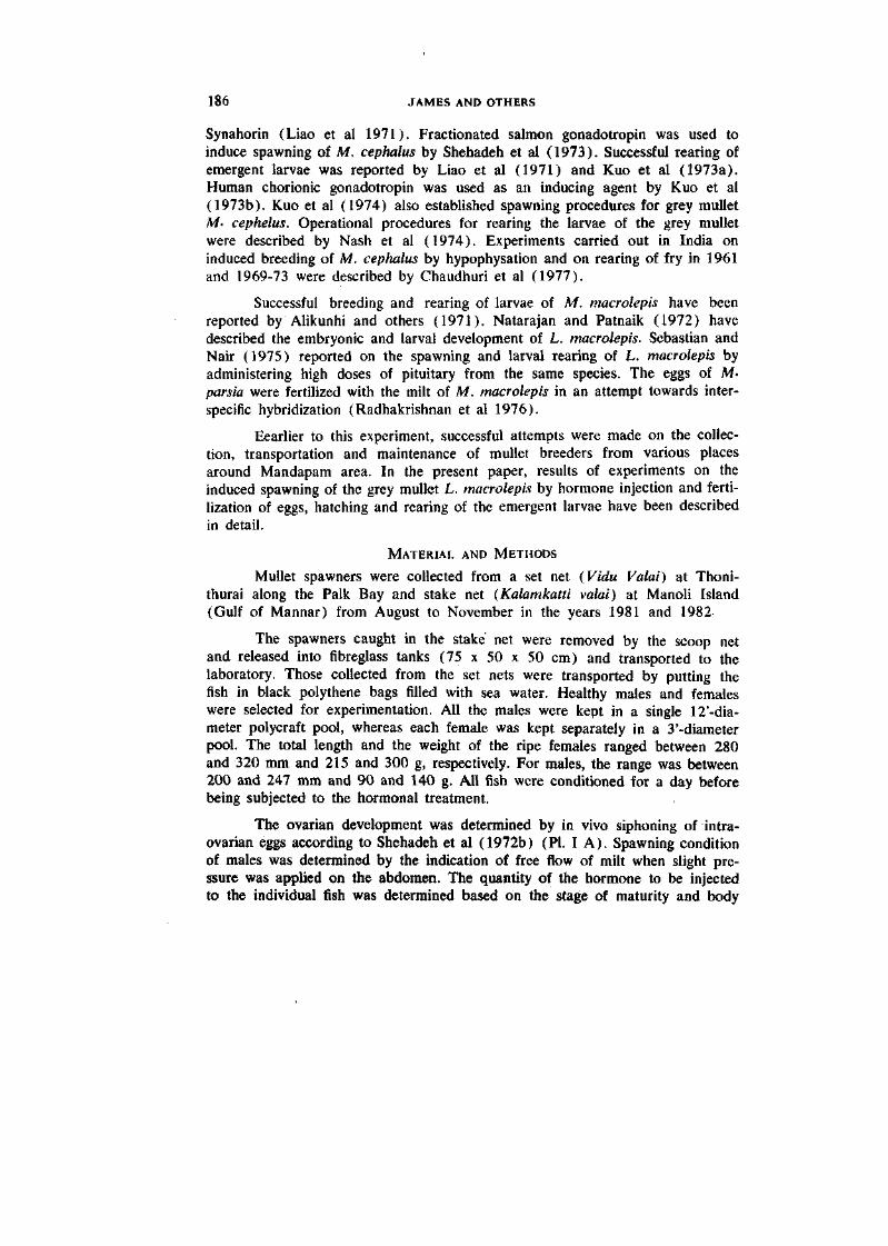
Synahorin (Liao et al 1971). Fractionated salmon gonadotropin was used to induce spawning of M. cephalus by Shehadeh et al (1973). Successful rearing of emergent larvae was reported by Liao et al (1971) and Kuo et al (1973a). Human chorionic gonadotropin was used as an inducing agent by Kuo et al (1973b). Kuo et al (1974) also established spawning procedures for grey mullet Af. cephelus. Operational procedures for rearing the larvae of the grey mullet were described by Nash et al (1974). Experiments carried out in India on induced breeding of M, cephalus by hypophysation and on rearing of fry in 1961 and 1969-73 were described by Chaudhuri et al (1977).
Successful breeding and rearing of larvae of M. macrolepis have been reported by Alikunhi and others (1971). Natarajan and Patnaik (1972) have described the embryonic and larval development of L. macrolepis. Sebastian and Nair (1975) reported on the spawning and larval rearing of L. macrolepis by administering high doses of pituitary from the same species. The eggs of M-parsia were fertilized with the milt of M. macrolepis in an attempt towards interspecific hybridization (Radhakrishnan et al 1976).
Eearlier to this experiment, successful attempts were made on the collection, transportation and maintenance of mullet breeders from various places around Mandapam area. In the present paper, results of experiments on the induced spawning of the grey mullet L. macrolepis by hormone injection and fertilization of eggs, hatching and rearing of the emergent larvae have been described in detail.
MATERIAL AND METHODS
Mullet spawners were collected from a set net {Vidu Valai) at Thoni-thurai along the Palk Bay and stake net (Kalamkatti valai) at Manoli Island (Gulf of Mannar) from August to November in the years 1981 and 1982-
The spawners caught in the stake net were removed by the scoop net and released into fibreglass tanks (75 x 50 x 50 cm) and transported to the laboratory. Those collected from the set nets were transported by putting the fish in black polythene bags filled with sea water. Healthy males and females were selected for experimentation. All the males were kept in a single 12'-dia-meter polycraft pool, whereas each female was kept separately in a 3'-diameter pool. The total length and the weight of the ripe females ranged between 280 and 320 mm and 215 and 300 g, respectively. For males, the range was between 200 and 247 mm and 90 and 140 g. All fish were conditioned for a day before being subjected to the hormonal treatment.
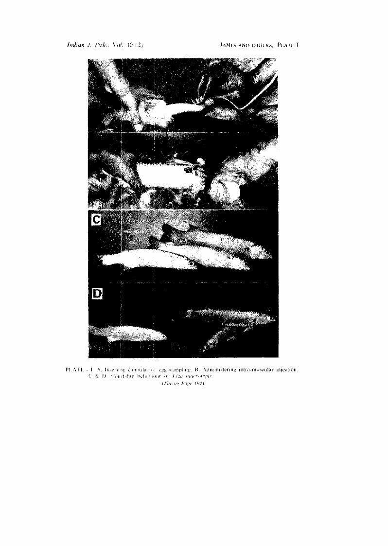
The ovarian development was determined by in vivo siphoning of intra-ovarian eggs according to Shehadeh et al (1972b) (PI. I A). Spawning condition of males was determined by the indication of free flow of milt when slight pressure was applied on the abdomen. The quantity of the hormone to be injected to the individual fish was determined based on the stage of maturity and body
INDUCED SPAWNING OF GREY MULLET 187
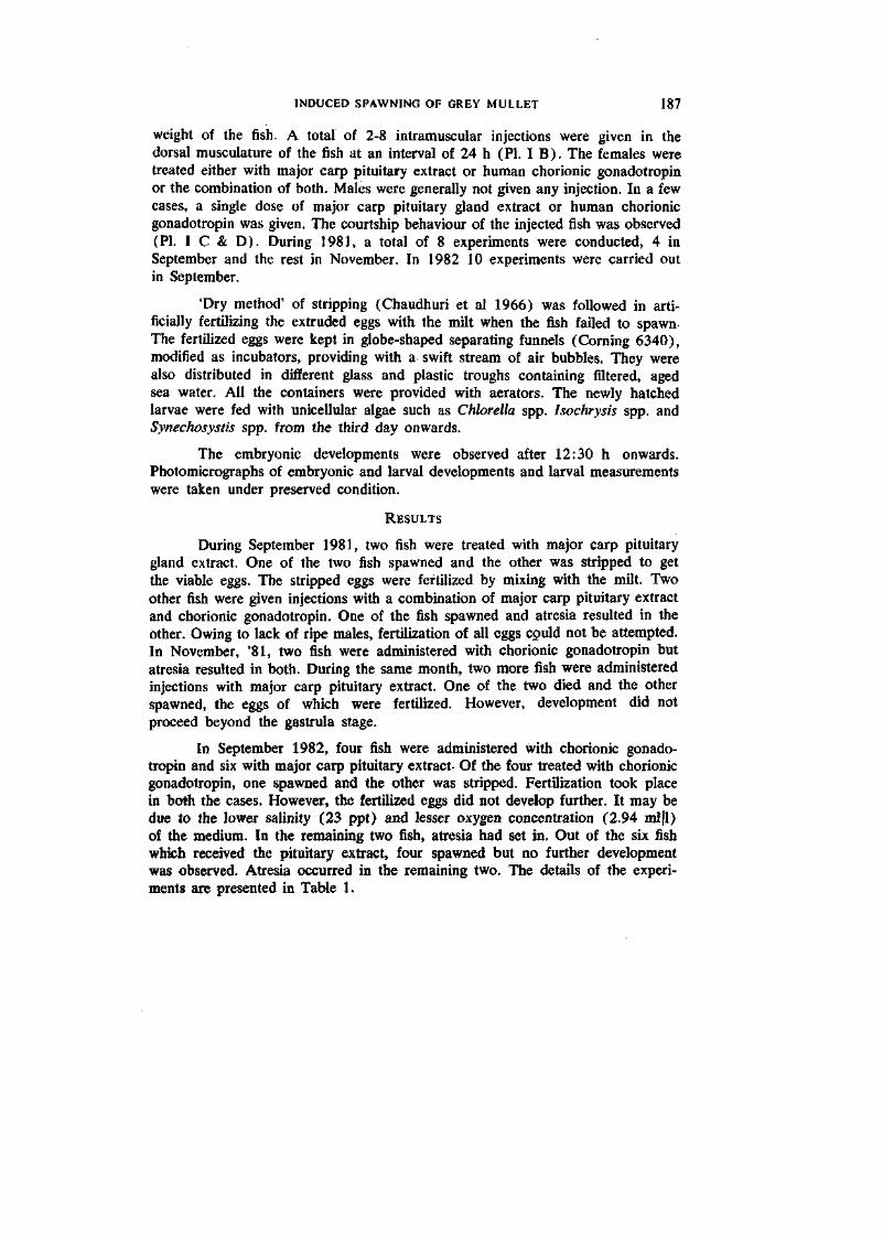
weight of the fish. A total of 2-8 intramuscular injections were given in the dorsal musculature of the fish at an interval of 24 h (PI. I B). The females were treated either with major carp pituitary extract or human chorionic gonadotropin or the combination of both. Males were generally not given any injection. In a few cases, a single dose of major carp pituitary gland extract or human chorionic gonadotropin was given. The courtship behaviour of the injected fish was observed (PI. I C & D). During 1981, a total of 8 experiments were conducted, 4 in September and the rest in November. In 1982 10 experiments were carried out in September.
'Dry method' of stripping (Chaudhuri et al 1966) was followed in artificially fertilizing the extruded eggs with the milt when the fish failed to spawn-The fertilized eggs were kept in globe-shaped separating funnels (Corning 6340), modified as incubators, providing with a swift stream of air bubbles. They were also distributed in different glass and plastic troughs containing filtered, aged sea water. All the containers were provided with aerators. The newly hatched larvae were fed with unicellular algae such as Chlorella spp. Isochrysis spp. and Synechosystis spp. from the third day onwards.
The embryonic developments were observed after 12:30 h onwards. Photomicrographs of embryonic and larval developments and larval measurements were taken under preserved condition.
RESULTS
During September 1981, two fish were treated with major carp pituitary gland extract. One of the two fish spawned and the other was stripped to get the viable eggs. The stripped eggs were fertilized by mixing with the milt. Two other fish were given injections with a combination of major carp pituitary extract and chorionic gonadotropin. One of the fish spawned and atresia resulted in the other. Owing to lack of ripe males, fertilization of all eggs could not be attempted. In November, '81, two fish were administered with chorionic gonadotropin but atresia resulted in both. During the same month, two more fish were administered injections with major carp pituitary extract. One of the two died and the other spawned, the eggs of which were fertilized. However, development did not proceed beyond the gastrula stage.
In September 1982, four fish were administered with chorionic gonadotropin and six with major carp pituitary extract- Of the four treated with chorionic gonadotropin, one spawned and the other was stripped. Fertilization took place in both the cases. However, the fertilized eggs did not develop further. It may be due to the lower salinity (23 ppt) and lesser oxygen concentration (2.94 ml|l) of the medium. In the remaining two fish, atresia had set in. Out of the six fish which received the pituitary extract, four spawned but no further development was observed. Atresia occurred in the remaining two. The details of the experiments are presented in Table 1.
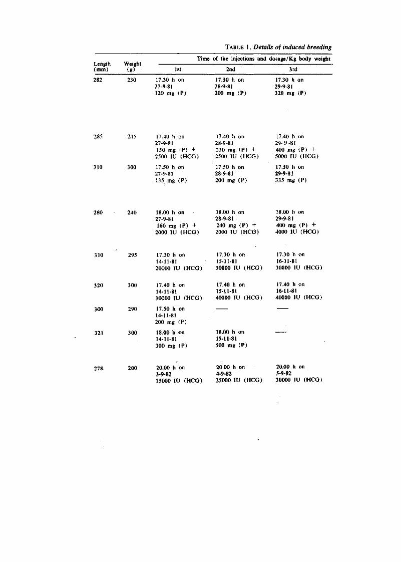
TABLE 1. Details of induced breeding
Length Weight (mm) (g)
Time of the injections and dosage/Kg body weight
1st 2nd 3rd
282 230 17.30 h on 27-9-81 120 mg (P)
17.30 h on 28-9-81 200 mg (P)
17.30 h on 29-9-81 320 mg (P)
285 215 17.40 h on 27-9-81 150 mg (P) + 2500 IV (HCG)
310 300 17.50 h on 27-9-81 135 mg (P)
17.40 h on 28-9-81 250 mg (P) + 2500 lU (HCG)
17.50 h on 28-9-81 200 mg (?)
17.40 h on 29-9-81 400 mg (P) + 5000 lU (HCG)
17.50 h on 29-9-81 335 mg (P)
280 240 18.00 h on 18.00 h on 18.00 h on 27-9-81 28-9-81 29-9-81 160 mg (P) + 240 mg (P) + 400 mg (P) +
2000 lU (HCG) 2000 lU (HCG) 4000 lU (HCG)
310 295 17.30 h on 14-11-81 20000 lU (HCG)
17.30 h on 15-11-81 30000 lU (HCG)
17.30 h on 16-11-81 30000 lU (HCG)
320 300 17.40 h on 14-11-81 30000 IV (HCG)
300 290 17.50 h on 14-11-81 200 mg (P)
321 300 18.00 h on 14-11-81 300 mg (P)
17.40 h on 15-11-81 40000 lU (HCG)
18.00 h on 15-11-81 500 mg (P)
17.40 h on 16-11-81 40000 lU (HCG)
278 200 20.00 h on 3-9-82 15000 lU (HCG)
20.00 h on 4-9-82 25000 lU (HCG)
20.00 h on 5-9-82 30000 lU (HCG)
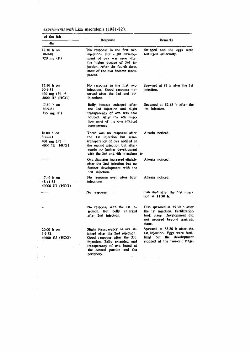
experiments with Liza macrolepis (1981-82).
of the fish
4th Response Remarks
17.30 h on 30-9-81 320 mg (P)
No response in the first two injections. But slight development of ova was seen nfter the higher dosage of 3rd injection. After the fourth dose, most of the ova became transparent.
Stripped and the eggs were fertilized artificially.
17.40 h on 30-9-81 400 mg (P) + 5000 lU (HCG)
17.50 h on 30-9-81 355 mg (P)
18.00 h on 30-9-81 400 mg (P) + 4000 lU (HCG)
17.40 h on 18-11-81 40000 lU (HCG)
No response in the first I wo injections. Good response observed after the 3rd and 4th injections.
Belly became enlarged after the 3rd injection and slight transparency of ova was nlso noticed. After the 4th injection most of the ova attained transparency.
There was no response after the 1st injection but seini-transparency of ova noticed at the second injection but afterwards no further development with the 3rd and 4th injections
Ova diameter increased slightly after the 2nd injection but no further development with the 3rd injection.
No response even after four injections).
No response.
Spawned at 83 h after the 1st injection.
Spawned at 82.45 h after the 1st injection.
Atresia noticed.
Atresia noticed.
Atresia noticed.
Fish died after the first injection at 11.30 h.
20.00 h on 6-9-82 40000 lU (HCG)
No response with the 1st injection. But belly enlarged after 2nd injection.
Slight transparency of ova attained after the 2nd injection. Good response after the 3rd injection. Belly extended and transparency of ova found at the central portion and the periphery.
Fish spawned at 35.30 h after the 1st injection. Fertilization took place. Development did not proceed beyond gastrula stage.
Spawned at 85.20 h after the 1st injection. Eggs were fertilized but the development stopped at the two-cell stage.
192 JAMES AND OTHERS
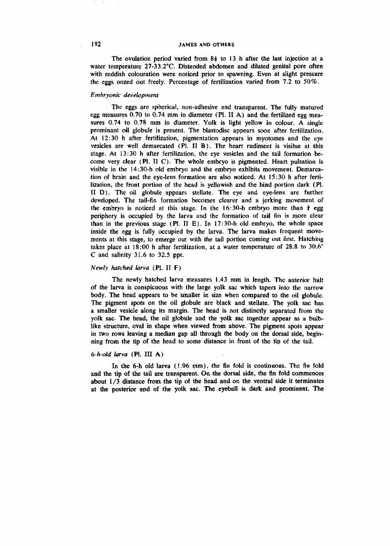
The ovulation period varied from 8i to 13 h after the last injection at a water temperature 27-33.2°C. Distended abdomen and dilated genital pore often with reddish colouration were noticed prior to spawning. Even at slight pressure the eggs oozed out freely. Percentage of fertilization varied from 7.2 to 50%.
Embryonic development
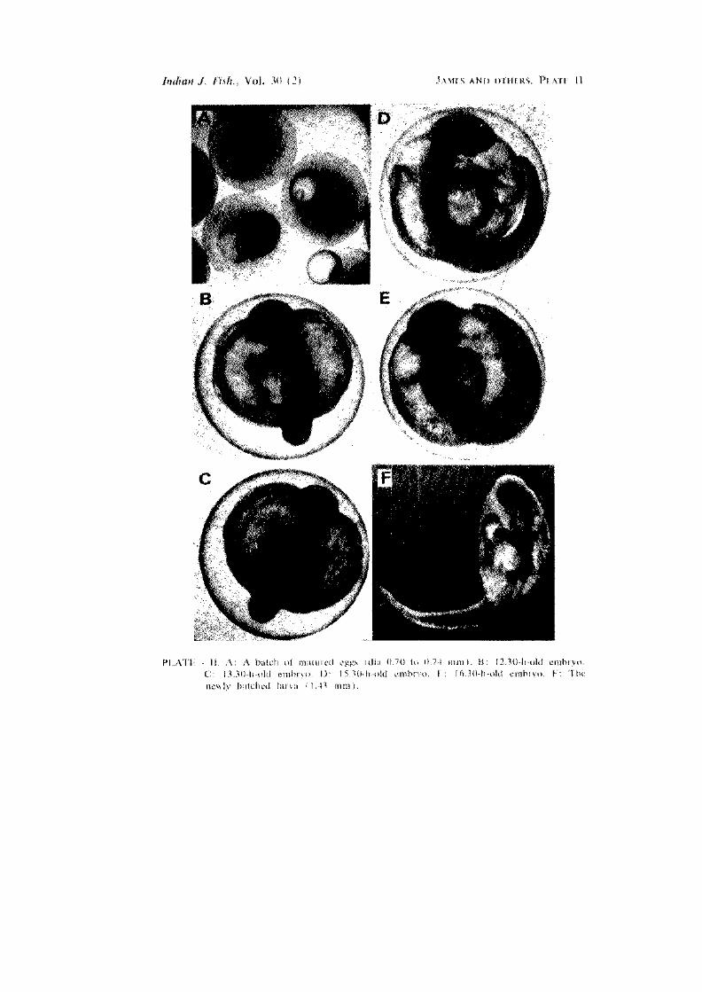
The eggs are spherical, non-adhesive and transparent. The fully matured egg measures 0.70 to 0.74 mm in diameter (PI. II A) and the fertilized egg measures 0.74 to 0.78 mm in diameter. Yolk is light yellow in colour. A single prominant oil globule is present. The blastodisc appears soon after fertilization. At 12:30 h after fertilization, pigmentation appears in myotomes and the eye vesicles are well demarcated (PI. II B). The heart rudiment is visible at this stage. At 13:30 h after fertilization, the eye vesicles and the tail formation become very clear (PI. II C). The whole embryo is pigmented. Heart pulsation is visible in the 14:30-h old embryo and the embryo exhibits movement. Demarcation of brain and the eye-lens formation are also noticed. At 15:30 h after fertilization, the front portion of the head is yellowish and the hind portion dark (PI. II D). The oil globule appears stellate. The eye and eye-lens are further developed. The tail-fin formation becomes clearer and a jerking movement of the embryo is noticed at this stage. In the 16:30-h embryo more than i egg periphery is occupied by the larva and the formation of tail fin is more clear than in the previous stage (PI. II E). In 17:30-h old embryo, the whole space inside the egg is fully occupied by the larva. The larva makes frequent movements at this stage, to emerge out with the tail portion coming out first. Hatching takes place at 18:00 h after fertilization, at a water temperature of 28.8 to 30.6° C and salinity 31.6 to 32.5 ppt.
Newly hatched larva (PI. II F)
The newly hatched larva measures 1.43 mm in length. The anterior halt of the larva is conspicuous with the large yolk sac which tapers into the narrow body. The head appears to be smaller in size when compared to the oil globule. The pigment spots on the oil globule are black and stellate. The yolk sac has a smaller vesicle along its margin. The head is not distinctly separated from the yolk sac- The head, the oil globule and the yolk sac together appear as a bulblike structure, oval in shape when viewed from above. The pigment spots appear in two rows leaving a median gap all through the body on the dorsal side, beginning from the tip of the head to some distance in front of the tip of the tail.
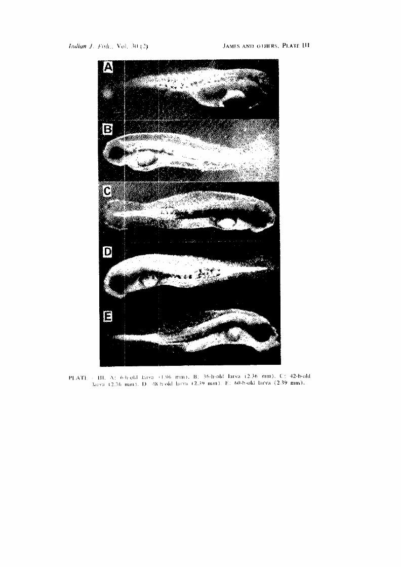
6-h-old larva (PI. Ill A)
In the 6-h old larva (1.96 mm), the fin fold is continuous. The fin fold and the tip of the tail are transparent. On the dorsal side, the fin fold commences about 1/3 distance from the tip of the head and on the ventral side it terminates at the posterior end of the yolk sac. The eyeball is dark and prominent. The
INDUCED SPAWNING OF GREY MULLET 193
alimentary tract is distinct. Pigmentation of the body extends from behind the head up to a little distance short of the tip of the taU. Pigment spots on the oil globule are not seen, except for some yellow tinge. The other pigmentation is in the form of black, branching pattern from the hind border of eye to a little distance short of tip of tail. Yolk sac occupies more than 1/3 of the body from the anterior end. Oil globule is distinctly spherical.
12-h-old larva
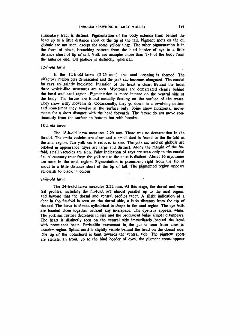
In the 12-h-old larva (2.25 mm) the anal opening is formed. The olfactory region gets demarcated and the yolk sac becomes elongated. The caudal fin rays are faintly indicated. Pulsation of the heart is clear. Behind the heart three vesicle-like structures are seen. Myotomes are demarcated clearly behind the head and anal region. Pigmentation is more intense on the ventral side of the body. The larvae are found casually floating on the surface of the water. They show jerky movements. Occasionally, they go down in a revolving pattern and sometimes they revolve at the surface only. Some show horizontal movements for a short distance with the head forwards. The larvae do not move continuously from the surface to bottom but with breaks.
IS-h-old larva
The 18-h-old larva measures 2.29 mm- There was no demarcation in the fin-old. The optic vesicles are clear and a small dent is found in the fin-fold at the anal region. The yolk sac is reduced in size. The yolk sac and oil globule are bilobed in appearance. Eyes are large and distinct. Along the margin of the fin-fold, small vacuoles are seen. Faint indication of rays are seen only in the caudal fin. Alimentary tract from the yolk sac to the anus is distinct. About 16 myotomes are seen in the anal region. Pigmentation is prominent right from the tip of snout to a little distance short of the tip of tail. The pigmented region appears yellowish to black in colour-
lA-h'-old larva
The 24-h-old larva measures 2.32 mm. At this stage, the dorsal and ventral profiles, including the fin-fold, are almost parallel up to the anal region, and beyond that the dorsal and ventral profiles taper. A slight indication of a dent in the fin-fold is seen on the dorsal side, a little distance from the tip of the tail- The larva is almost cylindrical in shape in the anal region. The eye-balls are located close together without any interspace. The eye-lens appears white. The yolk sac further decreases in size and the prominent bulge almost disappears. The heart is distinctly seen on the ventral side immediately behind the head with prominent beats. Peristaltic movement in the gut is seen from anus to anteriOT region. Spinal cord is slightly visible behind the head on the dorsal side. The tip of the notochord is bent towards the ventral side. The pigment spots are stellate. In front, up to the hind border of eyes, the pigment spots appear
194 JAMES AND OTHERS
as a dark line, unlike the posterior region. The pigment spots are located more or less in paraUel rows behind the oil globule up to a little distance short of tail-The larva responds to external stimuli as evidenced by the movement away from the point of disturbance.
30-h-old larva
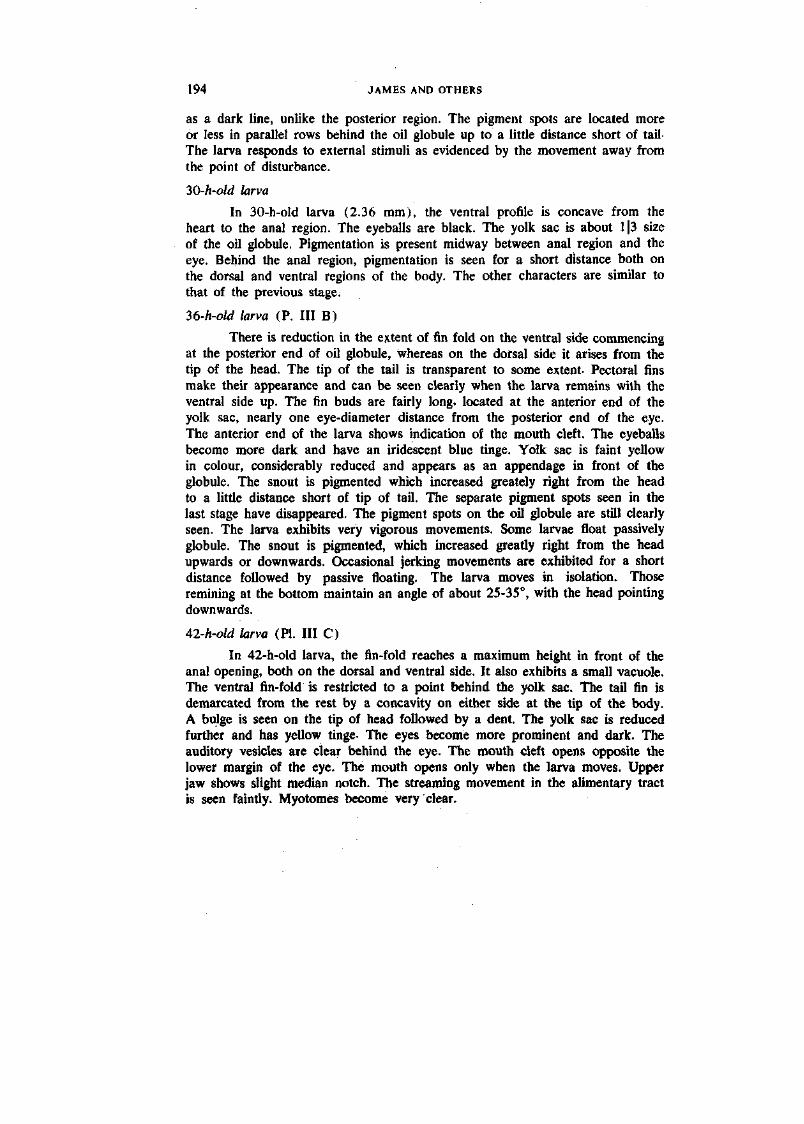
In 30-h-old larva (2.36 mm), the ventral profile is concave from the heart to the anal region. The eyeballs are black. The yolk sac is about 1|3 size of the oil globule. Pigmentation is present midway between anal region and the eye. Behind the anal region, pigmentation is seen for a short distance both on the dorsal and ventral regions of the body. The other characters are similar to that of the previous stage.
36-h-old larva (P. Ill B)
There is reduction in the extent of fin fold on the ventral side commencing at the posterior end of oil globule, whereas on the dorsal side it arises from the tip of the head. The tip of the tail is transparent to some extent. Pectoral fins make their appearance and can be seen clearly when the larva remains with the ventral side up. The fin buds are fairly long, located at the anterior end of the yolk sac, nearly one eye-diameter distance from the posterior end of the eye. The anterior end of the larva shows indication of the mouth cleft. The eyeballs become more dark and have an iridescent blue tinge. Yolk sac is faint yellow in colour, considerably reduced and appears as an appendage in front of the globule. The snout is pigmented which increased greately right from the head to a little distance short of tip of tail. The separate pigment spots seen in the last stage have disappeared. The pigment spots on the oil globule are still clearly seen. The larva exhibits very vigorous movements. Some larvae float passively globule. The snout is pigmented, which increased greatly right from the head upwards or downwards. Occasional jerking movements are exhibited for a short distance followed by passive floating. The larva moves in isolation. Those remining at the bottom maintain an angle of about 25-35°, with the head pointing downwards.
42-h-old larva (PI. Ill C)
In 42-h-old larva, the fin-fold reaches a maximum height in front of the anal opening, both on the dorsal and ventral side. It also exhibits a small vacuole. The ventral fin-fold is restricted to a point behind the yolk sac. The tail fin is demarcated from the rest by a concavity on either side at the tip of the body. A bulge is seen on the tip of head followed by a dent. The yolk sac is reduced further and has yellow tinge- The eyes become more prominent and dark. The auditory vesicles are clear behind the eye. The mouth cleft opens opposite the lower margin of the eye. The mouth opens only when the larva moves. Upper jaw shows slight median notch. The streaming movement in the alimentary tract is seen faintly. Myotomes become very clear.
Indian J. Fixh... Vol. 30 (2) JAMES AND OTHERS, I'LATE !
:iilar irijecfion.
Indian J. fish., Vol. 311 12) JAMES AND OTHERS. PLATE II
I'l '•. i ( ' ibryu. F: The
Indian J. Fhh.. Vol. 3fi (2) JAMES AND OTHERS, PLATE HI
Indiait J. Fish.. ¥ol. 30 (2) JAMFS ANI> OTHERS. PLATE IV
PLATE ~ I I i ll-
INDUCED SPAWNING OF GREY MULLET 195
48-/i-oW larva (PI. Ill D)
The 48-h-old larva measures 2.39 nmi in legnth. At this stage a cleft forms in the fin-fold at the anal region indicating the demarcation of the origin of anal fin. In the tail fin-fold and in the central portion, indication of formation of rays could be seen at the tip of the fold. Pectoral fins further elongate and project beyond the ventral profile. They almost cover the greater part of the oil globule. The jaws are functional, the lower jaw movement being conspicuous. The larva is able to move the jaw even in stationary condition. Mouth cleft becomes deep. The eye-balls are oval in shape. The streaming movement in the gut is seen very clearly passing forwards under the oil globule towards the heart. The pigment spots are stellate but less intense in the anterior region. The larvae do not exhibit quick jerks but move continuously for short distances.
54-h-old larv(i
In the 54-h-old larva the fin-fold undergoes further differentiation- One more cleft in the ventral side of the fin-fold is seen below the lower jaw. Pectoral fins are developed further. The other characters are similar to those of the previous stage.
60-h-old larva (PI. Ill E).
At this stage the ventral fin-fold reduces further and starts only behind the oil globule, whereas the dorsal fin-fold arises from the top of the head. The lower jaw exhibits prominent movements compared to upper jaw. It is also longer than the upper jaw. The eye-ball appears bright bluish and iris jet black in colour. Free movement of the eye-ball can be seen. Yolk sac has almost completely reduced. The size of the oil globule gets further reduced. The mouth is kept open constantly. From behind the eye the vertebral column shows an arch over the position of the oil globule and then descends down to run almost in a straight line. The pigmentation spreads from the posterior end of the head up to a little distance short of tip of the tail which is transparent. The larvae tend to congregate into two large groups. In each group majority are found at the bottom, while a few occupy different levels up to the surface. The larvae move in various directions. Those at the bottom appear to crawl for short distances, touching the bottom of the container.
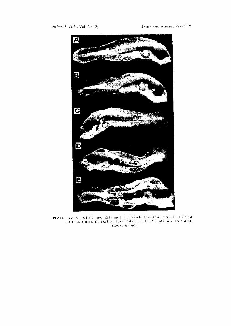
66-h-old larva (PI. IV A)
In the 66-h-old larva the ventral fin-fold starts much behind the oil globule- The cleft in the ventral fin-fold at the anal region becomes deeper. The lower jaw shows slight flickering movements. It also shows angular projections under the eye. Slight indication of the formation of the opercle is seen. The head appears quite robust. Oil globule is still prominent. Heart beat becomes quite fast. At the tip of the vertebral column, demarcation of individual vertebrae can be seen. Pigmentation appears branched.

196 ' JAMES AND OTHERS
12-h-old larva
The 72-h-old larva also measures 2.39 mm in length as does the 48-h-old larva. The entire fin-fold shows a peculiar pattern of vacuoles and marking. The dent in the fin-fold is distinct. Pectoral fins are prominent and rounded. When erect, they cross the dorsal margin of the body. Caudal fin rays become more prominent. The oil globule is still reduced. The whole body of the larva gets covered with some vacuole-like pattern. This may be the beginning of future pigmentation. The body is uniformly pigmented light brown from the head region up to a little distance short of the tip of the vertebral column. The main change at this stage seems to be the general pigmentation of the larva without any distinct branching type of pigmentation.
78-h-old larva (PI- IV B)
In the 78-h-old larva (2.43 mm), the fin-fold is transparent commencing in front of the notch on the head. The ventral fin-fold arises a little distance in front of the anus where it shows a dent and proceeds backwards. Slight concavities in the fin-fold are found in the posterior region where the transparent part of body begins. The head is prominent, followed by a notch after which the body is convex, and then gently slopes towards the posterior end. The body is opaque except the last portion. Lower jaw is concave below. Vertebrae become clear from the portion just above the oil globule and up to the end of tail. Pigmentation extends from behind the eye up to the transparent region. The margins of the jaws are also slightly pigmented.
S4-h-old larva
Over the upper jaw, a slight projection has appeared. More caudal rays are seen at this stage. The oil globule gets further reduced in size. Pigmentation spreads all over the body except the tip of the tail. Most of the larvae are found at the bottom with the head downwards and making an angle of 70-90°. While in that position, they show jerky movements, back and forth, as if searching for food and grazing. A few larvae are also seen floating in the mid-water column with the head downwards. They also show jerking movements. Sometimes, they even rise up to the surface with circulating movement of water.
90-h-old larva
In the 90-h-old larva, the body arches at the beginning and slopes down to the tail. Pigment spots spread all over the body and are branched-
96-h-old larva •
The size of the 96-h-old larva remains the same as in the 84-h-old larva. The caudal fin-fold shows a round shape. The dorsal fin-fold appears in front of the notch on the head and the ventral fin-fold behind the oil globule. Internal organs are distinctly demarcated.
INDUCED SPAWNING OF GREY MULLET 197
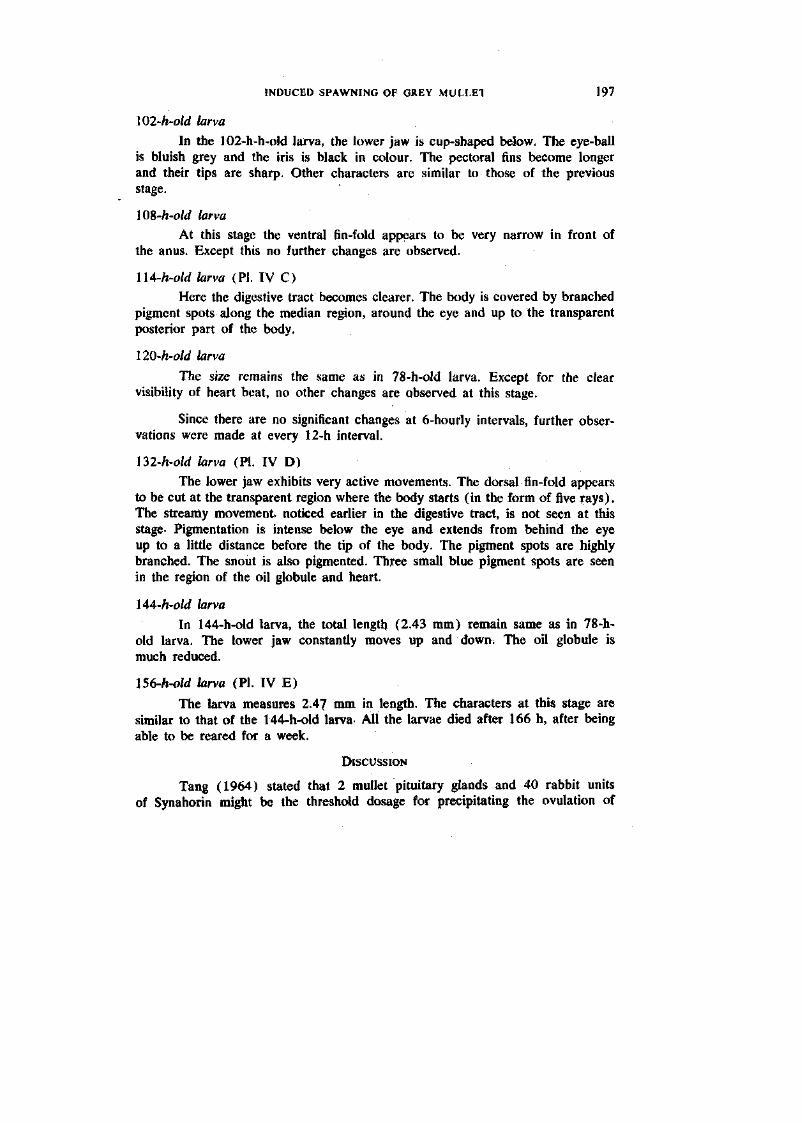
102-h-old larva In the 102-h-h-old larva, the lower jaw is cup-shaped below. The eye-ball
is bluish grey and the iris is black in colour. The pectoral fins become longer and their tips are sharp. Other characters are similar to those of the previous stage.
108-ft-oW larva At this stage the ventral fin-fold appears to be very narrow in front of
the anus. Except this no further changes are observed.
lU-h-old larva (PI. IV C) Here the digestive tract becomes clearer. The body is covered by branched
pigment spots along the median region, around the eye and up to the transparent posterior part of the body,
llO-h-old larva
The size remains the same as in 78-h-old larva. Except for the clear visibility of heart beat, no other changes are observed at this stage.
Since there are no significant changes at 6-hourly intervals, further observations were made at every 12-h interval.
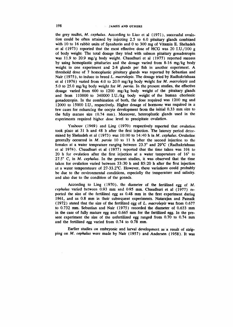
ni-h-old larva (PI. IV D) The lower jaw exhibits very active movements. The dorsal fin-fold appears
to be cut at the transparent region where the body starts (in the form of five rays). The streamy movement, noticed earlier in the digestive tract, is not seen at this stage- Pigmentation is intense below the eye and extends from behind the eye up to a little distance before the tip of the body. The pigment spots are highly branched. The snout is also pigmented. Three small blue pigment spots are seen in the region of the oil globule and heart.
\AA-h-old larva In 144-h-old larva, the total length (2.43 mm) remain same as in 78-h-
old larva. The lower jaw constantly moves up and down. The oil globule is much reduced.
156-ft-oW larva (PI. IV E)
The larva measures 2.47 mm in length. The characters at this stage are similar to that of the 144-h-old larva- All the larvae died after 166 h, after being able to be reared for a week.
DISCUSSION
Tang (1964) stated that 2 mullet pituitary glands and 40 rabbit units of Synahorin might be the threshdd dosage for precipitating the ovulation of
198 JAMES AND OTHERS
the grey mullet, M. cephalus. According to Liao et al (1971), successful ovulation could be often attained by injecting 2.5 to 6.0 pituitary glands combined with 10 to 16 rabbit units of Synahorin and 0 to 300 mg of Vitamin E. Shehadeh et al (1973) reported that the most effective dose of HCG was 20 I.U./IOO g of body weight- The total dosage they tried with salmon pituitary gonadotropin was 11.9 to 20.9 mg/g body weight. Chaudhuri et al (1977) reported success by using homoplastic pituitaries and the dosage varied from 8-16 mg/kg body weight in one experiment and 2-8 glands per fish in another experiment. A threshold dose of 7 homoplastic pituitary glands was reported by Sebastian and Nair (1975), to induce to breed L. macrolepis. The dosage tried by Radhakrishnan et al (1976) varied from 4.0 to 20.0 mg/kg body weight for M. macrolepis and 5.0 to 25.0 mg/kg body weight for M. parsia. In the present studies, the effective dosage varied from 600 to 1200 mg/kg body weight of the pituitary glands and from 110000 to 340000 I.U./kg body weight of the human chorionic gonadotropin. In the combination of both, the dose required was 1200 mg and 12000 to 15000 I.U., respectively. Higher dosage of hormone was required in a few cases for enhancing the oocyte development from the initial 0.33 mm size to the fully mature size (0.74 mm). Moreover, heteroplastic glands used in the experiments required higher dose level to precipitate ovulation.
Yashouv (1969) and Ling (1970) respectively reported that ovulation took place at 31 h and 48 h after the first injection. The latency period determined by Shehadeh et al (1973) was 10:00 to 14:40 h in M. cephalus. Ovulation generally occurred in M. parsia 10 to 11 h after the second injection to the females at a water temprature ranging between 23.3° and 29°C (Radhakrishnan et al 1976). Chaudhuri et al (1977) reported that the time taken was lOi to 20 h for ovulation after the first injection at a water temperature of 16° to 27.5° C, in M. cephalus. In the present studies, it was observed that the time taken for ovulation varied between 33:30 h and 85:20 h after the first injection at a water temperatuure of 27-33.2°C. However, these variations could probably be due to the environmental conditions, especially the temperature and salinity, and also due to the condition of the gonads.
According to Ling (1970), the diameter of the fertilized egg of M. cephalus varied between 0.93 mm and 0-95 mm. Chaudhuri et al (1977) reported the size of the fertilized egg as 0.48 mm in the first experiment during 1961, and as 0.8 mm in their subsequent experiments. Natarajan and Patnaik (1972) stated that the size of the fertilized egg of L. macrolepis was from 0.677 to 0.732 mm. Sebastian and Nair (1975) recorded the diameter of 0.633 mm in the case of fully mature egg and 0.665 mm for the fertilized egg. In the present experiment the size of the unfertilized egg ranged from 0.70 to 0.74 mm and the fertilized egg varied from 0.74 to 0.78 mm.
Earlier studies on embryonic and larval development as a result of stripping on M. cephalus were made by Nair (1957) and Anderson (1958). It was
INDUCED SPAWNING OF GREY MULLET 199
observed that the eye vesicles are well demarcated in 12:30-h-old embryo and the pigment cells appeared on the whole body of the embryo at 13:30 h after fertilization- These observations are almost in agreement with the observations of Sebastian and Nair (1975). Kuo et al (1973a) reported that in M. cephalus the heart began to beat at 25:10 h after fertilization. In the present observations, the pulsation of heart began at 14:30 h after fertilization and the eye-lens formation was also noticed at this stage.
Tang (1964) reported that the developing egg of M. cephalus sank in the standing water and, so, was incubated in suspension by circulating water. Yashouv and Berner Sampsonov (1970) described that the developing eggs of M. capita floated at the surface. They also reported that the eggs of M. cephalus lost their buoyancy after 20 h of fertilization and sank, but hatched successfully. Kuo et al (1973a) stated that, in aerated sea water (salinity 32 ppt), majority of the eggs collected from the bottom of the incubation tank were found to be either unfertilized or undeveloped. When these were prevented from setting down by strong aeration, high hatching rate of 90-95% was obtained. Liao (1975) reported that the fertile eggs of M. cephalus were buoyant and floated near the surface. Chaudhuri et al (1977) stated that the eggs floated immediately after fertilization, but subsequently sank to the bottom. Sebastian and Nair (1975) observed that the eggs of M. macrolepis floated in the shallow hatching trays. In the present study it was observed that the fertilized eggs floated in the glass trough and the unfertilized eggs settled at the bottom of the trough.
Liao (1975) reported that the hatching times were 34-38 h at 23-24.5°C and 49-54 h at 22.5-23.7°C in salinities ranging from 30.1 to 33.8 ppt. Kuo et al (1973a) stated that the hatching was evident at 36-38 h after fertilization for M. cephalus at 24°C and 48-50 h at 22°C in well-aerated sea water with a salinity of 32 ppt. Nash et al (1974) showed that 25°C and above were definitely inhibitory to successful incubation. Chaudhuri et al (1977) recorded the incubation period of 32 to 52i h and more hatchlings were obtained at 33f h. According to Sebastian and Nair (1975). most of the developing eggs hatched out at 23 h after fertilization. In the present study, the fertilized eggs hatched out after 18 h of incubation at a water temperature varying between 27 to 30.6°C and salinity of 32 ppt. Percentage of hatching was 47.2.
Ling (1970) reported that the size of the hatchlings of M. cephalus varied from batch to batch (2.1-3.1 mm). Similar findings were reported by Chaudhuri et al (1977). Natarajan and Patnaik (1972) recorded a size of 1.519-1.556 mm for the newly hatched larva of Liza macrolepis. Sebastian and Nair (1975) indicated a size less than 2 mm for the newly hatched larva of M. macrolepis. In the present study, the size of the newly hatched larva was 1.43 mm.
Pakrasi and Alikunhi (1952) reported the formation of mouth at 48 h after hatching in M. corsula. The mouth formation was observed in the two-day-
2 0 0 JAMES AND OTHERS
old larva of L. macrokpis by Natarajan and Patnaik (1972). Sebastian and Nair stated that the mouth formed in the two-day-old larva and jaws functioned on the fourth day in L. macrolepis. In the present studies, the mouth formation was observed at 42 h and jaws became functional at 48 h after hatching.
According to Natarajan and Patnaik (1972), the 1-day-, 2-day- and 3-day-old larvae of L. macrolepis measured 2.123 mm, 2-215 mm and 2.342 mm, respectively. In the present studies, the 1-day-old larva measured 2.32 mm; the 2-day-old larva 240 mm and 6-day-old larva 2.43 mm.
Larval feeding is the major problem in rearing the fry (Kuo et al 1973a. Nash and Kuo 1975). Liao et al (1971) reported that the three-day-old larvae, when offered oyster veligers, showed better survival. Sebastian and Nair (1975) observed in their initial experiments that the artificial feed such as milk powder, boiled yolk of egg and powdered oats, when supplied to the hatchlings, made the water foul. In their later experiments, they found the 3-4 days old hatchlings introduced in a cement tank fed actively on the microscopic organisms developed naturally in the tank, and the larvae grew very fast. Chaudhuri et al (1977) reported that the fry, which were given natural feed collected from the lake mouth, ensured better results. In the present study, C^hlorella spp, Isochrysis spp. and SynechocysHs spp. cultured in the laboratory were provided as feed to the larvae from the third day onwards.
Liao (1975) stated that the young larvae did not like strong illumination and tended to congregate in places of law light intensity and older larvae swam in schools. This behaviour of the larvae was also noticed in the present study, with the sides of all the rearing troughs covered with black paper to avoid excess light.
Liao et al (1971) observed two critical periods for M. cephalus larvae, one on the third day and the other on the eleventh day. Kuo et al (1973 a) reported two similar critical periods involving very high mortality, the first coinciding with the opening of the mouth (2nd to 3rd day) and the second preceding and overlapping the second phase (8th to 12th day). Chaudhuri et al (1971) mentioned that high mortality of hatchlings is a frequent occurrence, especially during the critical period, when the mouth is formed and the larvae start feeding when they are three days old. In the present experiments also mortality was observed on the third day. However, apart from the environmental factors, the absence of proper feed may be the cause for high mortality.
There were certain constraints in carrying out the present study. The non-availability of ripe males in good numbers in commercial catches was the major one. Always ripe females dominated the catches. Thonithurai (Palk Bay), from where the spawners were collected from set nets, was found to be the ideal place for this purpose. But the operation of that gear is seasonal (July-
INDUCED SPAWNING OF GREY MULLET 201
October), limiting the scope of the experiments. To overcome these problems attempts are being made for raising the brood fish in coastal ponds, close to the laboratory. The long-distance transportation results in high mortality. For a short distance, the transportation method followed in the present study was successful. The loosening of the body scales due to handling leads to bacterial and fungal infections, thereby resulting in mortality of the fish. This problem has been minimised by putting the fish in a transparent polythene bag filled with sea water, whenever the fish was handled.
REFERENCES
ALIKUNHI, K. H. , M. J. SEBASTIAN, K. K. SUKUMARAN, V. A. NAIR AND T. J. VINCENT.
1971. Induced spawning of the grey mullet (Mugil macrolepis) and observations on rearing hatchlings to fingerlings stage. Workshop on induced breeding of Carps. Institute of Fisheries Education, Bombay, Working Paper, 33, p. 2 (cyclostyled).
ANDERSON, W . W . 1958. Larval development, growth and spawning of striped mullet (Mugil cephalus) along the South Atlantic Coast of the United States. Fish. Bull. U.S. Fish. Wildl. Serv., 58 (144): 501.519.
BARRACKPORE. Central Inland Fisheries Research Institute, Annual Report, 1961-62: 55 p.
CHAUDHURI, H. , J. B. SiNOH AND K. K. SUKUMARAN. 1966. Experiments on large-scale production of fish seed of the Chinese grass carp, Ctenopharyngodon idella (C & V) and the silver carp, Hypophthalmichthys molitrix (C & V) by induced breeding in India. Proc. Indian. Acad. Sci. (b) 63: 80-95.
CHAUDHURI, H. , R . M . BHOWMICK, G . V. KOWTAL, M . M . BAOCHI, R. K. JANA AND S. D. GuPTHA. 1977. Experiments in artificial propagation and larval development of Mugil cephalus Linnaeus in India. / . Inland Fish. Soci. India, 9: 30-41.
Kuo, C. M., Z. H. SHEHADEH AND K. K. MILISEN. 1973a. A preliminary report on the development, growth and survival of laboratory reared larvae of the grey mullet, Mugil cephalus J. Fish. Biol, 5: 449-470.
Kuo, C. M., Z. H SHEHADEH AND C . E. NASH. 1973b. Induced spawning of captive grey mullet (Mugil cephalus L.) females by injection of human chorionic gonadotropin. Aquaculture, 1: 429-432.
Kuo, C. M., C. E. NASH AND Z. H . SHEHADEH. 1974. A procedural guide to induce spawning in grey mullet (Mugil cephalus L.). Aquaculture, 3: 1-14.
LiAo, I. C , Y. J. Lu, T. L. HUANG AND M . C. LIN. 1971. Experiments on induced breeding of the grey mullet, Mugil cephalus Linnaeus. Aquaculture, 1: 15-34.
LMO, I. C. 1975. Experiments on the induced breeding of the grey mullet in Taiwan from 1963-1973. Aquaculture, 6(1): 31-58.
LING, S. N . 1970. A brief review of the work done on the induced breeding of M. cephalus in Taiwan. /. Inland. Fish. Soc. India, 1: 1-12.
LUTHER, G . 1963. Some observations on the biology of Liza macrolepis (Smith) and Mugil cephalus Linnaeus (Mugilidae) with notes on the fishery of grey mullets near Mandapam. Indian ]. Fish., 10(2A): 642-666.
NAIR, G . S. 1957. Notes on the early development of Mugil cephalus Linnaeus. Bull. Res. Inst. Univ. Travancore, C5( l ) : 77-84.
202 JAMES AND OTHERS
NASH, C . E., CHING-MINO Kuo AND SUSAN C. MCCONNEL. 1974. Operational procedure for rearing larvae of the grey mullet (Mugil cephalus L.). Aquaculture, 3(1): 15-24.
NASH, C . E. AND C . M. KUO. 1975. Hypothesis for problems impeding the mass propagation of grey mullet and other fin fish. Aquaculture, 5: 119-133.
NATARAJAN, A. V. AND S. P. PATNAIK. 1972. Embryonic and larval development of Chilka mullet Liza macrolepis (Smith). / . Inland Fish. Soc. India, 4: 15-19.
PAKRASI, B . AND K. H . ALIKUNHI. 1952. On the development of grey mullet, Mugil corsula (Hamilton). /. Zoo/. Soc. India, 4(2): 123-140.
RADHAKRISHNAN, S., K. V. RAMAKRISHNA, G . R. M . RAO AND K. RAMAN. 1976. Breeding
of mullet by hormone stimulation. Matsya, 2: 28-31.
SEBASTIN, M . J. AND V. A. NAIR. 1975. The induced spawning of the grey mullet, Mugil macrolepis (Aguas) Smith and the large-scale rearing of its larvae. Aquaculture, 5: 41-52.
SHEHADEH, Z. H . AND J. N. ELLIS. 1970. Induced spawning of grey mullet, Mugil cephalus L. ;. Fish. Biol, 2: 355-360.
SHEHADEH, Z. H. , C . M . Kuo AND K. K. MILISEN. 1972b. Validation of an in vivo method for monitoring ovarian development in the grey mullet [Mugil cephalus L.). / . Fish. Biol, 5: 489-496.
SHEHADEH, Z . H. , C . M . KUO AND K. K. MILISEN. 1973. Induced spawning of grey mullet, Mugil cephalus L. with fractionated salmon pituitary extract. / . Fish. Biol, 5: 471-478.
TANG, Y . A. 1964. Induced spawning of striped mullet by hormone injection. Japan J. Ichthyol, 12: 23-28.
YASHOUV, A. 1969. Preliminary report on induced spawning of Mugil cephalus (L.) reared in captivity in fresh water ponds. Bamidgeh, Bull. Fish. Fish. Cul, Israel, 21(1): 19-24.
YASHOUV, A. AND E . BERNER SAMPSONOV, 1970. Contribution to the knowledge of eggs and early stages of mullet (Mugilidae) along the Israeli coast. Bamidgeh, 22(3): 72-89.
![Rearing Healthy Calves Manual 2nd ed (1)[2] copy](https://static.fdokumen.com/doc/165x107/6326a762051fac18490ddddd/rearing-healthy-calves-manual-2nd-ed-12-copy.jpg)