The Importance of Correlation between CBCT Analysis ... - MDPI
Upload
independentCategory
view
2download
0
This content has been downloaded from IOPscience. Please scroll down to see the full text.
Download details:
IP Address: 128.178.22.58
This content was downloaded on 27/03/2014 at 18:26
Please note that terms and conditions apply.
A comparison of rigid registration methods for prostate localization on CBCT and the
dependence on rectum distension
View the table of contents for this issue, or go to the journal homepage for more
2014 J. Phys.: Conf. Ser. 489 012025
(http://iopscience.iop.org/1742-6596/489/1/012025)
Home Search Collections Journals About Contact us My IOPscience

A comparison of rigid registration methods for
prostate localization on CBCT and the dependence
on rectum distension ∗
C Boydev1,2, D Pasquier3,4, F Derraz1,5, L Peyrodie6, ATaleb-Ahmed1 and JP Thiran2,7
1Laboratoire d’Automatique, de Mecanique et d’Informatique Industrielles et Humaines(LAMIH), Universite de Valenciennes et du Hainaut-Cambresis, France2Signal Processing Laboratory (LTS5), Ecole Polytechnique Federale de Lausanne, Switzerland3Centre de Radiotherapie et d’Oncologie Galilee de Lille, France4Departement Universitaire de Radiotherapie, Centre Oscar Lambret, Lille, France5Unite de Traitements de Signaux Biomedicaux (UTSB), Faculte Libre de Medecine, Lille,France6Unite de Traitements de Signaux Biomedicaux (UTSB), Hautes Etudes d’Ingenieur, Lille,France7Department of Radiology, University Hospital Center (CHUV) and University of Lausanne(UNIL), Switzerland{christine.boydev, jp.thiran}@epfl.ch, [email protected],{foued.derraz,taleb}@univ-valenciennes.fr, [email protected]
E-mail: [email protected]
Abstract. We evaluated automatic three-dimensional intensity-based rigid registration (RR)methods for prostate localization on CBCT scans and studied the impact of rectum distensionon registration quality. 106 CBCT scans of 9 prostate patients were used. Each one wasregistered to the planning computed tomography (CT) scan using different methods: (a) globalregistration, (b) pelvis bony structure registration, (c) bony registration refined by a localprostate registration using the CT clinical target volume (CTV) expanded with 1, 3, 5, 8,10, 12, 15 or 20-mm margin. Automatic CBCT contours were generated after propagationof the manual CT contours. To evaluate results, a radiation oncologist was asked to manuallydelineate the CTV on the CBCT scans (gold standard). The Dice similarity coefficients betweenpropagated and manual CBCT contours were calculated.
1. IntroductionDaily image guidance, such as cone-beam computed tomography (CBCT) image-guidedradiotherapy (IGRT) systems [1], has become a widely-used tool for patient repositioning inthe treatment of prostate cancer. The prostate gland is known to be a moving and deformablegland under influence of rectal and bladder filling changes [2, 3, 4], which limits the effectivenessof skin marks in patient setup. Prostate localization on CBCT scans is challenging due to therelatively poor image quality [5, 6].
∗This work was financially supported by ELEKTA SAS, Boulogne Billancourt, France.
XVII International Conference on the Use of Computers in Radiation Therapy (ICCR 2013) IOP PublishingJournal of Physics: Conference Series 489 (2014) 012025 doi:10.1088/1742-6596/489/1/012025
Content from this work may be used under the terms of the Creative Commons Attribution 3.0 licence. Any further distributionof this work must maintain attribution to the author(s) and the title of the work, journal citation and DOI.
Published under licence by IOP Publishing Ltd 1

Fig. 1: Comparison of the image quality between (left) a CT scan and (right) a CBCT scan. The manualdelineated contours of the prostate (number 1) and the rectum (number 2) are displayed. The displaysoftware used is VV (http://vv.creatis.insa-lyon.fr/).
This paper aims at evaluating different rigid registration (RR) methods for the purpose ofprostate position verification. CBCT scans were rigidly registered to the planning CT scan andthe manual delineated prostate contours were propagated from the CT scan to each CBCT scanto match the treatment anatomy. Deurloo et al. reported that the deformation of prostateduring the course of radiotherapy is small compared to organ motion, and therefore in IGRTof prostate cancer, in first order, only setup error and organ motion need to be corrected for,whereas prostate deformation can be considered as a second-order effect [7]. RR accounts forfirst-order inter-fraction prostate motion. In our work, we tested different types of CT/CBCTRRs: global, bony, and local soft-tissue RRs. We also evaluated the impact of rectal distensionon registration quality. Finally we drew up a couple of recommendations for clinical practice forthe use of automatic RR for prostate localization on CBCT scans.
In this paper, we will use the terms clinical target volume (CTV) and planning target volume(PTV) as defined by the ICRU [8].
2. Material and methodsTo automatically localize the prostate on the daily treatment CBCT scan, a CT/CBCT RR wasperformed and the resulting displacement was then applied to the contours manually drawn onthe planning CT scan to generate the automatic CBCT contours. Three types of RR methodswere tested:
(a) global RR,
(b) RR of the pelvis bony structures of CT and CBCT scans,
(c) bony RR followed by local soft-tissue RR. The latter was conducted using a region of interestdefined by the CT CTV expanded with a margin among 1, 3, 5, 8, 10, 12, 15 and 20 mm.The CTV represents the whole prostate gland and was manually delineated by the physicianin the planning process, prior to treatment.
In the following, for the sake of simplicity, the combination of bony RR with local RR (method(c)) will be referred to as local RR.
2.1. Data collectionIn total, 106 images of 9 prostate cancer patients were analyzed. All patients were instructedto follow a dietary protocol in order to have a full bladder and an empty air-free rectum at thetime of the planning CT acquisition and during treatment. The planning CT data was acquiredusing a General Electrics Light Speed scanner. The treatment system was an ELEKTA Synergylinear accelerator (LINAC) equipped with CBCT imaging. During CT (CBCT) acquisition, thepeak-voltage, X-ray tube current and exposure time were 120 kVp (120 kVp), 300 mA (40 mAor 64 mA) and 1000 ms (40 ms), respectively.
XVII International Conference on the Use of Computers in Radiation Therapy (ICCR 2013) IOP PublishingJournal of Physics: Conference Series 489 (2014) 012025 doi:10.1088/1742-6596/489/1/012025
2

For clinical requirements, the prostate CTV was manually delineated on each planning CTscan by a radiation oncologist (this step is always required in clinical practice for treatmentplanning). For the purposes of this study, for validation, the same radiation oncologist manuallydelineated the CTV on each CBCT scan. The CT contours were used in the definition of thelocal registration mask. The CBCT contours were only used as ground truth in quantitative RRvalidation.
2.2. Registration AlgorithmAlthough the context is not strictly monomodal image registration, the relationship betweenthe intensities on the CT image and those on the CBCT image is given by a linear equation.Hence the normalized-cross correlation metric was chosen as a suitable advanced cost function.The similarity between images was intensity-based, allowing registration to be fully automatic.Optimization was performed with the regular step gradient method. Transformations were rigid(six degrees of freedom, allowing translations and rotations). Linear interpolation was used inall our experiments. Three resolution levels were used.
For this study, all the data processing and visualization were performed on a Linux computerwith distribution openSUSE 11.4 x86 64, with an Intel Dual Core i5-560M 2.66 GHz processor,3MB L2 cache, 4 threads, and 8GB RAM.
For our implementation, the following open-source software, based on C++, was used:
• the Insight Toolkit ITK [9], freely available at www.itk.org,
• the ITK-based Command Line Image Toolkit clitk, freely available at http://www.
creatis.insa-lyon.fr/rio/clitk.
The software versions used were ITK 3.20.1, CMake 2.8.3 and gcc 4.5.1.
2.3. ValidationTo evaluate the RR results, we calculated the Dice similarity coefficient between the propagedCBCT contours and the manual CBCT contours (referred to as ground truth) [10]. Ideally,when the two volumes perfectly overlap, the Dice coefficient equals 1. A null Dice coefficientcorresponds to two disjoint volumes.
2.4. Statistical analysisDifferences in the Dice results across the multiple RR methods were tested for significanceusing the inferential non-parametric Friedman statistical test (with α set to 0.05). TheWilcoxon-Nemenyi-McDonald-Thompson post-hoc test was conducted to decide which methodsare significantly different from each other [11, page 295]. Software R was used [12].
2.5. Impact of rectal distension on local RR qualityThe performance of RR is deteriorated when the size or the shape of an organ changes. Whenperforming local RR on the prostate region of interest (ROI), the mask we use necessarilyincludes a portion of the rectum. However the rectum is highly prone to changes in size andshape due to its ever-changing filling (gaseous and solid contents). Our hypothesis we wish tovalidate is that unsuccessful local RRs are caused by the difference in rectal distension betweenthe CT and the CBCT scans. In this study, we correlate unsuccessful local RRs to the differencein rectum filling between the images. For this purpose, on the CT scan, we calculated theaverage intensity, ICT,r, in the rectum portion included in the registration mask (or ROI). Weused the manual segmentations to generate this region, Rpartial rectum, corresponding to theintersection of the ROI and the rectum volume on the CT scan. The rest of the rectum willnot influence the registration process as only voxels that are inside the mask will be considered
XVII International Conference on the Use of Computers in Radiation Therapy (ICCR 2013) IOP PublishingJournal of Physics: Conference Series 489 (2014) 012025 doi:10.1088/1742-6596/489/1/012025
3

for the calculation of the metric. We also calculated the average intensity, ICBCT,r, insideregion Rpartial rectum on the CBCT scan after rigidly aligning the bony structures of the CBCTand the CT scans. Because the overall range of intensities on a reconstructed CBCT scan canbe shifted, we calculated ICBCT,p that represents the CBCT average intensity inside the regioncorresponding to the CT manual prostate segmentation after rigidly aligning the bony structuresof the CBCT and the CT scans, and we substracted from the ICBCT,r. We used the followingvariable to quantify rectum filling variation:
F = |(ICBCT,r − ICBCT,p)− (ICT,r − ICT,p)| (1)
The F number given by Equation 1 consistently reflects the rectum distension. On a scan,an air-free empty rectum and a prostate, being both soft tissue, have the same range of pixelintensities. The rectum volume increases if its filling increases, i.e. when gas and solid contentsappear. Gas and solid contents in the rectum have lower intensities than those in an air-freeempty rectum.
We plotted the cumulative number of failed registrations, arranging the 106 CBCT scans inorder of increasing F number. A registration was assessed as not successful if the Dice coefficientafter registration was found to be lower than 95% of the Dice coefficient without registration.
3. Results3.1. Statistical analysisThe statistical analysis showed that there was a highly significant difference between thefollowing RR methods: (c)5mm vs (a) (p = 2.0 10−5), (c)5mm vs (b) (p = 6.6 10−8), (c)5mm vs(c)1mm (p = 1.1 10−6), (c)8mm vs (a) (p = 2.0 10−4), (c)8mm vs (b) (p = 4.0 10−7), (c)8mmvs (c)1mm (p = 2.8 10−5), and (c)10mm vs (b) (p = 6.1 10−3). All RR methods were found toyield Dice results significantly different from those obtained without registration. Statistically,RR gave the best agreement between the manual and the propagated contours when performedlocally on soft tissue, with 5-mm or 8-mm CTV expansion. Table 1 shows the Dice medians,standard deviations (SD) and the number of failed registrations for each RR method. The Dicemedian without registration was found to be 0.742. The highest Dice medians were obtainedwith local RR with 5-mm and 8-mm margins. Conversely, bony RR appeared to be more robustthan local RR methods as it counted the lowest number of failed registrations (5 cases over106). When local RR with small margins failed, it could be caused by the lack of contrastand/or the frequently observed presence of (moving or not) gas pockets situated in the rectumand contiguous with the prostate membrane. We examined the few cases where bony RR failed,and it turned out that the Dice coefficients were all between 90% and 95% of the Dice coefficientswithout registration. We chose to focus, in the following, on the local RR with 8-mm marginas it produced the second best median after local RR with 5-mm margin and the second lowestnumber of failed registrations after bony RR.
RR method Global Bony Local
1 mm 3 mm 5 mm 8 mm 10 mm 12 mm 15 mm 20 mm
Dice median 0.786 0.785 0.787 0.806 0.823 0.819 0.806 0.796 0.799 0.796Dice SD 0.071 0.066 0.097 0.140 0.088 0.075 0.084 0.064 0.076 0.068Failed registrations 5 5 22 20 11 7 9 11 12 7
Table 1: Registration results. The median and the standard deviation (SD) of the Dice coefficientswithout registration were 0.742 and 0.109, respectively.
XVII International Conference on the Use of Computers in Radiation Therapy (ICCR 2013) IOP PublishingJournal of Physics: Conference Series 489 (2014) 012025 doi:10.1088/1742-6596/489/1/012025
4

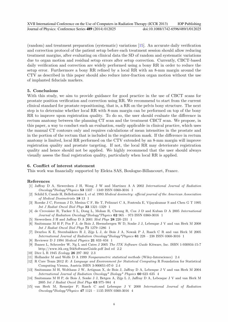
3.2. Impact of rectal distension on local RR qualityWe investigated in which cases local RR with 8-mm margin failed. Figure 2 illustrates theimpact of the variation of rectal filling between the images to be registered on the quality oflocal RR with 8-mm margin. We observed with our database that if the F factor as defined inEq. 1 is lower than or equal to 112.6, registrations were all successful. All failed registrationsappeared for values of F higher than 147.6. The range of F values obtained was from 0.2 to406.6.
0 20 40 60 80 100
# CBCT sorted in order of increasing F number
0
1
2
3
4
5
6
7
8
Cum
ulat
ive
failu
re c
ount
Failu
re c
ount
Local RR with an 8-mm margin
0
1
2
3
4
5
6
7
8
Fig. 2: Plot of (blue) the cumulative number of failed registrations and (red) its derivative, i.e. thefailure frequency, obtained with local RR with 8-mm margin, against the CBCT scans arranged in orderof increasing F number.
4. DiscussionLocal RR with 8-mm margin was able to improve upon the Dice results obtained with bony RRas long as the rectal distension, that is the difference in the rectum anatomy between the planningCT scan and the treatment CBCT scan, is limited. For low values of F , results exceeded globalRR or bony RR results, the latter being the current clinical standard for prostate repositioning.Smitsmans et al. found that 5-mm was the optimum margin for prostate CT/CT local RR[13]. In our study, we showed that indeed this method yielded the highest Dice median but itcounted 11 failed registrations over 106, against only 7 failed registrations for local RR with8-mm margin. Local RR with 8-mm has also a smaller SD than and a Dice median very close tothose obtained with the 5-mm margin. That is why we preconize to use an 8-mm margin. In alater study, Smitsmans et al. also reported that local CT/CBCT RRs with 5-mm margin mainlyfailed because of streaks in the CBCT scans caused by moving gas pockets in the rectum duringCBCT acquisition [14]. They implemented some improvements to their local RR algorithm,including the removal of the gray values of the pubic bone from the ROI. In our study, we couldnot find any correlation between the F factor defined in Eq. 1 and registration failures forlocal RR with margins lower than or including 5 mm. This is consistent with the fact that thelower the margin, the smaller the rectum portion included in the mask, and so the smaller theinfluence of the rectum portion on the registration process. Our assumption is that for marginslower than or including 5 mm, registration is not influenced by rectum distension.
Geometrical deviations occuring during treatment lead to target underdosage. To account forthese geometrical deviations such as target volume delineation, organ motion and setup errors, amargin between the CTV and the PTV must be added. Van Herk et al. derived a margin recipefor prostate cancer radiotherapy, separating geometrical deviations into treatment execution
XVII International Conference on the Use of Computers in Radiation Therapy (ICCR 2013) IOP PublishingJournal of Physics: Conference Series 489 (2014) 012025 doi:10.1088/1742-6596/489/1/012025
5
(random) and treatment preparation (systematic) variations [15]. An accurate daily verificationand correction protocol of the patient setup before each treatment session should allow reducingtreatment margins, after evaluating on clinical data the SD of random and systematic variationsdue to organ motion and residual setup errors after setup correction. Currently, CBCT-baseddaily verification and correction are widely performed using a bony RR in order to reduce thesetup error. Furthermore a bony RR refined by a local RR with an 8-mm margin around theCTV as described in this paper should also reduce inter-fraction organ motion without the useof implanted fiducials markers.
5. ConclusionsWith this study, we aim to provide guidance for good practice in the use of CBCT scans forprostate position verification and correction using RR. We recommend to start from the currentclinical standard for prostate repositioning, that is, a RR on the pelvis bony structure. The nextstep is to determine whether local RR with 8-mm margin can be performed on top of the bonyRR to improve upon registration quality. To do so, the user should evaluate the difference inrectum anatomy between the planning CT scan and the treatment CBCT scan. We propose, inthis paper, a way to conduct such an evaluation, easily applicable in clinical practice, which usesthe manual CT contours only and requires calculations of mean intensities in the prostate andin the portion of the rectum that is included in the registration mask. If the difference in rectumanatomy is limited, local RR performed on the CTV extended by an 8-mm margin will improveregistration quality and prostate targeting. If not, the local RR may deteriorate registrationquality and hence should not be applied. We highly recommend that the user should alwaysvisually assess the final registration quality, particularly when local RR is applied.
6. Conflict of interest statementThis work was financially supported by Elekta SAS, Boulogne-Billancourt, France.
References[1] Jaffray D A, Siewerdsen J H, Wong J W and Martinez A A 2002 International Journal of Radiation
Oncology*Biology*Physics 53 1337 – 1349 ISSN 0360-3016 1[2] Schild S, Casale H, Bellefontaine L et al. 1993 Medical dosimetry: official journal of the American Association
of Medical Dosimetrists 18 13 1[3] Roeske J C, Forman J D, Mesina C F, He T, Pelizzari C A, Fontenla E, Vijayakumar S and Chen G T 1995
Int J Radiat Oncol Biol Phys 33 1321–1329 1[4] de Crevoisier R, Tucker S L, Dong L, Mohan R, Cheung R, Cox J D and Kuban D A 2005 International
Journal of Radiation Oncology*Biology*Physics 62 965 – 973 ISSN 0360-3016 1[5] Siewerdsen J H and Jaffray D A 2001 Med Phys 28 220–231 1[6] Smitsmans M H P, Pos F J, de Bois J, Heemsbergen W D, Sonke J J, Lebesque J V and van Herk M 2008
Int J Radiat Oncol Biol Phys 71 1279–1286 1[7] Deurloo K E, Steenbakkers R J, Zijp L J, de Bois J A, Nowak P J, Rasch C R and van Herk M 2005
International Journal of Radiation Oncology*Biology*Physics 61 228 – 238 ISSN 0360-3016 1[8] Reviewer D J 1994 Medical Physics 21 833–834 1[9] Ibanez L, Schroeder W, Ng L and Cates J 2005 The ITK Software Guide Kitware, Inc. ISBN 1-930934-15-7
http://www.itk.org/ItkSoftwareGuide.pdf 2nd ed 2.2[10] Dice L R 1945 Ecology 26 297–302 2.3[11] Hollander M and Wolfe D A 1999 Nonparametric statistical methods (Wiley-Interscience) 2.4[12] R Core Team 2012 R: A Language and Environment for Statistical Computing R Foundation for Statistical
Computing Vienna, Austria ISBN 3-900051-07-0 2.4[13] Smitsmans M H, Wolthaus J W, Artignan X, de Bois J, Jaffray D A, Lebesque J V and van Herk M 2004
International Journal of Radiation Oncology* Biology* Physics 60 623–635 4[14] Smitsmans M H P, de Bois J, Sonke J J, Betgen A, Zijp L J, Jaffray D A, Lebesque J V and van Herk M
2005 Int J Radiat Oncol Biol Phys 63 975–984 4[15] van Herk M, Remeijer P, Rasch C and Lebesque J V 2000 International Journal of Radiation
Oncology*Biology*Physics 47 1121 – 1135 ISSN 0360-3016 4
XVII International Conference on the Use of Computers in Radiation Therapy (ICCR 2013) IOP PublishingJournal of Physics: Conference Series 489 (2014) 012025 doi:10.1088/1742-6596/489/1/012025
6
Copyright © 2022 FDOKUMEN