Xenopus LAP2 β protein knockdown affects location of lamin B and nucleoporins and has effect on...
14
ORIGINAL ARTICLE Xenopus LAP2β protein knockdown affects location of lamin B and nucleoporins and has effect on assembly of cell nucleus and cell viability Magda Dubińska-Magiera 1,2 & Magdalena Chmielewska 1,3 & Katarzyna Kozioł 1 & Magdalena Machowska 1 & Christopher J. Hutchison 4 & Martin W. Goldberg 4 & Ryszard Rzepecki 1 Received: 19 June 2015 /Accepted: 15 July 2015 # The Author(s) 2015. This article is published with open access at Springerlink.com Abstract Xenopus LAP2β protein is the single isoform expressed in XTC cells. The protein localizes on heterochro- matin clusters both at the nuclear envelope and inside a cell nucleus. The majority of XLAP2β fraction neither colocalizes with TPX2 protein during interphase nor can be immunoprecipitated with XLAP2β antibody. Knockdown of the XLAP2β protein expression in XTC cells by synthetic siRNA and plasmid encoded siRNA resulted in nuclear ab- normalities including changes in shape of nuclei, abnormal chromatin structure, loss of nuclear envelope, mislocalization of integral membrane proteins of INM such as lamin B2, mislocalization of nucleoporins, and cell death. Based on timing of cell death, we suggest mechanism associated with nucleus reassembly or with entry into mitosis. This confirms that Xenopus LAP2 protein is essential for the maintenance of cell nucleus integrity and the process of its reassembly after mitosis. Keywords LAP2 . Knockdown . Cell nucleus . Nucleoporin . Lamin B . TPX2 . Xenopus laevis Introduction Lamina-associated polypeptide 2 (LAP2) proteins are alterna- tively spliced proteins belonging to the family of LEM do- main proteins (for review, see (Schirmer and Foisner 2007, Dorner et al. 2007, Wagner and Krohne 2007). The LEM domain family in humans was originally composed of three proteins (LAP2, emerin, and MAN1). Since then, four addi- tional LEM domain proteins have been discovered in mam- mals. These are NET-25 (Lem2), Lem3, Lem4, and Lem5 (Lee and Wilson 2004, Berk et al. 2013). Mutations in genes coding for proteins of the nuclear lamina (also members of a LEM-domain family) lead to laminopathies, a group of rare genetic disorders (for review, see Zaremba-Czogalla et al. 2011a, Dubinska-Magiera et al. 2013). LAP2 proteins are expressed in metazoans, excluding Caenorhabditis elegans but including Drosophila melanogaster , Danio rerio, Xenopus laevis, Mus musculus, and Homo sapiens. LAP2 proteins are alternatively spliced products of a single gene, resulting in integral membrane or nucleoplasmic (or rarely cytoplasmic) proteins (Harris et al. 1994). Their unique feature is the similar conservative struc- ture, especially among vertebrate orthologues. LAP2 proteins comprise N-terminal Bcommon^ domain containing LEM do- main and LEM-like domain, both interacting with barrier to autointegration factor (BAF) (Furukawa 1999, Shumaker Handling Editor: Reimer Stick Magda Dubińska-Magiera, Magdalena Chmielewska, and Katarzyna Kozioł are first joined authors. Electronic supplementary material The online version of this article (doi:10.1007/s00709-015-0861-y) contains supplementary material, which is available to authorized users. * Ryszard Rzepecki [email protected] 1 Laboratory of Nuclear Proteins, Faculty of Biotechnology, University of Wrocław, Joliot- Curie 14a, 50-383 Wrocław, Poland 2 Department of Animal Developmental Biology, Institute of Experimental Biology, University of Wroclaw, Sienkiewicza 21, 50-335 Wroclaw, Poland 3 Department of Evolutionary Biology and Vertebrate Conservation, University of Wroclaw, Sienkiewicza 21, 50-335 Wroclaw, Poland 4 Integrative Cell Biology Laboratory, School of Biological and Biomedical Sciences, The University of Durham, South Road, Durham DH1 3LE, UK Protoplasma DOI 10.1007/s00709-015-0861-y
Transcript of Xenopus LAP2 β protein knockdown affects location of lamin B and nucleoporins and has effect on...
ORIGINAL ARTICLE
Xenopus LAP2β protein knockdown affects location of lamin Band nucleoporins and has effect on assembly of cell nucleusand cell viability
Magda Dubińska-Magiera1,2 & Magdalena Chmielewska1,3 & Katarzyna Kozioł1 &
Magdalena Machowska1 & Christopher J. Hutchison4& Martin W. Goldberg4 &
Ryszard Rzepecki1
Received: 19 June 2015 /Accepted: 15 July 2015# The Author(s) 2015. This article is published with open access at Springerlink.com
Abstract Xenopus LAP2β protein is the single isoformexpressed in XTC cells. The protein localizes on heterochro-matin clusters both at the nuclear envelope and inside a cellnucleus. The majority of XLAP2β fraction neither colocalizeswith TPX2 protein during interphase nor can beimmunoprecipitated with XLAP2β antibody. Knockdown ofthe XLAP2β protein expression in XTC cells by syntheticsiRNA and plasmid encoded siRNA resulted in nuclear ab-normalities including changes in shape of nuclei, abnormalchromatin structure, loss of nuclear envelope, mislocalizationof integral membrane proteins of INM such as lamin B2,mislocalization of nucleoporins, and cell death. Based ontiming of cell death, we suggest mechanism associated withnucleus reassembly or with entry into mitosis. This confirms
that Xenopus LAP2 protein is essential for the maintenance ofcell nucleus integrity and the process of its reassembly aftermitosis.
Keywords LAP2 . Knockdown . Cell nucleus .
Nucleoporin . Lamin B . TPX2 . Xenopus laevis
Introduction
Lamina-associated polypeptide 2 (LAP2) proteins are alterna-tively spliced proteins belonging to the family of LEM do-main proteins (for review, see (Schirmer and Foisner 2007,Dorner et al. 2007, Wagner and Krohne 2007). The LEMdomain family in humans was originally composed of threeproteins (LAP2, emerin, and MAN1). Since then, four addi-tional LEM domain proteins have been discovered in mam-mals. These are NET-25 (Lem2), Lem3, Lem4, and Lem5(Lee and Wilson 2004, Berk et al. 2013). Mutations in genescoding for proteins of the nuclear lamina (also members of aLEM-domain family) lead to laminopathies, a group of raregenetic disorders (for review, see Zaremba-Czogalla et al.2011a, Dubinska-Magiera et al. 2013).
LAP2 proteins are expressed in metazoans, excludingCaenorhabditis elegans but including Drosophilamelanogaster, Danio rerio, Xenopus laevis, Mus musculus,and Homo sapiens. LAP2 proteins are alternatively splicedproducts of a single gene, resulting in integral membrane ornucleoplasmic (or rarely cytoplasmic) proteins (Harris et al.1994). Their unique feature is the similar conservative struc-ture, especially among vertebrate orthologues. LAP2 proteinscomprise N-terminal Bcommon^ domain containing LEM do-main and LEM-like domain, both interacting with barrier toautointegration factor (BAF) (Furukawa 1999, Shumaker
Handling Editor: Reimer Stick
Magda Dubińska-Magiera, Magdalena Chmielewska, and KatarzynaKozioł are first joined authors.
Electronic supplementary material The online version of this article(doi:10.1007/s00709-015-0861-y) contains supplementary material,which is available to authorized users.
* Ryszard [email protected]
1 Laboratory of Nuclear Proteins, Faculty of Biotechnology,University of Wrocław, Joliot- Curie 14a, 50-383 Wrocław, Poland
2 Department of Animal Developmental Biology, Institute ofExperimental Biology, University of Wroclaw, Sienkiewicza 21,50-335 Wroclaw, Poland
3 Department of Evolutionary Biology and Vertebrate Conservation,University of Wroclaw, Sienkiewicza 21, 50-335 Wroclaw, Poland
4 Integrative Cell Biology Laboratory, School of Biological andBiomedical Sciences, The University of Durham, South Road,Durham DH1 3LE, UK
ProtoplasmaDOI 10.1007/s00709-015-0861-y
et al. 2001) or DNA (chromatin) (Cai et al. 2001). The rest ofthe particular LAP2 protein structure and function depends onwhat exons were incorporated into mature mRNA during al-ternative splicing of the primary transcript. The Bvariable^domain typically contains lamin binding domain or specifical-ly lamin B-binding domain and domains responsible for bind-ing to germ-cell-less (GCL) (Nili et al. 2001), HA95 protein(Martins et al. 2003), and histone deacetylase 3 (HDAC3)protein (Somech et al. 2005). Alternative splicing also gener-ates protein isoforms with or without a transmembrane do-main. This results in an appearance of integral membrane orBsoluble^ protein isoform. In mammals, six LAP2 proteinisoforms have been discovered (α, β, γ, δ, ε, and ζ). LAP2αand LAP2 ζ proteins lack transmembrane domains and aresoluble nucleoplasmic and cytoplasmic proteins, respectively(Shaklai et al. 2008). The most thoroughly studied isoformwas mammalian LAP2α protein due to its ability to modulatesignaling pathway associated with Rb protein (LAP2α, laminA, pRb complex) and regulate gene transcription associatedinter alia with cell cycle progression into S-phase (Gant et al.1999, Markiewicz et al. 2002, Yang et al. 1997, Pekovic et al.2007). Mammalian LAP2β isoforms are also involved in gen-eral gene regulation and as transcriptional repressors (Somechet al. 2005). They play a crucial role in attachment of chroma-tin to the nuclear envelope (NE) and nuclear lamina (NL)during interphase and nuclear reassembly after mitosis.
In X. laevis, there have been five cDNA sequences identi-fied that are presumably translated into three different LAP2proteins discovered so far,ω,β, and γ (Gant et al. 1999, Langet al. 1999, Lang and Krohne 2003, Chmielewska et al. 2011).The Xenopus LAP2β (66 kDa somatic polypeptide) is theonly one found in adult animals, whereas two other isoforms,ω and γ (86 and 40 kDa), are present in oocytes and eggs andare downregulated during embryogenesis (Chmielewska et al.2011). The N-terminal common fragment of XLAP2, whenadded to the in vitro nuclear assembly reaction, inhibits chro-matin decondensation and nuclear growth similarly to the hu-man LAP2β N-terminal fragment (Gant et al. 1999). More-over, XLAP2 β (66 kDa polypeptide) coprecipitates withlamin B2 and A from adult Xenopus tissues and A6 cells(Lang and Krohne 2003). The N-terminal domain of XLAP2(aa 1-165), like other vertebrate LAPs, interacts with BAF andBAF-DNA complexes. The in vitro interaction of XLAP2protein isoforms from X. laevis egg extracts—XLAP2ω andXLAP2γ with a spindle assembly factor—TPX2 (targetingprotein for Xklp2) was confirmed. XLAP2-TPX2 complexis therefore thought to be required for proper assembly ofpostmitotic nuclei in X. laevis in vitro nuclear assembly sys-tem (O'Brien and Wiese 2006).
Recently, we confirmed the presence of at least threeXLAP2 isoforms, ω, β, and γ, that were developmentallyregulated (Chmielewska et al. 2011). XLAP2 proteins colo-calize with lamin B2 and B3 during development and lamin
B2 in adult tissues. We also demonstrated that XenopusLAP2β localizes both at the NE and inside the nucleus inclusters of heterochromatin. The intranuclear clusters ofXLAP2β on heterochromatin were found to be partly inde-pendent of NE invaginations. We also demonstrated that inXTC cells, LAP2β is the sole LAP2 isoform expressed.
In this study, we examined the effect of knockdown ofLAP2β protein synthesis in X. laevis XTC cells and its effecton cell viability and cell nucleus structure and function.
Materials and methods
Plasmids, cDNAs and siRNAs, and tissue culture
Sequences of siRNAs for XLAP2 knockdown were designedusing the online Invitrogen tool [https://rnaidesigner.invitrogen.com/rnaiexpress/?CID=TN-Tools-BlockiT] on thetemplate of XLAP2 clone2 cDNA sequence, AF048815 (Gantet al. 1999): 15 XLAP2 sense strain: 5′- GCAAGACCCGUCGGUACUUACUAAA -3′, 98 XLAP2 sensestrain:, 5′- GGAAAGAUGUGUAUGUGCAACUCUA-3′,a nd 237 XLAP2 sen s e s t r a i n , 5 ′ - GAAGACCGACAAACCUAGAGCAGAA-3′. Scrambled controlsiRNA sequence was: control 15 (c15) sense, 3 ′-GCACCAUGCGGCCAUAUUUCAGAAA-5′.
Xenopus laevisXTC cells (Pudney et al. 1973) were grownin 54 % L-15 Leibovitz medium containing 10 % FBS, 2 mML-glutamine, 10 I.U./ml penicillin, 10 μg/ml streptomycin,and 0.025 μg/ml amphotericin B at 22–26 °C in normal airconditions as described previously (Chmielewska et al. 2011).
Cells were transfected with 100 nM specific or control(scrambled) siRNAs using Oligofectamine reagent (LifeTechnologies). For plasmid-based siRNA knockdown, plas-mid pFIV-H1/U6-copGFP (SBI System Bioscience, USA)with inserted sequences of control (C15) and active (15) se-quences for siRNA was used. Transfection was withMetafectene Pro (Biontex, Germany) with 1 μg of plasmidDNA per 6 μl of transfection reagent. Cells were grown on24-well plates at an initial density of 6×10⁴ per well. Cellswere collected after 24, 48, 72, or 96 h after transfection,depending on the experiment.
Microscope procedures
For imaging, confocal microscope LSM 510METAwith FCSsystem was used. For imaging in non-confocal mode, fluores-cence microscope (Olympus IX70) was used. Any brightnessand contrast adjustments were performed in AdobePhotoshop, Zen 2007 (Zeiss) or ImageJ (Schneider et al.2012).
M. Dubińska-Magiera et al.
Statistical analysis
For statistical analyses of cellular phenotypes, the followingprocedure was used: Samples of XTC cells grown on cover-slips were fixed for immunofluorescence microscopy andstained for XLAP2. Ten representative fields from each sam-ple (three independent experiments) were imaged under ×10objective. In the digital images, the total cell number wascounted. The statistical analysis was performed usingStatistica software (StatSoft, Poland). Groups of data werecompared utilizing the Student’s t test. Statistical significancewas assumed at values of P<0.05. Cell phenotypes were ex-amined in the digital images distinguished on the basis ofstructural criteria of shape and size of cells and nuclei, orga-nization of microtubular network, cell adherence to the sur-face, degree of cell shrinking, and apoptotic phenotype (deter-mined by abnormal chromatin condensation, the occurrenceof vesicles within the nucleus, and lack of NE staining).
For calculations of number of cells with knockdownXLAP2 protein XTC cells, 48 h after transfection withplasmid-based siRNA constructs Bc15^ or B15^, werefixed with 4 % paraformaldehyde (PFA), stained withanti-XLAP2 antibody and DAPI, and visualized with con-focal microscope. Fifty cells demonstrated GFP expres-sion were counted for each specimen. When counting,the XLAP2 expression level and shape of nuclei wereestimated also. Counting was performed for three seriesof transfections and averaged. Experiments on knock-down of XLAP2β protein (and controls) were always per-formed simultaneously, exactly at the same session on aconfocal microscope. We used the same aliquots of anti-bodies and the same settings of the microscope. Whendifferences in optical density of the staining were ques-tionable, we performed optical density measurements ofNE staining. As a cell with XLAP2 knockdown, wecounted cells with an optical density lower than 50 % ofa density of control from the same slide. Typically, valuesfor normal NE were about 150 or higher. Random viewsfrom slides were selected for observations. For GFP fluo-rescence (reporter protein for transfection), the thresholdwas estimated at the background level increased by100 %. Typically, background level was about 20–25 de-pending on the slide, which means that the threshold wasset at 40 to 50 depending on the measured background ofcurrently analyzed slides. Only cells from the same slideswere compared. All calculations were made on cells fixedwith PFA only since other fixation methods did not pre-serve the GFP in order to allow reproducible measure-ments (see Fig. 6).
For calculation of cells number, expressing a detectablelevel of TPX2 protein, exponentially growing XTC cells werefixed with 4 % PFA, stained with anti-TPX2 antibody andDAPI, and visualized with a confocal microscope. 200 cells
were counted. Percentage of cells with the detectable TPX2signal was calculated, and phases of the cell cycle for thosecells were estimated using FACS analyses.
Colocalization studies
For qualitative colocalization analysis (relative fluorescenceintensities measurements), images acquisition were performedon a Zeiss LSM 510 Meta confocal microscope. Distributionof fluorescence intensities along the cross-section of the typi-cal cell was done using BProfile^ function of the Zeiss ZEN2009 software (Carl Zeiss MicroImaging). Resulting graphshows signal intensities in accordance with chosen section(red arrow). To set threshold, point marker (cyan dot or line)was chosen. To show differences in XLAP2 and TPX2 fluo-rescence intensities in the nuclear envelope, the second pointmarker (violet dot or line) was set. The calculations for thesechosen points are presented in tables.
Quantitative colocalization analysis
For quantification of colocalization, image acquisition wasperformed on a Zeiss LSM 510 Meta confocal microscope.Colocalization of proteins’ signals was determined using ZeissZEN 2009 software (Carl Zeiss MicroImaging). The thresholdwas set for each pair of channels by selecting region of interest(ROI) outside the cells. Subsequently, the ROIs at theintranuclear area, at the nuclear envelope, or in the cytoplasmwere chosen, and colocalizations were calculated. For eachcase, five ROIs, coming from separate cells, were analyzed.Finally, averaged calculations, graphs, and statistical analysiswere done based on percentage of signals from lamin B2channel that are colocalized with signals from XLAP2 andTPX2 in the analyzed ROI and is insensitive to differencesin signal intensities.
Antibodies, immunofluorescence, and Western blotting
For immunofluorescence (IF), XTC cells were grown on cov-erslips, fixed with 4 % PFA in phosphate buffered saline(PBS) for 15 min, permeabilized with 0.5 % Triton X-100 inPBS (v/v) for 5 min, and blocked with 1 % fetal bovine serum(FBS) in PBS (for 30 min at room temperature). Alternatively,to use selected antibodies, cells were fixed with ice-cold meth-anol for 10 min in −20 °C without separated permeabilizationstep. All wash steps were done with PBS. Primary antibodieswere used for overnight incubation at 4 °C and secondaryantibodies for 60 min at room temperature. DNAwas stainedwith DAPI (4,6-diamidino-2-phenylindole, 0.2 μg/ml inmounting medium with Mowiol, and DABCO). For imaging,confocal microscope LSM 510 METAwith FCS system wasused. Any brightness and contrast adjustments were
Xenopus LAP2β protein knockdown
performed in Adobe Photoshop, Zen 2007 (Zeiss) or ImageJ(Schneider et al. 2012).
The specific serum and antibodies for an N-terminal frag-ment of XLAP2 protein were produced as described previous-ly (Rzepecki et al. 1998).
The following antibodies were used: rabbit affinity purifiedanti-N-TPX2 antibody (1:100 IF) was a kind gift from Prof. Y.Zheng (Tsai et al. 2006), antibodies anti-XBAF protein (1:50IF) were from Prof. P.A. Fisher and Prof. K. Furukawa(Dechat et al. 2004, Furukawa et al. 2003), and affinity puri-fied IgGs: anti-XLAP2 (1:100 WB, 1:60 IF) (Salpingidouet al. 2008, Chmielewska et al. 2011). Mouse monoclonalantibodies: anti-X. laevis lamin B2 ab (1:25 IF, Santa CruzBiotechnology sc-56147), Ab 414 against nucleoporins withF/G repeats (1:100 IF, Covance MMS-120P), Ac-40 actin Ab(1:800 WB, Sigma), rat monoclonals anti-alpha-tubulin YL1/2 (1:60, Serotec), anti-β-tubulin (1:150 IF, Sigma T4026), andanti-γ-tubulin (1:100 IF, Sigma T5326). For F-actin identifi-cation, Alexa Fluor® 546 phalloidin was used (at a concentra-tion of 2 U/ml; Invitrogen). Cellular lipid membranes werestained with DHCC (3,3″-dihexyloxacarbocyanine iodide ata concentration of 7.5 μg/ml; Invitrogen). Secondary antibod-ies for immunoblotting and fluorescence were from JacksonsImmunoResearch.
Proteins were separated on 10 or 12% SDS-PAGE gels andelectro-transferred onto nitrocellulose filters. For XLAP2
silencing analyses, optical density measurements of the pro-tein bands in immunoblots were performed with the BIO-PROFIL Bio-1D Windows Application V99.01. Protein con-tent in protein bands was normalized according to the actincontent in each lane. Western blotting analysis of the XLAP2protein silencing in XTC cells was monitored by staining withantibodies against XLAP2 and actin stainingwithmAb to betaactin (AC-40, SIGMA) was used as loading control. Opticaldensity of XLAP2 and actin bands was measured with theBIO-PROFIL Bio-1D Windows Application V99.01, andXLAP2 content was normalized versus actin for each lane.
Immunoprecipitation and mass spectroscopy
Immunoprecipitation (IP) and mass spectroscopy (MS) wereperformed essentially as described previously (Rzepecki et al.1998, Chmielewska et al. 2011, Zaremba-Czogalla et al.2011b) with minor modifications. All co-IP experiments wereperformed under native conditions under different ionicstrength in order to extract fractions of XLAP2β protein withdifferent solubility. Co-IP procedure number 1 was performedwith extraction, binding and washing the beads with buffercontaining 0.3 M NaCl. The procedure number 2 was per-formed with 0.6 M NaCl and procedure number 3 with0.6 M NaCl in extraction buffer and 0.3 M NaCl during therest of the procedure. For each co-IP procedure, we used 9×
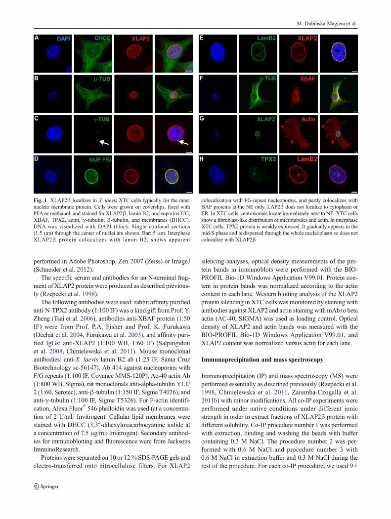
Fig. 1 XLAP2β localizes in X. laevis XTC cells typically for the innernuclear membrane protein. Cells were grown on coverslips, fixed withPFA or methanol, and stained for XLAP2β, lamin B2, nucleoporins F/G,XBAF, TPX2, actin, γ-tubulin, β-tubulin, and membranes (DHCC).DNA was visualized with DAPI (blue). Single confocal sections(1.5 μm) through the center of nuclei are shown. Bar: 5 μm. InterphaseXLAP2β protein colocalizes with lamin B2, shows apparent
colocalization with FG-repeat nucleoporins, and partly colocalizes withBAF proteins at the NE only. LAP2β does not localize to cytoplasm orER. In XTC cells, centrosomes locate immediately next to NE. XTC cellsshow a fibroblast-like distribution ofmicrotubules and actin. In interphaseXTC cells, TPX2 protein is weakly expressed. It gradually appears in themid-S phase and is dispersed through the whole nucleoplasm so does notcolocalize with XLAP2β
M. Dubińska-Magiera et al.
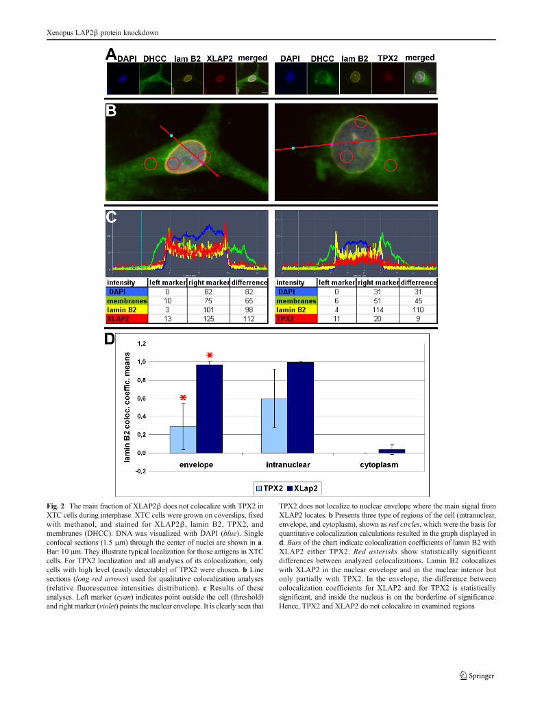
Fig. 2 The main fraction of XLAP2β does not colocalize with TPX2 inXTC cells during interphase. XTC cells were grown on coverslips, fixedwith methanol, and stained for XLAP2β, lamin B2, TPX2, andmembranes (DHCC). DNA was visualized with DAPI (blue). Singleconfocal sections (1.5 μm) through the center of nuclei are shown in a.Bar: 10 μm. They illustrate typical localization for those antigens in XTCcells. For TPX2 localization and all analyses of its colocalization, onlycells with high level (easily detectable) of TPX2 were chosen. b Linesections (long red arrows) used for qualitative colocalization analyses(relative fluorescence intensities distribution). c Results of theseanalyses. Left marker (cyan) indicates point outside the cell (threshold)and right marker (violet) points the nuclear envelope. It is clearly seen that
TPX2 does not localize to nuclear envelope where the main signal fromXLAP2 locates. b Presents three type of regions of the cell (intranuclear,envelope, and cytoplasm), shown as red circles, which were the basis forquantitative colocalization calculations resulted in the graph displayed ind. Bars of the chart indicate colocalization coefficients of lamin B2 withXLAP2 either TPX2. Red asterisks show statistically significantdifferences between analyzed colocalizations. Lamin B2 colocalizeswith XLAP2 in the nuclear envelope and in the nuclear interior butonly partially with TPX2. In the envelope, the difference betweencolocalization coefficients for XLAP2 and for TPX2 is statisticallysignificant, and inside the nucleus is on the borderline of significance.Hence, TPX2 and XLAP2 do not colocalize in examined regions
Xenopus LAP2β protein knockdown
106 cells, and for Western blot analyses, equivalents of 2×10⁵
cells/lane were loaded. Only proteins with Bidentificationscore^ of 100 % in at least one IP procedure were shown.
Mass spectrometry
For identification of coimmunoprecipitated proteins, tandemmass spectrometry was conducted essentially as describedpreviously (Chmielewska et al. 2011, Zaremba-Czogallaet al. 2011b).
Results
Localization of XLAP2β in cultured XTC cellsduring interphase
In order to get an insight into the cell cycle-dependent local-ization of XLAP2β in comparison to other important cellularproteins, we used XTC cells that express this single isoformand have a fibroblast-like phenotype (Chmielewska et al.2011).
Interphase XLAP2β protein shows typical location for in-tegral protein of inner nuclear membrane (INM), colocalizeswith lamin B2, shows apparent colocalization with FG-repeatnucleoporins, and does not localize to cytoplasm or endoplas-mic reticulum (ER) (Fig. 1). Centrosomes (MTOCs) locateimmediately next to NE in XTC cells. BAF protein is distrib-uted at the NE and surrounding cytoplasm. LAP2β partlycolocalizes only with BAF protein at the NE. Please note thatXTC cells show a fibroblast-like distribution of microtubulesand actin (Fig. 1).
We also analyzed the distribution of TPX2 protein in inter-phase XTC cells (Fig. 1) due to the previous report thatXLAP2 ω and γ proteins interact with TPX2 protein fromXenopus egg extracts (O'Brien and Wiese 2006). We foundthat most of XTC cells demonstrated weak or no staining withanti-TPX2 antibodies (Additional file 1: Figure S1B). TPX2protein is distributed through the nuclear interior. Detailedanalyses revealed that only about 40 % of interphase cellsdemonstrate clearly detectable by immunofluorescence stain-ing, higher level of TPX2 protein and that the location of theprotein is intranuclear (Additional file 1: Figure S1). The anal-yses of the cell cycle in XTC cells using FACS method indi-cated that this 40 % of cells correlates roughly with a percent-age of cells in S-phase together with G2/M-phase (not shown).This feature of frog TPX2 reflects the behavior of mammalianTPX2 protein (Stewart and Fang 2005). Since our XLAP2βantibody and TPX2 antibody were raised in rabbits, we couldnot perform direct colocalization studies between these twoproteins. That is why we used indirect approach and studiedXLAP2β and TPX2 protein colocalization with lamin B2 andanalyzed the distribution of these proteins through the nuclear
compartment. Figure 2a demonstrates typical example forstaining of XTC cells for DNA, membranes, XLAP2, TPX2,and lamin B2. Figure 2b demonstrates merged images withline sections taken for fluorescence intensities analyses shownin Fig. 2c. Figure 2b also indicates a marked different type ofregions used for calculation of colocalization coefficientsillustrated in Fig. 2d. The demonstrated data indicatethat there is no significant fraction of XLAP2β andTPX2 protein colocalizing with each other, particularlyat the nuclear envelope location where, additionally, itlocalizes only residual fraction of TPX2. This suggeststhat major fractions of both proteins may not interactwith each other.
In order to detect potential interactions between XLAP2and TPX2 proteins, we used immunoprecipitation for XLAP2followed by mass spectroscopy for identification of co-immunoprecipitated proteins from unsynchronized XTC cellextracts. The results of such experiments performed underthree different ionic conditions are demonstrated in Additionalfile 2: Figure S2. TPX2 protein was not detected in any of thesamples with XLAP2β co-immunoprecipitated proteins. Al-so, our previous studies on XLAP2β protein using IP andmass spectrometry did not show TPX2 as an interacting part-ner (Chmielewska et al. 2011) (22). Thus, our data indicatethat most of Xenopus TPX2 do not colocalize with the major-ity of LAP2β in XTC cells during interphase. This means that
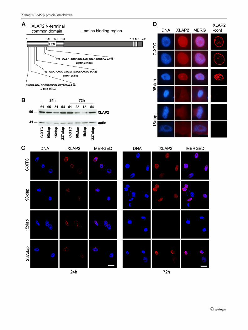
�Fig. 3 Analysis of the XLAP2 protein knockdown in tissue culturedX. laevis XTC cells. XTC cells were plated at 7.5×10⁴ cells/well,transfected with 100 nM siRNA sequences, and grown for 24 and 72 h.C-XTC, untreated cells; 15, 98, 237xlap, cells treated with differentsiRNAs. a Schematic diagram of the Xenopus LAP2β protein. Thepositions of amino acid residues are marked above the diagram.Locations of predicted domains are marked by different patterns:striped box common domain of all potential isoforms, white box LEMdomain, hatched box the transmembrane region. cDNA sequences givenbelow the diagram were used to design 25 nt ds siRNAs for XLAP2protein expression silencing. Numbers denote a position of nucleotides.b Western blotting analysis of the XLAP2 protein silencing in tissuecultured X. laevis XTC cells after 24 and 72 h post- transfection. Equalamounts of the protein were loaded. Numbers denoted above each lanerepresent the relative amounts of XLAP2 protein. Molecular masses ofthe proteins (kDa) are marked on the left side of the picture. XLAP2knockdown was most efficient with 98xlap and 15xlap siRNAs after3 days post-transfection. c, d Effect of XLAP2 protein knockdown onthe morphology of XTC cells was also analyzed by confocal andimmunofluorescence microscopy. XTC cells were stained for XLAP2(red), DNA was visualized with DAPI (blue). c General view of thecontrol, non-treated, and siRNA transfected XTC cells. Note thecomplete loss of XLAP2 signal after 72 h post-transfection with 15xlapsiRNA. dHigher magnification of the control and silenced nuclei imagedin non-confocal (3 left columns) or confocal mode (right column)visualize that the degrading XLAP2 protein is no longer localized in theNE. DNA is amorphous (98xlap). Note the apoptotic nucleus withlobulations (15xlap lower photograph). Images were combined andprocessed in Adobe Photoshop. Single confocal sections (1.5 μm)through the center of nuclei are shown. Bars, 20 μm (c), 10 μm (d)
M. Dubińska-Magiera et al.
Xenopus LAP2β protein knockdown
XLAP2β protein (unlike XLAP2ω and γ) is not responsiblefor TPX2 protein retention in cell nucleus during interphase.
siRNA mediated knockdown of XLAP2β resultsin nuclear and cellular abnormalities and cell death
In order to unveil the cellular function of LAP2β protein inX. laevis, we used siRNA to knockdown the expression of a
single XLAP2β isoform present in XTC cells. Figure 3 showsa schematic diagram of the protein, sequences of the siRNAfragments, their location in N-terminal Bcommon domain,^and the results of the knockdown experiment. The most effi-cient knockdown effect, as assayed by Western blot, showed15xlap siRNA (80 %) and the lesser 98xlap (64 %), while237xlap siRNA showed no effect (Fig. 3b). Similar data wereobtained by IF and confocal analyses, where it was possible to
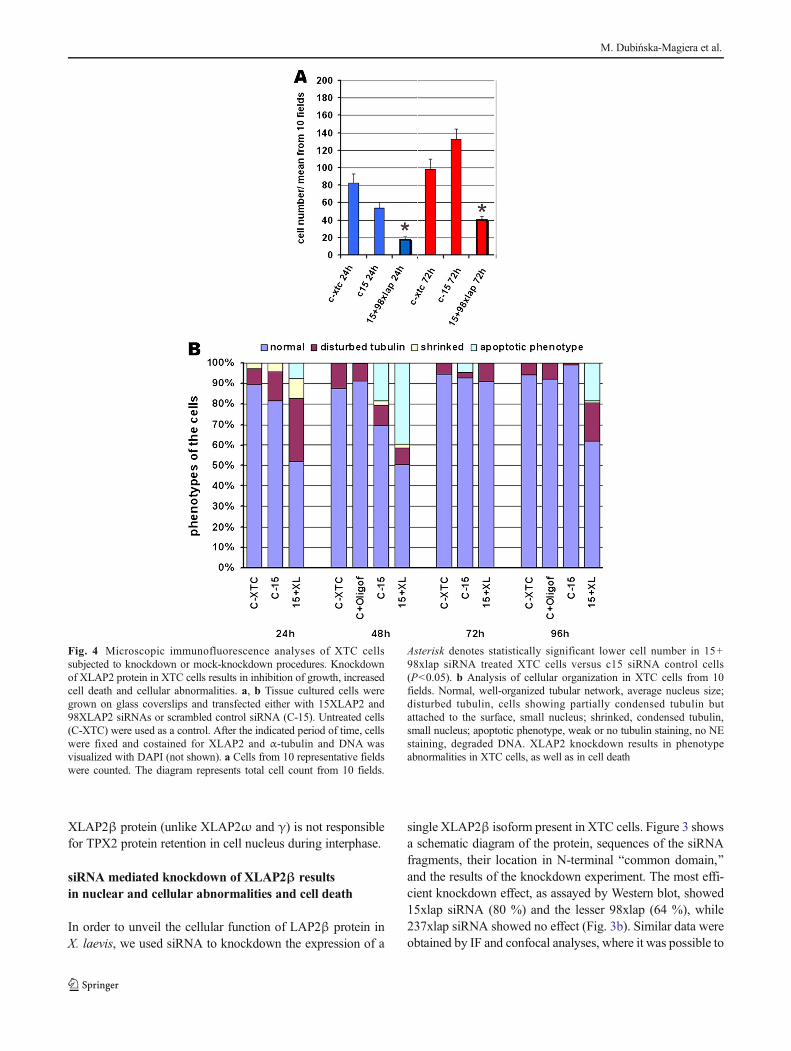
Fig. 4 Microscopic immunofluorescence analyses of XTC cellssubjected to knockdown or mock-knockdown procedures. Knockdownof XLAP2 protein in XTC cells results in inhibition of growth, increasedcell death and cellular abnormalities. a, b Tissue cultured cells weregrown on glass coverslips and transfected either with 15XLAP2 and98XLAP2 siRNAs or scrambled control siRNA (C-15). Untreated cells(C-XTC) were used as a control. After the indicated period of time, cellswere fixed and costained for XLAP2 and α-tubulin and DNA wasvisualized with DAPI (not shown). a Cells from 10 representative fieldswere counted. The diagram represents total cell count from 10 fields.
Asterisk denotes statistically significant lower cell number in 15+98xlap siRNA treated XTC cells versus c15 siRNA control cells(P<0.05). b Analysis of cellular organization in XTC cells from 10fields. Normal, well-organized tubular network, average nucleus size;disturbed tubulin, cells showing partially condensed tubulin butattached to the surface, small nucleus; shrinked, condensed tubulin,small nucleus; apoptotic phenotype, weak or no tubulin staining, no NEstaining, degraded DNA. XLAP2 knockdown results in phenotypeabnormalities in XTC cells, as well as in cell death
M. Dubińska-Magiera et al.
observe the decreased level of XLAP2 protein (Fig. 3c,237lap, 98lap) or its apparent disappearance after 72 h(Fig. 3c, 15xlap). Detailed analyses of the cell nuclei revealedalmost complete loss of the XLAP2 protein from the NE incells treated with 15xlap siRNA. This was associated with thenuclear shape abnormalities (Fig. 3d), similar to lamin A/Cknockout fibroblasts or EDMD (Emery-Dreifuss musculardystrophy) patients (Sullivan et al. 1999).
Subsequent large-scale analyses of the siRNA transfectedcells revealed the toxic effect of the XLAP2 protein knock-down and a drastically decreased growth of XTC cultures.Figure 4 demonstrates the representative results of one seriesof such experiments. When cell number was analyzed(Fig. 4a), we clearly observed the toxic effect of XLAP2knockdown on XTC cells after 24 and 72 h comparing tocontrol cells and control siRNA treatment. The analyses ofthe cell phenotypes after XLAP2 knockdown also demonstrat-ed increased percentage of the cell shape abnormalities(Fig. 4b). Cells treated with siRNA had increased a percentageof apoptotic phenotype, were shrinked, and/or had disturbedmicrotubule network.
Based on the analyses of cells still attached to cover glass atthe time of observation (Fig. 4), the first visible effect to ap-pear (after 24 h) is the disturbed cytoskeleton and shrinking ofthe cells with only a fraction of cells with apoptoticphenotype.
At 48 h, almost exactly the same number of cells showedabnormal phenotype, but most of them were apoptotic. Simi-lar temporal effect of the appearance of abnormal and apopto-tic phenotype is observed in the cycle between 72 and 96 h(Fig. 4) but obscured by proliferating normal (non-transfected) cells (Fig. 4a).
Based on the fact that we have detected only a few mitoticcells with LAP2 knockdown, we may conclude that, in gen-eral, knockdown of XLAP2β in XTC cells either stops thecells from entry into mitosis or cells detach themselves fromthe surface of culture vessel/cover glass before mitotic entry orboth. We propose that this effect is the result of abnormalnucleus re-assembly when cells lack XLAP2β protein.
XLAP2β knockdown affect cell nuclei morphologyand location of lamin B2 and nucleoporins
Xenopus XTC cells are not easy to transfect; therefore, inmost of our experiments, we could not get sufficient efficiencyof transfection for detailed studies of cell cycle or pathwaysaffected using bulk tissue culture transfection. Due to the tox-icity of XLAP2β knockdown, we were unable to select stablecell line with constitutive protein knockdown as well.
Therefore, in order to perform knockdown experiments inmore controllable system allowing for easy detection oftransfected cells under microscope, presumably with de-creased level of XLAP2β protein, we switched to plasmid-
based siRNA transfection system (pFIV-H1/U6-copGFP)with GFP (green fluorescent protein) reporter protein as amarker for transfected cells. Plasmid encoded siRNA systemdue to the GFP reporter protein expression makes availablethe distinction of the particular transfected cell for study. Ad-ditionally, when possible, the level and distribution of XLAP2protein were visualized. Thus, we were able to monitor theknockdown effect in transfected cells only (Fig. 5) and mon-itor the level and location of XLAP2β protein together withother proteins in multichannel confocal microscopy.
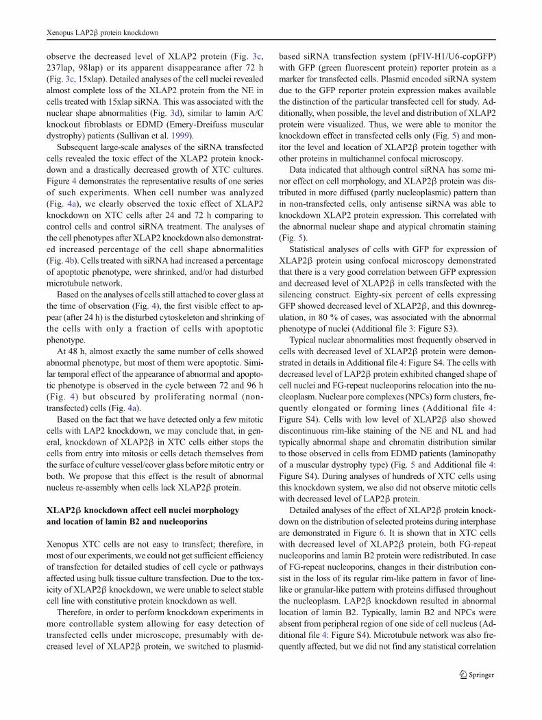
Data indicated that although control siRNA has some mi-nor effect on cell morphology, and XLAP2β protein was dis-tributed in more diffused (partly nucleoplasmic) pattern thanin non-transfected cells, only antisense siRNA was able toknockdown XLAP2 protein expression. This correlated withthe abnormal nuclear shape and atypical chromatin staining(Fig. 5).
Statistical analyses of cells with GFP for expression ofXLAP2β protein using confocal microscopy demonstratedthat there is a very good correlation between GFP expressionand decreased level of XLAP2β in cells transfected with thesilencing construct. Eighty-six percent of cells expressingGFP showed decreased level of XLAP2β, and this downreg-ulation, in 80 % of cases, was associated with the abnormalphenotype of nuclei (Additional file 3: Figure S3).
Typical nuclear abnormalities most frequently observed incells with decreased level of XLAP2β protein were demon-strated in details in Additional file 4: Figure S4. The cells withdecreased level of LAP2β protein exhibited changed shape ofcell nuclei and FG-repeat nucleoporins relocation into the nu-cleoplasm. Nuclear pore complexes (NPCs) form clusters, fre-quently elongated or forming lines (Additional file 4:Figure S4). Cells with low level of XLAP2β also showeddiscontinuous rim-like staining of the NE and NL and hadtypically abnormal shape and chromatin distribution similarto those observed in cells from EDMD patients (laminopathyof a muscular dystrophy type) (Fig. 5 and Additional file 4:Figure S4). During analyses of hundreds of XTC cells usingthis knockdown system, we also did not observe mitotic cellswith decreased level of LAP2β protein.
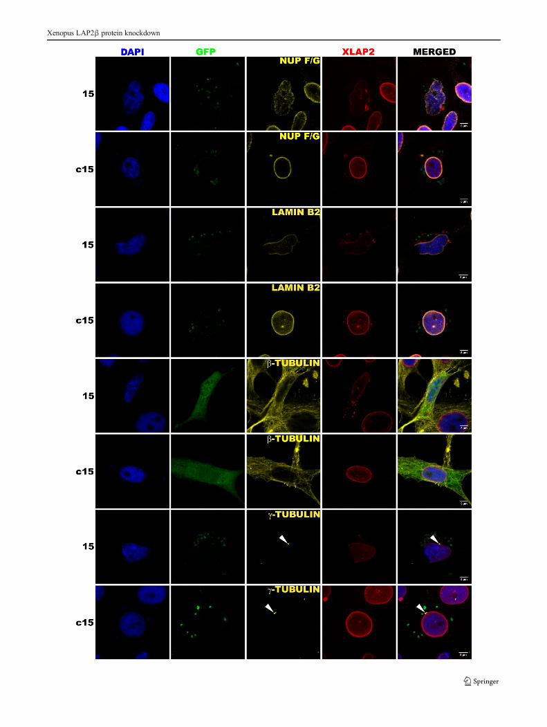
Detailed analyses of the effect of XLAP2β protein knock-down on the distribution of selected proteins during interphaseare demonstrated in Figure 6. It is shown that in XTC cellswith decreased level of XLAP2β protein, both FG-repeatnucleoporins and lamin B2 protein were redistributed. In caseof FG-repeat nucleoporins, changes in their distribution con-sist in the loss of its regular rim-like pattern in favor of line-like or granular-like pattern with proteins diffused throughoutthe nucleoplasm. LAP2β knockdown resulted in abnormallocation of lamin B2. Typically, lamin B2 and NPCs wereabsent from peripheral region of one side of cell nucleus (Ad-ditional file 4: Figure S4). Microtubule network was also fre-quently affected, but we did not find any statistical correlation
Xenopus LAP2β protein knockdown
between XLAP2β knockdown and location and distance ofcentrosomes to the NE.
Discussion
XLAP2 distribution in XTC cells and question of TPX2
Our data demonstrated that XLAP2β protein localizes mostlyto the NE of XTC cells during interphase. During this phase,XLAP2 colocalizes with typical NE and NL antigens as laminB2 (Fig. 1a). Apparent colocalization with nucleoporins(Fig. 1a) does not indicate their interaction. Higher resolutionimages (STED) or SEM/TEM studies demonstrated that they
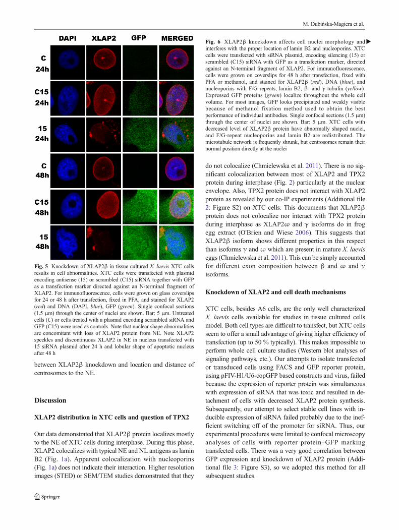
do not colocalize (Chmielewska et al. 2011). There is no sig-nificant colocalization between most of XLAP2 and TPX2protein during interphase (Fig. 2) particularly at the nuclearenvelope. Also, TPX2 protein does not interact with XLAP2protein as revealed by our co-IP experiments (Additional file2: Figure S2) on XTC cells. This documents that XLAP2βprotein does not colocalize nor interact with TPX2 proteinduring interphase as XLAP2ω and γ isoforms do in frogegg extract (O'Brien and Wiese 2006). This suggests thatXLAP2β isoform shows different properties in this respectthan isoforms γ and ω which are present in mature X. laeviseggs (Chmielewska et al. 2011). This can be simply accountedfor different exon composition between β and ω and γisoforms.
Knockdown of XLAP2 and cell death mechanisms
XTC cells, besides A6 cells, are the only well characterizedX. laevis cells available for studies in tissue cultured cellsmodel. Both cell types are difficult to transfect, but XTC cellsseem to offer a small advantage of giving higher efficiency oftransfection (up to 50 % typically). This makes impossible toperform whole cell culture studies (Western blot analyses ofsignaling pathways, etc.). Our attempts to isolate transfectedor transduced cells using FACS and GFP reporter protein,using pFIV-H1/U6-copGFP based constructs and virus, failedbecause the expression of reporter protein was simultaneouswith expression of siRNA that was toxic and resulted in de-tachment of cells with decreased XLAP2 protein synthesis.Subsequently, our attempt to select stable cell lines with in-ducible expression of siRNA failed probably due to the inef-ficient switching off of the promoter for siRNA. Thus, ourexperimental procedures were limited to confocal microscopyanalyses of cells with reporter protein–GFP markingtransfected cells. There was a very good correlation betweenGFP expression and knockdown of XLAP2 protein (Addi-tional file 3: Figure S3), so we adopted this method for allsubsequent studies.
Fig. 5 Knockdown of XLAP2β in tissue cultured X. laevis XTC cellsresults in cell abnormalities. XTC cells were transfected with plasmidencoding antisense (15) or scrambled (C15) siRNA together with GFPas a transfection marker directed against an N-terminal fragment ofXLAP2. For immunofluorescence, cells were grown on glass coverslipsfor 24 or 48 h after transfection, fixed in PFA, and stained for XLAP2(red) and DNA (DAPI, blue), GFP (green). Single confocal sections(1.5 μm) through the center of nuclei are shown. Bar: 5 μm. Untreatedcells (C) or cells treated with a plasmid encoding scrambled siRNA andGFP (C15) were used as controls. Note that nuclear shape abnormalitiesare concomitant with loss of XLAP2 protein from NE. Note XLAP2speckles and discontinuous XLAP2 in NE in nucleus transfected with15 siRNA plasmid after 24 h and lobular shape of apoptotic nucleusafter 48 h
�Fig. 6 XLAP2β knockdown affects cell nuclei morphology andinterferes with the proper location of lamin B2 and nucleoporins. XTCcells were transfected with siRNA plasmid, encoding silencing (15) orscrambled (C15) siRNA with GFP as a transfection marker, directedagainst an N-terminal fragment of XLAP2. For immunofluorescence,cells were grown on coverslips for 48 h after transfection, fixed withPFA or methanol, and stained for XLAP2β (red), DNA (blue), andnucleoporins with F/G repeats, lamin B2, β- and γ-tubulin (yellow).Expressed GFP proteins (green) localize throughout the whole cellvolume. For most images, GFP looks precipitated and weakly visiblebecause of methanol fixation method used to obtain the bestperformance of individual antibodies. Single confocal sections (1.5 μm)through the center of nuclei are shown. Bar: 5 μm. XTC cells withdecreased level of XLAP2β protein have abnormally shaped nuclei,and F/G-repeat nucleoporins and lamin B2 are redistributed. Themicrotubule network is frequently shrunk, but centrosomes remain theirnormal position directly at the nuclei
M. Dubińska-Magiera et al.
Xenopus LAP2β protein knockdown
We observed that cells with decreasing levels of XLAP2βprotein gradually accumulated abnormalities in size, shape,and microtubule network (Fig. 4, Additional file 4:Figure S4), leading finally to cell detachment from the surface.These changes were accompanied by changes in chromatinand nuclear morphology, NE structure, and distribution ofnuclear and NPC proteins (Figs. 5 and 6). A surprising featureof cells with a low level of XLAP2β was that we could notdetect mitotic cells. This suggests that lack of LAP2β preventscells from mitotic entry or that abnormally re-assembled nu-clei (and chromatin) in such cells resulted in cell cycle block atG0/G1 or G1/S/G2. The major effect of XLAP2β knockdownwas structural abnormalities of the cells, which were followedby appearance of apoptotic phenotype. This would favor thehypothesis that apoptosis is a secondary effect of XLAP2βknockdown in XTC cells.
Since isoformβ is the only LAP2 protein in XTC cells, thismay account for the severity of knockdown phenotype thoughthere may be several plausible mechanisms involved. InX. laevis, at least three different isoforms were detected duringdevelopment (Chmielewska et al. 2011). Since they share sev-eral common domains, some of the functions may be con-served between them. Domains with no similarity betweenthem may be responsible for different properties, unique forparticular isoform. XLAP2β was found to be associated withclusters of heterochromatin both next to NE and internally incell nuclei. As we have previously shown, XLAP2βwas pres-ent inside the cell nucleus both in nuclear envelope invagina-tions and without visible nuclear membranes in interphasecells (Chmielewska et al. 2011). Since mammalian LAP2βinteracts with HDAC3 (Somech et al. 2005), this proteinmay be anchored on heterochromatin by Xenopus LAP2βprotein and, thus, maintain the inactive status of heterochro-matin. The knockdown of XLAP2β may result in the releaseof HDAC3 from heterochromatin loci and change in locationand transcription activity, or both. Similar results were shownfor lamin B receptor (LBR), inner nuclear membrane proteinresponsible for heterochromatin positioning at nuclear periph-ery, present in NE, and membrane invaginations inside the cellnucleus (Ellenberg et al. 1997). Knockdown of LBR bymorpholino antisense nucleotides in 1–2 cell zebrafish embry-os caused reduced viability and severe morphological alter-ations in surviving embryos (Schild-Prufert et al. 2006).RNAi-mediated knockdown of LBR in HeLa cells resultedin failure of NE reassembly and abnormal chromatindecondensation after mitosis leading to the apoptotic celldeath (Lu et al. 2010). On the other hand, disruption of LBRfunction in differentiating cells was responsible for the loss ofperipheral heterochromatin and inverted nuclear morphologywith heterochromatin present in nuclear interior, which in turnaltered gene expression profile (Solovei et al. 2013).
Mechanism that may function during mitosis is associatedwith the structural role of LAP2β protein in the maintenance
of nuclear structure and reassembly during mitosis. The lackof XLAP2β – LEM domain integral membrane of INM andloss of one of the links between chromatin and NE (chroma-tin-BAF-LAP2β at NE) may result in abnormal nucleus andNE reassembly observed in our studies (Figs. 5 and 6). Sincein Xenopus cells, many different LEM domain proteins areexpressed besides LAP2β, the severity of phenotype is lowerthan LBR knockdown.
Proper assembly of lamin B and nucleoporins is necessaryfor DNA replication, transcription, and nuclear pore spacing.Defects in their distribution affect all these processes (Smytheet al. 2000, Moir et al. 2000, Spann et al. 2002, Margalit et al.2005). XLAP2β knockdown disrupts lamin B2 and NPC pro-tein location at NE and NL, which in turn can affect theseprocesses. On top of the above mechanisms lack of LAP2βleads to the redistribution of lamin B2 and probably the relo-cation of lamin A since both proteins interact with LAP2 pro-teins. This may further enhance the above mechanisms thatfinally results in the cells death after XLAP2β expressionknockdown.
Acknowledgments We would like to thank Prof. P.A. Fisher and Prof.K. Furukawa for anti-BAF antibodies and Prof. Y. Zheng for rabbit antiTPX2 antibodies. This work was partially supported by grants: EuropeanSocial Fund, Lower Silesia Research Grants for PhD students and Eras-mus training fellowship to M. Ch. and M. D-M. This work was partiallysupported by the Polish Ministry of Science and Higher Education Stat-utory Grant (1013/S/WB/2011-2013), Wrocław Research Center EIT+under the project: Biotechnologies and Advanced Medical TechnologiesBioMed (POIG.01.01.02-02-003/08) and COST Action BM1002Nanonet: Nanomechanics of Intermediate Filament Network (R.R.). Pub-lication costs were partially covered by Wroclaw Centre of Biotechnolo-gy, programme The Leading National Research Centre (KNOW) foryears 2014–2018.
Conflict of interests The authors declare that they have no conflict ofinterests.
Open Access This article is distributed under the terms of the CreativeCommons At t r ibut ion 4 .0 In te rna t ional License (h t tp : / /creativecommons.org/licenses/by/4.0/), which permits unrestricted use,distribution, and reproduction in any medium, provided you giveappropriate credit to the original author(s) and the source, provide a linkto the Creative Commons license, and indicate if changes were made.
References
Berk JM, Tifft KE, Wilson KL (2013) The nuclear envelope LEM-domain protein emerin. Nucleus 4(4):298–314. doi:10.4161/nucl.25751
Cai M, Huang Y, Ghirlando R, Wilson KL, Craigie R, Clore GM (2001)Solution structure of the constant region of nuclear envelope proteinLAP2 reveals two LEM-domain structures: one binds BAF and theother binds DNA. EMBO J 20(16):4399–4407. doi:10.1093/emboj/20.16.4399
M. Dubińska-Magiera et al.
Chmielewska M, Dubinska-Magiera M, Sopel M, Rzepecka D,Hutchison CJ, Goldberg MW, Rzepecki R (2011) Embryonic andadult isoforms of XLAP2 form microdomains associated with chro-matin and the nuclear envelope. Cell Tissue Res 344(1):97–110. doi:10.1007/s00441-011-1129-2
Dechat T, Gajewski A, Korbei B, Gerlich D, Daigle N, Haraguchi T,Furukawa K, Ellenberg J, Foisner R (2004) LAP2alpha and BAFtransiently localize to telomeres and specific regions on chromatinduring nuclear assembly. J Cell Sci 117(Pt 25):6117–6128. doi:10.1242/jcs.01529
Dorner D, Gotzmann J, Foisner R (2007) Nucleoplasmic lamins and theirinteraction partners, LAP2alpha, Rb, and BAF, in transcriptionalregulation. FEBS J 274(6):1362–1373
Dubinska-Magiera M, Zaremba-Czogalla M, Rzepecki R (2013) Muscledevelopment, regeneration and laminopathies: how lamins orlamina-associated proteins can contribute to muscle development,regeneration and disease. Cell Mol Life Sci 70(15):2713–2741. doi:10.1007/s00018-012-1190-3
Ellenberg J, Siggia ED, Moreira JE, Smith CL, Presley JF, WormanHJ, Lippincott-Schwartz J (1997) Nuclear membrane dynamicsand reassembly in living cells: targeting of an inner nuclearmembrane protein in interphase and mitosis. J Cell Biol138(6):1193–1206
Furukawa K (1999) LAP2 binding protein 1 (L2BP1/BAF) is a candidatemediator of LAP2-chromatin interaction. J Cell Sci 112(Pt 15):2485–2492
Furukawa K, Sugiyama S, Osouda S, Goto H, Inagaki M, Horigome T,Omata S, McConnell M, Fisher PA, Nishida Y (2003) Barrier-to-autointegration factor plays crucial roles in cell cycle progressionand nuclear organization in Drosophila. J Cell Sci 116(Pt 18):3811–3823. doi:10.1242/jcs.00682
Gant TM, Harris CA, Wilson KL (1999) Roles of LAP2 proteins innuclear assembly and DNA replication: truncated LAP2beta pro-teins alter lamina assembly, envelope formation, nuclear size, andDNA replication efficiency in Xenopus laevis extracts. J Cell Biol144(6):1083–1096
Harris CA, Andryuk PJ, Cline S, Chan HK, Natarajan A, Siekierka JJ,Goldstein G (1994) Three distinct human thymopoietins are derivedfrom alternatively spliced mRNAs. Proc Natl Acad Sci U S A91(14):6283–6287
Lang C, Krohne G (2003) Lamina-associated polypeptide 2beta(LAP2beta) is contained in a protein complex together with A-and B-type lamins. Eur J Cell Biol 82(3):143–153
Lang C, Paulin-Levasseur M, Gajewski A, Alsheimer M, Benavente R,Krohne G (1999) Molecular characterization and developmentallyregulated expression of Xenopus lamina-associated polypeptide 2(XLAP2). J Cell Sci 112(Pt 5):749–759
Lee KK, Wilson KL (2004) All in the family: evidence for four newLEM-domain proteins Lem2 (NET-25), Lem3, Lem4 and Lem5 inthe human genome. Symp Soc Exp Biol 56:329–339
Lu X, Shi Y, Lu Q, Ma Y, Luo J, Wang Q, Ji J, Jiang Q, Zhang C (2010)Requirement for lamin B receptor and its regulation by importin{beta} and phosphorylation in nuclear envelope assembly duringmitotic exit. J Biol Chem 285(43):33281–33293. doi:10.1074/jbc.M110.102368
Margalit A, Vlcek S, Gruenbaum Y, Foisner R (2005) Breaking andmaking of the nuclear envelope. J Cell Biochem 95(3):454–465.doi:10.1002/jcb.20433
Markiewicz E, Dechat T, Foisner R, Quinlan RA, Hutchison CJ (2002)Lamin A/C binding protein LAP2alpha is required for nuclear an-chorage of retinoblastoma protein. Mol Biol Cell 13(12):4401–4413. doi:10.1091/mbc.E02-07-0450
Martins S, Eikvar S, Furukawa K, Collas P (2003) HA95 and LAP2 betamediate a novel chromatin-nuclear envelope interaction implicatedin initiation of DNA replication. J Cell Biol 160(2):177–188. doi:10.1083/jcb.200210026
Moir RD, Spann TP, Herrmann H, Goldman RD (2000) Disruption ofnuclear lamin organization blocks the elongation phase of DNAreplication. J Cell Biol 149(6):1179–1192
Nili E, Cojocaru GS, Kalma Y, Ginsberg D, Copeland NG, Gilbert DJ,Jenkins NA, Berger R, Shaklai S, Amariglio N, Brok-Simoni F,Simon AJ, Rechavi G (2001) Nuclear membrane proteinLAP2beta mediates transcriptional repression alone and togetherwith its binding partner GCL (germ-cell-less). J Cell Sci 114(Pt18):3297–3307
O'Brien LL, Wiese C (2006) TPX2 is required for postmitotic nuclearassembly in cell-free Xenopus laevis egg extracts. J Cell Biol173(5):685–694. doi:10.1083/jcb.200512107
Pekovic V, Harborth J, Broers JL, Ramaekers FC, van Engelen B,Lammens M, von Zglinicki T, Foisner R, Hutchison C,Markiewicz E (2007) Nucleoplasmic LAP2alpha-lamin A com-plexes are required to maintain a proliferative state in human fibro-blasts. J Cell Biol 176(2):163–172. doi:10.1083/jcb.200606139
Pudney M, Varma MG, Leake CJ (1973) Establishment of a cell line(XTC-2) from the South African clawed toad, Xenopus laevis.Experientia 29(4):466–467
Rzepecki R, Bogachev SS, Kokoza E, Stuurman N, Fisher PA (1998)In vivo association of lamins with nucleic acids in Drosophilamelanogaster. J Cell Sci 111(Pt 1):121–129
Salpingidou G, Rzepecki R, Kiseleva E, Lyon C, Lane B, Fusiek K,Golebiewska A, Drummond S, Allen T, Ellis JA, Smythe C,Goldberg MW, Hutchison CJ (2008) NEP-A and NEP-B both con-tribute to nuclear pore formation in Xenopus eggs and oocytes. JCell Sci 121(Pt 5):706–716. doi:10.1242/jcs.019968
Schild-Prufert K, Giegerich M, Schafer M, Winkler C, Krohne G (2006)Structural and functional characterization of the zebrafish lamin Breceptor. Eur J Cell Biol 85(8):813–824. doi:10.1016/j.ejcb.2006.04.009, S0171-9335(06)00079-3
Schirmer EC, Foisner R (2007) Proteins that associate with lamins: manyfaces, many functions. Exp Cell Res 313(10):2167–2179. doi:10.1016/j.yexcr.2007.03.012, S0014-4827(07)00120-6
Schneider CA, Rasband WS, Eliceiri KW (2012) NIH Image to ImageJ:25 years of image analysis. Nat Methods 9(7):671–675
Shaklai S, Somech R, Gal-Yam EN, Deshet-Unger N, Moshitch-Moshkovitz S, Hirschberg K, Amariglio N, Simon AJ, Rechavi G(2008) LAP2zeta binds BAF and suppresses LAP2beta-mediatedtranscriptional repression. Eur J Cell Biol 87(5):267–278. doi:10.1016/j.ejcb.2008.01.014, S0171-9335(08)00040-X
Shumaker DK, Lee KK, Tanhehco YC, Craigie R, Wilson KL (2001)LAP2 binds to BAF.DNA complexes: requirement for the LEMdomain and modulation by variable regions. EMBO J 20(7):1754–1764. doi:10.1093/emboj/20.7.1754
Smythe C, Jenkins HE, Hutchison CJ (2000) Incorporation of the nuclearpore basket protein nup153 into nuclear pore structures is dependentupon lamina assembly: evidence from cell-free extracts of Xenopuseggs. EMBO J 19(15):3918–3931. doi:10.1093/emboj/19.15.3918
Solovei I, Wang AS, Thanisch K, Schmidt CS, Krebs S, Zwerger M,Cohen TV, Devys D, Foisner R, Peichl L, Herrmann H, Blum H,Engelkamp D, Stewart CL, Leonhardt H, Joffe B (2013) LBR andlamin A/C sequentially tether peripheral heterochromatin and in-versely regulate differentiation. Cell 152(3):584–598. doi:10.1016/j.cell.2013.01.009, S0092-8674(13)00012-3
Somech R, Shaklai S, Geller O, Amariglio N, Simon AJ, Rechavi G, Gal-Yam EN (2005) The nuclear-envelope protein and transcriptionalrepressor LAP2beta interacts with HDAC3 at the nuclear periphery,and induces histone H4 deacetylation. J Cell Sci 118(Pt 17):4017–4025. doi:10.1242/jcs.02521, 118/17/4017
Spann TP, Goldman AE, Wang C, Huang S, Goldman RD (2002)Alteration of nuclear lamin organization inhibits RNA polymeraseII-dependent transcription. J Cell Biol 156(4):603–608. doi:10.1083/jcb.200112047
Xenopus LAP2β protein knockdown

Stewart S, Fang G (2005) Anaphase-promoting complex/cyclosome con-trols the stability of TPX2 during mitotic exit. Mol Cell Biol 25(23):10516–10527. doi:10.1128/MCB.25.23.10516-10527.2005
Sullivan T, Escalante-Alcalde D, Bhatt H, Anver M, Bhat N, NagashimaK, Stewart CL, Burke B (1999) Loss of A-type lamin expressioncompromises nuclear envelope integrity leading to muscular dystro-phy. J Cell Biol 147(5):913–920
Tsai MY, Wang S, Heidinger JM, Shumaker DK, Adam SA, GoldmanRD, Zheng Y (2006) A mitotic lamin B matrix induced by RanGTPrequired for spindle assembly. Science 311(5769):1887–1893. doi:10.1126/science.1122771
Wagner N, Krohne G (2007) LEM-Domain proteins: new insights intolamin-interacting proteins. Int Rev Cytol 261:1–46. doi:10.1016/S0074-7696(07)61001-8
Yang L, Guan T, Gerace L (1997) Lamin-binding fragment of LAP2inhibits increase in nuclear volume during the cell cycle and pro-gression into S phase. J Cell Biol 139(5):1077–1087
Zaremba-Czogalla M, Dubinska-Magiera M, Rzepecki R (2011a)Laminopathies: the molecular background of the disease and theprospects for its treatment. Cell Mol Biol Lett 16(1):114–148. doi:10.2478/s11658-010-0038-9
Zaremba-Czogalla M, Gagat P, Koziol K, Dubinska-Magiera M,Sikora J, Dadlez M, Rzepecki R (2011b) Identification ofnew in vivo phosphosites on lamin Dm—the evidence ofheterogenei ty of phosphorylat ion s i tes in differentDrosophila tissues. Nucleus 2(5):478–488. doi:10.4161/nucl.2.5.17864
M. Dubińska-Magiera et al.