Involvement of the Lamin Rod Domain in Heterotypic Lamin Interactions Important for Nuclear...
11
The Rockefeller University Press, 0021-9525/2001/04/479/11 $5.00 The Journal of Cell Biology, Volume 153, Number 3, April 30, 2001 479–489 http://www.jcb.org/cgi/content/full/153/3/479 479 Involvement of the Lamin Rod Domain in Heterotypic Lamin Interactions Important for Nuclear Organization Eric C. Schirmer, Tinglu Guan, and Larry Gerace Department of Cell Biology, The Scripps Research Institute, La Jolla, California 92037 Abstract. The nuclear lamina is a meshwork of inter- mediate-type filament proteins (lamins) that lines the inner nuclear membrane. The lamina is proposed to be an important determinant of nuclear structure, but there has been little direct testing of this idea. To inves- tigate lamina functions, we have characterized a novel lamin B1 mutant lacking the middle z4/5 of its a-heli- cal rod domain. Though retaining only 10 heptads of the rod, this mutant assembles into intermediate fila- ment-like structures in vitro. When expressed in cul- tured cells, it concentrates in patches at the nuclear en- velope. Concurrently, endogenous lamins shift from a uniform to a patchy distribution and lose their com- plete colocalization, and nuclei become highly lobu- lated. In vitro binding studies suggest that the internal rod region is important for heterotypic associations of lamin B1, which in turn are required for proper organi- zation of the lamina. Accompanying the changes in lamina structure induced by expression of the mutant, nuclear pore complexes and integral membrane pro- teins of the inner membrane cluster, principally at the patches of endogenous lamins. Considered together, these data indicate that lamins play a major role in or- ganizing other proteins in the nuclear envelope and in determining nuclear shape. Key words: intermediate filament • nuclear lamina • nuclear pore complex • lamina-associated polypeptide • nuclear shape Introduction The nuclear envelope (NE) 1 is a double membrane system continuous with the ER that forms the nuclear boundary. It is punctuated by nuclear pore complexes (NPCs), which regulate molecular transport into and out of the nucleus (reviewed in Gorlich and Kutay, 1999). Underlying the in- ner surface of the NE is the nuclear lamina, a fibrous meshwork consisting of lamins (Aebi et al., 1986) and a number of more minor lamina-associated polypeptides (LAPs; reviewed in Gerace and Foisner, 1994; Stuurman et al., 1998). Lamins are type V intermediate filament (IF) proteins (McKeon et al., 1986). The B1 and B2 lamin subtypes are present in nuclei of nearly all vertebrate cells, whereas the A and C subtypes appear only at or after differentiation (Rober et al., 1989). All lamin subtypes colocalize by light microscopy (reviewed in Gerace and Foisner, 1994; Stuur- man et al., 1998). Moreover, A and B subtypes can interact in vitro (Georgatos et al., 1988; Ye and Worman, 1995), al- though there is no in vivo indication of the relevance or ex- istence of such heterotypic associations in the assembled lamina. In addition to interacting with LAPs, lamins bind to chromatin (Glass and Gerace, 1990), and this may be re- sponsible for anchoring interphase chromosomes to the NE. The best characterized LAPs are integral membrane proteins of the inner nuclear membrane (INM). These in- clude LAPs 1 and 2 (Senior and Gerace, 1988; Foisner and Gerace, 1993), LBR (Ye and Worman, 1994), and emerin (Fairley et al., 1999). Collectively, these integral proteins appear to help connect the lamina to the NE (reviewed in Collas and Courvalin, 2000). The importance of the lamina has recently been underscored by the finding that certain point mutants of emerin in humans cause Emery-Dreifuss muscular dystrophy, and that some lamin point mutations cause lipodystrophy and cardiac dysfunction as well as Emery-Dreifuss muscular dystrophy (reviewed in Wilson, 2000; see also Cao and Hegele, 2000). How these defects cause such different diseases remains unclear. Unraveling this mystery will require a further understanding of the complex structure of the assembled lamina and of its func- tions, many of which remain speculative. A role for the lamins in NE structure is inferred from their abundance and uniform distribution at the nuclear Address correspondence to Larry Gerace, 10550 N. Torrey Pines Rd., IMM10, R209, La Jolla, CA 92037. Tel.: (858) 784-8514. Fax: (858) 784- 9132. E-mail: [email protected] 1 Abbreviations used in this paper: FTIR, Fourier transform infrared spectroscopy; GFP, green fluorescent protein; HA, hemagglutinin; IF, in- termediate filament; INM, inner nuclear membrane; LAP, lamina-associ- ated polypeptide; NE, nuclear envelope; NLS, nuclear localization signal; NPC, nuclear pore complex; NRK, normal rat kidney; WT, wild type.
-
Upload
independent -
Category
Documents
-
view
6 -
download
0
Transcript of Involvement of the Lamin Rod Domain in Heterotypic Lamin Interactions Important for Nuclear...

The Rockefeller University Press, 0021-9525/2001/04/479/11 $5.00The Journal of Cell Biology, Volume 153, Number 3, April 30, 2001 479–489http://www.jcb.org/cgi/content/full/153/3/479 479
Involvement of the Lamin Rod Domain in Heterotypic Lamin Interactions Important for Nuclear Organization
Eric C. Schirmer, Tinglu Guan, and Larry Gerace
Department of Cell Biology, The Scripps Research Institute, La Jolla, California 92037
Abstract.
The nuclear lamina is a meshwork of inter-mediate-type filament proteins (lamins) that lines theinner nuclear membrane. The lamina is proposed to bean important determinant of nuclear structure, butthere has been little direct testing of this idea. To inves-tigate lamina functions, we have characterized a novel
lamin B1 mutant lacking the middle
z
4/5 of its
a
-heli-cal rod domain. Though retaining only 10 heptads ofthe rod, this mutant assembles into intermediate fila-ment-like structures in vitro. When expressed in cul-tured cells, it concentrates in patches at the nuclear en-velope. Concurrently, endogenous lamins shift from auniform to a patchy distribution and lose their com-plete colocalization, and nuclei become highly lobu-lated. In vitro binding studies suggest that the internal
rod region is important for heterotypic associations oflamin B1, which in turn are required for proper organi-zation of the lamina. Accompanying the changes inlamina structure induced by expression of the mutant,nuclear pore complexes and integral membrane pro-teins of the inner membrane cluster, principally at thepatches of endogenous lamins. Considered together,these data indicate that lamins play a major role in or-ganizing other proteins in the nuclear envelope and indetermining nuclear shape.
Key words: intermediate filament • nuclear lamina •nuclear pore complex • lamina-associated polypeptide• nuclear shape
Introduction
The nuclear envelope (NE)
1
is a double membrane systemcontinuous with the ER that forms the nuclear boundary.It is punctuated by nuclear pore complexes (NPCs), whichregulate molecular transport into and out of the nucleus(reviewed in Gorlich and Kutay, 1999). Underlying the in-ner surface of the NE is the nuclear lamina, a fibrousmeshwork consisting of lamins (Aebi et al., 1986) and anumber of more minor lamina-associated polypeptides(LAPs; reviewed in Gerace and Foisner, 1994; Stuurmanet al., 1998).
Lamins are type V intermediate filament (IF) proteins(McKeon et al., 1986). The B1 and B2 lamin subtypes arepresent in nuclei of nearly all vertebrate cells, whereas the Aand C subtypes appear only at or after differentiation(Rober et al., 1989). All lamin subtypes colocalize by lightmicroscopy (reviewed in Gerace and Foisner, 1994; Stuur-man et al., 1998). Moreover, A and B subtypes can interact
in vitro (Georgatos et al., 1988; Ye and Worman, 1995), al-though there is no in vivo indication of the relevance or ex-istence of such heterotypic associations in the assembledlamina. In addition to interacting with LAPs, lamins bind tochromatin (Glass and Gerace, 1990), and this may be re-sponsible for anchoring interphase chromosomes to the NE.
The best characterized LAPs are integral membraneproteins of the inner nuclear membrane (INM). These in-clude LAPs 1 and 2 (Senior and Gerace, 1988; Foisner andGerace, 1993), LBR (Ye and Worman, 1994), and emerin(Fairley et al., 1999). Collectively, these integral proteinsappear to help connect the lamina to the NE (reviewed inCollas and Courvalin, 2000). The importance of the laminahas recently been underscored by the finding that certainpoint mutants of emerin in humans cause Emery-Dreifussmuscular dystrophy, and that some lamin point mutationscause lipodystrophy and cardiac dysfunction as well asEmery-Dreifuss muscular dystrophy (reviewed in Wilson,2000; see also Cao and Hegele, 2000). How these defectscause such different diseases remains unclear. Unravelingthis mystery will require a further understanding of thecomplex structure of the assembled lamina and of its func-tions, many of which remain speculative.
A role for the lamins in NE structure is inferred fromtheir abundance and uniform distribution at the nuclear
Address correspondence to Larry Gerace, 10550 N. Torrey Pines Rd.,IMM10, R209, La Jolla, CA 92037. Tel.: (858) 784-8514. Fax: (858) 784-9132. E-mail: [email protected]
1
Abbreviations used in this paper:
FTIR, Fourier transform infraredspectroscopy; GFP, green fluorescent protein; HA, hemagglutinin; IF, in-termediate filament; INM, inner nuclear membrane; LAP, lamina-associ-ated polypeptide; NE, nuclear envelope; NLS, nuclear localization signal;NPC, nuclear pore complex; NRK, normal rat kidney; WT, wild type.
The Journal of Cell Biology, Volume 153, 2001 480
periphery and from their structural properties, such astheir ability to assemble into IF-related structures in vitro(Stuurman et al., 1998), the resistance of the polymer toextraction by Triton X-100, and the skeleton-like appear-ance of the isolated lamina (Dwyer and Blobel, 1976; Aebiet al., 1986). The only functional analysis of the role oflamins in nuclear structure comes from depletion studies.Fragile, growth-inhibited nuclei result from in vitro nu-clear assembly with lamin-depleted
Xenopus
egg extracts,though no conspicuous perturbation of nuclear shape isobserved (reviewed in Lourim and Krohne, 1994; see alsoBenavente and Krohne, 1986). Furthermore, minor aber-rations in nuclear shape in some cell types result from re-duction of lamin B levels in
Drosophila
(Lenz-Bohme etal., 1997) and from disruption of the mouse lamin A/Cgene (Sullivan et al., 1999). However, it is not clear fromthese studies whether the role of the lamina in nuclearstructure is direct or indirect (e.g., by effects on NPCs orchromosomes).
Similar to other IF proteins, lamins contain a long cen-tral
a
-helical rod domain flanked by nonhelical head andtail domains. Lamins differ from other IF proteins by hav-ing an additional six heptads in the rod domain (resultingin a rod of 354 vs. 310 residues), a nuclear localization sig-nal (NLS), and a carboxyl-terminal CaaX box for farnesy-lation (reviewed in Stuurman et al., 1998). The basic unitof lamin assembly is a two-stranded coiled coil formed byparallel, unstaggered association of the rod domains dueto heptad repeats (reviewed in Stuurman et al., 1998). Thisdimer further assembles into higher order structures, suchas 10-nm filaments, by head-to-tail interactions and bystaggered antiparallel lateral interactions (Moir et al.,1991; Heitlinger et al., 1992; Stuurman et al., 1996). Thelamina polymer continuously increases in mass during in-terphase in cycling cells as the NE increases in surface area(reviewed in Gerace and Foisner, 1994). It is unclear, how-ever, how new lamin subunits are integrated into the lam-ina during this process. In contrast, nuclear reassembly atthe end of mitosis apparently involves de novo assembly ofthe lamins at the NE via interactions with nuclear mem-brane proteins and chromosomes (reviewed in Collas andCourvalin, 2000).
Although the precise interactions involved in higher or-der lamin assembly are not clear, it is apparent that thehead, tail, and rod domains all make important contribu-tions. Mutational analysis of lamins and other IF proteinshas shown that the highly conserved sequences at bothends of the rod domain are particularly important forassembly in vitro (Coulombe et al., 1990; Heald andMcKeon, 1990; Hatzfeld and Weber, 1991; Stuurman etal., 1998). The only analysis of more internal regions of thelamin rod involves overexpression of a mutant lacking asix-heptad region that is absent from other IF proteins,which yields no significant nuclear structural alterations(Mical and Monteiro, 1998).
To understand more about the functions of the laminrod domain, we engineered a deletion of the internal
z
4/5of the lamin B1 rod. This mutant is predicted to dimerizeand engage in head-to-tail interactions, but would be ex-pected to have reduced lateral interactions involved inhigher-order assembly. To our surprise this mutant formedIF-like structures in vitro. Furthermore, when overex-
pressed in cultured cells, it became enriched in patches atthe NE. At the same time, endogenous lamins and otherNE proteins also became concentrated in patches, whichto varying degrees were separated from the mutant. Ac-companying the reorganization of the lamina into a patch-work, nuclear shape was grossly altered by lobulation.This is the first lamin mutant described that perturbs thesubstructure of the assembled lamina and that causes dras-tic alterations in nuclear shape. Our findings provide newinsight on the role of the lamina in NE organization andthe importance of the lamin rod domain for lamina stabil-ity and for heterotypic associations among lamin subtypes.
Materials and Methods
Plasmid Construction
The human lamin B1 coding sequence was amplified by PCR with primersthat added 5
9
BamHI/ Nde1 and 3
9
Not1 sites. To produce B1
D
rod, theseprimers were used with internal primers containing Hind3 sites that fusednucleotides 207 and 1017 via an added alanine codon. The individual laminfragments were also subcloned (B
1
N and B
1
C). To produce A/B1
D
rod, theequivalent 5
9
sequence for human lamin A was fused to the 3’ sequencefrom lamin B1. These genes were moved to vectors for mammalian trans-fection [CMV-driven pHHS10B; with a hemagglutinin (HA) epitope tag]and protein expression (pET28a; Novagen, with a 6
3
His tag).
Transient Transfection in Cultured Cells
Adherent HeLa, COS-7, or normal rat kidney (NRK) cells were plated onpolylysine-coated slides in DMEM supplemented with 10% FBS and
L
-glutamine at low confluency (
z
15%) as cells were usually harvested aslong as 60 h after transfection. Roughly 12 h after plating, DNA was trans-fected using Fugene 6 (Roche) according to the manufacturer’s instruc-tions. To assess cell division, cells were plated onto CELLocate coverslips(Eppendorf) and cotransfected with pEGFP-F (CLONTECH Laborato-ries, Inc.). Fluorescent cells were counted at 20 and 60 h after transfection.
Antibody Production
Polyclonal antibodies were produced in rabbits and guinea pigs from pep-tides comprising the principal chromatin binding site of human lamin A(residues 396–429; Taniura et al., 1995), and the predicted chromatin bind-ing sites of human lamin B1 (residues 391–428) and human lamin B2(SPSPSSRVTVSRATSSSSGSLSATGRLGRSKRKRLEVEEPLGSGPS-VLGTGTGGR—this differs from the reported sequence (Genbank M94362),but was repeatedly recovered by PCR from a HeLa cDNA library). Pep-tides were generated using the pPEP-T vector system (Kammerer et al.,1998). Western blot analysis indicated that each serum was specific for itslamin isotype.
Immunofluorescence Microscopy and Apoptosis Analysis
Cells were fixed for 7 min in 3.7% formaldehyde, permeabilized for 6 minin 0.2% Triton X-100, blocked with 4% BSA in PBS, and reacted for 40min at room temperature with antibodies to lamins (above) and/or to theNPC (mAb RL1, Snow et al., 1987; affinity purified Tpr polyclonal IgG,gift of P. Frosst, The Scripps Research Institute), LAP2 (mAb RL29; Fois-ner and Gerace, 1993), LAP1C, (mAb RL13; Senior and Gerace, 1988),LBR (rabbit serum provided by H. Worman, Columbia University, NewYork, NY), and the HA tag of the transfected proteins (mAb HA.11; Co-vance, or purified polyclonal; Upstate Biotechnology). After washing, fluo-rophore-conjugated secondary antibodies were added for 30 min, followedby 10 min with DAPI to visualize DNA, and mounting in fluoromount G(EM Sciences). Apoptosis was assessed at 60 and 74 h after transfection us-ing the In Situ Cell Death Detection Kit (TUNEL assay; Roche).
Images were obtained using an Axiovert S100TV microscope (CarlZeiss, Inc.) interfaced to a confocal system (MRC1024; Bio-Rad Labora-tories) with 488, 568, and 647 nm krypton/argon laser lines or using an mi-croscope (1
X
70; Olympus) interfaced with a DeltaVision system (AppliedPrecision, Inc.) and subsequently deconvolved with DeltaVision version

Schirmer et al.
Role of the Lamin Rod Domain in Nuclear Structure
481
2.0 software. All fluorescence images and micrographs shown in this studywere prepared for figures using Photoshop 5.0.
Nuclear Import Assays
To assess nuclear import, Cy5-labeled BSA conjugated to an NLS was mi-croinjected into the cytoplasm of B1
D
rod-transfected cells. This was coin-jected with Texas red–labeled BSA (Jackson ImmunoResearch Laborato-ries) that lacked an NLS or FITC-dextran (150 kD; Molecular Probes) toassess nuclear integrity. After 45 min, cells were fixed and processed for im-munofluorescence microscopy. The import competence of transfected cellswas also examined using a permeabilized cell assay (Adam et al., 1990).
Thin Section EM
HeLa cells cotransfected with B1
D
rod and pEGFP-F were harvested bytrypsinization at 60 h after transfection, washed, and resuspended in 1
3
PBS (Ca
2
1
, Mg
2
1
free), 1 mM EDTA, 25 mM Hepes, pH 7.0, 1% dialyzedFBS. Fluorescent cells were collected by FACS
®
, fixed with 2% glutaral-dehyde and 1% osmium tetroxide, embedded in Epon, and sectioned ac-cording to standard protocols. Micrographs were recorded with a PhilipsEM-208 at 70 kV.
Protein Purification
Lamin B1 and B1
D
rod were purified from inclusion bodies. The proteinswere induced in Bl21-(DE3) cells at A
595
0.7 for 3 h at 37
8
C that werelysed by sonication in PBS containing 1.5 mM
b
-mercaptoethanol andprotease inhibitors. The pellets from a 7-min centrifugation at 10,000
g
were washed with 0.2% Triton X-100, resuspended in 20 mM Tris, pH 8.0,300 mM NaCl, 8 M urea, 3 mM
b
-mercaptoethanol, incubated with nickelresin (QIAGEN), and eluted with the same buffer containing 200 mM im-idazole, and dialyzed into 20 mM Tris-HCl, pH 8.0, 8 M urea, 2 mM DTT,1 mM EDTA with protease inhibitors for storage. B
1
N and B
1
C were pre-pared similarly except that cells were lysed in urea as these proteins didnot form inclusion bodies.
In Vitro Assembly of Lamins
The solubility properties of the proteins were compared as in Glass andGerace (1990). In brief, they were diluted in urea to 250, 100, 30, 10, and 3
m
g/ml, and then dialyzed into 10 mM Tris, pH 8.8, 250 mM KCl, 1 mMDTT with protease inhibitors followed by 10 mM Tris, pH 8.8, 70 mMKCl, 1 mM DTT, 5 mM MgCl
2
, followed by 50 mM Hepes, pH 7.3, 70mM KCl, 1 mM DTT, 5 mM MgCl
2
. This was subjected to centrifugationat 10,000
g
for 7 min and pellets resuspended in the same volume as super-natants before applying equal volumes to SDS-PAGE.
For cross linking, proteins were diluted in urea storage buffer to 0.05mg/ml, and then dialyzed into 20 mM Na
2
HPO
4
, pH 9, 300 mM KCl, 0.5mM EDTA, 1 mM DTT. Aggregated material was removed by centrifuga-tion at 10,000
g
for 20 min and the supernatants were incubated with0.01% glutaraldehyde for 0, 2, 10, or 20 min. Reactions were stopped byaddition of 1 M glycine and precipitated with 15% TCA before analysis bySDS-PAGE.
To investigate filament assembly, purified lamin B1 and B1
D
rod weredialyzed out of urea into 20 mM Tris pH 8.8, 300 mM NaCl, 1 mM EDTA,1 mM DTT. Aliquots were removed at different times (5 min to 1 h), ap-plied to glow discharged carbon-coated grids, negatively stained with 1-2%uranyl acetate, and viewed on a Philips CM100 at 100 kV.
Lamin Binding Assays
Purified wild-type (WT) lamin B1 and B1
D
rod were coupled in urea toAffi-gel 15 matrix (Bio-Rad Laboratories) as in Georgatos et al. (1988) with
z
3 mg bound/ml matrix. Lamins A and C (prepared as in Glass and Ger-ace, 1990) or lamin B1 were diluted to 20
m
g/ml and dialyzed out of ureainto 25 mM Hepes, pH 7.5, 300 mM NaCl, 5 mM
b
-mercaptoethanol. Thiswas centrifuged (10,000
g
for 10 min) to remove aggregated proteins and in-cubated with the matrix overnight at 4
8
C. The columns were washed in thesame buffer and eluted with increasing concentrations of urea. Fractionswere analyzed by Western blotting with the antibodies described above.
Biophysical Analysis
For circular dichroism analysis, proteins at 0.3 mg/ml were dialyzed fromurea storage buffer into 20 mM Na
2
HPO
4
, pH 9, 150 mM KCl to maintain
most lamins in solution. Insoluble material was removed by centrifugationat 10,000
g
for 10 min shortly before determination of the final proteinconcentration for conversion to mean residue ellipticity and recording ofdichroic spectra with an AVIV CD spectrometer using a 1-nm bandwidth,4-s scan time, and 5-mm cuvette at 25
8
C. An average of four scans withbuffer subtracted is shown.
For Fourier transform infrared spectroscopic (FTIR) analysis, fila-ments prepared as above were pelleted by centrifugation at 10,000
g
for 10min and washed in water. The pellet was disrupted with a pipette tip, lay-ered onto a 5-mm-thick CaF lens, dried under nitrogen, and viewed in aNicolet MAGNA-IR 550 Spectrometer Series II with a spectral resolutionof 4 cm
2
1
using OMNIC analysis software. A water blank was subtractedfrom the spectra (128 interferograms).
Results
In Vitro Assembly of B1
D
Rod
The WT rod domain of lamins contains 48 heptads, whichare subdivided into four
a
-helical regions separated byshort spacers (Fig. 1). To analyze the role of the rod do-main in lamin assembly and function, we constructed a de-letion mutant of lamin B1 containing the entire head andtail domains, but lacking the rod domain except for thefirst and last five heptads, which were fused in register(Fig. 1, B1
D
rod). We also generated a chimeric
D
rod mu-tant by fusing the head and first five heptads of the laminA/C rod with the last five rod heptads and tail of lamin B1(Fig. 1, A/B1
D
rod). These forms are predicted to have asingle
a
-helix of 10 heptads that should form a coiled coildimer by computer modeling (Lupas, 1996). Consistentwith this, recombinant B1
D
rod (Fig. 2 A), like WT laminB1, forms a dimer in solution at pH 9 as determined bycross linking (Fig. 2 B; note this is the same materialshown in A) and yields a strong
a
-helical signal by circulardichroism (Fig. 2 C).
In addition to WT lamin B1 and B1
D
rod, we preparedseparately the two parts of lamin B1 that are fused to formB1
D
rod (Fig. 2 A, B
1
N and B
1
C) and compared the solubil-ity properties of these fragments and the longer lamin con-structs. Proteins were dialyzed from urea into buffers thatsupport lamin assembly in vitro (i.e., an equilibration bufferfollowed by a polymerization buffer; see Materials andMethods), and insoluble material was pelleted. SomeB1
D
rod pelleted at the lowest concentration tested (3
m
g/ml, data not shown). In contrast, even at 250
m
g/ml (whereall of B1
D
rod and most of WT lamin B1 was pelletable), thetwo lamin B1 fragments that are fused in B1
D
rod werecompletely soluble (Fig. 2 D). This suggests that the indi-vidual NH
2
- and COOH-terminal segments of B1
D
rod donot have a strong capacity for self assembly by themselves,but strongly promote assembly when they are linked to-gether in B1
D
rod, which would allow them to drive head-to-tail polymerization of the mutant lamin (see Discussion).
When assembly of the WT and mutant lamins was ana-lyzed by EM using negative staining,
z
10-nm filamentswere observed for both proteins, even after dialysis intothe equilibration buffer alone (Fig. 3, A–B). These fila-ments were similar to one another and to filaments formedby other B-type lamins (Heitlinger et al., 1991; Sasse et al.,1997). When WT lamin B1 and B1
D
rod were mixed to-gether and dialyzed into the assembly buffers, filamentssimilar to those formed by each alone were observed (datanot shown), indicating that B1
D
rod does not disassemble

The Journal of Cell Biology, Volume 153, 2001 482
WT lamin B1 filaments nor block their formation. How-ever, we could not distinguish whether these filamentswere homo- or heterotypic because we lacked specific re-agents to distinguish the two proteins.
Certain short peptides derived from the ends of the roddomain of desmin (a type III IF protein) could assembleinto filaments in vitro, but these filaments had a
b
sheetsubstructure as revealed by FTIR (Geisler et al., 1993). Asan additional criterion to determine whether filamentsformed from B1
D
rod were IF-like, we analyzed the assem-bled material by FTIR. The filaments formed by bothB1
D
rod and WT lamin B1 had a strong, wide band at
z
1,650 cm
2
1
(Fig. 3 C). This is near the band at 1,658 thatis characteristic of
a
-helical structure, and is distinct fromthe bands at 1,620 and 1,680 cm
2
1
that characterize
b
sheet. The only other IF analyzed by FTIR is desmin, forwhich the center of the wide peak was shifted to 1,640cm
2
1
(Heimburg et al., 1996). The band we see is roughlybetween these two, indicative of
a
-helical and inconsistentwith
b
-sheet structures. In summary, based on biochemi-cal, biophysical, and structural criteria, B1
D
rod is able toform IF-related filaments in vitro despite the absence of
z
4/5 of the rod domain.
Expression of the Lamin B1
D
rod Mutant Leads to Aberrant Nuclear Shape
To examine the effect of B1
D
rod polymers on the endoge-nous lamins, we expressed WT lamin B1 and B1
D
rod(both fused to a HA tag) in COS-7, HeLa, and NRK cellsby transient transfection. At intermediate to long times af-ter transfection (see below), the nuclei of all cell typestransfected with B1
D
rod were highly lobulated and irregu-larly shaped (Fig. 4). Much of the mutant lamin (Fig. 4,red) became localized to the NE, which was identified asthe boundary of the DNA staining region (Fig. 4 A, gray)and the border of the nucleus seen by differential interfer-ence contrast microscopy (Fig. 4 B). As commonly ob-served for proteins expressed by transfection, the mutant
Figure 1. Design of mutant lamins. Mammalianlamins contain an NH2-terminal head domain(z33 amino acids) followed by a coiled-coil roddomain (354 amino acids) and a COOH-terminaltail domain (200–300 amino acids). The rod ispredicted to contain four separate a-helices: coil1A, 1B, 2A, and 2B of 6, 20, 6, and 16 heptads,respectively (a slash delineates each heptad).
The B1Drod mutant lacks most of the rod, retaining only five heptads at each end that are fused in register (via an added alanine residuereplacing the valine in position g of the fifth NH2-terminal heptad). The A/B1Drod mutant contains the head and first five heptads oflamin A (black) fused to the last five heptads and tail of lamin B1 (white).
Figure 2.
The B1
D
rod mutant has characteristics of IF proteins.(A) WT lamin B1, B1
D
rod, and the two lamin B1 fragments thatwere fused in B1
D
rod (B
1
N and B
1
C) were expressed in
Esche-richia coli,
and were analyzed by SDS PAGE and staining withCoomassie blue. (B) The B1
D
rod mutant forms dimers. SolubleWT lamin B1 or B1
D
rod (the same material shown in A) werecross linked with glutaraldehyde for 0–20 min and analyzed bySDS-PAGE and staining with silver. No monomeric (m) proteinwas observed at any time after incubation with cross linker:rather, a diffuse band at the expected Mr for the dimer species(d) was observed that did not increase over time, indicating thestability of this soluble population. An apparent cross-linked tet-ramer also was seen for B1
D
rod, but a tetrameric form of WTlamin B1 was too large to fully migrate into the gel. Note that sil-ver staining does not give a linear representation of the proteinspecies (compare 0-min samples in B to Coomassie blue–stainedsamples in A). (C) The B1
D
rod mutant has considerable
a
-heli-
cal structure. The dichroic spectra of soluble WT lamin B1 andB1
D
rod proteins (in a similar buffer as in B) contained strongminima at 208 and 222 nm indicative of a-helical structure. (D)Relative solubility of B1Drod and its individual segments. Pro-teins at 250 mg/ml were dialyzed into buffers known to promotelamin assembly. Insoluble material was pelleted (P) and com-pared with the supernatant (S) and the total starting material (T).This demonstrated that the B1N and B1C fragments are ex-tremely soluble compared with their fusion product, B1Drod, andWT lamin B1.

Schirmer et al. Role of the Lamin Rod Domain in Nuclear Structure 483
also appeared in cytoplasmic (Fig. 4 B, top, arrow) as wellas intranuclear (not shown) aggregates in some cells. Themutant lamin frequently was concentrated in distinct NEpatches of variable sizes, rather than being uniformly dis-tributed throughout the NE (Fig. 4 B, compare the twolobules with arrowheads). In contrast to cells transfectedwith B1Drod, most cells transfected with WT lamin B1 hadnormally shaped, ovoid nuclei (see Fig. 6 A, below). Somehad morphological deformations and NE invaginations,but these were relatively minor compared with cells trans-fected with the mutant.
The B1Drod phenotype developed over time. At 20 h af-ter transfection, the mutant protein was uniformly distrib-uted at the NE and nuclei still had a normal (ovoid) shape(Fig. 4 C). By 30 h, the distribution of the mutant had be-come patchy in many cells, but the nuclei were still largelyovoid. By 40 h, most nuclei had a patchy distribution ofB1Drod and had assumed an aberrant, highly lobulatedshape, which was yet more pronounced at 70 h (Fig. 4 C).Regardless of their aberrant nuclear shape, none of .100cells analyzed that were expressing B1Drod exhibited thefragmented and/or condensed DNA characteristic of ap-optosis and necrosis (see Materials and Methods).
This time course suggested that the NE deformationsoccur during interphase nuclear growth rather than upon
Figure 3. In vitro assembly of B1Drod. (A–B) WT lamin B1 andB1Drod that were dialyzed out of urea-formed filaments. Thestructures formed by each protein were similar in appearance(bars, 100 nm). (C) FTIR indicates a-helical structures occur inthe filamentous form of B1Drod, similar to WT lamin B1. Theproteins were dialyzed using conditions that had yielded fila-ments by EM analysis. Polymerized material was pelleted and an-alyzed by FTIR. The scans of B1Drod and WT lamin B1 werevery similar although the band around 1650 was wider for B1Drodthan for WT lamin B1. The position of the bands is consistentwith a-helical rather than b-sheet characteristics (see text).
Figure 4. The B1Drod mutant lamin grossly alters nuclear mor-phology. (A) Volume projections of deconvolved optical sectionsreveal that DNA (gray) is bounded by the mutant lamin (red).(B) Differential interference contrast (DIC) images of cells trans-fected with B1Drod (left) and confocal sections of the same cellsimmunostained for the mutant’s HA epitope tag (middle, red).The lobules observed by DIC imaging correspond to those ob-served with mutant lamin staining (Merge). Bars, 10 mm. (C) Thelobulation phenotype increases over time. Representative fieldsof cells immunostained for the HA epitope from a COS7 trans-fection with B1Drod are shown at 20, 30, 40, and 70 h after trans-fection. Initially, the mutant lamin is evenly distributed at theNE, but over time its distribution becomes patchy. The appear-ance of nuclear lobules correlates with the redistribution of themutant into patches. This time course was slower for transfec-tions with efficiencies ,10%. Bar, 50 mm.

The Journal of Cell Biology, Volume 153, 2001 484
postmitotic NE reassembly. To test whether cells express-ing B1Drod could still undergo division, HeLa cells wereplated onto marked coverslips, WT or mutant lamins werecotransfected with a green fluorescent protein (GFP)marker, and the fluorescent cells were counted at 20 and60 h after transfection. Cells transfected with WT lamin B1or GFP alone doubled, while cells transfected withB1Drod only slightly increased in number (Table I). Thisindicates that the latter are growth inhibited, implying thatthe mutant lamin causes disruption of nuclear shape dur-ing interphase growth.
The morphology of cells expressing B1Drod was exam-ined in detail by thin-section EM. Consistent with the lightmicroscopy, the nucleus of some of the transfected cellswas highly lobulated (Fig. 5 A), even though chromatinwas surrounded by a NE with a double membrane (Fig. 5,B, C, and E) and NPCs (Fig. 5, arrowheads). Unusualmembrane-delimited structures were observed in the cyto-plasm, some of which contained several concentric layersof membranes (Fig. 5 A, *, and D). One such structure wasobserved within the NE (Fig. 5 E, *), possibly reflecting anintermediate in their formation. By contrast, mock or WTlamin B1-transfected cells had an ovoid nucleus, no un-usual cytoplasmic membrane structures, and only a thin,uniform zone of condensed chromatin underlying the NE(data not shown). Thus, cells expressing the B1Drod mu-tant maintain a double membrane NE with NPCs, eventhough nuclear shape is drastically altered.
Organization of Lamins in Cells Expressing Drod Mutants
To gain further insight into the mechanism by which ex-pression of the mutant lamin alters nuclear shape, we com-pared the localization of endogenous and mutant lamins inB1Drod-transfected cells. In cells transfected with WTlamin B1, all endogenous and exogenous lamins exhibitedthe continuous distribution throughout the NE that is nor-mally seen in untransfected cells, even if minor aberrationsin shape occurred (Fig. 6 A, yellow denotes colocaliza-tion). Other NE proteins (lamin B2, LAP2, LBR, and NPCantigens) also exhibited a normal uniform NE distributionin cells transfected with WT lamin B1 (data not shown).
In contrast, lamins A/C were concentrated in NEpatches in B1Drod-transfected cells, and these patchesonly partially overlapped with B1Drod patches (Fig. 6 B).The patches varied in size and distribution. In some cells,large NE domains encompassing entire lobules were dom-inated by one or the other protein (Fig. 6 B, top). In othercells, small patches of one or the other protein alternatedwithin a single lobule (Fig. 6 B, arrowheads). The alternat-ing patches were sometimes seen more clearly in volumeprojections of nuclei (Fig. 6 C, left).
We also transfected cells with a mutant in which thehead domain and first five heptads of lamins A/C werefused to the last five heptads of the lamin B1 rod plus theB1 tail domain (A/B1Drod; Fig. 1). The A/B1Drod hybrid(Fig. 6 C, right) was very similar to the B1Drod mutant interms of its patchy localization pattern and its effects onnuclear shape. Thus, incorporating the head domain oflamin A in the mutant did not alter the extent of mutantcolocalization with endogenous lamins A/C.
Endogenous lamin B2 also was concentrated in patchesin cells expressing the B1Drod mutant. However, the sepa-ration of the patches from the mutant was not as pro-nounced as for lamins A/C. Either lamin B2 or B1Drodwere enriched in some patches (Fig. 6 D, arrows), whereasboth proteins appeared to be colocalized in others at theresolution of light microscopy (Fig. 6 D, arrowheads). To
Table I. Cell Division in WT Lamin B1 and B1Drod-transfected Cells, Cotransfected with a GFP Marker
Plasmids transfected No. cells No. cells Fold increase
20* 60*
GFP 211 445 2.1GFP 1 WT Lamin B1 160 385 2.4GFP 1 B1Drod 143 180 1.2
*Hours after transfection.
Figure 5. Ultrastructural analysis of a HeLa cell expressingB1Drod by thin section EM. (A) Low magnification view of awhole nucleus. Bar, 1 mm. *Concentric membrane structure. (B–E)High magnification views. Bars, 300 nm. (B and C) A doublemembrane encloses the dense chromatin regions within lobules.Cytoplasm (C) and nucleus (N) are indicated. Arrowheads delin-eate nuclear pores. (D) Views of two concentric membrane struc-tures in the cytoplasm. (E) Arrowheads delineate nuclear pores;*concentric membrane contained within the NE.
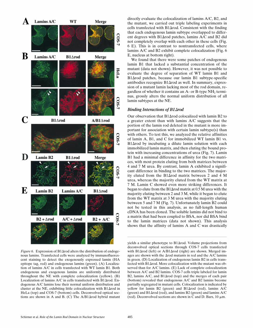
Schirmer et al. Role of the Lamin Rod Domain in Nuclear Structure 485
directly evaluate the colocalization of lamins A/C, B2, andthe mutant, we carried out triple labeling experiments incells transfected with B1Drod. Consistent with the findingthat each endogenous lamin subtype overlapped to differ-ent degrees with B1Drod patches, lamins A/C and B2 didnot completely overlap with each other in these cells (Fig.6 E). This is in contrast to nontransfected cells, wherelamins A/C and B2 exhibit complete colocalization (Fig. 6E, nucleus at bottom right).
We found that there were some patches of endogenouslamin B1 that lacked a substantial concentration of themutant (data not shown). However, it was not possible toevaluate the degree of separation of WT lamin B1 andB1Drod patches, because our lamin B1 subtype-specificantibodies recognize B1Drod as well. In summary, expres-sion of a mutant lamin lacking most of the rod domain, re-gardless of whether it contains an A- or B-type NH2 termi-nus, grossly alters the normal uniform distribution of alllamin subtypes at the NE.
Binding Interactions of B1Drod
Our observation that B1Drod colocalized with lamin B2 toa greater extent than with lamins A/C suggests that theportion of the lamin rod deleted in the mutant is more im-portant for association with certain lamin subtype(s) thanwith others. To test this, we analyzed the relative affinitiesof lamin A, B1, and C for immobilized WT lamin B1 vs.B1Drod by incubating a dilute lamin solution with eachimmobilized lamin matrix, and then eluting the bound pro-tein with increasing concentrations of urea (Fig. 7). LaminB1 had a minimal difference in affinity for the two matri-ces, with most protein eluting from both matrices between4 and 7 M urea. By contrast, lamin A exhibited a signifi-cant difference in binding to the two matrices. The major-ity eluted from the B1Drod matrix between 2 and 4 Murea, whereas the majority eluted from the WT matrix at7 M. Lamin C showed even more striking differences. Itbegan to elute from the B1Drod matrix at 0.5 M urea with themajority eluting between 2 and 3 M, while it began to elutefrom the WT matrix at 3 M urea with the majority elutingbetween 5 and 7 M (Fig. 7). Unfortunately lamin B2 couldnot be tested in this analysis, as no full-length humancDNA has been cloned. The soluble lamins did not bind toa matrix that had been coupled to BSA, nor did BSA bindto the lamin matrices (data not shown). This analysisshows that the affinity of lamins A and C was drastically
Figure 6. Expression of B1Drod alters the distribution of endoge-nous lamins. Transfected cells were analyzed by immunofluores-cent staining to detect the exogenously expressed lamin (HAepitope tag, red) and endogenous lamins (green). (A) Localiza-tion of lamins A/C in cells transfected with WT lamin B1. Bothendogenous and exogenous lamins are uniformly distributedthroughout the NE with complete colocalization (yellow). (B)Localization of lamins A/C in cells transfected with B1Drod. En-dogenous A/C lamins lose their normal uniform distribution andcluster at the NE, exhibiting little colocalization with B1Drod inHeLa (top) and COS-7 (bottom) cells. Deconvolved optical sec-tions are shown in A and B. (C) The A/B1Drod hybrid mutant
yields a similar phenotype to B1Drod. Volume projections fromdeconvolved optical sections through COS-7 cells transfectedwith B1Drod (left) or A/B1Drod (right) are shown. Merged im-ages are shown with the Drod mutants in red and the A/C laminsin green. (D) Localization of endogenous lamin B2 in cells trans-fected with B1Drod. More colocalization with the mutant was ob-served than for A/C lamins. (E) Lack of complete colocalizationbetween A/C and B2 lamins. COS-7 cells triple labeled for laminB2, lamins A/C, and B1Drod (top) and the merges of each pair(bottom) revealed that endogenous A/C and B2 lamins becomepartially segregated in mutant cells. Colocalization is indicated byyellow for lamin B2 (green) and B1Drod (red), lamins A/C(green) and B1Drod (red), and lamins B2 (green) and lamins A/C(red). Deconvolved sections are shown in C and D. Bars, 10 mm.

The Journal of Cell Biology, Volume 153, 2001 486
reduced for B1Drod compared with WT lamin B1, corre-lating with the minimal degree of overlap between laminA/C patches and mutant patches. By contrast, the affinityof WT lamin B1 is similar for itself and the mutant. Thissuggests that the internal region of the lamin B1 rod ismore important for heterotypic interactions with laminsA/C than for homotypic interactions.
Dominant Role of Lamins in Organizing other NE Proteins
The localization of integral membrane proteins of theINM also was examined in cells expressing the B1Drodmutant. Immunofluorescent staining revealed that LAP2b,like the endogenous lamins, lost its normal uniform distri-bution throughout the NE (Foisner and Gerace, 1993) andcollected in patches. The distribution of LAP2b in the NEvaried in different cells. In some cells, it was enriched indiscrete patches that had very little overlap with B1Drod-enriched patches (Fig. 8 A), similar to the pattern seen forlamins A/C. However, in other cells there were regions ofthe NE with extensive colocalization between LAP2b andthe mutant lamin, although these also contained endoge-nous lamin B2 (data not shown). In some cells, a smallamount of the LAP2b appeared to be outside of the NEand this colocalized with ER dyes (data not shown). Thepatchy localization of two other inner membrane proteins,LAP1C (Fig. 8 B) and LBR (data not shown), in cellstransfected with B1Drod was similar to that of LAP2b.Thus, several INM proteins become enriched in patches atthe NE of B1Drod-transfected cells.
The finding that LAP2b was often depleted in regions ofthe NE containing patches of B1Drod is consistent withthe finding that LAP2b binds to a central part of the laminB1 rod (Furukawa and Kondo, 1998) and that this bindingis important for its localization at the NE (Furukawa et al.,1998). Nevertheless, in all cases examined, we observedthat chromatin uniformly lined the NE of cells transfectedwith B1Drod, and was as strongly associated with regionsof the NE deficient in LAP2b (Fig. 8 C, arrow) as withthose enriched in LAP2b (arrowhead). Similarly, chroma-tin was not preferentially associated with patches ofLAP1C that arose in B1Drod-transfected cells (data notshown). Since the major chromatin binding site of laminB1 is present in the tail domain (Taniura et al., 1995) and
thus is retained in B1Drod, this suggests that chromatinbinding to the interphase NE is determined primarily bylamins (mutant plus wild type), which collectively line theNE in the transfected cells, rather than integral proteins ofthe INM, which assume a highly patchy distribution in thetransfected cells.
The distribution of NPC antigens also was examined incells transfected with B1Drod using the RL1 monoclonalantibody (Snow et al., 1987). RL1 recognizes multipleO-glycosylated NPC proteins on both the nucleoplasmicand cytoplasmic faces and characteristically shows an evenlydispersed, but punctate, distribution at the NE. In cells ex-pressing B1Drod, NPC antigens lost this distribution and clus-tered in patches (Fig. 9, left; compare staining in the untrans-fected cells). The same pattern was found for Tpr, an NPCcomponent that is localized to the nucleoplasmic face ofNPCs (data not shown). Interestingly, as was observed forlamins A/C, the NPC-enriched patches were largely distinctfrom B1Drod-enriched patches (Fig. 9 A, Merge) and, in fact,exhibited considerable colocalization with the lamin A/C-enriched patches (B, Merge). Thus, like INM proteins, NPCstend to cluster with endogenous lamins.
Finally, we functionally analyzed B1Drod-expressingcells for signal-mediated protein import and the integrityof the NE as a barrier between the nucleus and cytoplasm.Fluorescently labeled BSA conjugated to an NLS was in-jected into the cytoplasm of HeLa and COS-7 cells ex-pressing the mutant (Fig. 9 C, red). The import substrate(Fig. 9 C, blue) was efficiently concentrated in the nucleusin both cell types, indicating that the cells were competentfor signal-mediated import. A coinjected marker proteinthat lacked an NLS remained outside the nucleus (Fig. 9C, green), confirming that the NE was intact despite thedeformation of the nucleus. Similarly, we found that cellsexpressing B1Drod accumulated NLS-BSA in the nucleusin an ATP-dependent manner when analyzed in vitro afterdigitonin permeabilization (data not shown). This furthervalidates the functional integrity of the NE and NPCs inthe transfected cells.
DiscussionWe found that the lamin B1Drod mutant, when expressedin cultured cells, becomes efficiently incorporated in theNE, where it causes the redistribution of other NE pro-teins including endogenous lamins into patches, and in-duces dramatic nuclear lobulation. As the NE remains in-tact and functional for nuclear import in cells expressingB1Drod and the cells are not undergoing apoptosis, thechanges in nuclear structure induced by the mutant arelikely to be a direct consequence of changes in NE struc-ture. Our results strongly argue for a role of lamins in con-trolling the organization of other NE proteins in inter-phase and in defining nuclear shape. Furthermore, theyprovide in vivo data indicating the importance of hetero-typic interactions in the assembled lamina and suggest thatthe rod domain contributes to the specificity of hetero-typic lamin binding.
The B1Drod mutant stands out among the many laminmutants tested in the past as being the only protein thatdisrupts the uniform distribution of endogenous laminsubtypes at the NE and that drastically alters nuclear
Figure 7. Relative affinities of lamins for B1Drod and WT laminB1. WT or mutant lamin B1 were coupled with an Affi-gel matrixto which soluble WT lamins were bound. Shown are Westernblots of fractions eluted with increasing concentrations of urea,probed for each specific lamin isotype.

Schirmer et al. Role of the Lamin Rod Domain in Nuclear Structure 487
shape. The only two other lamin mutants tested that had asignificant effect on the endogenous lamina involved dele-tions of the head and tail domains (Ellis et al., 1997; Spannet al., 1997). These caused the disassembly of endogenouslamins and the formation of intranuclear lamin aggregates,but did not induce a conspicuous perturbation of nuclearshape and the cells were not tested for redistribution ofother NE proteins.
The Role of the Lamin Rod Domain in Lamina Structure
The ends of the lamin rod, which are intact in the B1Drodmutant, have been suggested to be important for dimeriza-tion and, together with the head and tail domains, forhead-to-tail polymerization of dimers (Stuurman et al.,1998). The importance of head-to-tail interactions in laminassembly is supported by our finding that the NH2- andCOOH-terminal segments derived from B1Drod do notself assemble into filaments when analyzed individually,but strongly promote self assembly when they are linkedtogether in B1Drod. The more internal regions of the rodthat are absent in B1Drod are thought to contribute tothe lateral packing of dimers in filaments (reviewed inMcLean and Lane, 1995; Stuurman et al., 1998), yet themutant still could assemble in vitro into long filaments andfilament bundles similar to those formed by WT lamin B1.Moreover, these filaments had biophysical propertiescharacteristic of IFs. This indicates that the central z4/5 ofthe rod is less critical than other regions of the lamin mole-cule for self assembly into filaments per se.
Nevertheless, the internal rod region of lamin B1 that isdeleted in B1Drod appears to be important for interac-tions with other lamin molecules, especially with otherlamin subtypes. However, it doesn’t contribute to thesame degree to interactions with different subtypes, as isapparent from the in vivo and in vitro studies reportedhere. In B1Drod-transfected cells, different lamin subtypesoverlapped to varying degrees with the mutant laminpatches. In vitro, the affinity of different lamin subtypesfor the mutant as compared with WT lamin B1 was re-duced to varying degrees. The reduction in the associationof B1Drod with the A/C lamins cannot be due simply tomismatched rod lengths (z10 nm for B1Drod vs. 48 nm forWT lamins) because WT lamin B1 bound both mutant andWT lamin B1 with similar affinity in vitro. Furthermore,the similarity of the in vivo phenotypes for B1Drod andthe A/B1Drod hybrid mutant suggests that these internalregions are more important than the head domain for dis-tinguishing heterotypic interactions. This concurs with avery recent study to address lamin heterotypic interactionsin vivo. This involved expression of a head deletion oflamin A, which caused disassembly of lamin A, but notlamin B, from the NE (Izumi et al., 2000). Previous workhad indicated that A and B lamin heterotypic interactionsare stronger than their homotypic interactions in vitro(Georgatos et al., 1988; Ye and Worman, 1995). Here wehave extended these studies by showing that the lamin roddomain contributes to heterotypic interactions of lamin B1and, further, have demonstrated for the first time thatthese interactions are relevant to lamina structure and or-ganization in vivo.
Figure 8. Changes in localiza-tion of integral membrane pro-teins of the INM induced byB1Drod. (A) Immunofluores-cent staining to detect LAP2(green) and B1Drod (red). Simi-lar results were observed whenLAP2 was visualized with apolyclonal antiserum, a mono-clonal antibody, and using aLAP2-YFP fusion protein (datanot shown). A single decon-volved optical section is shown.(B) NRK cells transfected withB1Drod (red) indicated thatLAP1 (green) also is redistrib-uted as a result of expressing themutant lamin. A deconvolvedvolume projection is shown.Bars, 10 mm. (C) Distribution ofNE proteins relative to chroma-tin. NE proteins LAP2 in greenand B1Drod in red are on theleft and DAPI staining for DNAon the right of the deconvolvedsection shown. Bar, 5 mm.

The Journal of Cell Biology, Volume 153, 2001 488
Role of the Lamina in NE Stability and Nuclear Organization
NPCs clustered in cells expressing B1Drod, yet remainedfunctional for import. The clustering of both NPCs andintegral membrane proteins of the INM into patchesthat largely colocalized with endogenous lamins providesstrong evidence that WT lamins play a key role in anchor-ing these components at the NE during interphase. Thiscomplements subcellular fractionation studies indicating aphysical connection between lamins and NPCs (Dwyerand Blobel, 1976), and in vitro binding studies showing aninteraction between lamins and various integral proteinsof the INM (Foisner and Gerace, 1993; Ye and Worman,1994; Fairley et al., 1999).
Several integral membrane proteins of the INM havebeen shown to bind directly to chromatin as well as tolamins (reviewed in Collas and Courvalin, 2000). Despitethe clustering of three INM proteins into patches in theB1Drod-transfected cells, including two (LAP2b and LBR)that bind to chromatin, chromatin was uniformly associ-ated with the NE and did not segregate preferentially withthe INM protein patches. Conversely, the endogenouslamins and B1Drod, although often segregated in patches,together formed a continuous lamin zone around the NE.These data argue that the principal basis for association ofchromatin with the NE is binding to lamins, not to integralproteins of the INM. Consistent with this notion, the ma-jor chromatin binding site of lamin B1 (Taniura et al.,1995) was retained in B1Drod.
Interestingly, although the NE was intact in B1Drod-expressing cells, we observed a fraction of the LAP2b incytoplasmic foci by fluorescence microscopy and also sawunusual concentric membrane structures in the cytoplasmby EM. We suggest that these structures might arise by thebudding off of NE membranes from regions overlayingB1Drod patches, where tethering of the membrane to thelamina might be weakened. This is consistent with the no-tion that the structural scaffolding of the lamina is essen-tial for stabilizing the INM, analogous to functions of spec-trin scaffolds at the plasma and Golgi membranes (Lorraand Huttner, 1999).
The distortion of nuclear structure caused by assemblyof B1Drod in the NE argues that lamins directly influencenuclear shape. We suggest that the extensive lobulation ofnuclei that occurs in these cells is due to the reduction inheterotypic lamin–lamin interactions and the patchworklamina that is formed as a consequence. Although theB1Drod mutant could self assemble into filamentous struc-tures, these structures would lack most of the interactionsthat normally occur along the length of the rod, and arepredicted to be less thermodynamically stable than WTlamin filaments. Thus, mutant-enriched patches of thelamina would be expected to have lower mechanicalstrength than WT patches. Moreover, interfaces betweenthe mutant and WT lamin patches are expected to beweaker on the basis of our in vitro binding results. Theweakened areas of the lamina would be expected to bemore sensitive to forces imposed over the surface of theNE by the dynamics of attached chromatin and the cyto-plasmic cytoskeleton. This could lead to disruption of thenormal curvature of the NE, causing the lobulation andgross distortion of nuclear shape that occurred in cells ex-pressing the mutant. Future studies using stably trans-fected cells may discern whether a specific concentrationof the mutant is required for these effects and if levels ofendogenous NE proteins are altered by expression of theB1Drod mutant.
Although no notable changes in nuclear shape were re-ported in previous studies in which the normal lamin orga-nization at the NE was disrupted, these results are not in-consistent with a role of the lamina in nuclear shapedetermination. In one type of study, lamin-depleted nucleiwere assembled in vitro using Xenopus egg extracts (re-viewed in Lourim and Krohne, 1994; see also Ellis et al.,1997; Spann et al., 1997). Since these nuclei were replica-tion deficient, transcriptionally inactive, and not associ-
Figure 9. Effects of B1Drod expression on NPCs. (A) Immuno-fluorescent staining to detect the NPC (green) and the mutantprotein (red). (B) Immunofluorescent staining of the NPC(green) and endogenous lamins A/C (red). (Merge) Colocaliza-tion is indicated by yellow. Deconvolved volume projections areshown. (C) B1Drod cells are capable of nuclear import. HeLacells (left) were microinjected in the cytoplasm with a fluores-cently labeled NLS-containing import substrate (blue) and a fluo-rescent 150-kD dextran (green). COS-7 cells (right) were injectedwith the same import substrate (blue) and fluorescent BSA thatdid not carry an NLS (green). HA staining for the B1Drod mu-tant (red) traces the nuclear boundary. Single confocal sectionsare shown. Bars, 10 mm.

Schirmer et al. Role of the Lamin Rod Domain in Nuclear Structure 489
ated with a cytoplasmic cytoskeleton, the unevenly dis-tributed forces from attached nuclear and cytoplasmiccomponents that would distort nuclear shape in our model(above) would be absent. In another type of study, a par-ticular lamin subtype was depleted from the nuclei of cer-tain cells by a lamin gene disruption (Lenz-Bohme et al.,1997; Sullivan et al., 1999). Gross distortions in nuclearshape would not be predicted with our model in thesecases since the cells still retained an assembled lamina inwhich the remaining lamins were uniformly distributed atthe NE. By altering the substructure of the intact, assem-bled lamina rather than by depleting lamins from the NE,expression of the B1Drod mutant provides a novel ap-proach to the question of how nuclear shape is deter-mined. Our results directly support the notion that hetero-typic associations among lamin subtypes are important forthe molecular organization of the NE and, correspond-ingly, for higher order nuclear architecture.
We thank S. Lyman for nuclear import reagents, and S. Lyman and H.Wodrich for critical reading of the manuscript. We especially thank R.Ghadiri and D. Bonn for assistance with circular dichroism and FTIRanalyses, M. Wood for EM assistance, U. Aebi for the pPEPT vector sys-tem, and H. Worman for LBR antiserum.
This work was supported by a National Institutes of Health (NIH)postdoctoral fellowship to E.C. Schirmer (F32 GM19085) and an NIHgrant to L. Gerace (GM28521).
Submitted: 15 August 2000Revised: 20 March 2001Accepted: 20 March 2001
References
Adam, S.A., R.S. Marr, and L. Gerace. 1990. Nuclear protein import in perme-abilized mammalian cells requires soluble cytoplasmic factors. J. Cell Biol.111:807–816.
Aebi, U., J. Cohn, L. Buhle, and L. Gerace. 1986. The nuclear lamina is a mesh-work of intermediate-type filaments. Nature. 323:560–564.
Benavente, R., and G. Krohne. 1986. Involvement of nuclear lamins in postmi-totic reorganization of chromatin as demonstrated by microinjection oflamin antibodies. J. Cell Biol. 103:1847–1854.
Cao, H., and R.A. Hegele. 2000. Nuclear lamin A/C R482Q mutation in Cana-dian kindreds with Dunnigan-type familial partial lipodystrophy. Hum. Mol.Genet. 9:109–112.
Collas, I., and J.C. Courvalin. 2000. Sorting nuclear membrane proteins at mito-sis. Trends Cell Biol. 10:5–8.
Coulombe, P.A., Y.M. Chan, K. Albers, and E. Fuchs. 1990. Deletions in epi-dermal keratins leading to alterations in filament organization in vivo and inintermediate filament assembly in vitro. J. Cell Biol. 111:3049–3064.
Dwyer, N., and G. Blobel. 1976. A modified procedure for the isolation of apore complex-lamina fraction from rat liver nuclei. J. Cell Biol. 70:581–591.
Ellis, D.J., H. Jenkins, W.G. Whitfield, and C.J. Hutchison. 1997. GST-lamin fu-sion proteins act as dominant negative mutants in Xenopus egg extract and re-veal the function of the lamina in DNA replication. J. Cell Sci. 110:2507–2518.
Fairley, E.A., J. Kendrick-Jones, and J.A. Ellis. 1999. The Emery-Dreifuss mus-cular dystrophy phenotype arises from aberrant targeting and binding ofemerin at the inner nuclear membrane. J. Cell Sci. 112:2571–2582.
Foisner, R., and L. Gerace. 1993. Integral membrane proteins of the nuclear en-velope interact with lamins and chromosomes, and binding is modulated bymitotic phosphorylation. Cell. 73:1267–1279.
Furukawa, K., C.E. Fritze, and L. Gerace. 1998. The major nuclear envelopetargeting domain of LAP2 coincides with its lamin binding region, but is dis-tinct from its chromatin interaction domain. J. Biol. Chem. 273:4213–4219.
Furukawa, K., and T. Kondo. 1998. Identification of the lamina-associated-polypeptide-2-binding domain of B-type lamin. Eur. J. Biochem. 251:729–733.
Geisler, N., T. Heimburg, J. Schunemann, and K. Weber. 1993. Peptides fromthe conserved ends of the rod domain of desmin disassemble intermediatefilaments and reveal unexpected structural features: a circular dichroism,fourier transform infrared, and electron microscopic study. J. Struct. Biol.110:205–214.
Georgatos, S.D., C. Stournaras, and G. Blobel. 1988. Heterotypic and homo-typic associations between the nuclear lamins: site-specificity and control byphosphorylation. Proc. Natl. Acad. Sci. USA. 85:4325–4329.
Gerace, L., and R. Foisner. 1994. Integral membrane proteins and dynamic or-ganization of the nuclear envelope. Trends Cell Biol. 4:127–131.
Glass, J.R., and L. Gerace. 1990. Lamins A and C bind and assemble at the sur-face of mitotic chromosomes. J. Cell Biol. 111:1047–1057.
Gorlich, D., and U. Kutay. 1999. Transport between the cell nucleus and the cy-toplasm. Annu. Rev. Cell. Dev. Biol. 15:607–660.
Hatzfeld, M., and K. Weber. 1991. Modulation of keratin intermediate filamentassembly by single amino acid exchanges in the consensus sequence at theC-terminal end of the rod domain. J. Cell Sci. 99:351–362.
Heald, R., and F. McKeon. 1990. Mutations of phosphorylation sites in lamin Athat prevent nuclear lamina disassembly in mitosis. Cell. 61:579–589.
Heimburg, T., J. Schunemann, K. Weber, and N. Geisler. 1996. Specific recog-nition of coiled coils by infrared spectroscopy: analysis of the three structuraldomains of type III intermediate filament proteins. Biochemistry. 35:1375–1382.
Heitlinger, E., M. Peter, M. Haner, A. Lustig, U. Aebi, and E.A. Nigg. 1991.Expression of chicken lamin B2 in Escherichia coli: characterization of itsstructure, assembly, and molecular interactions. J. Cell Biol. 113:485–495.
Heitlinger, E., M. Peter, A. Lustig, W. Villiger, E.A. Nigg, and U. Aebi. 1992.The role of the head and tail domain in lamin structure and assembly: analy-sis of bacterially expressed chicken lamin A and truncated B2 lamins. J.Struct. Biol. 108:74–89.
Izumi, M., O.A. Vaughan, C.J. Hutchison, and D.M. Gilbert. 2000. Head and/orCaaX domain deletions of lamin proteins disrupt preformed lamin A and Cbut not lamin B structure in mammalian cells. Mol. Biol. Cell. 11:4323–4337.
Kammerer, R.A., T. Schulthess, R. Landwehr, A. Lustig, D. Fischer, and J. En-gel. 1998. Tenascin-C hexabrachion assembly is a sequential two-step pro-cess initiated by coiled-coil alpha-helices. J. Biol. Chem. 273:10602–10608.
Lenz-Bohme, B., J. Wismar, S. Fuchs, R. Reifegerste, E. Buchner, H. Betz, andB. Schmitt. 1997. Insertional mutation of the Drosophila nuclear lamin Dm0gene results in defective nuclear envelopes, clustering of nuclear pore com-plexes, and accumulation of annulate lamellae. J. Cell Biol. 137:1001–1016.
Lorra, C., and W.B. Huttner. 1999. The mesh hypothesis of Golgi dynamics.Nat. Cell Biol. 1:E113–E115.
Lourim, D., and G. Krohne. 1994. Lamin-dependent nuclear envelope reassem-bly following mitosis: an argument. Trends Cell Biol. 4:314–318.
Lupas, A. 1996. Prediction and analysis of coiled-coil structures. Methods Enzy-mol. 266:513–525.
McKeon, F.D., M.W. Kirschner, and D. Caput. 1986. Homologies in both pri-mary and secondary structure between nuclear envelope and intermediatefilament proteins. Nature. 319:463–468.
McLean, W.H., and E.B. Lane. 1995. Intermediate filaments in disease. Curr.Opin. Cell Biol. 7:118–125.
Mical, T.I., and M.J. Monteiro. 1998. The role of sequences unique to nuclearintermediate filaments in the targeting and assembly of human lamin B: evi-dence for lack of interaction of lamin B with its putative receptor. J. Cell Sci.111:3471–3485.
Moir, R.D., A.D. Donaldson, and M. Stewart. 1991. Expression in Escherichiacoli of human lamins A and C: influence of head and tail domains on assem-bly properties and paracrystal formation. J. Cell Sci. 99:363–372.
Rober, R.A., K. Weber, and M. Osborn. 1989. Differential timing of nuclearlamin A/C expression in the various organs of the mouse embryo and theyoung animal: a developmental study. Development (Camb.). 105:365–378.
Sasse, B., A. Lustig, U. Aebi, and N. Stuurman. 1997. In vitro assembly ofDrosophila lamin Dm0—lamin polymerization properties are conserved.Eur. J. Biochem. 250:30–38.
Senior, A., and L. Gerace. 1988. Integral membrane proteins specific to the in-ner nuclear membrane and associated with the nuclear lamina. J. Cell Biol.107:2029–2036.
Snow, C.M., A. Senior, and L. Gerace. 1987. Monoclonal antibodies identify agroup of nuclear pore complex glycoproteins. J. Cell Biol. 104:1143–1156.
Spann, T.P., R.D. Moir, A.E. Goldman, R. Stick, and R.D. Goldman. 1997. Dis-ruption of nuclear lamin organization alters the distribution of replicationfactors and inhibits DNA synthesis. J. Cell Biol. 136:1201–1212.
Stuurman, N., S. Heins, and U. Aebi. 1998. Nuclear lamins: their structure, as-sembly, and interactions. J. Struct. Biol. 122:42–66.
Stuurman, N., B. Sasse, and P.A. Fisher. 1996. Intermediate filament proteinpolymerization: molecular analysis of Drosophila nuclear lamin head-to-tailbinding. J. Struct. Biol. 117:1–15.
Sullivan, T., D. Escalante-Alcalde, H. Bhatt, M. Anver, N. Bhat, K. Nagashima,C.L. Stewart, and B. Burke. 1999. Loss of A-type lamin expression compro-mises nuclear envelope integrity leading to muscular dystrophy. J. Cell Biol.147:913–920.
Taniura, H., C. Glass, and L. Gerace. 1995. A chromatin binding site in the taildomain of nuclear lamins that interacts with core histones. J. Cell Biol. 131:33–44.
Wilson, K.L. 2000. The nuclear envelope, muscular dystrophy and gene expres-sion. Trends Cell Biol. 10:125–129.
Ye, Q., and H.J. Worman. 1994. Primary structure analysis and lamin B andDNA binding of human LBR, an integral protein of the nuclear envelope in-ner membrane. J. Biol. Chem. 269:11306–11311.
Ye, Q., and H.J. Worman. 1995. Protein–protein interactions between humannuclear lamins expressed in yeast. Exp. Cell Res. 219:292–298.