Context and hand posture modulate the neural dynamics of tool–object perception
Upload
independentCategory
view
8download
0
Mutations in LMNA Modulate the Lamin A - Nesprin-2Interaction and Cause LINC Complex AlterationsLiu Yang1, Martina Munck1, Karthic Swaminathan1, Larisa E. Kapinos2, Angelika A. Noegel1*,
Sascha Neumann1*
1 Institute for Biochemistry I, Medical Faculty, University of Cologne, and Center for Molecular Medicine Cologne (CMMC) and Cologne Cluster on Cellular Stress
Responses in Aging-Associated Diseases (CECAD), Medical Faculty, University of Cologne, Cologne, Germany, 2 Biozentrum and the Nanoscience Institute, University of
Basel, Basel, Switzerland
Abstract
Background: In eukaryotes the genetic material is enclosed by a continuous membrane system, the nuclear envelope (NE).Along the NE specific proteins assemble to form meshworks and mutations in these proteins have been described in agroup of human diseases called laminopathies. Laminopathies include lipodystrophies, muscle and cardiac diseases as wellas metabolic or progeroid syndromes. Most laminopathies are caused by mutations in the LMNAgene encoding lamins A/C.Together with Nesprins (Nuclear Envelope Spectrin Repeat Proteins) they are core components of the LINC complex (Linkerof Nucleoskeleton and Cytoskeleton). The LINC complex connects the nucleoskeleton and the cytoskeleton and plays a rolein the transfer of mechanically induced signals along the NE into the nucleus, and its components have been attributedfunctions in maintaining nuclear and cellular organization as well as signal transduction.
Results: Here we narrowed down the interaction sites between lamin A and Nesprin-2 to aa 403–425 in lamin A and aa6146–6347 in Nesprin-2. Laminopathic mutations in and around the involved region of lamin A (R401C, G411D, G413C,V415I, R419C, L421P, R427G, Q432X) modulate the interaction with Nesprin-2 and this may contribute to the diseasephenotype. The most notable mutation is the lamin A mutation Q432X that alters LINC complex protein assemblies andcauses chromosomal and transcription factor rearrangements.
Conclusion: Mutations in Nesprin-2 and lamin A are characterised by complex genotype phenotype relations. Our datashow that each mutation in LMNAanalysed here has a distinct impact on the interaction among both proteins thatsubstantially explains how distinct mutations in widely expressed genes lead to the formation of phenotypically differentdiseases.
Citation: Yang L, Munck M, Swaminathan K, Kapinos LE, Noegel AA, et al. (2013) Mutations in LMNA Modulate the Lamin A - Nesprin-2 Interaction and Cause LINCComplex Alterations. PLoS ONE 8(8): e71850. doi:10.1371/journal.pone.0071850
Editor: Alfred Lewin, University of Florida, United States of America
Received April 9, 2013; Accepted July 3, 2013; Published August 20, 2013
Copyright: � 2013 Yang et al. This is an open-access article distributed under the terms of the Creative Commons Attribution License, which permitsunrestricted use, distribution, and reproduction in any medium, provided the original author and source are credited.
Funding: The work was supported by the Center for Molecular Medicine, University of Cologne, the Cologne Excellence Cluster on Cellular Stress Responses inAging-associated Diseases. LY is supported by a fellowship from the China Scholarship Council (CSC). The funders had no role in study design, data collection andanalysis, decision to publish, or preparation of the manuscript.
Competing Interests: The authors have declared that no competing interests exist.
* E-mail: [email protected] (AAN); [email protected] (SN)
Introduction
A hallmark of eukaryotes is the compartmentalization of the cell
and the presence of organelles. Among them, the largest one is the
nucleus that harbors the genetic material. The nucleus is
surrounded by a continuous membrane system, the nuclear
envelope (NE), that consists of two concentric membranes, inner
(INM) and outer (ONM) nuclear membrane. They are separated
by the perinuclear space and integrated along the nuclear pore
complexes. The ONM is continuous with the endoplasmic
reticulum (ER). Membrane invaginations that might consist of
one or both nuclear membranes can reach into the nucleus to form
the nucleoplasmic reticulum [1]. Research in the past decade
revealed the existence of large macromolecular protein complexes
present along or across the nuclear membranes [2–6]. The
biological importance of these assemblies is becoming more and
more evident since the number of human diseases that are known
to arise from mutations in their components is continuously
increasing. However, the majority is due to mutations in lamin A/
C proteins which are encoded by the LMNA gene.
Lamins are type V intermediate filament proteins that form a
meshwork underlying the INM. They are grouped into A- and B-
type lamins. The latter ones are encoded by the LMNB1 (lamin B1)
and LMNB2 (lamin B2) gene [7]. The LMNA gene encodes four A-
type lamins (A, AD10, C and C2) that are generated by alternative
splicing and posttranslational modifications. Lamins A and C
represent the major isoforms. The structure of lamins follows a
tripartide organization into an N-terminal head, a central rod
domain and a C-terminal tail. Lamins assemble by forming
parallel head to tail dimers stabilized by coiled coil structures in
the central rod domain. Dimers then assemble into a head to tail
polymer. Higher order structures are achieved by anti-parallel,
lateral polymer assemblies, the protofilaments [8]. Three to four
protofilaments assemble to intermediate filaments with a diameter
of about 10 nm [9].
PLOS ONE | www.plosone.org 1 August 2013 | Volume 8 | Issue 8 | e71850
So far more than 400 disease causing mutations have been
reported spread along the entire LMNAgene [9,10]. These
mutations cause several distinct diseases that include forms of
muscular dystrophy and cardiomyopathy, metabolic syndrome
and lipodystrophy, neuropathies and progeroid syndromes. This is
surprising as lamin A/C proteins are ubiquitously expressed
proteins and points towards tissue or development specific
interactions [9].
Currently three mutually non-exclusive hypotheses exist to
explain how mutations in nuclear envelope proteins might result in
the formation of laminopathies [10]. The first one is the structural
hypothesis that refers to the role of LINC complex components in
maintaining nuclear architecture and mechanotransduction across
the nuclear envelope. Mutations in LINC complex components
might cause weakened or strengthened interactions that lead to
misshapen nuclei, a hallmark of laminopathies, and impaired
transfer of mechanically induced signals across the NE [11,12].
The second hypothesis explains the role of NE proteins in the
formation in laminopathies by their ability to regulate signaling
events in a way distinct from mechanotransduction. Various NE
proteins have been shown to interact, sequester and regulate
transcription factor accessibility to the nucleus and chromatin and
therefore control the gene expression profile of a cell [13,14]. The
third hypothesis explains the formation of laminopathies by the
accumulation of mutated lamin proteins and the accompanied cell
toxic gain of function of these aggregates [15].
Along the nuclear envelope lamins A/C interact with integral
proteins of the NE like SUN-1/-2, Emerin, with transcription
factors like Fos, SREBP1 and with organizers of chromatin
organization like histones or the cell cycle regulator Cyclin D3
[16–21]. Additionally, lamins A/C interact with Nesprin-2 [22].
Nesprins (Nuclear Envelope Spectrin Repeat Proteins) are
proteins that mainly reside along the INM and ONM. So far
four Nesprins have been described in mammals (Nesprin-1, -2, -3
and -4). Each is encoded by a single gene that gives rise to multiple
isoforms. The number of identified isoforms is still increasing. So
far 21 isoforms have been identified for Nesprin-1 and 14 for
Nesprin-2 [23,24]. For Nesprin-3 two variants are known and one
for the epithelial specific Nesprin-4 [25,26]. Nesprins broadly
differ in their molecular masses from the smaller Nesprin-3
(,100 kDa) and 4 (,40 kDa) to the biggest, the so called giant
isoforms of Nesprin-1 and -2 that reach up to 1 MDa or 800 kDa,
respectively. Structural characteristics that are shared by most
Nesprin isoforms are the C-terminal KASH (Klarsicht/ANC-1/
Syne homology) domain that is a transmembrane domain and
spectrin repeats that form the basis of the central rod segment.
Even the largest Nesprins contain only one transmembrane
segment that is sufficient for connecting the proteins with the
membrane. The KASH domain additionally contains a consensus
motif at the C-terminus for the interaction with integral proteins of
the INM, the SUN (Sad1/UNC-84) proteins. The interaction of
the KASH domain of the Nesprins and the SUN domain of the
SUN proteins in the PNS forms the core of the LINC complex.
The LINC complex is a protein complex that traverses the nuclear
envelope for the direct and mechanic coupling of nucleoplasmic
and cytoplasmic components [6]. At their N-termini the different
Nesprins contain binding sites that mediate direct or indirect
connections to the actin filament system, microtubules or the
intermediate filament system and therefore an integration of the
nucleus into the cytoskeleton of cells.
Knowledge about nuclear envelope proteins and their involve-
ment in the formation of large macromolecular protein complexes
along the nuclear envelope is continuously increasing. However,
the exact nature of the interaction sites between certain LINC
complex components and the effect of distinct mutations for
particular interactions are still incomplete. A detailed understand-
ing of protein interactions will help to understand the pathological
role of disease causing mutations. In the present study we have
narrowed down the interaction sites between lamin A/C and
Nesprin-2 and explored the impact of eight laminopathic LMNA
mutations [R401C (1201C.T) [9,27,28], G411D (1232G.A)
[29], G413C (1237G.T) (www.umd.be/LMNA), V415I
(1243G.A) [9,30], R419C (1255C.T) [31], L421P (1262T.C)
[32,33], R427G (1279C.G) (www.umd.be/LMNA), Q432X
(1294C.T) [9,34]] on the binding capacities of Nesprin-2 for
lamin A/C and the formation of LINC complex assemblies.
Mutations in lamin A/C affected the interaction with Nesprin-2 in
a spectrum from increasing to decreasing. However most of the
described mutations had no obvious effect on the localization of
NE proteins along the NE. An exception was the truncation
mutation Q432X that caused severe aggregate formations of the
mutated lamin A/C protein and the sequestration of nuclear
envelope proteins.
Materials and Methods
Plasmids and site directed mutagenesisThe amino acid positions of GST and GFP Nesprin-2 proteins
used in this study refer to Nesprin-2 giant and have been described
elsewhere [14,22]. The lamin A fragments 1–263, 264–402, 345–
425 cloned into pPET-TEV expression vector are described in
[35]. The Lamin A sequence 436–548 is inserted into pET24d
(Novagen). WT lamin A amino acids 403–425 were cloned into
pEGFP-C2 (Clontech) via EcoRI and BamHI restriction sites and
the following primers: LA 403–425 For 59 AATTTCCTCT-
CACTCATCCCAGACACAGGGTGGGGGCAGCGTCA-
CAAAAAGCGCAAACTGGAGTCCACTGAG 39, Rev 59
GATCCTCAGTGGACTCCAGTTTGCGCTTTTTGGT-
GACGCTGCCCCCACCCTGTGTCTGGGATGAGTGA-
GAGGA 39. Site directed mutagenesis was performed by
QuikChange site directed mutagenesis (Stratagene) according to
the manufacturers instructions and by using full length lamin A
[36] as a template and primers carrying the corresponding point
mutations to generate GFP lamin A R401C (1201C.T), G411D
(1232G.A), G413C (1237G.T), V415I (1243G.A), R419C
(1255C.T), L421P (1262T.C), R427G (1279C.G), Q432X
(1294C.T).
Cell culture and transfectionHaCaT, COS7 cells [37,38], murine myoblasts (ATCC CRL-
1772) and primary human fibroblasts obtained from Dr. M.
Wehnert (Greifswald) [2] were grown in a humidified atmosphere
at 37uC, 5% CO2 in high Glucose Dulbeccos modified Eagle’s
medium (DMEM) (SIGMA) supplemented with 2 mM glutamine,
2 mM penicillin/streptomycin and 10% FBS. Cells were tran-
siently transfected by electroporation using Gene-PulserHII
(BioRad) at 180 V, 950 mF or by using the Amaxa cell line
NucleofectorH kit (Lonza) according to the manufacturers
instructions.
His- and GST-tag pull down assaysGST or His tagged fusion proteins were expressed in E. coli XL1
blue. Bacteria were grown to an OD600 between 0.6 and 0.8 and
protein expression over night at 20uC was induced by the addition
of 0.5 mM Isopropyl-1-thio-D-galactopyranoside (IPTG). The
bacteria were harvested, washed and lysed with STE buffer
(10 mM Tris-HCl, pH 8.0, 50 mM NaCl, 1 mM EDTA) supple-
mented with protease inhibitors. Lysis was performed by
Nesprin Lamin Interaction
PLOS ONE | www.plosone.org 2 August 2013 | Volume 8 | Issue 8 | e71850
mechanical shearing in a dounce homogenizer in the presence of
100 mg/ml lysozyme followed by 15 min incubation on ice.
Lysates from bacteria expressing GST fusion proteins were
additionally supplemented with Sarkosyl to a final concentration
of 1.5%. All samples were sonicated and centrifuged at
16.000 rpm for 30 min. Supernatants were transferred into a
new tube and lysates from bacteria expressing GST fusion proteins
were supplemented with Triton X-100 to a final concentration of
2%. GST and His-tag fusion proteins were concentrated from
bacterial lysates by adding glutathione Sepharose 4B or Ni-NTA
beads on at 4uC. Beads were washed five times with PBS to
remove unspecifically bound proteins. COS7 cells expressing the
corresponding GFP plasmids were lysed with lysis buffer (50 mM
Tris-HCl, pH 7.5, 150 mM NaCl, 1% Nonidet-P40, and 0.5%
sodium deoxycholate) supplemented with protease inhibitors. For
preclearing, lysates of COS7 cells expressing GFP fusion proteins
were incubated with beads for one hour at 4uC followed by an on
incubation with the corresponding GST- or Ni-NTA-beads bound
fusion proteins. Finally beads were washed five times with PBS
supplemented with protease inhibitors. SDS loading buffer was
added and samples were heated for 5 min at 95uC and analysed by
SDS-PAGE followed by Coomassie Blue staining or western blot.
Immunofluorescence and microscopyCells were fixed on coverslips for 15 min in 4% paraformalde-
hyde in PBS followed by 5 min permeabilization in 0.5% Triton
X-100 in PBS. Fixed samples were incubated for 30 min to 1 h in
phosphate-buffered gelatine for blocking (PBS, 0.1% fish gelatine
and 0.5% BSA) followed by incubation with primary antibodies or
TRITC Phalloidin (Sigma) for 1 h at RT or overnight at 4uCfollowed by three washing steps with PBS, 5 min each. Appropri-
ate secondary antibodies conjugated to Alexa 568 or Alexa 488
were applied. Nuclei were stained with 4,6-diamidino-2-pheny-
lindole (DAPI). Samples were again extensively washed with PBS
and fixed with gelvatol. Immunofluorescence analysis was
performed as described [2].
AntibodiesThe following antibodies were used in this study; polyclonal
rabbit anti Nesprin-2 pAbK1 [22], mouse monoclonal anti GFP
K3-148-2, mouse mAb specific for Emerin (4G5, abcam),
polyclonal rabbit anti lamin B1 (abcam), polyclonal rabbit anti
lamin A (H-102, Santa Cruz).
Quantification of western blot and Coomassie Bluestained gels
Quantification was performed by using the AlphaEaseFC
software, version 4.0.0, Alpha Innotech Corporation. GFP lamin
A signals were normalized against GST Nesprin-2 SR52,53 by
generating the ratio of western blot GFP lamin A and Coomassie
Blue GST Nesprin-2 signals. The ratio between WT GFP lamin A
SR52,53 and GST Nesprin-2 was set to 100%. The percentage of
enhanced or decreased binding between mutated GFP lamins and
WT GST Nesprin-2 was set in correlation to the WT ratio.
Heat shock experimentsHeat shock was performed by incubating cells plated on cover
slips in 24 well plates in an incubator at 42uC for 15 min under
otherwise normal cell culture conditions in a humidified atmo-
sphere with 5% CO2. Afterwards cells were fixed by the addition
of methanol and 10 min incubation at 220uC, followed by
extensive washing and immunofluorescence analysis as described
above.
Protein structure predictionaa sequences of human lamin A (P02545), Musmusculuslamin A
(P48678) and lamin B (P20700) were taken from the UniProt
database and aligned using the clustalW2 online program [39].
Aligned sequences were further processed by using ESPript 2.2
[40] for representation. For lamin A structural model construction,
lamin A amino acids 351–490 that encompass a part of the N-
terminal coil2B and the C-terminal globular domain were used in
the MULTICOM server [41]. The templates used for modeling
1UFGA [42] (C-terminal immunoglobulin like domain of mouse
Lamin A), 1IFRA (globular tail of human Lamin A), and 2LLA
[43] (chain A of Mannose-6-phosphate/insulin-like growth factor
II receptor) were taken from the PDB (Protein Data Bank).
Detailed amino acid sequences and exchanges were generated by
using SwissPDB Viewer v4.1.0 [44] and the molecular surfaces
were generated by using pyMOL v1.3.
Results
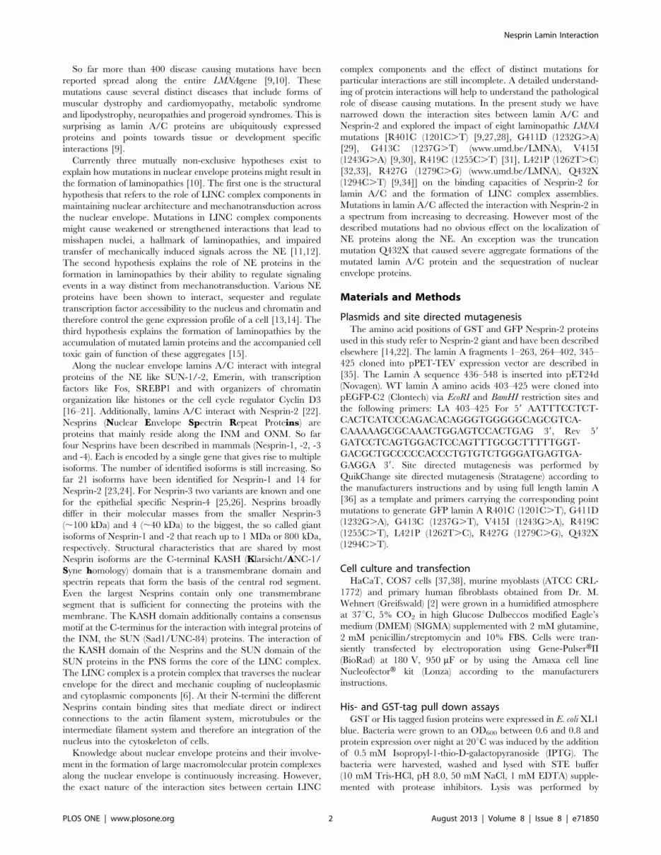
C-termini of Nesprin-2 and lamin A interactWe defined here the exact binding sites between lamin A/C and
Nesprin-2 by performing a series of in vitro pull down experiments
with purified recombinant Nesprin-2 and lamin A proteins
(Fig. 1A, B). GST tagged Nesprin-2 polypeptides we used for
our studies were derived from the C-terminus encompassing aa
6146–6799 of human Nesprin-2 giant (Fig. 1A). We have
described these polypeptides before as Nesprin-2 SRs 19–22
[22], however here we have renamed them according to a recent
detailed structural analysis predicting almost the entire Nesprin-2
sequence is made from more or less well conserved spectrin repeats
[24]. For lamin A we used polypeptides spanning amino acids 1–
548 of human lamin A (Fig. 1B). Nesprin-2 SR 52–56
coprecipitated with lamin A amino acids 345–425 (Fig. 1C).
Using lamin A polypeptides 264–402 and 345–425 which share an
overlapping sequence (Fig. 1B) we found that Nesprin-2 SR 52–56
did not coprecipitate with lamin A 264–402, letting us conclude
that the interaction site of lamin A for Nesprin-2 lies within the
amino acids 403–425 of lamin A. To further define the interaction
site lamin A fragments were incubated with GFP Nesprin-2
polypeptides expressed in COS7 cells. Nesprin-2 SR 52,53 showed
the strongest interaction to lamin A, whereas no interaction was
found for SR55, 56. A weak signal was detected for SR53, 54, 55
(Fig. 1D). To confirm the afore described interaction, we cloned
the sequences containing the lamin A interaction site for Nesprin-2
(lamin A aa 403–425) into a GFP expression vector and incubated
these proteins with purified Nesprin-2 polypeptides, which
confirmed the interaction of lamin A to Nesprin-2 SR52,53
(Fig. 1E). Finally, we addressed the question if lamin A binds to
SR52 or SR53 and found that Nesprin-2 SR53 is sufficient to
precipitate GFP lamin A aa 403–425 from total cell lysates. The
GFP lamin A 403–425 signal for SR53 was always much weaker
compared to Nesprin-2 SR52,53 (Fig. 1F). Taken together aa 403–
425 in lamin A and aa 6146–6347 corresponding to SR52,53 in
Nesprin-2 mediate the interaction between both proteins.
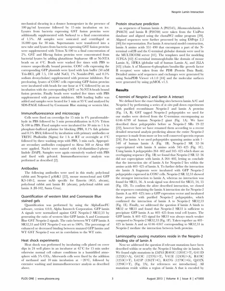
Laminopathy causing mutations reside in the Nesprin-2binding site of lamin A
Next we addressed the question if relevant mutations have been
described within or nearby the Nesprin-2 binding site in lamin A.
We found eight mutations in LMNA [R401C (1201C.T), G411D
(1232G.A), G413C (1237G.T), V415I (1243G.A), R419C
(1255C.T), L421P (1262T.C), R427G (1279C.G), Q432X
(1294C.T), (Fig. 2A), for references see introduction]. All
mutations reside within a region of lamin A that is encoded by
Nesprin Lamin Interaction
PLOS ONE | www.plosone.org 3 August 2013 | Volume 8 | Issue 8 | e71850
Figure 1. Nesprin-2 SR52,53 (aa 6146–6347) interacts with lamin A residues extending from position 403 to 425. (A) Schematic of GSTNesprin-2 fusion proteins used in this study. Aa positions refer to Nesprin-2 giant. (B) Schematic of His tag lamin A fusion proteins. (C) Bacteriallyexpressed lamin A proteins bound to beads were incubated with lysates of COS7 cells expressing GFP Nesprin-2 SR52-56. The polypeptides used forpull down are shown in the upper panel. GFP-tagged Nesprin-2 SR52-56 was detected with mAb K3-184 (WB: anti GFP). The molecular weightmarkers are indicated on the left. GFP-tagged Nesprin-2 SR52-56 precipitates with lamin A polypeptides 345–425. No signals were detected for laminA 264–402. For this reason we used these two lamin A proteins for our following experiments, one as a positive control and one as a negative control,to narrow the binding site of Nesprin-2 to lamin A. (D) Identification of the lamin A binding site in Nesprin-2. GFP Nesprin-2 polypeptides harboringindividual SRs were expressed as GFP tagged proteins in COS7 cells (lower panels) and incubated with lamin A fusion proteins (upper panel,Coomassie Blue stained SDS PAGE, 15% acrylamide). (E) Interaction of GST Nesprin-2 fusion proteins bound to beads with GFP Lamin A aa 403–425.(F) Determination of the Nesprin-2 SR domain for interaction with lamin. Nesprin-2 SR52 and SR53 were expressed as GST fusion proteins and bindingto GFP lamin A 403–425 was probed. For all experiments the use of equal protein amounts is demonstrated by Coomassie Blue stained SDS-PAGE.Equal amounts of GFP fusion proteins are shown by western blots of supernatants after coupling the GST and GFP fusion proteins. Nes-2 SR –Nesprin-2 Spectrin Repeats, LA – lamin A, WB – western blot, SPN – supernatant, P – pellet.doi:10.1371/journal.pone.0071850.g001
Nesprin Lamin Interaction
PLOS ONE | www.plosone.org 4 August 2013 | Volume 8 | Issue 8 | e71850
LMNA exon 7 and all are point mutations causing single nucleotide
exchanges. Seven mutations result in amino acid changes whereas
mutation Q432X (1294C.T) results in a stop codon (Fig. 2A).
The predicted molecular weight for GFP WT lamin A and all
mutations is ,100 kDa, for the truncated GFP lamin A Q432X
protein the predicted molecular weight is ,76 kDa. In western
blots all lamin A variants are detectable at the predicted molecular
weights (Fig. 2B).
Influence of LMNA mutations on the distribution ofinteraction partners
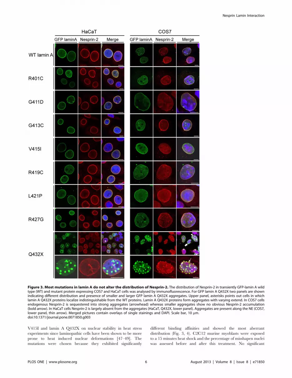
To test if mutations in lamin A cause changes in NE protein
assemblies, the distribution of WT and mutated GFP lamin A and
endogenous LINC complex components was studied in immuno-
fluorescence analysis. We utilized HaCaT and COS7 cells which
differ with respect to their morphology and the expression levels of
Nesprins. Previous work from our group demonstrated that
HaCaT keratinocytes express higher Nesprin-2 protein levels
compared to fibroblast cells [11,45]. The fibroblasts like COS7
cells express lower levels of Nesprin-2 as compared to HaCaT
keratinocytes. We found all GFP fusion proteins localizing along
the nuclear envelope like WT lamin A. GFP lamin A Q423X
proteins showed a different distribution. In COS7 as well as in
HaCaT cells Q432X localized along the NE (Fig. 3, asterisk),
additionally, GFP lamin A Q432X proteins formed aggregates
that seemed to appear along the NE rather than inside the nucleus
(Fig. 3, thin arrow). In HaCaT cells Nesprin-2 was largely
unaffected by these aggregates and showed the typical rim staining
(Fig. 3). Only when large aggregates were present Nesprin-2 was
also recruited to these aggregates (data not shown). By contrast, in
COS7 cells Nesprin-2 was sequestered into large lamin A
aggregates (Fig. 3, arrowhead). In cells with intermediate sized
aggregates one can observe both, a recruitment (Fig. 3, thin arrow)
and an absence of Nesprin-2 (Fig. 3, bold arrow) in the aggregates.
Different distributions were observed for further NE components
like lamin B1 (Fig. S1) or Emerin (Fig. S2). In HaCaT cells lamin
B1 was present only in large aggregates, whereas in COS7 it
appeared in both small and large aggregates. Emerin was present
in large aggregates formed in both cell lines to comparable extend.
Endogenous lamin A was also sequestered into GFP lamin A
Q432X aggregates (Fig. S3).
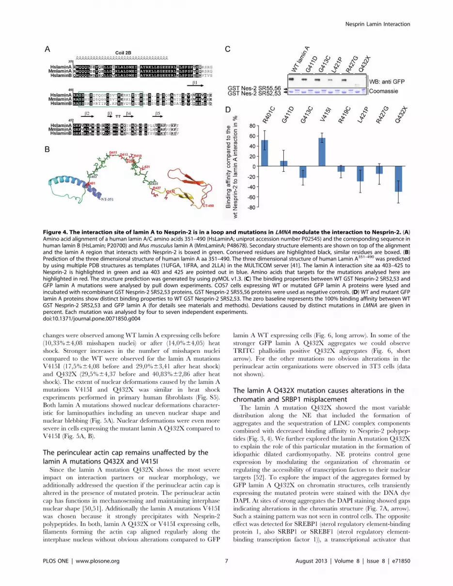
Mutations in LMNA modulate binding affinities of lamin Afor Nesprin-2
Next we addressed the question if the lamin A mutations
analysed here modulate the affinity to GST Nesprin-2 protein
complexes. For this, we first made structural predictions of the
region in lamin A that interacts with Nesprin-2 and found that the
interaction site in lamin A is located in a loop (Fig. 4B). This loop
connects the a helical rod domain and the globular domain at the
C-terminus (Fig. 4A). The affected amino acids should therefore
be accessible for interactions. The impact of the amino acid
exchanges caused by the lamin A mutations analysed here reach
from charge neutrality to the generation of positively or negatively
charged residues (Fig. S4). Recombinantly expressed GST Ne-
sprin-2 SR 52,53 was used as positive and GST Nesprin-2
SR55,56 as negative control. The proteins were incubated with
lysates of COS7 cells expressing the corresponding GFP lamin A
proteins (Fig. 4C). Mutated lamin A proteins were precipitated in
varying amounts with GST Nesprin-2 SR52,53 (Fig. 4C, shown
for lamin A mutations G411D, G413C, L421P, R427G and
Q432X). The lamin A mutations R401C and V415I were
precipitated at higher amounts compared to WT GFP lamin A,
whereas only small amounts of Q432X were precipitated by GST
Nesprin-2 SR52,53 (Fig. 4D). All further mutations were
precipitated at levels comparable to WT GFP lamin A.
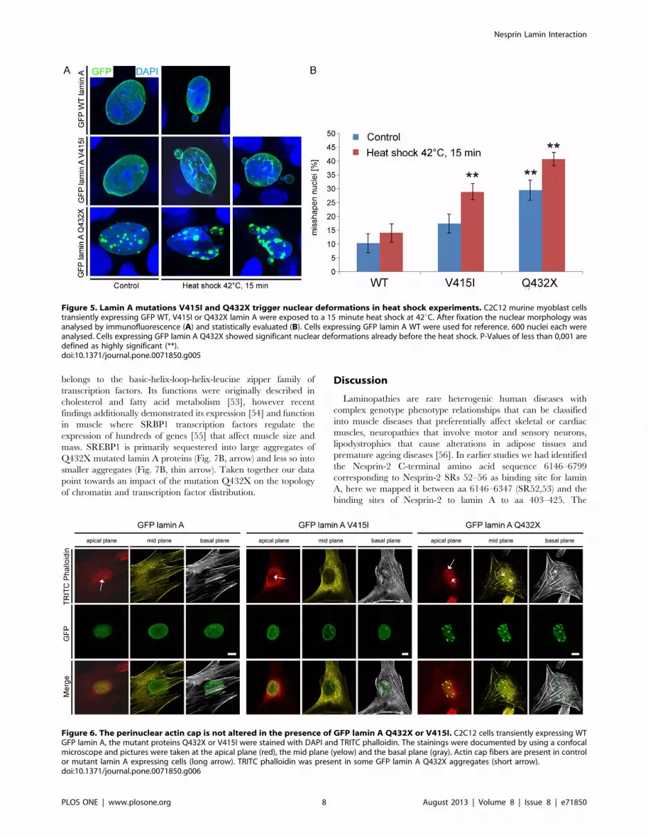
Mutations in lamin A cause the formation of misshapennuclei after heat exposure
Laminopathies often affect tissues that are under pronounced
mechanical strain like skeletal muscle or the heart. A hallmark of
laminopathies is the presence of dysmorphic nuclei, impaired
mechanical properties and stiffness of the nucleus [46]. We
exemplarily explored the effect of two lamin A mutations, lamin A
Figure 2. Schematic of lamin A mutations. (A) aa residues 399 to 434 of WT lamin A are shown. The interaction site of lamin A to Nesprin-2 isgiven in green. Below, the WT sequence mutations analysed in the present study are highlighted. Similarities to the WT sequence are shown as blacklines, exchanged aa are indicated in red. The mutation Q432X leads to a stop codon (X). (B) Western blot analysis of GFP lamin A fusion proteins. Theproteins were transiently expressed in COS7 cells, lysates were analysed by SDS-PAGE followed by western blot with mAb K3-184 that confirmed thepredicted molecular weights (given in kDa on the left). WB – western blot.doi:10.1371/journal.pone.0071850.g002
Nesprin Lamin Interaction
PLOS ONE | www.plosone.org 5 August 2013 | Volume 8 | Issue 8 | e71850
V415I and lamin A Q432X on nuclear stability in heat stress
experiments since laminopathic cells have been shown to be more
prone to heat induced nuclear deformations [47–49]. The
mutations were chosen because they exhibited significantly
different binding affinities and showed the most aberrant
distribution (Fig. 3, 4). C2C12 murine myoblasts were exposed
to a 15 minutes heat shock and the percentage of misshapen nuclei
was assessed before and after this treatment. No significant
Figure 3. Most mutations in lamin A do not alter the distribution of Nesprin-2. The distribution of Nesprin-2 in transiently GFP-lamin A wildtype (WT) and mutant protein expressing COS7 and HaCaT cells was analysed by immunofluorescence. For GFP lamin A Q432X two panels are shownindicating different distribution and presence of smaller and larger GFP lamin A Q432X aggregates. Upper panel, asterisks points out cells in whichlamin A Q432X proteins localize indistinguishable from the WT proteins. Lamin A Q432X proteins form aggregates with varying extend. In COS7 cellsendogenous Nesprin-2 is sequestered into strong aggregates (arrowhead) whereas smaller aggregates show no obvious Nesprin-2 accumulation(bold arrow). In HaCaT cells Nesprin-2 is largely absent from the aggregates (HaCaT, Q432X, lower panel). Aggregates are present along the NE (COS7,lower panel, thin arrow). Merged pictures contain overlays of single stainings and DAPI. Scale bar, 10 mm.doi:10.1371/journal.pone.0071850.g003
Nesprin Lamin Interaction
PLOS ONE | www.plosone.org 6 August 2013 | Volume 8 | Issue 8 | e71850
changes were observed among WT lamin A expressing cells before
(10,33%64,08 misshapen nuclei) or after (14,0%64,05) heat
shock. Stronger increases in the number of misshapen nuclei
compared to the WT were observed for the lamin A mutations
V415I (17,5%64,08 before and 29,0%63,41 after heat shock)
and Q432X (29,5%64,37 before and 40,83%62,86 after heat
shock). The extent of nuclear deformations caused by the lamin A
mutations V415I and Q432X was similar in heat shock
experiments performed in primary human fibroblasts (Fig. S5).
Both lamin A mutations showed nuclear deformations character-
istic for laminopathies including an uneven nuclear shape and
nuclear blebbing (Fig. 5A). Nuclear deformations were even more
severe in cells expressing the mutant lamin A Q432X compared to
V415I (Fig. 5A, B).
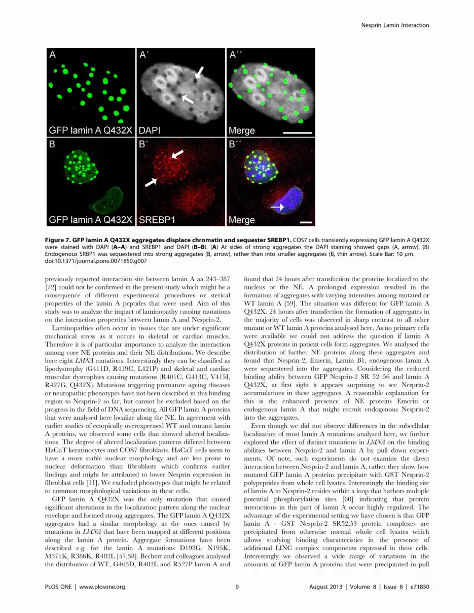
The perinculear actin cap remains unaffected by thelamin A mutations Q432X and V415I
Since the lamin A mutation Q432X shows the most severe
impact on interaction partners or nuclear morphology, we
additionally addressed the question if the perinuclear actin cap is
altered in the presence of mutated protein. The perinuclear actin
cap has functions in mechanosensing and maintaining interphase
nuclear shape [50,51]. Additionally the lamin A mutations V415I
was chosen because it strongly precipitates with Nesprin-2
polypeptides. In both, lamin A Q432X or V415I expressing cells,
filaments forming the actin cap aligned regularly along the
interphase nucleus without obvious alterations compared to GFP
lamin A WT expressing cells (Fig. 6, long arrow). In some of the
stronger GFP lamin A Q432X aggregates we could observe
TRITC phalloidin positive Q432X aggregates (Fig. 6, short
arrow). For the other mutations no obvious alterations in the
perinuclear actin organizations were observed in 3T3 cells (data
not shown).
The lamin A Q432X mutation causes alterations in thechromatin and SRBP1 misplacement
The lamin A mutation Q432X showed the most variable
distribution along the NE that included the formation of
aggregates and the sequestration of LINC complex components
combined with decreased binding affinity to Nesprin-2 polypep-
tides (Fig. 3, 4). We further explored the lamin A mutation Q432X
to explain the role of this particular mutation in the formation of
idiopathic dilated cardiomyopathy. NE proteins control gene
expression by modulating the organization of chromatin or
regulating the accessibility of transcription factors to their nuclear
targets [52]. To explore the impact of the aggregates formed by
GFP lamin A Q432X on chromatin structures, cells transiently
expressing the mutated protein were stained with the DNA dye
DAPI. At sites of strong aggregates the DAPI staining showed gaps
indicating alterations in the chromatin structure (Fig. 7A, arrow).
Such a staining pattern was not seen in control cells. The opposite
effect was detected for SREBP1 (sterol regulatory element-binding
protein 1, also SRBP1 or SREBF1 (sterol regulatory element-
binding transcription factor 1)), a transcriptional activator that
Figure 4. The interaction site of lamin A to Nesprin-2 is in a loop and mutations in LMNA modulate the interaction to Nesprin-2. (A)Amino acid alignment of a human lamin A/C amino acids 351–490 (HsLaminA; uniprot accession number P02545) and the corresponding sequence inhuman lamin B (HsLamin; P20700) and Mus musculus lamin A (MmLaminA; P48678). Secondary structure elements are shown on top of the alignmentand the lamin A region that interacts with Nesprin-2 is boxed in green. Conserved residues are highlighted black, similar residues are boxed. (B)Prediction of the three dimensional structure of human lamin A aa 351–490. The three dimensional structure of human Lamin A351–490 was predictedby using multiple PDB structures as templates (1UFGA, 1IFRA, and 2LLA) in the MULTICOM server [41]. The lamin A interaction site aa 403–425 toNesprin-2 is highlighted in green and aa 403 and 425 are pointed out in blue. Amino acids that targets for the mutations analysed here arehighlighted in red. The structure prediction was generated by using pyMOL v1.3. (C) The binding properties between WT GST Nesprin-2 SR52,53 andGFP lamin A mutations were analysed by pull down experiments. COS7 cells expressing WT or mutated GFP lamin A proteins were lysed andincubated with recombinant GST Nesprin-2 SR52,53 proteins. GST Nesprin-2 SR55,56 proteins were used as negative controls. (D) WT and mutant GFPlamin A proteins show distinct binding properties to WT GST Nesprin-2 SR52,53. The zero baseline represents the 100% binding affinity between WTGST Nesprin-2 SR52,53 and GFP lamin A (for details see materials and methods). Deviations caused by distinct mutations in LMNA are given inpercent. Each mutation was analysed by four to seven independent experiments.doi:10.1371/journal.pone.0071850.g004
Nesprin Lamin Interaction
PLOS ONE | www.plosone.org 7 August 2013 | Volume 8 | Issue 8 | e71850
belongs to the basic-helix-loop-helix-leucine zipper family of
transcription factors. Its functions were originally described in
cholesterol and fatty acid metabolism [53], however recent
findings additionally demonstrated its expression [54] and function
in muscle where SRBP1 transcription factors regulate the
expression of hundreds of genes [55] that affect muscle size and
mass. SREBP1 is primarily sequestered into large aggregates of
Q432X mutated lamin A proteins (Fig. 7B, arrow) and less so into
smaller aggregates (Fig. 7B, thin arrow). Taken together our data
point towards an impact of the mutation Q432X on the topology
of chromatin and transcription factor distribution.
Discussion
Laminopathies are rare heterogenic human diseases with
complex genotype phenotype relationships that can be classified
into muscle diseases that preferentially affect skeletal or cardiac
muscles, neuropathies that involve motor and sensory neurons,
lipodystrophies that cause alterations in adipose tissues and
premature ageing diseases [56]. In earlier studies we had identified
the Nesprin-2 C-terminal amino acid sequence 6146–6799
corresponding to Nesprin-2 SRs 52–56 as binding site for lamin
A, here we mapped it between aa 6146–6347 (SR52,53) and the
binding sites of Nesprin-2 to lamin A to aa 403–425. The
Figure 5. Lamin A mutations V415I and Q432X trigger nuclear deformations in heat shock experiments. C2C12 murine myoblast cellstransiently expressing GFP WT, V415I or Q432X lamin A were exposed to a 15 minute heat shock at 42uC. After fixation the nuclear morphology wasanalysed by immunofluorescence (A) and statistically evaluated (B). Cells expressing GFP lamin A WT were used for reference. 600 nuclei each wereanalysed. Cells expressing GFP lamin A Q432X showed significant nuclear deformations already before the heat shock. P-Values of less than 0,001 aredefined as highly significant (**).doi:10.1371/journal.pone.0071850.g005
Figure 6. The perinuclear actin cap is not altered in the presence of GFP lamin A Q432X or V415I. C2C12 cells transiently expressing WTGFP lamin A, the mutant proteins Q432X or V415I were stained with DAPI and TRITC phalloidin. The stainings were documented by using a confocalmicroscope and pictures were taken at the apical plane (red), the mid plane (yelow) and the basal plane (gray). Actin cap fibers are present in controlor mutant lamin A expressing cells (long arrow). TRITC phalloidin was present in some GFP lamin A Q432X aggregates (short arrow).doi:10.1371/journal.pone.0071850.g006
Nesprin Lamin Interaction
PLOS ONE | www.plosone.org 8 August 2013 | Volume 8 | Issue 8 | e71850
previously reported interaction site between lamin A aa 243–387
[22] could not be confirmed in the present study which might be a
consequence of different experimental procedures or sterical
properties of the lamin A peptides that were used. Aim of this
study was to analyze the impact of laminopathy causing mutations
on the interaction properties between lamin A and Nesprin-2.
Laminopathies often occur in tissues that are under significant
mechanical stress as it occurs in skeletal or cardiac muscles.
Therefore it is of particular importance to analyze the interaction
among core NE proteins and their NE distributions. We describe
here eight LMNA mutations. Interestingly they can be classified as
lipodystrophy (G411D, R419C, L421P) and skeletal and cardiac
muscular dystrophies causing mutations (R401C, G413C, V415I,
R427G, Q432X). Mutations triggering premature ageing diseases
or neuropathic phenotypes have not been described in this binding
region to Nesprin-2 so far, but cannot be excluded based on the
progress in the field of DNA sequencing. All GFP lamin A proteins
that were analysed here localize along the NE. In agreement with
earlier studies of ectopically overexpressed WT and mutant lamin
A proteins, we observed some cells that showed altered localiza-
tions. The degree of altered localization patterns differed between
HaCaT keratinocytes and COS7 fibroblasts. HaCaT cells seem to
have a more stable nuclear morphology and are less prone to
nuclear deformation than fibroblasts which confirms earlier
findings and might be attributed to lower Nesprin expression in
fibroblast cells [11]. We excluded phenotypes that might be related
to common morphological variations in these cells.
GFP lamin A Q432X was the only mutation that caused
significant alterations in the localization pattern along the nuclear
envelope and formed strong aggregates. The GFP lamin A Q432X
aggregates had a similar morphology as the ones caused by
mutations in LMNA that have been mapped at different positions
along the lamin A protein. Aggregate formations have been
described e.g. for the lamin A mutations D192G, N195K,
M371K, R386K, R482L [57,58]. Bechert and colleagues analysed
the distribution of WT, G465D, R482L and R527P lamin A and
found that 24 hours after transfection the proteins localized to the
nucleus or the NE. A prolonged expression resulted in the
formation of aggregates with varying intensities among mutated or
WT lamin A [59]. The situation was different for GFP lamin A
Q432X. 24 hours after transfection the formation of aggregates in
the majority of cells was observed in sharp contrast to all other
mutant or WT lamin A proteins analysed here. As no primary cells
were available we could not address the question if lamin A
Q432X proteins in patient cells form aggregates. We analysed the
distribution of further NE proteins along these aggregates and
found that Nesprin-2, Emerin, Lamin B1, endogenous lamin A
were sequestered into the aggregates. Considering the reduced
binding ability between GFP Nesprin-2 SR 52–56 and lamin A
Q432X, at first sight it appears surprising to see Nesprin-2
accumulations in these aggregates. A reasonable explanation for
this is the enhanced presence of NE proteins Emerin or
endogenous lamin A that might recruit endogenous Nesprin-2
into the aggregates.
Even though we did not observe differences in the subcellular
localization of most lamin A mutations analysed here, we further
explored the effect of distinct mutations in LMNA on the binding
abilities between Nesprin-2 and lamin A by pull down experi-
ments. Of note, such experiments do not examine the direct
interaction between Nesprin-2 and lamin A, rather they show how
mutated GFP lamin A proteins precipitate with GST Nesprin-2
polypeptides from whole cell lysates. Interestingly the binding site
of lamin A to Nesprin-2 resides within a loop that harbors multiple
potential phosphorylation sites [60] indicating that protein
interactions in this part of lamin A occur highly regulated. The
advantage of the experimental setting we have chosen is that GFP
lamin A - GST Nesprin-2 SR52,53 protein complexes are
precipitated from otherwise normal whole cell lysates which
allows studying binding characteristics in the presence of
additional LINC complex components expressed in these cells.
Interestingly we observed a wide range of variations in the
amounts of GFP lamin A proteins that were precipitated in pull
Figure 7. GFP lamin A Q432X aggregates displace chromatin and sequester SREBP1. COS7 cells transiently expressing GFP lamin A Q432Xwere stained with DAPI (A–A) and SREBP1 and DAPI (B–B). (A) At sides of strong aggregates the DAPI staining showed gaps (A, arrow). (B)Endogenous SRBP1 was sequestered into strong aggregates (B, arrow), rather than into smaller aggregates (B, thin arrow). Scale Bar: 10 mm.doi:10.1371/journal.pone.0071850.g007
Nesprin Lamin Interaction
PLOS ONE | www.plosone.org 9 August 2013 | Volume 8 | Issue 8 | e71850
down experiments that reached from enhanced (e.g. R401C,
V415I) to decreased signals (e.g. Q432X) compared to WT GFP
lamin A. Further mutations analysed here showed mild alterations.
The truncation in Q432X does not include the amino acids 403–
425 that mediate the lamin A interaction to Nesprin-2. However
lamin A Q432X precipitates in lower amounts with GST tagged
Nesprin-2 polypeptides. A reasonable explanation for this is that
the loss of the C-terminus of lamin A causes alterations in the three
dimensional protein structure which might make the interacting
amino acids less accessible. Over all these findings contribute to
the explanation how distinct mutations in ubiquitously expressed
proteins like lamin A lead to the formation of diseases with strong
variations in their clinical manifestations as it is known for
laminopathies. Distinct mutations in LMNA have distinct effects on
the formation of NE protein assemblies as it is shown here for
interaction between the core NE components Nesprin-2 and lamin
A. However the impact of distinct mutations on the interaction
between Nesprin-2 and lamin A is not reflected in the aspect of
nuclear stability. On the protein level lamin A V415I causes an
enhanced interaction with Nesprin-2, whereas the lamin A Q432X
interaction is weaker compared to the WT. At the immunofluo-
rescence level they show differential distributions as well. Lamin A
V415I is indistinguishable from the control, lamin A Q432X
causes the formation of aggregates (Fig. 3). However, both
mutations cause cardiomyopathic phenotypes, lone atrial fibrilla-
tion is caused by lamin A V415I and idiopathic dilated
cardiomyopathy by lamin A Q432X. When we expressed both
mutations in murine myoblasts or primary fibroblasts and treated
them with a heat shock at 42uC both mutations led to a significant
increase in the numbers of misshapen nuclei which shows that
each mutation has additional distinct effects on NE protein
assemblies. The underlying molecular mechanisms for the
discrepancy between the contradictory behavior of the mutations
V415I and Q432X in pull down experiments and a similar
tendency in the heat stress experiments might be related to the
complexity of NE protein assemblies. Lamins exhibit tripartite
organization and are composed of an N-terminal head, a central
rod and a C-terminal tail. The tail region that is missing in Q432X
might impair the assembly of lamins into a meshwork and thus
lead to pronounced nuclear fragility. On the other side the
mutation V415I precipitates stronger with GST Nesprin-2
polypeptides indicating that this mutation modulates the binding
properties among the NE protein network that finally results in
enhanced nuclear fragility.
Nesprin-2 is an actin binding protein that connects the NE to
the perinuclear cytoskeleton. Recently the Wirtz group identified a
perinuclear actin cap that consists of highly organized actin fibers
closely connected to the apical surface of the interphase nucleus
[51]. LINC complexes physically connect the actin cap to the NE,
and the actin cap is required to maintain the interphase nuclear
shape and is likely involved in mechanosensing and mechan-
otransduction. Malfunctions in the actin cap might explain the
pathological role of distinct lamin A mutations. An intact lamina
and LINC complexes are necessary for a functional actin cap [50].
In GFP lamin A Q432X and V415I expressing cells the overall
actin cap structure remained unaffected (Fig. 6). In lamin A
Q432X expressing cells we observed a subset of phalloidin positive
lamin A Q432X aggregates. The fact that not all aggregates were
phalloidin positive might be explained with the highly dynamic
nature of the actin cap fibers that might form only in response to
mechanical strain [51]. It remains to be determined if the observed
changes in the actin structure lead to disturbed mechanotransduc-
tion from the extracellular milieu to the nucleus and thus
contribute to the disease phenotype.
Alterations in the binding affinities between Nesprin-2 and
lamin A have consequences on the NE protein network that are
transmitted to further binding partners. Since lamins localize along
the inner surface of the INM and Nesprins as type II
transmembrane proteins can be integrated into the INM or the
ONM, the interaction between both proteins occurs along the
INM and the functional impact of altered affinities reaches into the
nucleus. Interestingly most lamin interactions have been mapped
to the C-terminus [8] and therefore overlap with the interaction
side for Nesprin-2. Alterations in the binding affinities between
both proteins therefore make lamins more or less accessible for
other interactions. NE proteins have been described in maintain-
ing chromosomal organization to provide a chromatin environ-
ment that is favorable or unfavorable for gene expression in which
the position of a gene is related to its expression level [52,61].
Lamins directly interact with DNA or Histones. The DNA binding
site of lamins A/C was mapped to aa 411–553 [62] and aa 396–
430 interact with histones [20]. Both sites are close to or part of aa
403–425 that interact with Nesprin-2 and variations in the lamin
adhesion to Nesprin-2 could thus affect chromosomal arrange-
ments and consequentially gene expression. Mutated Q432X
lamin A proteins show the most severe impact on chromosome
structures by the formation of gaps that are formed in the
otherwise even DNA staining at sides of aggregates.
NE proteins additionally have functions in controlling the
temporal and spatial accessibility of transcription factors to their
nuclear targets. Lamins for example play a role in FOS or
SREBP1 [18,19] signaling and Nesprins are part of SMAD, FOS
and Wnt signal transduction [13,14]. Mutated lamin A Q432X
proteins form aggregates and sequester LINC complex compo-
nents. To further explore the pathological role of this mutation, we
analysed the impact of the mutated proteins on the subcellular
distribution of the transcription factor SREBP1 that is a known
interaction partner of lamin A. The interaction site of lamin A to
SREBP1 lies between amino acids 389–664 and therefore overlaps
with the binding lamin A site to Nesprin-2 [19]. SREBP1 belongs
to a group of ubiquitously expressed transcription factors with a
wide spectrum of functions reaching from cholesterol and fatty
acid metabolism to the regulation of muscle size and protein
content [53,63]. Furthermore SREBP1 transcription factor
regulate the expression of proteins involved in controlling muscle
contractility like Titin or Troponins [63,64]. SREBP1 exists as an
inactive precursor attached to the ER membrane and the NE.
Cleavage of the SREBP1 precursor is initiated by sterol deficiency
that releases the N-terminal part as a mature protein from the
membrane that is translocated into the nucleus where it binds to
the SRE1 (sterol regulatory element-1) DNA sequence in the
promotor region of target genes [65,66]. Malfunctions in SREBP1
target genes lead to the formation of dilated cardiomyopathies
[67,68]. It remains to be determined to which degree nuclear
instability or aggregates formed by lamin A Q432X proteins
contribute to the pathological role of this particular mutation in
the formation of idiopathic dilated cardiomyopathy.
Supporting Information
Figure S1 Most mutations in lamin A do not affect thedistribution of Lamin B1. The distribution of endogenous
lamin B1 was analysed in HaCaT and Cos7 cells transiently
expressing GFP lamin A WT or mutated proteins. All mutated
GFP lamin A proteins are present at the nuclear envelope like WT
lamin. An exception is the truncation mutation Q432X that
additionally forms aggregates of varying size. In HaCaT cells
endogenous lamin B1 appears in strong aggregates. In COS7 cells
Nesprin Lamin Interaction
PLOS ONE | www.plosone.org 10 August 2013 | Volume 8 | Issue 8 | e71850
the endogenous lamin B1 protein is sequestered into smaller
aggregates. The merge contains the overlay of the single stainings
and DAPI. Scale bar, 10 mm.
(TIF)
Figure S2 Most mutations in lamin A do not affect thedistribution of Emerin. Distribution patterns of endogenous
Emerin were analysed in HaCaT and COS7 cells transiently
expressing WT or mutated GFP lamin A proteins. Merged
pictures contain overlays of the single stainings and DAPI. Scale
bar, 10 mm.
(TIF)
Figure S3 Endogenous lamin A colocalizes with GFPlamin A Q432X. HaCaT cells transiently expressing GFP lamin
A Q432X were stained for lamin A with antibody. The epitope of
this antibody is located in the C-terminus of lamin A that is
missing in lamin A Q432X. The merge consists of the green, red
signal and DAPI. Scale bar, 10 mm.
(TIF)
Figure S4 Molecular surface properties of WT lamin Aand laminopathy causing lamin A mutantions. The figure
shows a comparison of the surface rendering of WT lamin A aa
403–425 (left) and the same sequence including all lamin A
mutations analysed here (right). Highly positive and negatively
charged residues are shown in blue and red, respectively. Black
arrowheads point on positively charged groups that are lost due to
mutations. The red arrowhead points on a negatively charged
group that is inserted by the lamin A mutation G411D.
(TIF)
Figure S5 Lamin A mutations V415I and Q432X causenuclear deformations in heat shock experiments. Human
fibroblasts transiently expressing GFP WT, V415I or Q432X
lamin A were exposed to a 15 minute heat shock at 42uC and fixed
immediately to evaluate nuclear morphology by immunofluores-
cence (A) followed by and statistic analysis (B). Cells transiently
expressing GFP lamin A WT were used as a reference. Two
independent experiments were performed and 300 nuclei each
were analysed. Nuclei from cells expressing GFP lamin A Q432X
showed significantly higher amounts of deformations already
before heat shock. P-Values of less than 0,01 are defined as
significant (*) and below 0,001 as highly significant (**).
(TIF)
Acknowledgments
We thank Rolf Muller for help with cloning.
Author Contributions
Conceived and designed the experiments: AAN SN. Performed the
experiments: LY MM KS SN. Analyzed the data: LY SN AAN.
Contributed reagents/materials/analysis tools: LEK AAN SN. Wrote the
paper: AAN SN.
References
1. Malhas A, Goulbourne C, Vaux DJ (2011) The nucleoplasmic reticulum: form
and function. Trends Cell Biol 21: 362–373.
2. Taranum S, Sur I, Muller R, Lu W, Rashmi RN et al. (2012) Cytoskeletal
interactions at the nuclear envelope mediated by nesprins. Int J Cell Biol 2012:
736524.
3. Lu W, Schneider M, Neumann S, Jaeger VM, Taranum S et al. (2012) Nesprin
interchain associations control nuclear size. Cell Mol Life Sci.
4. Mellad JA, Warren DT, Shanahan CM (2011) Nesprins LINC the nucleus and
cytoskeleton. Curr Opin Cell Biol 23: 47–54.
5. Lu W, Gotzmann J, Sironi L, Jaeger VM, Schneider M et al. (2008) Sun1 forms
immobile macromolecular assemblies at the nuclear envelope. Biochim Biophys
Acta 1783: 2415–2426.
6. Crisp M, Liu Q, Roux K, Rattner JB, Shanahan C et al. (2006) Coupling of the
nucleus and cytoplasm: role of the LINC complex. J Cell Biol 172: 41–53.
7. Worman HJ, Bonne G (2007) ‘‘Laminopathies’’: a wide spectrum of human
diseases. Exp Cell Res 313: 2121–2133.
8. Ho CY, Lammerding J (2012) Lamins at a glance. J Cell Sci 125: 2087–2093.
9. Dittmer TA, Misteli T (2011) The lamin protein family. Genome Biol 12: 222.
10. Bertrand AT, Chikhaoui K, Yaou RB, Bonne G (2011) Clinical and genetic
heterogeneity in laminopathies. Biochem Soc Trans 39: 1687–1692.
11. Kandert S, Luke Y, Kleinhenz T, Neumann S, Lu W et al. (2007) Nesprin-2
giant safeguards nuclear envelope architecture in LMNA S143F progeria cells.
Hum Mol Genet 16: 2944–2959.
12. Lammerding J, Schulze PC, Takahashi T, Kozlov S, Sullivan T et al. (2004)
Lamin A/C deficiency causes defective nuclear mechanics and mechanotrans-
duction. J Clin Invest 113: 370–378.
13. Rashmi RN, Eckes B, Glockner G, Groth M, Neumann S et al. (2012) The
nuclear envelope protein Nesprin-2 has roles in cell proliferation and
differentiation during wound healing. Nucleus 3: 172–186.
14. Neumann S, Schneider M, Daugherty RL, Gottardi CJ, Eming SA et al. (2010)
Nesprin-2 interacts with {alpha}-catenin and regulates Wnt signaling at the
nuclear envelope. J Biol Chem 285: 34932–34938.
15. Capanni C, Mattioli E, Columbaro M, Lucarelli E, Parnaik VK et al. (2005)
Altered pre-lamin A processing is a common mechanism leading to
lipodystrophy. Hum Mol Genet 14: 1489–1502.
16. Clements L, Manilal S, Love DR, Morris GE (2000) Direct interaction between
emerin and lamin A. Biochem Biophys Res Commun 267: 709–714.
17. Haque F, Lloyd DJ, Smallwood DT, Dent CL, Shanahan CM et al. (2006)
SUN1 interacts with nuclear lamin A and cytoplasmic nesprins to provide a
physical connection between the nuclear lamina and the cytoskeleton. Mol Cell
Biol 26: 3738–3751.
18. Ivorra C, Kubicek M, Gonzalez JM, Sanz-Gonzalez SM, Alvarez-Barrientos A
et al. (2006) A mechanism of AP-1 suppression through interaction of c-Fos with
lamin A/C. Genes Dev 20: 307–320.
19. Lloyd DJ, Trembath RC, Shackleton S (2002) A novel interaction between
lamin A and SREBP1: implications for partial lipodystrophy and other
laminopathies. Hum Mol Genet 11: 769–777.
20. Taniura H, Glass C, Gerace L (1995) A chromatin binding site in the tail
domain of nuclear lamins that interacts with core histones. J Cell Biol 131: 33–
44.
21. Mariappan I, Gurung R, Thanumalayan S, Parnaik VK (2007) Identification of
cyclin D3 as a new interaction partner of lamin A/C. Biochem Biophys Res
Commun 355: 981–985.
22. Libotte T, Zaim H, Abraham S, Padmakumar VC, Schneider M et al. (2005)
Lamin A/C-dependent localization of Nesprin-2, a giant scaffolder at the
nuclear envelope. Mol Biol Cell 16: 3411–3424.
23. Rajgor D, Mellad JA, Autore F, Zhang Q, Shanahan CM (2012) Multiple novel
nesprin-1 and nesprin-2 variants act as versatile tissue-specific intracellular
scaffolds. PLoS One 7: e40098.
24. Simpson JG, Roberts RG (2008) Patterns of evolutionary conservation in the
nesprin genes highlight probable functionally important protein domains and
isoforms. Biochem Soc Trans 36: 1359–1367.
25. Roux KJ, Crisp ML, Liu Q, Kim D, Kozlov S et al. (2009) Nesprin 4 is an outer
nuclear membrane protein that can induce kinesin-mediated cell polarization.
Proc Natl Acad Sci U S A 106: 2194–2199.
26. Wilhelmsen K, Litjens SH, Kuikman I, Tshimbalanga N, Janssen H et al. (2005)
Nesprin-3, a novel outer nuclear membrane protein, associates with the
cytoskeletal linker protein plectin. J Cell Biol 171: 799–810.
27. Vytopil M, Ricci E, Dello Russo A, Hanisch F, Neudecker S et al. (2002)
Frequent low penetrance mutations in the Lamin A/C gene, causing Emery
Dreifuss muscular dystrophy. Neuromuscul Disord 12: 958–963.
28. Capanni C, Cenni V, Mattioli E, Sabatelli P, Ognibene A et al. (2003) Failure of
lamin A/C to functionally assemble in R482L mutated familial partial
lipodystrophy fibroblasts: altered intermolecular interaction with emerin and
implications for gene transcription. Exp Cell Res 291: 122–134.
29. Dutour A, Roll P, Gaborit B, Courrier S, Alessi MC et al. (2011) High
prevalence of laminopathies among patients with metabolic syndrome. Hum
Mol Genet 20: 3779–3786.
30. Brauch KM, Chen LY, Olson TM (2009) Comprehensive mutation scanning of
LMNA in 268 patients with lone atrial fibrillation. Am J Cardiol 103: 1426–
1428.
31. Haque WA, Oral EA, Dietz K, Bowcock AM, Agarwal AK et al. (2003) Risk
factors for diabetes in familial partial lipodystrophy, Dunnigan variety. Diabetes
Care 26: 1350–1355.
32. Caron M, Auclair M, Donadille B, Bereziat V, Guerci B et al. (2007) Human
lipodystrophies linked to mutations in A-type lamins and to HIV protease
inhibitor therapy are both associated with prelamin A accumulation, oxidative
stress and premature cellular senescence. Cell Death Differ 14: 1759–1767.
Nesprin Lamin Interaction
PLOS ONE | www.plosone.org 11 August 2013 | Volume 8 | Issue 8 | e71850
33. Decaudain A, Vantyghem MC, Guerci B, Hecart AC, Auclair M et al. (2007)
New metabolic phenotypes in laminopathies: LMNA mutations in patients withsevere metabolic syndrome. J Clin Endocrinol Metab 92: 4835–4844.
34. Moller DV, Pham TT, Gustafsson F, Hedley P, Ersboll MK et al. (2009) The
role of Lamin A/C mutations in Danish patients with idiopathic dilatedcardiomyopathy. Eur J Heart Fail 11: 1031–1035.
35. Kapinos LE, Schumacher J, Mucke N, Machaidze G, Burkhard P et al. (2010)Characterization of the head-to-tail overlap complexes formed by human lamin
A, B1 and B2 ‘‘half-minilamin’’ dimers. J Mol Biol 396: 719–731.
36. Broers JL, Machiels BM, van Eys GJ, Kuijpers HJ, Manders EM et al. (1999)Dynamics of the nuclear lamina as monitored by GFP-tagged A-type lamins.
J Cell Sci 112 (Pt 20): 3463–3475.37. Gluzman Y (1981) SV40-transformed simian cells support the replication of
early SV40 mutants. Cell 23: 175–182.38. Boukamp P, Petrussevska RT, Breitkreutz D, Hornung J, Markham A et al.
(1988) Normal keratinization in a spontaneously immortalized aneuploid human
keratinocyte cell line. J Cell Biol 106: 761–771.39. Larkin MA, Blackshields G, Brown NP, Chenna R, McGettigan PA et al. (2007)
Clustal W and Clustal X version 2.0. Bioinformatics 23: 2947–2948.40. Gouet P, Courcelle E, Stuart DI, Metoz F (1999) ESPript: analysis of multiple
sequence alignments in PostScript. Bioinformatics 15: 305–308.
41. Wang Z, Eickholt J, Cheng J (2010) MULTICOM: a multi-level combinationapproach to protein structure prediction and its assessments in CASP8.
Bioinformatics 26: 882–888.42. Dhe-Paganon S, Werner ED, Chi YI, Shoelson SE (2002) Structure of the
globular tail of nuclear lamin. J Biol Chem 277: 17381–17384.43. Williams C, Hoppe HJ, Rezgui D, Strickland M, Forbes BE et al. (2012) An
exon splice enhancer primes IGF2: IGF2R binding site structure and function
evolution. Science 338: 1209–1213.44. Guex N, Peitsch MC (1997) SWISS-MODEL and the Swiss-PdbViewer: an
environment for comparative protein modeling. Electrophoresis 18: 2714–2723.45. Zhen YY, Libotte T, Munck M, Noegel AA, Korenbaum E (2002) NUANCE, a
giant protein connecting the nucleus and actin cytoskeleton. J Cell Sci 115:
3207–3222.46. Dahl KN, Ribeiro AJ, Lammerding J (2008) Nuclear shape, mechanics, and
mechanotransduction. Circ Res 102: 1307–1318.47. Vigouroux C, Auclair M, Dubosclard E, Pouchelet M, Capeau J et al. (2001)
Nuclear envelope disorganization in fibroblasts from lipodystrophic patients withheterozygous R482Q/W mutations in the lamin A/C gene. J Cell Sci 114:
4459–4468.
48. Taranum S, Vaylann E, Meinke P, Abraham S, Yang L et al. (2012) LINCcomplex alterations in DMD and EDMD/CMT fibroblasts. Eur J Cell Biol 91:
614–628.49. Paradisi M, McClintock D, Boguslavsky RL, Pedicelli C, Worman HJ et al.
(2005) Dermal fibroblasts in Hutchinson-Gilford progeria syndrome with the
lamin A G608G mutation have dysmorphic nuclei and are hypersensitive to heatstress. BMC Cell Biol 6: 27.
50. Chambliss AB, Khatau SB, Erdenberger N, Robinson DK, Hodzic D et al.(2013) The LINC-anchored actin cap connects the extracellular milieu to the
nucleus for ultrafast mechanotransduction. Sci Rep 3: 1087.51. Khatau SB, Hale CM, Stewart-Hutchinson PJ, Patel MS, Stewart CL et al.
(2009) A perinuclear actin cap regulates nuclear shape. Proc Natl Acad Sci U S A
106: 19017–19022.
52. Heessen S, Fornerod M (2007) The inner nuclear envelope as a transcription
factor resting place. EMBO Rep 8: 914–919.
53. Horton JD, Goldstein JL, Brown MS (2002) SREBPs: activators of the complete
program of cholesterol and fatty acid synthesis in the liver. J Clin Invest 109:
1125–1131.
54. Guillet-Deniau I, Mieulet V, Le Lay S, Achouri Y, Carre D et al. (2002) Sterol
regulatory element binding protein-1c expression and action in rat muscles:
insulin-like effects on the control of glycolytic and lipogenic enzymes and UCP3
gene expression. Diabetes 51: 1722–1728.
55. Rome S, Lecomte V, Meugnier E, Rieusset J, Debard C et al. (2008) Microarray
analyses of SREBP-1a and SREBP-1c target genes identify new regulatory
pathways in muscle. Physiol Genomics 34: 327–337.
56. Zaremba-Czogalla M, Dubinska-Magiera M, Rzepecki R (2011) Laminopathies:
the molecular background of the disease and the prospects for its treatment. Cell
Mol Biol Lett 16: 114–148.
57. Hubner S, Eam JE, Wagstaff KM, Jans DA (2006) Quantitative analysis of
localization and nuclear aggregate formation induced by GFP-lamin A mutant
proteins in living HeLa cells. J Cell Biochem 98: 810–826.
58. Sylvius N, Hathaway A, Boudreau E, Gupta P, Labib S et al. (2008) Specific
contribution of lamin A and lamin C in the development of laminopathies. Exp
Cell Res 314: 2362–2375.
59. Bechert K, Lagos-Quintana M, Harborth J, Weber K, Osborn M (2003) Effects
of expressing lamin A mutant protein causing Emery-Dreifuss muscular
dystrophy and familial partial lipodystrophy in HeLa cells. Exp Cell Res 286:
75–86.
60. Maraldi NM, Capanni C, Cenni V, Fini M, Lattanzi G (2011) Laminopathies
and lamin-associated signaling pathways. J Cell Biochem 112: 979–992.
61. Akhtar A, Gasser SM (2007) The nuclear envelope and transcriptional control.
Nat Rev Genet 8: 507–517.
62. Stierle V, Couprie J, Ostlund C, Krimm I, Zinn-Justin S et al. (2003) The
carboxyl-terminal region common to lamins A and C contains a DNA binding
domain. Biochemistry 42: 4819–4828.
63. Dessalle K, Euthine V, Chanon S, Delarichaudy J, Fujii I et al. (2012) SREBP-1
transcription factors regulate skeletal muscle cell size by controlling protein
synthesis through myogenic regulatory factors. PLoS One 7: e50878.
64. Lecomte V, Meugnier E, Euthine V, Durand C, Freyssenet D et al. (2010) A
new role for sterol regulatory element binding protein 1 transcription factors in
the regulation of muscle mass and muscle cell differentiation. Mol Cell Biol 30:
1182–1198.
65. Wang X, Sato R, Brown MS, Hua X, Goldstein JL (1994) SREBP-1, a
membrane-bound transcription factor released by sterol-regulated proteolysis.
Cell 77: 53–62.
66. Wang X, Briggs MR, Hua X, Yokoyama C, Goldstein JL et al. (1993) Nuclear
protein that binds sterol regulatory element of low density lipoprotein receptor
promoter. II. Purification and characterization. J Biol Chem 268: 14497–14504.
67. Harvey PA, Leinwand LA (2011) The cell biology of disease: cellular
mechanisms of cardiomyopathy. J Cell Biol 194: 355–365.
68. Herman DS, Lam L, Taylor MR, Wang L, Teekakirikul P et al. (2012)
Truncations of titin causing dilated cardiomyopathy. N Engl J Med 366: 619–
628.
Nesprin Lamin Interaction
PLOS ONE | www.plosone.org 12 August 2013 | Volume 8 | Issue 8 | e71850
Copyright © 2022 FDOKUMEN