Mutations in squirrel monkey glucocorticoid receptor impair nuclear translocation

Antidepressants, but not antipsychotics, modulate GR function inhuman whole blood: An insight into molecular mechanisms
L.A. Carvalhoa,*, B.A. Garnerb, T. Dewc, H. Fazakerleya, and C.M. Parianteaa Section of Perinatal Psychiatry & Stress, Psychiatry and Immunology Laboratory, King's CollegeLondon, Institute of Psychiatry, London, UKb Melbourne Neuropsychiatry Centre, Department of Psychiatry, The University of Melbourne, RoyalMelbourne Hospital, Melbourne, Australiac Laboratory of Biochemistry, King's College London, London, UK
AbstractClinical studies have demonstrated an impairment of glucocorticoid receptor (GR)-mediated negativefeedback on the hypothalamic–pituitary–adrenal (HPA) axis in patients with major depression (GRresistance), and its resolution by antidepressant treatment. Recently, we showed that this impairmentis indeed due to a dysfunction of GR in depressed patients (Carvalho et al., 2009), and that the abilityof the antidepressant clomipramine to decrease GR function in peripheral blood cells is impaired inpatients with major depression who are clinically resistant to treatment (Carvalho et al. 2008). Tofurther investigate the effect of antidepressants on GR function in humans, we have compared theeffect of the antidepressants clomipramine, amytriptiline, sertraline, paroxetine and venlafaxine, andof the antipsychotics, haloperidol and risperidone, on GR function in peripheral blood cells fromhealthy volunteers (n=33). GR function was measured by glucocorticoid inhibition oflypopolysaccharide (LPS)-stimulated interleukin-6 (IL-6) levels. Compared to vehicle-treated cells,all antidepressants inhibited dexamethasone (DEX, 10–100 nM) inhibition of LPS-stimulated IL-6levels (p values ranging from 0.007 to 0.1). This effect was specific to antidepressants, asantipsychotics had no effect on DEX-inhibition of LPS-stimulated IL-6 levels. Thephosphodiesterase (PDE) type 4 inhibitor, rolipram, potentiated the effect of antidepressants on GRfunction, while the GR antagonist, RU-486, inhibited the effect of antidepressants on GR function.These findings indicate that the effect of antidepressants on GR function are specific for this classof psychotropic drugs, and involve second messenger pathways relevant to GR function andinflammation. Furthermore, it also points towards a possible mechanism by which one maybe ableto overcome treatment-resistant depression. Research in this field will lead to new insights into thepathophysiology and treatment of affective disorders.
© 2010 Elsevier B.V. and ECNP. All rights reserved.* Corresponding author. Section of Perinatal Psychiatry & Stress, Psychiatry and Immunology Laboratory, Centre for the Cellular Basisof Behaviour, The James Black Centre, King's College London, Institute of Psychiatry, 125 Coldharbour Lane, SE5 9NU, London, UK.Tel.: +44 20 7848 0352; fax: +44 20 7848 0986. [email protected] (L.A. Carvalho)..ContributorsCarvalho LA planned and conducted the experiments, analyzed the data and wrote the paper. Garner conducted the experiments andanalyzed the data on antipsychotics. Dew conducted all the IL-6 ELISA measurements. Fazakerley conducted the experiments onparoxetine and antipsychotics. Pariante revised the paper before submission.Conflict of interestThe authors have no relevant financial interest to disclose.
UKPMC Funders GroupAuthor ManuscriptEur Neuropsychopharmacol. Author manuscript; available in PMC 2010 November 16.
Published in final edited form as:Eur Neuropsychopharmacol. 2010 June ; 20(6): 379–387. doi:10.1016/j.euroneuro.2010.02.006.
UKPM
C Funders G
roup Author Manuscript
UKPM
C Funders G
roup Author Manuscript

KeywordsHypothalamic–pituitary–adrenal axis; Mood disorders; cAMP; Corticosteroids; Corticosteroidsensitivity; Protein-kinase A; Schizophrenia; Neuroinflammation; TNF
1. IntroductionMajor depression is often associated with alterations in hypothalamic–pituitary–adrenal (HPA)regulation, including increased plasma cortisol levels, and enlarged anterior pituitary andadrenals (Carvalho and Pariante, 2008; Gold et al. 1988; Pariante et al. 2008). A dysregulationof the HPA axis in patients with major depression is seen after the dexamethasone suppressiontest (DST) (Pariante 2004) and the DEX/corticotrophin releasing hormone (CRH) (Holsboer2000; Nemeroff 1996) test, indicating in depressed patients a relative impairment ofglucocorticoid receptor (GR)-mediated negative feedback (glucocorticoid resistance). Infurther support to the notion of relative glucocorticoid resistance in depressed patients is thefact that the increased cortisol levels are not accompanied by physical signs of Cushing'sSyndrome (Holsboer et al. 1992; Murphy 1991). Glucocorticoid resistance has also beendescribed in the peripheral blood immune cells from depressed patients (Holsboer 2000;Pariante et al., 2001). Peripheral glucocorticoid resistance might lead to the decreasedimmunosuppressive effect of glucocorticoids and the increased levels of inflammation seen inthese patients (Carvalho et al., 2008; Dantzer et al., 2008; Pariante et al. 2001). Interestingly,some studies indicate effective antidepressant treatment is associated with resolution of theHPA axis negative feedback disturbance (Linkowski et al. 1987)(Aihara et al. 2007; Binder etal. 2004; Hennings et al. 2009; Ising et al. 2005). In accordance, we have shown that citalopramincreases HPA axis negative feedback by glucocorticoids (measured as cortisol suppressionby prednisolone) after as little as four days of administration in healthy subjects (Pariante etal. 2004).
Recent studies show that antidepressants may reverse glucocorticoid receptor changes indepression by direct effects on the GR (Holsboer 2000). In laboratory animals, antidepressantsincrease GR receptor expression and increase negative feedback on the HPA axis (Carvalhoand Pariante, 2008). Our own work over the last few years, in cellular and animal models, hascontributed to elucidate the mechanisms by which antidepressants directly influence GRfunction. First, we demonstrated that antidepressants increase GR translocation in fibroblasts(Pariante et al. 1997), a finding later replicated by Funato et al. (2006), Heiske et al. (2003)and Okuyama-Tamura et al. (2003). Second, we showed that this GR translocation is associatedwith GR downregulation (Pariante et al. 2003a). Third, we described that antidepressantsincrease the intracellular concentrations of some glucocorticoids by inhibiting membranetransporters that actively expel glucocorticoid from cells, indirectly compensating for the GRdownregulation and ultimately increasing GR function in cells where these transporters arepresent (Pariante et al. 2001; Pariante et al. 2003a; Mason and Pariante, 2006). In contrast, inthe absence of functional membrane transporters, or in the presence of glucocorticoids that arenot substrates for these transporters, the antidepressants-induced GR activation anddownregulation leads to a reduced GR function (Carvalho and Pariante, 2008; Mason andPariante, 2006; Pariante et al. 1997; Pariante et al., 2001). Again, these findings have been laterreplicated by Augustyn et al. (2005), Budziszewska (2002) and Budziszewska et al. (2005).Fourth, we have recently found that the antidepressant clomipramine decreases GR functionin human peripheral blood cells – that is, in cells that do not express functional glucocorticoidtransporters (Park et al., 2003). These findings are consistent with the notion thatantidepressants induce GR translocation, GR downregulation, and hence reduce GR functionin cells that do not express membrane transporters. Finally, we have recently found that theability of clomipramine to decrease GR function in peripheral blood cells is impaired in patients
Carvalho et al. Page 2
Eur Neuropsychopharmacol. Author manuscript; available in PMC 2010 November 16.
UKPM
C Funders G
roup Author Manuscript
UKPM
C Funders G
roup Author Manuscript

with major depression who are clinically resistant to treatment, thus suggesting that the abilityof antidepressants to regulate GR function is related to their therapeutic action (Carvalho etal., 2008). These patients also had reduced GR function in peripheral blood cells (Carvalho etal. 2009) and increased levels of interleukin-6 possibly reflecting a pro-inflammatory state ofmonocytes (Carvalho et al., 2008). Taking together we hypothesize antidepressants correct theglucocorticoid resistance and pro-inflammatory state of monocytes in major depressivedisorders. Clinical non-responsiveness to antidepressants is possibly reflected by an inabilityof antidepressants to exert these corrective functions.
As mentioned above, we have used clomipramine in our previous work in human peripheralblood cells, a tricyclic antidepressant with predominantly serotonergic, but also noradrenergic,reuptake inhibition ability (Carvalho et al., 2008). To further understand the effect ofantidepressants on GR function in humans, we have evaluated the effect of otherantidepressants with different mechanisms of action on GR function in peripheral whole bloodcells of healthy volunteers, and compared to those of clomipramine. Moreover, we haveinvestigated whether the GR regulation is specific to antidepressants by comparing them toantipsychotics. Finally, we have examined a possible molecular mechanisms underlying theseeffects, with particular reference to a pathway that has been described as relevant for GRregulation by antidepressants and by inflammation, cAMP (Chen and Rasenick, 1995a,1995b; Nestler et al. 1989; Nibuya et al. 1996).
2. Experimental proceduresThe study protocol was approved by the Research Ethics Committee of the Institute ofPsychiatry, King's College London and Maudsley Hospital (London). All subjects gave theirwritten and informed consent.
2.1. Healthy subjectsThirty three healthy subjects (18 female, 15 male) participated in this study. They wererecruited through department staff, students and members of the local community. Subjectshad (average ±SEM) age of 33.3±2.3 years and BMI of 22.9±0.9. Subjects were recruited usingMindsearch database (http://www.mindsearch.iop.kcl.ac.uk/). Mindsearch is a service whichconnects academic researchers at the Institute of Psychiatry (IoP) with healthy volunteers fromthe general public to participate in their research projects. Mindsearch contains lists of healthyvolunteers who have previously participated in research studies at King's College London, UK.Complete history was taken before the study as assessed by open interview. Subjects withpsychiatric, immune or endocrine disorders were excluded from the study. Healthy subjectswere free from acute infections or allergic reactions, as well as from any psychotropicmedication or drugs known to modify immune and endocrine functions for at least one monthbefore blood sampling. We have successfully used this protocol before (Carvalho et al.,2008) although none of the subjects in the present study had participated in our previous work.
2.2. Blood collectionSubjects abstained from food, caffeine, tea, alcohol and cigarettes during the night before thestudy. On the study day, subjects were admitted to a research suite and blood was collected at10:00 AM (±30 min). A heparinised vacutainer tube was collected and used immediately asdescribed below for glucocorticoid function assay.
2.3. Glucocorticoid function assay reagents and drugsPBS Saline Gibco, 500 mL, ref. 2012-019, sterile, Invitrogen; RPMI-1640 Medium Sigma,500 mL, sterile, R8758; Dexamethasone Sigma, D4902; Clomipramine Sigma, C7291;Penicillin/Streptomycin Sigma, 500 mL, sterile, P4458; Lypopolysaccharide (LPS) Gibco, cat.
Carvalho et al. Page 3
Eur Neuropsychopharmacol. Author manuscript; available in PMC 2010 November 16.
UKPM
C Funders G
roup Author Manuscript
UKPM
C Funders G
roup Author Manuscript

20012-019, Lot L-2880, Amitriptyline Sigma A8404, Rolipram Sigma R6520-10MG, RU-486Sigma M8046-100MG, Sertraline Sigma S6319, Venlafaxine Sigma V7264-10MG,Paroxetine Sigma P9623-10MG, Haloperidol Sigma H1512-5G, Risperidone MP BiomedicalsEurope 0219371410.
2.4. Glucocorticoid function assay protocolThe protocol has been previously published (Carvalho et al., 2008), and it is based on the workby Rohleder et al. (2001), (2003). Five different antidepressants were used in the experiments:two tricyclics (clomipramine and amitriptyline), two selective serotonin reuptake inhibitors(SSRI; sertraline and paroxetine), and one serotonin and noradrenaline reuptake inhibitor(SNRI; venlafaxine). Furthermore, two antipsychotics (haloperidol and risperidone) were alsotested.
Glucocorticoid function was measured by dexamethasone (DEX) inhibition of LPS-stimulatedIL-6 levels. Whole blood was diluted tenfold with RPMI-0640 medium. All solutions wereprepared in pyrogene-free sterile saline (NaCl 0.9%) in order to achieve final concentrationsin the cultures of: 20 ng/mg for LPS; 10 μM for all antidepressants, antipsychotics, and forrolipram; 10 nM and 100 nM for DEX; 40 μM for RU-486. A total of 510 μL of diluted blood(in RPMI-1640 medium with L-glutamine supplemented with 100 IU/mL penicillin and 100mg/mL streptomycin) was added onto 48-well cell culture plates (Falcon, No 3078). LPS,antidepressants and DEX, or other drugs, were subsequently added to the wells. Samples wereincubated for 24 h in a humidified atmosphere containing 5% CO2. After the incubation, plateswere centrifuged (1000 g, 10min, 4°C) and the supernatant carefully collected and kept at −20°C until analysis. Determinations of IL-6 in the supernatant were performed in duplicates andalways by the same researcher.
The concentration of DEX used in this study was based on the average IC50=4.5 nM found inthis study. This value is within the normal values of cortisol found in the plasma of healthysubjects from 11 AM to midnight (Conceptual Framework of Adrenal Stress Index, 2010). Itis of note that this concentration of antidepressants and antipsychotics is somewhat in the upperrange of the values found in the plasma. However, brain concentrations of tricyclics in humans– largely derived from post-mortem studies after overdose, have described brain-to-plasmaconcentration ratios ranging from 8 to 125 fold at lower or higher plasma concentrationsrespectively (Avella et al. 2004; Sunshine and Baeumler, 1963). Therefore, considering thatthe plasma concentrations of tricyclics in patients taking therapeutic doses range 100–250 ng/mL (that is, approximately 0.3–0.8 micromolar for clomipramine), even a conservativeestimate of a brain-to-plasma concentration of 10 fold would lead to micromolar concentrationsof antidepressants in the brain of patients.
2.5. Sample measurementsIL-6 analysis was carried out using a commercially available ELISA kit (QuantikineAdiponectin ELISA kit distributed by R & D Systems Europe). The coefficient of variation(CV) for IL-6 analysis was 4.7% within run and 6.5% in between runs, and the detection limitwas 0.7 pg/mL.
2.6. Statistical analysis and data presentationData were expressed as mean ±standard error of the mean (SEM). We used Student's t-test orone-way analysis of variance (ANOVA) followed by Dunnet Comparison to examinedifferences between two or more groups respectively and as appropriate. In each experimentdata was compared to its appropriate control.
Carvalho et al. Page 4
Eur Neuropsychopharmacol. Author manuscript; available in PMC 2010 November 16.
UKPM
C Funders G
roup Author Manuscript
UKPM
C Funders G
roup Author Manuscript

LPS-stimulated levels are expressed as mean±SEM of raw IL-6 levels. Glucocorticoid (GC)suppression was calculated by normalising all data to LPS-stimulated IL-6 levels in the absenceof glucocorticoids expressed as 100%. Specifically, the calculation of the percentage inhibitionin glucocorticoids condition was as follows:
In the presence of the antidepressants (AD) or antipsychotics, the calculation of glucocorticoidinhibition was corrected by the presence of clomipramine as follows:
The significance level was set to p<0.05 (two-tailed) and computer statistical packages (Graph-Pad 4.1 Software Inc., San Diego, California, USA and SPSS 15.0 Inc. Software, Chicago,Illinois, USA) were used for this study.
3. ResultsIn the absence of LPS (unstimulated), cells showed IL-6 levels values in the lower limit ofdetection or undetectable. As expected, LPS stimulated a large increase of IL-6 production(around 2500 fold-induction). Tricyclics antidepressants like clomipramine and amitryptilineinhibited LPS-stimulated IL-6 levels when compared to vehicle-treated cells in the absence ofglucocorticoids (Fig. 1). On the other hand, this effect was not shown in the presence ofsertraline, venlafaxine or paroxetine (F(1,3)=0.093, p=0.91, Fig. 2).
The synthetic glucocorticoid DEX (1–1000 nM) suppressed in a concentration dependentmanner LPS-stimulated IL-6 levels (Fig. 3). The IC50 of DEX suppression of LPS-stimulatedIL-6 levels was 4.5 nM.
In order to investigate the effect of antidepressants on GR function, we evaluated the effect ofantidepressants on DEX suppression of LPS-stimulated IL-6 levels. As shown in Fig. 4,tricyclic antidepressants – clomipramine and amitriptyline – decreased DEX suppression ofLPS-stimulated IL-6 levels (DEX 100 nM and clomipramine, p=0.04; DEX 10 nM andclomipramine, p=0.03; DEX 10 nM and amitriptyline, p=0.02). Thus, tricyclic antidepressantsdecreased GR function in human whole blood.
To examine whether this effect could also be seen with non-tricyclic antidepressants, we testedtwo selective serotonin reuptake inhibitors – sertraline and paroxetine – and one dual serotoninand noradrenaline reuptake inhibitor venlafaxine. As shown in Fig. 5, sertraline, venlafaxineand paroxetine, as the tricyclics above, decreased glucocorticoid function, that is, decreasedDEX suppression of LPS-stimulated IL-6 levels (DEX 100 nM and sertraline, p=0.02; DEX10 nM and sertraline, p=0.007; DEX 10 nM and venlafaxine, p=0.05). Paroxetine had a similareffect of decreasing DEX inhibition of LPS-stimulated IL-6 levels, but the results reachedstatistical significance only for the highest concentration of dexamethasone (DEX 100 nM andparoxetine, p=0.01) and only trend significance for the lowest concentration (DEX 10 nM andparoxetine, p=0.075).
To examine whether this effect was specific to antidepressants, we then investigated the effectof two antipsychotics on GR function. The typical antipsychotic, haloperidol, and the atypical,risperidone, were used. In the absence of glucocorticoids antipsychotics did not change LPS-
Carvalho et al. Page 5
Eur Neuropsychopharmacol. Author manuscript; available in PMC 2010 November 16.
UKPM
C Funders G
roup Author Manuscript
UKPM
C Funders G
roup Author Manuscript

stimulated IL-6 levels when compared to control-treated cells (LPS alone 2381±372,risperidone 2889±342, haloperidol 3199±361, F(1,3)=1.3, p=0.28). In the presence ofdexamethasone (10–100 nM), haloperidol and risperidone did not show an effect on the DEXsuppression of LPS-stimulated IL-6 levels (DEX 100 nM and risperidone, p=0.19; DEX 10nM and risperidone, p=0.60; DEX 100 nM and haloperidol, p=0.31; DEX 10 nM andhaloperidol, p=0.91, Fig. 6).
To examine receptor specificity of the effects of antidepressants on DEX suppression of LPS-stimulated IL-6, the effect of clomipramine plus DEX was measured in the presence andabsence of the glucocorticoid receptor antagonist RU-486 (40 μM). RU-486 in the absence ofglucocorticoids did not inhibit LPS-stimulated IL-6 levels (LPS 1819±87, LPS +RU-486 1768±197, p=0.68). In the presence of RU-486, clomipramine still inhibited LPS-stimulated IL-6levels (LPS+clomipramine 1684±169, LPS+clomipramine+RU-486 1461±189, p=0.09). Asshown in Fig. 7, drug treatment (clomipramine and RU-486) had a significant main effect onthe DEX inhibition of LPS-stimulated IL-6 levels (F1,4=39.51, p=0.0001). RU-486significantly reduced the effects of DEX suppression of LPS-stimulated IL-6 levels (DEX34.42±3.58, DEX + RU-486 101.9±4.49, p=0.0001). Relevant to the role of clomipramine-mediated IL-6 effects, in the presence of RU-486, clomipramine was unable to modify GRfunction (DEX+RU-486 101.9±4.49, DEX+RU-486+clomipramine 96.24±7.75, p=0.54).
Because the effect of antidepressants on GR function in laboratory animals have been shownto involve the cAMP pathway (Miller et al. 2002), we investigated whether antidepressantinhibition of GR function in humans involved activation of cAMP and downstream cAMPdependent protein kinases. The effect of rolipram, a phosphodiesterase (PDE) type 4 inhibitorthat hinder cAMP breakdown, was used to investigate the effects of clomipramine on GR.Specifically, whole blood cells were incubated with rolipram (10 μM) alone or in combinationwith DEX (10 nM) and clomipramine (10 μM). As shown in Fig. 8, rolipram in the absenceof DEX did not change IL-6 levels. Drug treatment had a significant main effect on the DEXinhibition of LPS-stimulated levels (F1,3=7.613, p=0.001). Rolipram, in the presence of DEX,inhibited DEX inhibition of LPS-stimulated IL-6 levels (DEX 34.42±3.58, DEX and rolipram56.75±10.79, p=0.02). More-over, rolipram had a synergistic effect with clomipramine, leadingto further inhibition of GR function (DEX 34.42±3.58, DEX, rolipram and clomipramine 66.90±8.70, p=0.0005).
4. DiscussionIn this study, we show that antidepressants of different mechanisms of action all inhibitglucocorticoid receptor function in whole blood cells of healthy subjects. This effect appearsto be specific to antidepressants, as antipsychotics did not have the same effect. Furthermore,we have shown that increasing cAMP by the inhibition of phosphodiesterase with rolipramexert a similar effect as that of antidepressants — that of inhibiting GR function — and togetherrolipram and clomipramine lead to an even larger inhibition of GR function.
Antidepressants decrease GR function in whole blood of healthy subjects. We have previouslydescribed the effects of clomipramine on GR function (Carvalho et al., 2008), and this is afurther replication in a new set of subjects. Our study is also in agreement with previous studiesshowing that antidepressants decrease GR function in cells/conditions associated with lowerexpression of membrane transporters (Carvalho et al., 2008; Carvalho and Pariante, 2008;Miller et al. 2002; Pariante et al. 1997; Pariante and Miller, 2001; Yau et al. 2001). Our resultsare in line with the above mentioned evidence, as peripheral blood cells express low levels ofthe transporter (Park et al. 2003). Short-term in vitro incubation with antidepressants leads toactivation of GR translocation, which in turn leads to acute downregulation of GR expression,which can then be measured functionally as reduced GR function (Pariante et al. 1997; Pariante
Carvalho et al. Page 6
Eur Neuropsychopharmacol. Author manuscript; available in PMC 2010 November 16.
UKPM
C Funders G
roup Author Manuscript
UKPM
C Funders G
roup Author Manuscript

et al. 2003b; Pariante et al. 2003a). Previous work on the effect of antidepressant on GRexpression in rats has shown that GR downregulation is transient, and is followed by GRupregulation upon chronic treatment (Lai et al. 2003; Yau et al. 2001). It is thus possible thatalso in the periphery short-term antidepressant treatment leads to GR down-regulation andreduces GR function. On the other hand, chronic antidepressant treatment may induce GRupregulation and a subsequent increase in GR function. Of note, in cells or conditionsassociated with high levels of steroid membrane transporters, antidepressants “in vitro” havebeen shown to increase GR function (Pariante et al. 1997; Pariante et al. 2003b). Thus, the neteffect of antidepressants on GR function “in vitro” is likely to be mediated by the balancebetween intracellular concentration of glucocorticoids, following inhibition by antidepressantsof steroid transporters that expel some glucocorticoids from cells, and changes in GRexpression (for review please refer to Carvalho and Pariante (2008).
Apart from interacting with membrane steroid transporters, antidepressants may also exertsome of their therapeutic efficacy by altering a component of the membrane or cytoskeletonthat is associated with lipid rafts (Allen et al. 2007). Lipid rafts are specialized structures onthe plasma membrane that have an altered lipid composition as well as links to the cytoskeleton.Although chronic antidepressant treatment does not alter membrane cholesterol content,treatment with chemically distinct antidepressants results in the movement of Gαs (stimulatorysubunit of Protein G) out of lipid rafts and into a closer association with adenylyl cyclase(Donati and Rasenick, 2005; Menkes et al. 1983; Toki et al. 1999). This might contribute tothe increased cAMP tone and synaptic changes that are observed subsequent to chronicantidepressant treatment (Donati and Rasenick, 2003). cAMP/protein kinase A (PKA) signaltransduction pathways are reportedly involved in enhancing GR function (Pace et al. 2007).Several studies have demonstrated that PKA agonists, including forskolin and 8-Br-cAMP,can increase GR mRNA stability and GR mRNA levels, and enhance GR transcription andfunction (Dong et al. 1989; Penuelas et al. 1998).
Indeed, the data herein provides evidence for the involvement of cAMP as a potential molecularmechanism by which antidepressants decrease GR function in whole blood. Rolipram aprototypic phosphodiesterase (PDE4) inhibitor reduces cAMP catabolism (Meyers et al.2007). The effects of rolipram on GR function is in the same direction and synergistic to thatof antidepressants. This data, however, is inconsistent to the literature demonstrating thatdownstream elements of the cAMP cascade enhance GR function (Edgar et al. 1999;Rangarajan et al. 1992; Thome et al. 2000). Only one other work has evaluated the effect ofantidepressants and rolipram on GR function in the same experimental condition (Miller et al.,2002). Interestingly, albeit in the opposite direction to what was found here, they have alsofound synergistic effect of antidepressants and rolipram in relation to GR function (Miller etal. 2002). As mentioned above, the discrepancy is likely to be due to the antidepressant- androlipram-induced GR activation leading to different effects on GR expression and thus GRfunction in different experimental models. Unfortunately the paper by (Miller et al. 2002) didnot measure GR expression.
There is now evidence that antidepressants decrease inflammation, and that normalization ofcomponents of the innate immune system altered in depressed patients might constitute aprerequisite to accomplish remission (Hernandez et al. 2008; Piletz et al. 2008). Indeed,antidepressants decrease synthesis and release of the pro-inflammatory cytokine IL-1B(Castanon et al. 2002) and attenuates LPS-induced cFOS expression (Castanon et al. 2003).Furthermore, cyclooxygenase 2 (COX-2) has shown therapeutic benefits in major depression(Muller et al. 2006). Consistent with this, we have shown that treatment-resistant depressedpatients have increased inflammation as shown by elevated plasma interleukin-6 levels in thecontext of hypercortisolemia and glucocorticoid resistance in peripheral blood cells (Carvalhoet al., 2008; Carvalho and Pariante, 2008). Moreover, we also show a lack of effect of
Carvalho et al. Page 7
Eur Neuropsychopharmacol. Author manuscript; available in PMC 2010 November 16.
UKPM
C Funders G
roup Author Manuscript
UKPM
C Funders G
roup Author Manuscript

clomipramine on GR function in vitro (Carvalho et al. 2009). Based on the capacity ofantidepressants to modulate GR function and inflammation, one might wonder whetherantidepressants' effect on inflammation is in part via GR. A few studies support this possibility.First, the GR antagonist RU-486 reverses the inhibitory effect of rolipram on TNF-alphainduction (Laemontetal. 1999). Second, metyrapone — aglucocorticoid synthesis inhibitorattenuated rolipram-induced inhibition of eosinophilia (Kung et al. 2000). These results arestrengthened by others that show complementary effect of glucocorticoid and antidepressantson immunologic effects like decreasing cytokine tumor necrosis factor alpha release (Klemmet al. 1995; Roumestan et al. 2007; Taler et al. 2007). Our study, however, did not support thispossibility. The GR antagonist RU-486 did not inhibit LPS-stimulated levels in the absence ofDEX, nor reversed the clomipramine inhibition of LPS-stimulated IL-6 levels.
In conclusion, our data shows that antidepressants modulate GR function in human blood cells,via glucocorticoid receptors and possibly involving cAMP signalling transduction pathway.Our study further indicates that indeed GR function might be involved in the therapeutic actionof antidepressants. In vitro studies in humans are particularly relevant to understand themolecular mechanisms underlying GR abnormalities and its regulation by antidepressanttreatment.
AcknowledgmentsThis research has been funded by the UK Medical Research Council, the NARSAD, the South London and MaudsleyNHS Foundation Trust & Institute of Psychiatry NIHR Biomedical Research Centre for Mental Health, and theCommission of European Communities 7th Framework Programme Collaborative Project Grant Agreement no. 22963(Mood Inflame). Dr. Livia A Carvalho is funded by the NARSAD Young Investigator Award 2009.
Role of the funding source
This research has been funded by the UK Medical Research Council, the NARSAD, the South London and MaudsleyNHS Foundation Trust & Institute of Psychiatry NIHR Biomedical Research Centre for Mental Health, and theCommission of European Communities 7th Frame-work Programme Collaborative Project Grant Agreement no.22963 (Mood Inflame). Dr. Livia A Carvalho is also funded by the NARSAD Young Investigator Award.
ReferencesAihara M, Ida I, Yuuki N, Oshima A, Kumano H, Takahashi K, Fukuda M, Oriuchi N, Endo K, Matsuda
H, Mikuni M. HPA axis dysfunction in unmedicated major depressive disorder and its normalizationby pharmacotherapy correlates with alteration of neural activity in prefrontal cortex and limbic/paralimbic regions. Psychiatry Res 2007;155:245–256. [PubMed: 17587554]
Allen JA, Halverson-Tamboli RA, Rasenick MM. Lipid raft microdomains and neurotransmittersignalling. Nat. Rev. Neurosci 2007;8:128–140. [PubMed: 17195035]
Augustyn M, Otczyk M, Budziszewska B, Jagla G, Nowak W, Basta-Kaim A, Jaworska-Feil L, KuberaM, Tetich M, Leskiewicz M, Lason W. Effects of some new antidepressant drugs on the glucocorticoidreceptor-mediated gene transcription in fibroblast cells. Pharmacol. Rep 2005;57:766–773. [PubMed:16382195]
Avella J, Lehrer M, Katz M, Minden E. Two cases involving clomipramine intoxication. J. Anal. Toxicol2004;28:504–508. [PubMed: 15516304]
Binder EB, Salyakina D, Lichtner P, Wochnik GM, Ising M, Putz B, Papiol S, Seaman S, Lucae S, KohliMA, Nickel T, Kunzel HE, Fuchs B, Majer M, Pfennig A, Kern N, Brunner J, Modell S, Baghai T,Deiml T, Zill P, Bondy B, Rupprecht R, Messer T, Kohnlein O, Dabitz H, Bruckl T, Muller N, PfisterH, Lieb R, Mueller JC, Lohmussaar E, Strom TM, Bettecken T, Meitinger T, Uhr M, Rein T, HolsboerF, Muller-Myhsok B. Polymorphisms in FKBP5 are associated with increased recurrence of depressiveepisodes and rapid response to antidepressant treatment. Nat. Genet 2004;36:1319–1325. [PubMed:15565110]
Budziszewska B. Effect of antidepressant drugs on the hypothalamic–pituitary–adrenal axis activity andglucocorticoid receptor function. Pol. J. Pharmacol 2002;54:343–349. [PubMed: 12523487]
Carvalho et al. Page 8
Eur Neuropsychopharmacol. Author manuscript; available in PMC 2010 November 16.
UKPM
C Funders G
roup Author Manuscript
UKPM
C Funders G
roup Author Manuscript

Budziszewska B, Basta-Kaim A, Kubera M, Jaworska L, Leskiewicz M, Tetich M, Otczyk M, ZajicovaA, Holan V, Lason W. Effect of lipopolysaccharide and antidepressant drugs on glucocorticoidreceptor-mediated gene transcription. Pharmacol. Rep 2005;57:540–544. [PubMed: 16129923]
Carvalho LA, Pariante CM. In vitro modulation of the glucocorticoid receptor by antidepressants. Stress2008;11:411–424. [PubMed: 19065455]
Carvalho LA, Juruena MF, Papadopoulos A, Poon L, Kerwin RW, Cleare A, Pariante CM. Clomipraminein vitro reduces glucocorticoid receptor function in healthy subjects but not in patients with majordepression. Neuropsychopharmacology 2008;33:3182–3189. [PubMed: 18368033]
Carvalho LA, Juruena MF, Papadopoulos A, Poon L, Cleare A, Pariante CM. Clomipramine andglucocorticoid receptor function. Neuropsychopharmacology 2009;34:2194–2195.
Castanon N, Konsman JP, Medina C, Chauvet N, Dantzer R. Chronic treatment with the antidepressanttianeptine attenuates lipopolysaccharide-induced Fos expression in the rat paraventricular nucleusand HPA axis activation. Psychoneuroendocrinology 2003;28:19–34. [PubMed: 12445834]
Castanon N, Leonard BE, Neveu PJ, Yirmiya R. Effects of antidepressants on cytokine production andactions. Brain Behav. Immun 2002;16:569–574. [PubMed: 12401470]
Chen J, Rasenick MM. Chronic antidepressant treatment facilitates G protein activation of adenylylcyclase without altering G protein content. J. Pharmacol. Exp. Ther 1995a;275:509–517. [PubMed:7562593]
Chen J, Rasenick MM. Chronic treatment of C6 glioma cells with antidepressant drugs increasesfunctional coupling between a G protein (Gs) and adenylyl cyclase. J. Neurochem 1995b;64:724–732. [PubMed: 7830066]
Conceptual Framework of Adrenal Stress Index. 2010. Index. Diagnos-Techs Web site.http://www.diagnostechs.com/main.htm
Dantzer R, O'Connor JC, Freund JJ, Johnson RW, Kelley KW. From inflammation to sickness anddepression: when the immune system subjugates the brain. Nat. Rev. Neurosci 2008;9:46–57.[PubMed: 18073775]
Donati RJ, Rasenick MM. G protein signaling and the molecular basis of antidepressant action. Life Sci2003;73:1–17. [PubMed: 12726882]
Donati RJ, Rasenick MM. Chronic antidepressant treatment prevents accumulation of gsalpha incholesterol-rich, cytoskeletal-associated, plasma membrane domains (lipid rafts).Neuropsychopharmacology 2005;30:1238–1245. [PubMed: 15726116]
Dong Y, Aronsson M, Gustafsson JA, Okret S. The mechanism of cAMP-induced glucocorticoid receptorexpression. Correlation to cellular glucocorticoid response. J. Biol. Chem 1989;264:13679–13683.[PubMed: 2547771]
Edgar VA, Sterin-Borda L, Cremaschi GA, Genaro AM. Role of protein kinase C and cAMP in fluoxetineeffects on human T-cell proliferation. Eur. J. Pharmacol 1999;372:65–73. [PubMed: 10374716]
Funato H, Kobayashi A, Watanabe Y. Differential effects of antidepressants on dexamethasone-inducednuclear translocation and expression of glucocorticoid receptor. Brain Res 2006;1117:125–134.[PubMed: 16956592]
Gold PW, Goodwin FK, Chrousos GP. Clinical and biochemical manifestations of depression. Relationto the neurobiology of stress (1). N. Engl. J. Med 1988;319:348–353. [PubMed: 3292920]
Heiske A, Jesberg J, Krieg JC, Vedder H. Differential effects of antidepressants on glucocorticoidreceptors in human primary blood cells and human monocytic u-937 cells.Neuropsychopharmacology 2003;28:807–817. [PubMed: 12655328]
Hennings JM, Owashi T, Binder EB, Horstmann S, Menke A, Kloiber S, Dose T, Wollweber B, SpielerD, Messer T, Lutz R, Kunzel H, Bierner T, Pollmacher T, Pfister H, Nickel T, Sonntag A, Uhr M,Ising M, Holsboer F, Lucae S. Clinical characteristics and treatment outcome in a representativesample of depressed inpatients – findings from the Munich Antidepressant Response Signature(MARS) project. J. Psychiatr. Res 2009;43:215–229. [PubMed: 18586274]
Hernandez ME, Mendieta D, Martinez-Fong D, Loria F, Moreno J, Estrada I, Bojalil R, Pavon L.Variations in circulating cytokine levels during 52 week course of treatment with SSRI for majordepressive disorder. Eur. Neuropsychopharmacol 2008;18:917–924. [PubMed: 18805677]
Holsboer F. The corticosteroid receptor hypothesis of depression. Neuropsychopharmacology2000;23:477–501. [PubMed: 11027914]
Carvalho et al. Page 9
Eur Neuropsychopharmacol. Author manuscript; available in PMC 2010 November 16.
UKPM
C Funders G
roup Author Manuscript
UKPM
C Funders G
roup Author Manuscript

Holsboer F, Spengler D, Heuser I. The role of corticotropin-releasing hormone in the pathogenesis ofCushing's disease, anorexia nervosa, alcoholism, affective disorders and dementia. Prog. Brain Res1992;93:385–417. [PubMed: 1336204]
Ising M, Kunzel HE, Binder EB, Nickel T, Modell S, Holsboer F. The combined dexamethasone/CRHtest as a potential surrogate marker in depression. Prog. Neuropsychopharmacol. Biol. Psychiatry2005;29:1085–1093. [PubMed: 15950349]
Klemm P, Harris HJ, Perretti M. Effect of rolipram in a murine model of acute inflammation: comparisonwith the corticoid dexamethasone. Eur. J. Pharmacol 1995;281:69–74. [PubMed: 8566119]
Kung TT, Crawley Y, Luo B, Young S, Kreutner W, Chapman RW. Inhibition of pulmonary eosinophiliaand airway hyperresponsiveness in allergic mice by rolipram: involvement of endogenously releasedcorticosterone and catecholamines. Br. J. Pharmacol 2000;130:457–463. [PubMed: 10807686]
Laemont KD, Schaefer CJ, Juneau PL, Schrier DJ. Effects of the phosphodiesterase inhibitor rolipramon streptococcal cell wall-induced arthritis in rats. Int. J. Immunopharmacol 1999;21:711–725.[PubMed: 10576617]
Lai M, McCormick JA, Chapman KE, Kelly PAT, Seckl JR, Yau JLW. Differential regulation ofcorticosteroid receptors by monoamine neurotransmitters and antidepressant drugs in primaryhippocampal culture. Neuroscience 2003;118:975–984. [PubMed: 12732243]
Linkowski P, Mendlewicz J, Kerkhofs M, Leclercq R, Golstein J, Brasseur M, Copinschi G, Van CauterE. 24-hour profiles of adrenocorticotropin, cortisol, and growth hormone in major depressive illness:effect of antidepressant treatment. J. Clin. Endocrinol. Metab 1987;65:141–152. [PubMed: 3034952]
Mason BL, Pariante CM. The effects of antidepressants on the hypothalamic–pituitary–adrenal axis. DrugNews Perspect 2006;19:603–608. [PubMed: 17299602]
Menkes DB, Rasenick MM, Wheeler MA, Bitensky MW. Guanosine triphosphate activation of brainadenylate cyclase: enhancement by long-term antidepressant treatment. Science 1983;219:65–67.[PubMed: 6849117]
Meyers JA, Taverna J, Chaves J, Makkinje A, Lerner A. Phosphodiesterase 4 inhibitors augment levelsof glucocorticoid receptor in B cell chronic lymphocytic leukemia but not in normal circulatinghematopoietic cells. Clin. Cancer Res 2007;13:4920–4927. [PubMed: 17699872]
Miller AH, Vogt GJ, Pearce BD. The phosphodiesterase type 4 inhibitor, rolipram, enhancesglucocorticoid receptor function. Neuropsychopharmacology 2002;27:939–948. [PubMed:12464451]
Muller N, Schwarz MJ, Dehning S, Douhe A, Cerovecki A, Goldstein-Muller B, Spellmann I, Hetzel G,Maino K, Kleindienst N, Moller HJ, Arolt V, Riedel M. The cyclooxygenase-2 inhibitor celecoxibhas therapeutic effects in major depression: results of a double-blind, randomized, placebo controlled,add-on pilot study to reboxetine. Mol. Psychiatry 2006;11:680–684. [PubMed: 16491133]
Murphy BE. Steroids and depression. J. Steroid Biochem. Mol. Biol 1991;38:537–559. [PubMed:1645586]
Nemeroff CB. The corticotropin-releasing factor (CRF) hypothesis of depression: new findings and newdirections. Mol. Psychiatry 1996;1:336–342. [PubMed: 9118360]
Nestler EJ, Terwilliger RZ, Duman RS. Chronic antidepressant administration alters the subcellulardistribution of cyclic AMP-dependent protein kinase in rat frontal cortex. J. Neurochem1989;53:1644–1647. [PubMed: 2795022]
Nibuya M, Nestler EJ, Duman RS. Chronic antidepressant administration increases the expression ofcAMP response element binding protein (CREB) in rat hippocampus. J. Neurosci 1996;16:2365–2372. [PubMed: 8601816]
Okuyama-Tamura M, Mikuni M, Kojima I. Modulation of the human glucocorticoid receptor functionby antidepressive compounds. Neurosci. Lett 2003;342:206–210. [PubMed: 12757900]
Pace TW, Hu F, Miller AH. Cytokine-effects on glucocorticoid receptor function: relevance toglucocorticoid resistance and the pathophysiology and treatment of major depression. Brain Behav.Immun 2007;21:9–19. [PubMed: 17070667]
Pariante CM. Glucocorticoid receptor function in vitro in patients with major depression. Stress2004;7:209–219. [PubMed: 16019586]
Pariante CM, Miller AH. Glucocorticoid receptors in major depression: relevance to pathophysiologyand treatment. Biol. Psychiatry 2001;49:391–404. [PubMed: 11274650]
Carvalho et al. Page 10
Eur Neuropsychopharmacol. Author manuscript; available in PMC 2010 November 16.
UKPM
C Funders G
roup Author Manuscript
UKPM
C Funders G
roup Author Manuscript

Pariante CM, Pearce BD, Pisell TL, Owens MJ, Miller AH. Steroid-independent translocation of theglucocorticoid receptor by the antidepressant desipramine. Mol. Pharmacol 1997;52:571–581.[PubMed: 9380019]
Pariante CM, Makoff A, Lovestone S, Feroli S, Heyden A, Miller AH, Kerwin RW. Antidepressantsenhance glucocorticoid receptor function in vitro by modulating the membrane steroid transporters.Br. J. Pharmacol 2001;134:1335–1343. [PubMed: 11704655]
Pariante CM, Hye A, Williamson R, Makoff A, Lovestone S, Kerwin RW. The antidepressantclomipramine regulates cortisol intracellular concentrations and glucocorticoid receptor expressionin fibroblasts and rat primary neurones. Neuropsychopharmacology 2003a;28:1553–1561. [PubMed:12784111]
Pariante CM, Kim RB, Makoff A, Kerwin RW. Antidepressant fluoxetine enhances glucocorticoidreceptor function in vitro by modulating membrane steroid transporters. Br. J. Pharmacol 2003b;139:1111–1118. [PubMed: 12871829]
Pariante CM, Papadopoulos AS, Poon L, Cleare AJ, Checkley SA, English J, Kerwin RW, Lightman S.Four days of citalopram increase suppression of cortisol secretion by prednisolone in healthyvolunteers. Psychopharmacology (Berl) 2004;177:200–206. [PubMed: 15179544]
Park SW, Lomri N, Simeoni LA, Fruehauf JP, Mechetner E. Analysis of P-glycoprotein-mediatedmembrane transport in human peripheral blood lymphocytes using the UIC2 shift assay. CytometryA 2003;53:67–78. [PubMed: 12766968]
Penuelas I, Encio IJ, Lopez-Moratalla N, Santiago E. cAMP activates transcription of the humanglucocorticoid receptor gene promoter. J. Steroid Biochem. Mol. Biol 1998;67:89–94. [PubMed:9877208]
Piletz JE, Halaris A, Iqbal O, Hoppensteadt D, Fareed J, Zhu H, Sinacore J, Lindsay DC. Pro-inflammatory biomakers in depression: treatment with venlafaxine. World J. Biol. Psychiatry2008:1–11. [PubMed: 19117270]
Rangarajan PN, Umesono K, Evans RM. Modulation of glucocorticoid receptor function by proteinkinase A. Mol. Endocrinol 1992;6:1451–1457. [PubMed: 1435789]
Rohleder N, Schommer NC, Hellhammer DH, Engel R, Kirschbaum C. Sex differences in glucocorticoidsensitivity of proinflammatory cytokine production after psychosocial stress. Psychosom. Med2001;63:966–972. [PubMed: 11719636]
Rohleder N, Wolf JM, Kirschbaum C. Glucocorticoid sensitivity in humans-interindividual differencesand acute stress effects. Stress 2003;6:207–222. [PubMed: 13129814]
Roumestan C, Michel A, Bichon F, Portet K, Detoc M, Henriquet C, Jaffuel D, Mathieu M. Anti-inflammatory properties of desipramine and fluoxetine. Respir. Res 2007;8:35. [PubMed: 17477857]
Sunshine I, Baeumler J. A fatal case of poisoning with amitriptyline. Nature 1963;199:1103–1104.[PubMed: 14066956]
Taler M, Gil-Ad I, Lomnitski L, Korov I, Baharav E, Bar M, Zolokov A, Weizman A. Immunomodulatoryeffect of selective serotonin reuptake inhibitors (SSRIs) on human T lymphocyte function and geneexpression. Eur. Neuropsychopharmacol 2007;17:774–780. [PubMed: 17499975]
Thome J, Sakai N, Shin K, Steffen C, Zhang YJ, Impey S, Storm D, Duman RS. cAMP response element-mediated gene transcription is upregulated by chronic antidepressant treatment. J. Neurosci2000;20:4030–4036. [PubMed: 10818138]
Toki S, Donati RJ, Rasenick MM. Treatment of C6 glioma cells and rats with antidepressant drugsincreases the detergent extraction of G(s alpha) from plasma membrane. J. Neurochem1999;73:1114–1120. [PubMed: 10461902]
Yau JL, Noble J, Hibberd C, Seckl JR. Short-term administration of fluoxetine and venlafaxine decreasescorticosteroid receptor mRNA expression in the rat hippocampus. Neurosci. Lett 2001;306:161–164.[PubMed: 11406320]
Carvalho et al. Page 11
Eur Neuropsychopharmacol. Author manuscript; available in PMC 2010 November 16.
UKPM
C Funders G
roup Author Manuscript
UKPM
C Funders G
roup Author Manuscript

Figure 1.Tricyclic antidepressants clomipramine (n=16) and Amitryptiline (n=8) inhibited LPS-stimulated IL-6 levels. Results are expressed by MEAN±SEM of IL-6 levels (ng/mL). *p<0.05.
Carvalho et al. Page 12
Eur Neuropsychopharmacol. Author manuscript; available in PMC 2010 November 16.
UKPM
C Funders G
roup Author Manuscript
UKPM
C Funders G
roup Author Manuscript

Figure 2.SSRI/SNRI sertraline (n=10, venlafaxine n=9, and paroxetine n=14) do not inhibit LPS-stimulated IL-6 levels. Results are expressed by MEAN±SEM of IL-6 levels (ng/mL). *p<0.05.
Carvalho et al. Page 13
Eur Neuropsychopharmacol. Author manuscript; available in PMC 2010 November 16.
UKPM
C Funders G
roup Author Manuscript
UKPM
C Funders G
roup Author Manuscript

Figure 3.Dexamethasone (1–1000 nM) suppression of LPS-stimulated IL-6 levels in whole blood fromhealthy subjects (n=5–20) IC50DEX=4.5nM.
Carvalho et al. Page 14
Eur Neuropsychopharmacol. Author manuscript; available in PMC 2010 November 16.
UKPM
C Funders G
roup Author Manuscript
UKPM
C Funders G
roup Author Manuscript

Figure 4.Dexamethasone (100 and 10 nM) suppression of LPS-stimulated IL-6 levels in healthy subjects(n = 9) with (hashed columns) or without (white columns) clomipramine (10 μM) andamitryptiline (10 μM). Results are expressed by MEAN ± SEM of the % dexamethasonesuppression (LPS-stimulated IL-6 levels with glucocorticoid divided by LPS-stimulated IL-6levels without glucocorticoids). *p<0.05.
Carvalho et al. Page 15
Eur Neuropsychopharmacol. Author manuscript; available in PMC 2010 November 16.
UKPM
C Funders G
roup Author Manuscript
UKPM
C Funders G
roup Author Manuscript

Figure 5.Dexamethasone (100 and 10 nM) inhibition of LPS-stimulated IL-6 levels in healthy subjectswith (hashed columns) or without (white columns) A) Sertraline (10 μM, n = 10), B)Venlafaxine (10 μM, n = 9, and C) Paroxetine (10 μM=14). Results are expressed by MEAN± SEM of the % dexamethasone suppression (LPS-stimulated IL-6 levels with glucocorticoiddivided by LPS-stimulated IL-6 levels without glucocorticoids). *p<0.05.
Carvalho et al. Page 16
Eur Neuropsychopharmacol. Author manuscript; available in PMC 2010 November 16.
UKPM
C Funders G
roup Author Manuscript
UKPM
C Funders G
roup Author Manuscript

Figure 6.A) Dexamethasone (100 and 1 nM) inhibition of LPS-stimulated IL-6 levels in whole bloodfrom healthy subjects (n = 17–19) with (hashed/dark columns) or without (white columns)haloperidol 10 μM or risperidone (10 μM). Results are expressed by MEAN ± SEM of the %dexamethasone inhibition (LPS-stimulated IL-6 levels with glucocorticoid divided by LPS-stimulated IL-6 levels without dexamethasone).
Carvalho et al. Page 17
Eur Neuropsychopharmacol. Author manuscript; available in PMC 2010 November 16.
UKPM
C Funders G
roup Author Manuscript
UKPM
C Funders G
roup Author Manuscript

Figure 7.A) LPS-stimulated IL-6 levels in whole blood of healthy subjects (n = 8–14) in the presenceor absence of Dexamethasone (100 nM), RU-486 (40 μM) and clomipramine 10 μM. Resultsare expressed by MEAN ± SEM of the % glucocorticoid inhibition (LPS-stimulated IL-6 levelswith glucocorticoid divided by LPS-stimulated IL-6 levels without glucocorticoids). *p<0.05.
Carvalho et al. Page 18
Eur Neuropsychopharmacol. Author manuscript; available in PMC 2010 November 16.
UKPM
C Funders G
roup Author Manuscript
UKPM
C Funders G
roup Author Manuscript
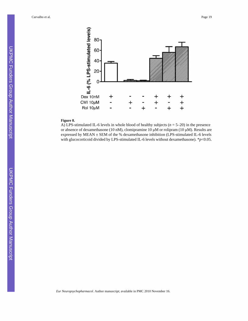
Figure 8.A) LPS-stimulated IL-6 levels in whole blood of healthy subjects (n = 5–20) in the presenceor absence of dexamethasone (10 nM), clomipramine 10 μM or rolipram (10 μM). Results areexpressed by MEAN ± SEM of the % dexamethasone inhibition (LPS-stimulated IL-6 levelswith glucocorticoid divided by LPS-stimulated IL-6 levels without dexamethasone). *p<0.05.
Carvalho et al. Page 19
Eur Neuropsychopharmacol. Author manuscript; available in PMC 2010 November 16.
UKPM
C Funders G
roup Author Manuscript
UKPM
C Funders G
roup Author Manuscript
Copyright © 2022 FDOKUMEN