Effects of chronic antidepressants and electroconvulsive shock on serotonergic neurotransmission in...
11
Review article Effects of chronic antidepressants and electroconvulsive shock on serotonergic neurotransmission in the rat hippocampus Eliyahu Dremencov a, * , Eitan Gur b , Bernard Lerer b , Michael E. Newman b a Life Sciences Faculty, Bar-Ilan University, Ramat-Gan 52900, Israel b Biological Psychiatry Laboratory, Department of Psychiatry, Hebrew University Hadassah Medical Center, Jerusalem, Israel Accepted 22 April 2003 Abstract The hippocampus may play a critical role in the pathophysiology and treatment of depression. There are two main lines of evidence for this: firstly, many of its functions correspond to those altered in depression, and secondly, many hippocampal functions are regulated by the serotonergic (5-HT) system, which is a common target of antidepressant treatments. Chronic effects of antidepressants and electroconvulsive shock (ECS) have been studied by various methods using electrophysiology, in vivo microdialysis or ex vivo neurochemical measurements. The aim of the current review is to point out possible correlations between these studies based on different methods and to suggest neurochemical mechanisms that result in the observed changes in hippocampal physiology and neurogenesis. These changes in hippocampal neurochemistry are reviewed and compared with the abnormalities associated with stress, corticosterone or depression. D 2003 Elsevier Science Inc. All rights reserved. Keywords: Antidepressants; Electroconvulsive shock; Hippocampus; 5-HT 1A autoreceptor; 5-HT 1B autoreceptor; Postsynaptic 5-HT 1A receptor; cAMP 1. Introduction Major depression is a severe disorder that involves disturbances of emotional, cognitive, autonomic and endo- crine functions. The pathophysiology of the disease is still unknown and may include functional abnormalities of different brain regions and circuits. The hippocampal forma- tion is one of those brain systems that was suggested to play a critical role in depressive disorder. There are two reasons for interest in the hippocampus. First, this region controls many of the brain functions that are disturbed in depressed patients, such as altered regulation of neuroendocrine and autonomic functions, mood and cognition difficulties, adverse responses to stressful stimuli and others. Second, hippocampal functions are highly regulated by serotonergic (5-HT) systems, the involvement of which in the pathology and treatment of depression has been shown in many studies (Hjorth et al., 2000). The effects of chronic antidepressant and electroconvulsive shock (ECS) administration on the hippocampal 5-HT system, measured using electrophysio- logical methods, have been described by Blier and de Montigny (1998, 1999), Blier et al. (1998) and Mongeau et al. (1997). These authors postulated that different classes of antidepressants and ECS facilitate 5-HT neurotransmis- sion in the hippocampus using different mechanisms. Thus, chronic administration of tricyclic antidepressants (TCA) and ECS causes sensitization of postsynaptic 5-HT 1A recep- tors, monoamine oxidase inhibitors (MAOIs) desensitize presynaptic 5-HT 1A receptors within the raphe nuclei and selective serotonin reuptake inhibitors (SSRIs) desensitize both nerve terminal 5-HT 1B autoreceptors within the hip- pocampus and somatodendritic 5-HT 1A autoreceptors. Thus, in the presence of 5-HT reuptake or monoamine oxidase inhibition, desensitization of 5-HT autoreceptors results in increased 5-HT input to the hippocampus. 0278-5846/03/$ – see front matter D 2003 Elsevier Science Inc. All rights reserved. doi:10.1016/S0278-5846(03)00123-4 Abbreviations: AC, adenylate cyclase; BDNF, brain-derived neuro- trophic factor; CA, cornu ammonis; cAMP, cyclic adenosine mono- phosphate; CREB, cAMP response element binding protein; DG, dentate gyrus; HPA axis, hypothalamo – pituitary – adrenal axis; IP 3 , inositol triphosphate; PLC, phospholipase C; ECS, electroconvulsive shock; LTP, long-term potentiation; MAOI, monoamine oxidase inhibitor; PKA, protein kinase A; SSRI, selective serotonin reuptake inhibitor; SNRI, serotonin and noradrenaline reuptake inhibitor; TMS, transcranial magnetic stimulation; TCA, tricyclic antidepressant; GR 127935, N-[4-methoxy-3-(4-methyl-1- piperizinyl)phenyl]-2 0 -methyl-4 0 -(5-methyl-1,2,4-oxadiazole-3-yl)[1,1 0 -bi- phenyl]-carboxamide; 8-OH-DPAT, 8-hydroxy-2-(di-n-propylamino)tetra- lin; 5-HT, 5-hydroxytryptamine, serotonin. * Corresponding author. Tel.: +972-3-531-8123; fax: +972-3-535- 1824. E-mail address: [email protected] (E. Dremencov). www.elsevier.com/locate/pnpbp Progress in Neuro-Psychopharmacology & Biological Psychiatry 27 (2003) 729 – 739
Transcript of Effects of chronic antidepressants and electroconvulsive shock on serotonergic neurotransmission in...
www.elsevier.com/locate/pnpbp
Progress in Neuro-Psychopharmacology & Biological Psychiatry 27 (2003) 729–739
Review article
Effects of chronic antidepressants and electroconvulsive shock on
serotonergic neurotransmission in the rat hippocampus
Eliyahu Dremencova,*, Eitan Gurb, Bernard Lererb, Michael E. Newmanb
aLife Sciences Faculty, Bar-Ilan University, Ramat-Gan 52900, IsraelbBiological Psychiatry Laboratory, Department of Psychiatry, Hebrew University Hadassah Medical Center, Jerusalem, Israel
Accepted 22 April 2003
Abstract
The hippocampus may play a critical role in the pathophysiology and treatment of depression. There are two main lines of evidence for
this: firstly, many of its functions correspond to those altered in depression, and secondly, many hippocampal functions are regulated by the
serotonergic (5-HT) system, which is a common target of antidepressant treatments. Chronic effects of antidepressants and electroconvulsive
shock (ECS) have been studied by various methods using electrophysiology, in vivo microdialysis or ex vivo neurochemical measurements.
The aim of the current review is to point out possible correlations between these studies based on different methods and to suggest
neurochemical mechanisms that result in the observed changes in hippocampal physiology and neurogenesis. These changes in hippocampal
neurochemistry are reviewed and compared with the abnormalities associated with stress, corticosterone or depression.
D 2003 Elsevier Science Inc. All rights reserved.
Keywords: Antidepressants; Electroconvulsive shock; Hippocampus; 5-HT1A autoreceptor; 5-HT1B autoreceptor; Postsynaptic 5-HT1A receptor; cAMP
1. Introduction
Major depression is a severe disorder that involves
disturbances of emotional, cognitive, autonomic and endo-
crine functions. The pathophysiology of the disease is still
unknown and may include functional abnormalities of
different brain regions and circuits. The hippocampal forma-
tion is one of those brain systems that was suggested to play
a critical role in depressive disorder. There are two reasons
for interest in the hippocampus. First, this region controls
0278-5846/03/$ – see front matter D 2003 Elsevier Science Inc. All rights reserv
doi:10.1016/S0278-5846(03)00123-4
Abbreviations: AC, adenylate cyclase; BDNF, brain-derived neuro-
trophic factor; CA, cornu ammonis; cAMP, cyclic adenosine mono-
phosphate; CREB, cAMP response element binding protein; DG, dentate
gyrus; HPA axis, hypothalamo–pituitary–adrenal axis; IP3, inositol
triphosphate; PLC, phospholipase C; ECS, electroconvulsive shock; LTP,
long-term potentiation; MAOI, monoamine oxidase inhibitor; PKA, protein
kinase A; SSRI, selective serotonin reuptake inhibitor; SNRI, serotonin and
noradrenaline reuptake inhibitor; TMS, transcranial magnetic stimulation;
TCA, tricyclic antidepressant; GR 127935, N-[4-methoxy-3-(4-methyl-1-
piperizinyl)phenyl]-20-methyl-40-(5-methyl-1,2,4-oxadiazole-3-yl)[1,10-bi-
phenyl]-carboxamide; 8-OH-DPAT, 8-hydroxy-2-(di-n-propylamino)tetra-
lin; 5-HT, 5-hydroxytryptamine, serotonin.
* Corresponding author. Tel.: +972-3-531-8123; fax: +972-3-535-
1824.
E-mail address: [email protected] (E. Dremencov).
many of the brain functions that are disturbed in depressed
patients, such as altered regulation of neuroendocrine and
autonomic functions, mood and cognition difficulties,
adverse responses to stressful stimuli and others. Second,
hippocampal functions are highly regulated by serotonergic
(5-HT) systems, the involvement of which in the pathology
and treatment of depression has been shown in many studies
(Hjorth et al., 2000). The effects of chronic antidepressant
and electroconvulsive shock (ECS) administration on the
hippocampal 5-HT system, measured using electrophysio-
logical methods, have been described by Blier and de
Montigny (1998, 1999), Blier et al. (1998) and Mongeau
et al. (1997). These authors postulated that different classes
of antidepressants and ECS facilitate 5-HT neurotransmis-
sion in the hippocampus using different mechanisms. Thus,
chronic administration of tricyclic antidepressants (TCA)
and ECS causes sensitization of postsynaptic 5-HT1A recep-
tors, monoamine oxidase inhibitors (MAOIs) desensitize
presynaptic 5-HT1A receptors within the raphe nuclei and
selective serotonin reuptake inhibitors (SSRIs) desensitize
both nerve terminal 5-HT1B autoreceptors within the hip-
pocampus and somatodendritic 5-HT1A autoreceptors. Thus,
in the presence of 5-HT reuptake or monoamine oxidase
inhibition, desensitization of 5-HT autoreceptors results in
increased 5-HT input to the hippocampus.
ed.
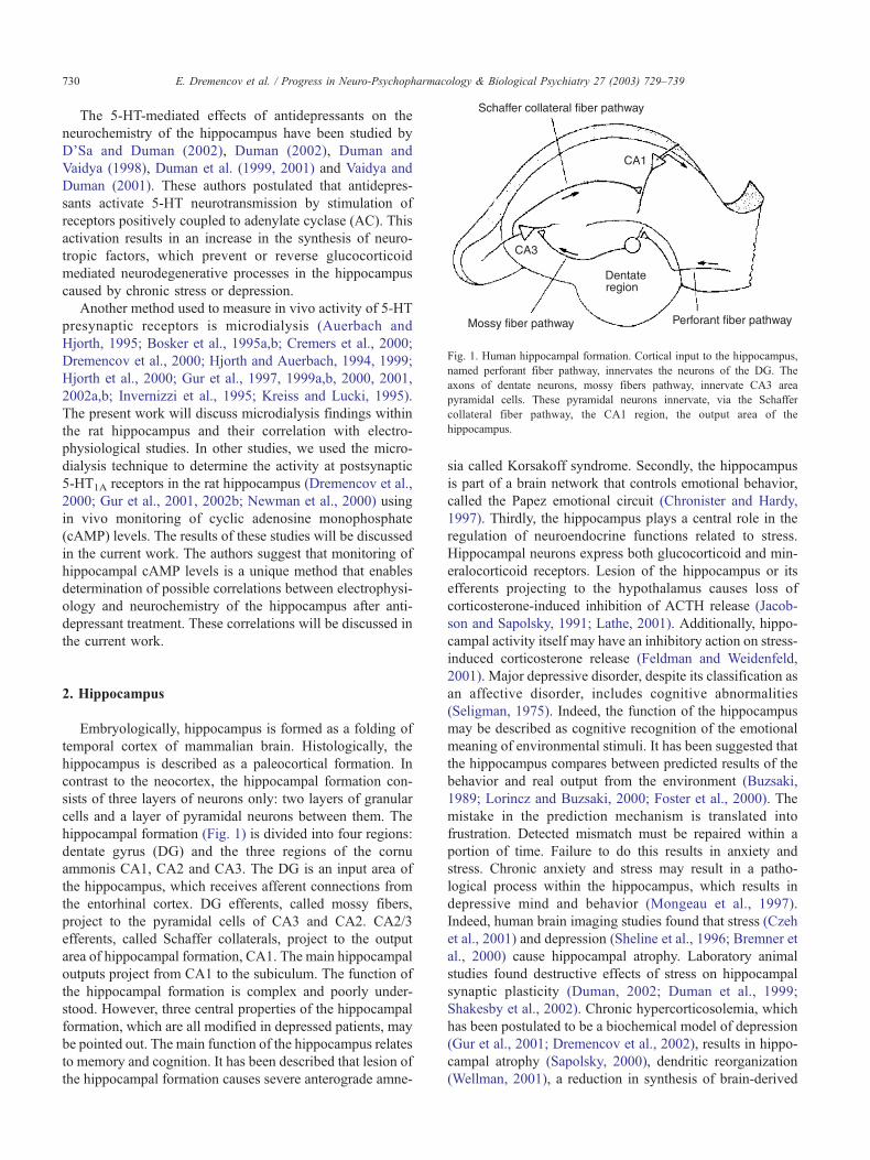
Fig. 1. Human hippocampal formation. Cortical input to the hippocampus,
named perforant fiber pathway, innervates the neurons of the DG. The
axons of dentate neurons, mossy fibers pathway, innervate CA3 area
pyramidal cells. These pyramidal neurons innervate, via the Schaffer
collateral fiber pathway, the CA1 region, the output area of the
hippocampus.
E. Dremencov et al. / Progress in Neuro-Psychopharmacology & Biological Psychiatry 27 (2003) 729–739730
The 5-HT-mediated effects of antidepressants on the
neurochemistry of the hippocampus have been studied by
D’Sa and Duman (2002), Duman (2002), Duman and
Vaidya (1998), Duman et al. (1999, 2001) and Vaidya and
Duman (2001). These authors postulated that antidepres-
sants activate 5-HT neurotransmission by stimulation of
receptors positively coupled to adenylate cyclase (AC). This
activation results in an increase in the synthesis of neuro-
tropic factors, which prevent or reverse glucocorticoid
mediated neurodegenerative processes in the hippocampus
caused by chronic stress or depression.
Another method used to measure in vivo activity of 5-HT
presynaptic receptors is microdialysis (Auerbach and
Hjorth, 1995; Bosker et al., 1995a,b; Cremers et al., 2000;
Dremencov et al., 2000; Hjorth and Auerbach, 1994, 1999;
Hjorth et al., 2000; Gur et al., 1997, 1999a,b, 2000, 2001,
2002a,b; Invernizzi et al., 1995; Kreiss and Lucki, 1995).
The present work will discuss microdialysis findings within
the rat hippocampus and their correlation with electro-
physiological studies. In other studies, we used the micro-
dialysis technique to determine the activity at postsynaptic
5-HT1A receptors in the rat hippocampus (Dremencov et al.,
2000; Gur et al., 2001, 2002b; Newman et al., 2000) using
in vivo monitoring of cyclic adenosine monophosphate
(cAMP) levels. The results of these studies will be discussed
in the current work. The authors suggest that monitoring of
hippocampal cAMP levels is a unique method that enables
determination of possible correlations between electrophysi-
ology and neurochemistry of the hippocampus after anti-
depressant treatment. These correlations will be discussed in
the current work.
2. Hippocampus
Embryologically, hippocampus is formed as a folding of
temporal cortex of mammalian brain. Histologically, the
hippocampus is described as a paleocortical formation. In
contrast to the neocortex, the hippocampal formation con-
sists of three layers of neurons only: two layers of granular
cells and a layer of pyramidal neurons between them. The
hippocampal formation (Fig. 1) is divided into four regions:
dentate gyrus (DG) and the three regions of the cornu
ammonis CA1, CA2 and CA3. The DG is an input area of
the hippocampus, which receives afferent connections from
the entorhinal cortex. DG efferents, called mossy fibers,
project to the pyramidal cells of CA3 and CA2. CA2/3
efferents, called Schaffer collaterals, project to the output
area of hippocampal formation, CA1. The main hippocampal
outputs project from CA1 to the subiculum. The function of
the hippocampal formation is complex and poorly under-
stood. However, three central properties of the hippocampal
formation, which are all modified in depressed patients, may
be pointed out. The main function of the hippocampus relates
to memory and cognition. It has been described that lesion of
the hippocampal formation causes severe anterograde amne-
sia called Korsakoff syndrome. Secondly, the hippocampus
is part of a brain network that controls emotional behavior,
called the Papez emotional circuit (Chronister and Hardy,
1997). Thirdly, the hippocampus plays a central role in the
regulation of neuroendocrine functions related to stress.
Hippocampal neurons express both glucocorticoid and min-
eralocorticoid receptors. Lesion of the hippocampus or its
efferents projecting to the hypothalamus causes loss of
corticosterone-induced inhibition of ACTH release (Jacob-
son and Sapolsky, 1991; Lathe, 2001). Additionally, hippo-
campal activity itself may have an inhibitory action on stress-
induced corticosterone release (Feldman and Weidenfeld,
2001). Major depressive disorder, despite its classification as
an affective disorder, includes cognitive abnormalities
(Seligman, 1975). Indeed, the function of the hippocampus
may be described as cognitive recognition of the emotional
meaning of environmental stimuli. It has been suggested that
the hippocampus compares between predicted results of the
behavior and real output from the environment (Buzsaki,
1989; Lorincz and Buzsaki, 2000; Foster et al., 2000). The
mistake in the prediction mechanism is translated into
frustration. Detected mismatch must be repaired within a
portion of time. Failure to do this results in anxiety and
stress. Chronic anxiety and stress may result in a patho-
logical process within the hippocampus, which results in
depressive mind and behavior (Mongeau et al., 1997).
Indeed, human brain imaging studies found that stress (Czeh
et al., 2001) and depression (Sheline et al., 1996; Bremner et
al., 2000) cause hippocampal atrophy. Laboratory animal
studies found destructive effects of stress on hippocampal
synaptic plasticity (Duman, 2002; Duman et al., 1999;
Shakesby et al., 2002). Chronic hypercorticosolemia, which
has been postulated to be a biochemical model of depression
(Gur et al., 2001; Dremencov et al., 2002), results in hippo-
campal atrophy (Sapolsky, 2000), dendritic reorganization
(Wellman, 2001), a reduction in synthesis of brain-derived

E. Dremencov et al. / Progress in Neuro-Psychopharmacology & Biological Psychiatry 27 (2003) 729–739 731
neurotrophic factor (BDNF) and memory difficulties (Schaaf
et al., 2000).
3. 5-HT system of the hippocampus
The hippocampus is innervated by 5-HT terminals ori-
ginating from the dorsal and median raphe nuclei (Bruinvels
et al., 1993). Hippocampal cells express postsynaptic
5-HT1A (Banerjee et al., 1993; el Mestikawy et al., 1989),
5-HT2A (Vaidya et al., 1997), 5-HT2B (Sanden et al., 2000),
5-HT2C (Clemett et al., 2000; Abramowski et al., 1995),
5-HT3 (Hewlett et al., 1998; Mitchell and Pratt, 1991; Tecott
et al., 1993), 5-HT4 (Vilaro et al., 1996), 5-HT5A, 5-HT5B,
5-HT6 and 5-HT7 (Kinsey et al., 2001; Neumaier et al., 2001)
receptors. 5-HT3 receptors are coupled to Na+ ion channels;
their activation results in membrane depolarization (Barnes
and Sharp, 1999). In the hippocampus, 5-HT3 receptors
activate cholinergic (Consolo et al., 1994) and glutamatergic
(Ohno and Watanabe, 1997) neurotransmission; blocking of
hippocampal 5-HT3 receptors decreases the efficiency of
working memory (Ohno and Watanabe, 1997). Postsynaptic
5-HT1A receptors and presynaptic 5-HT1A and 5-HT1B
autoreceptors are Gi protein-coupled receptors. Their activa-
tion induces dissociation of Gi, which results in inhibition of
AC (a subunit action), opening of K + channels (bg subunits
action) and cell membrane hyperpolarization (Raymond et
al., 2001). Indeed, activation of 5-HT1B autoreceptors
expressed on the 5-HT nerve terminals within the hippocam-
pus and presynaptic 5-HT1A autoreceptors expressed on the
5-HT cell bodies within the dorsal raphe nuclei inhibits 5-HT
neuronal activity and 5-HT release (Kikvadze and Foster,
1995; Hervas et al., 2000; Adell et al., 2001). However, no
effect of presynaptic 5-HT1A autoreceptors on cAMP con-
centration was observed (Clarke et al., 1996). In in vivo
studies, postsynatic 5-HT1A receptor activation was found to
activate cAMP formation (Cadogan et al., 1994). The para-
doxical activation of cAMP synthesis by Gi/Go-coupled
postsynaptic 5-HT1A receptors may be explained by the type
of AC expressed in the hippocampal cells. Thus, hippo-
campal neurons express type II of AC, which is not inhibited
by G protein ai or ao units but is stimulated by bg units
(Tang and Gilman, 1991; Federman et al., 1992). In the
hippocampus, postsynaptic 5-HT1A receptor activation
increases local cholinergic (Wilkinson et al., 1994; Fujii et
al., 2000) and dopaminergic (Sakaue et al., 2000) neuro-
transmission. Hippocampal application of a 5-HT1A receptor
agonist was found to attenuate cognitive deficits caused by
brain injury (Kline et al., 2001). On the other hand, antagon-
ist application ameliorates traumatic cognitive impairment
(Harder et al., 1996).
5-HT2 receptors constitute a family of Gq-coupled recep-
tors. Their activation results in phospholipase C (PLC)-
mediated inositol triphosphate (IP3) formation and in cell
membrane depolarization by opening of Ca2 + channels
(Raymond et al., 2001). However, activation of 5-HT2A
receptors in the hippocampus was found to inhibit synthesis
of BDNF, in contrast to the cortex where 5-HT2A receptor
activation stimulates BDNF formation (Vaidya et al., 1997).
Hippocampal 5-HT2 receptors also regulate activity of the
hypothalamo–pituitary–adrenal axis (HPA axis). Thus,
local administration of a 5-HT2A receptor antagonist
decreases the hypothalamic hormonal response to sexual
stimuli, while a 5-HT2C receptor antagonist increases it
(Popova and Amstislavskaya, 2002). Hippocampal 5-HT2A
receptor activation was found to increase working memory
efficiency (Williams et al., 2002).
5-HT4, 5-HT6 and 5-HT7 receptors are Gs coupled. Their
activation enhances cAMP formation (Raymond et al.,
2001). Hippocampal 5-HT4 receptors positively autoregu-
late 5-HT neurotransmission (Ge and Barnes, 1996) and
increase acetylcholine outflow (Siniscalchi et al., 1999). A
correspondence between 5-HT4 receptor-induced increase in
hippocampal cholinergic neurotransmission and cognitive
functions was established (Matsumoto et al., 2001). 5-HT4
receptors were found to modulate long-term potentiation
(LTP) formation within the hippocampus (Kulla and Man-
ahan-Vaughan, 2002). Hippocampal 5-HT6 receptor antag-
onists were found to enhance excitatory neurotransmission
(Dawson et al., 2001). Activation of 5-HT7 receptors was
found to synchronize neuronal activity within the hippocam-
pus (Gill et al., 2002). Despite initial uncertainty, the
5-HT5A receptor is now known to be negatively coupled
to AC together with the members of the 5-HT1 family
(Thomas et al., 2000). The hippocampal 5-HT system is
affected by stress, depression or hypercorticosolemia. Rosel
et al. (2000) demonstrated altered 5-HT2A receptor density
and IP3 levels within the hippocampus of depressive suicide
victims. Neumaier et al. (2002) found that stress alters
dorsal raphe 5-HT1B mRNA levels, while hypercorticosole-
mia affects hippocampal 5-HT1A mRNA levels. Stress-
induced desensitization of hippocampal 5-HT1A receptors
and sensitization of 5-HT4 receptors was found using ex
vivo electrophysiology (Bijak et al., 2001).
A recent electrophysiological study (Matsumoto et al.,
2002) has shown that three of the above-mentioned receptors
are involved in endogenous 5-HT-mediated synaptic trans-
mission in the CA1 field of the hippocampus. 5-HT1A
receptor activation was found to mediate by 5-HT-induced
inhibition, while 5-HT4 and 5-HT7 receptor activation was
associated with facilitation. In the CA3 field, however,
facilitatory effects of endogenous 5-HT were mediated by
both 5-HT1A and 5-HT7 receptors. The variety of 5-HT
receptors thus plays an important region-specific role in
modulating hippocampal responses to incoming stimuli.
4. Chronic effects of antidepressants and ECS on
hippocampal 5-HT neurotransmission
The chronic effects of different antidepressant drugs and
treatments on presynaptic regulation of hippocampal 5-HT

E. Dremencov et al. / Progress in Neuro-Psychopharmacology & Biological Psychiatry 27 (2003) 729–739732
release have been determined in various laboratories using
in vivo electrophysiological or microdialysis techniques
(Hjorth et al., 2000). Kreiss and Lucki (1995) demonstrated
a reduction of 5-HT1A autoreceptor-mediated inhibition of
hippocampal 5-HT release after chronic administration of
fluoxetine (15 mg/kg/day for 14 days), while our study
(Dremencov et al., 2000) showed that 7 days of fluoxetine
administration at 10 mg/kg/day reduced hippocampal
5-HT1B autoreceptor activity (Fig. 2). Cremers et al.
(2000), using in vivo microdialysis, showed that chronic
14-day administration of the SSRI, citalopram (20 mg/kg),
caused a significant decrease in 5-HT1A autoreceptor-medi-
ated inhibition of hippocampal 5-HT release. These findings
correlate with previous electrophysiological findings (Mon-
geau et al., 1997). However, other investigators (Bosker et
al., 1995a,b; Hjorth and Auerbach, 1994, 1999; Invernizzi et
al., 1995; Auerbach and Hjorth, 1995) were unable to show
any change in autoreceptor sensitivity in the hippocampus
after chronic administration of the SSRIs citalopram or
fluvoxamine. SSRI-mediated desensitization of 5-HT autor-
eceptors cannot be explained by alteration of receptor
density, which is normally unchanged by the treatment
(Hensler et al., 1991; Li et al., 1997; Gobbi et al., 1997;
LePoul et al., 2000), although in one study (Welner et al.,
1989) a decrease in 5-HT1A receptor number in the dorsal
raphe was found after chronic fluoxetine. However, the
decrease in 5-HT autoreceptor activity could be explained
Fig. 2. Effect of systemic N-[4-methoxy-3-(4-methyl-1-piperizinyl)phenyl]-
20-methyl-40-(5-methyl-1,2,4-oxadiazole-3-yl)[1,10-biphenyl]-carboxamide
(GR 127935) (5 mg/kg sc) on 5-HT levels in hippocampus of saline- and
fluoxetine-treated rats. Results are means ± S.E.M. of observations from
seven animals treated with saline and five animals treated subchronically
with fluoxetine (10 mg/kg/day ip for 6 days). Measurements were made 72
h after the last fluoxetine injection. Two-way analysis of variance on the
data from fractions 4 – 10 showed a significant effect of time
[ F(df = 6,60) = 3.62, P=.0039], an almost significant effect of treatment
[ F(df = 1,10) = 4.21, P=.067] and a significant interaction between time and
treatment [ F(df = 6,60) = 2.76, P=.019]. Post hoc Newman–Keuls tests
showed a significant difference ( *P < .006) between saline- and fluoxetine-
treated rats at fraction 6, i.e., 1 h after injection of GR 127935.
by chronic fluoxetine-induced Gi/Go protein down-regu-
lation within raphe nuclei, as shown by Li et al. (1996).
Alternatively, a reduction in 5-HT1A receptor–G protein
coupling in the raphe nuclei after chronic administration of
fluoxetine has been shown by several groups using 5-HT1A
receptor agonist-stimulated [35S]GTPgS binding (Castro et
al., 2003; Pejchal et al., 2002; Shen et al., 2002; Hensler,
2002).
Concerning other drugs, an effect of the reversible MAOI
befloxatone (0.75 mg/kg/day) to reduce 5-HT1A autorecep-
tor-mediated inhibition of firing activity of 5-HT neurons
was shown in the electrophysiological study of Haddjeri et
al. (1998b). A 3-week administration of the serotonin and
noradrenaline reuptake inhibitor (SNRI) venlafaxine was
shown in both in vivo electrophysiological studies and in
vitro [3H]5-HT release studies using hippocampal slices to
reduce 5-HT1A autoreceptor activity at a dose of 10 mg/kg/
day but to affect 5-HT1B autoreceptor activity only at the
much higher dose of 40 mg/kg/day (Beique et al., 2000a,b).
A recent study with the SNRI milnacipran (Mochizuki et al.,
2002) indicated that this drug given per os at 30 mg/kg b.i.d.
for 7 or 14 days also desensitized 5-HT1A autoreceptors, as
shown by a reduction in the potency of 5-HT to inhibit
firing of 5-HT neurons in slices of dorsal raphe, although no
such effect was observed in an earlier in vivo study by
Mongeau et al. (1998). On the other hand, microdialysis
studies from our laboratory (Gur et al., 1999a, 2002a)
showed no effect of chronic venlafaxine at a dose of 5
mg/kg/day for 4 weeks on either 5-HT1A or 5-HT1B auto-
receptor activity. Similarly, the tricyclic drugs clomipramine
(Newman et al., 2000), desipramine and mianserin (Kreiss
and Lucki, 1995) and ECS (Gur et al., 1997) did not alter
autoreceptor-mediated changes in 5-HT release in the hip-
pocampus. The absence of the effect of clomipramine and
desipramine on presynaptic regulation of 5-HT release may
be explained by the pharmacological properties of these
drugs. Desipramine acts mainly as a noradrenaline reuptake
inhibitor, and clomipramine dissociates rapidly to a metab-
olite that is inactive at the 5-HT reuptake site (Fujita et al.,
1991).
The chronic effects of antidepressants on postsynaptic
hippocampal 5-HT receptors have principally been deter-
mined in electrophysiological studies. An increase in post-
synaptic 5-HT1A receptor sensitivity was originally shown
by de Montigny and Aghajanian (1978) after chronic
administration of a variety of tricyclic drugs and by Chaput
et al. (1991) after chronic ECS. These effects may be due to
an increase in the 5-HT1A receptor number. Such an increase
has been found in the hippocampus after chronic imipr-
amine (Klimek et al., 1994; Burnet et al., 1994; Papp et al.,
1994; Moreau et al., 2001) and after chronic ECS (Hay-
akawa et al., 1994; Burnet et al., 1995) although other
studies have not confirmed these reports. The in vivo studies
of de Montigny’s group have been confirmed by ex vivo
studies performed with hippocampal slices (Bijak et al.,
1996, 2001). In these experiments, both chronic imipramine
E. Dremencov et al. / Progress in Neuro-Psychopharmacology & Biological Psychiatry 27 (2003) 729–739 733
and ECS enhanced the effects of 5-HT1A receptor activation
to inhibit the firing of pyramidal CA1 neurons, although
imipramine did not affect 5-HT1A receptor number. Interest-
ingly, 7-day administration of corticosterone, which as
mentioned above can be considered as a biochemical model
of depression, had an opposite action and attenuated the
inhibitory effect of 5-HT1A receptor activation. An altern-
ative mechanism, which could explain the increased func-
tional sensitivity of postsynaptic 5-HT1A receptors in the
hippocampus after chronic imipramine administration, is the
increase in receptor–G protein coupling recently demon-
strated by Shen et al. (2002) using the [35S]GTPgS binding
technique. It is of interest that although two studies (Welner
et al., 1989; Hensler, 2002) have shown increased hippo-
campal 5-HT1A receptor number after chronic administra-
tion of the tricyclic drug amitriptyline, there was no change
in 8-hydroxy-2-(di-n-propylamino)tetralin (8-OH-DPAT)-
stimulated [35S]GTPgS binding after this treatment, sug-
gesting that the extra receptors were uncoupled from G
proteins (Hensler, 2002). Electrophysiological studies have
also shown that the SNRIs venlafaxine and milnacipran
(Beique et al., 2000a; Mochizuki et al., 2002) as well as the
5-HT and specific noradrenergic antidepressant mirtazapine
(Haddjeri et al., 1998c) did not modify the sensitivity of
postsynaptic 5-HT1A receptors in the hippocampus. Regard-
ing the MAOIs, Blier et al. (1986) showed that the sens-
itivity of hippocampal pyramidal neurons to microiontopho-
retic applications of 5-HT was decreased after long-term
treatment with clorgyline but not with phenelzine. However,
these authors concluded that the increased effectiveness of
stimulation of firing of 5-HT neurons induced by chronic
MAOI administration overcomes the desensitization at the
postsynaptic level, so that the net effect is an increase in
5-HT transmission.
The TCA imipramine and ECS increase 5-HT4 receptor
activity (Bijak et al., 2001). On the other hand, antidepres-
sants and ECS showed opposite effects on 5-HT3 receptor
activity. 5-HT3 receptor activity was increased by ECS
(Ishihara and Sasa, 2001) but reduced by paroxetine (Mon-
geau et al., 1997). Regarding 5-HT2 receptor number,
although opposite effects are seen in cortex, with chronic
ECS up-regulate 5-HT2 receptors while antidepressants
reduce their density (Mongeau et al., 1997), there have
been few studies in hippocampus and only in the autoradio-
graphic study of Biegon and Israeli (1987) was a highly
localized increase in 5-HT2 receptor binding in the CA1
region seen in male rats only.
5. Chronic effects of antidepressants and ECS on
hippocampal cAMP formation
As mentioned above, activation of hippocampal 5-HT4,
5-HT6 and 5-HT7 receptors increases local cAMP synthesis.
Although the 5-HT1A receptor when cloned and transfected
induces inhibition of cAMP formation, in vivo studies as
described above have shown 8-OH-DPAT-induced stimu-
lation of cAMP formation. In addition, in vitro studies
(Markstein et al., 1999; Thomas et al., 1999) indicate that
the effect of 5-HT to stimulate AC activity in rat hippo-
campal homogenates and guinea pig hippocampal mem-
branes is mediated at least in part by activation of 5-HT1A
receptors. cAMP activates protein kinase A (PKA), which
may phoshorylate a cytosolic protein named cAMP
response element binding protein (CREB) (Duman et al.,
1997). CREB phosphorylation results in expression of
BDNF, which prevents or restores destructive morpho-
logical processes caused by corticosteroids, stress or depres-
sion (Duman et al., 1999). Indeed, cAMP-induced neuro-
genesis was demonstrated by Nakagawa et al. (2002), and
the regulation of neurogenesis by antidepressants (Duman et
al., 2001; Malberg et al., 2000) and ECS (Duman and
Vaidya, 1998; Vaidya et al., 1999) is an established phe-
nomenon. Shirayama et al. (2002) demonstrated antidepres-
sant effects of BDNF in a behavioral animal model of
depression. Additionally to alteration of gene expression,
cAMP may increase synaptic plasticity via PKA-mediated
channel phosphorylation, resulting in changes in ion con-
ductivity (Kandel et al., 1983) or cytoskeleton reorganiza-
tion, resulting in alterations in properties of dendritic spines
(Murphy and Segal, 1997; Segal and Murphy, 1998).
Indeed, chronic ECS and fluoxetine (Stewart and Reid,
2000) as well as chronic desipramine, mianserin and trans-
cranial magnetic stimulation (TMS) (Levkovitz et al., 2001)
showed positive effects on hippocampal LTP formation. Our
studies (Dremencov et al., 2000; Gur et al., 2001, 2002b;
Newman et al., 2000) determined hippocampal 5-HT1A
receptor-mediated cAMP formation using in vivo micro-
dialysis in freely moving rats. Postsynaptic 5-HT1A receptor
activation was found to increase hippocampal cAMP levels,
as previously described (Cadogan et al., 1994). Addition-
ally, we found that chronic treatment with clomipramine (10
mg/kg/day for 28 days) sensitized 5-HT1A-mediated cAMP
formation (Fig. 3) (Newman et al., 2000). Thus, the pro-
posed cAMP-related therapeutic effect of antidepressants
may be mediated via postsynaptic 5-HT1A receptors in the
hippocampus. These results correlate with the sensitization
of postsynaptic 5-HT1A receptor activity by TCAs previ-
ously described in electrophysiological studies (Mongeau et
al., 1997; Bijak et al., 2001). Sensitization of 5-HT1A-
mediated cAMP synthesis cannot be explained by alteration
of AC, because forskolin-induced cAMP formation was not
altered by clomipramine (Fig. 3). It is also unlikely that the
clomipramine-induced activation of 5-HT1A-mediated
cAMP formation is due to elevation of the 5-HT1A receptor
number, since Srinivas et al. (2001) found no effect of
clomipramine (10 mg/kg/day for 40 days) on 5-HT1A
receptor density in the hippocampus, although Lesch et al.
(1992) did show an increase in 5-HT1A receptor mRNA
levels after administration of clomipramine at 5 mg/kg/day
for 3 weeks. However, chronic administration of the tricy-
clics imipramine and clomipramine resulted in an increase

Fig. 3. Effect of systemic 8-OH-DPAT (0.2 mg/kg sc) and forskolin (50
mM) on cAMP levels in hippocampus of saline- and clomipramine-treated
rats. Results are means ± S.E.M. of observations from five animals treated
with saline and seven animals treated chronically with clomipramine (10
mg/kg/day for 28 days, administered by osmotic minipump). Measurements
were performed 24 h after the last day of clomipramine administration.
Two-way analysis of variance of cAMP levels over fractions 3–9, the
period from injection of 8-OH-DPAT until cAMP levels returned to
baseline, showed a significant effect of treatment [ F(df = 1,10) = 5.712,
P=.037], a significant effect of time (fraction number) [ F(df = 6,60) = 2.32,
P=.044] but no interaction between time and treatment [ F(df = 6,60) = 1.06,
P=.39]. Two-way analysis of variance of cAMP levels over fractions 9–14,
from the end of the period during which forskolin was infused until cAMP
levels returned to baseline, showed a significant effect of time
[ F(df = 5,40) = 3.398, P=.011] but no effect of treatment [ F(df = 1,8) =
0.005, P=.98] or interaction [ F(df = 5,40) = 0.918, P=.479].
Fig. 4. Effect of 8-OH-DPAT (0.2 mg/kg sc) and forskolin (50 mM) on
cAMP levels in hippocampus of sham- and ECS-treated rats. Results are
means ± S.E.M. of observations from 9 sham-treated rats and 14 ECS-
treated rats. Two-way analysis of variance of cAMP levels over fractions
4–10, the period from injection of 8-OH-DPAT until cAMP levels returned
to baseline, showed a significant effect of time (fraction number)
[ F(df = 6,144) = 3.96, P=.001] but no significant effect of treatment
[ F(df = 1,24) = 2.84, P=.104] or interaction between time and treatment
[ F(df = 6,144) = 1.69, P=.13]. Two-way analysis of variance of cAMP
levels over fractions 10–16, from the period during which forskolin was
infused until cAMP levels returned to baseline, showed a significant effect
of time [ F(df = 6,144) = 3.29, P=.004], a significant effect of treatment
[ F(df = 1,24) = 5.27, P=.03] and a significant interaction between time and
treatment [ F(df = 6,144) = 3.22, P=.005].
E. Dremencov et al. / Progress in Neuro-Psychopharmacology & Biological Psychiatry 27 (2003) 729–739734
in the level of the a subunit of Go (Lesch et al., 1991), while
imipramine also decreased the level of the a subunit of the
G protein Gi. Since Go dissociation releases bg units, the
changes in Gi/Go expression could provide an explanation
for clomipramine-induced activation of 8-OH-DPAT-medi-
ated cAMP synthesis, according to the mechanism proposed
above (Section 2).
In contrast to clomipramine, the majority of studies
indicate that repetitive administration of SSRIs does not
change postsynaptic 5-HT1A receptor activity in the hip-
pocampus, as determined by electrophysiological analysis
(Mongeau et al., 1997; LePoul et al., 2000). Beck et al.
(1997), however, found that fluoxetine (6.7 mg/kg/day for 3
weeks) increased the potency of 5-HT for the 5-HT1A
receptor-mediated hyperpolarization in area CA1 but not
in area CA3 of the hippocampus. Similarly, Castro et al.
(2003) and Shen et al. (2002) both found an increase in 5-
HT1A receptor agonist-mediated [35S]-GTPgS binding in
both DG and CA1 areas of the hippocampus after chronic
administration of fluoxetine (10 mg/kg/day for 2 or 3
weeks), indicative of an increase in receptor–G protein
coupling, although two other studies (Hensler, 2002; Pejchal
et al., 2002) did not find such an effect. It is of interest that
studies performed in rat hippocampal membranes (Newman
et al., 1992; Valdizan et al., 2002) showed that chronic
fluoxetine (10 mg/kg/day for 2 weeks or 15 mg/kg/day for 3
weeks) resulted in a decrease in the degree of 5-HT- or 8-
OH-DPAT-induced inhibition of forskolin-stimulated AC.
This would be expected to raise intracellular cAMP levels
and thus stimulate expression of CREB and BDNF. How-
ever, no such increase in cAMP levels was observed in
microdialysis studies after administration of fluoxetine at 10
mg/kg/day for 7 days (Dremencov et al., 2000). Further-
more, recent work (Miro et al., 2002) has shown that 14
days of treatment with 3 mg/kg fluoxetine down-regulated
expression of BDNF in hippocampus, in contrast to the
results of Duman et al. (1997, 1999).
Chronic ECS, on the other hand, reduced hippocampal
AC activity in response to local administration of forskolin,
as measured by cAMP monitoring using microdialysis (Fig.
4) (Gur et al., 2002b). This effect may be explained by ECS-
induced Gs down-regulation (McGowan et al., 1996). How-
ever, chronic ECS did not change the cAMP response to
8-OH-DPAT administration. Because the 5-HT1A-induced
cAMP response is mediated via AC, we postulated that
repetitive ECS actually increases postsynaptic 5-HT1A
receptor activity in the hippocampus, and the unchanged
response to 8-OH-DPAT is due to canceling out of the
increased receptor activity and decreased AC activity (Gur
et al., 2002b). This correlates with previous findings of in
vivo electrophysiological (Mongeau et al., 1997) and ex
vivo receptor density studies (for review, see Newman et al.,
E. Dremencov et al. / Progress in Neuro-Psychopharmacology & Biological Psychiatry 27 (2003) 729–739 735
1998). Direct effects on 5-HT1A receptor-mediated cAMP
formation of other antidepressant drugs and treatments, e.g.,
SNRIs, reversible MAOIs, noradrenergic and specific 5-HT
antidepressants, TMS, etc., have not according to our
knowledge been demonstrated. Antidepressant-induced
sensitization of hippocampal 5-HT1A receptors is an oppos-
ite process to the antidepressant-induced receptor desens-
itization observed in hypothalamus (Dremencov et al.,
2002) or raphe nuclei (Hjorth et al., 2000; Mongeau et al.,
1997). A possible reason for the difference is the subtype of
Gi/Go protein expressed in the hippocampus. Indeed, hippo-
campal neurons express only Go protein, while raphe nuclei
cells express only Gi3 and hypothalamus Gi1, Gi3 and Gz
proteins (Hensler, 2003).
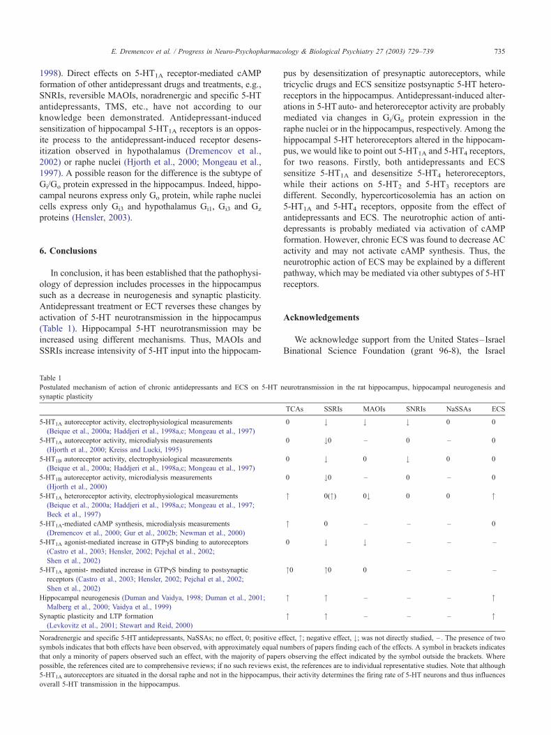
6. Conclusions
In conclusion, it has been established that the pathophysi-
ology of depression includes processes in the hippocampus
such as a decrease in neurogenesis and synaptic plasticity.
Antidepressant treatment or ECT reverses these changes by
activation of 5-HT neurotransmission in the hippocampus
(Table 1). Hippocampal 5-HT neurotransmission may be
increased using different mechanisms. Thus, MAOIs and
SSRIs increase intensivity of 5-HT input into the hippocam-
Table 1
Postulated mechanism of action of chronic antidepressants and ECS on 5-HT n
synaptic plasticity
5-HT1A autoreceptor activity, electrophysiological measurements
(Beique et al., 2000a; Haddjeri et al., 1998a,c; Mongeau et al., 1997)
5-HT1A autoreceptor activity, microdialysis measurements
(Hjorth et al., 2000; Kreiss and Lucki, 1995)
5-HT1B autoreceptor activity, electrophysiological measurements
(Beique et al., 2000a; Haddjeri et al., 1998a,c; Mongeau et al., 1997)
5-HT1B autoreceptor activity, microdialysis measurements
(Hjorth et al., 2000)
5-HT1A heteroreceptor activity, electrophysiological measurements
(Beique et al., 2000a; Haddjeri et al., 1998a,c; Mongeau et al., 1997;
Beck et al., 1997)
5-HT1A-mediated cAMP synthesis, microdialysis measurements
(Dremencov et al., 2000; Gur et al., 2002b; Newman et al., 2000)
5-HT1A agonist-mediated increase in GTPgS binding to autoreceptors
(Castro et al., 2003; Hensler, 2002; Pejchal et al., 2002;
Shen et al., 2002)
5-HT1A agonist- mediated increase in GTPgS binding to postsynaptic
receptors (Castro et al., 2003; Hensler, 2002; Pejchal et al., 2002;
Shen et al., 2002)
Hippocampal neurogenesis (Duman and Vaidya, 1998; Duman et al., 2001;
Malberg et al., 2000; Vaidya et al., 1999)
Synaptic plasticity and LTP formation
(Levkovitz et al., 2001; Stewart and Reid, 2000)
Noradrenergic and specific 5-HT antidepressants, NaSSAs; no effect, 0; positive e
symbols indicates that both effects have been observed, with approximately equal n
that only a minority of papers observed such an effect, with the majority of paper
possible, the references cited are to comprehensive reviews; if no such reviews exi
5-HT1A autoreceptors are situated in the dorsal raphe and not in the hippocampus,
overall 5-HT transmission in the hippocampus.
pus by desensitization of presynaptic autoreceptors, while
tricyclic drugs and ECS sensitize postsynaptic 5-HT hetero-
receptors in the hippocampus. Antidepressant-induced alter-
ations in 5-HT auto- and heteroreceptor activity are probably
mediated via changes in Gi/Go protein expression in the
raphe nuclei or in the hippocampus, respectively. Among the
hippocampal 5-HT heteroreceptors altered in the hippocam-
pus, we would like to point out 5-HT1A and 5-HT4 receptors,
for two reasons. Firstly, both antidepressants and ECS
sensitize 5-HT1A and desensitize 5-HT4 heteroreceptors,
while their actions on 5-HT2 and 5-HT3 receptors are
different. Secondly, hypercorticosolemia has an action on
5-HT1A and 5-HT4 receptors, opposite from the effect of
antidepressants and ECS. The neurotrophic action of anti-
depressants is probably mediated via activation of cAMP
formation. However, chronic ECS was found to decrease AC
activity and may not activate cAMP synthesis. Thus, the
neurotrophic action of ECS may be explained by a different
pathway, which may be mediated via other subtypes of 5-HT
receptors.
Acknowledgements
We acknowledge support from the United States–Israel
Binational Science Foundation (grant 96-8), the Israel
eurotransmission in the rat hippocampus, hippocampal neurogenesis and
TCAs SSRIs MAOIs SNRIs NaSSAs ECS
0 # # # 0 0
0 #0 – 0 – 0
0 # 0 # 0 0
0 #0 – 0 – 0
" 0(") 0# 0 0 "
" 0 – – – 0
0 # # – – –
"0 "0 0 – – –
" " – – – "
" " – – – "
ffect, "; negative effect, #; was not directly studied, – . The presence of two
umbers of papers finding each of the effects. A symbol in brackets indicates
s observing the effect indicated by the symbol outside the brackets. Where
st, the references are to individual representative studies. Note that although
their activity determines the firing rate of 5-HT neurons and thus influences
E. Dremencov et al. / Progress in Neuro-Psychopharmacology & Biological Psychiatry 27 (2003) 729–739736
Science Foundation (grant 692/0) and the National Institutes
of Health, USA (grant MH 60580).
References
Abramowski, D., Rigo,M., Duc, D., Hoyer, D., Staufenbiel,M., 1995. Local-
ization of the 5-hydroxytryptamine2C receptor protein in human and rat
brain using specific antisera. Neuropharmacology 34, 1635–1645.
Adell, A., Celada, P., Artigas, F., 2001. The role of 5-HT1B receptors in the
regulation of serotonin cell firing and release in the rat brain. J. Neuro-
chem. 79, 172–182.
Auerbach, S.B., Hjorth, S., 1995. Effect of chronic administration of the
selective serotonin uptake inhibitor citalopram on extracellular 5-HT
and apparent autoreceptor sensitivity in rat forebrain in vivo. Naunyn-
Schmiedeberg’s Arch. Pharmacol. 352, 597–606.
Banerjee, P., Berry-Kravis, E., Bonafede-Chhabra, D., Dawson, G., 1993.
Heterologous expression of the serotonin 5-HT1A receptor in neural and
non-neural cell lines. Biochem. Biophys. Res. Commun. 192, 104–110.
Barnes, N.M., Sharp, T., 1999. A review of central 5-HT receptors and their
function. Neuropharmacology 38, 1083–1152.
Beck, S.G., Birnstiel, S., Choi, K.C., Pouliot, W., 1997. Fluoxetine selec-
tively alters 5-hydroxytryptamine1A and g-aminobutyric acidB receptor-
mediated hyperpolarization in area CA1, but not area CA3, hippocam-
pal pyramidal cells. J. Pharmacol. Exp. Ther. 281, 115–122.
Beique, J., de Montigny, C., Blier, P., Debonnel, G., 2000a. Effects of
sustained administration of the serotonin and norepinephrine reuptake
inhibitor venlafaxine: I. In vivo electrophysiological studies in the rat.
Neuropharmacology 39, 1800–1812.
Beique, J., de Montigny, C., Blier, P., Debonnel, G., 2000b. Effects of
sustained administration of the serotonin and norepinephrine reuptake
inhibitor venlafaxine: II. In vitro studies in the rat. Neuropharmacology
39, 1813–1822.
Biegon, A., Israeli, M., 1987. Quantitative autoradiographic analysis of the
effects of electroconvulsive shock on serotonin-2 receptors in male and
female rats. J. Neurochem. 48, 1386–1391.
Bijak, M., Tokarski, K., Czyrak, A., Mackowiak, M., Wedzony, K., 1996.
Imipramine increases the 5-HT1A receptor-mediated inhibition of hippo-
campal neurons without changing the 5-HT1A receptor binding. Eur. J.
Pharmacol. 305, 79–85.
Bijak, M., Zahorodna, A., Tokarski, K., 2001. Opposite effects of antide-
pressants and corticosterone on the sensitivity of hippocampal CA1
neurons to 5-HT1A and 5-HT4 receptor activation. Naunyn-Schmiede-
berg’s Arch. Pharmacol. 363, 491–498.
Blier, P., de Montigny, C., 1998. Possible serotonergic mechanisms under-
lying the antidepressant and anti-obsessive-compulsive disorder re-
sponses. Biol. Psychiatry 44, 313–323.
Blier, P., de Montigny, C., 1999. Serotonin and drug-induced therapeutic
responses in major depression, obsessive-compulsive and panic disor-
ders. Neuropsychopharmacology 21 (Suppl. 2), 91–98.
Blier, P., de Montigny, C., Azzaro, A.J., 1986. Modification of serotonergic
and noradrenergic neurotransmissions by repeated administration of
monoamine oxidase inhibitors: electrophysiological studies in the rat
central nervous system. J. Pharmacol. Exp. Ther. 237, 987–994.
Blier, P., Pineyro, G., el Mansari, M., Bergeron, R., de Montigny, C., 1998.
Role of somatodendritic 5-HT autoreceptors in modulating 5-HT neuro-
transmission. Ann. N. Y. Acad. Sci. 861, 204–216.
Bosker, F.J., Klompmakers, A.A., Westenberg, H.G.M., 1995a. Effects of
single and repeated oral administration of fluvoxamine on extracellular
serotonin in the median raphe nucleus and dorsal hippocampus of the
rat. Neuropharmacology 34, 501–508.
Bosker, F.J., van Essevelt, K.E., Klompmakers, A.A., Westenberg, H.G.M.,
1995b. Chronic treatment with fluvoxamine by osmotic minipumps
fails to induce persistent functional changes in central 5-HT1A and 5-
HT1B receptors, as measured by in vivo microdialysis in dorsal hippo-
campus of conscious rats. Psychopharmacology 117, 358–363.
Bremner, J.D., Narayan, M., Anderson, E.R., Staib, L.H., Miller, H.L.,
Charney, D.S., 2000. Hippocampal volume reduction in major depres-
sion. Am. J. Psychiatry 157, 115–118.
Bruinvels, A.T., Palacios, J.M., Hoyer, D., 1993. Autoradiographic char-
acterization and localization of 5-HT1D compared to 5-HT1B binding
sites in rat brain. Naunyn-Schmiedeberg’s Arch. Pharmacol. 347,
569–582.
Burnet, P.W.J., Michelson, D., Smith, M.A., Gold, P.W., Sternberg, E.M.,
1994. The effect of chronic imipramine administration on the densities
of 5-HT1A and 5-HT2 receptors and the abundancies of 5-HT receptor
and transporter mRNA in the cortex, hippocampus and dorsal raphe of
three strains of rat. Brain Res. 638, 311–324.
Burnet, P.W.J., Mead, A., Eastwood, S.L., Lacey, K., Harrison, P.J., Sharp,
T., 1995. Repeated ECS differentially affects brain 5-HT1A and 5-HT2A
receptor expression. NeuroReport 6, 901–904.
Buzsaki, S., 1989. Two-stage model of memory trace formation: a role for
‘‘noisy’’ brain states. Neuroscience 31, 551–570.
Cadogan, A.K., Kendall, D.A., Marsden, C.A., 1994. Serotonin 5-HT1A
receptor activation increases cyclic AMP formation in the rat hippo-
campus in vivo. J. Neurochem. 62, 1816–1821.
Castro, E., Diaz, A., Del Olmo, E., Pazos, A., 2003. Chronic fluoxetine
induces opposite changes in G protein coupling at pre and postsynaptic
5-HT1A receptors in rat brain. Neuropharmacology 44, 93–101.
Chaput, Y., de Montigny, C., Blier, P., 1991. Presynaptic and postsynaptic
modifications of the serotonin system by long-term administration of
antidepressant treatments: an in vivo electrophysiological study in the
rat. Neuropsychopharmacology 5, 219–229.
Chronister, R.B., Hardy, S.G.P., 1997. The limbic system. Fundamental
Neuroscience. Churchill Livingstone, New York, pp. 443–454.
Clarke, W.P., Yocca, F.D., Maayani, S., 1996. Lack of 5-hydroxytrypta-
mine1A-mediated inhibition of adenylyl cyclase in dorsal raphe of male
and female rats. J. Pharmacol. Exp. Ther. 277, 1259–1266.
Clemett, D.A., Punhani, T., Duxon, M.S., Blackburn, T.P., Fone, K.C.,
2000. Immunohistochemical localisation of the 5-HT2C receptor protein
in the rat CNS. Neuropharmacology 39, 123–132.
Consolo, S., Bertorelli, R., Russi, G., Zambelli, M., Ladinsky, H., 1994.
Serotonergic facilitation of acetylcholine release in vivo from rat dor-
sal hippocampus via serotonin 5-HT3 receptors. J. Neurochem. 62,
2254–2261.
Cremers, T.I.F.H., Spoelstra, E.N., De Boer, P., Bosker, F.J., Mork, A., Den
Boer, J., Westerink, B.H.C., Wikstrom, H.V., 2000. Desensitization of
5-HT autoreceptors upon pharmacokinetically monitored chronic treat-
ment with citalopram. Eur. J. Pharmacol. 397, 351–357.
Czeh, B., Michaelis, T., Watanabe, T., Frahm, J., de Biurrun, G., van
Kampen, M., Bartolomucci, A., Fuchs, E., 2001. Stress-induced
changes in cerebral metabolites, hippocampal volume, and cell prolif-
eration are prevented by antidepressant treatment with tianeptine.
Proc. Natl. Acad. Sci. U. S. A. 98, 12796–12801.
Dawson, L.A., Nguyen, H.Q., Li, P., 2001. The 5-HT6 receptor antagonist
SB-271046 selectively enhances excitatory neurotransmission in the
rat frontal cortex and hippocampus. Neuropsychopharmacology 25,
662–668.
de Montigny, C., Aghajanian, G.K., 1978. Tricyclic antidepressants: long-
term treatment increases responsivity of rat forebrain neurons to sero-
tonin. Science 202, 1303–1306.
Dremencov, E., Gur, E., Lerer, B., Newman, M.E., 2000. Subchronic fluox-
etine administration to rats: effects on 5-HT autoreceptor activity as
measured by in vivo microdialysis. Eur. Neuropsychopharmacol. 10,
229–236.
Dremencov, E., Gur, E., Lerer, B., Newman, M.E., 2002. Effects of chronic
antidepressants and electroconvulsive shock on serotonergic neurotrans-
mission in the rat hypothalamus. Prog. Neuropsychopharmacol. Biol.
Psychiatry 26, 1029–1034.
D’Sa, C., Duman, R.S., 2002. Antidepressants and neuroplasticity. Bipolar
Disord. 4, 183–194.
Duman, R.S., 2002. Synaptic plasticity and mood disorders. Mol. Psychia-
try 7 (Suppl. 1), 29–34.

E. Dremencov et al. / Progress in Neuro-Psychopharmacology & Biological Psychiatry 27 (2003) 729–739 737
Duman, R.S., Vaidya, V.A., 1998. Molecular and cellular actions of chronic
electroconvulsive seizures. J. ECT 14, 81–93.
Duman, R.S., Heninger, G.R., Nestler, E.J., 1997. A molecular and cellular
theory of depression. Arch. Gen. Psychiatry 54, 597–606.
Duman, R.S., Malberg, J., Thome, J., 1999. Neural plasticity to stress and
antidepressant treatment. Biol. Psychiatry 46, 1181–1191.
Duman, R.S., Nakagawa, S., Malberg, J., 2001. Regulation of adult neuro-
genesis by antidepressant treatment. Neuropsychopharmacology 25,
836–844.
el Mestikawy, S., Taussig, D., Gozlan, H., Emerit, M.B., Ponchant, M.,
Hamon, M., 1989. Chromatographic analyses of the serotonin 5-HT1A
receptor solubilized from the rat hippocampus. J. Neurochem. 53,
1555–1566.
Federman, A.D., Conklin, B.R., Schrader, K.A., Reed, R.R., Bourne, H.R.,
1992. Hormonal stimulation of adenylyl cyclase through Gi-protein
beta gamma subunits. Nature 356, 159–161.
Feldman, S., Weidenfeld, J., 2001. Electrical stimulation of the dorsal
hippocampus caused a long lasting inhibition of ACTH and adreno-
cortical responses to photic stimuli in freely moving rats. Brain Res.
911, 22–26.
Foster, D.J., Morris, R.G., Dayan, P., 2000. A model of hippocampally
dependent navigation, using the temporal difference learning rule. Hip-
pocampus 10, 1–16.
Fujii, T., Nakai, K., Nakajima, Y., Kawashima, K., 2000. Enhancement of
hippocampal cholinergic neurotransmission through 5-HT1A receptor-
mediated pathways by repeated lithium treatment in rats. Can. J. Phys-
iol. Pharmacol. 78, 392–399.
Fujita, K., Kobayashi, A., Suzuki, S., Nakazawa, K., 1991. Changes of
serotonin and catecholamines are related to pharmacokinetic alterations
of clomipramine in rat brain. Eur. J. Pharmacol. 204, 227–233.
Ge, J., Barnes, N.M., 1996. 5-HT4 receptor-mediated modulation of 5-
HT release in the rat hippocampus in vivo. Br. J. Pharmacol. 117,
1475–1480.
Gill, C.H., Soffin, E.M., Hagan, J.J., Davies, C.H., 2002. 5-HT7 receptors
modulate synchronized network activity in rat hippocampus. Neuro-
pharmacology 42, 82–92.
Gobbi, M., Crespi, D., Foddi, M.C., Fracasso, C., Mancini, L., Parotti,
L., Mennini, T., 1997. Effects of chronic treatment with fluoxetine
and citalopram on 5-HT uptake, 5-HT1B autoreceptors, 5-HT3 and 5-
HT4 receptors in rats. Naunyn-Schmiedeberg’s Arch. Pharmacol. 356,
22–28.
Gur, E., Lerer, B., Newman, M.E., 1997. Chronic electroconvulsive shock
and 5-HT autoreceptor activity in rat brain: an in vivo microdialysis
study. J. Neural Transm. 104, 795–804.
Gur, E., Dremencov, E., Lerer, B., Newman, M.E., 1999a. Venlafaxine:
acute and chronic effects on 5-hydroxytryptamine levels in rat brain
in vivo. Eur. J. Pharmacol. 372, 17–24.
Gur, E., Lerer, B., Newman, M.E., 1999b. Chronic clomipramine and triio-
dothyronine increase serotonin levels in rat frontal cortex in vivo: rela-
tionship to serotonin autoreceptor activity. J. Pharmacol. Exp. Ther.
288, 81–87.
Gur, E., Lerer, B., Dremencov, E., Newman, M.E., 2000. Chronic repet-
itive transcranial magnetic stimulation induces subsensitivity of pre-
synaptic serotonergic autoreceptor activity in rat brain. NeuroReport
11, 2925–2929.
Gur, E., Dremencov, E., Lerer, B., Newman, M.E., 2001. Functional effects
of corticosterone on 5-HT1A and 5-HT1B receptor activity in rat brain: in
vivo microdialysis studies. Eur. J. Pharmacol. 411, 115–122.
Gur, E., Dremencov, E., Van de Kar, L.D., Lerer, B., Newman, M.E.,
2002a. Effects of chronically administered venlafaxine on serotonin
receptor activity in rat hippocampus and hypothalamus. Eur. J. Pharma-
col. 436, 57–65.
Gur, E., Dremencov, E., Garcia, F., Van de Kar, L.D., Lerer, B., Newman,
M.E., 2002b. Functional effects of chronic electroconvulsive shock on
serotonergic 5-HT1A and 5-HT1B receptor activity in rat hippocampus
and hypothalamus. Brain Res. 952, 52–60.
Haddjeri, N., Blier, P., de Montigny, C., 1998a. Long-term antidepressant
treatments result in a tonic activation of forebrain 5-HT1A receptors.
J. Neurosci. 18, 10150–10156.
Haddjeri, N., De Montigny, C., Curet, O., Blier, P., 1998b. Effect of
the reversible monoamine oxidase-A inhibitor befloxatone on the rat
5-hydroxytryptamine neurotransmission. Eur. J. Pharmacol. 343,
179–192.
Haddjeri, N., Blier, P., de Montigny, C., 1998c. Acute and long-term actions
of the antidepressant drug mirtazapine on central 5-HT neurotransmis-
sion. J. Affect. Disord. 51, 255–266.
Harder, J.A., Maclean, C.J., Alder, J.T., Francis, P.T., Ridley, R.M., 1996.
The 5-HT1A antagonist, WAY 100635, ameliorates the cognitive impair-
ment induced by fornix transection in the marmoset. Psychopharmacol-
ogy 127, 245–254.
Hayakawa, H., Shimizu, M., Nishida, A., Motohashi, N., Yamawaka, S.,
1994. Increase in serotonin 1A receptor in the dentate gyrus as
revealed by autoradiographic analysis following repeated electrocon-
vulsive shock but not imipramine treatment. Neuropsychobiology 30,
53–56.
Hensler, J.G., 2002. Differential regulation of 5-HT1A receptor–G protein
interactions in brain following chronic antidepressant administration.
Neuropsychopharmacology 26, 565–573.
Hensler, J.G., 2003. Regulation of 5-HT1A receptor function in brain
following agonist or antidepressant administration. Life Sci. 72,
1665–1682.
Hensler, J.G., Kovachich, G.B., Frazer, A., 1991. A quantitative radio-
graphic study of serotonin1A receptor regulation. Effect of 5,7-dihy-
droxytryptamine and antidepressant treatments. Neuropsychopharma-
cology 4, 131–144.
Hervas, I., Queiroz, C.M., Adell, A., Artigas, F., 2000. Role of uptake
inhibition and autoreceptor activation in the control of 5-HT release
in the frontal cortex and dorsal hippocampus of the rat. Br. J. Pharma-
col. 130, 160–166.
Hewlett, W.A., Fridman, S., Trivedi, B.L., Schmidt, D.E., de Paulis, T.,
Ebert, M.H., 1998. Characterization of desamino-5-[125I]iodo-3-me-
thoxy-zacopride ([125I]MIZAC) binding to 5-HT3 receptors in the rat
brain. Prog. Neuropsychopharmacol. Biol. Psychiatry 22, 397–410.
Hjorth, S., Auerbach, S.B., 1994. Lack of 5-HT1A autoreceptor desensitiza-
tion following chronic citalopram treatment, as determined by in vivo
microdialysis. Neuropharmacology 33, 331–334.
Hjorth, S., Auerbach, S.B., 1999. Autoreceptors remain functional after
prolonged treatment with a serotonin reuptake inhibitor. Brain Res.
835, 224–228.
Hjorth, S., Bengtsson, H.J., Kullberg, A., Carlzon, D., Peilot, H., Auerbach,
S.B., 2000. Serotonin autoreceptor function and antidepressant drug
action. J. Psychopharmacol. 14, 177–185.
Invernizzi, R., Bramante, M., Samanin, R., 1995. Extracellular concentra-
tions of serotonin in the dorsal hippocampus after acute and chronic
treatment with citalopram. Brain Res. 696, 62–66.
Ishihara, K., Sasa, M., 2001. Potentiation of 5-HT3 receptor functions in the
hippocampal CA1 region of rats following repeated electroconvulsive
shock treatments. Neurosci. Lett. 307, 37–40.
Jacobson, L., Sapolsky, R., 1991. The role of the hippocampus in feedback
regulation of the hypothalamic–pituitary–adrenocortical axis. Endocr.
Rev. 12, 118–134.
Kandel, E.R., Abrams, T., Bernier, L., Carew, T.J., Hawkins, R.D.,
Schwartz, J.H., 1983. Classical conditioning and sensitization share
aspects of the same molecular cascade in Aplysia. Quant. Biol. 48,
821–830.
Kikvadze, I., Foster, G.A., 1995. Action potential-dependent output of 5-
hydroxytryptamine in the anaesthetized rat amygdalopiriform cortex is
strongly inhibited by tonic 5-HT1B-receptor stimulation. Brain Res. 692,
111–117.
Kinsey, A.M., Wainwright, A., Heavens, R., Sirinathsinghji, D.J., Oliver,
K.R., 2001. Distribution of 5-HT5A, 5-HT5B, 5-HT6 and 5-HT7 receptor
mRNAs in the rat brain. Brain Res. Mol. Brain Res. 88, 194–198.
Klimek, V., Zak-Knapik, J., Mackowiak, M., 1994. Effects of repeated
treatment with fluoxetine and citalopram. 5-HT uptake inhibitors, on

E. Dremencov et al. / Progress in Neuro-Psychopharmacology & Biological Psychiatry 27 (2003) 729–739738
5-HT1A and 5-HT2 receptors in the rat brain. J. Psychiatr. Neurosci. 19,
63–67.
Kline, A.E., Yu, J., Horvath, E., Marion, D.W., Dixon, C.E., 2001. The
selective 5-HT1A receptor agonist repinotan HCl attenuates histopathol-
ogy and spatial learning deficits following traumatic brain injury in rats.
Neuroscience 106, 547–555.
Kreiss, D.S., Lucki, I., 1995. Effects of acute and repeated administration of
antidepressant drugs on extracellular levels of 5-hydroxytryptamine
measured in vivo. J. Pharmacol. Exp. Ther. 274, 866–876.
Kulla, A., Manahan-Vaughan, D., 2002. Modulation by serotonin 5-HT4
receptors of long-term potentiation and depotentiation in the dentate
gyrus of freely moving rats. Cereb. Cortex 12, 150–162.
Lathe, R., 2001. Hormones and the hippocampus. J. Endocrinol. 169,
205–231.
LePoul, E., Boni, C., Hanoun, N., Laporte, A.-M., Laaris, N., Chauveau, J.,
Hamon, M., Lanfumey, L., 2000. Differential adaptation of brain 5-
HT1A and 5-HT1B receptors and 5-HT transporter in rats chronically
treated with fluoxetine. Neuropharmacology 39, 110–122.
Lesch, K.P., Aulakh, C.S., Tolliver, T.J., Hill, J.L., Murphy, D.L., 1991.
Regulation of G proteins by chronic antidepressant drug treatment in rat
brain: tricyclics but not clorgyline increase Go alpha subunits. Eur. J.
Pharmacol. 207, 361–364.
Lesch, K.P., Aulakh, C.S., Wolozin, B.L., Murphy, D.L., 1992. Serotonin
(5-HT) receptor, 5-HT transporter and G protein–effector expression:
implications for depression. Pharmacol. Toxicol. 71 (Suppl. 1), 49–60.
Levkovitz, Y., Grisaru, N., Segal, M., 2001. Transcranial magnetic stimu-
lation and antidepressive drugs share similar cellular effects in rat hip-
pocampus. Neuropsychopharmacology 24, 608–616.
Li, Q., Muma, N.A., Van de Kar, L.D., 1996. Chronic fluoxetine induces a
gradual desensitization of 5-HT1A receptors: reduction in hypothalamic
and midbrain GI and GO proteins and in neuroendocrine responses to a
5-HT1A agonist. J. Pharmacol. Exp. Ther. 279, 1035–1042.
Li, Q., Battaglia, G., Van de Kar, L.D., 1997. Autoradiographic evidence
for differential G-protein coupling of 5-HT1A receptors in rat brain:
lack of effect of repeated injections of fluoxetine. Brain Res. 769,
141–151.
Lorincz, A., Buzsaki, G., 2000. Two-phase computational model training
long-term memories in the entorhinal–hippocampal region. Ann. N. Y.
Acad. Sci. 911, 83–111.
Malberg, J.E., Eisch, A.J., Nestler, E.J., Duman, R.S., 2000. Chronic anti-
depressant treatment increases neurogenesis in adult rat hippocampus.
J. Neurosci. 20, 9104–9110.
Markstein, R., Matsumoto, M., Kohler, C., Togashi, H., Yoshioka, M.,
Hoyer, D., 1999. Pharmacological characterisation of 5-HT receptors
positively coupled to adenylyl cyclase in the rat hippocampus. Naunyn-
Schmiedeberg’s Arch. Pharmacol. 359, 454–459.
Matsumoto, M., Togashi, H., Mori, K., Ueno, K., Ohashi, S., Kojima, T.,
Yoshioka, M., 2001. Evidence for involvement of central 5-HT4 recep-
tors in cholinergic function associated with cognitive processes: behav-
ioral, electrophysiological, and neurochemical studies. J. Pharmacol.
Exp. Ther. 296, 676–682.
Matsumoto, M., Kojima, T., Togashi, H., Mori, K., Ohashi, S., Ueno, K.,
Yoshioka, M., 2002. Differential characteristics of endogenous seroto-
nin-mediated synaptic transmission in the hippocampal CA1 and CA3
fields of anaesthetized rats. Naunyn-Schmiedeberg’s Arch. Pharmacol.
366, 570–577.
McGowan, S., Eastwood, S.L., Mead, A., Burnet, P.W.J., Smith, C., Flani-
gan, T.P., Harrison, P.J., 1996. Hippocampal and cortical G protein
(Gsa, Goa and Gi2a) mRNA expression after electroconvulsive shock
or lithium carbonate treatment. Eur. J. Pharmacol. 306, 249–255.
Miro, X., Perz-Torres, S., Artigas, F., Puigdomenech, P., Palacios, J.M.,
Mengod, G., 2002. Regulation of cAMP phosphodiesterase mRNAs
expression in rat brain by acute and chronic fluoxetine treatment. An
in situ hybridization study. Neuropharmacology 43, 1148–1157.
Mitchell, E.A., Pratt, JA., 1991. Neuroanatomical structures involved in the
action of the 5-HT3 antagonist ondansetron: a 2-deoxyglucose autora-
diographic study in the rat. Brain Res. 538, 289–294.
Mochizuki, D., Hokonohara, T., Kawasaki, K., Miki, N., 2002. Repeated
administration of milnacipran induces rapid desensitization of somato-
dendritic 5-HT1A autoreceptors but not postsynaptic 5-HT1A receptors.
J. Psychopharmacol. 16, 253–260.
Mongeau, R., Blier, P., De Montigny, C., 1997. The serotonergic and nor-
adrenergic systems of the hippocampus: their interactions and the ef-
fects of antidepressant treatment. Brain Res. Rev. 23, 145–195.
Mongeau, R., Weiss, M., De Montigny, C., Blier, P., 1998. Effect of acute,
short- and long-term milnacipran administration on rat locus coeruleus
noradrenergic and dorsal raphe serotonergic neurons. Neuropharmacol-
ogy 37, 905–918.
Moreau, X., Jeanningros, R., Mazzola-Pomietto, P., 2001. Chronic effects
of triiodothyronine in combination with imipramine on 5-HT transport-
er, 5-HT1A and 5-HT2A receptors in adult rat brain. Neuropsychophar-
macology 24, 652–662.
Murphy, D.D., Segal, M., 1997. Morphological plasticity of dendritic
spines in central neurons is mediated by activation of cAMP re-
sponse element binding protein. Proc. Natl. Acad. Sci. U. S. A.
94, 1482–1487.
Nakagawa, S., Kim, J.E., Lee, R., Malberg, J.E., Chen, J., Steffen, C.,
Zhang, Y.J., Nestler, E.J., Duman, R.S., 2002. Regulation of neurogen-
esis in adult mouse hippocampus by cAMP and the cAMP response
element-binding protein. J. Neurosci. 22, 3673–3682.
Neumaier, J.F., Sexton, T.J., Yracheta, J., Diaz, A.M., Brownfield, M.,
2001. Localisation of 5-HT7 receptors in rat brain by immunocyto-
chemistry, in situ hybridization, and agonist stimulated cFos expression.
J. Chem. Neuroanat. 21, 63–73.
Neumaier, J.F., Edwards, E., Plotsky, P.M., 2002. 5-HT1B mRNA regulation
in two animal models of altered stress reactivity. Biol. Psychiatry 51,
902–908.
Newman, M.E., Shapira, B., Lerer, B., 1992. Regulation of 5-HT1A receptor
function in rat hippocampus by short- and long-term administration of
5-HT1A agonists and antidepressants. J. Pharmacol. Exp. Ther. 260,
16–20.
Newman, M.E., Gur, E., Shapira, B., Lerer, B., 1998. Neurochemical mech-
anisms of action of ECS: evidence from in vivo studies. J. ECT 14,
153–171.
Newman, M.E., Gur, E., Dremencov, E., Garcia, F., Lerer, B., Van de Kar,
L.D., 2000. Chronic clomipramine alters presynaptic 5-HT1B and post-
synaptic 5-HT1A receptor sensitivity in rat hypothalamus and hippo-
campus, respectively. Neuropharmacology 39, 2309–2317.
Ohno, M., Watanabe, S., 1997. Differential effects of 5-HT3 receptor antag-
onism on working memory failure due to deficiency of hippocampal
cholinergic and glutamatergic transmission in rats. Brain Res. 762,
211–215.
Papp, M., Klimek, V., Willner, P., 1994. Effects of imipramine on seroto-
nergic and beta-adrenergic receptor binding in a realistic animal model
of depression. Psychopharmacology 114, 309–314.
Pejchal, T., Foley, M.A., Kosofsky, B.E., Waeber, C., 2002. Chronic
fluoxetine treatment selectively uncouples raphe 5-HT1A receptors as
measured by [35S]-GTPgS autoradiography. Br. J. Pharmacol. 135,
1115–1122.
Popova, N.K., Amstislavskaya, T.G., 2002. 5-HT2A and 5-HT2C serotonin
receptors differentially modulate mouse sexual arousal and the hypo-
thalamo–pituitary– testicular response to the presence of a female. Neu-
roendocrinology 76, 28–34.
Raymond, J.R., Mukhin, Y.V., Gelasco, A., Turner, J., Collinsworth, G.,
Gettys, T.W., Grewal, J.S., Garnovskaya, M.N., 2001. Multiplicity of
mechanisms of serotonin receptor signal transduction. Pharmacol. Ther.
92, 179–212.
Rosel, P., Arranz, B., San, L., Vallejo, J., Crespo, J.M., Urretavizcaya, M.,
Navarro, M.A., 2000. Altered 5-HT2A binding sites and second mes-
senger inositol triphosphate (IP3) levels in hippocampus but not in
frontal cortex from depressed suicide victims. Psychiatry Res. 99,
173–181.
Sakaue, M., Somboonthum, P., Nishihara, B., Koyama, Y., Hashimoto, H.,
Baba, A., Matsuda, T., 2000. Postsynaptic 5-hydroxytryptamine1A re-

E. Dremencov et al. / Progress in Neuro-Psychopharmacology & Biological Psychiatry 27 (2003) 729–739 739
ceptor activation increases in vivo dopamine release in rat prefrontal
cortex. Br. J. Pharmacol. 129, 1028–1034.
Sanden, N., Thorlin, T., Blomstrand, F., Persson, P.A., Hansson, E., 2000.
5-Hydroxytryptamine2B receptors stimulate Ca2 + increases in cultured
astrocytes from three different brain regions. Neurochem. Int. 36,
427–434.
Sapolsky, R.M., 2000. Glucocorticoids and hippocampal atrophy in neuro-
psychiatric disorders. Arch. Gen. Psychiatry 57, 925–935.
Schaaf, M.J., De Kloet, E.R., Vreugdenhil, E., 2000. Corticosterone effects
on BDNF expression in the hippocampus. Implications for memory
formation. Stress 3, 201–208.
Segal, M., Murphy, D.D., 1998. CREB activation mediates plasticity in
cultured hippocampal neurons. Neural Plast. 6, 1–7.
Seligman, M.E.P., 1975. Helplessness. Depression, Development and Death.
Freeman, San Francisco.
Shakesby, A.C., Anwyl, R., Rowan, M.J., 2002. Overcoming the effects of
stress on synaptic plasticity in the intact hippocampus: rapid actions of
serotonergic and antidepressant agents. J. Neurosci. 22, 3638–3644.
Sheline, Y.I., Wang, P.W., Gado, M.H., Csernansky, J.G., Vannier, M.W.,
1996. Hippocampal atrophy in recurrent major depression. Proc. Natl.
Acad. Sci. U. S. A. 93, 3908–3913.
Shen, C., Li, H., Meller, E., 2002. Repeated treatment with antidepressants
differentially alters 5-HT1A agonist-stimulated [35S]GTPgS binding in
rat brain regions. Neuropharmacology 42, 1031–1038.
Shirayama, Y., Chen, A.C., Nakagawa, S., Russell, D.S., Duman, R.S.,
2002. Brain-derived neurotrophic factor produces antidepressant effects
in behavioral models of depression. J. Neurosci. 22, 3251–3261.
Siniscalchi, A., Badini, I., Beani, L., Bianchi, C., 1999. 5-HT4 receptor
modulation of acetylcholine outflow in guinea pig brain slices. Neuro-
Report 10, 547–551.
Srinivas, B.N., Subhash, M.N., Vinod, K.Y., 2001. Cortical 5-HT1A recep-
tor downregulation by antidepressants in rat brain. Neurochem. Int. 38,
573–579.
Stewart, C.A., Reid, I.C., 2000. Repeated ECS and fluoxetine administra-
tion have equivalent effects on hippocampal synaptic plasticity. Psycho-
pharmacology 148, 217–223.
Tang, W.J., Gilman, A.G., 1991. Type-specific regulation of adenylyl cy-
clase by G protein beta gamma subunits. Science 254, 1500–1503.
Tecott, L.H., Maricq, A.V., Julius, D., 1993. Nervous system distribution of
the serotonin 5-HT3 receptor mRNA. Proc. Natl. Acad. Sci. U. S. A. 90,
1430–1444.
Thomas, D.R., Middlemiss, D.N., Taylor, S.G., Nelson, P., Brown, A.M.,
1999. 5-CT stimulation of adenylyl cyclase in guinea-pig hippocampus:
evidence for involvement of 5-HT7 and 5-HT1A receptors. Br. J. Phar-
macol. 128, 158–164.
Thomas, E.A., Matli, J.R., Hu, J.L., Carson, M.J., Sutcliffe, J.G., 2000.
Pertussis toxin treatment prevents 5-HT5A receptor-mediated inhibition
of cyclic AMP accumulation in rat C6 glioma cells. J. Neurosci. Res.
61, 75–81.
Vaidya, V.A., Duman, R.S., 2001. Depression—emerging insights from
neurobiology. Br. Med. Bull., 61–79.
Vaidya, V.A., Marek, G.J., Aghajanian, G.K., Duman, R.S., 1997. 5-HT2A
receptor-mediated regulation of brain-derived neurotrophic factor
mRNA in the hippocampus and the neocortex. J. Neurosci. 17,
2785–2795.
Vaidya, V.A., Siuciak, J.A., Du, F., Duman, R.S., 1999. Hippocampal
mossy fiber sprouting induced by chronic electroconvulsive seizures.
Neuroscience 89, 157–166.
Valdizan, E.M., Lopez de Jesus, M., Zaldequio, A., Castro, M.E., Salles, J.,
Pazos, A., 2002. Modifications in G-protein mediated serotonergic sig-
nal transduction after chronic fluoxetine treatment. Program No. 306.7.
Abstract Viewer/Itinerary Planner, Washington, DC: Society for Neuro-
science, online.
Vilaro, M.T., Cortes, R., Gerald, C., Branchek, T.A., Palacios, J.M., Men-
god, G., 1996. Localization of 5-HT4 receptor mRNA in rat brain by
in situ hybridization histochemistry. Brain Res. Mol. Brain Res. 43,
356–360.
Wellman, C.L., 2001. Dendritic reorganization in pyramidal neurons in
medial prefrontal cortex after chronic corticosterone administration.
J. Neurobiol. 49, 245–253.
Welner, S.A., de Montigny, C., desRoches, J., desJardins, P., Suranyi-Ca-
dotte, B.E., 1989. Autoradiographic quantitation of serotonin1A recep-
tors in rat brain following antidepressant drug treatment. Synapse 4,
347–352.
Wilkinson, L.O., Middlemiss, D.N., Hutson, P.H., 1994. 5-HT1A receptor
activation increases hippocampal acetylcholine efflux and motor activ-
ity in the guinea pig: agonist efficacy influences functional activity in
vivo. J. Pharmacol. Exp. Ther. 270, 656–664.
Williams, G.V., Rao, S.G., Goldman-Rakic, P.S., 2002. The physiolog-
ical role of 5-HT2A receptors in working memory. J. Neurosci. 22,
2843–2854.