Vasopressin neurotransmission and the control of circadian rhythms in the suprachiasmatic nucleus
14
I.J.A. Urban, J.P.H. Burbach and D. De Wied (Eds.) Progress in Brain Research, Vol. 119 0 1998 Elsevier Science B.V. All rights reserved. CHAPTER 4.2 351 Vasopressin neurotransmission and the control of circadian rhythms in the suprachiasmatic nucleus C.D. Ingram”.*, R. Ciobanub, I.L. Coculescub, R. Tanasescub, M. Coculescub, R. Mihaib Neuroendocrine Research Group, Depanment of Anatomy, University of Bristol, Bristol BS8 IlD, UK bDepartment of Endocrinology, Carol Davila’ University of Medicine and Pharmacy, Bucharest 71279, Romania Vasopressin (VP) is one of the principal transmitters in the suprachiasmatic nucleus (SCN). Approximately 20% of neurones in the dorsomedial division of the SCN synthesize the peptide and a high proportion of SCN neurones (>40%) are excited by VP acting through the V1 receptor. This suggests that VP may act as a feedback regulator of elec- trical activity within the nucleus. Such an intrinsic excita- tory signal can be demonstrated by perifusion with a V1 antagonist which reduces spontaneous neural activity. As the synthesis and release of VP occurs in a circadian manner, this leads to a variable feedback excitation which may contribute to the circadian pattern of activity of the neural clock. This role in amplifying rhythmicity is supported by observations that animals deficient in VP show a reduced circadian amplitude of behavioural rhythms (e.g. locomotor and cortical electroencephalographic rhythms). VP expression declines during ageing and although aged animals show no change in the proportion of SCN neurones excited by VP, the rhythm of spontaneous electrical activity shows a progressive decline, consistent with the reduced endogenousexcitatory feedback. However, the homozygous Brattleboro rat which lacks any VP expres- sion still maintains rhythms of electrical activity, indicating that VP is not the sole factor generating circadian activity. The generation of this rhythmicity may depend upon the interaction of VP with other transmitter systems, such as the inhibitory transmitters somatostatin and GABA which show a circadian variation in efficacy. In addition to its role in feedback amplificationof the endogenousrhythm of elec- trical activity, VP also functions as part of the efferent signal to the rest of the CNS where it potentially regulates a number of behavioural and physiological rhythms, includ- ing the circadian activity of the hypothalam+pituitary- adrenal axis. Thus, the combined amplification and signal- ling functions makes VP an important component of the neuronal clock function in mammals. Introduction In mammals, circadian rhythms are dependent upon signals generated within the hypothalamic suprachiasmatic nuclei (SCN) and, as such, the SCN is considered as the principal neural clock. There are several features of the endogenous clock mechanism which are important for the generation of normal circadian rhythms, and these include: (i) the generation of an approxi- mately 24 h rhythm; (ii) the amplification of this rhythm (i.e. increasing the difference between nadir and acrophase j to produce a biologically * Corresponding author. Tel.: +44-117-928-7411; fax: +44- 1 17-929-1687; e-mail: [email protected]. significant signal; (iii) the control of the relation- ship between internal and external (environmen- tal) rhythms (i.e. entrainment); and (iv) the transmission of the rhythm to other parts of the brain. Each of these features may be regulated by separate neural circuits within the SCN. Expres- sion of the activity of the clock may be measured by several indices including: the behaviour (e.g. locomotion, feeding, drinking) or physiology (e.g. body temperature) of the intact animal; the expression of both cell regulatory genes or neuro- transmitter-related genes and their particular products; or, more directly, the electrical activity of the neurones themselves. In respect of the latter, neurones of the SCN display a circadian
-
Upload
nottingham -
Category
Documents
-
view
4 -
download
0
Transcript of Vasopressin neurotransmission and the control of circadian rhythms in the suprachiasmatic nucleus
I.J.A. Urban, J.P.H. Burbach and D. De Wied (Eds.) Progress in Brain Research, Vol. 119 0 1998 Elsevier Science B.V. All rights reserved.
CHAPTER 4.2
351
Vasopressin neurotransmission and the control of circadian rhythms in the suprachiasmatic nucleus
C.D. Ingram”.*, R. Ciobanub, I.L. Coculescub, R. Tanasescub, M. Coculescub, R. Mihaib “Neuroendocrine Research Group, Depanment of Anatomy, University of Bristol, Bristol BS8 IlD, UK
bDepartment of Endocrinology, ‘Carol Davila’ University of Medicine and Pharmacy, Bucharest 71279, Romania
Vasopressin (VP) is one of the principal transmitters in the suprachiasmatic nucleus (SCN). Approximately 20% of neurones in the dorsomedial division of the SCN synthesize the peptide and a high proportion of SCN neurones (>40%) are excited by VP acting through the V1 receptor. This suggests that VP may act as a feedback regulator of elec- trical activity within the nucleus. Such an intrinsic excita- tory signal can be demonstrated by perifusion with a V1 antagonist which reduces spontaneous neural activity. As the synthesis and release of VP occurs in a circadian manner, this leads to a variable feedback excitation which may contribute to the circadian pattern of activity of the neural clock. This role in amplifying rhythmicity is supported by observations that animals deficient in VP show a reduced circadian amplitude of behavioural rhythms (e.g. locomotor and cortical electroencephalographic rhythms). VP expression declines during ageing and although aged animals show no change in the proportion
of SCN neurones excited by VP, the rhythm of spontaneous electrical activity shows a progressive decline, consistent with the reduced endogenous excitatory feedback. However, the homozygous Brattleboro rat which lacks any VP expres- sion still maintains rhythms of electrical activity, indicating that VP is not the sole factor generating circadian activity. The generation of this rhythmicity may depend upon the interaction of VP with other transmitter systems, such as the inhibitory transmitters somatostatin and GABA which show a circadian variation in efficacy. In addition to its role in feedback amplification of the endogenous rhythm of elec- trical activity, VP also functions as part of the efferent signal to the rest of the CNS where it potentially regulates a number of behavioural and physiological rhythms, includ- ing the circadian activity of the hypothalam+pituitary- adrenal axis. Thus, the combined amplification and signal- ling functions makes VP an important component of the neuronal clock function in mammals.
Introduction
In mammals, circadian rhythms are dependent upon signals generated within the hypothalamic suprachiasmatic nuclei (SCN) and, as such, the SCN is considered as the principal neural clock. There are several features of the endogenous clock mechanism which are important for the generation of normal circadian rhythms, and these include: (i) the generation of an approxi- mately 24 h rhythm; (ii) the amplification of this rhythm (i.e. increasing the difference between nadir and acrophase j to produce a biologically
* Corresponding author. Tel.: +44-117-928-7411; fax: +44- 1 17-929-1687; e-mail: [email protected].
significant signal; (iii) the control of the relation- ship between internal and external (environmen- tal) rhythms (i.e. entrainment); and (iv) the transmission of the rhythm to other parts of the brain. Each of these features may be regulated by separate neural circuits within the SCN. Expres- sion of the activity of the clock may be measured by several indices including: the behaviour (e.g. locomotion, feeding, drinking) or physiology (e.g. body temperature) of the intact animal; the expression of both cell regulatory genes or neuro- transmitter-related genes and their particular products; or, more directly, the electrical activity of the neurones themselves. In respect of the latter, neurones of the SCN display a circadian
352
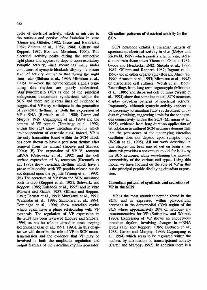
cycle of electrical activity, which is intrinsic to the nucleus and persists after isolation in vitro (Green and Gillette, 1982; Groos and Hendricks, 1982; Shibata et al., 1982, 1984; Gillette and Reppert, 1987; Bos and Mirmiran, 1990). This electrical activity peaks during the subjective light phase and appears to depend upon excitatory synaptic activity, since recordings made under conditions of synaptic blockade display a constant level of activity similar to that during the night time nadir (Shibata et al., 1984; Mirmiran et al., 1995). However, the neurochemical signals regu- latin$ this rhythm are poorly understood. [Arg ]vasopressin (VP) is one of the principal endogenous transmitters synthesized within the SCN and there are several lines of evidence to suggest that VP may participate in the generation of circadian rhythms: (i) Both the expression of VP mRNA (Burbach et al., 1988; Carter and Murphy, 1989; Cagampang et al., 1994) and the content of VP peptide (Tominaga et al., 1992) within the SCN show circadian rhythms which are independent of extrinsic cues. Indeed, VP is the only transmitter found within the SCN which has been shown to have a persistent rhythm after removal from the animal (Inouye and Shibata, 1994); (ii) The expression of VP V1 receptor mRNA (Ostrowski et al., 1992) and the cell surface expression of V1 receptors (Kremarik et al., 1995) show circadian rhythms which have a phase relationship with VP peptide release but do not depend upon the peptide (Young et al., 1993); (iii) The secretion of VP from the SCN measured both in vivo (Reppert et al., 1981; Schwartz and Reppert, 1985; Kalsbeek et al., 1995) and in vitro (Earnest and Sladek, 1987; Gillette and Reppert, 1987; Earnest et al., 1991; Murakami et al., 1991; Watanabe et al., 1993; Shinohara et al., 1994; Tominaga et al., 1994) show circadian cycles which again have a phase relationship with VP synthesis. The regulation of VP expression in the SCN has been reviewed (Inouye and Shibata, 1994) as has its role in circadian time keeping (Reghunandanan et al., 1991, 1993). In this chap- ter we will describe the role of VP in SCN neuro- transmission and the evidence that VP may be involved in both the amplitude regulation and output features of the circadian rhythm generator.
Circadian patterns of electrical activity in the SCN
SCN neurones exhibit a circadian pattern of spontaneous electrical activity in vivo (Meijer and Rietveld, 1989) which persists after in vitro isola- tion in brain tissue slices (Green and Gillette, 1982; Groos and Hendricks, 1982; Shibata et al., 1982, 1984; Gillette and Reppert, 1987; Ingram et al., 1996) and in either organotypic (Bos and Mirmiran, 1990; Aronson et al., 1993; Mirmiran et al., 1995) or dissociated cell cultures (Welsh et al., 1995). Recordings from long-term organotypic (Mirmiran et al., 1995) and dispersed cell cultures (Welsh et al., 1995) show that some but not all SCN neurones display Circadian patterns of electrical activity. Importantly, although synaptic activity appears to be necessary to maintain fully synchronized circa- dian rhythmicity, suggesting a role for the endogen- ous connectivity within the SCN (Mirmiran et al., 1995), evidence from long duration application of tetrodotoxin to cultured SCN neurones demonstrate that the persistence of the underlying circadian oscillator does not depend upon synaptic input (Welsh et al., 1995). All our work described in this chapter has been carried out on brain slices since this provides a convenient model for isolating the SCN neurones, while maintaining the intrinsic connectivity of the various cell types. Using this model we have focused on the role of VP as this is the principal peptide displaying circadian expres- sion.
Circadian pattern of synthesis and secretion of VP in the SCN
VP is the most abundant peptide found in the SCN, and is expressed within parvocellular neurones in the dorsomedial (DM) region of the SCN where approximately 20% of neurones are immunoreactive for VP (Sofroniew and Weindl, 1980). Expression of VP shows an endogenous circadian rhythm, involving changes in mRNA levels (Uhl and Reppert, 1986; Burbach et al., 1988; Carter and Murphy, 1989; Cagampang et al., 1994) which seem to be regulated within the nucleus by attenuation of transcriptional activity (Carter and Murphy, 1992). In addition there is a
353
diurnal variation in molecular size due to differen- tial polyadenylation, with the longer and more stable 740 nucleotide mRNA expressed during the light phase (Robinson et al., 1988; Carter and Murphy, 1989). Correlated with and dependent upon these cycles of transcription and polyadenyla- tion is a rhythm of VP release which is maintained in vitro and peaks during the subjective light phase (Earnest and Sladek, 1987; Gillette and Reppert, 1987; Earnest et al., 1991; Murakami et al., 1991; Watanabe et al., 1993; Shinohara et al., 1994; Tominaga et al., 1994). The remarkable correlation between the in vitro cycles of VP release and of electrical activity (both peaking in the middle of the light phase), has led to the hypothesis that the endogenous cycle of VP release contributes to the cycle of electrical activity and, thereby, functions as integral part of the circadian clock mechanism. However, a causal link between these two rhythms has been previously dismissed as the peak of elec- trical activity occurs slightly out of phase with the peak of peptide secretion, peptide content or VP mRNA levels. However, this assumes that no other factors are playing a part in the response, such as cyclical changes in receptor expression or interactions with inhibitory transmitters. In the following sections these possibilities will be considered.
Role of VP in intranuclear neurotransmission and circadian patterns of activity
There is considerable evidence that VP synthe- sized within the SCN is involved in intranuclear circuits, which have the potential to participate in the generation and integration of circadian signals. Immunoelectronmicroscopy has shown the presence of VP-immunoreactive synapses which contact a number of neurochemically different cell types (van den Pol and Gorcs, 1986; Castel et al., 1990; Daikoku et al., 1992). VP synapses have been seen on VP neurones and may participate either in the synchronization of the population of VP-producing neurones, or in recurrent control that may be important for the resetting of the clock. Local VP circuits exist within the SCN involving soma-somatic appositions of VP neurones and an extensive axo-dendritic network innervating
neurones in both the ipsilateral and contralateral SCN (Caste1 et al., 1990). Although VP fibres appear to be concentrated in the DM division of the SCN (Daikoku et al., 1992), V1 receptors are equally distributed throughout the nucleus (Kremarik et al., 1995). This is in agreement with our observation that there is no regional localisation of VP responsive neurones (unpublished observa- tions), although others have reported that a greater proportion of responsive neurones are found in the DM division of the hamster SCN (Liou and Albers, 1989).
Both our own studies in rat (Mihai et al., 1994a,b; Ingram et al., 1996) and those of others in rat (Shibata and Moore, 1988) and hamster (Liou and Albers, 1989) have shown that a high propor- tion of SCN neurones (approximately 40%) show excitatory responses to VP in vitro (Fig. 1). In general, perfusion with doses of M or greater are required in order to observe significant excita- tion, although lower doses may be effective during the subjective dark phase when endogenous peptide release is reduced. Indeed, both ourselves and others (Liou and Albers, 1989) have shown larger responses and a greater proportion of SCN neurones responding to VP during the dark phase of the circa- dian cycle. This difference may arise either from the circadian variation in VP receptor mRNA expression (Young et al., 1993), or from the fact that endogenous excitatory tone is reduced during the dark phase. The excitatory effects of VP can be blocked by co-administration of the receptor antagonists d( CH& [D-Tyr( OMe) 2,Va14,Cit 8] VP (Fig. 1C; Mihai et al., 1994a) or d(CH2)5[Tyr(O- Me)2]VP (Liou and Albers, 1989), indicating a receptor-mediated effect. A much smaller popula- tion of SCN neurones (<5%) show inhibitory responses to VP (Mihai et al., 1994a) and the possi- bility that these effects may be mediated via inhi- bitory interneurones is indicated by the small increase in activity following application of a receptor antagonist (Mihai et al., 1994b). Such interneurones may include those synthesizing either somatostatin or GABA (see below).
Autoradiographic studies have shown the presence of binding sites for either [3H]VP (Freund-Mercier et al., 1987, 1988; Tribollet et al., 1988) or the highly selective linear V, antago-
354
A. AVT POVT AVP, TGOT AVP 15 0 0 0 0
.i &
.m c* AVP AVT TGOT AVT AVP AVT 0 0 0 0 0
c4
l 5 To
- 10 min
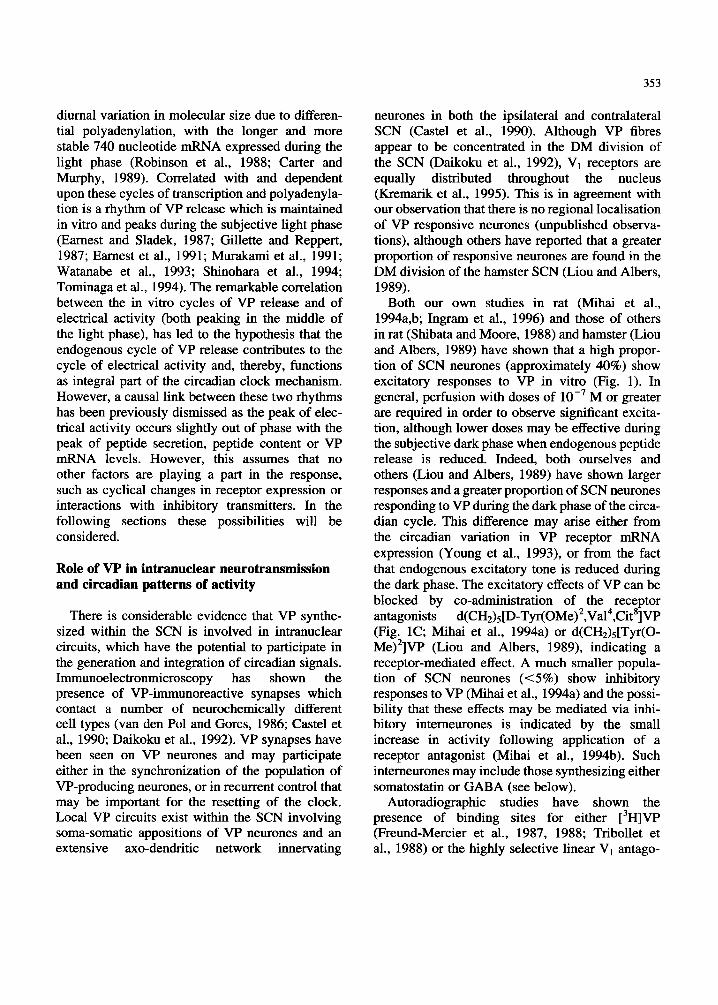
Fig. 1. Electrophysiological actions of VP and related peptides in the SCN. Neurones showing excitatory responses to VP, [Arg8]vasotocin (AVT) and the V,, agonist, [Phe2,0m8]vasotocin (POW) at (A,B) or
M (C). These neurones showed no response to the OT agonist [Thr4,Gly7]oxytocin (TGOT) (A,C) or the VP metabolite w 1 u 4 , ~ y t 6 ] ~ 4-9 (VP 4-9) (A). III a propor- tion of neurones application of the VP antagonist d(CH& ET~I-(OE~)~,V~~~,C~~*]VP (5 X lo-’ M) caused a marked suppression of basal activity, indicative of an endogenous excitatory drive in the tissue (B). This dose of antagonist was able to block the excitatory effects of exogenous VP and AVT (C).
nist Phaa,D-Tyr(Me),Phe,Gln,Asn,Arg,Pro,Arg,- Tyr-NH2 (Kremarik et al., 1995), and V1, (but not V2) receptor mRNA transcripts has been detected in the SCN (Ostrowski et al., 1992; Young et al., 1993). Consistent with the presence of V1, recep- tors, SCN neurones show excitatory responses to the V1, selective agonist [Phe2,0m8]vasotocin (Fig.lA; Liou and Albers, 1989, Mihai et al., 1994a), but not the V2 agonist [Va14,D-Arg8]VP (Liou and Albers, 1989). Interestingly, despite the fact that the oxytocin receptor-selective agonist
[Thr4,Gly7]oxytocin (TGOT) has no effect on most SCN neurones (e.g. Fig.lA,C), a small sub- population of neurones are excited (Mihai et al., 1994a), consistent with the presence of a low density of binding sites for the iodinated OT antagonist d(CH2)5 [Tyr(Me) *,Thr4,0m 8,Tyr-NH;] - vasotocin (Kremarik et al., 1995).
In addition to showing the excitatory effects of TGOT (Tolchard and Ingram, 1993) and VP, our studies in the brain-stem dorsal vagal complex have shown that the ancestral hybrid peptide [Arg8]va- sotocin (VT) can have excitatory effects, apparently independent of OT or VP receptors (Ingram and Tolchard, 1994). VP-responsive neurones in the SCN are also excited by VT (e.g. Fig. 1) and a few neurones show highly preferential responses to this peptide (Mihai et al., 1994a). This suggests that a receptor for VT may be expressed in several parts of the mammalian brain including the SCN. However, this receptor appears not to be that acti- vated by the common metabolite [pGlu4,Cyt6]VP 4-9 as we have been unable to detect any effect of this peptide on SCN neurones (Fig. 1A; Mihai et al., 1994a). Our recordings have provided two important
pieces of evidence which suggest that VP may be contributing to the circadian cycle of electrical activity. Firstly, during the application of a V1 antagonist the otherwise stable basal activity showed a reversible decline in activity, indicative of an endogenous excitatory tone (Fig. 1B; Mihai et al., 1994a). Interestingly, the magnitude of this effect was greater during the light phase when endogenous VP secretion is highest than during the dark phase (Mihai et al., 1994b). Secondly, the circadian variation in tonic basal activity is much greater for those neurones which are respon- sive to VP than for those which are insensitive. This is consistent with the observations of both circadian and non-circadian neurones in culture (Mirmiran et al., 1995; Welsh et al., 1995), and suggests that the ability to respond to VP (i.e., expression of VP receptors) may be a marker for this sub-population of highly circadian neurones. However, we currently have no evidence for the identity of these neurones.
The ability of VP to interact with rhythm gener- ating mechanisms is supported by in vivo beha-
355
vioural data. Injection of an VP antagonist into the SCN causes a shift in the ratio of water and food intake between light and dark phases of the cycle, without abolishing the circadian difference (Reghu- nandanan et al., 1987, 1992). Similarly, continuous i.c.v. infusion of anti-VP antiserum or a V1 receptor antagonist significantly reduces the power of the locomotor rhythm, reducing the active time and increasing slow-wave sleep (Arnauld et al., 1989), while the power of the rhythm is increased by VP (Kruisbrink et al., 1987). Thus, while not essential for rhythm generation, these data suggest that VP may be important for controlling the amplitude of circadian rhythms.
Circadian rhythms in the absence of endogenous VP
Some of the best evidence for the role(s) of VP in the chronobiology of the SCN has come from examination of models of altered peptide expres- sion. Unfortunately, no transgenic animal bearing the VP gene has been shown to express VP in the SCN (Zeng et al., 1994, Waller et al., 1996), and there is no VP-knockout rat with which to study changes in rhythmicity. However, there are several natural states of absence of VP, including the homozygous Brattleboro rat and the mink.
The Brattleboro strain of Long-Evans rat carries a single base deletion in Exon B of the VP gene. In the homozygous animal, this recessive mutation results in an inability to process the translated gene product to VP, and immunocytochemical staining has shown the complete absence of VP in the SCN (van Leeuwen et al., 1989). Nevertheless, in spite of this deficiency, homozygous Brattleboro rats continue to display circadian rhythms of corti- cal EEG (Brown and Nunez, 1989), motor activity (Peterson et al., 1980; Groblewski et al., 1981), drinking behaviour (Peterson et al., 1980; Stoinev and Ikonomov, 1990) and pineal N-acetyltransfer- ase levels (Peterson et al., 1980; Schroder et al., 1988). Furthermore, the rhythms of expression of VP and V1, receptor genes in homozygous animals are identical with controls, despite the absence of any translated VP which may feedback on the pace- maker (Uhl and Reppert, 1986; Young et d., 1993). These observations have led to the conclusion that,
despite its marked circadian expression, VP has no role in the generation of circadian rhythms by the SCN.
We have studied VP effects in the SCN of Brat- tleboro rats in order to determine any possible role of VP in controlling circadian rhythms (Ingram et al., 1996). The density of [3H]VP binding sites is much greater in the homozygous Brattleboro rat than in rats expressing VP (Freund-Mercier et al., 1988; Snijdewint et al., 1989), and this was reflected by a significantly greater proportion of neurones responding to VP, and in the observation of responses to lower doses of VP than normally seen in Wistar rats. This difference in receptor expression appears not to be due to differences in transcription rates (Young et al., 1993) but may reflect the availability of free receptors or homolo- gous receptor down-regulation which might occur in the presence of endogenous peptide. A similar proportion of neurones (4%) were inhibited by VP, suggesting that effects of VP mediated through inhibitory interneurones would also be affected by the absence of the peptide.
Compared to Wistar rats, mean basal activity of VP-responsive neurones was lower in the Brattle- boro rats, consistent with the absence of a tonic excitatory drive. Furthermore, when the population of neurones was classified on the basis of the response to VP, the difference in spontaneous activ- ity between the light and dark phases of the cycle was much greater for the responsive neurones. This suggests that, although the excitation by endogen- ous VP is not the principal mechanism driving the circadian rhythm of electrical activity, like in normal animals (Mihai et al., 1994a), VP sensitivity appears to be a marker for a sub-population of highly circadian SCN neurones. Finally, when we compared the spontaneous activity between hetero- zygous and homozygous rats, the peak activity during the subjective light phase was significantly lower in the homozygous animals, consistent with a lack of VP feedback at this time. Interestingly the absolute difference in firing rate in the light phase was very similar to the antagonist-induced decrease in firing rate seen in Wistar rats (Mihai et al., 1994b). Furthermore, in this study of the effects of the receptor antagonist (Mihai et al., 1994b) only half the SCN neurones showed a decrease in
356
spontaneous activity in response to the peptide. However, despite the apparent absence of any tonic excitation, the antagonist-insensitive neurones still showed a significant circadian varia- tion in spontaneous activity suggesting that, like in the homozygous Brattleboro rat, VP was not the principal generator of the circadian rhythm. Thus, all these data point to the fact that VP is not essen- tial for generation of circadian rhythms but serves to amplify the light-dark differences.
The effect of the absence of VP on the power of circadian rhythms of electrical activity are also reflected in the other behaviours by Brattleboro rats. Although homozygous animals maintain free-running rhythms of drinking and locomotor activity under constant low light (Peterson et al., 1980; Groblewski et al., 1981), the diurnal differ- ence is much less marked compared to heterozy- gous controls. Furthermore, homozygous animals show a dampening of EEG rhythms with a greater proportion of total slow-wave and paradoxical sleep during the dark (normally active) period (Brown and Nunez, 1989), a difference which is maintained even after correcting their drinking by i.v. VP or water (Groblewski et al., 1981; Danguir, 1983).
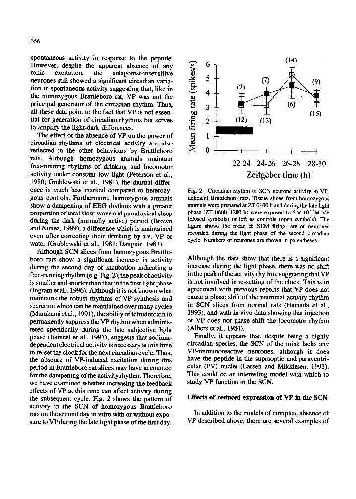
Although SCN slices from homozygous Brattle- boro rats show a significant increase in activity during the second day of incubation indicating a free-running rhythm (e.g. Fig. 2), the peak of activity is smaller and shorter than that in the first light phase (Ingram et al., 1996). Although it is not known what maintains the robust rhythms of VP synthesis and secretion which can be maintained over many cycles (Murakami et al., 1991), the ability of tetrodotoxin to permanently suppress the VP rhythm when adminis- tered specifically during the late subjective light phase (Earnest et al., 1991), suggests that sodium- dependent electrical activity is necessary at this time to re-set the clock for the next circadian cycle. Thus, the absence of VP-induced excitation during this period in Brattleboro rat slices may have accounted for the dampening of the activity rhythm. Therefore, we have examined whether increasing the feedback effects of VP at this time can affect activity during the subsequent cycle. Fig. 2 shows the pattern of activity in the SCN of homozygous Brattleboro rats on the second day in vitro with or without expo- sure to VP during the late light phase of the first day.
n . 6 T
1
22-24 24-26 26-28 28-30
Zeitgeber time (h)
Fig. 2. Circadian rhythm of SCN neurone activity in VP- deficient Brattleboro rats. Tissue slices from homozygous animals were prepared at ZT 0100 h and during the late light phase (ZT 0600-1200 h) were exposed to 5 X lO-*M VP (closed symbols) or left as controls (open symbols). The figure shows the mean 2 SEM firing rate of neurones recorded during the light phase of the second circadian cycle. Numbers of neurones are shown in parentheses.
Although the data show that there is a significant increase during the light phase, there was no shift in the peak of the activity rhythm, suggesting that VP is not involved in re-setting of the clock. This is in agreement with previous reports that VP does not cause a phase shift of the neuronal activity rhythm in SCN slices from normal rats (Hamada et al., 1993), and with in vivo data showing that injection of VP does not phase shift the locomotor rhythm (Albers et al., 1984).
Finally, it appears that, despite being a highly circadian species, the SCN of the mink lacks any VP-immunoreactive neurones, although it does have the peptide in the supraoptic and paraventri- cular (PV) nuclei (Larsen and Mikklesen, 1993). This could be an interesting model with which to study VP function in the SCN.
Effects of reduced expression of VP in the SCN
In addition to the models of complete absence of VP described above, there are several examples of
357
reduced expression of VP which indicate its invol- vement in SCN neurobiology.
Ageing is associated with a decrease in the amplitude of circadian rhythms (van Goo1 and Minniran, 1986), and this may relate to a gradual ‘winding down’ of the biological clock. Indications of the changes in the organisation of the SCN which occur during ageing include, an increased number of degenerating neurones in the DM division of the rat SCN (Woods et al., 1993), and a simultaneous decrease in the number of VP-immunoreactive neurones in this region (Roozendaal et al., 1987). Although a similar decline in numbers appears not to occur in humans, comparison of the number of VP-immunoreactive neurones in the human SCN with time of death shows a clear circadian fluctua- tion in young subjects (<50 years) with a peak during the day, while older subjects (>50 years) show no such variation (Hofman and Swaab, 1994). Interestingly, in rats, both this age-related decline of the number of VP-immunoreactive neurones (Lucassen et al., 1995) and the amplitude of sleep-wake rhythms (Witting et al., 1993) can be prevented by increasing the intensity of the light stimulus.
We have recently been examining whether there are changes in the electrical activity rhythm expressed by aged animals. Our data have shown that, when maintained in constant light (LL), aged male Wistar rats (15-18 months) are capable of displaying a free-running rhythm of electrical activ- ity, and that this can be re-entrained to 12 h lighVl2 h dark (LD) after a period of about 3 weeks. Furthermore, consistent with previous reports that in both aged hamsters (24 months: Watanabe et al., 1995) and rats (23-28 months: Satinoff et al., 1993) there is a decrease in the circadian rhythm of spon- taneous activity of SCN neurones, we have observed a reduction in the light phase peak of activity compared to young ( 3 4 month) animals. More importantly however, when we divided the population of SCN neurones on the basis of their ability to respond to VP, this attenuation of activity was found to be selective for the neurones respon- sive to VP. This suggests that the age-related decline is either an inherent property of this sub- population of neurones or that the afferent drive to these neurones was declining. In this respect, we
found no significant difference in the proportion of VP-sensitive SCN neurones or in the magnitude of the VP-induced excitation, suggesting that by mid-age there has been no change in the expression of V1 receptors. Therefore, we conclude that the declining expression of VP in the SCN underlies an age-related decrease in the power of the activity rhythm. Although the animals we have used are younger (15-18 months) than those shown to have effects on neurone number (>30 months: Roozendaal et al., 1987; Lucassen et al., 1995), it is quite possible that changes in the levels of expression of VP precede the changes in number of VP-immunoreactive neurones. However, it will be necessary to quantify mRNA levels in order to substantiate this conclusion.
A second model showing the importance of differential expression of VP in the SCN, is that of the inter-individual variation in circadian rhyth- micity shown by male voles, a measure which is correlated with VP expression in the SCN (Gerkema et al., 1994). Under constant light condi- tions VP immunoreactivity is suppressed in the SCN of all animals, and this was associated with a complete loss of a circadian pattern of locomotor activity in some animals. Although this rhythm was regained when these animals were placed in LD this group of animals had a significantly higher number of VP neurones in the SCN than those which main- tained rhythmicity throughout. A similar correla- tion has been observed in selected mouse lines in which a high amplitude rhythm of wheel running activity was associated with increased density of VP-immunoreactive neurones (Bult et al., 1993). However, in both these latter cases it remains to be determined whether these effects are mediated through changes in electrical activity rhythms or the output system of the SCN.
Transmitter interactions in the SCN
Although VP is the principal peptide found in the SCN, a large number of other transmitters are expressed in this nucleus (Inouye and Shibata, 1994) and can potentially participate in rhythm generation through interactions with VP-sensitive neurones. It has been demonstrated that a propor- tion of VP-sensitive neurones will also show exci-

358
tatory responses to NPY (Shibata and Moore, 1988) but, although neurotensin-binding sites have been detected in the VL division of the SCN (Francois- Bellan et al., 1992), we have not detected any effect of this peptide on VP-sensitive or -insensitive neurones (unpublished data). More recently we have been examining the main inhibitory transmit- ters in the SCN, somatostatin and GABA.
Double immunostaining has shown synaptic associations between VP axons and somatostatin perikarya and vice versa, suggesting a reciprocal relation between these two cell types (Daikoku et al., 1992). Our recordings show that application of somatostatin- 14 causes dose-dependent and long lasting inhibition of a high proportion of SCN neurones (Fig.3; Ingram et al., 1995), and studies using the SSTR2-selective agonist BIM 23027 has shown that this is the most predominant receptor sub-type in the SCN. Comparison of the effects in the different phases of the circadian cycle showed that a significantly greater proportion of neurones respond to somatostatin during the subjective dark phase compared to the light phase (48% vs. 79%). Furthermore, the pattern and magnitude of responses varied in the different phases of the cycle: inhibitions being significantly greater and of longer duration during the dark phase, suggesting either a rhythm of receptor expression or a variable interaction with endogenous peptide, similar to the situation for VP.
This circadian variation in sensitivity to somatos- tatin is important in view of the marked circadian changes in somatostatin synthesis and content in the SCN. The cycle of somatostatin content p k a k s at around CT 4 (Shinohara et al., 1991; Fukuhara et al., 1993) and somatostatin mRNA also fluctuates during the circadian cycle with a peak at CT 0 (Takeuchi et al., 1992). Neurones of the SCN express an identical 24 h rhythm of somatostatin- like immunoreactivity in both sighted and blind rats (Shinohara et al., 1991) and animals maintained under constant darkness (DD) (Fukuhara et al., 1993). In all cases the peak content at CT 4 which declines prior to lights off reaching a nadir at CT 16 during the subjective night. Unfortunately, it is not known whether this increased content during the light phase is accompanied by a reduction or increased in peptide release and, until this can be
A. GABA SS(-7) Sq-6) 0 0 0
T
AVP 0
n rn . d
AVP SS(-7) SS(-6) GABA 0 0 0 0 3 B.
v 9 1 2 T
.a - c4
10 min
Fig. 3. Interaction between VP and inhibitory transmitters in the SCN. Somatostatin (SS) causes long duration and dose-dependent inhibition of SCN neurones particularly at
M (SS(-6)). This inhibition is both on neurones excited by M VP (A,C) and neurones insensitive to VP (B). The effect of somatostatin is greater than that of GABA M).
resolved, it will not be possible to determine the effect on the activity of SCN neurones.
The inhibitory effects of somatostatin are likely to contribute to the phase regulating actions of the peptide. Previous studies have shown that somatos- tatin can cause either phase delays or phase advances to the rhythm of electrical activity depending on the time of administration (Hamada et al., 1993), while depletion of somatostatin with cysteamine caused only phase advances (Fukuhara et al., 1994). Although these authors showed that the phase shifting effect of somatostatin could be blocked by the purported antagonist, cyclo-(Pro- Phe-D-Trp-Lys-Thr-Phe), we have been unable to block the inhibitory effects of somatostatin with this compound (unpublished data).
In view of the observation that VP sensitivity
359
appears to characterise highly circadian neurones, we have looked for interactions between these p$tides. While some somatostatin-sensitive neurones were excited by M VP (Fig. 3A,C) others were without effect (Fig. 3B), suggesting at least two target populations. Furthermore, inhibi- tion by somatostatin did not block the excitatory response to VP, indicating that they act through separate post-receptor mechanisms. Although the convergent effects indicates that somatostatin is likely to modulate the activity of those VP-sensitive neurones displaying the greatest circadian variation in firing rate, the ability of both peptides to affect firing rate but only somatostatin to effect phase changes (Hamada et al., 1993) strongly suggests that phase shifting is not mediated through simple changes in neuronal activity but may reside in the ability of second messengers to interact with a molecular oscillator.
The other main inhibitory transmitter in the SCN is GABA, and a large number of synapses and peri- karya within the SCN are immunoreactive for the GABA synthesis enzyme glutamic acid decarbox- ylase (van den Pol and Gorcs, 1986). This is consis- tent with the mainly inhibitory synaptic potentials recorded in cultured SCN neurones (Welsh et al., 1995), and the ability of application of GABA to inhibit a large number of SCN neurones both in slices (Liou et al., 1990; Strecker et al., 1995) and organotypic culture (Bos and Mirmiran, 1993). We have examined the possible interaction between GABA and the neurones responsive to VP and/or somatostatin (Fig. 3). In agreement with previous studies (Liou et al., 1990), our data show that appli- cation of M GABA has very rapid inhibitory effects although these rarely cause total suppression of neuronal activity (Fig. 3A,B) and not all neurones were affected (Fig. 3C). Comparison of the effects of somatostatin and GABA showed that the peptide was far more potent and long last- ing than GABA. There was no direct correlation between responsiveness to either VP or somatosta- tin and responses to GABA, further suggesting that VP acts on a number of pharmacologically distinct populations of SCN neurones. Future studies will require that these populations become more accu- rately defined in order to elucidate their role in rhythm-generating circuitry.
Role of VP in the efferent pathways of the SCN
In addition to synapsing and releasing VP within the nucleus, the SCN is one of the major sources of central VP which can signal circadian rhythms to the rest of the CNS. This signal can take the form of a humoral signal in the cerebrospinal fluid (CSF), and measurement of VP levels in the CSF (Reppert et al., 1981; Mens et al., 1982; Schwartz and Reppert, 1985) and the SCN (Kalsbeek et al., 1995) show circadian patterns of release. However, although these are dependent on intact SCN there is no direct evidence to show that the peptide found in the CSF is of SCN origin. On the other hand, the output signal may be neuronal, and pathways have been described projecting princi- pally to mid-line diencephalic areas (Watts and Swanson, 1987). This output function of VP may play an important role in determining circadian patterns of motor (Kruisbrink et al., 1987; Arnauld et al., 1989) and appetitive (Reghunandanan et al., 1987, 1992) behaviours. However, some of the best evidence for a functional role of extranuclear VP is in the transduction pathway controlling circadian activity of the hypothalamepituitary- adrenal (HPA) axis. Lesions of the SCN increase both basal and stress-induced HPA activity, abol- ishing the diurnal variation in plasma corticoster- one concentration (Buijs et al., 1993). Infusion of VP into the dorsomedial hypothalamic/paraventri- cular region of SCN-lesioned animals will decrease the levels of corticosterone, while infu- sion of an antagonist into intact animals causes a marked increase (Kalsbeek and Buijs, 1992; Kals- beek et al., 1992). Furthermore, analogous to our studies on the time-dependent effect of a V1 antagonist on electrical activity (Mihai et al., 1994b), the magnitude of the effect of antagonist infusion varied with the circadian cycle, being greatest during the latter half of the light phase when endogenous VP secretion will have been maximal (Kalsbeek et al., 1996b). Finally, if the levels of endogenous VP were prevented from fall- ing during the late light phase by exogenous infu- sion, then the normal rise in corticosterone seen at the light-dat-k transition was blocked (Kalsbeek et al., 1996a).
Thus, VP appears to fulfil the role of the circa-
360
dian inhibitor of HPA function, although it remains to be demonstrated whether this W projection runs directly to the PVN and mediates some of the inhi- bitory and excitatory responses seen following SCN stimulation (Hermes and Renaud, 1993), or is mediated via the dorsomedial hypothalamic nucleus (Kalsbeek et al., 1996a). In either case it is unclear why microdialysis in the paraventricular/ dorsomedial hypothalamic region fails to show a circadian rhythm of VP release (Kalsbeek et al., 1995).
Finally, it is interesting to note that, in respect of the transmission of circadian signals to the rest of the brain, it has recently been shown in hamsters that a diffusible factor produd by SCN grafts is able to impose strain-specific periodicity on the host (Silver et al., 1996). Whether this factor is (at least in part) VP remains to be determined.
Model for the role of VP in the SCN
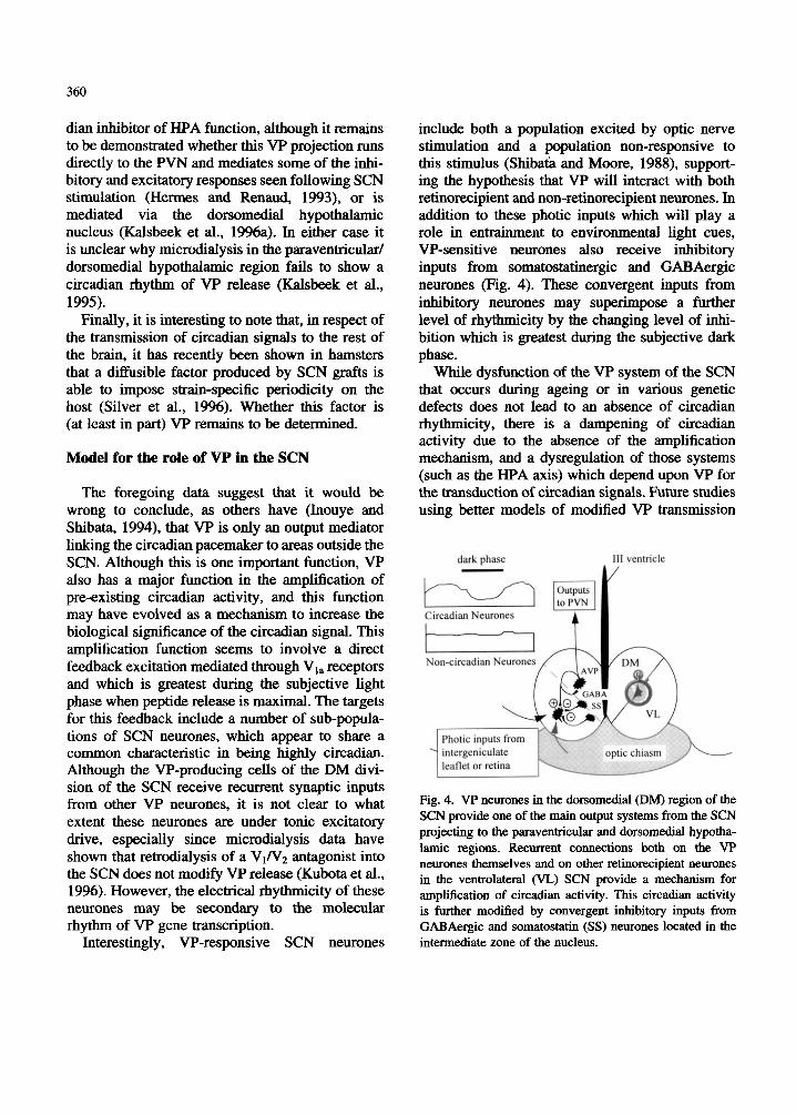
The foregoing data suggest that it would be wrong to conclude, as others have (Inouye and Shibata, 1994), that VP is only an output mediator linking the circadian pacemaker to areas outside the SCN. Although this is one important function, VP also has a major function in the amplification of pre-existing circadian activity, and this function may have evolved as a mechanism to increase the biological significance of the circadian signal. This amplification function seems to involve a direct feedback excitation mediated through V1, receptors and which is greatest during the subjective light phase when peptide release is maximal. The targets for this feedback include a number of sub-popula- tions of SCN neurones, which appear to share a common characteristic in being highly circadian. Although the VP-producing cells of the DM divi- sion of the SCN receive recurrent synaptic inputs from other VP neurones, it is not clear to what extent these neurones are under tonic excitatory drive, especially since microdialysis data have shown that retrodialysis of a VINz antagonist into the SCN does not modify VP release (Kubota et al., 1996). However, the electrical rhythmicity of these neurones may be secondary to the molecular rhythm of VP gene transcription.
Interestingly, VP-responsive SCN neurones
include both a population excited by optic nerve stimulation and a population non-responsive to this stimulus (Shibata and Moore, 1988), support- ing the hypothesis that VP will interact with both retinorecipient and non-retinorecipient neurones. In addition to these photic inputs which will play a role in entrainment to environmental light cues, VP-sensitive neurones also receive inhibitory inputs from somatostatinergic and GABAergic neurones (Fig. 4). These convergent inputs from inhibitory neurones may superimpose a further level of rhythrmcity by the changing level of inhi- bition which is greatest during the subjective dark phase.
While dysfunction of the VP system of the SCN that occurs during ageing or in various genetic defects does not lead to an absence of circadian rhythmicity, there is a dampening of circadian activity due to the absence of the amplification mechanism, and a dysregulation of those systems (such as the HPA axis) which depend upon VP for the transduction of circadian signals. Future studies using better models of modified VP transmission
I11 vartricle A /
Fig. 4. W neurones in the dorsomedial (DM) region of the SCN provide one of the main output systems from the SCN projecting to the paraventricular and dorsomedial hyptha- lamic regions. Recurrent connections both on the VP neurones themselves and on other retinorecipient neurones in the ventrolateral (VL) SCN provide a mechanism for amplification of circadian activity. This circadian activity is further modified by convergent inhibitory inputs from GABAergic and somatostatin (SS) neurones located in the intermediate zone of the nucleus.

36 1
will allow us to further define the role of VP in the SCN.
Acknowledgements
Original work described in this chapter has been made possible through support from the Royal Society, the Romanian Academy, the Biotechnol- ogy and Biological Sciences Research Council International Scientific Interchange Scheme, TEMPUS. and the British Council.
References
Albers, H.E., Fems, C.F., Leeman, S.E. and Goldman, B.D. (1984) Avian pancreatic polypeptide phase shifts hamster circadian rhythms when microinjected into suprachiasmatic region. Science, 223: 833-835.
Amauld, E., Bibene, V., Meynard, J., Rodriguez, F. and Vincent, J.D. (1989) Effects of chronic icv infusion of vasopressin on sleep-waking cycle of rats. Am. J. Physiol., 256: R674R684.
Aronson, B.D., Bell-Pedersen, D., Block, G.D., Dunlap, J.C., Eskin, A., Garceau, N.Y., Geusz, M.E., Johnson, K.A., Khalsa, S.B.S., Koster-van Hoffen, G.C., Koume- nis, C., Lee, T.M., Lesauter, J., Lindgren, K.M., Liu, Q.Y., Loros, J.J., Michel, S.H., Mirmiran, M., Moore, R.Y., Ruby, N.F., Silver, R., Turek, F.W., Zatz, M. and Zucker, I. (1993) Circadian rhythms. Bruin Res. Rev. 18: 320-322.
Bos, N.P.A. and Mirmiran, M. (1990) Circadian rhythms in spontaneous neuronal discharges of cultured suprachias- matic nucleus. Bruin Res., 511: 158-162.
Bos, N.P.A. and Mirmiran, M. (1993) Effect of excitatory and inhibitory amino acids on neuronal discharges in the cultured suprachiasmatic nucleus. Bruin Res. Bull., 31:
Brown, M.H. and Nunez, A.A. (1989) Vasopressin-deficient rats show a reduced amplitude of the circadian sleep rhythm. Physiol. Behuv., 46: 759-762.
Buijs, R.M., Kalsbeek, A., van der Woude, T.P., van Heer- ikhuize, J.J. and Shinn, S. (1993) Suprachiasmatic nucleus lesion increases corticosterone secretion. Am. J. Physiol., 264: R1186-RII96.
Bult, A., Hiestand, L., van der Zee, E.A. and Lynch, C.B. (1993) Circadian rhythms differ between selected mouse lines: a model to study the role of vasopressin neurons in the suprachiasmatic nuclei. Bruin Res. Bull., 32: 623- 627.
Burbach, J.P.H., Liu, B., Voorhuis, T.A.M. and van Tol,
67-72.
H.H.M. (1988) Diurnal variation in vasopressin and oxytocin messenger RNAs in the hypothalamic nuclei of the rat. Mol. Bruin Res., 4: 157-160.
Cagampang, F.R.A., Yang, J., Nakayama, Y., Fukuhara, C. and Inouye, S-I.T. (1994) Circadian variation of argi- nine-vasopressin messenger RNA in the rat suprachias- matic nucleus. Mol. Bruin. Res., 24: 179-184.
Carter, D.A. and Murphy, D. (1989) Diurnal rhythm of vasopressin mRNA species in the rat suprachiasmatic nucleus: interdependence of neuroendocrine modulation and maintenance in explant culture. Mol. Bruin Res., 6:
Carter, D.A. and Murphy, D. (1992) Nuclear mechanisms mediate rhythmic changes in vasopressin mRNA expres- sion in the rat suprachiasmatic nucleus. Mol. Bruin Res.,
Castel, M., Feinstein, N., Cohen, S. and Harari, N. (1990) Vasopressinergic innervation of the mouse suprachias- matic nucleus: An immuno-electron microscopic analy- sis. J. Comp. Neurol., 298: 172-187.
Daikoku, S., Hisano, S. and Kagotani, Y. (1992) Neuronal associations in the rat suprachiasmatic nucleus demon- strated by immunoelectron microscopy. J. Comp. Neurol., 325: 559-571.
Danguir, J. (1983) Sleep deficits in rats with hereditary diabetes insipidus. Nature, 304: 163-164.
Earnest, D.J. and Sladek, C.D. (1987) Circadian vasopressin release from perifused rat suprachiasmatic explants in vitro: effects of acute stimulation. Bruin Res., 422: 398402.
Earnest, D.J., Digiorgio, S.M. and Sladek, C.D. (1991) Effects of tetrodotoxin on the circadian pacemaker mechanism in suprachiasmatic explants in vitro. Bruin Res. Bull., 26: 677-682.
Francois-Bellan, A.M., Bosler, O., Tonon, M.C., Wei, L.T. and Beaudet, A. (1992) Association of neurotensin receptors with VIP-containing neurons and serotonin- containing axons in the suprachiasmatic nucleus of the rat. Synapse,, 10: 282-290.
Freund-Mercier, M.J., Stoeckel, M.E., Palacios, J.M., Pazos, A., Reichhart, J.M., Porte, A. and Richard, Ph. (1987) Pharmacological characteristics and anatomical distribu- tion of [3H]oxytocin-binding sites in the Wistar rat brain by autoradiography. Neuroscience,, 20: 599-614.
Freund-Mercier, M.J., Dietl, M.M., Stoeckel, M., Palacios, J.M. and Richard, Ph. (1988) Quantitative autoradio- graphic mapping of the neurohypophysial hormone bind- ing sites in the rat forebrain and pituitary gland. II. Comparative study of the Long-Evans and Brattleboro strains. Neuroscience,, 21 : 273-281.
Fukuhara, C., Shinohara, K., Tominaga, K., Otori, Y. and Inouye, S.4.T. (1993) Endogenous circadian rhythmicity
233-239.
1 2 315-321.
362
of somatostatin-like immunoreactivity in the rat supra- chiasmatic nucleus. Brain Res., 606: 28-35.
Fukuhara, C., Hamada, T., Shibata, S., Watanabe, S., Aoki, K. and Inouye, S.4.T. (1994) Phase advances of circa- dian rhythms in somatostatin depleted rats: Effects of cysteamine on rhythms of locomotor activity and elec- trical discharge of the suprachiasmatic nucleus. J. Comp. Physiol. A, 175: 677-685.
Gerkema, M.P., van der Zee, E. and Feitsma, L.E. (1994) Expression of circadian rhythmcity correlates with the number of arginine vasopressin-immunoreactive cells in the suprachiasmatic nucleus of common voles, Microtus arvalis. Brain Res., 639: 93-101.
Gillette, M.U. and Reppert, S.M. (1987) The hypothalamic suprachiasmatic nuclei: Circadian patterns of vasopres- sin secretion and neuronal activity in vitro. Brain Res.
Green, D.J. and Gillette, R. (1982) Circadian rhythm of firing rate recorded from single cells in the rat supra- chiasmatic brain slice. Brain Res., 245: 198-200.
Groblewski, T.A., Nunez, A.A. and @Id, R.M. (1981) Circadian rhythms in vasopressin deficient rats. Brain Res. Bull., 6: 125-130.
Groos, G.A. and Hendricks, J. (1982) Circadian rhythms of electrical discharge of rat suprachiasmatic neurones recorded in vitro. Neurosci. Lett., 34 283-288.
Hamada, T., Shibata, S., Tsunyoshi, A., Tominaga, K. and Watanabe, S. (1993) Effect of somatostatin on circadian rhythms of firing and 2-deoxyglucose uptake in rat suprachiasmatic slices. Am. J. Physiol., 265: R1199- R1204.
Hermes, M.L.H.J. and Renaud, L.P. (1993) Differential responses of identified rat hypothalamic paraventricular neurones to suprachiasmatic nucleus stimulation. Neuroscience, 56: 823-832.
Hofman, M.A. and Swaab, D.F. (1994) Alterations in circa- dian rhythrmcity of the vasopressin-producing neurons of the human suprachiasmatic nucleus (SCN) with aging. Brain Res., 651: 134-142.
Ingram, C.D. and Tolchard, S. (1994) [Args]vasotocin excites neurones in the dorsal vagal complex in vitro: Evidence for an action though novel class(es) of CNS receptors. J. Neuroendocrinol., 6 415422.
Ingram, C.D., Tanasescu, R. and Mihai, R. (1995) Circadian variation in the in vitro effect of somatostatin (SS) on the electrical activity of suprachiasmatic nucleus (SCN) neurones of the rat. J. Endocrinol., 147 (Suppl.): P62.
Ingram, C.D., Snowball, R.K. and Mihai, R. (1996) Circa- dian rhythm of neuronal activity in suprachiasmatic nucleus slices from the vasopressin-deficient Brattleboro rat. Neuroscience, 75: 635-641.
Inouye, S.T. and Shibata, S. (1994) Neurochemical organi-
Bull., 19: 135-139.
zation of circadian rhythm in the suprachiasmatic nucleus. Neurosci. Res., 20: 109-1 30.
Kalsbeek, A. and Buijs, R.M. (1992) Peptidergic transmit- ters of the suprachiasmatic nuclei and the control of circadian rhythmicity. Prog. Brain Res., 92: 321-333.
Kalsbeek, A., Buijs, R.M., van Heerikhuize, J.J., Arts, M. and van der Woude, T.P. (1992) Vasopressin-containing neurons of the suprachiasmatic nuclei inhibit corticoster- one release. Brain Res., 580: 62-67.
Kalsbeek, A., Buijs, R.M., Engelmann, M., Wotjak, C.T. and Landgraf, R. (1995) In vivo measurement of a diG- nal variation in vasopressin release in the rat suprachias, matic nucleus. Brain Res., 682: 75-82.
Kalsbeek, A., van der Vliet, J. and Buijs, R.M. (1996a) Decrease of endogenous vasopressin release necessary for expression of the circadian rise in plasma corticoster- one: A reverse microdialysis study. J. Neuroendocrinol.,
Kalsbeek, A., van Heerikhuize, J.J., Wortel, J. and Buijs, R.M. (1996b) A dirurnal rhythm of stimulatory input to the hypothalamo-pituitary-adrenal system as revealed by timd intrahypothdamic administration of the vasopres- sin V1 antagonist J. Neurosci., 16 5555-5565.
Kremarik, P., Freund-Mercier, M.J. and Stoeckel, M.E. (1995) Oxytocin and vasopressin binding sites in the hypothalamus of the rat: Histoautoradiographic detec- tion. Brain Res. Bull., 36: 195-203.
Kruisbrink, J., Mirmiran, M., van der Woude, T.P. and Boer, G.J. (1987) Effects of enhanced cerebrospinal fluid level of vasopressin, vasopressin-antagonist and vasoactive intestinal polypeptide on circadian sleep-wake rhythm in the rat. Brain Res., 419 76-86.
Kubota, M., Landgraf, R. and Wotjak, C.T. (1996) Release of vasopressin within the rat suprachiasmatic nucleus: No effects of a VlN2 antagonist. NeuroRepon, 7:
Larsen, P.J. and Mikklesen, J.D. (1993) The suprachias- matic nucleus of the mink (Mustela vison): Apparent absence of vasopressin-immunoreactive neurons. Cell Tissue Res., 273: 239-247.
Liou, S.Y. and Albers, H.E. (1989) Single unit response of suprachiasmatic nucleus neurones to arginine vasopres- sin in mediated by a V,-like receptor in hamster. Brain Res., 477: 336-343.
Liou, S.Y., Shibata, S., Albers, H.E. and Ueki, S. (1990) Effects of GABA and anxiolytics on the single unit discharge of suprachiasmatic neurons in rat hypothala- mic slices. Brain Res. Bull., 25: 103-107.
Lucassen, P.J., Hofman, M.A. and Swaab, D.F. (1995) Increased light intensity prevents the age related loss of vasopressin-expressing neurons in the rat suprachias- matic nucleus. Brain Res., 693: 261-266.
8: 299-307.
1933-1936.
. 363
Meijer, J.H. and Rietveld, W.J. (1989) Neurophysiology of the suprachiasmatic circadian pacemaker in rodents. Physiol. Rev., 69: 671-707.
Mens, W.B.J., Adringa-Bakker, E.A.D. and van Wimersma Greidanus, T.B. (1982) Changes in cerebrospinal fluid levels of vasopressin and oxytocin during various light- dark regimes. Neurosci. Lett., 34: 51-56.
Mihai, R., Coculescu, M., Wakerley, J.B. and Ingram, C.D. (1994a) The effects of [Arg']vasopressin and [Arg'lva- sotocin on the firing rate of suprachiasmatic newones in vitro. Neuroscience, 62: 783-792.
Mihai, R., Juss, T.S. and Ingram, C.D. (1994b) Suppression of suprachiasmatic nucleus neurone activity with a vaso- pressin receptor antagonist: Possible role for endogenous vasopressin in circadian activity cycles in vitro. Neurosci. Lett., 179: 95-99.
Mirmiran, M., Koster-van Hoffen, G.C. and Bos, N.P.A. (1995) Circadian rhythm generation in the cultured suprachiasmatic nucleus. Brain Res. Bull., 38: 275-283.
Murakami, N., Takamure, M., Takahashi, K., Utunomiya, K., Kuroda, H. and Etoh, T. (1991) Long-term cultured neurons from rat suprachiasmatic nucleus retain the capacity for circadian oscillation of vasopressin release. Brain Rex, 545: 347-350.
Ostrowski, N.L., Lolait, S.J., O'Carroll, A.-M., Brownstein, M.J. and Young, W.S. HI (1992) Distribution of V,, and V2 vasopressin receptor messenger ribonucleic acids in rat liver, kidney, pituitary and brain. Endocrinology,
Peterson, G.M., Watkins, W.B. and Moore, R.Y. (1980) The suprachiasmatic hypothalamic nuclei of the rat: V, Vaso- pressin neurons and circadian rhythmicity. Behav. Neural Biol., 29: 236-245.
Reghunandanan, V., Badgaiyan, R.D., Marya, R.K. and Maini, B.K. (1987) Suprachiasmatic injection of a vaso- pressin antagonist modifies the circadian rhythm of food intake. Behav. Neural Biol., 48: 344-351.
Reghunandanan, V., Reghunandanan, R. and Marya, R.K. (1991) Vasopressin: Its role in circadian time keeping. Chronobiologica, 18: 3947.
Reghunandanan, V., Reghunandanan, R., Marya, R.K. and Singh, P.I. (1992) Vasopressin antagonist disrupts the circadian rhythm of water intake on suprachiasmatic injection. Chronobiol. Int., 9: 356-361.
Reghunandanan, V., Reghunandanan, R. and Singh, P.I. (1993) Neurotransmitters of the suprachiasmatic nucleus: Role in the regulation of circadian rhythms. Prog. Neurobiol., 41: 647-655.
Reppert, S.M., Artman, H.G., Swaminathan, S. and Fisher, D.A. (1981) Vasopressin exhibits a rhythmic daily pattern in the cerebrospinal fluid but not in blood. Science, 213: 1256-1258.
131: 533-535.
Robinson, B.G., Frim, D.M., Schwartz, W.J. and Majzoub, J.A. (1988) Vasopressin mRNA in the suprachiasmatic nuclei: Daily regulation of polyadenylate tail length. Science, 24 1 : 342-344.
Roozendaal, B., van Gool, W.A., Swaab, D.F., Hoogendijk, J.F. and Mirmiran, M. (1987) Changes in vasopressin cells of the rat suprachiasmatic nucleus with aging. Brain Res., 409: 259-264.
Satinoff, E., Li, H., Tcheng, T.K., Liu, C., McArthur, A.J., Medanic, M. and Gillette, M.U. (1993) Do the supra- chiasmatic nuclei oscillate in old rats as they do in young ones? Am. J. Physiol., 265: R1216-R1222.
Schrijder, H., Stehle, J. and Henschel, M. (1988) Twenty- four-hour pineal melatonin synthesis in the vasopressin- deficient Brattleboro rat. Brain Res., 459: 328-332.
Schwartz, W.J. and Reppert, S.M. (1985) Neural regulation of circadian vasopressin rhythm in cerebrospinal fluid: A preeminent role for the suprachiasmatic nucleus. J. Neurosci., 5: 2771-2778.
Shibata, S. and Moore, R.Y. (1988) Neuropeptide Y and vasopressin effects on suprachiasmatic nucleus neurons in vitro. J. Biol. Rhythms, 3: 265-276.
Shibata, S., Oomura, Y., Kita, H. and Hattori, K. (1982) Circadian rhythmic changes of neuronal activity in the suprachiasmatic nucleus of the rat hypothalamic slice. Brain Rex, 247: 154-158.
Shibata, S., Shiratsuchi, A., Liou, S.Y. and Ueki, S. (1984) The role of calcium ions in circadian rhythm of supra- chiasmatic nucleus neuron activity in rat hypothalamic slices. Neurosci. Lett., 52: 181-184.
Shinohara, K., Isobe, Y., Takeuchi, J. and Inouye, S.-I.T. (1991) Circadian rhythms of somatostatin-immunoreac- tivity in the suprachiasmatic nucleus of the rat. Neurosci. Lett., 129: 59-62.
Shinohara, K., Honma, S., Katsuno, Y., Abe, H. and Honma, K. (1994) Circadian rhythms in the release of vasoactive intestinal polypeptide and arginine-vasopressin in orga- notypic slice culture of rat suprachiasmatic nucleus. Neurosci. Lett., 170: 183-186.
Silver, R., LeSauter, J., Tresco, P.A. and Lehman, M.N. (1996) A diffusible coupling signal from the transplanted suprachiasmatic nucleus controlling circadian locomotor rhythms. Nature, 382: 810-813.
Snijedewint, F.G., vanLeeuwen, F.W. andBoer, G.J. (1989) Ontogeny of vasopressin and oxytocin binding sites in the brain of Wistar and Brattleboro rats as demonstrated by light microscopical autoradiography. J. Chem. Neuroanat., 2: 3-17.
Sofroniew, M.V. and Weindl, A. (1980) Identification of parvocellular vasopressin and neurophysin neurone in the suprachiasmatic nucleus of a variety of mammals including primates. J. Comp. Neurol., 193: 659-675.
364
Stoinev, A.G. and Ikonomov, 0.Kh. (1990) Effect of contin- uous vasopressin infusion on circadian rhythms of food and water intake, diuresis, and electrolyte excretion in Brattleboro rats. Bull. Exp. Biol. Med., 109: 122-124.
Strecker, G.J., Bouskila, Y. and Dudek, F.E. (1995) Neuro- transmission and electrophysiological mechanisms in the suprachiasmatic nucleus. Semin. Neurosci., 7: 43-5 1.
Takeuchi, J., Nagasaki, H., Shinohara, K. and Inouye, S.- I.T. (1992) A circadian rhythm of somatostatin messen- ger RNA levels, but not of vasoactive intestinal polypep tide/peptide histidine isoleucine messenger RNA in rat suprachiasmatic nucleus. Mol. Cell. Neurosci., 3: 29-35.
Tolchard, S. and Ingram, C.D. (1993) Electrophysiological action of oxytocin in the dorsal vagal complex of the female rat in vitro: Changing responsiveness during the oestrous cycle and after steroid treatment. Brain Res.,
Tominaga, K., Shinohara, K., Otori, Y., Fukuhara, C. and Inouye, S.T. (1992) Circadian rhythms in vasopressin content in the suprachiasmatic nucleus of the rat. NeuroReport, 3: 809-812.
Tominaga, K., Inouye, S.-I.T. and Okumura, H. (1994) Organotypic slice culture of the rat suprachiasmatic nucleus: Sustenance of cellular architecture and circa- dian rhythm. Neuroscience, 59: 1025-1042.
Tribollet, E., Barberis, C., Jard, S., Dubois-Dauphin, M. and Dreifuss, J.J. (1988) Localization and pharmacological characteristics of high aBinity binding sites for vasopres- sin and oxytocin in the rat brain by light microscopic autoradiography. Brain Res., 442: 105-1 18.
Uhl, G.R. and Reppert, S.M. (1986) Suprachiasmatic nucleus vasopressin messenger RNA: Circadian varia- tion in normal and Brattleboro rats. Science, 232 390- 393.
Van den Pol, A.N. and Gorcs, T. (1986) Synaptic relation- ships between neurones containing vasopressin, gastrin- releasing peptide, vasoactive intestinal polypeptide, and glutamate decarboxylase immunoreactivity in the supra- chiasmatic nucleus: Dual ultrastructural immunocyto- chemistry with gold-substituted silver peroxidase. J. Comp. Neurol., 252: 507-521.
Van Gool, W.A. and Mirmiran, M. (1986) Aging and circa- dian rhythms. Prog. Brain Res., 7 0 255-277.
609: 21-28.
Van Leeuwen, F., van der Beek, E., Seger, M., Burbach, P. and Ivell, R. (1989) Age-related development of a heterozygous phenotype in solitary neurons of the homo- zygous Brattleboro rat. Proc. Natl. Acad. Sci. USA, 8 6 6417-6420.
Waller, S., Fairhall, K.M., Xu, J., Robinson, I.C.A.F. and Murphy, D. (1996) Neurohypophyseal and fluid home- ostasis in transgenic rats expressing a tagged rat vaso- pressin prepropeptide in hypothalamic neurons.
Watanabe, K., Koibuchi, N., Ohtake, H. and Yamaoka, S. (1993) Circadian rhythm of vasopressin release in primary cultures of rat suprachiasmatic nucleus. Bruin Res., 624 115-120.
Watanabe, A., Shibata, S. and Watanabe, S. (1995) Circa- dian rhythm of spontaneous neuronal activity in the suprachiasmatic nucleus of old hamsters in vitro. Brain Res., 695: 237-239.
Watts, A.G. and Swanson, L.W. (1987) Efferent projections of the suprachiasmatic nucleus. 2. Studies using retro- grade transport of fluorescent dyes and simultaneous peptide immunohistochemistry in the rat. J. Comp. Neurol., 258: 230-252.
Welsh, D.K., Logothetis, D.E., Meister, M. and Reppert, S.M. (1995) Individual neurons dissociated from rat suprachiasmatic nucleus express independently phased circadian firing rhythms. Neuron, 14: 697-706.
Witting, W., Mirmiran , M., Bos, N.P.A. and Swaab, D.F. (1993) Effect of light intensity on diurnal sleep wake distribution in young and old rats. Brain Res. Bull., 3 0
Woods, W.H., Powell, E.W., Andrews, A. and Ford, C.W. (1993) Light and electron-microscopic analysis of two divisions of the suprachiasmatic nucleus in the young and aged rat. Anut. Rec., 237: 71-88.
Young, W.S. IJI, Kovacs, K. and Lolait, S.J. (1993) The diurnal rhythm in vasopressin V,, receptor expression in the suprachiasmatic nucleus is not dependent on vaso- pressin. Endocrimlogy, 133: 585-590.
Zeng, Q., Carter, D.A. and Murphy, D. (1994) Cell specific expression of a vasopressin transgene in rats. J. Neuroen- docrinol.. 6: 469-477.
Endocrimlogy, 137: 5068-5077.
157-1 62.