A novel homozygous p.Arg527Leu LMNA mutation in two unrelated Egyptian families causes overlapping...
7
ARTICLE A novel homozygous p.Arg527Leu LMNA mutation in two unrelated Egyptian families causes overlapping mandibuloacral dysplasia and progeria syndrome Mohammad Al-Haggar* ,1 , Agnieszka Madej-Pilarczyk 2 , Lukasz Kozlowski 3 , Janusz M Bujnicki 3,4 , Sohier Yahia 1 , Dina Abdel-Hadi 1 , Amany Shams 5 , Nermin Ahmad 6 , Sahar Hamed 7 and Monika Puzianowska-Kuznicka* ,8,9 Mandibuloacral dysplasia (MAD) is a rare disease resulting from a mutation of LMNA gene encoding lamins A and C. The most common mutation associated with this disease is a homozygous arginine 527 replacement by histidine. Three female patients originating from two unrelated families from Northeast Egypt were examined. Their growth was retarded; they had microcephaly, widened cranial sutures, prominent eyes and cheeks, micrognathia, dental crowding, hypoplastic mandible, acro-osteolysis of distal phalanges, and joint contractures. In addition, they presented some progeroid features, such as pinched nose, premature loss of teeth, loss of hair, scleroderma-like skin atrophy, spine rigidity, and waddling gait. The clinical presentation of the disease varied between the patient originating from Family 1 and patients from Family 2, suggesting that unknown, possibly epigenetic factors, modify the course of the disease. The first symptoms of the disease appeared at the age of 2.5 (a girl from Family 1), 5, and 3 years (girls from Family 2). All patients had the same, novel homozygous c.1580G4T LMNA mutation, resulting in the replacement of arginine 527 by leucine. Computational predictions of such substitution effects suggested that it might alter protein stability and increase the tendency for protein aggregation, and as a result, might influence its interaction with other proteins. In addition, restriction fragment-length polymorphism analysis performed in 178 unrelated individuals showed that up to 1.12% of inhabitants of Northeast Egypt might be heterozygous carriers of this mutation, suggesting the presence of a founder effect in this area. European Journal of Human Genetics (2012) 20, 1134–1140; doi:10.1038/ejhg.2012.77; published online 2 May 2012 Keywords: LMNA; lamin A/C; mutation; mandibuloacral dysplasia; progeria INTRODUCTION Mandibuloacral dysplasia type A (MAD type A (MADA); OMIM # 248370) is a rare disease with a recessive trait of inheritance. Its most common features are: postnatal growth retardation, skeletal abnormalities such as hypoplasia of the mandible and of clavicles, acro-osteolysis of the terminal phalanges, delayed closure of the cranial sutures, dental overcrowding, joint contractures, skin atrophy with mottled pigmentation, lipodystrophy limited to extremities, as well as metabolic abnormalities such as insulin- resistance, diabetes, and hypertriglyceridemia. Affected individuals might also present progeroid features such as bird-like face with micrognathia, pinched nose, prominent eyes, hair loss, scler- oderma-like skin changes, and nail dysplasia. MADA is associated with primary laminopathy resulting from mutation in the LMNA gene 1 encoding lamin A and C, the main components of the nuclear lamina. In addition, MAD type B, in which lipodystrophy is generalized, results from a secondary laminopathy caused by mutations in ZMPSTE-24 gene encoding zinc metallo- proteinase FACE-1 responsible for post-translational modification of prelamin A. 2 The most common mutation reported in patients with MADA is the homozygous p.Arg527His substitution in exon 9 of the LMNA gene. 1,3,4 Other LMNA mutations resulting in MAD are homozygous p.Ala529Val and compound p.Arg527His/Val440Met. 5,6 MAD with progeria-like features was described in relation to the homozygous p.Arg527Cys, homozygous p.Lys542Asn, homozygous p.Arg471Cys, compound p.Thr528Met/Met540Thr, and compound p.Arg471Cys/ Arg527Cys LMNA mutations. 7–11 In this work, we describe a novel homozygous p.Arg527Leu substitution mutation in the LMNA gene, resulting in MAD with progeroid features. We also suggest that a founder effect might be present in Northeast Egypt. 1 Genetics Unit, Department of Pediatrics, Faculty of Medicine, Mansoura University Children’s Hospital, Mansoura, Egypt; 2 Neuromuscular Unit, Mossakowski Medical Research Centre, Warsaw, Poland; 3 Laboratory of Bioinformatics and Protein Engineering, International Institute of Molecular and Cell Biology, Warsaw, Poland; 4 Institute of Molecular Biology and Biotechnology, Faculty of Biology, Adam Mickiewicz University, Poznan, Poland; 5 Department of Anatomy and Histology, Faculty of Medicine, Mansoura University, Mansoura, Egypt; 6 Department of Radiology, Mansoura University Children’s Hospital, Mansoura, Egypt; 7 Mansoura Urology and Nephrology Center, Mansoura, Egypt; 8 Department of Human Epigenetics, Mossakowski Medical Research Centre, Warsaw, Poland; 9 Department of Geriatrics and Gerontology, Medical Center of Postgraduate Education, Warsaw, Poland *Correspondence: Professor M Al-Haggar, Genetics Unit, Department of Pediatrics, Faculty of Medicine, Mansoura University, 35516 Mansoura, Egypt. Tel: þ 2011117 15 350; Fax: þ 2050 223 4092; E-mail: [email protected] or Professor M Puzianowska-Kuznicka, Department of Human Epigenetics, Mossakowski Medical Research Centre, Polish Academy of Sciences, 5 Pawinskiego Street, 02-106 Warsaw, Poland. Tel: þ 48 22 5991755; Fax: þ 48 22 668 55 22; E-mail [email protected] Received 7 November 2011; revised 6 March 2012; accepted 27 March 2012; published online 2 May 2012 European Journal of Human Genetics (2012) 20, 1134–1140 & 2012 Macmillan Publishers Limited All rights reserved 1018-4813/12 www.nature.com/ejhg
Transcript of A novel homozygous p.Arg527Leu LMNA mutation in two unrelated Egyptian families causes overlapping...
ARTICLE
A novel homozygous p.Arg527Leu LMNA mutation intwo unrelated Egyptian families causes overlappingmandibuloacral dysplasia and progeria syndrome
Mohammad Al-Haggar*,1, Agnieszka Madej-Pilarczyk2, Lukasz Kozlowski3, Janusz M Bujnicki3,4,Sohier Yahia1, Dina Abdel-Hadi1, Amany Shams5, Nermin Ahmad6, Sahar Hamed7 andMonika Puzianowska-Kuznicka*,8,9
Mandibuloacral dysplasia (MAD) is a rare disease resulting from a mutation of LMNA gene encoding lamins A and C. The most
common mutation associated with this disease is a homozygous arginine 527 replacement by histidine. Three female patients
originating from two unrelated families from Northeast Egypt were examined. Their growth was retarded; they had microcephaly,
widened cranial sutures, prominent eyes and cheeks, micrognathia, dental crowding, hypoplastic mandible, acro-osteolysis of
distal phalanges, and joint contractures. In addition, they presented some progeroid features, such as pinched nose, premature
loss of teeth, loss of hair, scleroderma-like skin atrophy, spine rigidity, and waddling gait. The clinical presentation of the
disease varied between the patient originating from Family 1 and patients from Family 2, suggesting that unknown, possibly
epigenetic factors, modify the course of the disease. The first symptoms of the disease appeared at the age of 2.5 (a girl from
Family 1), 5, and 3 years (girls from Family 2). All patients had the same, novel homozygous c.1580G4T LMNA mutation,
resulting in the replacement of arginine 527 by leucine. Computational predictions of such substitution effects suggested that
it might alter protein stability and increase the tendency for protein aggregation, and as a result, might influence its interaction
with other proteins. In addition, restriction fragment-length polymorphism analysis performed in 178 unrelated individuals
showed that up to 1.12% of inhabitants of Northeast Egypt might be heterozygous carriers of this mutation, suggesting the
presence of a founder effect in this area.
European Journal of Human Genetics (2012) 20, 1134–1140; doi:10.1038/ejhg.2012.77; published online 2 May 2012
Keywords: LMNA; lamin A/C; mutation; mandibuloacral dysplasia; progeria
INTRODUCTION
Mandibuloacral dysplasia type A (MAD type A (MADA); OMIM# 248370) is a rare disease with a recessive trait of inheritance. Itsmost common features are: postnatal growth retardation,skeletal abnormalities such as hypoplasia of the mandible and ofclavicles, acro-osteolysis of the terminal phalanges, delayed closure ofthe cranial sutures, dental overcrowding, joint contractures, skinatrophy with mottled pigmentation, lipodystrophy limited toextremities, as well as metabolic abnormalities such as insulin-resistance, diabetes, and hypertriglyceridemia. Affected individualsmight also present progeroid features such as bird-like facewith micrognathia, pinched nose, prominent eyes, hair loss, scler-oderma-like skin changes, and nail dysplasia. MADA is associatedwith primary laminopathy resulting from mutation in the LMNAgene1 encoding lamin A and C, the main components of the nuclearlamina. In addition, MAD type B, in which lipodystrophy is
generalized, results from a secondary laminopathy caused bymutations in ZMPSTE-24 gene encoding zinc metallo-proteinase FACE-1 responsible for post-translational modification ofprelamin A.2
The most common mutation reported in patients with MADA isthe homozygous p.Arg527His substitution in exon 9 of the LMNAgene.1,3,4 Other LMNA mutations resulting in MAD are homozygousp.Ala529Val and compound p.Arg527His/Val440Met.5,6 MAD withprogeria-like features was described in relation to the homozygousp.Arg527Cys, homozygous p.Lys542Asn, homozygous p.Arg471Cys,compound p.Thr528Met/Met540Thr, and compound p.Arg471Cys/Arg527Cys LMNA mutations.7–11
In this work, we describe a novel homozygous p.Arg527Leusubstitution mutation in the LMNA gene, resulting in MAD withprogeroid features. We also suggest that a founder effect might bepresent in Northeast Egypt.
1Genetics Unit, Department of Pediatrics, Faculty of Medicine, Mansoura University Children’s Hospital, Mansoura, Egypt; 2Neuromuscular Unit, Mossakowski Medical ResearchCentre, Warsaw, Poland; 3Laboratory of Bioinformatics and Protein Engineering, International Institute of Molecular and Cell Biology, Warsaw, Poland; 4Institute of MolecularBiology and Biotechnology, Faculty of Biology, Adam Mickiewicz University, Poznan, Poland; 5Department of Anatomy and Histology, Faculty of Medicine, Mansoura University,Mansoura, Egypt; 6Department of Radiology, Mansoura University Children’s Hospital, Mansoura, Egypt; 7Mansoura Urology and Nephrology Center, Mansoura, Egypt;8Department of Human Epigenetics, Mossakowski Medical Research Centre, Warsaw, Poland; 9Department of Geriatrics and Gerontology, Medical Center of PostgraduateEducation, Warsaw, Poland*Correspondence: Professor M Al-Haggar, Genetics Unit, Department of Pediatrics, Faculty of Medicine, Mansoura University, 35516 Mansoura, Egypt. Tel: þ2011117 15 350;Fax: þ 2050 223 4092; E-mail: [email protected] Professor M Puzianowska-Kuznicka, Department of Human Epigenetics, Mossakowski Medical Research Centre, Polish Academy of Sciences, 5 Pawinskiego Street, 02-106Warsaw, Poland. Tel: þ 48 22 5991755; Fax: þ 48 22 668 55 22; E-mail [email protected]
Received 7 November 2011; revised 6 March 2012; accepted 27 March 2012; published online 2 May 2012
European Journal of Human Genetics (2012) 20, 1134–1140& 2012 Macmillan Publishers Limited All rights reserved 1018-4813/12
www.nature.com/ejhg
SUBJECTS AND METHODS
Subjects
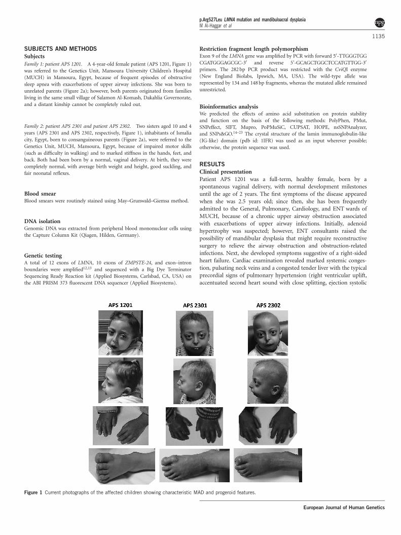
Family 1: patient APS 1201. A 4-year-old female patient (APS 1201, Figure 1)
was referred to the Genetics Unit, Mansoura University Children’s Hospital
(MUCH) in Mansoura, Egypt, because of frequent episodes of obstructive
sleep apnea with exacerbations of upper airway infections. She was born to
unrelated parents (Figure 2a); however, both parents originated from families
living in the same small village of Salamon Al-Komash, Dakahlia Governorate,
and a distant kinship cannot be completely ruled out.
Family 2: patient APS 2301 and patient APS 2302. Two sisters aged 10 and 4
years (APS 2301 and APS 2302, respectively, Figure 1), inhabitants of Ismalia
city, Egypt, born to consanguineous parents (Figure 2a), were referred to the
Genetics Unit, MUCH, Mansoura, Egypt, because of impaired motor skills
(such as difficulty in walking) and to marked stiffness in the hands, feet, and
back. Both had been born by a normal, vaginal delivery. At birth, they were
completely normal, with average birth weight and height, good suckling, and
fair neonatal reflexes.
Blood smearBlood smears were routinely stained using May–Grunwald–Giemsa method.
DNA isolationGenomic DNA was extracted from peripheral blood mononuclear cells using
the Capture Column Kit (Qiagen, Hilden, Germany).
Genetic testingA total of 12 exons of LMNA, 10 exons of ZMPSTE-24, and exon–intron
boundaries were amplified12,13 and sequenced with a Big Dye Terminator
Sequencing Ready Reaction kit (Applied Biosystems, Carlsbad, CA, USA) on
the ABI PRISM 373 fluorescent DNA sequencer (Applied Biosystems).
Restriction fragment length polymorphismExon 9 of the LMNA gene was amplified by PCR with forward 50-TTGGGTGG
CGATGGGAGCGC-30 and reverse 5’-GCAGCTGGCTCCATGTTGG-30
primers. The 282 bp PCR product was restricted with the CviQI enzyme
(New England Biolabs, Ipswich, MA, USA). The wild-type allele was
represented by 134 and 148 bp fragments, whereas the mutated allele remained
unrestricted.
Bioinformatics analysisWe predicted the effects of amino acid substitution on protein stability
and function on the basis of the following methods: PolyPhen, PMut,
SNPeffect, SIFT, Mupro, PoPMuSiC, CUPSAT, HOPE, nsSNPAnalyzer,
and SNPs&GO.14–23 The crystal structure of the lamin immunoglobulin-like
(IG-like) domain (pdb id: 1IFR) was used as an input wherever possible;
otherwise, the protein sequence was used.
RESULTS
Clinical presentationPatient APS 1201 was a full-term, healthy female, born by aspontaneous vaginal delivery, with normal development milestonesuntil the age of 2 years. The first symptoms of the disease appearedwhen she was 2.5 years old; since then, she has been frequentlyadmitted to the General, Pulmonary, Cardiology, and ENT wards ofMUCH, because of a chronic upper airway obstruction associatedwith exacerbations of upper airway infections. Initially, adenoidhypertrophy was suspected; however, ENT consultants raised thepossibility of mandibular dysplasia that might require reconstructivesurgery to relieve the airway obstruction and obstruction-relatedinfections. Next, she developed symptoms suggestive of a right-sidedheart failure. Cardiac examination revealed marked systemic conges-tion, pulsating neck veins and a congested tender liver with the typicalprecordial signs of pulmonary hypertension (right ventricular uplift,accentuated second heart sound with close splitting, ejection systolic
Figure 1 Current photographs of the affected children showing characteristic MAD and progeroid features.
p.Arg527Leu LMNA mutation and mandibuloacral dysplasiaM Al-Haggar et al
1135
European Journal of Human Genetics
with early diastolic murmurs). The electrocardiogram showed rightventricular hypertrophy with marked right-axis deviation. Echocar-diography revealed intact inter-ventricular and inter-atrial septa, poorcardiac muscle performance with reduced ejection fraction, andmarked elevation of pulmonary blood pressure (45 mmHg). Respira-tory function tests were normal. A diagnosis of sub-acutecor pulmonale due to upper airway obstruction was made, andtreatment with oxygen inhalation, diuretic therapy, and digitalis wassuccessful.
Symptoms detected at age 4 are described in Table 1. Basicbiochemical parameters and results of other tests are shown inTable 2. Notably, no metabolic abnormalities were present, mostprobably because of the young age of the patient. The patient remainsunder the control of the Cardiac Clinic; she is maintained on diureticsand load reducers. To relieve the upper airway compromise, untilmaxillofacial reconstructive surgery is performed, she receives (on an‘on-demand’ basis) a continuous positive airway pressure breathing(nasal CPAP) using a portable device.
Patient APS 2301 developed difficulties in leaning forward, in theability to perform fine movements of hands (such as feeding herselfand writing), and in movements of feet, all being a result of markedjoint stiffness, at age 5. Plain radiographs of her hands and chestshowed marked osteolysis (acro-osteolysis of terminal phalanges andabsent clavicles; Figures 3a and b, respectively). She attended theOrthopedic, Dermatology, and Rheumatology Clinics; however,regardless of the prescribed treatment, symptoms were not relievedand systematically progressed. Patient APS 2302 started to havesimilar symptoms at age 3. Neither girl had any symptoms relatedto the respiratory system, they were normotensive withoutdyslipidemia.
Symptoms observed in Patient APS 2301 at age 10 and in PatientAPS 2302 at age 4 are listed in Table 1, whereas the results ofbiochemical and of other tests are shown in Table 2. No metabolic
abnormalities were noticed. They currently receive palliative physicaltherapy to relieve joint stiffness. They also receive vitamins and adhereto a dietary regimen to prevent dyslipidemia.
Blood smearAs the parents of the affected children did not agree for a biopsy, weevaluated the patients’ blood smears. The majority (approximately80% in the patient APS 2301) of the peripheral blood lymphocytesdetected in routine blood smears stained with May–Grunwald–Giemsa method showed markedly irregular nuclear shape(Figures 3c and d).
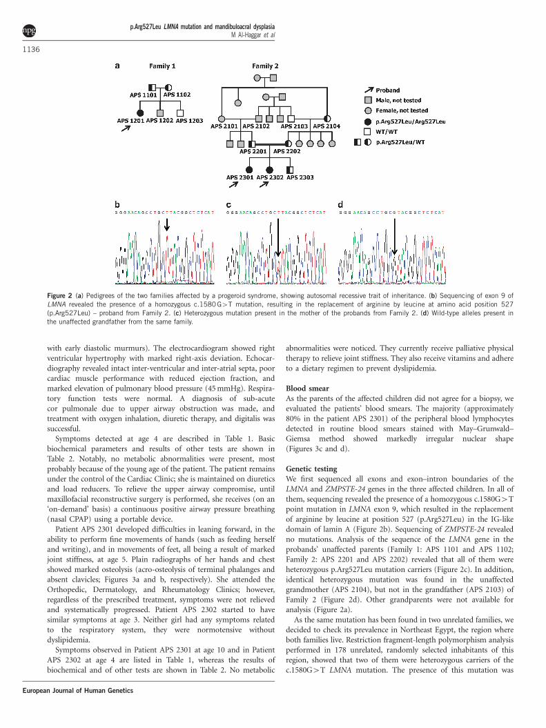
Genetic testingWe first sequenced all exons and exon–intron boundaries of theLMNA and ZMPSTE-24 genes in the three affected children. In all ofthem, sequencing revealed the presence of a homozygous c.1580G4Tpoint mutation in LMNA exon 9, which resulted in the replacementof arginine by leucine at position 527 (p.Arg527Leu) in the IG-likedomain of lamin A (Figure 2b). Sequencing of ZMPSTE-24 revealedno mutations. Analysis of the sequence of the LMNA gene in theprobands’ unaffected parents (Family 1: APS 1101 and APS 1102;Family 2: APS 2201 and APS 2202) revealed that all of them wereheterozygous p.Arg527Leu mutation carriers (Figure 2c). In addition,identical heterozygous mutation was found in the unaffectedgrandmother (APS 2104), but not in the grandfather (APS 2103) ofFamily 2 (Figure 2d). Other grandparents were not available foranalysis (Figure 2a).
As the same mutation has been found in two unrelated families, wedecided to check its prevalence in Northeast Egypt, the region whereboth families live. Restriction fragment-length polymorphism analysisperformed in 178 unrelated, randomly selected inhabitants of thisregion, showed that two of them were heterozygous carriers of thec.1580G4T LMNA mutation. The presence of this mutation was
Figure 2 (a) Pedigrees of the two families affected by a progeroid syndrome, showing autosomal recessive trait of inheritance. (b) Sequencing of exon 9 of
LMNA revealed the presence of a homozygous c.1580G4T mutation, resulting in the replacement of arginine by leucine at amino acid position 527
(p.Arg527Leu) – proband from Family 2. (c) Heterozygous mutation present in the mother of the probands from Family 2. (d) Wild-type alleles present in
the unaffected grandfather from the same family.
p.Arg527Leu LMNA mutation and mandibuloacral dysplasiaM Al-Haggar et al
1136
European Journal of Human Genetics
subsequently confirmed by sequencing. Therefore, approximately1.12% of inhabitants of Northeast Egypt might be heterozygouscarriers of the c.1580G4T-mutated LMNA allele, and the frequencyof the mutated allele in this region is approximately 0.56%.
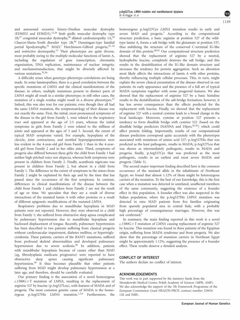
Computational predictions of the p.Arg527Leu LMNAsubstitution effectsComputational analysis showed that the p.Arg527Leu substitutionleads to the disruption of a salt bridge, introduction of a hydrophobicresidue on the protein surface, and to the formation of a cavity thatincreases the solvent-exposed hydrophobic surface (Figure 4). Wepredicted the effects of p.Arg527Leu substitution on protein stability
and function on the basis of a series of algorithms (Table 3).Predictions returned by most methods agree that the substitution ofthe positively charged arginine to the non-charged, hydrophobicleucine, results in destabilization of the lamin A IG-like domainstructure and may increase the protein aggregation tendency.The primary destabilization of the protein structure will causesecondary effects due to the alteration of lamin A interaction withits protein partners.
DISCUSSION
Mutations of the LMNA gene result in a wide range of diseases,termed ‘laminopathies’,24,25 such as autosomal dominant
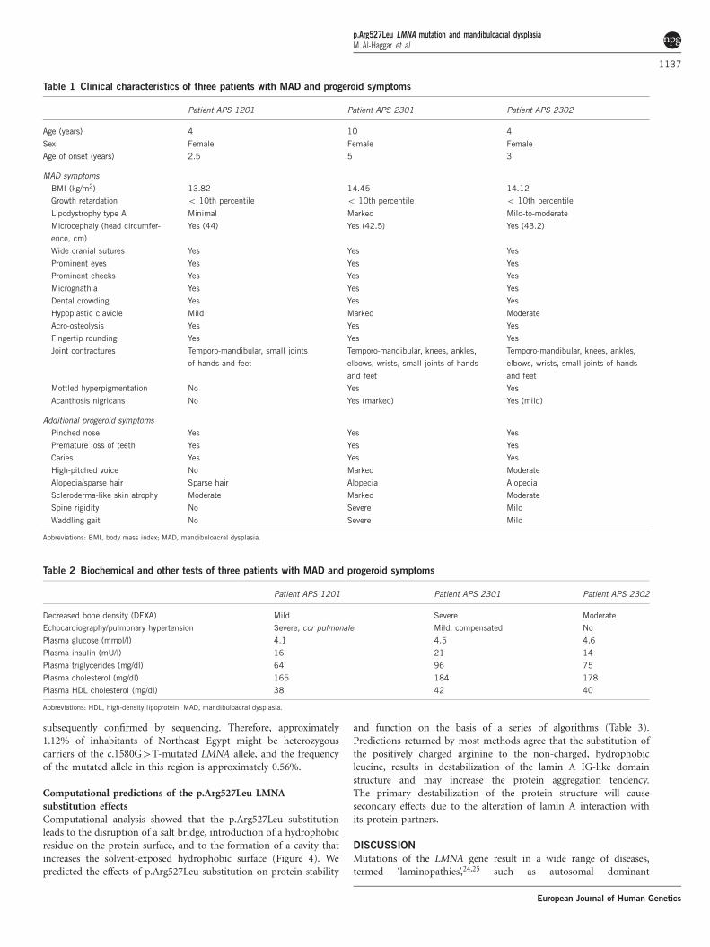
Table 1 Clinical characteristics of three patients with MAD and progeroid symptoms
Patient APS 1201 Patient APS 2301 Patient APS 2302
Age (years) 4 10 4
Sex Female Female Female
Age of onset (years) 2.5 5 3
MAD symptoms
BMI (kg/m2) 13.82 14.45 14.12
Growth retardation o 10th percentile o 10th percentile o 10th percentile
Lipodystrophy type A Minimal Marked Mild-to-moderate
Microcephaly (head circumfer-
ence, cm)
Yes (44) Yes (42.5) Yes (43.2)
Wide cranial sutures Yes Yes Yes
Prominent eyes Yes Yes Yes
Prominent cheeks Yes Yes Yes
Micrognathia Yes Yes Yes
Dental crowding Yes Yes Yes
Hypoplastic clavicle Mild Marked Moderate
Acro-osteolysis Yes Yes Yes
Fingertip rounding Yes Yes Yes
Joint contractures Temporo-mandibular, small joints
of hands and feet
Temporo-mandibular, knees, ankles,
elbows, wrists, small joints of hands
and feet
Temporo-mandibular, knees, ankles,
elbows, wrists, small joints of hands
and feet
Mottled hyperpigmentation No Yes Yes
Acanthosis nigricans No Yes (marked) Yes (mild)
Additional progeroid symptoms
Pinched nose Yes Yes Yes
Premature loss of teeth Yes Yes Yes
Caries Yes Yes Yes
High-pitched voice No Marked Moderate
Alopecia/sparse hair Sparse hair Alopecia Alopecia
Scleroderma-like skin atrophy Moderate Marked Moderate
Spine rigidity No Severe Mild
Waddling gait No Severe Mild
Abbreviations: BMI, body mass index; MAD, mandibuloacral dysplasia.
Table 2 Biochemical and other tests of three patients with MAD and progeroid symptoms
Patient APS 1201 Patient APS 2301 Patient APS 2302
Decreased bone density (DEXA) Mild Severe Moderate
Echocardiography/pulmonary hypertension Severe, cor pulmonale Mild, compensated No
Plasma glucose (mmol/l) 4.1 4.5 4.6
Plasma insulin (mU/l) 16 21 14
Plasma triglycerides (mg/dl) 64 96 75
Plasma cholesterol (mg/dl) 165 184 178
Plasma HDL cholesterol (mg/dl) 38 42 40
Abbreviations: HDL, high-density lipoprotein; MAD, mandibuloacral dysplasia.
p.Arg527Leu LMNA mutation and mandibuloacral dysplasiaM Al-Haggar et al
1137
European Journal of Human Genetics
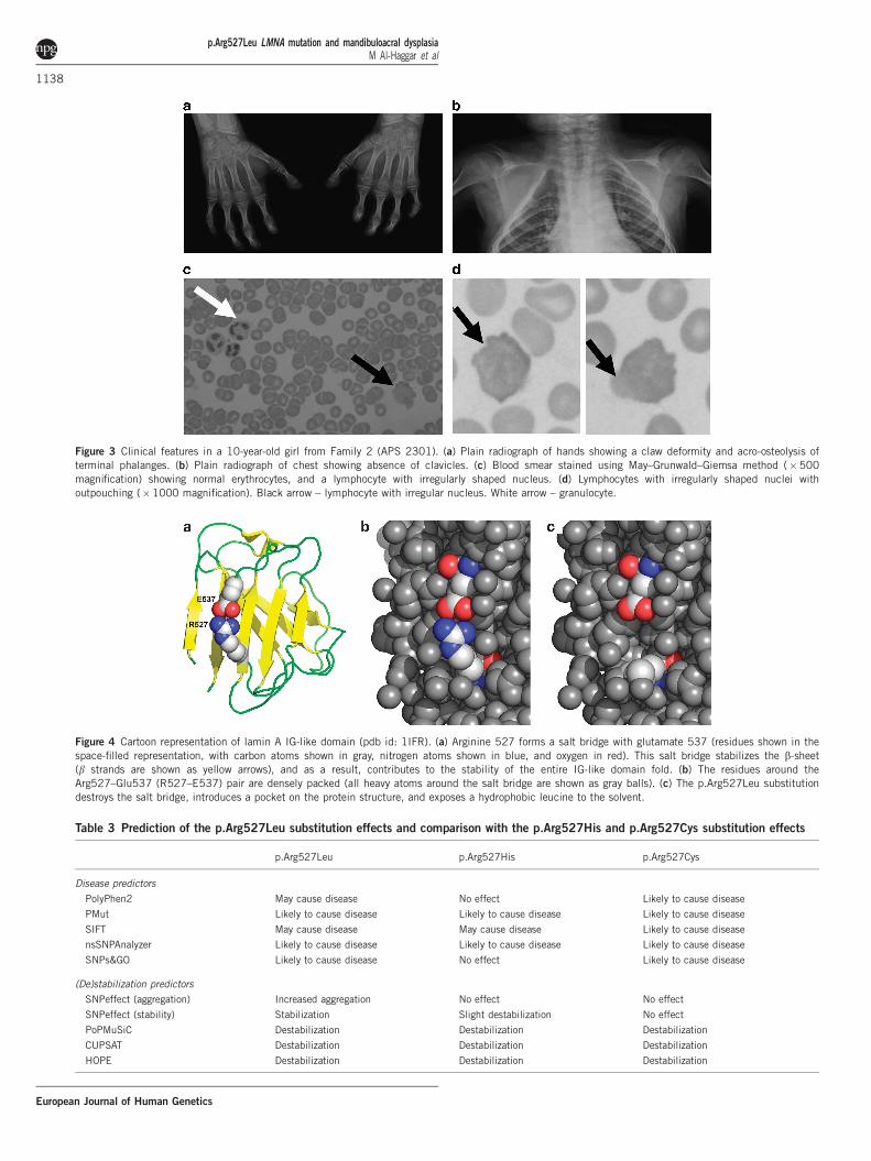
Figure 3 Clinical features in a 10-year-old girl from Family 2 (APS 2301). (a) Plain radiograph of hands showing a claw deformity and acro-osteolysis of
terminal phalanges. (b) Plain radiograph of chest showing absence of clavicles. (c) Blood smear stained using May–Grunwald–Giemsa method (�500
magnification) showing normal erythrocytes, and a lymphocyte with irregularly shaped nucleus. (d) Lymphocytes with irregularly shaped nuclei with
outpouching (�1000 magnification). Black arrow – lymphocyte with irregular nucleus. White arrow – granulocyte.
Figure 4 Cartoon representation of lamin A IG-like domain (pdb id: 1IFR). (a) Arginine 527 forms a salt bridge with glutamate 537 (residues shown in the
space-filled representation, with carbon atoms shown in gray, nitrogen atoms shown in blue, and oxygen in red). This salt bridge stabilizes the b-sheet
(b strands are shown as yellow arrows), and as a result, contributes to the stability of the entire IG-like domain fold. (b) The residues around theArg527–Glu537 (R527–E537) pair are densely packed (all heavy atoms around the salt bridge are shown as gray balls). (c) The p.Arg527Leu substitution
destroys the salt bridge, introduces a pocket on the protein structure, and exposes a hydrophobic leucine to the solvent.
Table 3 Prediction of the p.Arg527Leu substitution effects and comparison with the p.Arg527His and p.Arg527Cys substitution effects
p.Arg527Leu p.Arg527His p.Arg527Cys
Disease predictors
PolyPhen2 May cause disease No effect Likely to cause disease
PMut Likely to cause disease Likely to cause disease Likely to cause disease
SIFT May cause disease May cause disease Likely to cause disease
nsSNPAnalyzer Likely to cause disease Likely to cause disease Likely to cause disease
SNPs&GO Likely to cause disease No effect Likely to cause disease
(De)stabilization predictors
SNPeffect (aggregation) Increased aggregation No effect No effect
SNPeffect (stability) Stabilization Slight destabilization No effect
PoPMuSiC Destabilization Destabilization Destabilization
CUPSAT Destabilization Destabilization Destabilization
HOPE Destabilization Destabilization Destabilization
p.Arg527Leu LMNA mutation and mandibuloacral dysplasiaM Al-Haggar et al
1138
European Journal of Human Genetics
and autosomal recessive Emery–Dreifuss muscular dystrophy(EDMD2 and EDMD3),12,26 limb girdle muscular dystrophy type1B,27 congenital muscular dystrophy,28 dilated cardiomyopathy 1A,29
Charcot-Marie-Tooth disorder type 2B,30 Dunningan-type familialpartial lipodystrophy,31 MAD,1 Hutchinson–Gilford progeria,32–34
and restrictive dermopathy.13 Their phenotypes are quite diverse,most probably owing to the multiple molecular functions of lamin A,including the regulation of gene transcription, chromatinorganization, DNA replication, maintenance of nuclear integrity,and mechanotransduction, that might be diversely affected byvarious mutations.35,36
A difficulty arises when genotype–phenotype correlations are beingmade. In some laminopathies, there is a good correlation between thespecific mutations of LMNA and the clinical manifestations of thedisease; in others, multiple mutations present in distinct parts ofLMNA might all result in a similar phenotype; in still other cases, themutation of a single residue might result in a diverse phenotypes.37
Indeed, this was also true for our patients; even though they all hadthe same LMNA mutation, the symptomatology of their disease wasnot exactly the same. First, the initial, most pronounced symptoms ofthe disease in the girl from Family 1, were related to the respiratorytract and appeared at the age of 2.5 years, whereas the initialsymptoms in girls from Family 2 were related to the stiffness ofjoints and appeared at the ages of 3 and 5. Second, the extent oftypical MAD symptoms varied. For example, hypoplasia of theclavicle, joint contractures, and mottled hyperpigmentation wereless evident in the 4-year-old girl from Family 1 than in the 4-year-old girl from Family 2 and in her older sister. Third, symptoms ofprogeria also differed between these girls: the child from Family 1 hadneither high-pitched voice nor alopecia, whereas both symptoms werepresent in children from Family 2. Finally, acanthosis nigricans waspresent in children from Family 2, but absent in a child fromFamily 1. The difference in the extent of symptoms in the sisters fromFamily 2 might be explained by their age and by the time that haspassed since the occurrence of the first symptoms. In contrast,differences in clinical manifestations of the disease between thechild from Family 1 and children from Family 2 are not the resultof age or time. We speculate that they are a result of diverseinteractions of the mutated lamin A with other proteins or a resultof different epigenetic modifications of the mutated LMNA.
Respiratory problems due to mandibular hypoplasia in MADpatients were not reported. However, they were observed in a childfrom Family 1; she suffered from obstructive sleep apnea complicatedby pulmonary hypertension due to mandibular hypoplasia andbackward displacement of tongue. Recently, pulmonary hypertensionhas been described in two patients suffering from classical progeriawithout cardiovascular impairment, diabetes mellitus, or hypertrigly-ceridemia. These patients, carriers of the BANF1 mutations, sufferedfrom profound skeletal abnormalities and developed pulmonaryhypertension due to severe scoliosis.38 In addition, patientswith mandibular hypoplasia because of causes other than MAD(eg, fibrodysplasia ossificans progressiva) were reported to haveobstructive sleep apnea causing significant pulmonaryhypertension.39 It thus becomes plausible that other patientssuffering from MAD might develop pulmonary hypertension at alater age, and therefore, should be carefully evaluated.
Our primary finding is the association of a novel homozygousc.1580G4T mutation of LMNA, resulting in the replacement ofarginine 527 by leucine (p.Arg527Leu), with features of MADA and ofprogeria. The most common genetic cause of MADA is the homo-zygous p.Arg527His LMNA mutation.1,3,4 Furthermore, the
homozygous p.Arg527Cys LMNA mutation results in early andsevere MAD and progeria.7 According to the computationalstructure prediction, a basic arginine at position 527 of the wild-type lamin A, forms a salt bridge with the glutamate at position 537,thus stabilizing the structure of the conserved C-terminal IG-likedomain of this protein.40,41 Our computational structure predictionshowed that the replacement of arginine 527 by a neutral,hydrophobic leucine, completely destroys the salt bridge, and thisresults in the destabilization of the IG-like domain structure andincreases the tendency for protein aggregation. Such an alterationmost likely affects the interactions of lamin A with other proteins,thereby influencing multiple cellular processes. This, in turn, mightexplain the severe clinical presentation of the disease observed in ourpatients: its early appearance and the presence of a full set of typicalMADA symptoms together with some progeroid features. We alsoshowed that the replacement of arginine 527 by a basic histidineresults in the destabilization of the salt-bridge formation; however, ithas less severe consequences than the effects predicted for thereplacement with leucine. Finally, we showed that the replacementof arginine 527 with a neutral cysteine leads to a broad change in thelocal landscape. Moreover, cysteine at position 527 presents atendency to form disulfide bridge with cysteine 522 (based on thedisulfide bridge predictors DiANNA and DIpro,42,43) which couldaffect protein folding. Importantly, results of our computationaldisease prediction correspond quite accurately with the phenotypesassociated with mutations of amino acid 527: p.Arg527His that waspredicted as the least pathogenic, results in MADA; p.Arg527Leu thatwas shown as intermediately pathogenic, results in MADA andprogeria; finally, p.Arg527Cys that was predicted as the mostpathogenic, results in an earliest and most severe MADA andprogeria (Table 3).
The second major important finding described here is the commonoccurrence of the mutated allele in the inhabitants of NortheastEgypt; we found that almost 1.12% of them might be heterozygouscarriers of the mutation. To the best of our knowledge, this is the firstcase when a mutation was detected in unrelated, unaffected membersof the same community, suggesting the existence of a foundereffect in this population. A founder effect was also suspected in theItalian population, where the p.Arg527His LMNA mutation wasdetected in nine MAD patients from five families originatingfrom sparsely populated area in central Italy, with a probablyhigh percentage of consanguineous marriages. However, this wasnot confirmed.1
In summary, the main finding reported in this work is a novelc.1580G4T mutation of LMNA causing replacement of arginine 527by leucine. This mutation was found in three patients of the Egyptianorigin, suffering from MADA syndrome and from progeria. We alsoshow that the percentage of mutation carriers in Northeast Egyptmight be approximately 1.12%, suggesting the presence of a foundereffect. These results deserve a detailed analysis.
CONFLICT OF INTEREST
The authors declare no conflict of interest.
ACKNOWLEDGEMENTSThis work was in part supported by the statutory funds from the
Mossakowski Medical Centre, Polish Academy of Sciences (MPK, AMP).
We also acknowledge the support of the 7th Framework Programme of the
European Commission Grant HEALTH-PROT, contract number 229676
(LK and JMB).
p.Arg527Leu LMNA mutation and mandibuloacral dysplasiaM Al-Haggar et al
1139
European Journal of Human Genetics
1 Novelli G, Muchir A, Sangiuolo F et al: Mandibuloacral dysplasia is caused by amutation in LMNA encoding lamin A/C. Am J Hum Genet 2002; 71: 426–431.
2 Agarwal AK, Fryns JP, Auchus RJ, Garg A: Zinc metalloproteinase, ZMPSTE24, ismutated in mandibuloacral dysplasia. Hum Mol Genet 2003; 12: 1995–2001.
3 Shen JJ, Brown CA, Lupski JR, Potocki L: Mandibuloacral dysplasia causedby homozygosity for the R527H mutation in lamin A/C. J Med Genet 2003; 40:854–857.
4 Garavelli L, D’Apice MR, Rivieri F et al: Mandibuloacral dysplasia type A in childhood.Am J Med Genet A 2009; 149A: 2258–2264.
5 Garg A, Cogulu O, Ozkinay F, Onay H, Agarwal AK: A novel homozygous Ala529ValLMNA mutation in Turkish patients with mandibuloacral dysplasia. J Clin EndocrinolMetab 2005; 90: 5259–5264.
6 Lombardi F, Gullotta F, Columbaro M et al: Compound heterozygosity for mutations inLMNA in a patient with a myopathic and lipodystrophic mandibuloacral dysplasia typeA phenotype. J Clin Endocrinol Metab 2007; 92: 4467–4471.
7 Agarwal AK, Kazachkova I, Ten S, Garg A: Severe mandibuloacral dysplasia-associatedlipodystrophy and progeria in a young girl with a novel homozygous Arg527Cys LMNAmutation. J Clin Endocrinol Metab 2008; 93: 4617–4623.
8 Plasilova M, Chattopadhyay C, Pal P et al: Homozygous missense mutation in thelamin A/C gene causes autosomal recessive Hutchinson-Gilford progeria syndrome.J Med Genet 2004; 41: 609–614.
9 Zirn B, Kress W, Grimm T, Berthold LD et al: Association of homozygous LMNAmutation R471C with new phenotype: mandibuloacral dysplasia, progeria, and rigidspine muscular dystrophy. Am J Med Genet A 2008; 146A: 1049–1054.
10 Verstraeten VL, Broers JL, van Steensel MA et al: Compound heterozygosity formutations in LMNA causes a progeria syndrome without prelamin A accumulation.Hum Mol Genet 2006; 15: 2509–2522.
11 Cao H, Hegele RA: LMNA is mutated in Hutchinson-Gilford progeria (MIM 176670)but not in Wiedemann-Rautenstrauch progeroid syndrome (MIM 264090). Hum Genet2003; 48: 271–274.
12 Bonne G, Di Barletta MR, Varnous S et al: Mutations in the gene encoding lamin A/Ccause autosomal dominant Emery-Dreifuss muscular dystrophy. Nat Genet 1999; 21:285–288.
13 Navarro CL, De Sandre-Giovannoli A, Bernard R et al: Lamin A and ZMPSTE24 (FACE-1) defect cause nuclear disorganization and identify restrictive dermopathy as a lethalneonatal laminopathy. Hum Mol Genet 2004; 13: 2493–2503.
14 Ramensky V, Bork P, Sunyaev S: Human non-synonymous SNPs: server and survey.Nucleic Acids Res 2002; 30: 3894–3900.
15 Ferrer-Costa C, Gelpı JL, Zamakola L, Parraga I, de la Cruz X, Orozco M: PMUT: a web-based tool for the annotation of pathological mutations on proteins. Bioinformatics2005; 21: 3176–3178.
16 Reumers J, Maurer-Stroh S, Schymkowitz J, Rousseau F: SNPeffect v2.0: a new stepin investigating the molecular phenotypic effects of human non-synonymous SNPs.Bioinformatics 2006; 22: 2183–2185.
17 Ng PC, Henikoff S: Predicting deleterious amino acid substitutions. Genome Res2001; 11: 863–874.
18 Cheng J, Randall A, Baldi P: Prediction of protein stability changes for single-sitemutations using support vector machines. Proteins 2006; 62: 1125–1132.
19 Dehouck Y, Kwasigroch JM, Gilis D, Rooman M: PoPMuSiC 2.1: a web server forthe estimation of protein stability changes upon mutation and sequence optimality.BMC Bioinformatics 2011; 12: 151.
20 Parthiban V, Gromiha MM, Schomburg D: CUPSAT: prediction of protein stability uponpoint mutations. Nucleic Acids Res 2006; 34: W239–W242.
21 Venselaar H, Te Beek TA, Kuipers RK, Hekkelman ML, Vriend G: Protein structureanalysis of mutations causing inheritable diseases. An e-Science approach with lifescientist friendly interfaces. BMC Bioinformatics 2010; 11: 548.
22 Bao L, Zhou M, Cui Y: nsSNPAnalyzer: identifying disease-associated nonsynonymoussingle nucleotide polymorphisms. Nucleic Acids Res 2005; 33: W480–W482.
23 Calabrese R, Capriotti E, Fariselli P, Martelli PL, Casadio R: Functional annotationsimprove the predictive score of human disease-related mutations in proteins.. HumMutat 2009; 30: 1237–1244.
24 Broers JL, Ramaekers FC, Bonne G, Yaou RB, Hutchison CJ: Nuclear lamins:laminopathies and their role in premature ageing. Physiol Rev 2006; 86: 967–1008.
25 Worman HJ, Bonne G: ‘‘Laminopathies’’: a wide spectrum of human diseases. Exp CellRes 2007; 313: 2121–2133.
26 Raffaele Di Barletta M, Ricci E, Galluzzi G et al: Different mutations in the LMNA genecause autosomal dominant and autosomal recessive Emery-Dreifuss musculardystrophy. Am J Hum Genet 2000; 66: 1407–1412.
27 Muchir A, Bonne G, van der Kooi AJ et al: Identification of mutations in the geneencoding lamins A/C in autosomal dominant limb girdle muscular dystrophywith atrioventricular conduction disturbances (LGMD1B). Hum Mol Genet 2000; 9:1453–1459.
28 Quijano-Roy S, Mbieleu B, Bonnemann CG et al: De novo LMNA mutations cause anew form of congenital muscular dystrophy.. Ann Neurol 2008; 64: 177–186.
29 Fatkin D, MacRae C, Sasaki T et al: Missense mutations in the rod domain of thelamin A/C gene as causes of dilated cardiomyopathy and conduction-system disease.N Engl J Med 1999; 341: 1715–1724.
30 De Sandre-Giovannoli A, Chaouch M, Kozlov S et al: Homozygous defects in LMNA,encoding lamin A/C nuclear-envelope proteins, cause autosomal recessive axonalneuropathy in human (Charcot-Marie-Tooth disorder type 2) and mouse. Am J HumGenet 2002; 70: 726–736.
31 Shackleton S, Lloyd DJ, Jackson SN et al: LMNA, encoding lamin A/C, is mutated inpartial lipodystrophy.. Nat Genet 2000; 24: 153–156.
32 De Sandre-Giovannoli A, Bernard R, Cau P et al: Lamin A truncation in Hutchinson-Gilford progeria. Science 2003; 300: 2055.
33 Eriksson M, Brown WT, Gordon LB et al: Recurrent de novo point mutations in lamin Acause Hutchinson-Gilford progeria syndrome. Nature 2003; 423: 293–298.
34 Hennekam RC: Hutchinson-Gilford progeria syndrome: review of the phenotype.Am J Med Genet A 2006; 140: 2603–2624.
35 Verstraeten VL, Broers JL, Ramaekers FC, van Steensel MA: The nuclear envelope, akey structure in cellular integrity and gene expression. Curr Med Chem 2007; 14:1231–1248.
36 Maraldi NM, Capanni C, Cenni V, Fini M, Lattanzi G: Laminopathies and lamin-associated signaling pathways. J Cell Biochem 2011; 112: 979–992.
37 Scharner J: Gnocchi VF, Ellis JA, Zammit PS: Genotype-phenotype correlations inlaminopathies: how does fate translate? Biochem Soc Trans 2010; 38: 257–262.
38 Cabanillas R, Cadinanos J, Villameytide JAF et al: Nestor-Guillermo progeria syndrome:a novel premature aging condition with early onset and chronic development caused byBANF1 mutations. Am J Med Genet 2011A; 155: 2617–2625.
39 Carvalho DR, Pinnola GC, Ferreira DR et al: Mandibular hypoplasia in fibrodysplasiaossificans progressiva causing obstructive sleep apnoea with pulmonary hypertension.Clin Dysmorphol 2010; 19: 69–72.
40 Dhe-Paganon S, Werner ED, Chi YI, Shoelson SE: Structure of the globular tail ofnuclear lamin. J Biol Chem 2002; 277: 17381–17384.
41 Krimm I, Ostlund C, Gilquin B et al: The Ig-like structure of the C-terminal domain oflamin A/C, mutated in muscular dystrophies, cardiomyopathy, and partial lipodystro-phy. Structure 2002; 10: 811–823.
42 Ferre F, Clote P: DiANNA 1.1: an extension of the DiANNA web server forternary cysteine classification.. Nucleic Acids Res 2006; 34: (Web Server issue)W182–W185.
43 Cheng J, Saigo H, Baldi P: Large-scale prediction of disulphide bridges using kernelmethods, two-dimensional recursive neural networks, and weighted graph matching.Proteins 2006; 62: 617–629.
p.Arg527Leu LMNA mutation and mandibuloacral dysplasiaM Al-Haggar et al
1140
European Journal of Human Genetics