Altered pre-lamin A processing is a common mechanism leading to lipodystrophy
14
Altered pre-lamin A processing is a common mechanism leading to lipodystrophy Cristina Capanni 1 , Elisabetta Mattioli 2 , Marta Columbaro 2 , Enrico Lucarelli 3 , Veena K. Parnaik 4 , Giuseppe Novelli 5 , Manfred Wehnert 6 , Vittoria Cenni 2 , Nadir M. Maraldi 1,2 , Stefano Squarzoni 1 and Giovanna Lattanzi 1, * 1 ITOI, CNR, Unit of Bologna, c/o IOR, Bologna, Italy, 2 Laboratory of Cell Biology, Istituti Ortopedici Rizzoli, Bologna, Italy, 3 Regeneration and Tissue Engineering Laboratory of the Musculoskeletal Tissue Bank, IOR, Bologna, Italy, 4 Centre for Cellular and Molecular Biology, Hyderabad 500 007, India, 5 Department of Biopathology and Image Diagnostics, University of Rome Tor Vergata, Rome, Italy and 6 Institute of Human Genetics, University of Greifswald, Germany Received January 20, 2005; Revised and Accepted April 8, 2005 Lipodystrophies are a heterogeneous group of human disorders characterized by the anomalous distribution of body fat associated with insulin resistance and altered lipid metabolism. The pathogenetic mechanism of inherited lipodystrophies is not yet clear; at the molecular level they have been linked to mutations of lamin A/C, peroxisome proliferator-activated receptor (PPARg) and other seemingly unrelated proteins. In this study, we examined lamin A/C processing in three laminopathies characterized by lipodystrophic pheno- types: Dunnigan type familial partial lipodystrophy, mandibuloacral dysplasia and atypical Werner’s syndrome. We found that the lamin A precursor was specifically accumulated in lipodystrophy cells. Pre- lamin A was located at the nuclear envelope and co-localized with the adipocyte transcription factor sterol regulatory element binding protein 1 (SREBP1). Using co-immunoprecipitation experiments, we obtained the first demonstration of an in vivo interaction between SREBP1 and pre-lamin A. Binding of SREBP1 to the lamin A precursor was detected in patient fibroblasts as well as in control fibroblasts forced to accumu- late pre-lamin A by farnesylation inhibitors. In contrast, SREBP1 did not interact in vivo with mature lamin A or C in cultured fibroblasts. To gain insights into the effect of pre-lamin A accumulation in adipose tissue, we inhibited lamin A precursor processing in 3T3-L1 pre-adipocytes. Our results show that pre-lamin A seques- ters SREBP1 at the nuclear rim, thus decreasing the pool of active SREBP1 that normally activates PPARg and causing impairment of pre-adipocyte differentiation. This defect can be rescued by treatment with trogli- tazone, a known PPARg ligand activating the adipogenic program. INTRODUCTION Lipodystrophies are a heterogeneous group of human dis- orders characterized by the anomalous distribution of body fat or generalized loss of adipose tissue (1). Various degrees of insulin resistance are associated with these diseases. Several types of lipodystrophy have been characterized at the molecular genetic level, including Dunnigan-type familial partial lipodystrophy (FPLD) (2), partial lipodystrophy with mandibuloacral dysplasia (MAD) (3), syndromes of partial lipodystrophy with cardiomyopathy (4) and Berardinelli–Seip congenital generalized lipodystrophy (5). In FPLD and MAD, lamin A/C mutations have been linked to disease (2,3), whereas a form of partial lipodystrophy associated with PPARg mutations has also been described (6). Berardi- nelli – Seip congenital generalized lipodystrophy is due to mutations of seipin, an endoplasmic reticulum protein (5). Other lipodystrophies are acquired or drug-induced, such as the lipodystrophy syndrome that is associated with the use of highly active antiretroviral treatment (HAART) (7) (reviewed in 1). In addition, progeroid syndromes such as Hutchinson – Gilford progeria (HGPS) and atypical Werner’s # The Author 2005. Published by Oxford University Press. All rights reserved. For Permissions, please email: [email protected] *To whom correspondence should be addressed at: ITOI, CNR, Unit of Bologna, c/o IOR, Via di Barbiano 1/10, I-40136 Bologna, Italy. Tel: þ39 0516366768; Fax: þ39 051583593; Email: [email protected] Human Molecular Genetics, 2005, Vol. 14, No. 11 1489–1502 doi:10.1093/hmg/ddi158 Advance Access published on April 20, 2005 by guest on November 16, 2015 http://hmg.oxfordjournals.org/ Downloaded from
-
Upload
independent -
Category
Documents
-
view
0 -
download
0
Transcript of Altered pre-lamin A processing is a common mechanism leading to lipodystrophy
Altered pre-lamin A processing is a commonmechanism leading to lipodystrophy
Cristina Capanni1, Elisabetta Mattioli2, Marta Columbaro2, Enrico Lucarelli3, Veena K. Parnaik4,
Giuseppe Novelli5, Manfred Wehnert6, Vittoria Cenni2, Nadir M. Maraldi1,2, Stefano Squarzoni1
and Giovanna Lattanzi1,*
1ITOI, CNR, Unit of Bologna, c/o IOR, Bologna, Italy, 2Laboratory of Cell Biology, Istituti Ortopedici Rizzoli,
Bologna, Italy, 3Regeneration and Tissue Engineering Laboratory of the Musculoskeletal Tissue Bank,
IOR, Bologna, Italy, 4Centre for Cellular and Molecular Biology, Hyderabad 500 007, India, 5Department of
Biopathology and Image Diagnostics, University of Rome Tor Vergata, Rome, Italy and 6Institute of
Human Genetics, University of Greifswald, Germany
Received January 20, 2005; Revised and Accepted April 8, 2005
Lipodystrophies are a heterogeneous group of human disorders characterized by the anomalous distributionof body fat associated with insulin resistance and altered lipid metabolism. The pathogenetic mechanism ofinherited lipodystrophies is not yet clear; at the molecular level they have been linked to mutations of laminA/C, peroxisome proliferator-activated receptor (PPARg) and other seemingly unrelated proteins. In thisstudy, we examined lamin A/C processing in three laminopathies characterized by lipodystrophic pheno-types: Dunnigan type familial partial lipodystrophy, mandibuloacral dysplasia and atypical Werner’ssyndrome. We found that the lamin A precursor was specifically accumulated in lipodystrophy cells. Pre-lamin A was located at the nuclear envelope and co-localized with the adipocyte transcription factor sterolregulatory element binding protein 1 (SREBP1). Using co-immunoprecipitation experiments, we obtainedthe first demonstration of an in vivo interaction between SREBP1 and pre-lamin A. Binding of SREBP1 tothe lamin A precursor was detected in patient fibroblasts as well as in control fibroblasts forced to accumu-late pre-lamin A by farnesylation inhibitors. In contrast, SREBP1 did not interact in vivo with mature lamin Aor C in cultured fibroblasts. To gain insights into the effect of pre-lamin A accumulation in adipose tissue, weinhibited lamin A precursor processing in 3T3-L1 pre-adipocytes. Our results show that pre-lamin A seques-ters SREBP1 at the nuclear rim, thus decreasing the pool of active SREBP1 that normally activates PPARgand causing impairment of pre-adipocyte differentiation. This defect can be rescued by treatment with trogli-tazone, a known PPARg ligand activating the adipogenic program.
INTRODUCTION
Lipodystrophies are a heterogeneous group of human dis-orders characterized by the anomalous distribution of bodyfat or generalized loss of adipose tissue (1). Various degreesof insulin resistance are associated with these diseases.Several types of lipodystrophy have been characterized atthe molecular genetic level, including Dunnigan-type familialpartial lipodystrophy (FPLD) (2), partial lipodystrophy withmandibuloacral dysplasia (MAD) (3), syndromes of partiallipodystrophy with cardiomyopathy (4) and Berardinelli–Seip
congenital generalized lipodystrophy (5). In FPLD andMAD, lamin A/C mutations have been linked to disease(2,3), whereas a form of partial lipodystrophy associatedwith PPARg mutations has also been described (6). Berardi-nelli–Seip congenital generalized lipodystrophy is due tomutations of seipin, an endoplasmic reticulum protein (5).Other lipodystrophies are acquired or drug-induced, such asthe lipodystrophy syndrome that is associated with the useof highly active antiretroviral treatment (HAART) (7)(reviewed in 1). In addition, progeroid syndromes such asHutchinson–Gilford progeria (HGPS) and atypical Werner’s
# The Author 2005. Published by Oxford University Press. All rights reserved.
For Permissions, please email: [email protected]
*To whom correspondence should be addressed at: ITOI, CNR, Unit of Bologna, c/o IOR, Via di Barbiano 1/10, I-40136 Bologna, Italy. Tel: þ390516366768; Fax: þ39 051583593; Email: [email protected]
Human Molecular Genetics, 2005, Vol. 14, No. 11 1489–1502doi:10.1093/hmg/ddi158Advance Access published on April 20, 2005
by guest on Novem
ber 16, 2015http://hm
g.oxfordjournals.org/D
ownloaded from
syndrome (WS) show generalized lipodystrophy, oftencombined with insulin resistant diabetes mellitus (8–11).
In recent years, two major players have emerged as beingpossibly involved in the pathogenesis of lipodystrophies.The first is lamin A/C, the nuclear lamina constituentmutated in FPLD and MAD cells (2,3) as well as in progeroidsyndromes with lipodystrophy (8–11). Moreover, mutationsof ZMPSTE 24, the metalloprotease involved in laminA processing (12), cause diseases featuring a lipodystrophyphenotype such as MAD in humans (13), a MAD-resemblingphenotype in knockout mice (14,15) and a HGPS(Markquardt, personal communication).
The second emerging protein in the pathogenesis oflipodystrophy is the sterol regulatory element binding protein1 (SREBP1), a transcription factor whose localization and trans-activation ability appear to be altered in acquired lipodystrophy(7,16). In this context, it is noteworthy that mutations of PPARgtranscription factor, which is transcribed downstream ofSREBP1 activation and mediates adipocyte differentiation(17), are responsible for other forms of partial lipodystrophy(6,18). Moreover, a single point mutation in the PPARgpromoter has been recently associated with FPLD (19).
Lamins A and C are nuclear lamina proteins obtained byalternative splicing of LMNA and are almost ubiquitouslyexpressed in differentiated tissues (20). Before beingassembled in the nuclear lamina, lamin A undergoescomplex post-translational modifications including farnesyla-tion of the C-terminus and protease cleavage (12,13,21).Farnesylation of pre-lamin A is necessary for the followingsteps of protein cleavage, as metalloproteases fail to bindnon-farnesylated lamin A sequence (15). Mature lamin Aforms a heterodimeric complex with lamin C (22), whichappears to play structural and functional roles, not completelyelucidated (20). Lamin A/C interacts with lamin B and emerin(23) as well as constituents of the nuclear matrix includinglamina-associated polypeptide (LAP) 2 alpha (24), nuclearactin (25,26), transcription factor E2F and protein Rb (27).An increasing number of LMNA mutations give rise toseveral diseases grouped under the definition of laminopathies,but characterized by different tissue-specific defects (28).Besides the earlier-mentioned disorders affecting adiposetissue and/or causing pre-mature aging, LMNA-linked dis-orders include autosomal-dominant Emery–Dreifuss musculardystrophy (EDMD) (29), limb-girdle muscular dystrophy type1B (30), dilated cardiomyopathy with conduction systemdisease (31) affecting skeletal and/or cardiac muscle andother diseases affecting different tissues such as Charcot–Marie–Tooth neuropathy type 2 (32). The pathogenesisof EDMD and other laminopathies has been extensivelyinvestigated in recent years (33–37) and there is circumstan-tial evidence that lamin A/C interaction with either nuclearenvelope constituents or chromatin may be affected in thediseases (20,38–42).
SREBP/ADD1 was initially cloned from rat adipose tissue(43) and shown to be activated in cultured fibroblasts, adipo-cytes and liver (44,45). The human homolog of ADD1,SREBP1, plays a major role in the control of genes involvedin adipocyte differentiation, whereas a closely related factor,SREBP2, is mostly related to cholesterol metabolism (46).SREBP1 is synthesized as an 125 kDa precursor and it
is embedded in the endoplasmic reticulum membrane.Depletion of cholesterol causes proteolytic cleavage of thetranscriptionally active N-terminal portion of SREBP1 fromits position to allow translocation into the nucleus (47). Anumber of published data suggest that SREBP1 interactswith the nuclear envelope at an undefined site, while beingtransferred from the endoplasmic reticulum to the nuclearinterior (48). Interestingly, SREBP1 and lamin A/C do interactin vitro (34), suggesting that alteration of their interplay mayunderlie the pathogenic mechanism of lipodystrophies. Recentevidence shows that impairment of SREBP1 accumulationinside the nucleus occurs in pre-adipocytes treated with anagent employed in HAART and causative of acquired lipody-strophy (7,16). Retention of SREBP1 at the nuclear envelopeis observed under these experimental conditions, concomitantwith anomalous accumulation of unprocessed lamin A at thenuclear rim (7). Downstream of SREBP1 retention at thenuclear envelope, activation of the transcription factorPPARg, which regulates adipocyte differentiation, is impaired(16), indicating a possible pathogenetic pathway for acquiredlipodystrophy.
In this study, we evaluated lamin A precursor maturation andintermolecular interactions in LMNA-mutated FPLD, MAD andWS fibroblasts and control fibroblasts. Our results show thatpre-lamin A is processed to a reduced rate in FPLD, MADand WS cells leading to precursor protein accumulation. Pre-lamin A is bound to SREBP1 at the nuclear rim, thus limitingtranslocation of the transcription factor to the nuclear interior.By using 3T3-L1 pre-adipocytes as a cellular model, we alsoshow that forced pre-lamin A accumulation reduces therate of DNA-bound SREBP1 and lowers PPARg expression.The downstream effects of reduced PPARg expression can berescued by troglitazone (TZD) treatment.
RESULTS
Pre-lamin A accumulates in FPLD, MAD and WSfibroblasts
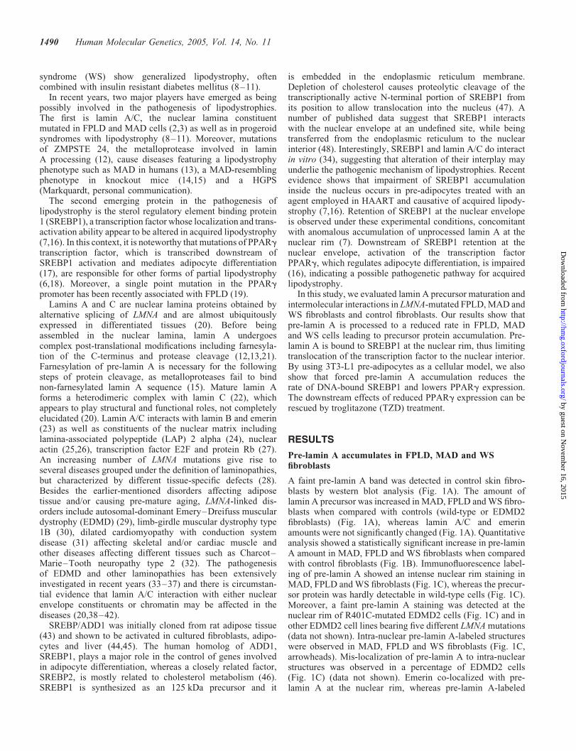
A faint pre-lamin A band was detected in control skin fibro-blasts by western blot analysis (Fig. 1A). The amount oflamin A precursor was increased in MAD, FPLD and WS fibro-blasts when compared with controls (wild-type or EDMD2fibroblasts) (Fig. 1A), whereas lamin A/C and emerinamounts were not significantly changed (Fig. 1A). Quantitativeanalysis showed a statistically significant increase in pre-laminA amount in MAD, FPLD and WS fibroblasts when comparedwith control fibroblasts (Fig. 1B). Immunofluorescence label-ing of pre-lamin A showed an intense nuclear rim staining inMAD, FPLD and WS fibroblasts (Fig. 1C), whereas the precur-sor protein was hardly detectable in wild-type cells (Fig. 1C).Moreover, a faint pre-lamin A staining was detected at thenuclear rim of R401C-mutated EDMD2 cells (Fig. 1C) and inother EDMD2 cell lines bearing five different LMNA mutations(data not shown). Intra-nuclear pre-lamin A-labeled structureswere observed in MAD, FPLD and WS fibroblasts (Fig. 1C,arrowheads). Mis-localization of pre-lamin A to intra-nuclearstructures was observed in a percentage of EDMD2 cells(Fig. 1C) (data not shown). Emerin co-localized with pre-lamin A at the nuclear rim, whereas pre-lamin A-labeled
1490 Human Molecular Genetics, 2005, Vol. 14, No. 11
by guest on Novem
ber 16, 2015http://hm
g.oxfordjournals.org/D
ownloaded from
intra-nuclear structures did not co-localize with emerin inlipodystrophy nuclei (Fig. 1C).
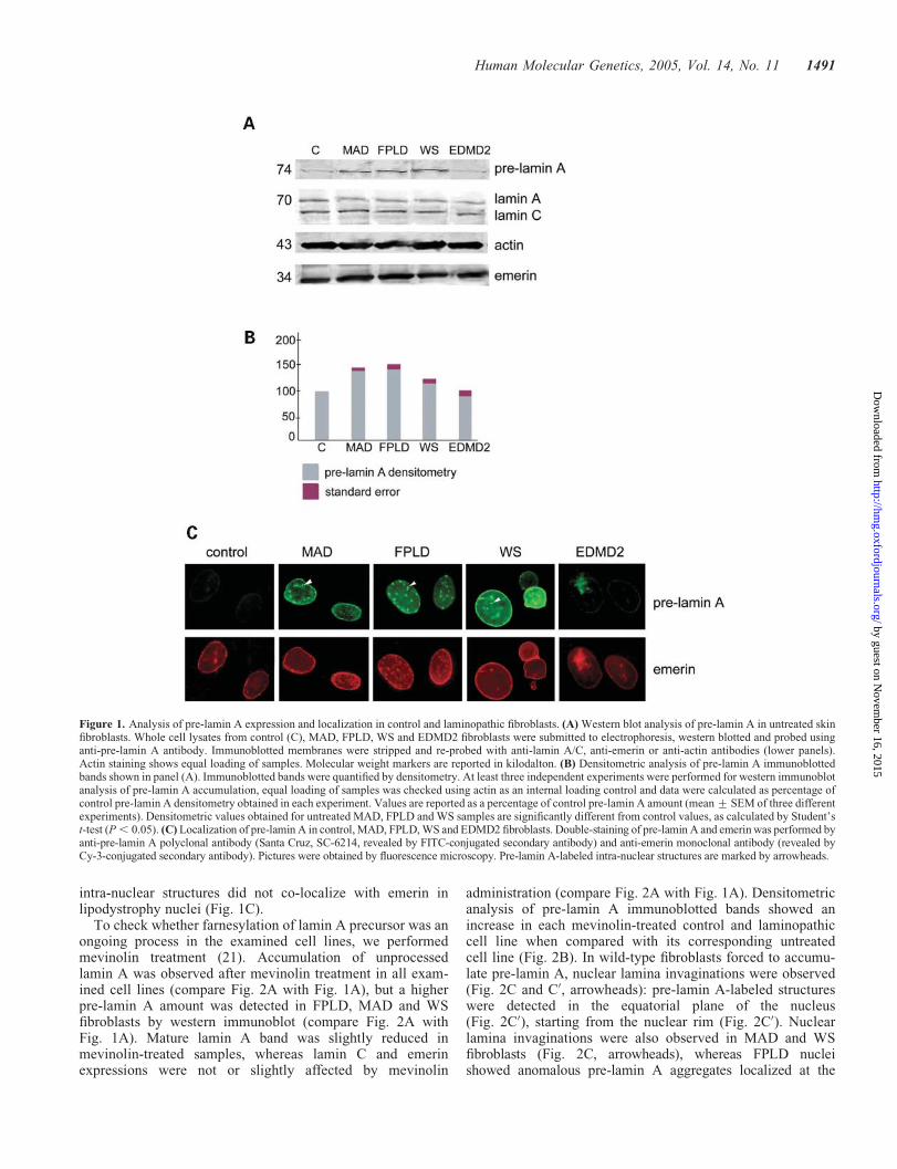
To check whether farnesylation of lamin A precursor was anongoing process in the examined cell lines, we performedmevinolin treatment (21). Accumulation of unprocessedlamin A was observed after mevinolin treatment in all exam-ined cell lines (compare Fig. 2A with Fig. 1A), but a higherpre-lamin A amount was detected in FPLD, MAD and WSfibroblasts by western immunoblot (compare Fig. 2A withFig. 1A). Mature lamin A band was slightly reduced inmevinolin-treated samples, whereas lamin C and emerinexpressions were not or slightly affected by mevinolin
administration (compare Fig. 2A with Fig. 1A). Densitometricanalysis of pre-lamin A immunoblotted bands showed anincrease in each mevinolin-treated control and laminopathiccell line when compared with its corresponding untreatedcell line (Fig. 2B). In wild-type fibroblasts forced to accumu-late pre-lamin A, nuclear lamina invaginations were observed(Fig. 2C and C0, arrowheads): pre-lamin A-labeled structureswere detected in the equatorial plane of the nucleus(Fig. 2C0), starting from the nuclear rim (Fig. 2C0). Nuclearlamina invaginations were also observed in MAD and WSfibroblasts (Fig. 2C, arrowheads), whereas FPLD nucleishowed anomalous pre-lamin A aggregates localized at the
Figure 1. Analysis of pre-lamin A expression and localization in control and laminopathic fibroblasts. (A) Western blot analysis of pre-lamin A in untreated skinfibroblasts. Whole cell lysates from control (C), MAD, FPLD, WS and EDMD2 fibroblasts were submitted to electrophoresis, western blotted and probed usinganti-pre-lamin A antibody. Immunoblotted membranes were stripped and re-probed with anti-lamin A/C, anti-emerin or anti-actin antibodies (lower panels).Actin staining shows equal loading of samples. Molecular weight markers are reported in kilodalton. (B) Densitometric analysis of pre-lamin A immunoblottedbands shown in panel (A). Immunoblotted bands were quantified by densitometry. At least three independent experiments were performed for western immunoblotanalysis of pre-lamin A accumulation, equal loading of samples was checked using actin as an internal loading control and data were calculated as percentage ofcontrol pre-lamin A densitometry obtained in each experiment. Values are reported as a percentage of control pre-lamin A amount (mean + SEM of three differentexperiments). Densitometric values obtained for untreated MAD, FPLD and WS samples are significantly different from control values, as calculated by Student’st-test (P, 0.05). (C) Localization of pre-lamin A in control, MAD, FPLD, WS and EDMD2 fibroblasts. Double-staining of pre-lamin A and emerin was performed byanti-pre-lamin A polyclonal antibody (Santa Cruz, SC-6214, revealed by FITC-conjugated secondary antibody) and anti-emerin monoclonal antibody (revealed byCy-3-conjugated secondary antibody). Pictures were obtained by fluorescence microscopy. Pre-lamin A-labeled intra-nuclear structures are marked by arrowheads.
Human Molecular Genetics, 2005, Vol. 14, No. 11 1491
by guest on Novem
ber 16, 2015http://hm
g.oxfordjournals.org/D
ownloaded from
nuclear rim (Fig. 2C) (data not shown) (42). In mevinolin-treatedwild-type fibroblasts, pre-lamin A-labeled intra-nuclear struc-tures partially co-localized with emerin, whereas they did notco-localize with emerin in MAD, FPLD or WS nuclei (Fig. 2C).
SREBP1 is bound to pre-lamin A in vivo
SREBP1 was detected in control and laminopathic fibroblastsas an 125 kDa precursor and as an active cleaved form(68 kDa) (Fig. 3A). An additional low molecular weight bandwas observed in mevinolin-treated controls and in FPLD,MAD and WS fibroblasts (Fig. 3A). In vivo binding of pre-lamin A to the active 68 kDa SREBP1 form was detected incontrol human fibroblasts following mevinolin-treatment(Fig. 3A) and in FPLD, MAD and WS fibroblasts eitheruntreated (Fig. 3A) or after mevinolin administration (data
not shown). Co-precipitation of SREBP1 and pre-lamin Awas observed using either anti-pre-lamin A or anti-SREBP1antibody to immunoprecipitate protein complexes (Fig. 3A).In contrast, we failed to co-immunoprecipitate lamin A/Cusing anti-SREBP1 antibody (Fig. 3A). Moreover, laminA/C was not co-precipitated by anti-pre-lamin A antibody(Fig. 3A). An emerin immunoprecipitation was performed asa control, which showed absence of co-precipitation ofSREBP1 (Fig. 3A). These results were confirmed by immuno-precipitation experiments using anti-lamin A/C antibody(Fig. 3B). SREBP1 was not detected in the protein complexprecipitated by anti-lamin A/C antibody (Fig. 3B). It shouldbe noted that only in fibroblasts expressing high pre-laminA levels, cross-reactivity of anti-lamin A/C antibody withpre-lamin A caused immunoprecipitation of pre-lamin A (andco-precipitation of a proportional amount of SREBP1) by
Figure 2. Analysis of pre-lamin A expression and localization in mevinolin-treated control and laminopathic fibroblasts. (A) Western blot analysis of pre-laminA in skin fibroblasts, following 18 h mevinolin treatment. Whole cell lysates from control (C), MAD, FPLD, WS and EDMD2 fibroblasts were submitted toelectrophoresis, western blotted and probed using anti-pre-lamin A antibody. Immunoblotted membranes were stripped and re-probed with anti-lamin A/C,anti-emerin or anti-actin antibodies (lower panels). Actin staining shows equal loading of samples. Molecular weight markers are reported in kilodaltons.(B) Densitometric analysis of pre-lamin A immunoblotted bands shown in panel (A). Immunoblotted bands were quantified by densitometry. At least three inde-pendent experiments were performed for western immunoblot analysis of pre-lamin A accumulation. Equal loading of samples was checked using actin as aninternal loading control and data were calculated as percentage of pre-lamin A densitometry measured in each untreated cell line. Values are reported as a per-centage of pre-lamin A amount in each corresponding untreated cell line (Fig. 1A and B) and they represent means + SEM of three different experiments.Western blotting of untreated fibroblasts (Fig. 1A) and mevinolin-treated fibroblasts (A) was routinely performed on the same membrane to allow comparisonof results. (C) Localization of pre-lamin A in mevinolin-treated control, MAD, FPLD and WS fibroblasts. Double-staining of pre-lamin A and emerin wasperformed by anti-pre-lamin A polyclonal antibody (Santa Cruz, SC-6214, revealed by FITC-conjugated secondary antibody) and anti-emerin monoclonal anti-body (revealed by Cy-3-conjugated secondary antibody (C0) Localization of pre-lamin A in mevinolin-treated wild-type nuclei (control). Pictures were obtainedat two different focal planes (lamina plane, equatorial plane). Pre-lamin A-labeled intra-nuclear structures shown in (C) and (C0) are marked by arrowheads.Pictures were obtained by fluorescence microscopy.
1492 Human Molecular Genetics, 2005, Vol. 14, No. 11
by guest on Novem
ber 16, 2015http://hm
g.oxfordjournals.org/D
ownloaded from
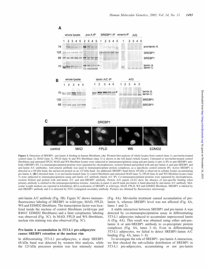
anti-lamin A/C antibody (Fig. 3B). Figure 3C shows immuno-fluorescence labeling of SREBP1 in wild-type, MAD, FPLD,WS and EDMD2 fibroblasts. The transcription factor was loca-lized inside the nucleus of control fibroblasts (wild-type andR401C EDMD2 fibroblasts) and a faint cytoplasmic labelingwas observed (Fig. 3C). In MAD, FPLD and WS fibroblasts,nuclear rim staining was also observed (Fig. 3C).
Pre-lamin A accumulation in 3T3-L1 pre-adipocytescauses SREBP1 retention at the nuclear rim
In differentiating 3T3-L1 pre-adipocytes, a sharp SREBP168 kDa band was detected by western blot analysis, whilethe 125 kDa precursor protein was less intensely stained
(Fig. 4A). Mevinolin treatment caused accumulation of pre-lamin A, whereas SREBP1 level was not affected (Fig. 4A,lanes 1 and 2).
A stable interaction between SREBP1 and pre-lamin A wasdetected by co-immunoprecipitation assay in differentiating3T3-L1 adipocytes induced to accumulate unprocessed laminA (Fig. 4A). This result was obtained using either anti-pre-lamin A or anti-SREBP1 antibody to co-precipitate proteincomplexes (Fig. 4A, lanes 3–6). Even in differentiating3T3-L1 adipocytes, we failed to detect SREBP1-lamin A/Cbinding (Fig. 4A, lanes 3–8).
To investigate the role of SREBP1–pre-lamin A interaction,we first checked the sub-cellular distribution of SREBP1 in3T3-L1 pre-adipocytes, accumulating or not pre-lamin
Figure 3. Detection of SREBP1–pre-lamin A binding in human fibroblasts. (A). Western blot analysis of whole lysates from control (lane 1), mevinolin-treatedcontrol (lane 2), MAD (lane 3), FPLD (lane 4) and WS fibroblasts (lane 5) is shown in the left panel (whole lysate). Untreated or mevinolin-treated controlfibroblasts and untreated FPLD, MAD and WS fibroblast lysates were subjected to immunoprecipitation using anti-pre-lamin A (pre-A IP) or anti-SREBP1 anti-body (SREBP1 IP). Co-immunoprecipitated proteins were separated by electrophoresis, western blotted and probed with anti-pre-lamin A and anti-SREBP1 andanti-lamin A/C antibodies. Anti-emerin antibody was used to immunoprecipitate protein complexes, as a specificity control (emerin IP). Active SREBP1 isdetected as a 68 kDa band, the uncleaved protein as an 125 kDa band. An additional SREBP1 band below 68 kDa is observed in cellular lysates accumulatingpre-lamin A. (B) Untreated (lane 1) or mevinolin-treated (lane 2) control fibroblasts and untreated MAD (lane 3), FPLD (lane 4) and WS fibroblast lysates (lane5) were subjected to immunoprecipitation using anti-lamin A/C antibody (lamin A/C IP). Co-immunoprecipitated proteins were separated by electrophoresis,western blotted and probed with anti-lamin A/C and anti-SREBP1 antibody. Protein A/G panels (A/G) show the absence of non-specific binding whenprimary antibody is omitted in the immunoprecipitation mixture. Asterisks in panel A and B mark pre-lamin A band detected by anti-lamin A/C antibody. Mol-ecular weight markers are reported in kilodaltons. (C) Localization of SREBP1 in wild-type, MAD, FPLD, WS and EDMD2 fibroblasts. SREBP1 is labeled byanti-SREBP1 antibody and it is detected by FITC-conjugated secondary antibody. Pictures are obtained by fluorescence microscopy.
Human Molecular Genetics, 2005, Vol. 14, No. 11 1493
by guest on Novem
ber 16, 2015http://hm
g.oxfordjournals.org/D
ownloaded from
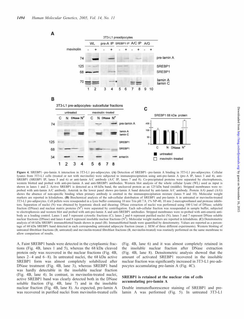
A. Faint SREBP1 bands were detected in the cytoplasmic frac-tions (Fig. 4B, lanes 1 and 5), whereas the 68 kDa cleavedprotein only was recovered in the nuclear fractions (Fig. 4B,lanes 2–4 and 6–8). In untreated nuclei, the 68 kDa activeSREBP1 form was almost completely solubilized afterDNase treatment (Fig. 4B, lane 3), whereas SREBP1 bandwas hardly detectable in the insoluble nuclear fraction(Fig. 4B, lane 4). In contrast, in mevinolin-treated nuclei,active SREBP1 band was clearly detected both in the DNasesoluble fraction (Fig. 4B, lane 7) and in the insolublenuclear fraction (Fig. 4B, lane 8). As expected, pre-lamin Awas recovered in purified nuclei from mevinolin-treated cells
(Fig. 4B, lane 6) and it was almost completely retained inthe insoluble nuclear fraction after DNase extraction(Fig. 4B, lane 8). Densitometric analysis showed that theamount of activated SREBP1 recovered in the insolublenuclear fraction was significantly increased in 3T3-L1 pre-adi-pocytes accumulating pre-lamin A (Fig. 4C).
SREBP1 is retained at the nuclear rim of cellsaccumulating pre-lamin A
Double immunofluorescence staining of SREBP1 and pre-lamin A was performed (Fig. 5). In untreated 3T3-L1
Figure 4. SREBP1–pre-lamin A interaction in 3T3-L1 pre-adipocytes. (A) Detection of SREBP1–pre-lamin A binding in 3T3-L1 pre-adipocytes. Cellularlysates from 3T3-L1 cells (treated or not with mevinolin) were subjected to immunoprecipitation using anti-pre-lamin A (pre-A IP, lanes 3 and 4), anti-SREBP1 (SREBP1 IP, lanes 5 and 6) or anti-lamin A/C antibody (A/C IP, lanes 7 and 8). Co-precipitated proteins were separated by electrophoresis,western blotted and probed with anti-pre-lamin A and anti-SREBP1 antibodies. Western blot analysis of the whole cellular lysate (WL) used as input isshown in lanes 1 and 2. Active SREBP1 is detected as a 68 kDa band, the uncleaved protein as an 125 kDa band (middle). Stripped membranes were re-probed with anti-lamin A/C antibody. Asterisk in the lower panel shows pre-lamin A band detected by anti-lamin A/C antibody. Protein A/G panel (A/G)shows the absence of non-specific binding when primary antibody is omitted in the immunoprecipitation mixture (lanes 9 and 10). Molecular weightmarkers are reported in kilodaltons. (B) Biochemical analysis of the sub-cellular distribution of SREBP1 and pre-lamin A in untreated or mevinolin-treated3T3-L1 pre-adipocytes. Cell pellets were resuspended in a lysis buffer containing 10 mM Tris pH 7.8, 1% NP-40, 10 mM 2-mercaptoethanol and protease inhibi-tors. Separation of nuclei (N) was obtained by hypotonic shock and shearing: DNase extraction of nuclei was performed using 100 U/ml of DNase; solublefraction (DNase) and nuclear matrix proteins (NP) were separated by centrifugation. Each sub-cellular fraction was resuspended in sample buffer, subjectedto electrophoresis and western blot and probed with anti-pre-lamin A and anti-SREBP1 antibodies. Stripped membranes were re-probed with anti-emerin anti-body as a loading control. Lanes 1 and 5 represent cytosolic fractions (C); lanes 2 and 6 represent purified nuclei (N); lanes 3 and 7 represent DNase solublenuclear fractions (DNase) and lanes 4 and 8 represent insoluble nuclear fractions (NP). Molecular weight markers are reported in kilodaltons. (C) Densitometricanalysis of 68 kDa SREBP1 immunoblotted bands shown in panel (B). Immunoblotted bands were quantified by densitometry. Values are reported as a percen-tage of 68 kDa SREBP1 band detected in each corresponding untreated adipocyte fraction (mean + SEM of three different experiments). Western blotting ofuntreated fibroblast fractions (B, untreated) and mevinolin-treated fibroblast fractions (B, mevinolin-treated) was routinely performed on the same membrane toallow comparison of results.
1494 Human Molecular Genetics, 2005, Vol. 14, No. 11
by guest on Novem
ber 16, 2015http://hm
g.oxfordjournals.org/D
ownloaded from
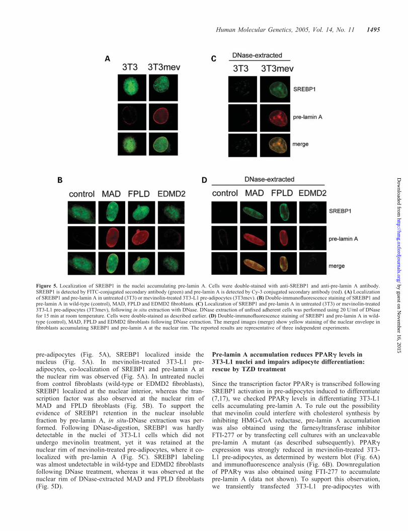
pre-adipocytes (Fig. 5A), SREBP1 localized inside thenucleus (Fig. 5A). In mevinolin-treated 3T3-L1 pre-adipocytes, co-localization of SREBP1 and pre-lamin A atthe nuclear rim was observed (Fig. 5A). In untreated nucleifrom control fibroblasts (wild-type or EDMD2 fibroblasts),SREBP1 localized at the nuclear interior, whereas the tran-scription factor was also observed at the nuclear rim ofMAD and FPLD fibroblasts (Fig. 5B). To support theevidence of SREBP1 retention in the nuclear insolublefraction by pre-lamin A, in situ-DNase extraction was per-formed. Following DNase-digestion, SREBP1 was hardlydetectable in the nuclei of 3T3-L1 cells which did notundergo mevinolin treatment, yet it was retained at thenuclear rim of mevinolin-treated pre-adipocytes, where it co-localized with pre-lamin A (Fig. 5C). SREBP1 labelingwas almost undetectable in wild-type and EDMD2 fibroblastsfollowing DNase treatment, whereas it was observed at thenuclear rim of DNase-extracted MAD and FPLD fibroblasts(Fig. 5D).
Pre-lamin A accumulation reduces PPARg levels in3T3-L1 nuclei and impairs adipocyte differentiation:rescue by TZD treatment
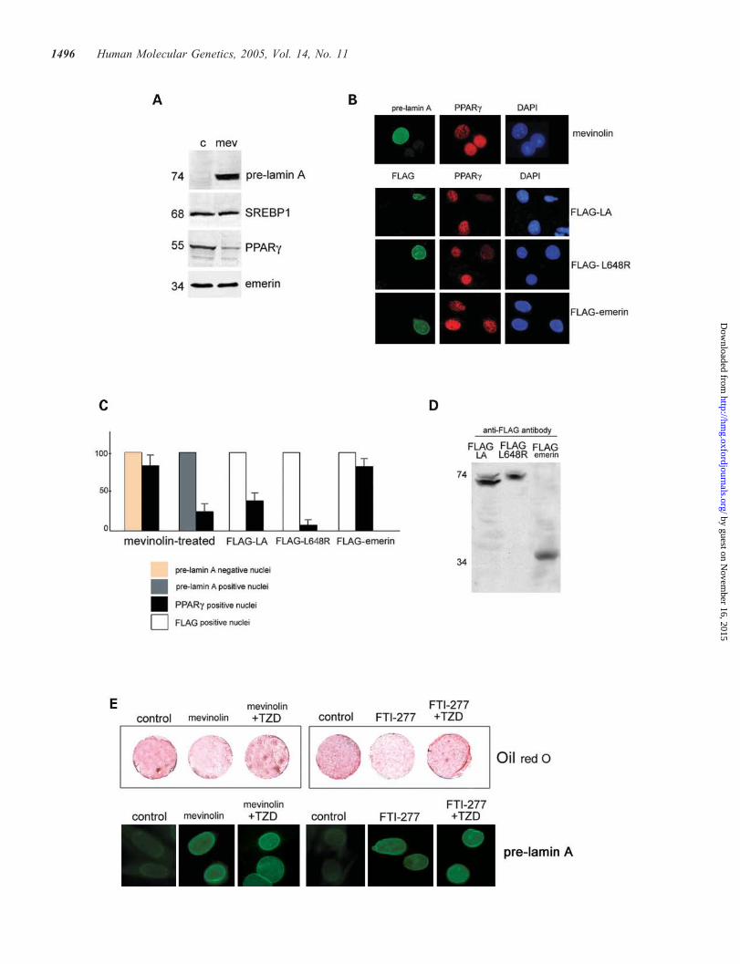
Since the transcription factor PPARg is transcribed followingSREBP1 activation in pre-adipocytes induced to differentiate(7,17), we checked PPARg levels in differentiating 3T3-L1cells accumulating pre-lamin A. To rule out the possibilitythat mevinolin could interfere with cholesterol synthesis byinhibiting HMG-CoA reductase, pre-lamin A accumulationwas also obtained using the farnesyltransferase inhibitorFTI-277 or by transfecting cell cultures with an uncleavablepre-lamin A mutant (as described subsequently). PPARgexpression was strongly reduced in mevinolin-treated 3T3-L1 pre-adipocytes, as determined by western blot (Fig. 6A)and immunofluorescence analysis (Fig. 6B). Downregulationof PPARg was also obtained using FTI-277 to accumulatepre-lamin A (data not shown). To support this observation,we transiently transfected 3T3-L1 pre-adipocytes with
Figure 5. Localization of SREBP1 in the nuclei accumulating pre-lamin A. Cells were double-stained with anti-SREBP1 and anti-pre-lamin A antibody.SREBP1 is detected by FITC-conjugated secondary antibody (green) and pre-lamin A is detected by Cy-3 conjugated secondary antibody (red). (A) Localizationof SREBP1 and pre-lamin A in untreated (3T3) or mevinolin-treated 3T3-L1 pre-adipocytes (3T3mev). (B) Double-immunofluorescence staining of SREBP1 andpre-lamin A in wild-type (control), MAD, FPLD and EDMD2 fibroblasts. (C) Localization of SREBP1 and pre-lamin A in untreated (3T3) or mevinolin-treated3T3-L1 pre-adipocytes (3T3mev), following in situ extraction with DNase. DNase extraction of unfixed adherent cells was performed using 20 U/ml of DNasefor 15 min at room temperature. Cells were double-stained as described earlier. (D) Double-immunofluorescence staining of SREBP1 and pre-lamin A in wild-type (control), MAD, FPLD and EDMD2 fibroblasts following DNase extraction. The merged images (merge) show yellow staining of the nuclear envelope infibroblasts accumulating SREBP1 and pre-lamin A at the nuclear rim. The reported results are representative of three independent experiments.
Human Molecular Genetics, 2005, Vol. 14, No. 11 1495
by guest on Novem
ber 16, 2015http://hm
g.oxfordjournals.org/D
ownloaded from
1496 Human Molecular Genetics, 2005, Vol. 14, No. 11
by guest on Novem
ber 16, 2015http://hm
g.oxfordjournals.org/D
ownloaded from
a FLAG-tagged lamin A construct overexpressing an unclea-vable L648R mutated pre-lamin A: PPARg was downregu-lated in the nuclei accumulating FLAG–L648R pre-lamin A(Fig. 6B and C). Overexpression of wild-type FLAG–laminA (FLAG–LA) was also effective in reducing PPARg level(Fig. 6B and C), whereas PPARg expression was notlowered in 3T3-L1 cells overexpressing the full-lengthemerin protein, here used as a control (Fig. 6B and C). Statisti-cal evaluation of these results showed that the percentage ofPPARg-labeled nuclei was significantly reduced in cellsaccumulating pre-lamin A (Fig. 6C). It should be noted thatthe 74 kDa pre-lamin A band only was detected in FLAG–L648R pre-lamin A-transfected cells by western immunoblot(Fig. 6D). On the other hand, accumulation of wild-type pre-lamin A along with mature lamin A occurred in pre-adipocytesoverexpressing FLAG–LA (Fig. 6D).
Accumulation of pre-lamin A reduced adipocyte differen-tiation, as detected by oil red O staining of mevinolin- orFTI-277-treated 3T3-L1 cultures (Fig. 6E). However, 3T3-L1 cells treated with the PPARg ligand TZD showed positiveoil red O staining even after accumulation of pre-lamin A bymevinolin or by FTI-277 treatment (Fig. 6E), indicating therescue of adipogenic differentiation.
DISCUSSION
The results reported here show that (i) accumulation of pre-lamin A specifically occurs in lipodystrophy-linked laminopa-thies, but not in EDMD2; (ii) in vivo binding of pre-lamin A tothe adipocyte transcription factor SREBP1 does occur and it isdetectable in cells accumulating pre-lamin A; (iii) pre-lamin Asequesters SREBP1 at the nuclear rim, thus reducing the poolof DNA-bound active transcription factor; (iv) retention ofSREBP1 by pre-lamin A causes downregulation of PPARgexpression and hence reduces the rate of pre-adipocyte differ-entiation and (v) adipogenic differentiation of pre-adipocytesaccumulating pre-lamin A may be rescued by PPARg ago-nists. The overall evaluation of these results shows that acommon pathogenic mechanism may be causative of differentlipodystrophic phenotypes.
The proposed mechanism involves primarily in the accumu-lation of pre-lamin A and selective binding of the lamin A pre-cursor to the adipocyte transcription factor SREBP1. In MAD,FPLD and WS cells, we observed an accumulation of thelamin A precursor. Interestingly, the amount of pre-lamin Awas not increased in EDMD2 fibroblast cell lines, indicatingthat impairment of lamin A precursor maturation is specifi-cally associated with lipodystrophy-linked lamin A mutations.However, the reason why lamin A processing is selectivelyaffected by R482L, R527H and S143F LMNA mutations isnot obvious. At present, we can rule out the possibility ofaltered interplay between pre-lamin A and farnesyltrans-ferases. In fact, mevinolin treatment was effective in increas-ing mutated pre-lamin A amount in MAD, FPLD and WSfibroblasts, suggesting that farnesylation of pre-lamin A isan ongoing mechanism in these cells (MAD being a recessivedisease model, only expressing mutated lamin A). On theother hand, altered interaction of mutated pre-lamin A withZPMSTE 24 endoprotease might affect protein processing. Itis noteworthy that ZPMSTE 24 mutations lead to accumu-lation of pre-lamin A due to impaired cleavage of the farnesy-lated protein and cause MAD (13), HGPS (Markquardt,personal communication) and restrictive dermopathy (49) inhumans and a MAD-resembling phenotype in mice (14,15).
We localized pre-lamin A-labeled intra-nuclear structuresboth in wild-type and in laminopathic cells accumulating thelamin A precursor. Such structures have been previouslyshown in wild-type cells induced to accumulate pre-lamin A(21,50). However, additional dominant negative effects ofmutations appear to affect differently both number and shapeof pre-lamin A-labeled structures in each laminopathic cellline. In the case of MAD and WS fibroblasts, pre-lamin-A-containing structures mostly protrude through the nuclearinterior, as was also observed in control nuclei, whereas inR482L FPLD nuclei they are mostly localized at the nuclearlamina level. We recently noticed that the number of pre-lamin-A-labeled intra-nuclear structures and the level oflamin A precursor were significantly increased in olderMAD patients, though carrying the same R527H LMNAmutation (data not shown). Noticeably, pre-lamin A-labeledstructures may co-localize with emerin in wild-type and
Figure 6. Analysis of PPARg expression and adipocyte differentiation in 3T3-L1 pre-adipocytes accumulating pre-lamin A. (A) 3T3-L1 pre-adipocytes were leftuntreated (c) or treated with mevinolin for 18 h (mev), thereafter allowed to differentiate for 4 days in differentiation medium. Nuclear lysates were obtained byhypotonic shock and shearing and subjected to electrophoresis and western blot analysis with anti-PPARg and anti-pre-lamin A antibody. Immunoblotted mem-branes were stripped and re-probed with anti-SREBP1 and anti-emerin antibody. The 68 kDa SREBP1 band was detected in the nuclear fractions. Emerin waslabeled as a loading control. Molecular weight markers are reported in kilodaltons. (B) 3T3-L1 pre-adipocytes grown on coverslips were treated with mevinolinor transfected with the indicated constructs and allowed to differentiate for 4 days. Double-immunofluorescence staining of pre-lamin A and PPARg was per-formed in mevinolin-treated samples (mevinolin). Double-staining with anti-FLAG and anti-PPARg antibodies was performed in FLAG–LA-transfected pre-adipocytes (FLAG–LA), L648R-lamin A-transfected adipocytes (FLAG–L648R) and emerin-transfected adipocytes (FLAG–emerin). DAPI staining allowsvisualization of all the nuclei. (C) Percentage of PPARg-positive nuclei in 3T3-L1 pre-adipocytes subjected to mevinolin treatment (mevinolin-treated), over-expressing wild-type FLAG–LA, mutated L648R FLAG–LA (FLAG–L648R) or FLAG–emerin. One-thousand nuclei were counted per sample and data arereported as percentage of counted nuclei (+SEM) of three separate countings performed in independent experiments. (D) Western blot analysis of exogenousproteins expressed in 3T3-L1 cells (FLAG–LA, FLAG-tagged wild-type lamin A; FLAG–L648R, FLAG-tagged L648R-mutated lamin A; FLAG–emerin,FLAG-tagged wild-type emerin). Each construct was transfected in 3T3-L1 cells. Cellular lysates were subjected to western immunoblot using anti-FLAG anti-body. The 74 kDa band corresponds to FLAG–pre-lamin A, the 70 kDa band corresponds to FLAG–LA, the 34 kDa band corresponds to FLAG–emerin. (E)Pre-adipocyte differentiation was evaluated by oil red O staining of cultured 3T3-L1 cells on day 2 of differentiation. 3T3-L1 cultures were left untreated(control) or treated with mevinolin for 18 h or with FTI-277 for 24 h. Thereafter, cells were allowed to differentiate for 2 days either in the presence or inthe absence of 10 mM TZD. An untreated culture (control) is shown on the left of each slide, a drug-treated culture (mevinolin or FTI-277) is shown in themiddle and a culture treated with mevinolin or FTI-277 and then with TZD for further 48 h (mevinolin þ TZD or FTI-277 þ TZD) is shown on the right.Oil red O staining is increased in differentiated cells bearing fat deposits. Pre-lamin A staining of representative nuclei from each 3T3-L1 sample is shownin the lower row (pre-lamin A). Each of these pictures is representative of three independent experiments.
Human Molecular Genetics, 2005, Vol. 14, No. 11 1497
by guest on Novem
ber 16, 2015http://hm
g.oxfordjournals.org/D
ownloaded from
EDMD2 nuclei, but not in FPLD, MAD and WS nuclei,suggesting disease-specific changes of intermolecular inter-actions (42).
Because we had determined a link between pre-lamin Aaccumulation and LMNA-associated lipodystrophies, wedecided to investigate whether pre-lamin A interacted withthe transcription factor SREBP1, which mediates adipocytedifferentiation (44). In vitro binding of SREBP1 to lamin A/C had been demonstrated by Lloyd et al. (34) and the site ofinteraction had been mapped between aminoacids 227 and487 of SREBP1 N-terminus sequence (34). In the presentstudy, we demonstrated for the first time in vivo binding ofthe lamin A precursor to SREBP1. As expected, the low-molecular-weight form of SREBP1 interacts with pre-laminA, indicating involvement of the nuclear lamina protein inthe localization of the active transcription factor. Interestingly,Lloyd et al. (34) performed a GST binding-assay with a laminA peptide spanning aminoacids 389–664, which includes pre-lamin A C-terminal sequence. Thus, our results confirm thatthe whole unprocessed lamin A molecule is required forSREBP1 interaction in vivo. The previously suggestedreduction of lamin A–SREBP1 binding affinity caused byR482 LMNA mutation (34) does not appear to affect in vivointeraction between pre-lamin A and SREBP1. This mightbe related to the increased availability of pre-lamin A inFPLD cells, which could overcome the reduced binding affi-nity caused by the mutation. In addition, we cannot rule outthe possibility that a different aminoacid substitution (R482Linstead of R482W) may differently affect protein interplay(42).
By treating pre-adipocytes with farnesylation inhibitors, weobtained a suitable cellular model that reproduced the situationof pre-lamin A accumulation in adipose tissue. This allowedus to investigate some biological mechanisms downstreamof reduced pre-lamin A maturation.
We showed that the retention of active SREBP1 at thenuclear lamina occurs in pre-adipocytes accumulatingpre-lamin A, as demonstrated by the significantly increasedproportion of mature SREBP1 found in the nuclear insolublefraction following DNase extraction. Immunofluorescencelabeling allowed us to show co-localization of SREBP1 andpre-lamin A in the nuclei accumulating pre-lamin A. In fact,DNA-bound transcription factor disappears from controlnuclei after the removal of DNA, whereas lamina-associatedSREBP1 is still retained in the nuclei accumulating pre-lamin A and shows a typical nuclear rim staining. Wesuggest that the reduced rate of lamin A precursor processingallows detection of pre-lamin A-bound SREBP1, which ishardly detectable in control cell lines undergoing rapid pre-lamin A maturation.
It is noteworthy that SREBP1 nuclear translocation isimpaired in Lmna-null mouse cardiomyocytes, but not in theadipose tissue from Lmna null mice (36), indicating thatnuclear import of SREBP1 in adipocytes does not requireLmna products. These data and our present results supportthe view that SREBP1–pre-lamin A interplay has a physio-logical role in the negative regulation of SREBP1 nucleartranslocation in adipose tissue, so that excess accumulationof pre-lamin A selectively sequesters SREBP1 at the adipo-cyte nuclear envelope. On the other hand, pre-lamin A
might play a positive regulation role for SREBP1 nuclearimport in other cell types such as cardiac myocytes (36). Inagreement with this hypothesis, pre-lamin A–SREBP1 inter-play, which is potentially disrupted by some LMNA mutationscausing AD-EDMD (34), might be involved in the nuclearimport of the transcription factor in skeletal muscle cells.
Retention of SREBP1 at the nuclear rim was previouslyreported in 3T3-F442A pre-adipocytes treated with Indinavir,a drug employed in HAART and causing lipodystrophy as aside-effect (7). Under these experimental conditions, 3T3-F442A fibroblasts show increased pre-lamin A level andreduced PPARg expression (7). These and our results stronglysuggest that retention of SREBP1 at the nuclear rim of lipody-strophy cells is associated with the presence of increased pre-lamin A level, irrespective of the occurrence of LMNAmutations. In fact, the identification of a selective and stableinterplay between pre-lamin A and SREBP1 helps to explainthe pathophysiology of both acquired and inherited lipodystro-phy, the latter due to either lamin A/C or ZMPSTE 24mutations. Moreover, it is noteworthy that farnesyltransferaseinhibitors (causing pre-lamin A accumulation) have been pre-viously reported to impair adipocyte differentiation (51,52).Our results help in explaining the biological mechanismunderlying this effect.
A key event of adipocyte differentiation is the induction ofthe transcription factor PPARg which is triggered by SREBP1(48). Downregulation of PPARg expression was obtained inpre-adipocytes accumulating the lamin A precursor either bymevinolin treatment or by overexpressing uncleavable pre-lamin A. Uncleavable mutants of lamin A have been shownto assemble and localize at the nuclear envelope with kineticscomparable with that of wild-type protein (53). The L648R-lamin A mutant did in fact localize at the nuclear envelope,where it could potentially bind SREBP1, as observed for de-farnesylated pre-lamin A. Interestingly, a reduced PPARglevel was also observed in adipocytes accumulating wild-type FLAG–pre-lamin A. In the latter case, the increase ofpre-lamin A level was directly attributable to overexpressionof the exogenous protein. These results support the view thatexcess accumulation of pre-lamin A is sufficient to sequesteran amount of SREBP1, even in the presence of maturelamin A. Thus, PPARg induction, a key step of adipocytedifferentiation, may be altered in laminopathies featuring lipo-dystrophy, as already proposed for acquired lipodystrophies(7,16).
In fact, adipocyte differentiation was impaired by pre-laminA accumulation, as determined by oil red O staining of mevi-nolin- or FTI-277-treated pre-adipocyte cultures. Given thatFTI-277 selectively affects pre-lamin A processing withoutany interference with ras protein farnesylation or with choles-terol synthesis (54), we conclude that reduced adipocytedifferentiation is a direct effect of pre-lamin A accumulation.It should be noted that highly differentiated cultures were notaffected by drug treatment (data not shown), in agreementwith the evidence that an early event in the adipocyte differen-tiation pathway, such as PPARg activation, is affected by pre-lamin A.
PPARg expression has been reported to be regulated depen-dent on the body district (55). This important observationsuggests that pre-lamin A accumulation may elicit different
1498 Human Molecular Genetics, 2005, Vol. 14, No. 11
by guest on Novem
ber 16, 2015http://hm
g.oxfordjournals.org/D
ownloaded from
effects in different fat depots depending on the extent ofPPARg activation required in that particular area. Thiscould, in part, explain the selective involvement of some butnot all adipose tissue districts in partial lipodystrophies.
Treatment of 3T3-L1 cells accumulating pre-lamin A withthe PPARg ligand TZD elicited rescue of the adipogenicprogram. The latter finding gives an important insight intopossible therapeutic approaches to lipodystrophy, whichhave been already reported to elicit some promising effects(56).
As MAD is a recessive disease, one would expect pre-laminA accumulation even in the heterozygous state, i.e. in a per-centage of cells from parents of affected individuals bearingone mutated allele (3). We did not observe a significantincrease of pre-lamin A amount in those cells. However, thisstudy deserves further deepening, since we found that overex-pression of R527H-mutated lamin A in transfected cells causesaccumulation of mutated pre-lamin A (but not endogenouspre-lamin A) (data not shown). On the other hand, MAD phe-notype is not observed in heterozygous individuals, stronglysupporting the view that a threshold pre-lamin A level isrequired to activate the pathogenic mechanism. Regardingthe pathophysiology of MAD and WS, two main questionsneed further investigation. The first is the effect elicited bythe accumulation of lamin A precursor protein in osteoblasts.In fact, bone resorption of the extremities is a specific featureof MAD phenotype (3) and osteoporosis is also observed inWS patients (11) (though it might be related to the agingprocess featuring this disease). We are now addressing thisproblem by evaluating the effect of high pre-lamin A levelin pre-osteocyte differentiation or survival. The other openquestion is the pathogenetic mechanism leading to a prematureaging phenotype in MAD patients and mostly in WS patients.We suggest that this phenotype could be dependent both on ananomalous amount of pre-lamin A and on altered interactionbetween mutated lamin A/C and its nuclear binding partners.
MATERIALS AND METHODS
Cell culture and differentiation
Skin biopsies were obtained from a MAD patient carrying anR527H lamin A/C mutation (3), an FPLD patient carryingan R482L lamin A/C mutation (42), an atypical WS patientcarrying an S143F LMNA mutation, an EDMD2 patient carry-ing R401C LMNA mutation and from unaffected controls fol-lowing their written consent. Fibroblast cultures wereestablished and cultured in Dulbecco’s modified Eagle’smedium (DMEM) supplemented with 10% fetal calf serum(FCS) and antibiotics. Cells at passages 10–15 wereemployed. 3T3-L1 pre-adipocytes were obtained from theEuropean Collection of Cell Cultures and cultured inDMEM plus 10% FCS. Pre-adipocytes approaching conflu-ence were induced to differentiate by incubation for 2 daysin a differentiation medium containing DMEM plus 10%FCS, 5 mg/ml insulin, 0.25 mM dexamethasone and 0.1 mM
3-isobutyl-1-methylxanthine. Thereafter, incubation was per-formed for an additional 2 days in the same medium excludingdexamethasone and 3-isobutyl-1-methylxanthine. Cells were
maintained for 8–10 days in DMEM with 10% FCS toattain maximal differentiation (57).
Transfection of pre-adipocyte cultures
A full-length rat lamin A cDNA encoding for pre-lamin A wasexpressed from the CMV promoter and tagged at the 50 endwith a sequence coding for FLAG epitope (FLAG–LA)(58). The L648R point mutation was introduced into thefull-length rat lamin A cDNA using the QuikChange site-directed mutagenesis kit (Stratagene, La Jolla, CA, USA).This mutation impairs cleavage of pre-lamin A byZMPSTE24 endoprotease and causes accumulation of farnesy-lated pre-lamin A (12). FLAG–LA or L648R–FLAG-lamin Awas transfected in 3T3-L1 pre-adipocytes at day 0 of differen-tiation. A FLAG-tagged wild-type emerin construct was usedto transfect pre-adipocytes as a control. Transfection of 3T3-L1 pre-adipocytes was performed using FuGene 6 reagentaccording to the manufacturer’s instructions. Cells werefixed at day 4 of differentiation.
Drug treatments
Pre-lamin A accumulation in human fibroblasts and 3T3-L1adipocytes was induced by inhibition of isoprenoid synthesiswith 25 mM Mevinolin (Sigma) for 18 h (21). This drug sub-tracts the substrate of farnesyltransferases, thus impairingpre-lamin A farnesylation which is required for furtherprotein processing (59). Alternatively, pre-lamin A farnesyla-tion was inhibited using the peptidomimetic drug FTI-277(Calbiochem), which selectively impairs pre-lamin A farnesy-lation, at the dosage used (20 mM for 24 h) (54). Treatmentwith TZD (Sigma) was performed according to the publishedprotocols to trigger PPARg activation (16). Briefly, 3T3-L1pre-adipocytes were induced to accumulate pre-lamin A by18 h mevinolin treatment or by 24 h FTI-277 treatment. There-after, cells were transferred in culture medium, containing ornot 10 mM TZD, and incubated for 48 h. Incubation wasstopped by formalin fixation, as described subsequently.
Sub-cellular fractioning and DNase treatment
Sub-cellular fractions were obtained as previously described(26). Briefly, the cell pellet was resuspended in a lysisbuffer containing 10 mM Tris, pH 7.8, 1% NP-40, 10 mM
2-mercaptoethanol and protease inhibitors. Separation ofnuclei was obtained by hypotonic shock and shearing; nucleiwere obtained as pellet by a 300g centrifugation at 48C.Other samples were treated by the same procedure andemployed to obtain the cytoplasmic fraction as supernatant.Cytoplasmic fractions were clarified by centrifugation at600g. DNase extraction of nuclei was performed accordingto published protocols (42) except that 100 U/ml of DNasewas applied for 30 min. The insoluble nuclear fraction (NP
in Fig. 4), containing nuclear matrix constituents, wasobtained as pellet. Each fraction was then resuspended inloading buffer, subjected to sodium dodecyl sulphate–poly-acrylamide gel electrophoresis (SDS–PAGE) and westernblot analysis.
Human Molecular Genetics, 2005, Vol. 14, No. 11 1499
by guest on Novem
ber 16, 2015http://hm
g.oxfordjournals.org/D
ownloaded from
In situ DNase extraction was performed as follows. Unfixedcell cultures were washed in phosphate-buffered saline (PBS),treated with a buffer containing 10 mM Tris-HCl (pH 7.4),150 mM NaCl, 5 mM MgCl2, 1% NP-40, 1 mM phenylmethyl-sulfonyl fluoride (PMSF), 10 mg/ml leupeptin and aprotininfor 15 min at room temperature. Detergent-treated cells werewashed twice in the absence of NP-40 and subjected toDNase treatment (20 U/ml for 15 min at room temperature).Samples were fixed with 4% paraformaldehyde and processedfor immunofluorescence labeling.
Antibodies
The antibodies employed for western blot analysis or immuno-fluorescence labeling were as follow: anti-lamin A/C, mono-clonal (Novocastra Laboratories, NCL-LAM-A/C); anti-laminA/C, goat polyclonal, (Santa Cruz, sc-6215); anti-pre-laminA, goat polyclonal (Santa Cruz, sc-6214) (15); anti-SREBP1,rabbit polyclonal (Santa Cruz, sc-8984); anti-emerin, mousemonoclonal (Novocastra Laboratories, NCL-emerin); anti-PPARg, mouse monoclonal (Santa Cruz, sc-7273); anti-actin,goat polyclonal (Santa Cruz, sc-1616) and anti-FLAG, rabbitpolyclonal (Sigma, F-7425).
Western blot and immunoprecipitation
Western blot analysis of cellular lysates from human fibrobastsand 3T3-L1 pre-adipocytes was done as follows. Cells werelysed in buffer containing 1% NP-40, 0.25% sodium deoxy-cholate, 150 mM NaCl, 1 mM EGTA, 1 mM PMSF, 1 mM
NaF, 1 mM aprotinin, leupeptin and pepstatin. Proteins wereloaded in Laemmli sample buffer and subjected to SDS–PAGE followed by immunochemical reactions. For immuno-precipitation analysis, human fibroblasts and 3T3-L1 pre-adipocytes were treated with a buffer containing 50 mM
HEPES–HCl pH 7.4, 100 mM NaCl, 1% NP-40, 1 mM
PMSF, 1 mM dithiothreitol, 1.5 mM MgCl2, 10 mM of eachleupeptin, pepstatin, aprotinin for 10 min at 48C. After soni-cation, cell extracts were clarified by 12 000g centrifugationat 48C. Protein A/G–Sepharose beads were incubated withanti-SREBP1 or with anti-pre-lamin A antibodies for 14 h at48C and unbound antibodies were removed by centrifugation.Cellular extracts were incubated with antibody-coupledSepharose beads for 3 h at 48C. Immunoprecipitated com-plexes were diluted in Laemmli buffer, subjected to SDS–PAGE (6–20%) and transferred to nitrocellulose membrane.Membranes were saturated with 5% dried skimmed milk–4% bovine serum albumin (BSA) and incubation withprimary antibodies was performed for 1 h at room tempera-ture. Immunoblotted bands were revealed by the AmershamECL detection system. Densitometry of immunoblottedbands was performed by a Biorad GS-800 calibrated densi-tometer. Statistical analysis was performed by Student’s t-test.
Immunofluorescence
Human fibroblasts and 3T3-L1 pre-adipocytes grown on cov-erslips were fixed in methanol at 208C. In situ DNase extractedsamples were fixed by 4% paraformaldehyde. Sampleswere incubated with PBS containing 4% BSA to saturate
non-specific binding. Primary antibodies were applied over-night at 48C, secondary antibodies were applied for 1 h atroom temperature. Slides were mounted with an anti-fadereagent in glycerol and observed with a Nikon E 600 fluor-escence microscope equipped with a digital camera. Pictureswere elaborated with Photoshop-6 software.
Oil red O staining
Oil red O staining of cultured 3T3-L1 pre-adipocytes was per-formed to stain lipid droplets. Cells were fixed for 1 h at roomtemperature with 10% formalin in isotonic buffer, washedthree times with distilled water, and then stained with 0.6%(wt/vol) oil red O solution (60% isopropanol, 40% water)for 1 h at 228C. After washing three times with distilledwater, the cells were examined under a microscope (16).
ACKNOWLEDGEMENTS
The authors thank Professor E. Nigg for collaboration. Thetechnical support of P. Sabatelli, A. Valmori, S. Grasso isgratefully acknowledged. This work was supported by grantsfrom the Italian Health Ministry (P.F. no. 2003/123), fromthe Italian Ministry of University and Research (FIRBProject no. RBNE01JJ45_005) and MIUR-Cofin 2004, by a‘Telethon’ grant to G.N. and by a grant from ‘FondazioneCarisbo’, Italy. M.W. was funded by the German networkon muscular dystrophies (MD-NET, research project R8,01GM0302) of the German Ministry of Education andResearch, Germany. V.K.P. was supported by the Council ofScientific and Industrial Research, India.
Conflict of Interest statement. None declared.
REFERENCES
1. Hegele, R.A. (2003) Monogenic forms of insulin resistance: apertures thatexpose the common metabolic syndrome. Trends Endocrinol. Metab., 14,371–377.
2. Shacketon, S., Lloyd, D.J., Jackson, SN., Evans, R., Niermeijer, M.F.,Singh, B.M., Schmidt, H., Brabant, G., Kumar, S. et al. (2000) LMNA,encoding lamin A/C, is mutated in partial lipodystrophy. Nat. Genet., 24,153–156.
3. Novelli, G., Muchir, A., Sangiuolo, F., Helbling-Leclerc, A., D’Apice,M.R., Massart, C., Capon, F., Sbraccia, P., Federici, M., Lauro, R. et al.(2002) Mandibuloacral dysplasia is caused by a mutation in LMNA-encoding lamin A/C. Am. J. Hum. Genet., 71, 426–431.
4. Bhayana, S., Siu, V.M., Joubert, G.I., Clarson, C.L., Cao, H. andHegele, R.A. (2002) Cardiomyopathy in congenital completelipodystrophy. Clin. Genet., 61, 283–287.
5. Magre, J.J., Delepine, M., Khallouf, E., Gedde-Dahl, T., Jr, VanMaldergem, L., Sobel, E., Papp, J., Meier, M., Megarbane, A., Bachy, A.et al. (2001) Identification of the gene altered in Berardinelli-Seipcongenital lipodystrophy on chromosome 11q13. Nat. Genet., 28,365–370.
6. Hegele, R.A., Cao, H., Frankowski, C., Mathews, S.T. and Leff, T. (2002)PPARG F388L, a transactivation-deficient mutant, in familial partiallipodystrophy. Diabetes, 51, 3586–3590.
7. Caron, M., Auclair, M., Sterlingot, H., Kornprobst, M. and Capeau, J.(2003) Some HIV protease inhibitors alter lamin A/C maturation andstability, SREBP-1 nuclear localization and adipocyte differentiation.AIDS, 17, 2437–2444.
1500 Human Molecular Genetics, 2005, Vol. 14, No. 11
by guest on Novem
ber 16, 2015http://hm
g.oxfordjournals.org/D
ownloaded from
8. de Sandre-Giovannoli, A., Bernard, R., Cau, P., Navarro, C., Amiel, J.,Boccaccio, I., Lyonnet, S., Stewart, C.L., Munnich, A., Le Merrer, M.et al (2003) Lamin a truncation in Hutchinson–Gilford progeria. Science,300, 2055.
9. Eriksson, M., Brown, W.T., Gordon, L.B., Glynn, M.W., Singer, J.,Scott, L., Erdos, M.R., Robbins, C.M., Moses, T.Y., Berglund, P. et al.(2003) Recurrent de novo point mutations in lamin A cause Hutchinson–Gilford progeria syndrome. Nature, 423, 293–298.
10. Cao, H. and Hegele, R.A. (2003) LMNA is mutated in Hutchinson–Gilford progeria (MIM 176670) but not in Wiedemann–Rautenstrauchprogeroid syndrome (MIM 264090). J. Hum. Genet., 48, 271–274.
11. Chen, L., Lee, L., Kudlow, B.A., Dos Santos, H.G., Sletvold, O.,Shafeghati, Y., Botha, E.G., Garg, A., Hanson, N.B., Martin, G.M. et al.(2003) LMNA mutations in atypical Werner’s syndrome. Lancet, 362,440–445.
12. Corrigan, D.P., Kuszczak, D., Rusinol, A.E., Thewke, D.P., Hrycyna, C.A.,Michaelis, S., Sinensky, M.S. (2005) Prelamin A endoproteolyticprocessing in vitro by recombinant Zmpste24. Biochem. J., 387, 129–138.
13. Agarwal, A.K., Fryns, J.P., Auchus, R.J. and Garg, A. (2003) Zincmetalloproteinase, ZMPSTE24, is mutated in mandibuloacral dysplasia.Hum. Mol. Genet., 12, 1995–2001.
14. Bergo, M.O., Gavino, B., Ross, J., Schmidt, W.K., Hong, C., Kendall, L.V.,Mohr, A., Meta, M., Genant, H., Jiang, Y. et al. (2002) Zmpste24deficiency in mice causes spontaneous bone fractures, muscle weakness,and a prelamin A processing defect. Proc. Natl Acad. Sci. USA, 99,13049–13054.
15. Pendas, A.M., Zhou, Z., Cadinanos, J., Freije, J.M., Wang, J., Hultenby, K.,Astudillo, A., Wernerson, A., Rodriguez, F., Tryggvason, K. et al(2002) Defective prelamin A processing and muscular and adipocytealterations in Zmpste24 metalloproteinase-deficient mice. Nat. Genet., 31,94–99.
16. Caron, M., Auclair, M., Vigouroux, C., Glorian, M., Forest, C. andCapeau, J. (2001) The HIV protease inhibitor indinavir impairs sterolregulatory element-binding protein-1 intranuclear localization, inhibitspreadipocyte differentiation, and induces insulin resistance. Diabetes, 50,1378–1388.
17. Spiegelman, B.M. (1998) PPAR-gamma: adipogenic regulator andthiazolidinedione receptor. Diabetes, 47, 507–514.
18. Agarwal, A.K. and Garg, A. (2002) A novel heterozygous mutation inperoxisome proliferator-activated receptor-gamma gene in a patient withfamilial partial lipodystrophy. J. Clin. Endocrinol. Metab., 87, 408–411.
19. Al-Shali, K., Cao, H., Knoers, N., Hermus, A.R., Tack, C.J., Hegele, R.A.(2004) A single-base mutation in the peroxisome proliferator-activatedreceptor fgammag promoter associated with altered in vitro expression andpartial lipodystrophy. J. Clin. Endocrinol. Metab., 89, 5655–5660.
20. Zastrow, M.S., Vlcek, S. and Wilson, K.L. (2004) Proteins that bindA-type lamins: integrating isolated clues. J. Cell. Sci., 117, 979–987.
21. Sasseville, A.M. and Raymond, Y. (1995) Lamin A precursor is localizedto intranuclear foci. J. Cell. Sci., 108, 273–285.
22. Stuurman, N., Heins, S. and Aebi, U. (1998) Nuclear lamins: theirstructure, assembly, and interactions. J. Struct. Biol., 122, 42–66.
23. Sakaki, M., Koike, H., Takahashi, N., Sasagawa, N., Tomioka, S.,Arahata, K. and Ishiura, S. (2001) Interaction between emerin and nuclearlamins. J. Biochem., 129, 321–327.
24. Markiewicz, E., Dechat, T., Foisner, R., Quinlan, R.A. and Hutchison, C.J.(2002) Lamin A/C binding protein LAP2alpha is required fornuclear anchorage of retinoblastoma protein. Mol. Biol. Cell, 13,4401–4413.
25. Sasseville, A.M. and Langelier, Y. (1998) In vitro interaction of thecarboxy-terminal domain of lamin A with actin. FEBS Lett., 425,485–489.
26. Lattanzi, G., Cenni, V., Marmiroli, S., Capanni, C., Mattioli, E., Merlini, L.,Squarzoni, S. and Maraldi, N.M. (2003) Association of emerin withnuclear and cytoplasmic actin is regulated in differentiating myoblasts.Biochem. Biophys. Res. Commun., 303, 764–770.
27. Mancini, M.A., Shan, B., Nickerson, J.A., Penman, S. and Lee, W.H.(1994) The retinoblastoma gene product is a cell cycle-dependent,nuclear matrix-associated protein. Proc. Natl Acad. Sci. USA, 91,418–422.
28. Novelli, G. and D’Apice, M.R. (2003) The strange case of the ‘lumper’lamin A/C gene and human premature ageing. Trends Mol. Med., 9,370–375.
29. Bonne, G., Raffaele-Di Barletta, M., Varnous, S., Becane, H.M.,Hammouda, E.H., Merlini, L., Muntoni, F., Greenberg, C.R., Gary, F.,Urtizberea, J.A. et al. (1999) Mutations in the gene encoding lamin A/Ccause autosomal dominant Emery–Dreifuss muscular dystrophy. Nat.Genet., 21, 285–288.
30. Muchir, A., Bonne, G., van der Kooi, A.J., van Meegen, M., Baas, F.,Bolhuis, P.A., de Visser, M. and Schwartz, K. (2000) Identification ofmutations in the gene encoding lamins A/C in autosomal dominant limbgirdle muscular dystrophy with atrioventricular conduction disturbances(LGMD1B). Hum. Mol. Genet., 9, 1453–1459.
31. Fatkin, D., MacRae, C., Sasaki, T., Wolff, M.R., Porcu, M., Frenneaux, M.,Atherton, J., Vidaillet, H.J., Jr, Spudich, S., de Girolami, U. et al. (1999)Missense mutations in the rod domain of the lamin A/C gene as causes ofdilated cardiomyopathy and conduction-system disease. N. Engl. J. Med.,341, 1715–1724.
32. de Sandre-Giovannoli, A., Chaouch, M., Kozlov, S., Vallat, J.M., Tazir, M.,Kassouri, N., Szepetowski, P., Hammadouche, T., Vandenberghe, A.,Stewart, C.L. et al. (2002) Homozygous defects in LMNA, encodinglamin A/C nuclear-envelope proteins, cause autosomal recessive axonalneuropathy in human (Charcot–Marie tooth disorder type 2) and mouse.Am. J. Hum. Genet., 70, 726–736.
33. Sullivan, T., Escalante-Alcalde, D., Bhatt, H., Anver, M., Bhat, N.,Nagashima, K., Stewart, C.L. and Burke, B. (1999) Loss of A-type laminexpression compromises nuclear envelope integrity leading to musculardystrophy. J. Cell. Biol., 147, 913–920.
34. Lloyd, D.J., Trembath, R.C. and Shackleton S. (2002) A novelinteraction between lamin A and SREBP1: implications forpartial lipodystrophy and other laminopathies. Hum. Mol. Genet.,11, 769–777.
35. Krimm, I., Ostlund, C., Gilquin, B., Couprie, J., Hossenlopp, P.,Mornon, J.P., Bonne, G., Courvalin, J.C., Worman, H.J. and Zinn-Justin, S.(2002) The Ig-like structure of the C-terminal domain of lamin A/C,mutated in muscular dystrophies, cardiomyopathy, and partiallipodystrophy. Structure, 10, 811–823.
36. Nikolova, V., Leimena, C., McMahon, A.C., Tan, J.C., Chandar, S.,Jogia, D., Kesteven, S.H., Michalicek, J., Otway, R., Verheyen, F. et al.(2004) Defects in nuclear structure and function promote dilatedcardiomyopathy in lamin A/C-deficient mice. J. Clin. Invest.,113, 357–369.
37. Lammerding, J., Schulze, P.C., Takahashi, T., Kozlov, S., Sullivan, T.,Kamm, R.D., Stewart, C.L. and Lee, R.T. (2004) Lamin A/C deficiencycauses defective nuclear mechanics and mechanotransduction. J. Clin.Invest., 113, 370–378.
38. Sabatelli, P., Lattanzi, G., Ognibene, A., Columbaro, M., Capanni, C.,Merlini, L. Maraldi, N.M. and Squarzoni, S. (2001) Nuclear alterations inautosomal-dominant Emery–Dreifuss muscular dystrophy. Muscle Nerve,24, 826–829.
39. Maraldi, N.M., Lattanzi, G., Marmiroli, S., Squarzoni, S. and Manzoli, F.A.(2004) New roles for lamins, nuclear envelope proteins and actin in thenucleus. Adv. Enzyme Regul., 44, 155–172.
40. Stierle, V., Couprie, J., Ostlund, C., Krimm, I., Zinn-Justin, S.,Hossenlopp, P., Worman, H.J., Courvalin, J.C. and Duband-Goulet,I. (2003) The carboxyl-terminal region common to lamins A and Ccontains a DNA binding domain. Biochemistry, 42, 4819–4828.
41. Muchir, A., van Engelen, B.G., Lammens, M., Mislow, J.M., McNally, E.,Schwartz, K. and Bonne, G. (2003) Nuclear envelope alterations infibroblasts from LGMD1B patients carrying nonsense Y259Xheterozygous or homozygous mutation in lamin A/C gene. Exp. Cell Res.,291, 352–362.
42. Capanni, C., Cenni, V., Mattioli, E., Sabatelli, P., Ognibene, A.,Columbaro, M., Parnaik, V.K., Wehnert, M., Maraldi, N.M., Squarzoni, S.et al. (2003) Failure of lamin A/C to functionally assemble in R482Lmutated familial partial lipodystrophy fibroblasts: altered intermolecularinteraction with emerin and implications for gene transcription. Exp. CellRes., 291, 122–134.
43. Tontonoz, P., Kim, J.B., Graves, R.A. and Spiegelman, B.M. (1993)ADD1: a novel helix-loop-helix transcription factor associated withadipocyte determination and differentiation. Mol. Cell. Biol., 13,4753–4759.
44. Kim, J.B. and Spiegelman, B.M. (1996) ADD1/SREBP1 promotesadipocyte differentiation and gene expression linked to fatty acidmetabolism. Genes Dev., 10, 1096–1107.
Human Molecular Genetics, 2005, Vol. 14, No. 11 1501
by guest on Novem
ber 16, 2015http://hm
g.oxfordjournals.org/D
ownloaded from
45. Ericsson, J., Jackson, S.M., Kim, J.B., Spiegelman, B.M. and Edwards, P.A.(1997) Identification of glycerol-3-phosphate acyltransferase as anadipocyte determination and differentiation factor 1- and sterol regulatoryelement-binding protein-responsive gene. J. Biol. Chem., 272,7298–7305.
46. Kim, J.B., Wright, H.M., Wright, M. and Spiegelman, B.M. (1998)ADD1/SREBP1 activates PPARgamma through the production ofendogenous ligand. Proc. Natl Acad. Sci. USA, 95, 4333–4337.
47. Wang, X., Sato, R., Brown, M.S., Hua, X. and Goldstein, J.L. (1994)SREBP-1, a membrane-bound transcription factor released bysterol-regulated proteolysis. Cell, 77, 53–62.
48. Hegele, R.A. (2001) Molecular basis of partial lipodystrophy andprospects for therapy. Trends Mol. Med., 7, 121–126.
49. Navarro, C.L., de Sandre-Giovannoli, A., Bernard, R., Boccaccio, I.,Boyer, A., Genevieve, D., Hadj-Rabia, S., Gaudy-Marqueste, C.,Smitt, H.S., Vabres, P., Faivre, L. et al. (2004) Lamin A and ZMPSTE24(FACE-1) defects cause nuclear disorganization and identify restrictivedermopathy as a lethal neonatal laminopathy. Hum. Mol. Genet., 13,2493–2503.
50. Maraldi, N.M., Squarzoni, S., Sabatelli, P., Capanni, C., Mattioli, E.,Ognibene, A. and Lattanzi, G. (2005) Laminopathies: involvement ofstructural nuclear proteins in the pathogenesis of an increasing number ofhuman diseases. J. Cell Physiol., 203, 319–327.
51. Nishio, E., Tomiyama, K., Nakata, H. and Watanabe, Y. (1996)3-Hydroxy-3-methylglutaryl coenzyme A reductase inhibitor impairs celldifferentiation in cultured adipogenic cells (3T3-L1). Eur. J. Pharmacol.,301, 203–206.
52. Solomon, C.S., Leitner, J.W. and Goalstone, M.L. (2003) Dominantnegative alpha-subunit of farnesyl- and geranylgeranyl-transferase Iinhibits insulin-induced differentiation of 3T3-L1 pre-adipocytes.Int. J. Obes. Relat. Metab. Disord., 27, 40–47.
53. Hennekes, H. and Nigg, E.A. (1994) The role of isoprenylation inmembrane attachment of nuclear lamins. A single point mutation preventsproteolytic cleavage of the lamin A precursor and confers membranebinding properties. J. Cell Sci., 107, 1019–1029.
54. Adjei, A.A., Davis, J.N., Erlichman, C., Svingen, P.A. and Kaufmann, S.H.(2000) Comparison of potential markers of farnesyltransferase inhibition.Clin. Cancer Res., 6, 2318–2325.
55. Sewter, C.P., Blows, F., Vidal-Puig, A. and O’Rahilly, S. (2002) Regionaldifferences in the response of human pre-adipocytes to PPARgamma andRXRalpha agonists. Diabetes, 51, 718–723.
56. Owen, K.R., Donohoe, M., Ellard, S. and Hattersley, A.T. (2003)Response to treatment with rosiglitazone in familial partial lipodystrophydue to a mutation in the LMNA gene. Diabet. Med., 20, 823–827.
57. Parpal, S., Karlsson, M., Thorn, H. and Stralfors, P. (2001) Cholesteroldepletion disrupts caveolae and insulin receptor signaling for metaboliccontrol via insulin receptor substrate-1, but not for mitogen-activatedprotein kinase control. J. Biol. Chem., 276, 9670–9678.
58. Kumaran, R.I., Muralikrishna, B. and Parnaik, V.K. (2002) Lamin A/Cspeckles mediate spatial organization of splicing factor compartments andRNA-polymerase II transcription. J. Cell Biol., 159, 783–793.
59. Sinensky, M., Beck, L.A., Leonard, S. and Evans, R. (1990) Differentialinhibitory effects of lovastatin on protein isoprenylation and sterolsynthesis. J. Biol. Chem., 265, 19937–19941.
1502 Human Molecular Genetics, 2005, Vol. 14, No. 11
by guest on Novem
ber 16, 2015http://hm
g.oxfordjournals.org/D
ownloaded from