Development of Polymer-based Gels for Multimodal Medical ...
Upload
independentCategory
view
1download
0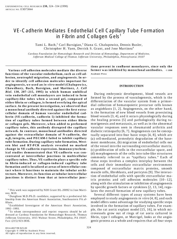
EXPERIMENTAL CELL RESEARCH 238, 324–334 (1998)ARTICLE NO. EX973844
VE-Cadherin Mediates Endothelial Cell Capillary Tube Formationin Fibrin and Collagen Gels1
Tami L. Bach,2 Carl Barsigian,3 Diana G. Chalupowicz, Dennis Busler,Christopher H. Yaen, Derrick S. Grant, and Jose Martinez4
Cardeza Foundation for Hematologic Research and Division of Hematology, Department of Medicine,Jefferson Medical College of Thomas Jefferson University, Philadelphia, Pennsylvania 19107
tions present in confluent monolayers, since only theformer was inhibited by monoclonal antibodies. q 1998Various cell adhesion molecules mediate the diverseAcademic Pressfunctions of the vascular endothelium, such as cell ad-
hesion, neutrophil migration, and angiogenesis. In or-der to identify cell adhesion molecules important forangiogenesis, we used an in vitro model (Chalupowicz, INTRODUCTIONChowdhury, Bach, Barsigian, and Martinez, J. CellBiol. 130, 207–215, 1995) in which human umbilical
During embryonic development, blood vessels arevein endothelial cell monolayers are induced to formformed by the process of vasculogenesis, which is thecapillary-like tubes when a second gel, composed ofdifferentiation of the vascular system from a primor-either fibrin or collagen, is formed overlying the apicaldial collection of hematopoietic precursor cells knownsurface. In the present investigation, we observed thatas angioblasts [1, 2]. Angiogenesis, on the other hand,a monoclonal antibody directed against the first extra-is the formation of new blood vessels from preexistingcellular domain of human vascular endothelial cad-blood vessels [3, 4], and it occurs physiologically duringherin (VE-cadherin, cadherin 5) inhibited the forma-the healing process [5] and pathologically during tu-tion of capillary tubes formed between either fibrinmorigenesis and metastasis, as well as in the abnormalor collagen gels. Moreover, when added to preformed
capillary tubes, this antibody disrupted the capillary vascular responses seen in rheumatoid arthritis andnetwork. In contrast, monoclonal antibodies directed diabetic retinopathy [6, 7]. Angiogenesis can be concep-against the extracellular domain of N-cadherin, the tually separated into four basic steps [4, 8], which areavb3 integrin, and PECAM-1 failed to inhibit capillary (a) cell-mediated, proteolytic degradation of the base-tube formation. During capillary tube formation, West- ment membrane, (b) migration of endothelial cells outern blot and RT-PCR analysis revealed no marked of the vessel into the surrounding extracellular matrix,change in VE-cadherin expression. Immunocytochem- (c) proliferation of cells in the extracellular space, andical studies demonstrated that VE-cadherin was con- (d) morphogenesis of the cells into tube-like structurescentrated at intercellular junctions in multicellular commonly referred to as ‘‘capillary tubes.’’ Each ofcapillary tubes. Thus, VE-cadherin plays a specific role these steps involves a complex interplay between thein fibrin-induced or collagen-induced capillary tube cells and their immediate extracellular environmentformation and is localized at areas of intercellular con- composed of extracellular matrix proteins, smoothtact where it functions to maintain the tubular archi- muscle cells, fibroblasts, and pericytes [9]. The interac-tecture. Moreover, its function at tubular intercellular
tion of endothelial cells with specific extracellular ma-junctions is distinct from that at intercellular junc-trix proteins and cell adhesion molecules [10–12],along with the stimulation or inhibition of angiogenesisby specific growth factors or cytokines [2, 13, 14], regu-
1 This work was supported by NIH Grant HL-20092 to Jose Marti- lates the overall formation of new capillary tubes.nez, M.D. Several different types of assay systems have been2 Dunglison M.D./Ph.D. candidate, supported by a predoctoral fel-
employed to study angiogenesis in vitro [4, 8], and eachlowship from the American Heart Association, Southeastern PA af-model offers some advantage for studying specific stepsfiliate.
3 Established Investigator of the American Heart Association, involved in the formation of capillary tubes. For exam-1992–1993. ple, the rat aortic explant model [15, 16], in which mi-4 To whom correspondence and reprint requests should be ad- crovessels grow out of rings of rat aorta cultured indressed at Cardeza Foundation for Hematologic Research, Thomas
fibrin, type I collagen, or Matrigel, looks at the angio-Jefferson University, 1015 Walnut Street, Philadelphia, PA 19107.Fax: (215) 923-3836. genic process as a whole and requires that all four steps
3240014-4827/98 $25.00Copyright q 1998 by Academic PressAll rights of reproduction in any form reserved.
AID ECR 3844 / 6i2d$$$621 01-08-98 08:00:32 eca

325VE-CADHERIN MEDIATES CAPILLARY TUBE FORMATION
in the process are operative. At the other extreme, gel- markedly inhibited the generation of capillary tubes inaddition to disrupting preformed tubes. Hoffman mod-over-gel or sandwich assays [17, 18] focus on the last
step, namely differentiation of the endothelial cells into ulation contrast optics defined the morphologic fea-tures of endothelial cells in the sandwich assays, thuscapillary tubes. Since the main activities assessed are
the cellular reorganization and adhesion/dysadhesion demonstrating their marked disorganization in thepresence of the VE-cadherin antibody. Immunoblot andevents required for tube formation, sandwich assays
may be useful for defining the mechanism through RT-PCR analyses demonstrated VE-cadherin expres-sion to be unchanged during capillary tube formation.which new capillaries are formed.
We recently showed that human umbilical vein endo- Immunofluorescent studies demonstrated that VE-cadherin was concentrated at intercellular junctions inthelial cell (HUVEC)5 monolayers established on a fi-
brin gel rapidly rearrange to form a network of capil- multicellular capillary tubes, similar to its localizationin monolayers. These findings therefore suggest thatlary tubes upon generation of a second fibrin gel overly-
ing the cells [18]. The interaction of the N-terminus VE-cadherin plays a specific role in capillary tube for-mation induced by two different matrix proteins which(amino acids 15–42) of the fibrin b chain of the overly-
ing gel with the apical surface of the monolayer was are important during the angiogenic process.essential for the induction of capillary tube formation[18]. In this regard, our findings are similar to those MATERIALS AND METHODSdemonstrating that the interaction of type I collagenfibrils with the apical surface of cultured HUVECs also Reagents and antibodies. Endothelial cell basal medium (EBM)results in the rapid formation of capillary tube-like was from Clonetics Corp. (San Diego, CA). Human recombinant basic
fibroblast growth factor (bFGF) and rat tail collagen (type I) werestructures [19, 20]. Although these studies demon-from Collaborative Biomedical Products (Bedford, MA). Insulin–strate that the interaction of the overlying fibrin ortransferrin–selenium supplement (1001) containing insulin (1.00 g/collagen with the apical cell membrane is importantL), transferrin (0.55 g/L), and sodium selenite (0.00067 g/L) was from
for the induction of tube formation, the identity of the Life Technologies (Gaithersburg, MD). Lyophilized human thrombinapical cell surface receptor(s) that mediates fibrin-in- was from Sigma Chemical Co. (St. Louis, MO). The monoclonal anti-
body (clone 75) against residues 26–194 of human VE-cadherin wasduced or collagen-induced tube formation is unknown.purchased from Transduction Laboratories (Lexington, KY). A mono-Several families of cytoadhesive proteins, such as theclonal antibody (clone GC-4) against human N-cadherin and a poly-integrins, the selectins, members of the immunoglobu- clonal pan-cadherin antibody against the conserved cytoplasmic do-
lin (Ig) superfamily, and the cadherins, play major roles main of the cadherin family were purchased from Sigma. Anotherin mediating cell–cell interactions and therefore are monoclonal antibody against N-cadherin (clone NCAD2) was pur-
chased from Becton Dickinson (Bedford, MA). Monoclonal anti-avb3likely candidates for receptors that are involved in cap-(clone LM 609) was purchased from Chemicon International, Inc.illary tube formation [8, 11, 12]. Several studies have(Temecula, CA). Monoclonal (clone 4G6) and polyclonal (WACO) anti-demonstrated that the association of matrix proteins PECAM-1 antibodies were gifts from Dr. Steven M. Albelda (Depart-
with integrins plays an essential role in the develop- ment of Medicine, University of Pennsylvania, Philadelphia). Thement of new blood vessels [21–26]. Recently, E-selectin ECL detection system was purchased from Amersham Life Sciences
(Arlington Heights, IL). TRIzol reagent and M-MLV reverse tran-has been shown to participate in capillary tube forma-scriptase were purchased from Life Technologies. Human aortiction by interacting with sialyl Lewis-X and sialylsmooth muscle cells were provided by Dr. A. Zalewski of the DivisionLewis-A carbohydrate moieties on adjacent cells [27], of Cardiology and Dr. Kerri Pratt of the Department of Surgery at
suggesting that these cell adhesion molecules may be Thomas Jefferson University. All other reagents were purchasedfrom Fisher Scientific Co. (Pittsburgh, PA), Sigma Chemical Co., orimportant for new capillary formation. These studiesLife Technologies.have begun to address the role of the integrins and
Cell culture. Primary cultures of HUVECs were isolated fromselectins in angiogenesis; however, relatively little isumbilical cords and maintained in culture as described [28]. For theknown about the involvement of the Ig superfamily ofcapillary tube assays, HUVECs (passages 1 to 4) were detached by
receptors and the cadherins [11]. incubation for 5 min in 0.05 % Trypsin/0.53 mM EDTA at roomIn our present study, we focused on the role of the temperature. The cells were centrifuged at 60g for 5 min and resus-
pended in serum-free EBM containing bovine serum albumin (1% w/known endothelial cell cadherins, VE-cadherin and N-v), bFGF (2 to 5 ng/ml), insulin (0.01 mg/ml), transferrin (5.5 mg/ml),cadherin, in capillary tube formation in both fibrin andand sodium selenite (6.7 ng/ml).collagen gel sandwich assays. We found that antibodies
Specificity of VE-cadherin antibody. To demonstrate the specific-directed against VE-cadherin, but not against N-cad- ity of the VE-cadherin antibody, HUVECs and aortic smooth muscleherin, PECAM-1 (Ig superfamily), or avb3 integrin, cells were extracted into boiling Laemmli sample buffer and run on
7.5% polyacrylamide gels under reducing conditions. The proteinswere transferred to nitrocellulose paper, and Western blots were
5 Abbreviations used: HUVEC, human umbilical vein endothelial performed using either the monoclonal antibody against human VE-cadherin or the polyclonal anti-pan-cadherin antibody, both at a con-cell; fibrin, fibrin II generated by thrombin treatment of fibrinogen;
EBM, endothelial basal medium; PMA, 4b-phorbol 12-myristate 13- centration of 10 mg/ml. Immunoreactive bands were visualized usinga secondary antibody conjugated with horseradish peroxidase andacetate; bFGF, basic fibroblast growth factor; DPBS, PBS containing
1 mM CaCl2 and 1 mM MgCl2; Ig, immunoglobulin. using the ECL detection system.
AID ECR 3844 / 6i2d$$$621 01-08-98 08:00:32 eca

326 BACH ET AL.
Capillary tube formation by endothelial cells sandwiched between Western blot analysis of cadherin expression during capillary tubeformation. Endothelial cell monolayers on fibrin or collagen matri-fibrin gels. Capillary tube formation was assessed essentially as
previously described [18]. Fibrinogen was purified from human ces and capillary tubes sandwiched between two fibrin or collagengels were analyzed by SDS–PAGE and Western blotting for differ-plasma [28] and was dialyzed into 0.05 M Tris/0.15 M NaCl at a
final concentration of 1–3 mg/ml. To make the underlying fibrin gel, ences in the expression of VE-cadherin. Confluent HUVEC mono-layers on fibrin or collagen were washed twice with methionine-free225 ml of fibrinogen solution was placed into each well of a 24-well
culture plate and human thrombin (10 to 30 U/ml in 101 minimum and serum-free EBM supplemented as above and incubated for 2 hin the same medium containing 10 mCi/ml [35S]methionine. The me-essential medium) was added to a final concentration of 1 Unit of
thrombin/mg of fibrinogen. After fibrin gel polymerization (at least dium was then aspirated, a second fibrin or collagen gel was formedoverlying the monolayer, and after 5 min of polymerization at 377C,5 min at 377C), 1-ml aliquots of HUVECs (250,000 cells/ml) sus-
pended in serum-free EBM, supplemented as indicated above, were the supplemented [35S]methionine EBM was readded. Capillarytubes at 0, 4, and 8 h as well as monolayers at these time pointsseeded onto each gel. After formation of a confluent monolayer (24
h after seeding), the culture medium was aspirated and the same were extracted in 10 mM Tris–HCl, 150 mM NaCl (TBS) with 2 mMCaCl2, 1 mM PMSF, 40 U/ml aprotinin, 15 mg/ml leupeptin, pH 7.5,procedure was used to generate a second fibrin gel overlying the
apical surface of the cells. After 5 min of polymerization at 377C, 1- with 0.3% SDS and 1% NP-40 for 30 min on ice with gentle agitation.The samples were centrifuged at 14,000g for 30 min at 47C to removeml aliquots of supplemented, serum-free EBM were added to each
well and tube formation was assessed. the insoluble fibrin or collagen polymers. The samples were thenconcentrated in a rotary evaporator, equal numbers of 35S countsCapillary tube formation by endothelial cells sandwiched betweenwere run on a 7.5% polyacrylamide gel (under reducing conditions),collagen gels. Type I collagen from rat tail was diluted to a concen-and the proteins were transferred to nitrocellulose paper. Westerntration of 1 mg/ml and the pH was neutralized by adding 1
10 of theblots were performed using the monoclonal antibody against humanvolume of 101 minimum essential medium. Aliquots of 250 ml wereVE-cadherin at a concentration of 10 mg/ml. Immunoreactive bandsadded to each well of 24-well culture plates and incubated at 377Cwere visualized using a secondary antibody conjugated with horse-until gelation occurred. HUVECs were seeded on the collagen-coatedradish peroxidase and using the ECL detection system.wells and monolayers established by incubation for 24 h at 377C.
Unattached cells were then aspirated and overlying collagen gels RT-PCR analysis of cadherin expression during capillary tube for-generated using the same procedure [18]. mation. In an attempt to identify changes in gene expression of
VE-cadherin during capillary tube formation, HUVEC monolayersPMA-induced capillary tube formation of endothelial cells on singlesandwiched between fibrin gels were analyzed by RT-PCR amplifica-fibrin gels. An assay system separate from the fibrin-sandwich tech-tion. Total RNA was obtained using a standard TRIzol extractionnique was used to assess the role of various cell adhesion moleculesprocedure as outlined in the manufacturer’s protocol (Gibco BRL),in the angiogenic process. In this system, HUVEC monolayers areand RT-PCR analysis was performed as described below. Total RNAinduced by the tumor promoter 4b-phorbol 12-myristate 13-acetate(100 ng) was reverse transcribed with 200 U M-MLV reverse tran-(PMA) to invade the underlying matrix and form tube-like structuresscriptase in the presence of 0.1 mg/ml random hexamer and 500 mM[21]. Endothelial cells were seeded onto fibrin gels and grown todNTP for 50 min at 377C, followed by a 757C incubation for 5 minconfluence overnight in supplemented, serum-free EBM, as describedto deactivate the M-MLV. The resulting cDNA was quantitated viaabove. After formation of a confluent monolayer (24 h after seeding),spectrophotometry at an absorbance of 260 nm and amplified in thethe culture media were aspirated and 1-ml aliquots of fresh mediapresence of a 0.1 mM upstream primer (CCCACCGGCGCCAAA-were added to each well along with 20 ng/ml of PMA (stock of 0.625AGAGAGATTGG), a 0.1 mM downstream primer (CTGGTTTTC-mg/ml in DMSO). For treatment wells, the indicated antibodies (5–CTTCAGCTGGAAGTGGT), 400 mM dNTP, and 5 U of Taq DNA10 mg/ml) were added to their respective wells and capillary tubepolymerase. PCR parameters were 947C for 2 min, 607C for 45 s, andformation was observed microscopically.727C for 2 min for 30 cycles, followed by an extension step at 727CInhibition of capillary tube formation by monoclonal and poly-for 5 min. The amplified samples were then loaded at equal volumesclonal antibodies. To study inhibition of capillary tube formationonto agarose gels. The RT-PCR products were visualized with ethid-by monoclonal and polyclonal antibodies against cell adhesion mole-ium bromide and their relative intensities were scanned and mea-cules, HUVEC monolayers prepared on either fibrin or collagen gelssured using NIH Image software.were preincubated for 30 min to an hour with fresh media containing
the indicated monoclonal antibody at 5–10 mg/ml. The media were Immunofluorescent detection of VE-cadherin. The distribution ofVE-cadherin in HUVEC monolayers cultured on single fibrin gelsthen aspirated and overlying fibrin or collagen gels were prepared
with the indicated monoclonal antibody (5–10 mg/ml) being included as well as in HUVEC capillary tubes formed in a fibrin–fibrin gelsandwich was assessed by immunofluorescent techniques using ain the fibrinogen or collagen solutions prior to gelation. Following
gel polymerization, fresh media containing the indicated monoclonal previously described procedure [29] with modifications. Cells wereor polyclonal antibody (5–10 mg/ml) were added over the gels and placed in Labtek multiwell slides and confluent HUVEC monolayerscapillary tube formation was observed microscopically. In order to were washed three times with PBS containing 1 mM CaCl2 and 1examine the effect of antibodies on preformed capillary tubes, anti- mM MgCl2 (DPBS) and then fixed with 4% formaldehyde at 07C forbodies were added to the media, at a final concentration of 5–10 mg/ 30 min. The formaldehyde was then aspirated and the cells wereml, several hours after the overlying gels were formed and well- washed three times with DPBS. The washed cells were then perme-established capillary tubes were evident. abilized with 0.5% Triton X-100 for 5 min at room temperature.
Nonspecific binding sites were blocked by incubation with 3% BSAMicroscopic analysis of capillary tubes. Endothelial cell mono-in DPBS for 5 min at room temperature. The BSA was then aspiratedlayers and capillary tubes were assessed by phase-contrast, immuno-and the cells were immediately incubated with primary mouse mono-fluorescence, and Hoffman modulation contrast microscopy. Hoffmanclonal antibodies against human VE-cadherin (10 mg/ml in DPBS)modulation contrast was used mainly to observe surface topographyfor 1 to 12 h at 377C in a humidified chamber. To detect VE-cadherinand texture of the endothelial cells upon incubation with variousin capillary tubes formed in a fibrin sandwich, the primary antibodyantibodies. Cells were observed at 801 magnification with a Nikonsolution was added and incubated at 377C for 1 h prior to fixationMicrophot-SA upright microscope (Nikon Corp., Tokyo, Japan)by layering of the solution on top of the overlying fibrin gel followingequipped with Hoffman modulation contrast optics. This form of mi-aspiration of the incubation medium and three washes with DPBS,croscopy enhanced our visualization of the capillary tubes and thusand the rest of the procedure was performed as described for theprovided us with a better insight into the cellular adhesion/dysadhe-
sion events which occur in the process of capillary tube formation. confluent monolayers. After aspiration of the primary antibody solu-
AID ECR 3844 / 6i2d$$$621 01-08-98 08:00:32 eca

327VE-CADHERIN MEDIATES CAPILLARY TUBE FORMATION
FIG. 1. Effect of cadherin antibodies on fibrin-induced capillary tube formation. Confluent HUVEC monolayers were established byovernight incubation of cells on fibrin gels. Capillary tube formation was induced, in the presence or absence of cadherin antibodies, bygeneration of an overlying fibrin gel and was assessed by phase-contrast microscopy at either 2 (A, B, and C) or 24 h (D, E, and F) afterformation of the overlying fibrin gel. (A and D) Control tubes in the absence of antibody; (B and E) tubes in the presence of a VE-cadherinantibody (5 mg/ml); (C and F) tubes in the presence of an N-cadherin antibody (5 mg/ml). Original magnification, 1001.
tions and three rinses with DPBS, the slides were incubated at 377C apparent effect on capillary tube formation after eitherfor 30 min with Cy3-conjugated sheep anti-mouse secondary anti- 2 h (Fig. 1C) or 24 h (Fig. 1F). The importance of VE-body diluted 1:200 in DPBS. The secondary antibody solution was
cadherin for tube formation was further emphasizedthen aspirated and the cells were washed three times with DPBS.by the finding that the addition of the VE-cadherinThe well fixtures and silicon gaskets on the Labtek slides were re-
moved, 1 to 2 drops of N-propyl galate (1:1 in DPBS) was added to antibody 5 h after placement of the overlying fibrin geleach well, and the slides were mounted with glass coverslips. Slides resulted in the disruption of preformed capillary tubeswere viewed in a Nikon fluorescence microscope and images recorded (Fig. 2B) when compared to the control in which nousing NIH image software on a Macintosh PPC 7100.
antibody was added (Fig. 2A). These data indicate thatVE-cadherin, but not N-cadherin, plays a critical role inRESULTSfibrin-induced endothelial cell capillary tube formation.
Immunoblots demonstrating the specificity of theWhen HUVEC monolayers are sandwiched betweenVE-cadherin antibody are shown in Fig. 3. The VE-two fibrin gels, they rapidly undergo morphogenesiscadherin antibody reacted with a single 130-kDa pep-to form a net of capillary tubes [18]. Capillary tubetide in Western blots of HUVEC whole-cell lysates (Fig.formation can be seen as early as 2 h (Fig. 1A) after3A), but did not react with a similar peptide in lysatesformation of the second fibrin gel overlying the conflu-of aortic smooth muscle cells (not shown), which do notent monolayer, and the tubes become more extensivecontain VE-cadherin [10]. A pan-cadherin antibody didand elongated by 24 h of incubation (Fig. 1D). In orderhowever react with a 130-kDa band in aortic smoothto determine whether the known endothelial cell cad-muscle cell lysates (Fig. 3B), demonstrating that theseherins—VE-cadherin and N-cadherin—are involvedcells contain members of the cadherin family otherin the differentiation of monolayers into capillarythan VE-cadherin.tubes, monoclonal antibodies to these cell adhesion
Detailed photomicrographs generated through Hoff-molecules were tested for their ability to inhibit capil-man modulation contrast optics facilitated observationlary tube formation induced by the overlying fibrin gel.of cellular differences between control and antibody-As shown in Fig. 1B, the VE-cadherin antibody mark-treated tubes. Depicted in Fig. 4 are fibrin-induced cap-edly inhibited the earliest phases of capillary tube for-illary tubes 4 h after formation of the overlying fibrinmation observed 2 h after addition of the overlying fi-gel. Control tubes are shown in Fig. 4A in which thebrin gel. This inhibitory effect of the antibody persistedendothelial cells have reorganized into a network ofwith even greater inhibition of tube formation at 24 h
(Fig. 1E). In contrast, the N-cadherin antibody had no branching and anastamosing capillary-like tubes. The
AID ECR 3844 / 6i2d$$$621 01-08-98 08:00:32 eca

328 BACH ET AL.
FIG. 2. Effect of VE-cadherin antibody on preformed fibrin-induced capillary tubes. Confluent HUVEC monolayers were establishedby overnight incubation of cells on fibrin gels. Capillary tube formation was induced by generation of an overlying fibrin gel. After 5h of exposure to the overlying fibrin gel, an antibody against VE-cadherin was added to the medium and the tubes were assessed byphase-contrast microscopy at 24 h. (A) Control tubes without antibody; (B) tubes with 5 mg/ml of VE-cadherin antibody. Originalmagnification, 1001.
cells appear to be spindle-shaped, elongated, and con- shown), the endothelial cell monolayers reorganizedinto an array of capillary-like tubes much like those ofnected through many intercellular contacts, thus form-
ing tightly adherent cords of cells. Figure 4B demon- the control. These data further support the role of VE-cadherin in the formation and maintenance of fibrin-strates the disorganization that occurs when the endo-
thelial cells are treated with a monoclonal antibody induced capillary tubes.We previously demonstrated that fibrin-induced cap-against VE-cadherin. Instead of differentiating into a
illary tubes are not disrupted by RGDS or the mono-capillary network, the endothelial cells appear to disas-clonal antibody 7E3 directed against avb3 integrin [18].sociate from one another and remain as individual cellsWe have now tested another monoclonal antibody,or small cellular aggregates. Similar cellular morphol-LM609, which has been shown to enhance [21] or in-ogy was observed when preformed tubes were treatedhibit [23–26] angiogenesis in other systems. Our re-with the VE-cadherin antibody (not shown). Whensults indicate that the functional antibody LM609 hadtreated with a monoclonal antibody to N-cadherin (notno apparent effect on capillary tube formation by cellssandwiched between fibrin gels when examined either2 h (Fig. 5B) or 24 h (Fig. 5D) after generation of theoverlying fibrin gel relative to the controls (Figs. 5Aand 5C, respectively). These data confirm our previousreport that avb3 does not play a major role in capillarytube formation when assessed using the fibrin gel sand-wich technique [18]. In contrast, the LM609 antibodydid interfere with capillary tube formation of PMA-induced endothelial cell monolayers plated onto singlefibrin gels (data not shown), a result that correlateswith those of other studies in which cell proliferationis necessary for angiogenesis [23–26].
The cell adhesion molecule PECAM-1 is concentratedat intercellular junctions in HUVEC monolayers [30,31]. We therefore examined the effect of antibodiesagainst PECAM-1 on fibrin-induced endothelial cellcapillary tube formation. For this purpose, we used a
FIG. 3. Specificity of VE-cadherin antibody for HUVEC. Conflu- monoclonal antibody as well as a polyclonal antiserument monolayers of HUVEC and aortic smooth muscle cells (SMC)[30, 31] widely used to study PECAM-1 function. Aswere extracted into boiling Laemmli sample buffer and run on 7.5%
polyacrylamide gels under reducing conditions. The proteins were shown in Fig. 6, neither the monoclonal antibody (Fig.transferred to nitrocellulose paper and Western blots were performed 6B) nor the polyclonal antiserum (Fig. 6C) altered cap-using either the monoclonal antibody against human VE-cadherin illary tube formation compared to the controls (Fig. 6A)(A, HUVEC) or the polyclonal anti-pan-cadherin antibody (B, SMC), examined 24 h after formation of the overlying fibrinboth at a concentration of 10 mg/ml. Immunoreactive bands were
gel, suggesting a lack of involvement of PECAM-1 invisualized using a secondary antibody conjugated with horseradishperoxidase and using the ECL detection system. fibrin-induced capillary tube formation.
AID ECR 3844 / 6i2d$$$621 01-08-98 08:00:32 eca

329VE-CADHERIN MEDIATES CAPILLARY TUBE FORMATION
FIG. 4. Effect of VE-cadherin antibody on fibrin-induced capillary tube formation. Confluent HUVEC monolayers were established byovernight incubation of cells on fibrin gels. Capillary tube formation was induced, in the presence or absence of the VE-cadherin antibody,by generation of an overlying fibrin gel and was assessed by Hoffman modulation contrast microscopy 4 h after formation of the overlyingfibrin gel. (A) Control tubes; (B) tubes in the presence of anti-VE-cadherin (5 mg/ml). Original magnification, 801.
Because VE-cadherin was the only HUVEC receptor herins in capillary tube formation by using a collagen gelsandwich. In these experiments, control cells started totested that had a significant role in fibrin-induced capil-
lary tube formation, we further assessed the role of cad- form a tubular network within 2 h of addition of the
FIG. 5. Effect of the anti-integrin antibody, LM609, on fibrin-induced capillary tube formation. Confluent HUVEC monolayers wereestablished by overnight incubation of cells on fibrin gels. Capillary tube formation was induced, in the presence (B and D) or absence (Aand C) of LM609 (6 mg/ml), by generation of an overlying fibrin gel and was assessed by phase-contrast microscopy either 2 h (A and B) or24 h (C and D) after formation of the overlying fibrin gel. Original magnification, 1001.
AID ECR 3844 / 6i2d$$$621 01-08-98 08:00:32 eca
330 BACH ET AL.
FIG. 6. Effect of PECAM-1 antibodies on fibrin-induced capillary tube formation. Confluent HUVEC monolayers were established byovernight incubation of cells on fibrin gels. Capillary tube formation was induced, in the presence or absence of PECAM-1 antibodies, bygeneration of an overlying fibrin gel and was assessed by phase-contrast microscopy 24 h after formation of the overlying fibrin gel. (A)Control tubes; (B) tubes in the presence of a monoclonal PECAM-1 antibody (10 mg/ml); (C) tubes in the presence of a polyclonal PECAM-1 antibody (10 mg/ml). Original magnification, 1001.
overlying collagen gel (Fig. 7A), and the inclusion of the mation, we compared its expression in HUVEC mono-layers with that in capillary tubes at various timeVE-cadherin antibody inhibited formation of the capillary
network as seen by a decrease in the extent of branching points. The level of VE-cadherin, as detected by West-ern blot analysis, did not noticeably change during 8 h(Fig. 7B). In contrast, the N-cadherin antibody had no
apparent effect on capillary tube formation even after 24 of capillary tube formation, regardless of whether thecells were cultured in fibrin gels or collagen gels (noth of incubation, at which time the collagen-induced tubes
appeared even more fully developed (Fig. 7C). Thus, as shown). Similarly, RT-PCR analysis showed no notice-able change in the level of VE-cadherin transcripts overdemonstrated for fibrin-induced capillary tube formation,
collagen-induced capillary tube formation was also inhib- the same 8-h period of fibrin-induced capillary tubeformation (Fig. 8). These results indicate that fibrin-ited by antibodies against VE-cadherin, but not N-cad-
herin, thereby suggesting an important role for VE-cad- induced capillary tube formation is not associated withquantitative changes in VE-cadherin expression in theherin in the angiogenic process.
Since VE-cadherin appeared to be essential in both fibrin gel-over-gel system.We also assessed the distribution of VE-cadherin byfibrin-induced and collagen-induced capillary tube for-
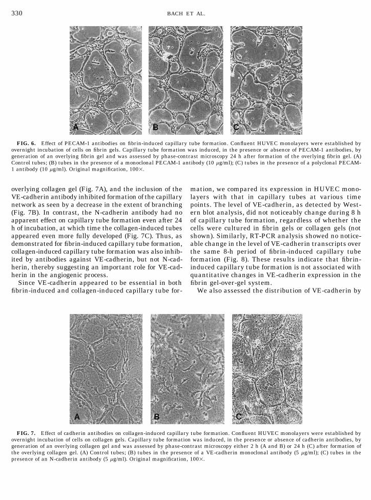
FIG. 7. Effect of cadherin antibodies on collagen-induced capillary tube formation. Confluent HUVEC monolayers were established byovernight incubation of cells on collagen gels. Capillary tube formation was induced, in the presence or absence of cadherin antibodies, bygeneration of an overlying collagen gel and was assessed by phase-contrast microscopy either 2 h (A and B) or 24 h (C) after formation ofthe overlying collagen gel. (A) Control tubes; (B) tubes in the presence of a VE-cadherin monoclonal antibody (5 mg/ml); (C) tubes in thepresence of an N-cadherin antibody (5 mg/ml). Original magnification, 1001.
AID ECR 3844 / 6i2d$$$621 01-08-98 08:00:32 eca

331VE-CADHERIN MEDIATES CAPILLARY TUBE FORMATION
the marked inhibition or disruption of tube formationelicited by a monoclonal antibody specific for HUVECVE-cadherin. In contrast, antibodies against N-cad-herin, PECAM-1, and avb3 had no apparent effect onfibrin-induced or collagen-induced generation of capil-lary tubes. Moreover, transcriptional expression andtranslational expression of VE-cadherin were quantita-tively similar throughout capillary tube formation. Fi-nally in this study, VE-cadherin was shown to be local-ized to intercellular junctions in capillary tubes in apattern similar to its distribution in HUVEC mono-layers, yet anti-VE-cadherin antibodies failed to dis-
FIG. 8. Quantitative RT-PCR of HUVEC RNA in fibrin-inducedcapillary tubes. Total HUVEC RNA was extracted with TRIzol re-agent from fibrin-induced capillary tubes at the time intervals of 0,2, 4, 8, and 12 h. RT-PCR analysis was performed, with upstreamand downstream VE-cadherin primers, using 4 mg of first-strandcDNA. The products were loaded in equal volumes onto agarose gelsand visualized at 1036 kb with ethidium bromide.
immunofluorescence. As shown in Fig. 9A, VE-cadherinwas diffusely distributed in HUVEC monolayers (red),but was greatly concentrated along cell borders (yel-low). As seen in Fig. 9B, the distribution of VE-cadherinin capillary tubes was similar to that in HUVEC mono-layers, with diffuse staining throughout the cells (red)and with intense staining along areas of intercellularcontact (yellow). These results indicate that VE-cad-herin remains quantitatively present as an essentialcomponent of the intercellular contacts even after HU-VEC monolayers have morphogenetically changed tocapillary tube structures.
DISCUSSION
Four major families of endothelial cell adhesion re-ceptors—the integrins, the selectins, the cadherins,and the Ig superfamily of receptors—are among themolecules that have been considered as potential cellu-lar receptors that may function during various steps ofthe angiogenic process [11, 12]. Although the involve-ment of the integrins [8, 18, 21–26] and selectins [8,27] in this process has been partly elucidated, the roleof the cadherins and the Ig superfamily of receptors ispoorly characterized [11]. In the present article, wehave addressed the potential role of the endothelial FIG. 9. Immunofluorescent distribution of VE-cadherin in HU-cell cadherins (VE-cadherin and N-cadherin), the Ig VEC monolayers and capillary tubes on fibrin. Established HU-
VEC monolayers or capillary tubes on fibrin were stained with asuperfamily receptor PECAM-1, and avb3 integrin inmonoclonal antibody against human VE-cadherin (10 mg/ml) fol-capillary tube formation. Our results demonstrate thatlowed by rhodaminated secondary antibody to mouse immunoglob-VE-cadherin plays a fundamental role in both the for- ulins. (A) VE-cadherin in HUVEC monolayer (original magnifica-
mation and the maintenance of capillary tubes in fibrin tion 201); (B) VE-cadherin in capillary tubes (original magnifica-tion 201; inset 601).and collagen gel-over-gel assays. This was apparent by
AID ECR 3844 / 6i2d$$$621 01-08-98 08:00:32 eca

332 BACH ET AL.
rupt monolayers, suggesting the existence of specific antibody may recognize epitopes of the extracellulardomain that do not participate in homotypic interac-differences in the intercellular role of VE-cadherin in
capillary tubes compared to monolayers. Thus, we have tions.The role of VE-cadherin in endothelium has been thedemonstrated a specific role for VE-cadherin in capil-
lary tube formation and maintenance induced by two subject of recent investigations. Cells naturally devoidof VE-cadherin but transfected to express VE-cadherindifferent matrix proteins which are known to play im-
portant roles in angiogenesis. show homotypic cell–cell associations [43]. Transfec-tion with C-terminus truncated mutants gave similarCadherins are a family of adhesive molecules, pres-
ent at adherens junctions, which mediate cell–cell con- results, indicating that cell–cell associations can occurwithout formation of the cadherin–catenin complextacts via their extracellular domains while anchoring
to cytoskeletal structures via their cytoplasmic tails [34]. Also, cadherin transfection of cells down-regulatescellular fibronectin and a3b1 [45].[32–36] and which play a major role in the morphogen-
esis of different organs [37]. Vascular endothelial cells Previous studies have shown that the association ofmatrix proteins with integrins plays an important roleexpress two different cadherins [12, 38]: N-cadherin,
a 135-kDa protein, which is diffusely distributed in the development of new blood vessels [11, 21–26,46]. However, in the fibrin sandwich assay, the interac-throughout the endothelial cell surface as well as at
intercellular junctions [39–41]; and VE-cadherin, a tion of fibrin with avb3 , although fundamental for cellspreading and monolayer formation, did not influence130-kDa protein, which is a specific endothelial cell
cadherin that is preferentially localized at interendo- the formation of capillary tubes, as shown by failure ofRGDS or the monoclonal antibody 7E3 against avb3 tothelial cell junctions [42, 43] and which is involved in
the development of blood vessels during embryogenesis inhibit tube formation [18]. In the present study, wehave corroborated this observation by showing that an-[44]. It is located at intercellular junctions of essen-
tially all types of endothelium, and reduction of the other monoclonal antibody (LM609) against avb3 alsofailed to inhibit fibrin-induced capillary tube forma-extracellular calcium concentration leads to its rapid
redistribution, accompanied by a slight increase in en- tion. Differences in the experimental models may ex-plain the requirement for integrin interactions in otherdothelial cell permeability, with no detectable change
in endothelial cell morphology as detected by immuno- assay systems [21–26] compared to the sandwichmodel [18]. Since the cells are rapidly sealed betweenfluorescence microscopy [43]. These characteristics of
cadherins provide a theoretical basis for their involve- two extracellular matrix gels in the sandwich model,integrin-mediated cell migration likely plays a minorment in the process of capillary tube formation.
Our studies clearly show that VE-cadherin plays a role in tube formation compared to single-gel assayswhere migration into the gel is required [21, 22].fundamental role in the formation of capillaries in a
fibrin or collagen matrix assay, indicating that it par- The Ig superfamily molecule known as PECAM-1is a 130-kDa protein that can participate in homo-ticipates in formation of specific cell–cell interactions
that are involved in the formation of capillaries. The typic or heterotypic cell–cell associations [30, 31].This receptor has been shown to play an importantsame antibody to VE-cadherin that inhibited formation
of capillaries did not disturb the morphology of the en- function in mediating the transmigration of leuko-cytes across interendothelial cell junctions [30, 31],dothelial cell monolayer, emphasizing the importance
of VE-cadherin-mediated intercellular forces in tube but the role of this molecule in the formation of capil-laries has not been delineated. Our present studystructures compared to flattened cells. In addition, VE-
cadherin may also be necessary for the maintenance of indicates that PECAM-1 does not directly participatein the process of capillary tube formation as assessedthe capillary structure as suggested by our findings
that the monoclonal antibody induced a disorganiza- using the fibrin sandwich technique. Furthermore,the finding that both PECAM-1 [30, 40] and VE-cad-tion of a preformed capillary net. Since this antibody
recognizes the first extracellular domain of VE-cad- herin [12, 42, 43] are almost exclusively localized tointercellular junctions in endothelial cells, but thatherin (amino acids 26–194) that participates in homo-
philic interactions that form the cell–cell junction [12, only disruption of VE-cadherin-based interactions in-hibited tube formation, suggests that these cell adhe-43], we conclude that this region of the molecule may
mediate homophilic interactions associated with the sion molecules may play distinct roles in mediatingcell–cell interactions for the development of capillaryformation and maintenance of capillary tubes. The fail-
ure of N-cadherin antibodies to inhibit capillary tube vessels. The fact that cadherins are spatially relatedto PECAM-1 at intercellular junctions, with cadher-formation suggests that N-cadherin, which is diffusely
distributed on the EC surface and not as localized to ins being located more apically and PECAM-1 morebasally [39, 40], supports this hypothesis.cell–cell junctions as VE-cadherin [41], does not partic-
ipate in the formation of capillaries and may serve an- A recent study has shown that antibodies against E-selectin or its ligands, sialyl-Lewis X or sialyl-Lewis A,other cellular function. Alternatively, the monoclonal
AID ECR 3844 / 6i2d$$$621 01-08-98 08:00:32 eca

333VE-CADHERIN MEDIATES CAPILLARY TUBE FORMATION
4. Cockerill, G. W., Gamble, J. R., and Vadas, M. A. (1995). Angio-inhibited formation of capillary tubes [27]. In thisgenesis: Models and modulators. Int. Rev. Cytol. 159, 113–160.study, capillary tube formation induced by seeding bo-
5. Arnold, F., and West, D. C. (1991). Angiogenesis in wound heal-vine capillary endothelial cells onto fibronectin-coateding. Pharm. Ther. 52, 407–422.dishes required 44 h for tube formation to occur and
6. Folkman, J. (1994). Angiogenesis and breast cancer. J. Clin.was associated with a two- to threefold increase in the Oncol. 12, 441–443.level of E-selectin mRNA in cells that formed tubes 7. Folkman, J. (1995). Angiogenesis in cancer, vascular, rheuma-compared to control monolayers [27]. In our study, tube toid and other disease. Nature Med. 1(1), 27–31.formation was readily apparent within only 2 h of gen- 8. Bischoff, J. (1995). Approaches to studying cell adhesion mole-
cules in angiogenesis. Trends Cell Biol. 5, 69–74.erating overlying fibrin or collagen gels and presum-ably did not involve participation of E-selectin, since 9. Nehls, V., Schuchardt, E., and Drenckhahn, D. (1994). The ef-
fect of fibroblasts, vascular smooth muscle cells, and pericytesunstimulated HUVECs do not express this cell adhe-on sprout formation of endothelial cells in a fibrin gel angiogene-sion molecule [27]. Due to the differences in the typessis system. Microvasc. Res. 48, 349–363.of cells used and in the experimental assays, it is diffi-
10. Dejana, E. (1996). Endothelial adherens junctions: Implicationscult to assess whether E-selectin participates in the in the control of vascular permeability and angiogenesis. J.formation of capillaries using the two-gel system. Clin. Invest. 98, 1949–1953.
The role of VE-cadherin in angiogenesis has been 11. Stromblad, S., and Cheresh, D. A. (1996). Cell adhesion andangiogenesis. Trends Cell Biol. 6, 462–467.hypothesized to involve limitation of endothelial cell
12. Dejana, E., Corada, M., and Lampugnani, M. G. (1995). Endo-migration and growth [10], since VE-cadherin presentthelial cell-to-cell junctions. FASEB J. 9, 910–918.at adherens junctions inhibits cell migration from a
13. Klagsbrun, M., and D’Amore, P. A. (1991). Regulators of angio-monolayer [35] and, when transfected into tumor cells,genesis. Annu. Rev. Physiol. 53, 217–239.elicits contact inhibition of growth [47]. Reversal of this
14. Maciag, T. (1990). Molecular and cellular mechanisms of angio-inhibitory effect has been reported to occur when vascu-genesis. Important Adv. Oncol. 85–98.lar endothelial growth factor, an angiogenic factor, in-
15. Nicosia, R. F., and Ottinetti, A. (1990). Modulation of microvas-teracts with endothelial cell monolayers and induces cular growth and morphogenesis by reconstituted basementtyrosine phosphorylation of adherens junctions pro- membrane gel in three-dimensional cultures of rat aorta: A com-
parative study of angiogenesis in matrigel, collagen, fibrin, andteins [10]. Also FGF-1-induced angiogenesis on singleplasma clot. In Vitro Cell. Dev. Biol. 26, 119–128.fibrin gels has been shown to be transcriptionally de-
16. Nicosia, R. F., and Ottinetti, A. (1990). Growth of microvesselspendent [48] and to involve regulation of cell migrationin serum-free matrix culture of rat aorta: A quantitative assaythrough the jagged-notch signaling pathway [49]. Ourof angiogenesis in vitro. Lab. Invest. 63, 115–122.results show that VE-cadherin is crucial for reestab-
17. Montesano, R., Orci, L., and Vassalli, P. (1983). In vitro rapidlishment and maintenance of cell–cell contacts neces- organization of endothelial cells into capillary-like networks issary for the formation of new capillary tubes and thus promoted by collagen matrices. J. Cell Biol. 97, 1648–1652.acts in a fashion to promote and sustain angiogenesis. 18. Chalupowicz, D. G., Chowdhury, Z. A., Bach, T. L., Barsigian,However, up-regulation of the VE-cadherin transcript C., and Martinez, J. (1995). Fibrin II induces endothelial cell
capillary tube formation. J. Cell Biol. 130, 207–215.did not occur during tube formation, indicating that19. Jackson, C., and Jenkins, K. L. (1991). Type I collagen fibrilsqualitative changes in the distribution of this cell-adhe-
promote rapid vascular tube formation upon contact with thesion molecule, rather than quantitative changes, areapical side of cultured endothelium. Exp. Cell Res. 192, 319–important. Further work will be necessary to clarify 323.
the role of VE-cadherin in physiologic angiogenic states 20. Jackson, C. J., Giles, I., Knop, A., Nethery, A., and Schrieber,such as wound healing and in pathological conditions, L. (1994). Sulfated polysaccharides are required for collagen-
induced vascular tube formation. Exp. Cell Res. 215, 294–302.such as diabetic retinopathy and tumor angiogenesis,in which dysregulation of angiogenesis occurs. 21. Gamble, J. R., Matthias, L. J., Meyer, G., Kaur, P., Russ, G.,
Faull, R., Berndt, M. C., and Vadas, M. A. (1993). Regulationof in vitro capillary tube formation by anti-integrin antibodies.
The authors thank Andrew S. Likens for the art work, Rosemarie J. Cell Biol. 121, 931–943.Silvano for preparation of the manuscript, and Bonny Lightner for 22. Brooks, P. C., Clark, R. A. F., and Cheresh, D. A. (1994). Re-her experience in cell culture. quirement of vascular integrin avb3 for angiogenesis. Science
264, 569–571.23. Brooks, P. C., Montgomery, A. M., Rosenfeld, M., Reisfeld,REFERENCES
T. H., Klier, G., and Cheresh, D. A. (1995). Integrin avb3 antago-nists promote tumor regression by inducing apoptosis of angio-
1. Risau, W., and Flamme, J. (1995). Vasculogenesis. Annu. Rev. genic blood vessels. Cell 79, 1157–1164.Cell Dev. Biol. 11, 73–91. 24. Brooks, P. C., Stromblad. S., Klemke, R., Visscher, D., Sarkar,
2. Breier, G., and Risau, W. (1996). The role of vascular endothe- F. H., and Cheresh, D. A. (1995). Anti-integrin avb3 blocks hu-lial growth factor in blood vessel formation. Trends Cell Biol. man breast cancer growth and angiogenesis in human skin. J.6, 454–456. Clin. Invest. 96, 1815–1822.
25. Friedlander, M., Brooks, P. C., Shaffer, R. W., Kincaid, C. M.,3. Folkman, J., and Shing, Y. (1992). Angiogenesis. J. Biol. Chem.267, 10931–10934. Varner, J. A., and Cheresh, D. A. (1995). Definition of two angio-
AID ECR 3844 / 6i2d$$$621 01-08-98 08:00:32 eca

334 BACH ET AL.
genic pathways by distinct av integrins. Science 270, 1500– S. (1994). Cloning of five human cadherins clarifies characteris-tic features of cadherin extracellular domain and provides fur-1502.ther evidence for two structurally different types of cadherin.26. Friedlander, M., Theesfeld, C. L., Sugita, M., Fruttiger, M.,Cell Adhes. Commun. 2, 15–26.Thomas, M. A., Chang, S., and Cheresh, D. A. (1996). Involve-
39. Heimark, R. L., Degner, M., and Schwartz, S. M. (1990). Identi-ment of integrins avb3 and avb5 in ocular neovascular diseases.fication of a Ca2/-dependent cell–cell adhesion molecule in en-Proc. Natl. Acad. Sci. USA 93, 9764–9769.dothelial cells. J. Cell Biol. 110, 1745–1756.27. Nguyen, M., Strubel, N. A., and Bischoff, J. (1993). A role for
40. Ayalon, O., Sabanai, H., Lampugnani, M. C., Dejana, E., andsialyl Lewis-X/A glycoconjugates in capillary morphogenesis.Geiger, B. (1994). Spatial and temporal relationships betweenNature 365, 267–269.cadherins and PECAM-1 in cell–cell junctions of human endo-28. Martinez, J., Rich, E., and Barsigian, C. (1989). Transglutami- thelial cells. J. Cell Biol. 126, 247–258.nase-mediated cross-linking of fibrinogen by human umbilical
41. Salomon, D., Ayalon, O., Patel-King, R., Hynes, R. O., and Gei-vein endothelial cells. J. Biol. Chem. 264, 20502–20508.ger, B. (1992). Extrajunctional distribution of N-cadherin in
29. Chowdhury, Z. A., Barsigian, C., Chalupowicz, G. D., Bach, cultured human endothelial cells. J. Cell Sci. 102, 1–11.T. L., Garcia-Manero, G., and Martinez, J. (1996). Co-localiza- 42. Lampugnani, M. G., Resnati, M., Raiteri, M., Pigott, R., Pisa-tion of tissue transglutaminase and stress fibers in human vas- cane, A., Houen, G., Ruco, L. P., and Dejana, E. (1992). A novelcular smooth muscle cells and human umbilical vein endothelial endothelial-specific membrane protein is a marker of cell–cellcells. (1997). Exp. Cell Res. 231, 38–49. contacts. J. Cell Biol. 118, 1511–1522.
30. Albelda, S. M., Oliver, P. D., Romer, L. H., and Buck, C. A. 43. Breviario, F., Caveda, L., Corada, M., Martin-Padura, I., Na-(1990). EndoCAM: A novel endothelial cell–cell adhesion mole- varro, P., Golay, J., Introna, M., Gulino, D., Lampugnani, M. G.,cule. J. Cell Biol. 110, 1227–1237. and Dejana, E. (1995). Functional properties of human vascular
31. Albelda, S. M., Muller, W. A., Buck, C. A., and Newman, P. J. endothelial cadherin (7B4/cadherin-5), an endothelium-specific(1991). Molecular and cellular properties of PECAM-1 (Endo- cadherin. Arterioscler. Thromb. Vasc. Biol. 15, 1229–1239.CAM; CD31): A novel vascular cell–cell adhesion molecule. J. 44. Breier, G., Breviario, F., Caveda, L., Berthier, R., Schnurch, H.,Cell Biol. 114, 1059–1068. Gotsch, U., Vestweber, D., Risau, W., and Dejana, E. (1996).
Molecular cloning and expression of murine vascular endothe-32. Geiger, B., and Ayalon, O. (1992). Cadherins. Annu. Rev. Celllial cadherin in early state development of cardiovascular sys-Biol. 8, 307–332.tem. Blood 87, 630–641.33. Suzuki, S. T. (1996). Structural and functional diversity of cad-
45. Finnemann, S., Kuhl, M., Otto, G., and Wedlich, D. (1995).herin superfamily: Are new4 members of cadherin superfamilyCadherin transfection of Xenopus XTC cells down-regulates ex-involved in the signal transduction pathway? J. Cell Biochem.pression of substrate adhesion molecules. Mol. Cell. Biol. 15,61, 531–542.5082–5091.34. Hinck, L., Nathke, I. S., Papkoff, J., and Nelson, W. J. (1994).
46. Grant, D. S., Tashiro, K-I., Segui-Real, B., Yamada, Y., Martin,Dynamics of cadherin/catenin complex formation: Novel proteinG. R., and Kleinman, H. K. (1989). Two different laminin do-interactions and pathways of complex assembly. J. Cell Biol.mains mediate the differentiation of human endothelial cells125, 1327–1340.into capillary-like structures in vitro. Cell 58, 933–943.
35. Lampugnani, M. G., Corada, M., Caveda, L., Breviario, F., Aya-47. Caveda, L., Martin-Padura, I., Navarro, P., Breviario, F., Cor-lon, O., Geiger, B., and Dejana, E. (1995). The molecular organi-
ada, M., Gulino, D., Lampugnani, M. G., and Dejana, E. (1996).zation of endothelial cell to cell junctions: Differential associa-Inhibition of cultured cell growth by vascular endothelial cad-tion of plakoglobin, b-catenin, and a-catenin with vascular en-herin (cadherin-5/VE-cadherin). J. Clin. Invest. 98, 886–893.dothelial cadherin (VE-cadherin). J. Cell Biol. 129, 203–217.
48. Zimrin, A. B., Villeponteau, B., and Maciag, T. (1995). Models36. Navarro, P., Caveda, L., Breviario, F., Mandoteanu, I., Lampug- of in vitro angiogenesis: Endothelial cell differentiation on fibrin
nani, M. G., and Dejana, E. (1995). Catenin-dependent and but not matrigel is transcriptionally dependent. Biochem. Bio--independent functions of vascular endothelial cadherin. J. Biol. phys. Res. Commun. 213, 630–638.Chem. 270, 30965–30972.
49. Zimrin, A. B., Pepper, M. S., McMahon, G. A., Nguyen, F., Mon-37. Ranscht, B. (1994). Cadherins and catenins: Interactions and tesano, R., and Maciag, T. (1996). An antisense oligonucleotide
functions in embryonic development. Curr. Opin. Cell Biol. 6, to the notch ligand jagged enhances fibroblast growth factor-740–746. induced angiogenesis in vitro. J. Biol. Chem. 271(51), 32499–
32502.38. Tanihara, H., Sano, K., Heimark, R. L., St. John, T., and Suzuki,
Received April 16, 1997Revised version received September 3, 1997
AID ECR 3844 / 6i2d$$$621 01-08-98 08:00:32 eca
Copyright © 2022 FDOKUMEN