
Doctoral Thesis Creating and validating UML class diagrams ...
Upload
independentCategory
view
1download
0
Validating a Strategy for Molecular Dynamics Simulations of Cyclodextrin InclusionComplexes through Single-Crystal X-ray and NMR Experimental Data: A Case Study
Giuseppina Raffaini,*,† Fabio Ganazzoli,† Luciana Malpezzi,† Claudio Fuganti,†
Giovanni Fronza,‡ Walter Panzeri,‡ and Andrea Mele*,†
Dipartimento di Chimica, Materiali e Ingegneria Chimica “G. Natta”, Politecnico di Milano, Via L. Mancinelli7, 20131 Milano, Italy, CNRsIstituto di Chimica del Riconoscimento Molecolare, Via L. Mancinelli 7,20131 Milano, Italy
ReceiVed: February 20, 2009; ReVised Manuscript ReceiVed: May 13, 2009
A theoretical and experimental study about the formation and structure of the inclusion complex (-)-menthyl-O-�-D-glucopyranoside 1 with �-cyclodextrin (�-CD) 2 is presented as paradigmatic case study to test theresults of molecular dynamics (MD) simulations. The customary methodological approachsthe use ofexperimental geometrical parameters as restraints for MD runssis logically reversed and the calculatedstructures are a posteriori compared with those obtained from NMR spectroscopy in D2O solution and singlecrystal X-ray diffraction so as to validate the simulation procedure. The guest molecule 1 allows for a broadrepertoire of intermolecular interactions (dipolar, hydrophobic, hydrogen bonds) concurring to stabilize thehost-guest complex, thus providing the general applicability of the simulation procedure to cyclodextrinphysical chemistry. Many starting geometries of the host-guest association were chosen, not assuming anya priori inclusion. The simulation protocol, involving energy minimization and MD runs in explicit water,yielded four possible inclusion geometries, ruling out higher-energy outer adducts. By analysis of the averageenergy at room temperature, the most stable geometry in solution was eventually obtained, while the kineticsof formation showed that it is also kinetically favored. The reliability of such geometry was thoroughly checkedagainst the NOE distances via the pair distribution functions, that is, the statistical distribution of intermoleculardistances among selected diagnostic atoms calculated from the MD trajectories at room temperature. Ananalogous procedure was adopted both with implicit solvent and in Vacuo. The most stable geometry matchedthat found with explicit solvent but major differences were observed in the relative stability of the metastablecomplexes as a consequence of the lack of hydration on the polar moiety of the guest. Finally, a control setof geometrical parameters of the thermodynamically favored complex matched the corresponding one obtainedfrom the X-ray structure, while local conformational differences were indicative of packing effects.
1. Introduction
Cyclodextrins (CD), such as cyclomaltohexaose, cyclomal-toheptaose, and cyclomaltooctaose (R-CD, �-CD, and γ-CD,respectively), are cyclic oligosaccharides with a truncated-coneshape, having an inner hydrophobic cavity and an externalhydrophilic surface. The narrow and the wide rims of themacrocycle comprise primary and secondary hydroxyl groups,respectively. Because of these features, apolar organic moleculesor polar molecules with an apolar region can be accommodatedinto the cavity of CDs to form energetically stable host-guestinclusion complexes.1 The formation of such supramolecularcomplexes is a well assessed phenomenon with applicationscovering a broad range of fields, including chiral separations,2
drug delivery and targeting,3 food chemistry,4 sensors.5
The driving forces for the formation of host-guest complexesof cyclodextrins are the energetically favorable nonbondinginteractions, such as hydrogen bonds (H-bonds), hydrophobiceffects, and van der Waals interactions (dispersion forces anddipolar interactions) of the guest included into the host CD
cavity. Additionally, in water the release of relatively isolatedsmall clusters of water molecules from the hydrophobic cavityentropically favors guest inclusion, largely compensating theentropy loss due to inclusion. Clearly, the extent of eachcontribution to the stabilization of the complex depends on thechemical nature of the guest and host molecules.6
Computational methods based on molecular mechanics (MM)and molecular dynamics (MD) have been extensively used inparallel to experimental structural methods such as NMR,calorimetry, and X-ray diffraction to obtain a comprehensivemodel of the complexation and molecular recognition phenom-ena. Typically, a first-approximation model deduced from theexperimental data is refined by simple energy minimization(MM methods), or, more recently, through MD simulations inVacuo or increasingly more often in explicit water, simulatingthe dynamic behavior on nanoseconds time-scale. Wheneveravailable, the atom coordinates obtained from single crystalX-ray diffraction structure of a given complex are used asstarting geometry for MD simulations in water aimed atsimulating or predicting the structure of the complex in solutionor in an environment mimicking physiological conditions. Theavailability of suitable force fields and the decreased costs ofcomputing prompted many investigators to massive use ofsimulations, thus raising the issue of control protocols for thereliability of calculated geometry and conformational properties.
* To whom correspondence should be addressed. (G.R.) Fax: 0039 0223993180. Tel.: 0039 02 23993024. E-mail: [email protected].(A.M) Fax: 0039 02 23993180. Tel.: 0039 02 23993006. E-mail:[email protected].
† Politecnico di Milano.‡ CNRsIstituto di Chimica del Riconoscimento Molecolare.
J. Phys. Chem. B 2009, 113, 9110–91229110
10.1021/jp901581e CCC: $40.75 2009 American Chemical SocietyPublished on Web 06/15/2009
Dow
nloa
ded
by C
ILE
A C
ON
SOR
TIA
IT
AL
Y o
n Ju
ly 2
, 200
9Pu
blis
hed
on J
une
15, 2
009
on h
ttp://
pubs
.acs
.org
| do
i: 10
.102
1/jp
9015
81e

In the present work a paradigmatic case study is reported anddiscussed where the results of MM and MD simulations carriedout in Vacuo, with implicit solvent and in explicit water on aspecifically tailored �-cyclodextrin inclusion complex arevalidated against solution NMR data and compared with solid-state X-ray results. To this end, the customary approachmentioned abovesfrom experiments to calculationssis logicallyreversed, going from calculations to experiments. For this study,the guest molecule, (-)-menthyl-O-�-D-glucopyranoside 1(Scheme 1) was encapsulated into �-cyclodextrin 2 (�-CD, seeScheme 1), and this process was theoretically modeled throughcomputer simulations. The inclusion complex of 1 and 2 wascharacterized in aqueous solution by NMR spectroscopy andin the solid state by single crystal X-ray diffraction, and theexperimental data were thoroughly compared with the simulationresults for validating the simulation procedure.
The model guest molecule 1, was chosen on the basis of twocriteria: (i) Structural suitability for the formation of an inclusioncomplex with CD exhibiting the broadest spectrum of interac-tions (hydrophobic, hydrogen bond, dipolar); indeed, 1 fulfillsthe requirements proposed by Saenger for the ideal guest,namely the presence, in the molecular frame, of “hydrophobicbody and hydrophilic “ends” which can form hydrogen bondsto the hydroxyl groups on both sides of the host molecule tostabilize the inclusion complex.”.7 (ii) The possibility ofachieving a thorough structural characterization of the inclusioncomplex both in solution and in the solid state. The quality ofthe structure determination in solution, in turn, depends primarilyon the number of intermolecular NOEs that can be collected,providing a sufficiently wide set of host-guest distances forthe formulation of a first-approximation geometry of thecomplex. As for the solid-state structure, it should be consideredthat most cyclodextrin inclusion complexes do not afford singlecrystals suitable for X-ray diffraction studies. Therefore, thechoice of the present model system was the result of a largenumber of attempts of crystallizing inclusion complexes of �-CDwith small-medium sized glycoconjugates.8
The plan of the paper is the following: we first report theexperimental and computational details, then we discuss theresults of the NMR experiments in solution and of the solid-state structure obtained by X-ray single-crystal analysis, andafterward we report the simulation results in solution (by MDruns at room temperature) and in vacuo (by MD runs and energyminimizations) to validate the simulation procedure in com-parison with the experimental data.
2. Materials, Experimental Methods, and ComputationalDetails
Cyclomaltoheptaose (�-cyclodextrin, 2) was purchased fromSigma-Aldrich and used without any further purification. (-)-Menthyl-O-�-D-glucopyranoside (1) was obtained from SanGiorgio Flavours (Torino, Italy) and used as such. Deuteriumoxide (99.97%) was obtained from Cortec (France).
2.1. Molecular Modeling and Data Analysis. The simula-tions were performed with InsightII/Discover 2000,9 using theconsistent valence force field CVFF.10 This force field describesnonbonded interactions through van der Waals and Coulombicterms only, with no extra term for H-bonds. CVFF, originallydesigned to model proteins, was later augmented to includeadditional functional groups, accounting also inter alia for theacetal moiety related with the anomeric effect in carbohydrates.11
Therefore, CVFF can be satisfactorily used for these molecules,12
even though this effect is not conformationally dominant in CDsbecause of the geometric constraint imposed by the macrocycle.Extensive tests carried out in comparison with NMR data dosupport the general accuracy of CVFF for oligosaccharides.13
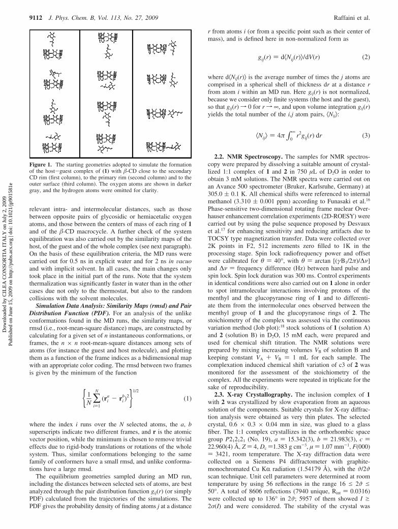
The geometries of �-CD and of 1 were generated with theavailable templates of InsightII, then subjected to an MD runin vacuo at room temperature and finally optimized up to anenergy gradient lower than 4 × 10⁻³ kJ mol-1 Å-1. 1 was thenplaced close to �-CD but outside the hydrophobic cavity,selecting 12 unbiased trial geometries (Figure 1) for the initialoptimizations. The arrangements were chosen so that all themain different sides of 1 could approach the two rims and theouter surface of �-CD. No inclusion complex was assumed inthis stage, unlike what it is often done.14 The whole system wasthen hydrated by adding more than one thousand watermolecules at the density of 1 g cm-3 in a cubic cell with edgesof 33 Å and periodic boundary conditions (constant-volumeconditions), and after full geometry optimization all the resultingadducts were subjected to independent MD simulations. Furthersimulations were carried out for the same starting geometriesin vacuo and with implicit solvent in an effective dielectricmedium to check whether these much faster procedures producesatisfactory results. The dynamic equations were integrated usingthe Verlet algorithm with a time step of 1 fs at a temperatureof 300 K, controlled through the Berendsen thermostat, and theinstantaneous coordinates were periodically saved for furtheranalysis. The system equilibration was monitored by the timechange of the potential energy and of its components, and of
SCHEME 1: Molecular Structures and Atom Numbering Scheme for Compounds 1 and 2a
a Hydrogen atoms of 1 and 2 are identified by using lower case letters and atom numbers as shown: g ) glucose of menthyl glucoside; m )menthyl residue; c ) glucose units of cyclodextrin (e.g., g3 refers to the H atom in position 3 of the glucose unit of compound 1, while c3 indicatesthe homologous proton of compound 2). Symmetry equivalent glucose units of 2 are labelled A-G according to Breslow.15
Validating MD Simulations with NMR and X-ray Data J. Phys. Chem. B, Vol. 113, No. 27, 2009 9111
Dow
nloa
ded
by C
ILE
A C
ON
SOR
TIA
IT
AL
Y o
n Ju
ly 2
, 200
9Pu
blis
hed
on J
une
15, 2
009
on h
ttp://
pubs
.acs
.org
| do
i: 10
.102
1/jp
9015
81e
relevant intra- and intermolecular distances, such as thosebetween opposite pairs of glycosidic or hemiacetalic oxygenatoms, and those between the centers of mass of each ring of 1and of the �-CD macrocycle. A further check of the systemequilibration was also carried out by the similarity maps of thehost, of the guest and of the whole complex (see next paragraph).On the basis of these equilibration criteria, the MD runs werecarried out for 0.5 ns in explicit water and for 2 ns in Vacuoand with implicit solvent. In all cases, the main changes onlytook place in the initial part of the runs. Note that the systemthermalization was significantly faster in water than in the othercases due not only to the thermostat, but also to the randomcollisions with the solvent molecules.
Simulation Data Analysis: Similarity Maps (rmsd) and PairDistribution Function (PDF). For an analysis of the unlikeconformations found in the MD runs, the similarity maps, orrmsd (i.e., root-mean-square distance) maps, are constructed bycalculating for a given set of n instantaneous conformations, orframes, the n × n root-mean-square distances among sets ofatoms (for instance the guest and host molecule), and plottingthem as a function of the frame indices as a bidimensional mapwith an appropriate color coding. The rmsd between two framesis given by the minimum of the function
where the index i runs over the N selected atoms, the a, bsuperscripts indicate two different frames, and r is the atomicvector position, while the minimum is chosen to remove trivialeffects due to rigid-body translations or rotations of the wholesystem. Thus, similar conformations belonging to the samefamily of conformers have a small rmsd, and unlike conforma-tions have a large rmsd.
The equilibrium geometries sampled during an MD run,including the distances between selected sets of atoms, are bestanalyzed through the pair distribution function gij(r) (or simplyPDF) calculated from the trajectories of the simulations. ThePDF gives the probability density of finding atoms j at a distance
r from atoms i (or from a specific point such as their center ofmass), and is defined here in non-normalized form as
where d⟨Nij(r)⟩ is the average number of times the j atoms arecomprised in a spherical shell of thickness dr at a distance rfrom atom i within an MD run. Here gij(r) is not normalized,because we consider only finite systems (the host and the guest),so that gij(r)f 0 for rf ∞, and upon volume integration gij(r)yields the total number of the i,j atom pairs, ⟨Nij⟩:
2.2. NMR Spectroscopy. The samples for NMR spectros-copy were prepared by dissolving a suitable amount of crystal-lized 1:1 complex of 1 and 2 in 750 µL of D2O in order toobtain 3 mM solutions. The NMR spectra were carried out onan Avance 500 spectrometer (Bruker, Karlsruhe, Germany) at305.0 ( 0.1 K. All chemical shifts were referenced to internalmethanol (3.310 ( 0.001 ppm) according to Funasaki et al.16
Phase-sensitive two-dimensional rotating frame nuclear Over-hauser enhancement correlation experiments (2D-ROESY) werecarried out by using the pulse sequence proposed by Desvauxet al.17 for enhancing sensitivity and reducing artifacts due toTOCSY type magnetization transfer. Data were collected over2K points in F2, 512 increments zero filled to 1K in theprocessing stage. Spin lock radiofrequency power and offsetwere calibrated for θ ) 40°, with θ ) arctan [(γB1/2π)/∆ν]and ∆ν ) frequency difference (Hz) between hard pulse andspin lock. Spin lock duration was 300 ms. Control experimentsin identical conditions were also carried out on 1 alone in orderto spot intramolecular interactions involving protons of thementhyl and the glucopyranose ring of 1 and to differenti-ate them from the intermolecular ones observed between thementhyl group of 1 and the glucopyranose rings of 2. Thestoichiometry of the complex was assessed via the continuousvariation method (Job plot):18 stock solutions of 1 (solution A)and 2 (solution B) in D2O, 15 mM each, were prepared andused for chemical shift titration. The NMR solutions wereprepared by mixing increasing volumes VB of solution B andkeeping constant VA + VB ) 1 mL for each sample. Thecomplexation induced chemical shift variation of c3 of 2 wasmonitored for the assessment of the stoichiometry of thecomplex. All the experiments were repeated in triplicate for thesake of reproducibility.
2.3. X-ray Crystallography. The inclusion complex of 1with 2 was crystallized by slow evaporation from an aqueoussolution of the components. Suitable crystals for X-ray diffrac-tion analysis were obtained as very thin plates. The selectedcrystal, 0.6 × 0.3 × 0.04 mm in size, was glued to a glassfiber. The 1:1 complex crystallizes in the orthorhombic spacegroup P212121 (No. 19), a ) 15.342(3), b ) 21.983(3), c )22.960(4) Å, Z ) 4, Dc )1.383 g cm-3, µ ) 1.07 mm-1, F(000)) 3421, room temperature. The X-ray diffraction data werecollected on a Siemens P4 diffractometer with graphite-monochromated Cu KR radiation (1.54179 Å), with the ϑ/2ϑ
scan technique. Unit cell parameters were determined at roomtemperature by using 56 reflections in the range 16 e 2ϑ e50°. A total of 8606 reflections (7940 unique, Rint ) 0.0316)were collected up to 136° in 2ϑ; 5957 of them showed I g2σ(I) and were considered. The stability of the crystal was
Figure 1. The starting geometries adopted to simulate the formationof the host-guest complex of (1) with �-CD close to the secondaryCD rim (first column), to the primary rim (second column) and to theouter surface (third column). The oxygen atoms are shown in darkergray, and the hydrogen atoms were omitted for clarity.
{ 1N ∑
i)1
N
(ria - ri
b)2}1/2
(1)
gij(r) ) d⟨Nij(r)⟩/dV(r) (2)
⟨Nij⟩ ) 4π∫0
∞r2gij(r) dr (3)
9112 J. Phys. Chem. B, Vol. 113, No. 27, 2009 Raffaini et al.
Dow
nloa
ded
by C
ILE
A C
ON
SOR
TIA
IT
AL
Y o
n Ju
ly 2
, 200
9Pu
blis
hed
on J
une
15, 2
009
on h
ttp://
pubs
.acs
.org
| do
i: 10
.102
1/jp
9015
81e

periodically monitored during data collection and no crystaldecay was observed. The structure was solved by direct methods(SIR97) providing almost all the non-H atoms of the �-CDcomplex. Subsequent difference Fourier syntheses and least-squares refinement (SHELX-97 program)19 converged to a finalR(F2) ) 0.0550 for the observed reflections and wR(F2) )0.1580 for all data. The hydrogen atoms were generated inidealized position by the XP module of SHELX-97 and refinedusing a riding model. Five fully positioned water molecules werefound on the electron density map, while minor peaks of electrondensity suggested partially occupied water sites: only those withoccupation factor refined to values g0.20 were retained in thefinal refinement. The H atoms of the water molecules were notintroduced. Because of the large number of parameters, the finalcycle of refinement was carried out in two blocks: one for �-CDalone (702 refined parameters) and one for guest and watermolecules (366 refined parameters). The maximum values forpositive and negative residual electron density were 0.31 and-0.30 e Å-3, respectively. The goodness of fit was S ) 1.040.
The full list of data collection, structure determination andrefinement is reported in the Supporting Information, Table S1.
3. Results and Discussion
3.1. The Solution Structure of the Complex: NMR. The1H NMR spectrum of a mixture of 1 and 2 in D2O showed thetypical complexation-induced chemical shift variations ofselected protons: deshielding of the signals of the guest moleculeprotons and shielding of c3 and c5 of cyclodextrin (see Scheme1). These findings provided the first evidence of the formationof a true inclusion complex in solution.20 The continuousvariation plot (Job plot) reported in Figure 2 shows a sharpmaximum at r ) [H]/([H] + [G]) ) 0.5, consistent with 1:1stoichiometry of the inclusion complex. A thorough character-ization of the solution geometry of the complex was obtainedby the analysis of the intermolecular nuclear Overhauserenhancements in the rotating frame (ROEs). An expansion ofthe 2D-ROESY experiment on the inclusion complex is shownin Figure 3. The one-dimensional projection on the F1 dimensionis related to signals assigned to the protons of the menthylmoiety, while the F2 projection displays the spectral region ofthe nonanomeric protons of CD. Intense cross-peaks relatingprotons c3 of cyclodextrin and m6eq, m3eq, and m2 of thementhyl residue of 1 indicate that the latter protons are in spatialproximity to the secondary OH rim of cyclodextrin. Interest-ingly, m6eq, m3eq, and m2 do not give rise to cross peaks withc5. In a complementary way, the cross peaks corresponding tothe pairs c5/m4eq and c5/m5ax point out that the molecular frameconsisting of the carbon atoms in position 4 and 5 (includingalso the methyl group 7) of the menthyl moiety is close in spaceto the primary OH rim of cyclodextrin. Once again, m4eq andm5ax do not show dipolar contact with c3. Furthermore and quitesurprisingly, also proton m8 shows no ROE with either c3 orc5. These findings are consistent with the menthyl residue of 1
Figure 2. Continuous variation plot (Job plot) for the formation ofthe inclusion complex of 1 and 2. The quantity ∆δ is the complexationinduced chemical shift variation of proton c3. [H] and [G] indicate themolar concentration of host 2 and guest 1, respectively.
Figure 3. Detail of the contour plot of 2D-ROESY of the inclusion complex. Horizontal (F2) and vertical (F1) projection are referred to thementhyl moiety of 1 and to the nonanomeric protons of 2, respectively. The assignment of relevant signals is given according to the atom labelingused in Scheme 1. Cross-peaks arising from intermolecular ROEs are framed in rectangular boxes (for ROEs of methyl groups see Figure 4).Significant intramolecular ROEs of proton m1 at 3.63 ppm (m1/m6eq, m1/m5ax, m1/m3ax, and m1/m10) are also marked with an asterisk.
Validating MD Simulations with NMR and X-ray Data J. Phys. Chem. B, Vol. 113, No. 27, 2009 9113
Dow
nloa
ded
by C
ILE
A C
ON
SOR
TIA
IT
AL
Y o
n Ju
ly 2
, 200
9Pu
blis
hed
on J
une
15, 2
009
on h
ttp://
pubs
.acs
.org
| do
i: 10
.102
1/jp
9015
81e

entering the hydrophobic cavity of 2 from the secondary OHrim and fully inserted into the cavity. The intermolecular ROEsdue to the methyl groups of 1 with the inner protons c3 and c5of 2 are displayed in Figure 4. The observed pattern is fullyconsistent with the complexation geometry inferred from thedata reported above. The two diastereotopic methyl groups m9and m10 of the isopropyl substituent in position 2 of the menthylresidue show strong correlations with c3, and no correlation atall with c5. The isopropyl substituent is therefore close to thelarge rim of the CD cavity. Conversely, the methyl group inposition 7, namely far away from the glycosidic junction, showsselective ROE with c5 and no correlation with c3. Moreinterestingly, a weak but significant ROE between m7 and bothc6 protons is clearly detectable, suggesting that such methylgroup is protruding out of the cavity of CD and close in spaceto the CH2OH tails of CD. The discussion so far assumed thatinter- and intramolecular ROEs could be unambiguously dis-tinguished. Any possible misinterpretations due to overlap ofinter- and intramolecular ROEs was double checked by controlexperiments carried out on the guest 1 alone. ROESY experi-ments on 1 showed that m1 and g1, falling in the glucopyranoseregion of the spectrum (3.2-4.5 ppm), are the only protonsgiving rise to strong intramolecular ROEs with the menthylprotons falling in the high field region of spectrum (0.7-2.2ppm). The signals of m1 (3.63 ppm) and g1 (4.49 ppm) arewell separated from those of c3 (3.91 ppm) and c5 (3.74 ppm),and therefore the intramolecular cross-peaks do not overlap theintermolecular ones.
The general solution geometry of the complex can besummarized as follows: (i) formation of a 1:1 inclusion complex;
(ii) the menthyl group of 1 is located inside the cavity of 2;(iii) the glycosidic linkage of 1 is on the secondary OH rim ofCD; (iv) the glucose moiety is outside the cavity, exposed towater solvent and likely to establish hydrogen bonds with OHgroups of CD. The latter conclusion can only be indirectlyinferred from points i-iii as no direct ROEs between hydrogenatoms of the glycan of 1 with H atoms of 2 could be observeddue to spectral overlap.
3.2. The Solid State Structure of the Complex: X-rayDiffraction. �-CD and (-)-menthyl-O-�-D-glucopyranosideform an host-guest inclusion complex in the 1:1 ratio. Thesolid-state structure of the complex is shown in Figure 5, whilesome geometrical parameters of �-CD are reported in Table 1,in comparison with analogous quantities obtained by thecomputer simulations (see later). The �-CD macrocyclic ring,characterized by the usual truncated-cone shape, shows a pseudo7-fold symmetry, apart from some tilt of the sugar ring, asindicated by the geometry of the almost planar heptagon definedby the O4 atoms (see Table 1): the sides of the heptagon rangefrom 4.315(6) to 4.512(6) Å (mean value 4.372 Å) and O4(n- 1) · · ·O4(n) · · ·O4(n + 1) angles range from 123.7(1) to132.4(2)° (mean value 128.2° to be compared with the idealvalue of 128.6° for regular heptagon). The perimeter of theheptagon defined by the O4 atoms is 30.60 Å. For the sake ofcomparison, the values of the geometrical parameters reportedabove are 4.36 ( 0.12 Å and 128.3 ( 3.7° for uncomplexed�-CD.21 Furthermore, the O4 atoms show relatively smalldeviations from planarity, as reported in Table 1 through thedistances from their mean plane, the largest deviations being+0.240(3) Å and -0.282(3) Å.
Figure 4. Detail of the contour plot of 2D-ROESY of the inclusion complex. The cross-peaks related to the methyl groups of 1 and the innerprotons of 2 are showed. The assignment of relevant signals is given according to the atom labeling used in Scheme 1. Cross-peaks arising fromintermolecular ROEs are framed in rectangular boxes. No contribution of intramolecular ROEs of 1 is detectable.
9114 J. Phys. Chem. B, Vol. 113, No. 27, 2009 Raffaini et al.
Dow
nloa
ded
by C
ILE
A C
ON
SOR
TIA
IT
AL
Y o
n Ju
ly 2
, 200
9Pu
blis
hed
on J
une
15, 2
009
on h
ttp://
pubs
.acs
.org
| do
i: 10
.102
1/jp
9015
81e

A good descriptor of the conical form of the �-CD moleculeis the average value of the dihedral angles between the O4 atomsplane and the least-squares plane of the C2, C3, C5, and O5atoms of each glucose residue: as shown in Table 1, these valuesrange from 67.9(1)° to 87.2(2)°, with a mean value of 78.9(2)°.The degree of inclination of each residue relative to themolecular axis is well measured by the tilt angle, defined asthe dihedral angles between the O4(n) plane and the planesdefined by the O4(n + 1)-C1(n) · · ·C4(n)-O(4)n atoms; itranges from 7.0° to 29.5°. The cyclic rigid shape of the �-CDmolecule is mainly stabilized, as expected, by the intramolecularH-bond between the O2-H group of a glucopyranoside residueand the O3-H group of the adjacent one, (see Table 2).
Within each glucosyl residue, bond angles and distances showno significant differences. The conformation of the glucosylresidues, all in the 4C1 conformation, is clarified by valuesreported in Table 1. The most relevant differences are in theorientation of the C6-O6 bonds. In fact, apart the O6G-Hgroup, all the primary hydroxyl groups point outside the cavityand show the preferred gauche-gauche conformation with meantorsion angles C4-C5-C6-O6 and O5-C5-C6-O6 of 56.2(6)and -65.0(6)°, respectively, as shown in Table 1. The C6G-O6Gbond is rotated inward and shows a gauche orientation withrespect to the O5G-C5G bond and a trans orientation withrespect to the C4G-C5G bond. The detailed values of the torsionangles involving the orientation of the C6-O6 bonds areaffected in general by crystal packing interactions (see later).
Accordingly, they show significant deviations from the theoreti-cal values obtained for the isolated complex by the simulationsin vacuo, a feature that shall be discussed in some detail in alater section.
In the complex, the guest is partially located inside the �-CDcavity showing the menthyl residue fully inserted into thehydrophobic cavity (Figure 5). The glucose residue of the guest1 protrudes out of the cavity and lays in the intermolecular spacein proximity of the secondary OH rim of cyclodextrin. Thestability of the inclusion complex is well ensured by twodifferent factors: the hydrophobic interaction of the menthylmoiety with CD cavity, and the host-guest hydrogen bonds.In the present complex, an unusually high number of hydrogenbonds has been found. Indeed, each host is connected to itsguest by one O-H · · ·O and two C-H · · ·O short interactions,as indicated in Figure 5.
In the crystal assembly of cyclodextrins inclusion complexes,the hydrogen bond network stabilizes both the complex and thecrystal packing. We have demonstrated8a that H-bonds betweenhost (H ) and guest (G ), either within the complex (Hi-Gi )or across two adjacent complexes (Hi-Gj ), are specific stabiliz-ing factors of the complex. Similarly, H-bonds between twoseparate host molecules close in the crystalline assembly(Hi-Hj ) or between guest molecules close in space butbelonging to distinct complexes (Gi-Gj ), mainly contribute tothe stability of the crystal packing. Further consolidation of thepacking structure is also achieved by a number of H-bondedwater molecules. The crystal packing of the title complex isshown in Figure 6 and the list of the observed H-bonds andtheir classification in the above-mentioned types is summarizedin Table 2. A sketch of the position of a given complex withthe closest in-plane neighbors is also reported in Scheme 2. Ascan be seen from Table 2, each inclusion complex interacts withother 10 neighboring complexes.
In the crystal, the �-CD complexes are arranged as monomericentities in an herringbone fashion, assuming a cage-typestructure and forming infinite chains parallel to the a axis. Inthe intermolecular space there are 10 water molecules per unitcell; 5 of them are affected by disorder and are distributed over13 positions. No water molecules are located within themacrocycle cavity. An extended hydrogen bonding clusteramong the water molecules and the primary and secondaryhydroxyl groups of the �-CD contributes to the stabilization ofthe crystal packing.
3.3. Computer Simulations. Computer simulations showthat in vacuo isolated �-CD has a truncated conical shape withlittle distortion from C7 symmetry, while minor distortions arepresent in water with a slight tilt of a few glucoside rings whichdoes not greatly affect the symmetry of the cavity, unlike whathappens for R-, γ-, or δ-CD.22 Compound 1 has an elongatedshape, and the MD runs in vacuo show that at room temperatureit has a limited flexibility, localized at the torsion angles aroundthe bonds adjacent to the glycosidic oxygen. These angles showlarge fluctuations, with full relative rotations of the two ringsystems. In the optimized geometry, 1 can approach �-CD withits different sides at either rim, or at the outer surface of �-CDas shown in Figure 1. No host-guest complex was assumed apriori, and guest inclusion was eventually obtained only throughMD runs. All the geometries shown in Figure 1 were used asstarting points for the initial energy minimizations in explicitwater, in vacuo, and with implicit solvent (see the ExperimentalSection).
In water, these minimizations produced high-energy outeradducts, with roughly the same arrangement as in Figure 1. The
Figure 5. The structure of the inclusion complex viewed along twodirections in order to illustrate the geometry of inclusion and theintermolecular hydrogen bonds. For the sake of clarity, all H atomsnot involved in hydrogen bonds have been omitted.
Validating MD Simulations with NMR and X-ray Data J. Phys. Chem. B, Vol. 113, No. 27, 2009 9115
Dow
nloa
ded
by C
ILE
A C
ON
SOR
TIA
IT
AL
Y o
n Ju
ly 2
, 200
9Pu
blis
hed
on J
une
15, 2
009
on h
ttp://
pubs
.acs
.org
| do
i: 10
.102
1/jp
9015
81e

MD runs at 300 K of these adducts allowed the systems tolargely explore the configurational space, and to optimize thegeometry of the inclusion complex in some local, deep energyminimum. To find the most stable arrangement at room
temperature, some MD runs were repeated two or three timeswith unlike random initial velocities to ensure a better sampling.When 1 was initially placed close to the outer surface of �-CD,it could simply “diffuse” on the outer surface, or fly away,
TABLE 1: Some Relevant Geometrical Parameters for the Host-Guest Complex of 1 with �-CD Obtained by the SingleCrystal X-ray Analysis (Bold-Face Values) and by the Computer Simulations for the Most Stable Arrangement Obtained inVacuo, Very Similar to IA in Figure 7 (Values in Italic)
glucosyl residue of �-CD A B C D E F G
distancesa (Å) O4n-O4(n + 1) 4.410 4.314 4.520 4.348 4.321 4.370 4.3284.585 4.539 4.473 4.507 4.605 4.479 4.480
anglesb (deg) O4n-O4(n+1)-O4(n + 2) 123.7 130.2 130.4 126.4 125.8 132.5 128.6129.1 130.4 128.3 125.9 130.2 130.8 124.9
distancesc (Å) O4n-plane Id 0.240 0.017 -0.082 -0.112 0.215 0.004 -0.282-0.064 0.123 0.011 -0.136 0.061 0.097 -0.092
dihedral angles (deg) between planes I and IId,e 77.6 83.2 81.4 83.3 67.9 87.2 71.887.6 91.4 83.9 96.5 86.0 93.0 98.0
dihedral (or tilt) angles (deg) between planes I and IIId,f 12.9 7.1 9.0 14.4 23.2 7.0 29.52.9 3.2 6.4 4.5 7.6 4.3 6.8
torsion angles (deg) O5n-C5n-C6n-O6n -76.5 -71.2 -52.0 -65.9 -62.7 -61.8 59.4172.5 169.3 171.3 169.6 168.0 172.7 65.6
torsion angles (deg) C4n-C5n-C6n-O6n 45.7 48.4 68.0 57.0 58.1 60.4 178.5-65.5 -68.5 -66.6 -68.4 -70.1 -65.5 -171.8
a Average values with standard deviations: 4.37 ( 0.07 Å from the X-ray results and 4.52 ( 0.05 Å from the computer simulations.b Average values with standard deviations: 128.2 ( 3.1° from the X-ray results and 128.5 ( 2.3° from the computer simulations. c Standarddeviations of ( 0.003 Å from the X-ray results. d Plane I is the average plane defined by the O4 atoms of �-CD. e Plane II is the plane definedby atoms C2, C3, C5, and O5. f Plane III is the plane defined by atoms O4′, C1, C4, O4.
TABLE 2: List of Hydrogen Bonds Involving a Complex Moiety with All Its First Neighboring Symmetry-Related Complexes(See Text for a Discussion of Types Interactions and Scheme 2 for a Graphical View). The Symbols H and G Indicate the Hostand Guest Molecules, Respectively
D-H · · ·A D-H (Å) H · · ·A (Å) D · · ·A (Å) ∠(DHA) (deg) code for interaction
Type of Interaction: Host-Host (H-H ), IntramolecularO2A-H2OA · · ·O3B 0.75 2.13 2.866(5) 165.1 H 1-H 1O2A-H2OA · · ·O4B 0.75 2.32 2.732(5) 115.8 H 1-H 1O3B-H3OB · · ·O2A 0.70 2.17 2.866(5) 177.1 H 1-H 1O3B-H3OB · · ·O4B 0.70 2.50 2.835(5) 111.6 H 1-H 1O3C-H3OC · · ·O2B 0.85 1.90 2.742(5) 174.4 H 1-H 1O2D-H2OD · · ·O3D 0.85 2.47 2.899(5) 112.5 H 1-H 1O3D-H3OD · · ·O2C 0.85 2.04 2.885(6) 174.3 H 1-H 1O3E-H3OE · · ·O2D 0.82 1.97 2.790(5) 173.9 H 1-H 1O2G-H2OG · · ·O3A 0.89 2.03 2.906(5) 168.0 H 1-H 1O2G-H2OG · · ·O4A 0.89 2.30 2.753(4) 111.4 H 1-H 1
Type of Interaction: Host-Guest (H-G ) within the Same ComplexO3A-H3OA · · ·O2 0.85 1.94 2.775(4) 166.5 H 1-G 1C3-H3C · · ·O2A 1.14 2.58 3.688(5) 163.9 G1-H 1C5-H5C · · ·O3B 0.94 2.57 3.478(5) 162.0 G1-H 1
Type of Interaction: Host-Guest (H-G ) of Adjacent ComplexesO6C-H6OC · · ·O3a 0.88 1.81 2.692(4) 171.4 H 1-G4O2D-H2OD · · ·O4b 0.85 2.49 2.823(4) 104.2 H 1-G 5O6E-H6OE · · ·O2c 0.95 2.40 3.340(5) 170.8 H 1-G 7C1E-H1CE · · ·O3c 1.07 2.35 3.297(5) 146.4 H 1-G 7O2-H2O · · ·O5Ed 1.01 2.03 2.951(3) 150.7 G1-H 8O2-H2O · · ·O6Ed 1.01 2.63 3.340(5) 127.2 G1-H 8O3-H3O · · ·O6Fd 0.85 1.84 2.671(4) 163.7 G1-H 8O4-H4O · · ·O2De 0.82 2.01 2.823(4) 171.6 G1-H 10O6-H6O · · ·O3Ee 0.85 1.78 2.635(4) 179.6 G1-H 10
Type of Interaction: Host-Host (H-H ), IntermolecularO6A-H6OA · · ·O3Gf 0.63 2.08 2.676(5) 156.5 H 1-H 2O6B-H6OB · · ·O6Fg 0.96 2.23 3.131(6) 156 H 1-H 3O6B-H6OB · · ·O5Fg 0.96 2.42 3.104(6) 128 H 1-H 3O6D-H6OD · · ·O6Ah 0.85 1.99 2.839(5) 179.5 H 1-H 6C1C-H1CC · · ·O6Gh 0.97 2.55 3.481(7) 161.0 H 1-H 6O3G-H3OG · · ·O6Ed 0.95 1.71 2.606(5) 155.1 H 1-H 8O6G-H6OG · · ·O6Di 0.85 1.95 2.799(6) 179.5 H 1-H 9C1F-H1CF · · ·O6Aj 1.10 2.31 3.399(6) 167.6 H 1-H 11
Symmetry codes: a 1/2 - x, -y, 1/2 + z. b 1/2 + x, -1/2 - y, 1 - z. c 1/2 - x, -y, 1/2 + z. d 1/2 -x, -y, z - 1/2. e x - 1/2, -y - 1/2, 1 - z. f x- 1/2, 1/2 - y, 1 - z. g x - 1, y, z. h -x, y - 1/2, 1/2 - z + 1. i -x, 1/2 + y, 1/2 -z + 1. j 1/2 + x, 1/2 - y, 1 - z.
9116 J. Phys. Chem. B, Vol. 113, No. 27, 2009 Raffaini et al.
Dow
nloa
ded
by C
ILE
A C
ON
SOR
TIA
IT
AL
Y o
n Ju
ly 2
, 200
9Pu
blis
hed
on J
une
15, 2
009
on h
ttp://
pubs
.acs
.org
| do
i: 10
.102
1/jp
9015
81e

maximizing the overall configurational entropy. Otherwise, theMD runs stabilized the system forming a small number ofinclusion complexes with a few unlike arrangements. The finaloptimizations eventually led to four basic inclusion types,labeled as I to IV, with two minor variants indicated with theletters A or B, for a total of six geometries shown in Figure 7.We note that the kinetics of inclusion is strongly affected bythe solvent: the water molecules act as a thermal bath and hasten
the thermalization of the system through random collisions,slowing down however the relaxation because of the solvent“viscosity”.
The lowest-energy complex shows inclusion of the menthylgroup in the hydrophobic cavity from the secondary rim: minordistortions are present, and the glucoside ring is well hydratedby the solvent (not shown in Figure 7) and forms an H-bondwith the secondary hydroxyls. However, two different arrange-ments are still possible, due to the possible variation of theglycosidic torsion angles of 1. In water, the most stablearrangement shows the isopropyl group of the menthyl moietyclose to the �-CD secondary rim (IA in Figure 7), while themethyl group buried in the cavity approaches the primary rim,as also found experimentally in solution and in the solid state.A further geometry (IB in Figure 7) shows a rotated menthylmoiety, so that the isopropyl and the methyl groups swap theirposition. In water, at room temperature such arrangement is lessstable by about 12.5 kJ/mol than the former one: this valuefavors the most stable geometry by a factor of about 150, asobtained through the Boltzmann statistical weights. The ar-rangement IA is robust, and the depth of the guest into the cavityremains almost constant through the major part of the MD run(see also later). Conversely, in higher-energy geometries thefluctuations are much larger, almost leading to the guestexpulsion from the cavity. For instance, in some MD runs thementhyl group entered the cavity with an unfavorable orienta-tion, and its center of mass showed large fluctuations withrespect to the center of mass of the macrocycle in the distancerange 2 j d j 5 Å, suggesting a dynamic equilibrium betweeninner and outer positions with weak attractive interactions.Similar large fluctuations were also observed within the MDruns for the inclusion complex formed in water by benzylalcohol in �-CD, where the small size of the guest comparedto the cavity ensured a significant conformational freedom.23
A few snapshots of the inclusion kinetics of 1 in �-CD inwater to the most stable arrangement IA are reported in Figure8. In the figure, we display the six water molecules initiallyclustered inside the cavity (the MD simulations indicate anaverage value of 6.3 in isolated �-CD22), while the bulk solventis not shown. These molecules are eventually set free upon guestinclusion, increasing the system entropy and favoring thecomplex formation. The kinetics of inclusion is summarized inFigure 9, where the potential energy Epot and the distance dbetween the centers of mass of the menthyl group and of themacrocycle are plotted as a function of time. In either case, aswell as in all the other MD runs, the inclusion kinetics followsa simple exponential decay described by
where y, y∞, can be Epot, Epot,∞, or d, d∞, the ∞ subscript denotesthe equilibrium values achieved at very long time, and τ is thecorresponding characteristic time (τE or τd). The relative stabilityof the various geometries in water at 300 K was establishedthrough the (average) values Epot,∞. The best-fit parameters arereported in the caption of Figure 9. The figure shows that theapolar moiety quickly reaches its final position in the cavity,with a characteristic time τd ) 11.5 ( 0.4 ps. On the otherhand, since τE ) 25.2 ( 1.4 ps, Epot settles to a constant valueat a somewhat longer time. We conclude that according to theMD simulations the guest quickly reaches its final, mostfavorable position, whereas minor, somewhat slower rearrange-ments of �-CD are required to optimize the host-guestinteractions. Interestingly, all the other runs producing higher-
Figure 6. A view of a two-dimensional section of the crystal packing,projected along the b axis. Hydrogen bonds are drawn as dashed lines.For the sake of clarity, all water molecules have been omitted.
SCHEME 2: Sketch of the Position of a Given Complexin the Crystal Structure with the Closest in-PlaneNeighbors. H ) host, G ) guest. The Numbers Refer toan Arbitrary Numbering of 1:1 Complexes
y ) y∞ + A exp(-t/τ) (4)
Validating MD Simulations with NMR and X-ray Data J. Phys. Chem. B, Vol. 113, No. 27, 2009 9117
Dow
nloa
ded
by C
ILE
A C
ON
SOR
TIA
IT
AL
Y o
n Ju
ly 2
, 200
9Pu
blis
hed
on J
une
15, 2
009
on h
ttp://
pubs
.acs
.org
| do
i: 10
.102
1/jp
9015
81e

energy systems did always show characteristic times in the range22-25 ps both for τE and for τd, indicating that the thermody-namically most stable state is also kinetically favored.
Other metastable inclusion complexes are possible. One ofthem (complex II in Figure 7) involves an incomplete inclusionof the glucose ring of 1 in the cavity of 2 through the secondary
rim, with an (average) potential energy larger by 25.1 kJ/molthan the most-stable state, and a distance between the centersof mass of the sugar ring of 1 and of the macrocycle that isabout twice as large. Thus, the glucoside moiety of 1 iseffectively close to the secondary hydroxyls of �-CD, whilethe menthyl group in water suffers a poor hydration, due to the
Figure 7. The geometries of inclusion obtained after the MD runs in water. The labels refer to the various inclusion arrangements as described inthe text. The most stable inclusion complex obtained in water corresponds to arrangement IA. In both panels, the guest is shown in a ball-and-stickrepresentation, and the �-CD by a line drawing for clarity. The oxygen atoms are in a darker gray color, and the hydrogen atoms were omitted forsimplicity, while the water molecules are not shown for clarity.
Figure 8. Snapshots of the inclusion of the guest 1 (shown with balls and sticks) in the �-CD cavity obtained in the MD trajectory in water andleading to the most stable geometry IA. The concomitant expulsion of the water molecules initially present in the hydrophobic cavity (shown asballs and sticks for clarity) is also shown. The bulk water outside the �-CD cavity is not shown for clarity. The simulation times of the snapshotsare: (a) 0 ps; (b) 1 ps; (c) 2.5 ps; (d) 5 ps; (e) 10 ps; (f) 12.5 ps; (g) 17.5 ps; (h) 25 ps; (i) 40 ps.
9118 J. Phys. Chem. B, Vol. 113, No. 27, 2009 Raffaini et al.
Dow
nloa
ded
by C
ILE
A C
ON
SOR
TIA
IT
AL
Y o
n Ju
ly 2
, 200
9Pu
blis
hed
on J
une
15, 2
009
on h
ttp://
pubs
.acs
.org
| do
i: 10
.102
1/jp
9015
81e

lack of H-bonds. Interestingly, this inclusion geometry was alsoobtained starting from the same initial arrangement (uppermostarrangement in the left column of Figure 1) yielding also themost stable final inclusion complex in a separate MD run. Inthe present case, the inclusion process, driven by a different setof initial velocities, showed at first a fleeting, shallow inclusionof the menthyl group, followed by its expulsion, an overallrotation of 1 with respect to �-CD, and a two-step inclusion ofthe sugar ring. Such kinetics is shown in Figure 10 through thetime dependence of the distances between the centers of massof the two ring systems of 1 and of the macrocycle, and throughthe similarity map (rmsd map, see section 2.1) of the wholesystem. The latter plot clearly shows the distance between theconformations in all the frame pairs saved in the MD trajectory,allowing detection of the different families of “conformers”clustered close to the main diagonal of the rmsd map with asmall rms distance. Such families effectively correspond to theinitial arrangement, the fleeting inclusion and then the expulsionof the menthyl group, the rotation of 1, and the final, two-stepinclusion of the sugar ring in a robust metastable geometry.
Further metastable inclusion geometries may involve menthylor glucoside insertion of the guest through the primary rim(geometries III and IV in Figure 7). This process is howeverless favorable than the previous ones, due in part to the smallersize of the opening. Actually, the size difference does not affectthe kinetics of inclusion, which shows again characteristic timesτE in the 22-25 ps range, but it does affect the complex energydue to the macrocycle strain: thus, insertion of the menthyl groupleads to a complex less stable than the best one by 21.0 kJ/mol, while a larger relative destabilization (amounting to 75.3kJ/mol) is found upon glucoside inclusion. Moreover, energyminimizations of instantaneous geometries taken during the MDtrajectories often lead to a partial expulsion of the included
glucoside, favored by H-bonds and dipolar interactions with thehydroxyl groups at the �-CD rim, and by hydration.
In conclusion, the simulations in explicit water show that thearrangement IA is thermodynamically the most favorable one,and that its formation is also kinetically favored. A further MDrun in water at room temperature lasting for additional 500 pswas then carried out for this arrangement to analyze thedistribution of the intermolecular distances among diagnostichydrogen atoms for a comparison with the results of the NOEexperiments discussed before. The usual threshold of 4 Å for avanishing NOE is considered, since the intermolecular cross-relaxation occurs within the long-lived inclusion complex (videsupra).24 The distribution of the intermolecular distances relatedto NOE contacts is best analyzed through the PDF plots (seesection 2.1), yielding the probability density of finding selectedsets of atoms as a function of their separation. In this way,thermal fluctuations and molecular motions at equilibrium aretaken into account in the calculation of statistical averages tobe compared with the experimental data.
Selected PDF obtained for geometry IA are reported in Figure11, showing results for the diagnostic �-CD hydrogens c3 andc5 (see Scheme 1 for the nomenclature), and for the mainhydrogens of the menthyl moiety of compound 1. Figures 11aand 11b report the PDF between hydrogens m9 and m10 andhydrogens c3 and c5. The first peak at r = 2.4 Å for thedistances among c3 and m9, m10 shows the close proximity ofthese atoms, indicating that the isopropyl methyl groups areclose to the secondary rim, as also found through the NOE data.Conversely, the much larger distance of protons c5 from thesame atoms (their PDF is nonvanishing only for r > 4 Å) aswell as the large separation of m8 from c3 and c5 shown inFigure 11c (m8 is closer to c3 than to c5, but the distances areanyway larger than 4 Å) are again consistent with the lack ofNOE among these atoms. Also, Figure 11d shows the closerproximity of hydrogens m7 to c5 than to c3, again in keepingwith the observed intense NOE. The same panel also shows
Figure 9. The time evolution of the potential energy Epot (upper plot)and of the distance d between the centers of mass of the macrocycleand of the menthyl group of compound (1) (lower plot), calculatedfrom the MD runs in water starting from the uppermost arrangementin the left column of Figure 1. The continuous gray lines are the best-fit curve obtained from eq 4 with the fitted values Epot,∞ ) -40480 (7 kJ/mol, AE ) 1523 ( 59 kJ/mol, and τE ) 25.2 ( 1.4 ps for Epot,and d∞ ) 0.87 ( 0.01 Å, Ad ) 6.0 ( 0.1 Å, and τd ) 11.5 ( 0.4 psfor d.
Figure 10. (i) Upper plot: the distance between the centers of massof the ring systems of 1 (indicated in the legend) and of the �-CDmacrocycle plotted as a function of time. (ii) Lower plot: the rmsdmap of the whole system calculated from the same MD run: a framewas saved every 0.5 ps, and the pixel color shows the distance (shownin the left scale in Å) between the frames plotted along the two axes.Darker pixels correspond to similar conformations with a small rmsdistance [as defined in eq 1], white pixels correspond to highly differentconformations separated by an rms distance lager than 2 Å.
Validating MD Simulations with NMR and X-ray Data J. Phys. Chem. B, Vol. 113, No. 27, 2009 9119
Dow
nloa
ded
by C
ILE
A C
ON
SOR
TIA
IT
AL
Y o
n Ju
ly 2
, 200
9Pu
blis
hed
on J
une
15, 2
009
on h
ttp://
pubs
.acs
.org
| do
i: 10
.102
1/jp
9015
81e

the broad distribution of distances between m7 and c6: the lowvalue of the PDF at small r values (2 Å < r < 4 Å) entails asmall probability of achieving a short distance between thesesets of atoms, hence a very weak NOE. The close separationbetween the menthyl axial proton m5 and proton c5 should alsobe noted: the corresponding PDF (see Figure 11e) is much largerthan that involving protons m5 and c3, also in agreement withthe NOE pattern. We further report in Figure 11f the PDFbetween the equatorial proton m4 and protons c3 and c5,showing the closer proximity of m4 with the latter �-CD atomthan with the former one, a pattern opposite to that shown bym2 (see Figure 11g), once more in agreement with the NOEresults. Other PDF plots for additional host-guest proton pairsare reported in the Supporting Information (Figure S1).
We should mention here that the formation of an inclusioncomplex formed by benzyl alcohol and �-CD was alreadystudied through MD runs in water starting from an outerarranngement of the guest.23 In this case, the main interest wason the conformational properties of the host after guest inclusion,for instance at the glycosidic bonds of the macrocycle, includingalso the distribution of the tilt angles. The inclusion geometrieswere clustered in families of conformers based on the depth ofinclusion, with minimal energy diferrences, however, whichsuggest large fluctuations favored by the small size of the guest.The analysis of the inclusion geometry was relatively limited,since the interest of the paper was mainly devoted to alternativemethodologies to sample the configurational space of the system,and therefore, for instance, the kinetics of the process was largely
Figure 11. The PDF of selected sets of diagnostic atoms (see text) shown in the legends of each panel, plotted as a function of their separationr (in Å). These plots were calculated from the MD runs in explicit water after full equilibration of the inclusion complex with the most stablegeometry IA shown in Figure 7.
9120 J. Phys. Chem. B, Vol. 113, No. 27, 2009 Raffaini et al.
Dow
nloa
ded
by C
ILE
A C
ON
SOR
TIA
IT
AL
Y o
n Ju
ly 2
, 200
9Pu
blis
hed
on J
une
15, 2
009
on h
ttp://
pubs
.acs
.org
| do
i: 10
.102
1/jp
9015
81e

ignored. Therefore, no meaninglful comparison can be madewith our metastable geometries or our calculated distributionof distances.
We now briefly comment on the simulations carried out invacuo and with implicit solvent starting again with the arrange-ments of Figure 1. In both cases, the initial energy minimizationsalready produced an inclusion complex, or sometimes a looselybound outer adduct. After the MD runs, the latter adducts couldalso form an inclusion complex. Interestingly, with implicitsolvent if the guest was initially placed close to the side surfaceof �-CD no inclusion took eventually place. This pattern is thesame as in water, but it differs from what found in vacuo, whereinclusion could also be obtained from this initial arrangement.
The MD runs carried out with implicit solvent yielded thesame inclusion geometries and relative stability as in explicitwater. In fact, the most stable geometry is the same as type IAreported before, which in turn fully agrees with the experimentalresults, both in solution and in the solid state. The maindifference in the host-guest arrangement compared to thesimulations in water consists in a slightly deeper inclusion withinthe cavity: the average distance between the centers of mass ofthe menthyl ring and of the macrocycle amounts to 0.314 (0.002 Å, less than in water, where the H-bonds formed withthe glucose ring favor a slightly shallower inclusion. The relativestability of the higher-energy metastable states, calculated byoptimization of many instantaneous conformations of the MDtrajectory, is again the same as in water. In implicit solvent,the inclusion complex having the menthyl group entering thecavity through the primary rim is less stable by 9.6 kJ/mol,whereas the complexes with an included glucoside ring have astill higher energy, amounting to 10.9 and 13.0 kJ/mol abovethe lowest energy minimum if inclusion takes place from thesecondary or the primary rim, respectively. These energydifferences should be compared with those calculated in water,where the included glucose ring yields a smaller stability thanwith implicit solvent due to lack of H-bonds with water.
The simulations in vacuo, after the MD runs and the geometryoptimizations of many instantaneous geometries, yielded thesame arrangements discussed before. The most stable inclusioncomplex basically shows the same geometry as the most stableone found in explicit water. On the other hand, noticeabledifferences are found for the relative stabilities of the metastableinclusion complexes. In fact, and quite surprisingly, inclusionof the glucose ring from the primary rim appears to be quitestable in vacuo, its energy being only larger by 1.3 kJ/mol thanthe most stable one. Inclusion of the glucose ring from thesecondary rim yields an energy that is larger than the most stableone by 5.9 kJ/mol, and inclusion of the menthyl group fromthe primary rim produces a complex with a still higher energyby 7.5 kJ/mol. This implies that in vacuo the relative populationsof these states are in the order 1:0.61:0.09:0.05, so that no uniquestate would really dominate. The relatively large stability ofthe host-guest complexes with an included glucose moiety isclearly related to the lack of hydration for the whole system,and with the strong H-bonds and dipolar interactions that thesugar can show at the two CD rims with a shallow inclusion.This result confirms also that the simulations in vacuo shouldbe treated with great care when a guest with a polar moiety isincluded in water.
The most stable geometry obtained in vacuo can be comparedwith the solid-state structure, if crystallization is under ther-modynamic control, as it is likely in the present case (slowevaporation of the solvent). This geometry is basically the sameas found in solution, and compares reasonably well with the
solid-state structure determined through the X-ray crystalanalysis reported in section 3.2. Differences between the twogeometries are indicative of crystal packing effects. Somegeometrical parameters of the most stable geometry calculatedin vacuo are reported in Table 1 in comparison with thecorresponding X-ray values. The crystal packing effects are mostevident in the details of the host conformation concerning inparticular the torsion angles of the primary hydroxyl groups(last two lines in Table 1). In fact, the simulations in vacuoindicate that these hydroxyls mainly form intramolecularH-bonds, whereas in the solid state structure they form inter-molecular H-bonds involving both the neighboring complexesand the water molecules trapped in the crystal, leading to largedifferences in the torsion angles (see Table 1) between calculatedand observed values. For the same reason, the distances betweenthe hydroxyl groups of adjacent rings within the host aresignificantly different, and also the tilt angles show smallervalues and a narrower distribution in the simulation results invacuo compared to the X-ray data (see Table 1) to optimizethe intramolecular H-bond at both rims. Interestingly, a differ-ence is also found for the torsion angle around the C5-C6 bondof the glucose moiety of the guest molecule: such difference isagain due to an intermolecular H-bond of the primary hydroxylin the crystal structure, which becomes a host-guest H-bondinvolving a secondary hydroxyl of �-CD in the simulationgeometry.
4. Conclusions
This paper reports a theoretical and experimental study of ahost-guest inclusion complex formed by a menthyl glycocon-jugate with a macrocyclic oligosaccharide, �-cyclodextrin (�-CD). The theoretical study was carried out with computersimulations in explicit water and did not assume any prior guestinclusion within the host molecule, but rather considered thepossible outer arrangements, and inclusion was only obtainedthrough molecular dynamics (MD) runs at room temperature.The possible inclusion geometries were ranked on the basis ofthe average energy at equilibrium achieved within the MD runs.The most favorable inclusion complex obtained in water by thissystematic ab initio procedure were fully validated by com-parison of the distribution of the intermolecular distances withthose inferred from the experimental NOE data in solution.Moreover, the analogous simulations performed in vacuo yieldeda similar arrangement, with a good agreement with the solid-state structure shown by single-crystal X-ray analysis. Localconformational differences with the latter geometry provided aclear indication of the intermolecular interactions due to crystalpacking, most notably through H-bonds with neighboringcomplexes and/or water molecules.
The simulations carried out in this paper also suggest that acomputationally efficient strategy can be adopted to establishthe preferred host-guest geometry and its stability comparedto other possible inclusion arrangements. Such strategy involvessimulations with implicit solvent through energy minimizationsand MD runs, and comparison of the resulting arrangementsranking them in terms of relative stability through their potentialenergy. However, the present results suggest that additional,shorter runs in explicit water should be carried out to check forthe relative stabilities and obtain the final energies with greateraccuracy. In this way, hydration effects due to the solvent, andin particular H-bonds with the water molecules, would be easilydetermined. For similar reasons, the in vacuo simulations shouldbe treated and interpreted with great care. Nevertheless, the moststable geometry eventually obtained in vacuo essentially yields
Validating MD Simulations with NMR and X-ray Data J. Phys. Chem. B, Vol. 113, No. 27, 2009 9121
Dow
nloa
ded
by C
ILE
A C
ON
SOR
TIA
IT
AL
Y o
n Ju
ly 2
, 200
9Pu
blis
hed
on J
une
15, 2
009
on h
ttp://
pubs
.acs
.org
| do
i: 10
.102
1/jp
9015
81e

the solid-state structure, and we suggest that it can provide abenchmark for assessing specific conformational effects due tothe intermolecular packing interactions.
Supporting Information Available: Figure S1 with furtherplots of the Pair Distribution Functions for selected atom pairscalculated from the MD runs. Table S1 with a summary of theX-ray data collection procedure. This material is available freeof charge via the Internet at at http://pubs.acs.org.
References and Notes
(1) (a) Saenger, W. Angew. Chem., Int. Ed. 1980, 19, 344–362. (b)Jozwiakowski, M. J.; Connors, K. A. Carbohydr. Res. 1985, 143, 51–59.(c) Szejtli, J. In ComprehensiVe Supramolecular Chemistry; Atwood, J. L.,Davies, J. E. D., Macnicol, G. D., Vogtle, F., Eds.; Pergamon: Oxford,1996; Chapter 5, p 189-203.
(2) Mitchell, C. R., Armstrong, D. W. In Chiral Separations; Gubitz,G., Schmid, M. G., Eds.; Springer: Berlin, 2004; Chapter 3, p 61-112.
(3) (a) Hirayama, F.; Uekama, K. AdV. Drug DeliVery ReV. 1999, 36,125–141. (b) Uekama, K.; Hirayama, F.; Irie, T. Chem. ReV. 1998, 98, 2045–2076.
(4) Szejtli J. Cyclodextrin Technology; Kluwer Academic: Dordrecht,The Netherlands, 1988; p 307-328.
(5) Liu, Y.; Shi, J.; Guo, D.-S. J. Org. Chem. 2007, 72, 8227–8234.(6) Rekharsky, M. V.; Inoue, Y. Chem. ReV. 1998, 98, 1875–1917.(7) Anibarro, M.; Ge�ler, K.; Uson, I.; Sheldrick, G. M.; Saenger, W.
Carbohydr. Res. 2001, 333, 251–256.(8) (a) Malpezzi, L.; Fronza, G.; Fuganti, C.; Mele, A.; Bruckner, S.
Carbohydr. Res. 2004, 339, 2117–2125. (b) Fronza, G.; Fuganti, C.; Genesio,E.; Mele, A. J. Incl. Phenom. Macrocyc. Chem. 2002, 44, 225–228.
(9) Accelrys Inc. InsightII 2000; San Diego, CA. See also the URL:http://www.accelrys.com/.
(10) Dauber-Osguthorpe, P.; Roberts, V. A.; Osguthorpe, D. J.; Wolff,J.; Genest, M.; Hagler, A. T. Proteins: Struct., Funct., Genet. 1988, 4, 31–47.
(11) Bush, C. A.; Martin-Pastor, M.; Imberty, A. Annu. ReV. Biophys.Biomol. Struct. 1999, 28, 269–293.
(12) von der Lieth, C.-W.; Kozar, T. J. Mol. Struct. (Theochem) 1996,368, 213–222.
(13) Asensio, J. L.; Martin-Pastor, M.; Jimenez-Barbero, J. Int. J. Biol.Macromol. 1995, 137–248.
(14) (a) Franchi, P.; Lucarini, M.; Mezzina, E.; Pedulli, G. F. J. Am.Chem. Soc. 2004, 126, 4343–4354. (b) Dodziuk, H.; Lukin, O. Chem. Phys.Lett. 2000, 327, 18–22. (c) Dodziuk, H.; Lukin, O.; Nowinski, K. S. J.Mol. Struct. (Theochem) 2000, 503, 221–230. (d) Zubiaur, M.; De Federico,M.; Burusco, K. K.; Bea, I.; Virgili, A.; Sanchez-Ferrando, F.; Jaime, C.J. Incl. Phenom. Macrocycl. Chem. 2005, 51, 241–247. (e) Nunez-Aguero,C.-J.; Escobar-Llanos, C.-M.; Dıaz, D.; Jaime, C.; Garduno-Juarez, R.Tetrahedron 2006, 62, 4162–4172.
(15) Breslow, R.; Doberty, J. B.; Guillot, G.; Hersh, C. L. J. Am. Chem.Soc. 1978, 100, 3227–3229.
(16) Funasaki, N.; Nomura, M.; Yamaguchi, H.; Ishikawa, S.; Neya, S.Bull. Chem. Soc. Jpn. 2000, 73, 2727–2728.
(17) Desvaux, H.; Berthault, P.; Birlirakis, N.; Goldman, M.; Piotto,M. J. Magn. Reson. A 1995, 113, 47–52.
(18) Connors, K. A. Binding Constants. The Measurement of MolecularComplex Stability; John Wiley & Sons: New York, 1987, p 24-28.
(19) Sheldrick, G. M.; Schneider, T. R. In Methods in Enzymology;Carter, C. W., Jr., Sweet, R. M., Eds.; Academic: San Diego, CA 1997;Vol. 277, pp 319-343.
(20) Inoue, Y. Annu. Rep. NMR Spectrosc. 1993, 27, 59–101.(21) Lindner, K.; Saenger, W. Carbohydr. Res. 1982, 99, 103–115.(22) Raffaini, G.; Ganazzoli, F. Chem. Phys. 2007, 333, 128–134.(23) Varady, J.; Wu, X.; Wang, S. J. Phys. Chem. B 2002, 106, 4863–
4872.(24) Bagno, A.; Rastrelli, F.; Saielli, G. Progress NMR Spectrosc. 2005,
47, 41–93.
JP901581E
9122 J. Phys. Chem. B, Vol. 113, No. 27, 2009 Raffaini et al.
Dow
nloa
ded
by C
ILE
A C
ON
SOR
TIA
IT
AL
Y o
n Ju
ly 2
, 200
9Pu
blis
hed
on J
une
15, 2
009
on h
ttp://
pubs
.acs
.org
| do
i: 10
.102
1/jp
9015
81e
Copyright © 2022 FDOKUMEN