Back to the future: covalent epitope-based HIV vaccine development
Upload
independentCategory
view
0download
0
A
cfftocM(©
K
1
pofsaarI
Cf
0d
Available online at www.sciencedirect.com
Journal of Pharmaceutical and Biomedical Analysis 46 (2008) 645–652
Non-covalent inclusion of ferulic acid with �-cyclodextrin improvesphoto-stability and delivery: NMR and modeling studies
Cecilia Anselmi a,b,∗, Marisanna Centini a,b, Maria Maggiore a,b, Nicola Gaggelli c,Marco Andreassi a,b, Anna Buonocore a,b, Giangiacomo Beretta d,
Roberto Maffei Facino d
a Dipartimento Farmaco Chimico Tecnologico, University of Siena, Via Aldo Moro, 53100 Siena, Italyb Centro Interdipartimentale di Scienza e Tecnologia Cosmetiche, University of Siena, Via della Diana 2, 53100 Siena, Italy
c Dipartimento di Chimica, University of Siena, Via Aldo Moro, 53100 Siena, Italyd Istituto di Chimica Farmaceutica e Tossicologica “Pietro Pratesi”, Faculty of Pharmacy,
University of Milan, Via Mangiagalli 25, 20133 Milan, Italy
Received 18 September 2007; received in revised form 16 November 2007; accepted 20 November 2007Available online 4 December 2007
bstract
Ferulic acid (FA) is a highly effective antioxidant and photo-protective agent, already approved in Japan as a sunscreen, but it is poorly suited forosmetic application because of its low physicochemical stability. We prepared the inclusion complex of FA with �-cyclodextrin by co-precipitationrom an aqueous solution, and used 1H NMR and molecular dynamics to investigate the most probable structure of the inclusion complex. In rotatingrame nuclear Overhouser effect spectroscopy (ROESY) experiments FA penetrated the �-CD hydrophobic cavity with the �,�-unsaturated part ofhe molecule and some of its aromatic skeleton. In proton chemical shift measurements of FA and �-cyclodextrins we determined the stoichiometry
−1
f the association complex (1:1) by Job’s method, and its stability constant (K1:1 1162 ± 140 M ) and described the molecular dynamics of theomplex on the basis of theoretical studies. Encapsulation with �-cyclodextrin improves (i) the chemical stability of FA against UVB stress (10ED [Minimal Erythemal Dose: 1 MED = 25 mJ/cm2 for skin phototype II: 30]), since no degradation products are formed after irradiation, andii) the bioavailability of FA on the skin, slowing its delivery (Strainer cell model).2007 Elsevier B.V. All rights reserved.
mode
aps[
crp
eywords: Ferulic acid; �-Cyclodextrin; Inclusion complex; NMR; Molecular
. Introduction
Ferulic acid (FA, 4-hydroxy-3-methoxycinnamic acid) is ahenolic acid ubiquitous in the plant kingdom, where it is onef the most abundant compounds in plants and plant-derivedoods [1–3]. It has received much attention in Chinese medicineince it exerts several biological effects: cholesterol-loweringctivity in animals (rats and hamsters) [4,5], antimicrobial and
nti-inflammatory activities (COX-2 inhibition) [6], and plays aole in the prevention of thrombosis and atherosclerosis [7,8].n addition it showed chemopreventive action against colon∗ Corresponding author at: Centro Interdipartimentale di Scienza e Tecnologiaosmetiche, Via della Diana 2, 53100 Siena, Italy. Tel.: +39 0577 232039;
ax: +39 0577 232070.E-mail address: [email protected] (C. Anselmi).
rrb
oa[
r
731-7085/$ – see front matter © 2007 Elsevier B.V. All rights reserved.oi:10.1016/j.jpba.2007.11.037
ling; Photo-stability; Antioxidant activity; Delivery
nd rectal cancer and is now under investigation as an anti-roliferative agent [9]. Some of its lipophilic esters are beingcreened for the prevention of UV light-induced skin tumors10].
As free radicals play an important role in the development ofancer, the anticancer effect of FA is partly attributed to its free-adical scavenging activity since it forms a resonance-stabilizedhenoxyl radical; it does in fact quench a large array of freeadicals: superoxide ion, alcoxyl, peroxyl and nitrogen dioxideadicals and prevents peroxynitrite-mediated attack of protein-ound and free tyrosine [11].
In view of all these promising properties, and the high degreef conjugated unsaturation (strong UV-absorption) and perme-
bility on the skin, FA has been approved in Japan as a sunscreen12].UV light is an exogenous factor that can cause skin damage,esulting in both precancerous and cancerous skin lesions, and

6 al an
arucbcbpcnfta
taFotcp
ttascs
tebWm
ed
2
2
(Ft(2pd(stfemg
(feeMn(S
2
(sfcv
wyc
2
tw
2
wltfil2t
2s
twvewr
a9u
46 C. Anselmi et al. / Journal of Pharmaceutic
ccelerating skin aging, through a mechanism involving free-adical formation. Therefore topical application of FA may beseful for preventing the different types of light-induced skinancer, but its application on the skin is limited by the poor sta-ility of the molecule, which under physical and thermal stressesan break down into inactive products [2]. The problem maye overcome by the use of cyclodextrins (CDs), which in theharmaceutical field have been shown to form stable inclusionomplexes with a variety of small lipophilic molecules throughon-covalent interactions. For ingredients used in cosmetics, theormation of an inclusion complex can increase the stability ofhe active principle, and improve its solubility, bioavailabilitynd delivery on the skin.
In a previous paper we reported the analytical characteriza-ion of the inclusion complex between FA and �-CD by differentnalytical techniques, and tentatively indicated by 1H NMR thatA can penetrate the �-CD cavity [13]. Similar results werebtained by Qi et al. who, using spectrofluorometry, showedhat FA binds to the cavity of �-CD, but with a weak bindingonstant (707 M−1) [14], so this cannot ensure a long lastinghotoprotection against UVB stress (unpublished data).
Since �-CD has a hydrophobic cavity smaller in diameterhan �-CD (0.45 A vs. 0.85 A), and in the light of the fact thathe size/shape fitted relationship and hydrogen bond interactionsre vital for stability of the guest/host inclusion complex, thistudy carried our earlier work forward by assessing whetheromplexation of FA with �-CD improved the physicochemicaltability of the inclusion complex.
In the first part of the study we obtained a detailed descrip-ion of the geometry of the complex (obtained through ROESYxperiments) and determined the complex stoichiometry andinding constant by investigating the H-f chemical shift of FA.e also did some theoretical investigations on the complexolecular dynamics (MD).In the second part, we evaluated the suitability of �-CD for
ncapsulating FA in order to improve its light-stability and slow-own the delivery in cosmetic sunscreen preparations.
. Experimental
.1. Materials
All the organic solvents used were of analytical gradeAldrich, Milan, Italy). FA was kindly supplied by Tsuno Riceine Chemicals (Wakayama, Japan). Trolox (6-hydroxy-2,5,7,8-
etramethylchroman-2-carboxylic acid) and �-cyclodextrin�-CD) were purchased from Aldrich (Aldrich, Milan, Italy).,2′-Azobis(2-amino-propane)-dihydrochloride (AAPH), R-hycoerythrin (R-PE) and 2′,7′-dichloro-dihydrofluoresceiniacetate (DCFH-DA) were purchased from Molecular ProbesMolecular Probes, Rome, Italy). Deuterium oxide (D2O),odium 2,2,3,3-2H4-trimethyl-silan-propionate (TSP) and deu-erium chloride (DCl) for 1H NMR experiments were purchased
rom Sigma–Aldrich (Milan, Italy). The excipients for themulsion were obtained as follows: tri-C12–13 alkyl citrate (Cos-acol ECI) from Sasol Italy S.p.A. (Milan, Italy); cetearyllucoside, cetearyl alcohol (Montanov 68) from Seppic S.A.
r2st
d Biomedical Analysis 46 (2008) 645–652
Paris, France); potassium cetyl phosphate (Amphysol K)rom ResPharma (Trezzo sull’Adda, Milan, Italy); disodiumthylenediaminotetra-acetic acid salt (disodium EDTA) and tri-thanolamine (TEA) from BASF Italia Spa (Cesano Maderno,ilan, Italy); methylchloroisothiazolinone, methylisothiazoli-
one (Kathon CG) from ROHM and HAAS Italia S.r.l.Mozzate, Como, Italy); imidazolydinyl urea (Gram 1) frominerga S.p.A. (Pero, Milan, Italy).
.2. Preparation of the ferulic acid–α-CD complex
FA and �-CD were co-precipitated at different molar ratios2:1 and 1:1) according to Chikuno and Terao [15]. A methanololution of FA was added to CD dissolved in water and stirredor 24 h in the dark at room temperature. Once the reaction wasomplete, the precipitate was collected by filtration, dried inacuum and washed with ether to remove residual FA.
The equimolecular mixture of FA and �-CD (1:1 molar ratio)as prepared by thoroughly mixing the two powders. Some anal-ses were done on the equimolecular mixture in parallel with theo-precipitate.
.3. HPLC analysis
HPLC analysis was done as previously reported [13,16]. Inhese conditions the retention time of FA was 10.5 min and thereas no interference from �-CD.
.4. X-ray diffraction (XRD) analysis
Powder X-ray diffraction (PXRD) patterns were obtainedith a PW 1710 Philips diffractometer (Lelyweg, The Nether-
ands) using Ni-filtered Cu K� radiation (λ = 1.5418 A) overhe interval 2–45◦/2θ, in the following conditions: target Cu;lter Ni; voltage 40 kV; current 20 mA; time constant 4 s; angu-
ar speed 1◦/min (2θ); 1◦, −0.1◦ and −1◦ slit; angular range◦ < 2θ < 45◦. Analyses were run in duplicate on pure �-CD, FA,he association complex and the �-CD/FA physical mixture.
.5. 1H NMR analysis and determination of complextoichiometry and stability constant (K1:1)
Spectra were recorded using a Bruker DRX Avance 600 spec-rometer operating at 14.1 T. The NMR data were processedith a SGI O2 R5000 workstation and the Bruker XWinNMR2.1 software. Chemical shifts were measured from TSP used asxternal standard (to avoid TSP signal shifting upon interactionith �-CD) with resonance set at 0 ppm. All the spectra were
ecorded in 99.96% D2O at 298 ± 0.1 ◦C.1H NMR spectra were acquired with a spectral width of 6 kHz
nd 32,768 data-points (acquisition time 2.730 s), a 90◦ pulse of.0 �s, relaxation delay of 2.0 s and 256 transients. The resid-al water signal was suppressed by pre-saturation during the
ecycling delay. FA and �-CD resonances were assigned usingD NMR correlation spectroscopy (COSY) spectra. The COSYpectrum was recorded in the phase-sensitive mode, over a spec-ral width of 6000 Hz, relaxation delay 1.0 s, 1024 points in F2
al an
dosrTanaz
apo
o(ci
vd�
CCpoacmFirokTm
mw
2
mattmsttoaBwTf
oicco
wCAntw2
2
ciC(z(Cctoo
Amamat3b
2
[tbdflrafio(ac
C. Anselmi et al. / Journal of Pharmaceutic
imension and 256 points in F1 dimension which was zero-fillednce before processing. The spectrum was acquired with 8 tran-ients for each of the 256 increments. Spin-lattice relaxationates were measured by inversion recovery pulse sequences.he same sequence was used to measure single-selective relax-tion rates with suitably shaped �-pulses instead of the usualon-selective �-pulse. All rates were calculated by regressionnalysis of the initial recovery curves of longitudinal magneti-ation components leading to errors not greater than ±2%.
For stoichiometric analysis, all the 1D proton spectra werecquired with a spectral width of 6000 Hz and 16,384 data-oints. Depending on the sample concentrations, the numberf scans ranged from 8 to 128 with 3 s relaxation delay.
All samples were adjusted to pH 4.0 using a minimal amountf DCl and pH was measured with an Orion 720A pH-meterOrion, Cambridge, England) coupled to a glass microelectrodealibrated on a standard buffer. The pH was not corrected forsotopic effects and was considered within ±0.05 confidence.
To determine the complex stoichiometry, the continuousariation method (Job plot) [17] was employed, based on theifferences in the chemical shift of FA in the presence of �-CD,δobs = δ − δ0, where δ represents the shift in the presence of �-D, and δ0 the shift attributed to the free FA in the absence of theD. FA solution (3 mM) and �-CD solutions (3 mM) were pre-ared in D2O and mixed, keeping the total molar concentrationsf FA and �-CD at 3 mM. Chemical shifts were then recordedt molar fractions of FA from 0.2 to 0.8 (0.1 increments), andhemical shift changes were plotted against r. r = m/(m + n) is theolar FA fraction and m and n the stoichiometric coefficients of
A and �-CD in the complex. An r value of 0.5 indicates a 1:1nclusion complex stoichiometry (i.e. m = n = 1). For any otheratio of the components, r is less than 0.5. For the determinationf the association constant (K1:1), the concentration of FA wasept at 0.2 mM and that of �-CD was raised from 0 to 2.4 mM.he change in the chemical shift of the H-f proton of FA wasonitored during the titration.Data analyses for stoichiometry and binding constant deter-
ination were done using GRACE software running on a Linuxorkstation.
.6. Molecular modeling
FA and �-CD structures were prepared in their optimal geo-etric conformation (in vacuo) using a molecular mechanics
pproach, running the conjugate gradient algorithm up to betterhan 10−3 kcal/mol A. The FA fragment was then placed insidehe �-CD cavity and the geometry of the complex was mini-
ized with the assumption of an effective dielectric medium,etting the dielectric constant at ε = 78, in order to simulatehe correct solvent value (water). The MD of the complex washen simulated in water (1416 TIP3P molecules), using peri-dic boundary conditions (30 A × 30 A × 30 A periodic box)nd with 1 atm pressure and temperature 300 K, controlled by a
erendsen thermostat. The dynamic equations were integratedith the Verlet algorithm with a constant time step of 1.0 fs.he MD run lasted 1.0 ns and the instantaneous coordinates (orrames) were periodically saved for further analysis or geometry
ecr
d Biomedical Analysis 46 (2008) 645–652 647
ptimization. Within these runs, the total and potential energynitially decreased, then fluctuated around a constant value, indi-ating that a state of equilibrium had been reached. Each frameollected during the MD run was then minimized up to a gradientf less than 1 × 10⁻³ kcal mol−1.
All simulation steps, including data analysis, were doneith the NAMD package (developed by the Theoretical andomputational Biophysics Group in the Beckman Institute fordvanced Science and Technology at the University of Illi-ois at Urbana-Champaign [18]), using a modified version ofhe AMBER force field [19–21] on a dual-PIII workstation,orking under LINUX RedHat 7.3 (SGI-SMP kernel version.4.18-4SGI-XFS-1.1).
.7. Photo-stability
Photo-stability was evaluated using an oil/water emulsionontaining 3% of FA or an equal amount of the antioxidantncluded in �-CD. The emulsion composition was tri-
12–13alkyl citrate (15%), cetearyl glucoside, cetearyl alcohol5%), potassium cetyl phosphate (0.5%), methylchloroisothia-olinone, methylisothiazolinone (0.02%), imidazolydinyl urea0.3%), disodium EDTA (0.15%), distilled water, FA or FA/�-D complex. FA dissolved in water/TEA (150 mM), or its CDomplex dispersed in water, was added to the cooling phase ofhe emulsion preparation at about 40 ◦C. A portion (10–15 mg)f the emulsion containing free or complexed FA was layerednto a quartz lamina.
Samples were irradiated using a solar simulator (Universalrc Lamp Housing model 66000 and Arc Lamp Power Supplyodel 68805, LOT ORIEL Italia, Milan, Italy) with a xenon
rc lamp calibrated with a radiometer (Goldilux Smart Meterodel 70234, LOT ORIEL Italia, Milan, Italy) equipped withUVB probe. The lamp was calibrated before each determina-
ion. Samples were kept 40 cm from the lamp, irradiated with00 mJ/cm2, 10 MED (Minimal Erythemal Dose), and analyzedy HPLC as described.
.8. Antioxidant activity: ORAC assay
The ORAC assay was carried out as described by Cao et al.22]. Peroxyl radicals were generated by thermal decomposi-ion of the radical initiator AAPH, and R-PE (16.7 nM in acetateuffer 75 mM adjusted to pH 5.5) was used as fluorescent oxi-ation probe. Trolox 20 �M was used as control standard. Alluorescence measurements were made using a Victor2 multiwelleader (Perkin Elmer Life Sciences, Milan, Italy) thermostatedt 37 ◦C. The excitation filter was set at 485 nm and emissionlter at 535 nm. The analyzer was programmed to record the flu-rescence of R-PE every 5 min for 3 h after addition of AAPH3 mg/mL). All compounds were dissolved in acetate buffer thendded to the R-PE. Blanks were all the reagents except the testompound.
The results were calculated as described by Cao et al. andxpressed as Trolox equivalents (�M) [22]. FA and the FA/�-CDomplex were tested for antioxidant activity in the concentrationange from 2.5 to 10 �M.
6 al and Biomedical Analysis 46 (2008) 645–652
2
ottCcptd0Af
2
Ss
3
3
nap
ppco1iln
otoo
3
wptupeasFC
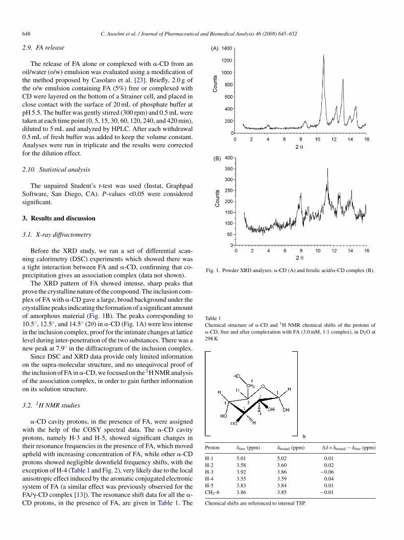
Fig. 1. Powder XRD analyses: �-CD (A) and ferulic acid/�-CD complex (B).
Table 1Chemical structure of �-CD and 1H NMR chemical shifts of the protons of�-CD, free and after complexation with FA (3.0 mM, 1:1 complex), in D2O at298 K
Proton δfree (ppm) δbound (ppm) �δ = δbound − δfree (ppm)
H-1 5.01 5.02 0.01H-2 3.58 3.60 0.02H-3 3.92 3.86 −0.06
48 C. Anselmi et al. / Journal of Pharmaceutic
.9. FA release
The release of FA alone or complexed with �-CD from anil/water (o/w) emulsion was evaluated using a modification ofhe method proposed by Casolaro et al. [23]. Briefly, 2.0 g ofhe o/w emulsion containing FA (5%) free or complexed withD were layered on the bottom of a Strainer cell, and placed inlose contact with the surface of 20 mL of phosphate buffer atH 5.5. The buffer was gently stirred (300 rpm) and 0.5 mL wereaken at each time point (0, 5, 15, 30, 60, 120, 240, and 420 min),iluted to 5 mL and analyzed by HPLC. After each withdrawal.5 mL of fresh buffer was added to keep the volume constant.nalyses were run in triplicate and the results were corrected
or the dilution effect.
.10. Statistical analysis
The unpaired Student’s t-test was used (Instat, Graphpadoftware, San Diego, CA). P-values <0.05 were consideredignificant.
. Results and discussion
.1. X-ray diffractometry
Before the XRD study, we ran a set of differential scan-ing calorimetry (DSC) experiments which showed there wastight interaction between FA and �-CD, confirming that co-
recipitation gives an association complex (data not shown).The XRD pattern of FA showed intense, sharp peaks that
rove the crystalline nature of the compound. The inclusion com-lex of FA with �-CD gave a large, broad background under therystalline peaks indicating the formation of a significant amountf amorphous material (Fig. 1B). The peaks corresponding to0.5◦, 12.5◦, and 14.5◦ (2θ) in �-CD (Fig. 1A) were less intensen the inclusion complex, proof for the intimate changes at latticeevel during inter-penetration of the two substances. There was aew peak at 7.9◦ in the diffractogram of the inclusion complex.
Since DSC and XRD data provide only limited informationn the supra-molecular structure, and no unequivocal proof ofhe inclusion of FA in �-CD, we focused on the 1H NMR analysisf the association complex, in order to gain further informationn its solution structure.
.2. 1H NMR studies
�-CD cavity protons, in the presence of FA, were assignedith the help of the COSY spectral data. The �-CD cavityrotons, namely H-3 and H-5, showed significant changes inheir resonance frequencies in the presence of FA, which movedpfield with increasing concentration of FA, while other �-CDrotons showed negligible downfield frequency shifts, with thexception of H-4 (Table 1 and Fig. 2), very likely due to the local
nisotropic effect induced by the aromatic conjugated electronicystem of FA (a similar effect was previously observed for theA/�-CD complex [13]). The resonance shift data for all the �-D protons, in the presence of FA, are given in Table 1. TheH-4 3.55 3.59 0.04H-5 3.83 3.84 0.01CH2-6 3.86 3.85 −0.01
Chemical shifts are referenced to internal TSP.

C. Anselmi et al. / Journal of Pharmaceutical and Biomedical Analysis 46 (2008) 645–652 649
Fc
ufittr
tdttlsaod
ptw
TC(
P
HHHHHC
C
Fc
rfah0twap
err
ig. 2. 1H NMR spectra of FA (lower), �-CD (middle) and ferulic acid/�-CDomplex (upper) in D2O.
pfield frequency shift of the H-3 and H-6 protons and down-eld shifts of H-5 in the �-CD cavity is an unequivocal indicator
hat FA fits deeply inside �-CD, resulting in the formation ofhe FA/�-CD inclusion complex, in accordance with previouseports [24].
All the FA protons showed significant frequency changes inhe presence of �-CD (Table 2). The signals of H-d and H-gisplayed upfield frequency changes, while the rest of the pro-on resonances moved downfield. Regarding the magnitude ofhe chemical shift changes, there was an important shift at theevel of H-f that was not present in the previous FA/�-CD inclu-ion complex studied [13]. These different interactions suggestdifferent type of inclusion of the molecule inside the cavity
r, as reported elsewhere, the inclusion of the guest might causeistortion of the host [25].
The movements of these proton signals suggested that the
enetration of FA involves insertion of the �,�-unsaturated por-ion and part of the aromatic skeleton of FA inside the cavity,ith the phenol and methoxyl groups projected outside the widerable 2hemical structure of FA and 1H NMR chemical shifts of the protons of FA, free
3.0 mM) and after complexation with �-CD (1:1 complex) in D2O at 298 K
roton δfree (ppm) δbound (ppm) �δ = δbound − δfree (ppm)
(c) 7.60 (d) 7.65 0.05(d) 7.28 (s) 7.20 −0.08(e) 7.18 (d) 7.28 0.01(f) 6.93 (d) 7.03 0.10(g) 6.40 (d) 6.33 −0.07
H3 (j) 3.88 (s) 3.94 0.06
hemical shifts are referenced to internal TSP.
edtds
T1
a
HHHHHHHH
�
ig. 3. ROESY spectrum of a solution containing the 1:1 FA/�-CD inclusionomplex (7.0 mM, D2O, pH 4.5).
im. This interpretation is supported by the strong H-c, H-d, H-and H-g cross-peaks with H-3, H-5 and H-6 protons of the
nnular interior of �-CD in the rotating frame nuclear Over-ouser effect spectroscopy (1H ROESY) spectrum (mixing time.5 ms) of the inclusion complex (Fig. 3), which indicates thathese protons are located inside the �-CD cavity. No cross-peaksere observed for the methoxyl protons and the �-CD protons
nd this is consistent with the location of the methoxyl group inroximity to either the primary or secondary rim of �-CD.
The other approach was to check FA binding to �-CD bystimating the selective and non-selective proton spin-latticeelaxation rates of the ligand [26]. The 1H spin-lattice relaxationates of FA and FA/�-CD are summarized in Table 3. FA protonsxperienced interacted differently with the macromolecule. The
ata suggest a strong hydrophobic interaction in the formation ofhe inclusion. F ratios suggest a slowing of the re-orientationalynamics of the ligand FA when going from the free to the boundtate, as in the inclusion with �-CD.able 3H NMR non-selective (Rnsel
1 ) and selective (Rsel1 ) spin-lattice relaxation rates
nd F ratios for selected protons of FA free and bound to �-CD in D2O at 298 K
Rnsel1 (s−1) Rsel
1 (s−1) F = Rnsel1 /Rsel
1
Fer Fer + �-CD Fer Fer + �-CD Fer Fer + �-CD
a – – – – – –
b – – – – – –
c 0.84 0.98 0.80 1.47 1.05 0.67
d 0.85 1.03 0.77 1.34 1.10 0.77
e 0.64 0.94 0.68 1.31 0.94 0.72
f 0.52 0.69 0.55 0.72 0.95 0.96
g 0.50 0.82 0.54 0.97 0.93 0.85
j 1.27 –a 1.21 –a 1.05 –
a In the bound state, the peak of CH3 in j position is overlapped, with some-CD resonances.
650 C. Anselmi et al. / Journal of Pharmaceutical an
3
awodF1
cf(
aoFc
�
wottp
Fp
kfwa
it[[
wCt[
3
pTpmpciE
tfa
Fivct
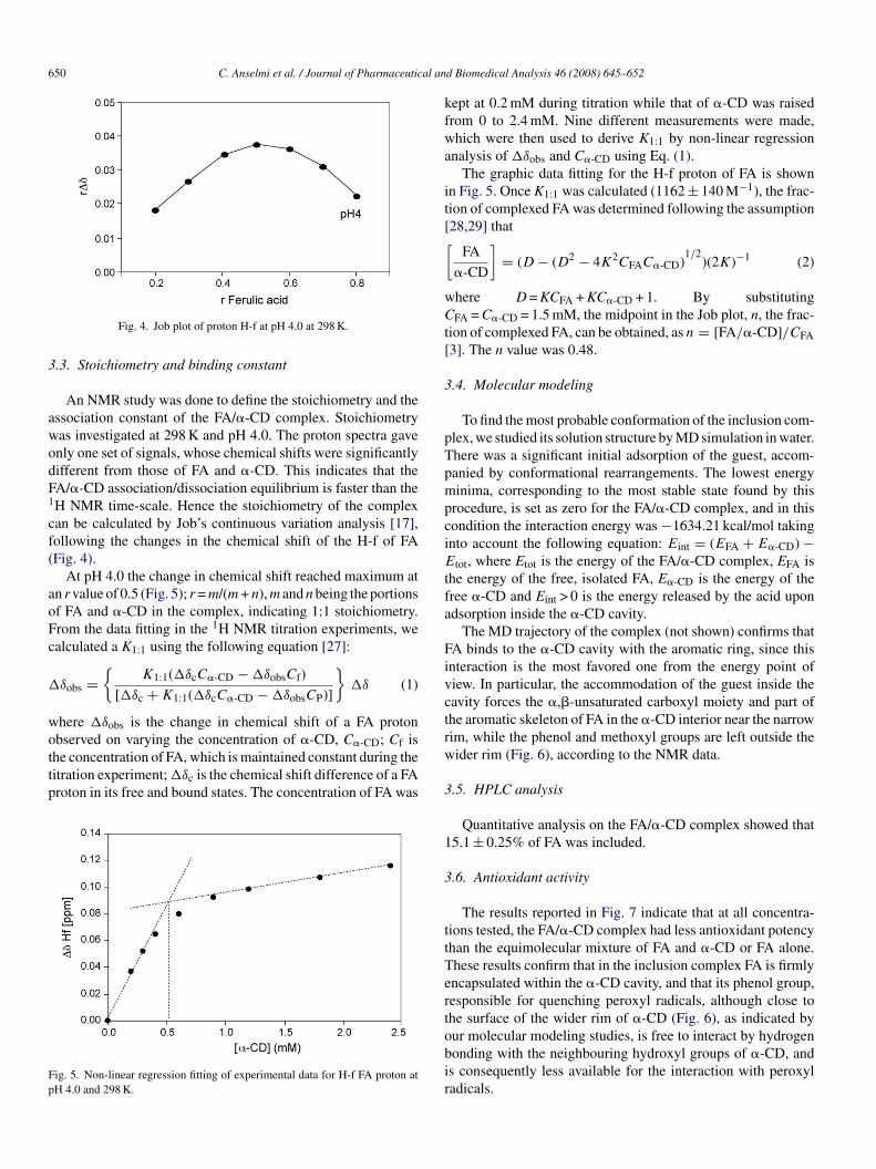
Fig. 4. Job plot of proton H-f at pH 4.0 at 298 K.
.3. Stoichiometry and binding constant
An NMR study was done to define the stoichiometry and thessociation constant of the FA/�-CD complex. Stoichiometryas investigated at 298 K and pH 4.0. The proton spectra gavenly one set of signals, whose chemical shifts were significantlyifferent from those of FA and �-CD. This indicates that theA/�-CD association/dissociation equilibrium is faster than theH NMR time-scale. Hence the stoichiometry of the complexan be calculated by Job’s continuous variation analysis [17],ollowing the changes in the chemical shift of the H-f of FAFig. 4).
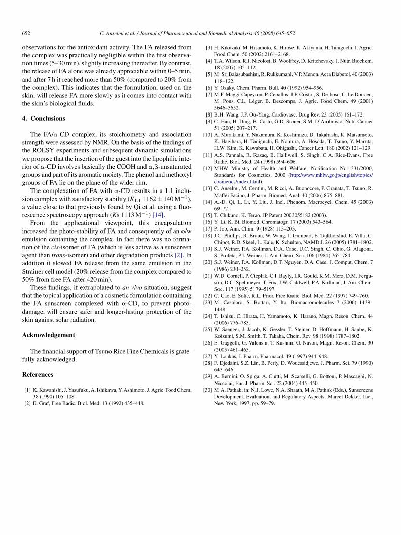
At pH 4.0 the change in chemical shift reached maximum atn r value of 0.5 (Fig. 5); r = m/(m + n), m and n being the portionsf FA and �-CD in the complex, indicating 1:1 stoichiometry.rom the data fitting in the 1H NMR titration experiments, wealculated a K1:1 using the following equation [27]:
δobs ={
K1:1(�δcC�-CD − �δobsCf)
[�δc + K1:1(�δcC�-CD − �δobsCP)]
}�δ (1)
here �δobs is the change in chemical shift of a FA proton
bserved on varying the concentration of �-CD, C�-CD; Cf ishe concentration of FA, which is maintained constant during theitration experiment; �δc is the chemical shift difference of a FAroton in its free and bound states. The concentration of FA wasig. 5. Non-linear regression fitting of experimental data for H-f FA proton atH 4.0 and 298 K.
rw
3
1
3
ttTertobir
d Biomedical Analysis 46 (2008) 645–652
ept at 0.2 mM during titration while that of �-CD was raisedrom 0 to 2.4 mM. Nine different measurements were made,hich were then used to derive K1:1 by non-linear regression
nalysis of �δobs and C�-CD using Eq. (1).The graphic data fitting for the H-f proton of FA is shown
n Fig. 5. Once K1:1 was calculated (1162 ± 140 M−1), the frac-ion of complexed FA was determined following the assumption28,29] that
FA
�-CD
]= (D − (D2 − 4K2CFAC�-CD)
1/2)(2K)−1 (2)
here D = KCFA + KC�-CD + 1. By substitutingFA = C�-CD = 1.5 mM, the midpoint in the Job plot, n, the frac-
ion of complexed FA, can be obtained, as n = [FA/�-CD]/CFA3]. The n value was 0.48.
.4. Molecular modeling
To find the most probable conformation of the inclusion com-lex, we studied its solution structure by MD simulation in water.here was a significant initial adsorption of the guest, accom-anied by conformational rearrangements. The lowest energyinima, corresponding to the most stable state found by this
rocedure, is set as zero for the FA/�-CD complex, and in thisondition the interaction energy was −1634.21 kcal/mol takingnto account the following equation: Eint = (EFA + E�-CD) −
tot, where Etot is the energy of the FA/�-CD complex, EFA ishe energy of the free, isolated FA, E�-CD is the energy of theree �-CD and Eint > 0 is the energy released by the acid upondsorption inside the �-CD cavity.
The MD trajectory of the complex (not shown) confirms thatA binds to the �-CD cavity with the aromatic ring, since thisnteraction is the most favored one from the energy point ofiew. In particular, the accommodation of the guest inside theavity forces the �,�-unsaturated carboxyl moiety and part ofhe aromatic skeleton of FA in the �-CD interior near the narrowim, while the phenol and methoxyl groups are left outside theider rim (Fig. 6), according to the NMR data.
.5. HPLC analysis
Quantitative analysis on the FA/�-CD complex showed that5.1 ± 0.25% of FA was included.
.6. Antioxidant activity
The results reported in Fig. 7 indicate that at all concentra-ions tested, the FA/�-CD complex had less antioxidant potencyhan the equimolecular mixture of FA and �-CD or FA alone.hese results confirm that in the inclusion complex FA is firmlyncapsulated within the �-CD cavity, and that its phenol group,esponsible for quenching peroxyl radicals, although close tohe surface of the wider rim of �-CD (Fig. 6), as indicated by
ur molecular modeling studies, is free to interact by hydrogenonding with the neighbouring hydroxyl groups of �-CD, ands consequently less available for the interaction with peroxyladicals.
C. Anselmi et al. / Journal of Pharmaceutical and Biomedical Analysis 46 (2008) 645–652 651
Fig. 6. Molecular modeling of ferulic acid/�-CD inclusion complex.
Fig. 7. Antioxidant protective effect of FA, FA/�-CD complex and of theirpm*
3
(i
Table 4FA content (free or complexed with �-CD) in an o/w emulsion before and after10 MED irradiation
Sample % FA beforeirradiation
% FA afterirradiation
Oil/water emulsion containing free FA 2.73 ± 0.25 1.90 ± 0.27Oil/water emulsion containing the
FA/�-CD complex2.73 ± 0.21 2.82 ± 0.11
Table 5Release of free and �-CD complexed FA from an o/w emulsion: Strainer cellmodel
Time (min) Release (%)
Free FA FA/�-CD
0 0 05 5.6 ± 0.5 1.2 ± 0.2
15 12.2 ± 1.4 2.6 ± 0.6230 16.9 ± 1.3 4.6 ± 1.260 22.3 ± 1.6 7.0 ± 0.5
120 31.7 ± 5.3 10.4 ± 0.424
scFtiUeies
3
The results reported in Table 5 indicate a time-dependent lin-
hysical mixture at different concentrations against free-radical stress. Data areean ± S.D. of four experiments, expressed as Trolox equivalent (TE, �M).
P < 0.05.
.7. Photo-stability
As indicated by the qualitative profile and the HPLC dataFig. 8 and Table 4), the exposure of an o/w emulsion contain-ng FA alone to 10 MED irradiation (1 MED = 25 mJ/cm2 for
eet
Fig. 8. HPLC profile after 10 MED UVB irradiation of an o/w emulsion
40 42.7 ± 4.4 16.0 ± 3.220 53.1 ± 6.2 20.6 ± 4.5
kin phototype II [30]), results in its partial conversion to theis-isomer. This degradative pathway does not take place whenA is included in �-CD, where there is only one sharp, dis-inct peak with the retention time of native FA (10.033). Thisndicates that inclusion makes FA insensitive to this level ofVB stress. By contrast, complexation with �-CD does not
nsure the same degree of FA protection (data not shown). Themprovement in stability of the sunscreen in complexed �-CD isxtremely important for long lasting skin protection and ensuresafer exposure of the consumer to UV rays.
.8. Delivery
ar release of FA, alone or complexed with �-CD, from the o/wmulsion but to a different extent on account of the tight inser-ion of FA inside the �-CD cavity, in accordance with previous
containing (a) 3% of free FA and (b) FA complexed with �-CD.
6 al an
otttatst
4
stwrgg
sar
ietaaS5
ttds
A
f
R
[
[
[
[
[
[[[[
[
[
[
[[
[
[
[
[[
52 C. Anselmi et al. / Journal of Pharmaceutic
bservations for the antioxidant activity. The FA released fromhe complex was practically negligible within the first observa-ion times (5–30 min), slightly increasing thereafter. By contrast,he release of FA alone was already appreciable within 0–5 min,nd after 7 h it reached more than 50% (compared to 20% fromhe complex). This indicates that the formulation, used on thekin, will release FA more slowly as it comes into contact withhe skin’s biological fluids.
. Conclusions
The FA/�-CD complex, its stoichiometry and associationtrength were assessed by NMR. On the basis of the findings ofhe ROESY experiments and subsequent dynamic simulationse propose that the insertion of the guest into the lipophilic inte-
ior of �-CD involves basically the COOH and �,�-unsaturatedroups and part of its aromatic moiety. The phenol and methoxylroups of FA lie on the plane of the wider rim.
The complexation of FA with �-CD results in a 1:1 inclu-ion complex with satisfactory stability (K1:1 1162 ± 140 M−1),value close to that previously found by Qi et al. using a fluo-
escence spectroscopy approach (Ks 1113 M−1) [14].From the applicational viewpoint, this encapsulation
ncreased the photo-stability of FA and consequently of an o/wmulsion containing the complex. In fact there was no forma-ion of the cis-isomer of FA (which is less active as a sunscreengent than trans-isomer) and other degradation products [2]. Inddition it slowed FA release from the same emulsion in thetrainer cell model (20% release from the complex compared to0% from free FA after 420 min).
These findings, if extrapolated to an vivo situation, suggesthat the topical application of a cosmetic formulation containinghe FA sunscreen complexed with �-CD, to prevent photo-amage, will ensure safer and longer-lasting protection of thekin against solar radiation.
cknowledgement
The financial support of Tsuno Rice Fine Chemicals is grate-ully acknowledged.
eferences
[1] K. Kawanishi, J. Yasufuku, A. Ishikawa, Y. Ashimoto, J. Agric. Food Chem.38 (1990) 105–108.
[2] E. Graf, Free Radic. Biol. Med. 13 (1992) 435–448.
[
[
d Biomedical Analysis 46 (2008) 645–652
[3] H. Kikuzaki, M. Hisamoto, K. Hirose, K. Akiyama, H. Taniguchi, J. Agric.Food Chem. 50 (2002) 2161–2168.
[4] T.A. Wilson, R.J. Nicolosi, B. Woolfrey, D. Kritchevsky, J. Nutr. Biochem.18 (2007) 105–112.
[5] M. Sri Balasubashini, R. Rukkumani, V.P. Menon, Acta Diabetol. 40 (2003)118–122.
[6] Y. Ozaky, Chem. Pharm. Bull. 40 (1992) 954–956.[7] M.F. Maggi-Capeyron, P. Ceballos, J.P. Cristol, S. Delbosc, C. Le Doucen,
M. Pons, C.L. Leger, B. Descomps, J. Agric. Food Chem. 49 (2001)5646–5652.
[8] B.H. Wang, J.P. Ou-Yang, Cardiovasc. Drug Rev. 23 (2005) 161–172.[9] C. Han, H. Ding, B. Casto, G.D. Stoner, S.M. D’Ambrosio, Nutr. Cancer
51 (2005) 207–217.10] A. Murakami, Y. Nakamura, K. Koshimizu, D. Takahashi, K. Matsumoto,
K. Hagihara, H. Taniguchi, E. Nomura, A. Hosoda, T. Tsuno, Y. Maruta,H.W. Kim, K. Kawabata, H. Ohigashi, Cancer Lett. 180 (2002) 121–129.
11] A.S. Pannala, R. Razaq, B. Halliwell, S. Singh, C.A. Rice-Evans, FreeRadic. Biol. Med. 24 (1998) 594–606.
12] MHW Ministry of Health and Welfare, Notification No. 331/2000,Standards for Cosmetics, 2000 (http://www.mhlw.go.jp/english/topics/cosmetics/index.html).
13] C. Anselmi, M. Centini, M. Ricci, A. Buonocore, P. Granata, T. Tsuno, R.Maffei Facino, J. Pharm. Biomed. Anal. 40 (2006) 875–881.
14] A.-D. Qi, L. Li, Y. Liu, J. Incl. Phenom. Macrocycl. Chem. 45 (2003)69–72.
15] T. Chikuno, K. Terao. JP Patent 2003055182 (2003).16] Y. Li, K. Bi, Biomed. Chromatogr. 17 (2003) 543–564.17] P. Job, Ann. Chim. 9 (1928) 113–203.18] J.C. Phillips, R. Braun, W. Wang, J. Gumbart, E. Tajkhorshid, E. Villa, C.
Chipot, R.D. Skeel, L. Kale, K. Schulten, NAMD J. 26 (2005) 1781–1802.19] S.J. Weiner, P.A. Kollman, D.A. Case, U.C. Singh, C. Ghio, G. Alagona,
S. Profeta, P.J. Weiner, J. Am. Chem. Soc. 106 (1984) 765–784.20] S.J. Weiner, P.A. Kollman, D.T. Nguyen, D.A. Case, J. Comput. Chem. 7
(1986) 230–252.21] W.D. Cornell, P. Cieplak, C.I. Bayly, I.R. Gould, K.M. Merz, D.M. Fergu-
son, D.C. Spellmeyer, T. Fox, J.W. Caldwell, P.A. Kollman, J. Am. Chem.Soc. 117 (1995) 5179–5197.
22] C. Cao, E. Sofic, R.L. Prior, Free Radic. Biol. Med. 22 (1997) 749–760.23] M. Casolaro, S. Bottari, Y. Ito, Biomacromolecules 7 (2006) 1439–
1448.24] T. Ishizu, C. Hirata, H. Yamamoto, K. Harano, Magn. Reson. Chem. 44
(2006) 776–783.25] W. Saenger, J. Jacob, K. Gessler, T. Steiner, D. Hoffmann, H. Sanbe, K.
Koizumi, S.M. Smith, T. Takaha, Chem. Rev. 98 (1998) 1787–1802.26] E. Gaggelli, G. Valensin, T. Kushnir, G. Navon, Magn. Reson. Chem. 30
(2005) 461–465.27] Y. Loukas, J. Pharm. Pharmacol. 49 (1997) 944–948.28] F. Djedaini, S.Z. Lin, B. Perly, D. Wouessidjewe, J. Pharm. Sci. 79 (1990)
643–646.
29] A. Bernini, O. Spiga, A. Ciutti, M. Scarselli, G. Bottoni, P. Mascagni, N.Niccolai, Eur. J. Pharm. Sci. 22 (2004) 445–450.30] M.A. Pathak, in: N.J. Lowe, N.A. Shaath, M.A. Pathak (Eds.), Sunscreens
Development, Evaluation, and Regulatory Aspects, Marcel Dekker, Inc.,New York, 1997, pp. 59–79.
Copyright © 2022 FDOKUMEN


![Chiral separation by a monofunctionalized cyclodextrin derivative: From selector to permethyl-[beta]-cyclodextrin bonded stationary phase](https://static.fdokumen.com/doc/165x107/63327b24576b626f850d70ad/chiral-separation-by-a-monofunctionalized-cyclodextrin-derivative-from-selector.jpg)