Preventing Disulphide Bond Formation Weakens Non-covalent Forces Among Lysozyme Aggregates
11
Preventing Disulfide Bond Formation Weakens Non-Covalent Forces among Lysozyme Aggregates Vijay Kumar Ravi 1 , Mohit Goel 1 , Hema Chandra Kotamarthi 2 , Sri Rama Koti Ainavarapu 2 , Rajaram Swaminathan 1 * 1 Department of Biotechnology, Indian Institute of Technology Guwahati, Guwahati, Assam, India, 2 Department of Chemical Sciences, Tata Institute of Fundamental Research, Homi Bhabha Road, Mumbai, Maharashtra, India Abstract Nonnative disulfide bonds have been observed among protein aggregates in several diseases like amyotrophic lateral sclerosis, cataract and so on. The molecular mechanism by which formation of such bonds promotes protein aggregation is poorly understood. Here in this work we employ previously well characterized aggregation of hen eggwhite lysozyme (HEWL) at alkaline pH to dissect the molecular role of nonnative disulfide bonds on growth of HEWL aggregates. We employed time-resolved fluorescence anisotropy, atomic force microscopy and single-molecule force spectroscopy to quantify the size, morphology and non-covalent interaction forces among the aggregates, respectively. These measurements were performed under conditions when disulfide bond formation was allowed (control) and alternatively when it was prevented by alkylation of free thiols using iodoacetamide. Blocking disulfide bond formation affected growth but not growth kinetics of aggregates which were ,50% reduced in volume, flatter in vertical dimension and non-fibrillar in comparison to control. Interestingly, single-molecule force spectroscopy data revealed that preventing disulfide bond formation weakened the non-covalent interaction forces among monomers in the aggregate by at least ten fold, thereby stalling their growth and yielding smaller aggregates in comparison to control. We conclude that while constrained protein chain dynamics in correctly disulfide bonded amyloidogenic proteins may protect them from venturing into partial folded conformations that can trigger entry into aggregation pathways, aberrant disulfide bonds in non-amyloidogenic proteins (like HEWL) on the other hand, may strengthen non-covalent intermolecular forces among monomers and promote their aggregation. Citation: Ravi VK, Goel M, Kotamarthi HC, Ainavarapu SRK, Swaminathan R (2014) Preventing Disulfide Bond Formation Weakens Non-Covalent Forces among Lysozyme Aggregates. PLoS ONE 9(2): e87012. doi:10.1371/journal.pone.0087012 Editor: Etienne Dague, LAAS-CNRS, France Received September 14, 2013; Accepted December 16, 2013; Published February 14, 2014 Copyright: ß 2014 Ravi et al. This is an open-access article distributed under the terms of the Creative Commons Attribution License, which permits unrestricted use, distribution, and reproduction in any medium, provided the original author and source are credited. Funding: This work was financially supported by Science and Engineering Research Board, Department of Science and Technology, Government of India under project SR/SO/BB-048/2009. VKR wishes to acknowledge financial support under Rajiv Gandhi National Fellowship scheme [Ref: F. 14-2 (SC)/2009 (SA-III)] of University Grants Commission, Government of India, New Delhi. The funders had no role in study design, data collection and analysis, decision to publish, or preparation of the manuscript. Competing Interests: The authors have declared that no competing interests exist. * E-mail: [email protected] Introduction Accumulation of ordered protein aggregates like amyloid is a symptom of several diseases like Alzheimer’s, Parkinson’s and human lysozyme amyloidosis [1]. The causative molecular mechanisms responsible for such diseases are topics of intense research currently. The role of aberrant disulfide bonds in promoting protein aggregation has assumed significance lately owing to observation of intermolecular disulfide bonds among aggregates of mutant Cu Zn superoxide dismutase (SOD1) in the spinal cord of transgenic mice [2] and in paired helical filaments of tau protein existing as neurofibrillary tangles in human brain [3]. The former is implicated in amyotrophic lateral sclerosis while the latter is the hallmark of Alzheimer’s disease. Nonnative disulfide bonds were also among the prominent covalent modifications of crystallins isolated from cataractous lenses [4]. Formation of disulfide bonds has been shown to influence aggregation of proteins in multiple ways. For example, intramo- lecular disulfide bonds in tau protein can result in conformations that lock up the protein chain such that further aggregation is slowed down. In contrast, intermolecular disulfide bonds in the same protein have been shown to accelerate aggregation [3]. Several instances where formation of disulfide bonds can alter the aggregation pathway [5], reduce toxicity of amyloid fibrils [6] and change material properties of the amyloid fibrils [5] have been reported. In this work, we investigate the role of disulfide bonds in controlling the aggregation of hen eggwhite lysozyme (HEWL) which has eight cysteine residues per polypeptide chain. Aggre- gation of HEWL has been well studied in the past [7] serving as model protein. It has been shown previously that zero cysteine mutant of HEWL can form amyloid fibrils at acidic and neutral pH [8]. However, in contrast, presence of DTT has been shown to abolish aggregation in HEWL in alkaline pH [9] by preventing formation of incorrect disulfide bonds. The mechanism by which such disulfide bonds influence aggregation of HEWL at alkaline pH is not clear and needs to be established. Here in this work, we show quantitatively that blocking disulfide bond formationin HEWL a) weakens non-covalent interactions among HEWL monomers in amorphous aggregates considerably as measured by single-molecule force spectroscopy; b) diminishes the size of aggregates as observed by nanosecond fluorescence anisotropy PLOS ONE | www.plosone.org 1 February 2014 | Volume 9 | Issue 2 | e87012
Transcript of Preventing Disulphide Bond Formation Weakens Non-covalent Forces Among Lysozyme Aggregates

Preventing Disulfide Bond Formation WeakensNon-Covalent Forces among Lysozyme AggregatesVijay Kumar Ravi1, Mohit Goel1, Hema Chandra Kotamarthi2, Sri Rama Koti Ainavarapu2,
Rajaram Swaminathan1*
1 Department of Biotechnology, Indian Institute of Technology Guwahati, Guwahati, Assam, India, 2 Department of Chemical Sciences, Tata Institute of Fundamental
Research, Homi Bhabha Road, Mumbai, Maharashtra, India
Abstract
Nonnative disulfide bonds have been observed among protein aggregates in several diseases like amyotrophic lateralsclerosis, cataract and so on. The molecular mechanism by which formation of such bonds promotes protein aggregation ispoorly understood. Here in this work we employ previously well characterized aggregation of hen eggwhite lysozyme(HEWL) at alkaline pH to dissect the molecular role of nonnative disulfide bonds on growth of HEWL aggregates. Weemployed time-resolved fluorescence anisotropy, atomic force microscopy and single-molecule force spectroscopy toquantify the size, morphology and non-covalent interaction forces among the aggregates, respectively. Thesemeasurements were performed under conditions when disulfide bond formation was allowed (control) and alternativelywhen it was prevented by alkylation of free thiols using iodoacetamide. Blocking disulfide bond formation affected growthbut not growth kinetics of aggregates which were ,50% reduced in volume, flatter in vertical dimension and non-fibrillar incomparison to control. Interestingly, single-molecule force spectroscopy data revealed that preventing disulfide bondformation weakened the non-covalent interaction forces among monomers in the aggregate by at least ten fold, therebystalling their growth and yielding smaller aggregates in comparison to control. We conclude that while constrained proteinchain dynamics in correctly disulfide bonded amyloidogenic proteins may protect them from venturing into partial foldedconformations that can trigger entry into aggregation pathways, aberrant disulfide bonds in non-amyloidogenic proteins(like HEWL) on the other hand, may strengthen non-covalent intermolecular forces among monomers and promote theiraggregation.
Citation: Ravi VK, Goel M, Kotamarthi HC, Ainavarapu SRK, Swaminathan R (2014) Preventing Disulfide Bond Formation Weakens Non-Covalent Forces amongLysozyme Aggregates. PLoS ONE 9(2): e87012. doi:10.1371/journal.pone.0087012
Editor: Etienne Dague, LAAS-CNRS, France
Received September 14, 2013; Accepted December 16, 2013; Published February 14, 2014
Copyright: � 2014 Ravi et al. This is an open-access article distributed under the terms of the Creative Commons Attribution License, which permits unrestricteduse, distribution, and reproduction in any medium, provided the original author and source are credited.
Funding: This work was financially supported by Science and Engineering Research Board, Department of Science and Technology, Government of India underproject SR/SO/BB-048/2009. VKR wishes to acknowledge financial support under Rajiv Gandhi National Fellowship scheme [Ref: F. 14-2 (SC)/2009 (SA-III)] ofUniversity Grants Commission, Government of India, New Delhi. The funders had no role in study design, data collection and analysis, decision to publish, orpreparation of the manuscript.
Competing Interests: The authors have declared that no competing interests exist.
* E-mail: [email protected]
Introduction
Accumulation of ordered protein aggregates like amyloid is a
symptom of several diseases like Alzheimer’s, Parkinson’s and
human lysozyme amyloidosis [1]. The causative molecular
mechanisms responsible for such diseases are topics of intense
research currently. The role of aberrant disulfide bonds in
promoting protein aggregation has assumed significance lately
owing to observation of intermolecular disulfide bonds among
aggregates of mutant Cu Zn superoxide dismutase (SOD1) in the
spinal cord of transgenic mice [2] and in paired helical filaments of
tau protein existing as neurofibrillary tangles in human brain [3].
The former is implicated in amyotrophic lateral sclerosis while the
latter is the hallmark of Alzheimer’s disease. Nonnative disulfide
bonds were also among the prominent covalent modifications of
crystallins isolated from cataractous lenses [4].
Formation of disulfide bonds has been shown to influence
aggregation of proteins in multiple ways. For example, intramo-
lecular disulfide bonds in tau protein can result in conformations
that lock up the protein chain such that further aggregation is
slowed down. In contrast, intermolecular disulfide bonds in the
same protein have been shown to accelerate aggregation [3].
Several instances where formation of disulfide bonds can alter the
aggregation pathway [5], reduce toxicity of amyloid fibrils [6] and
change material properties of the amyloid fibrils [5] have been
reported.
In this work, we investigate the role of disulfide bonds in
controlling the aggregation of hen eggwhite lysozyme (HEWL)
which has eight cysteine residues per polypeptide chain. Aggre-
gation of HEWL has been well studied in the past [7] serving as
model protein. It has been shown previously that zero cysteine
mutant of HEWL can form amyloid fibrils at acidic and neutral
pH [8]. However, in contrast, presence of DTT has been shown to
abolish aggregation in HEWL in alkaline pH [9] by preventing
formation of incorrect disulfide bonds. The mechanism by which
such disulfide bonds influence aggregation of HEWL at alkaline
pH is not clear and needs to be established. Here in this work, we
show quantitatively that blocking disulfide bond formationin
HEWL a) weakens non-covalent interactions among HEWL
monomers in amorphous aggregates considerably as measured by
single-molecule force spectroscopy; b) diminishes the size of
aggregates as observed by nanosecond fluorescence anisotropy
PLOS ONE | www.plosone.org 1 February 2014 | Volume 9 | Issue 2 | e87012

decay and atomic force microscopy (AFM); and c) inhibits amyloid
fibril formation as revealed by Thioflavin T (ThT) fluorescence, in
stark contrast to unblocked control samples.
Materials and Methods
MaterialsSodium dihydrogen phosphate, sodium bicarbonate, sodium
thiosulphate, L-cysteine, along with solvents like DMSO, Di-
methylformamide (DMF) were purchased from Merck Limited,
Mumbai. 8-Anilino-1-naphthalene sulfonic acid ammonium salt
(ANS), 2,29-dithiobis (5-nitropyridine), (DTNP), Hen eggwhite
lysozyme (HEWL), iodoacetamide, thioflavin T (ThT) were
obtained from Sigma-Aldrich Chemicals Pvt. Ltd., India. 2-
dimethyl amino naphthalene 6-sulphonyl chloride (dansyl
chloride) was purchased from Invitrogen, USA. AFM Cantilevers
PPP-NCL-50 (Point probe Plus/Non- contact/Long cantilever,
part 65-262P) was purchased from Molecular Imaging, USA.
Muscovite mica, V-1 quality was purchased from Electron
Microscopy Sciences, USA.
Protein LabelingHEWL was covalently labeled with dansyl chloride following
the protocol provided from Molecular Probes with minor
modification as reported previously [10]. The HEWL and dansyl
concentrations were measured using absorbance at 280 nm and
339 nm, respectively. The protein to dye labeling ratio in the
conjugates varied between 1 and 2.
Sample Preparation and IncubationStock solution of HEWL was freshly prepared in deionised
water (MilliQ, Millipore, India). For inducing aggregation, this
stock was diluted in 50 mM phosphate buffer at pH 12.2 to a final
concentration of 70 mM. The samples were incubated at room
temperature (22–28uC) and all measurements were performed at
25uC.
Cysteine Carboxymethylation Reaction by IodoacetamideIodoacetamide was dissolved in DMF to make a stock solution
of 1.5 M. 8 mL of this stock solution of iodoacetamide was directly
added in four installments of 2 mL each to 1 mL of HEWL sample
periodically after completion of 2, 6, 12 and 24 hours of
incubation in pH 12.2. No further addition of iodoacetamide
was made to HEWL sample thereafter. The above procedure was
chosen because previous results had shown that a significant
Figure 1. Changes observed in ANS emission spectra in presence of thiol-blocked HEWL and control HEWL samples incubated inpH 12.2 at 301 K for 48 and 144 hours is shown.doi:10.1371/journal.pone.0087012.g001
Figure 2. Thioflavin T fluorescence observed in thiol-blockedHEWL and control HEWL samples incubated in pH 12.2 at301 K for different durations as indicated in X axis is displayed.The error bars represent standard deviations of averaged data fromthree separate experiments.doi:10.1371/journal.pone.0087012.g002
Role of Disulphide Bonds in Lysozyme Aggregation
PLOS ONE | www.plosone.org 2 February 2014 | Volume 9 | Issue 2 | e87012
fraction of HEWL disulfide bonds break in pH 12.2 to yield free –
SH groups during 2–24 hours after incubation in alkaline pH [9].
Also this procedure is simpler to perform requiring no prior
unfolding by guanidine hydrochloride and reduction by DTT. For
the progress of reaction, sample was kept at room temperature in
dark condition.
The free thiols in HEWL with or without added iodoacetamide
in the medium were estimated using DTNP assay described
previously [9]. Briefly, DTNP (50 mM) reacts with free cysteine (0–
100 mM) at pH 7 to yield a product that absorbs at 387 nm. The
unknown concentration of free thiols in HEWL can be determined
from the standard plot generated with L-cysteine.
Our experiments with iodoacetamide following the procedure
above yielded on average modification of 60–70% of thiols among
8 cysteine residues of every HEWL polypeptide chain at pH 12.2.
The remaining thiols were not alkylated probably because they
were buried inside the core of HEWL and thus inaccessible to
iodoacetamide.
Steady-state Fluorescence MeasurementsAll steady-state fluorescence measurements were carried out
using Fluoromax-3 spectrofluorometer (Jobin-Yvon Horiba Inc.,
USA). To minimize the photobleaching, excitation light shutter
was kept closed when not recording fluorescence intensity.
ANS binding. The stock concentration of ANS (1 mg/mL in
deionised water) was measured using extinction coefficient of
4,950 M21 cm21 at 350 nm. Binding of ANS (10 mM) to HEWL
(diluted to 5 mM) in pH 12.2 buffer inside a cuvette was monitored
from fluorescence of ANS. Steady-state fluorescence emission
spectrumof ANS was recorded from 400 to 600 nm after
excitation at 380 nm (excitation slitwidth 1 nm and emission
slitwidth 10 nm). The background intensity from Raman scatter
and buffer were negligible (,5%) compared to sample fluores-
cence intensity under identical conditions. These were however
subtracted from sample emission spectra.
ThT binding. Stock solution of ThT was made by dissolving
3 mg in 3 mL of deionised water (MilliQ). This solution was
subsequently filtered through 0.45 mm filter. The filtered solution
was diluted in ethanol before taking absorbance at 416 nm. The
ThT concentration was calculated using extinction coefficient
26,620 M21 cm21. Aliquots of ThT stock solution were stored in
220uC. For fibril binding assays 1 mM ThT was prepared in
deionised water. This solution was diluted to 0.5 mM in 20 mM
Gly-Gly buffer, pH 8.5. HEWL protein aggregates were diluted to
7 mM and ,11 mM ThT was added into this protein sample,
while sample volume was made up to 1 mL using 20 mM Gly-Gly
pH 8.5 buffer. It was seen that fibrils bind strongly with ThT at
molar ratio of ,1:2 protein to ThT [11]. The spectrum of
vortexed sample was taken immediately by exciting at 450 nm
(1 nm slit width) and recording emission from 470 to 550 nm with
5 nm slit width. The background fluorescence intensity including
Raman scatter was subtracted from sample spectra. The
integrated fluorescence intensity of ThT alone measured in
absence of HEWL at 48 hours was normalized to unity. The
measured integrated fluorescence of all samples were scaled up
against this normalized value and plotted to reveal the observed
fold increase in ThT fluorescence against incubation time of
HEWL at pH 12.2 after averaging independent spectral scans.
Steady-state Fluorescence Anisotropy MeasurementsThe steady-state fluorescence anisotropy (rss) was measured after
G-factor correction and dark counts subtraction as described
previously [10]. For this measurement HEWL samples (,70 mM)
contained 2 mM of dansyl-conjugated HEWL while remaining
protein was unlabelled. The rss of dansyl-conjugated HEWL was
monitored after different time intervals of incubation.
Dansyl-conjugated HEWL was excited at 375 nm (slit
width = 1 nm) and emission at 440 nm was collected with a slit
width of 5 nm. Each measurement was done in duplicate, while
data reported are averages of three such measurements. The
increase in rss of dansyl-conjugated HEWL with incubation time in
pH 12.2 was fitted to the von Bertalanffy equation below:
rss(t)~r?ss { r?ss {r0ss
� �e{kt ð1Þ
Here, refers r?ss to rss at t = infinity, while r0ss refers same at
t = 0. k denotes the rate constant for increase in anisotropy.
Time-resolved Fluorescence MeasurementsTime-resolved fluorescence intensity and anisotropy (G-factor
corrected) measurements were performed in LIFE SPEC II
spectrometer (Edinburgh Instruments, Livingston, UK) operating
in TCSPC mode, collecting emission decay in 4096 channels using
a microchannel plate PMT. Dansyl-conjugated HEWL was
excited at 375 nm using EPL-375, picosecond pulsed diode laser
with instrument response function (IRF) fwhm ,150 ps. Dansyl
probe fluorescence was collected at 440 nm with a temporal
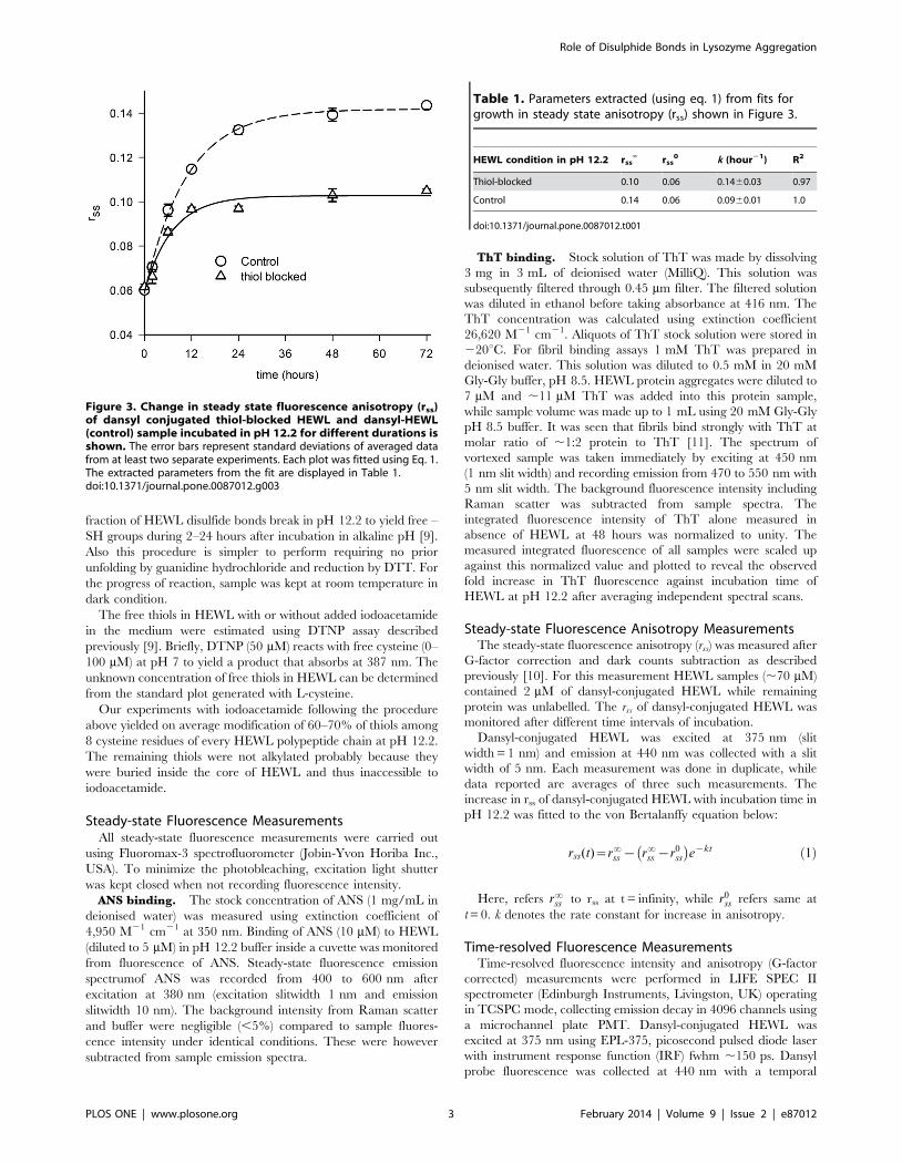
Figure 3. Change in steady state fluorescence anisotropy (rss)of dansyl conjugated thiol-blocked HEWL and dansyl-HEWL(control) sample incubated in pH 12.2 for different durations isshown. The error bars represent standard deviations of averaged datafrom at least two separate experiments. Each plot was fitted using Eq. 1.The extracted parameters from the fit are displayed in Table 1.doi:10.1371/journal.pone.0087012.g003
Table 1. Parameters extracted (using eq. 1) from fits forgrowth in steady state anisotropy (rss) shown in Figure 3.
HEWL condition in pH 12.2 rss‘ rss
0 k (hour21) R2
Thiol-blocked 0.10 0.06 0.1460.03 0.97
Control 0.14 0.06 0.0960.01 1.0
doi:10.1371/journal.pone.0087012.t001
Role of Disulphide Bonds in Lysozyme Aggregation
PLOS ONE | www.plosone.org 3 February 2014 | Volume 9 | Issue 2 | e87012
resolution of 24.414 ps/channel. For this measurement HEWL
samples (,70 mM) contained 2 mM of dansyl-conjugated HEWL
while remaining protein was unlabeled. Fluorescence lifetime data
reported are average of three measurements. Intensity decays were
analysed by iterative reconvolution using the Marquardt-Leven-
berg algorithm to extract lifetimes (ti) and amplitudes (ai) as given
in equation below.
I(t)~X
i
aie{t=ti where i~13 and
X
i
ai~1:0 ð2Þ
Mean fluorescence lifetime, tm~X
i
aiti ð3Þ
The raw (G-factor corrected) anisotropy decays were tail-fitted
using a sum of two exponentials (equation 4), yielding two
rotational correlation times.
r(t)~Azb1e{t=w1zb2e
{t=w2 ð4Þ
Here, A is a constant dependent on G-factor, bi denotes the
amplitude for wi, w1 and w2 refer to the fast and slow rotational
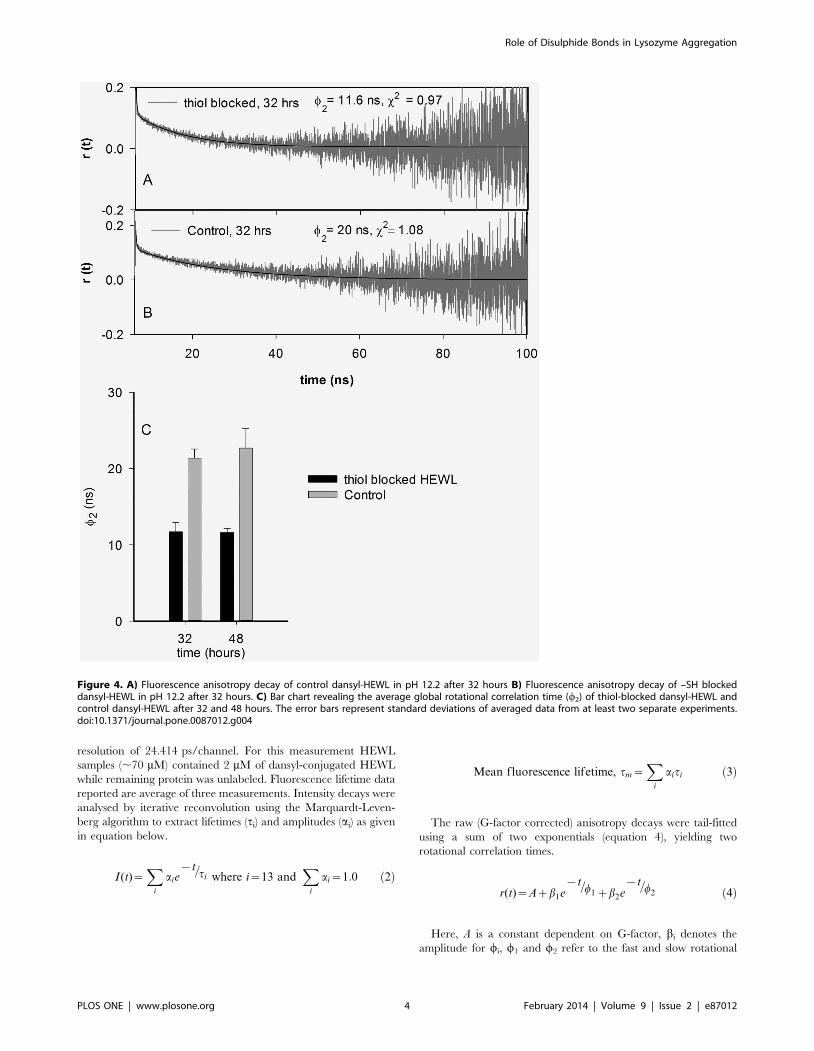
Figure 4. A) Fluorescence anisotropy decay of control dansyl-HEWL in pH 12.2 after 32 hours B) Fluorescence anisotropy decay of –SH blockeddansyl-HEWL in pH 12.2 after 32 hours. C) Bar chart revealing the average global rotational correlation time (w2) of thiol-blocked dansyl-HEWL andcontrol dansyl-HEWL after 32 and 48 hours. The error bars represent standard deviations of averaged data from at least two separate experiments.doi:10.1371/journal.pone.0087012.g004
Role of Disulphide Bonds in Lysozyme Aggregation
PLOS ONE | www.plosone.org 4 February 2014 | Volume 9 | Issue 2 | e87012
correlation times, respectively. The slower rotational correlation
time (w2) corresponds to global rotational motion of the whole
HEWL aggregate. As the 0.15 ns IRF pulse-width is negligibly
small in comparison to the time scale of protein rotational motion
(.4 ns), the extracted values of w2 by this tail-fit approach are not
affected by consequences of IRF convolution.
Atomic Force Microscopy ImagingThe HEWL aggregated samples (10 mL) were dropped on
freshly cleaved mica in the presence of 10 mM Mg2+ ions and left
to adsorb for 2 minutes. The samples were rinsed with 0.2 mm
filtered deionized water to remove unadsorbed sample and were
dried under nitrogen stream. Samples were imaged in air under
AAC MODE (non contact) in PICO PLUSTM AFM purchased
from Molecular Imaging, USA. Cantilever type PPP-NCL-50
(resonance frequency, 150 kHz Molecular Imaging) was used for
AAC mode. Images were acquired digitally at a scan speed of
1 line/second with 256 data points per line. The AFM images
were captured at least three times for every sample condition. The
shape and size reported are without tip geometry correction.
Pulling Molecules Using Atomic Force MicroscopyThe force-extension curves were measured using a custom built
AFM for pulling single molecules, whose details are given
elsewhere [12]. Gold coated cantilevers with silicon nitride tip
with spring constants around 40 pN/nm were used for the
experiments. In a typical experiment, a small volume (40 mL) of
sample solution (120 mM HEWL solution in 50 mM phosphate
buffer at pH 12.2) was added to a gold coated coverslip and
pulling experiments were performed on it. The pulling speed was
maintained at 400 nm/s.
Results
Our objective in this work was to assess the progress of HEWL
aggregation at pH 12.2 under two different conditions. In
condition 1, the free –SH groups generated upon exposure of
HEWL to pH 12.2 were derivatised by addition of iodoacetamide,
to yield S-carboxyamidomethyl cysteine derivative of HEWL
which we shall refer to as thiol-blocked HEWL. In condition 2, no
iodoacetamide was added to HEWL samples at pH 12.2 which we
refer to as control. Aside from addition of iodoacetamide, HEWL
samples in conditions 1 and 2 were identical in all other respects.
Exposed Hydrophobic Regions Probed by ANSPrevious work has shown that HEWL on exposure to pH 12.2
becomes partially unfolded initiating the process of aggregation
[9,13]. Exposed hydrophobic regions among the aggregates were
monitored using ANS, which serves as an indicator for the growth
of aggregates and its suppression [14]. Here we measured extent of
exposure among hydrophobic pockets in S-carboxyamidomethyl
cysteine derivative of HEWL along with unmodified HEWL after
48 and 144 hours of incubation in pH 12.2 (Figure 1). At 48
hours of incubation time both S-carboxyamidomethyl cysteine
derivative of HEWL and unmodified HEWL displayed enhanced
ANS fluorescence intensity and blue shifted lmax of 498 and
471 nm, respectively compared to ANS alone. At 144 hours, the –
SH blocked and unblocked HEWL bound ANS fluorescence
intensity decreased substantially while emission lmax of ANS was
nearly same as recorded for 48 hours of incubation time. ANS
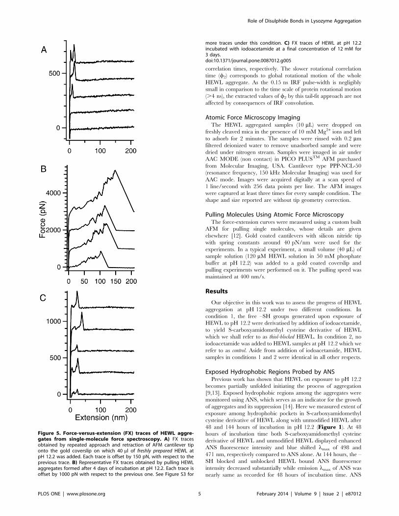
Figure 5. Force-versus-extension (FX) traces of HEWL aggre-gates from single-molecule force spectroscopy. A) FX tracesobtained by repeated approach and retraction of AFM cantilever tiponto the gold coverslip on which 40 ml of freshly prepared HEWL atpH 12.2 was added. Each trace is offset by 150 pN, with respect to theprevious trace. B) Representative FX traces obtained by pulling HEWLaggregates formed after 4 days of incubation at pH 12.2. Each trace isoffset by 1000 pN with respect to the previous one. See Figure S3 for
more traces under this condition. C) FX traces of HEWL at pH 12.2incubated with iodoacetamide at a final concentration of 12 mM for3 days.doi:10.1371/journal.pone.0087012.g005
Role of Disulphide Bonds in Lysozyme Aggregation
PLOS ONE | www.plosone.org 5 February 2014 | Volume 9 | Issue 2 | e87012
samples (10 mM) in absence of protein displays subdued fluores-
cence intensity and emission lmax,526 nm in pH 12.2 buffer.
The larger area below spectra observed with thiol-blocked HEWL
samples in comparison to unblocked samples demonstrate that
thiol-blocked HEWL harbors more bound ANS population. The
ANS peak shift from 526 nm to 498 nm indicates that ANS
molecules are buried inside moderately exposed hydrophobic
interior of –SH blocked HEWL [15]. Thus control samples possess
deeply buried, more non-polar environment for bound ANS
compared to thiol-blocked samples. Evidently blocking thiol
groups in HEWL causes the aggregates to be more open and
solvent exposed compared to unblocked HEWL samples owing
perhaps to absence of cross-linking disulfide bonds.
Presence of Amyloid Fibrils from Thioflavin TFormation of amyloid fibril represents the end point of protein
aggregation. It is worthwhile to know if the S-carboxyamido-
methyl cysteine derivative of HEWL forms only intermediates like
oligomers during incubation at pH 12.2 or does it also form
amyloid fibrils? To answer this question, ThT assay of HEWL S-
carboxyamidomethyl cysteine derivative was performed after 48
hours. Previous studies have shown that detectable increase in
ThT fluorescence in HEWL commences only after 48 hours [7,9].
At 48 hours, ThT fluorescence intensity from cysteine methylated
HEWL was more than 50% diminished compared to control
HEWL pH 12.2 samples (Figure 2). After a gentle rise till 96
hours, this trend is seen maintained steady till 240 hours. This
suggests a significant reduction of amyloid fibril population in
thiol-blocked samples compared to control. Residual ThT
fluorescence in thiol-blocked samples may arise from ThT binding
to oligomers or amorphous aggregates in sample [16–18].
However, the fluorescence quantum yield of ThT in such cases
is much lower compared to amyloid fibril bound ThT [18]. It has
also been reported that positive charge of ThT can interact with
negative charge of protein molecule (in this case HEWL whose pI
is ,11.3) in absence of fibrils [19].
The high standard deviations (error bars) noticed among ThT
fluorescence data points in presence of HEWL (Fig. 2) has been
observed previously too [9]. It indicates a heterogeneous
population of oligomers and fibrils among all HEWL samples.
Thus, the ThT fluorescence data clearly argue for a diminished
amyloid fibril and oligomer population among thiol-blocked
HEWL samples in comparison to control HEWL samples.
Size and Growth Kinetics of Oligomeric HEWLAggregates
The growth of HEWL aggregates was monitored by measuring
the Brownian rotational motion of fluorescently labeled aggregates
using steady state and time-resolved fluorescence anisotropy. For
this purpose, monomeric HEWL was labeled with dansyl chloride
(an amine reactive fluorescent naphthalene derivative). Dansyl
probe-conjugated to protein has a long fluorescence lifetime
(.10 ns) which provides an ideal time window of 0–100 ns to
monitor the slow rotational motion of large protein aggregates that
reveals their size [10,20]. Previous work by us has shown that
dansyl probe has no role in promoting aggregation of HEWL [9].
The growth of thiol-blocked and control HEWL aggregates in
pH 12.2 was monitored from the changes in measured steady-state
fluorescence anisotropy, rss of labeled dansyl probe against time
(Figure 3). While both the rss traces reveal a rise followed by
saturation after 24 hours of incubation in pH 12.2, the anisotropy
of thiol-blocked samples saturates at lower rss values in comparison
to control. The mean fluorescence lifetime (tm, Eq. 3) of the
dansyl-conjugated HEWL under both control and thiol-blocked
conditions remained fairly constant for the duration of experiment
(Figure S1, Figure S2 and Table S1) revealing no change in
fluorescence quantum yield thereby suggesting that rise in rss is
directly linked to increase in volume of dansyl-conjugated HEWL
arising from growth of HEWL aggregates as demonstrated
previously too [10]. The traces were fitted to a monomer
population restricted growth model using the von Bertalanffy
equation (Eq. 1). The values of fitted parameters are listed in
Table 1. It is observed that growth rate constant k is not
significantly different between thiol-blocked and control condi-
tions, however the saturation value of rss (rss‘) is significantly less in
thiol-blocked condition in comparison to control. This indicates
that size of HEWL aggregates formed after 48 hours under thiol-
blocked condition is smaller in comparison to control conditions.
However, this needs to be confirmed from nanosecond time-
resolved fluorescence anisotropy measurements. The typical
nanosecond time-resolved fluorescence anisotropy traces of
Figure 6. The observed distributions of detachment peak forces (A) and their distances (B) from FX traces of lysozyme aggregatesacquired under conditions similar to that described in Figure 5B. The average detachment force of lysozyme aggregates was 10006550 pNand the average distance at which detachment occurred was 126662 nm as measured from 69 different FX traces.doi:10.1371/journal.pone.0087012.g006
Role of Disulphide Bonds in Lysozyme Aggregation
PLOS ONE | www.plosone.org 6 February 2014 | Volume 9 | Issue 2 | e87012
Role of Disulphide Bonds in Lysozyme Aggregation
PLOS ONE | www.plosone.org 7 February 2014 | Volume 9 | Issue 2 | e87012
dansyl-conjugated HEWL aggregates incubated for 32 hours in
pH 12.2 under thiol-blocked and control conditions along with fits
(as per Eq. 4) are shown in Figures 4A and 4B respectively.
Figure 4C reveals the average value of rotational correlation time
component (w2) corresponding to global tumbling motion of whole
aggregate assembly observed after different times of incubation in
pH 12.2. It is evident that HEWL aggregates formed under thiol-
blocked condition show a nearly two-fold faster rotational motion
(lower w2) corresponding to a significantly smaller hydrodynamic
volume (from Stokes-Einstein equation) in comparison to HEWL
aggregates under control condition at both 32 and 48 hour
periods. This reaffirms the earlier conclusion reached from rss
data. Hence the effect of thiol-blocking on arresting the overall
growth and assembly of HEWL aggregates is clearly established
from the fluorescence anisotropy measurements.
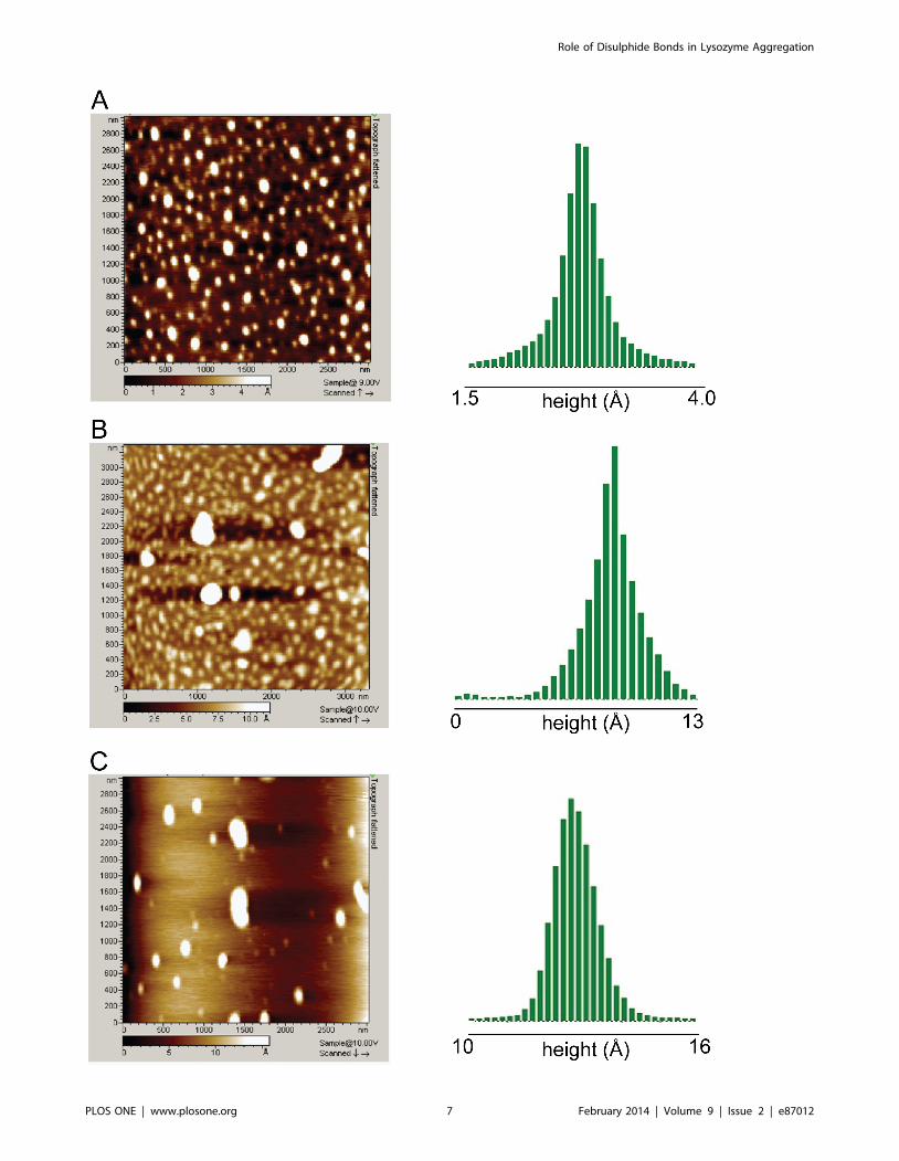
Figure 7. AFM images depicting topography (left) and their height distribution in Angstroms (right) for A) thiol-blocked HEWL inpH 12.2 after 72 hours, B) thiol-blocked HEWL in pH 12.2 after 11 days, C) control HEWL in pH 12.2 after 8 days. For heightdistribution, the threshold employed is identical to the leftmost value on the axis. The heights observed for maximum population in each image wereapproximately as follows: A) 2.7 A B) 8.5 A C) 12.7 A. Note the reduced height (flatness) of thiol-blocked samples in comparison to control.doi:10.1371/journal.pone.0087012.g007
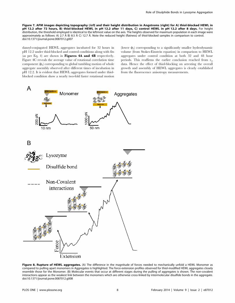
Figure 8. Rupture of HEWL aggregates. (A) The difference in the magnitude of forces needed to mechanically unfold a HEWL Monomer ascompared to pulling apart monomers in Aggregates is highlighted. The force-extension profiles observed for thiol-modified HEWL aggregates closelyresemble those for the Monomer. (B) Molecular events that occur at different stages during the pulling of aggregates is shown. The non-covalentinteractions appear as the weakest link between the monomers which are otherwise cross-linked by intermolecular disulfide bonds in the aggregate.doi:10.1371/journal.pone.0087012.g008
Role of Disulphide Bonds in Lysozyme Aggregation
PLOS ONE | www.plosone.org 8 February 2014 | Volume 9 | Issue 2 | e87012
Pulling Apart HEWL Aggregates by single-molecule ForceSpectroscopy
The strength of association between different monomers in the
HEWL aggregate is an important parameter to gauge the
robustness of aggregate. Measuring this strength for HEWL
aggregates formed under the thiol-blocked and control conditions
will shed light on the criticality of disulfide bonds in the assembly
of HEWL aggregates. Single-molecule force spectroscopy was
therefore employed to ascertain the robustness of HEWL
aggregates after thiol-blocking. The force-versus-extension (FX)
traces of un-aggregated (A), aggregated HEWL at pH 12.2 (B),
and aggregated HEWL at pH 12.2 after thiol-blocking (C) were
obtained using single-molecule atomic force microscope (SM-
AFM) as shown in Figure 5.
A) The FX traces clearly indicate that very often there is no
molecule adsorbed between the cantilever tip and the gold
surface due to the small sizeof protein compared to larger tip
diameter (,20–30 nm). Occasionally low force peaks were
observed at the beginning of FX traces, which couldbe due to
either unfolding of a single HEWL molecule or any non-
specific interaction between cantilever tip and gold surface.
Clearly no aggregates are found under this condition.
B) In contrast to the case A, here there are large aggregates
picked by the cantilever in FX traces. Multiple force peaks in
the FX traces are due to the forced dissociation of very strong
intermolecular interactions in these aggregates. These traces
clearly indicate the varying lengths and mechanical stabilities
of these aggregates (300–2600 pN) as evident in Figure 6from the observed distributions of detachment peak forces
(A) and detachment lengths (B). Force peaks in these FX
traces could be attributed to the unraveling of HEWL
molecules in the aggregate. It must be noted that the inter-
molecular disulfide bonds are still intact in the aggregate. It
was earlier shown that disulfide bonds do not rupture below
2 nN [21,22].
C) The FX traces resemble those of freshly dissolved HEWL
indicating the absence of any large aggregates in the solution.
Iodoacetamide blocks the free –SH groups in HEWL
preventing it from forming aggregates.
Morphology of AggregatesAfter monitoring for hydrophobicity, presence of fibril, changes
in size and mechanical strength of aggregates, it was essential to
observe their morphology. For this purpose AFM was employed.
The AFM images were acquired after incubation of 70 mM thiol-
blocked HEWL and 70 mM unblocked HEWL for various
durations in pH 12.2. Topography images of thiol-blocked HEWL
captured at 72 h (Figure 7A) reveal predominantly small globular
aggregates in large population. At longer incubation times (11
days), few larger aggregates of thiol-blocked HEWL are also visible
in a sea of smaller aggregates (Figure 7B). The morphology of
free thiol containing HEWL (control) revealed a mixture of large
elongated and small globular aggregates (Figure 7C). The
distribution of measured heights (along Z axis) on the right
column in Figure 7 clearly shows that thiol-blocked HEWL
samples possess shorter heights in comparison to control samples.
Thus thiol-modified HEWL aggregates appear more flat (probably
owing to less molecular packing) compared to control samples.
Interestingly, fibrils were never detected among thiol-blocked
HEWL samples even after several weeks while earlier work has
shown that 75 mM HEWL (control) formed matured fibrils in
pH 12.2 as revealed by AFM [7]. This substantiates ThT
fluorescence data which revealed diminished fluorescence in
thiol-blocked samples compared to control.
Discussion
In previous work it was shown that presence of DTT from the
beginning in the incubated HEWL sample at pH 12.2 significantly
abolished the growth of aggregates [9]. In presence of DTT when
all exposed disulfide bonds are reduced, HEWL chain has freedom
to sample a larger dynamic ensemble of conformation(s). Perhaps
such conformation(s) do not favour the same aggregation pathway
as in absence of DTT. For this reason iodoacetamide was added
two hours after initiation of aggregation when aggregation
competent conformations are active and aggregation is progress-
ing. This way it was ensured that same aggregation pathway was
chosen while the role of intermolecular disulfide bonds on
aggregate growth can be investigated.
The results show that preventing disulfide bond formation in
HEWL at pH 12.2 has significant consequences on the structure
and aggregation propensity of the protein. Blocking thiol groups
causes non-polar groups in HEWL aggregates to be more solvent
exposed and opened up (Fig. 1) probably owing to absence of
disulfide bonds that fasten the monomers together causing poor
packing and compaction. Also a smaller aggregate (see below)
arising from weak intermolecular interactions leading to poor
molecular packing can result in poor shielding of hydrophobic
groups. Intriguingly, the increased flexibility in the protein
conformation(s) under the thiol-modified state apparently does
NOT affect addition of monomers leading to growth of aggregates
as revealed by similar aggregate growth kinetics between control
and thiol-blocked samples (Fig. 3). However, the aggregates
formed in the thiol-blocked condition are not able to retain the
monomers together perhaps due to weak intermolecular forces
coupled with absence of cross-linking intermolecular disulfide
bonds, leading to their dissociation. This might explain why a) size
of aggregates are smaller (Fig. 4) after thiol modification; and b)
ordered aggregates like amyloid fibrils are not seen in same
condition (Fig. 2). The occasional large aggregate seen in AFM
images (Fig. 7) might be attributed to trace amounts of –SH groups
that may have escaped modification while the predominantly
small flat aggregates in the same images might represent the
limiting threshold size of aggregates in absence of disulfide bonds.
That the hydrophobic interaction forces keeping the monomers
together in these small aggregates in the absence of disulfide bonds
are significantly weaker in comparison to control samples is
corroborated by the force spectroscopy measurements (Fig. 5).
AFM based force spectroscopy has been used to study the
interactions between aggregating proteins like amyloid b [23,24],
alpha synuclein and lysozyme [25]. AFM is not only useful for
imaging of aggregates to measure their size and kinetics of
aggregation process, but it can also be used to measure the intra-
and intermolecular interactions at the single-molecule level
[26,27]. AFM based force spectroscopy has earlier been used to
study misfolding and aggregation of synuclein and amyloid beta
peptides [27,28]. Force spectroscopy directly measures the
interaction forces between molecules by picking individual
aggregates and mechanically stretching them at a constant velocity
to rupture the stabilizing intermolecular interactions. The
constant-velocity experiment provides the mechanical rupture
events in the form of a force-versus-extension trace (FX). The
rupture forces measured in the FX traces directly quantify the
strength of the intermolecular association in the aggregates.
Covalent interactions require typically many nanonewton rupture
Role of Disulphide Bonds in Lysozyme Aggregation
PLOS ONE | www.plosone.org 9 February 2014 | Volume 9 | Issue 2 | e87012
forces whereas the non-covalent interactions require very low
rupture forces.
Previous results of protein-protein interactions on lysozyme
molecules in aggregating acidic conditions revealed a maximum
force of ,1100 pN at pH 3 that was roughly ten times more than
force encountered under non-aggregating condition at pH 7 [25].
Here we have performed the pulling experiments on lysozyme at
pH12.2. The non-aggregated molecules (freshly exposed to
pH 12.2) are rarely picked up (due to their small size) and even
if they are picked up occasionally they tend to extend and detach
at very low forces (Figure 5A and Figure 8A (Monomer)). On the
contrary, the aggregated molecules (on day 4), owing to their large
size, attach frequently between cantilever and the gold surface and
have a very high mechanical stability (Figure 5B and Figure 8A(Aggregates)). The recorded FX traces suggest that the aggregates
unravel in piece-wise manner where individual monomeric units
or monomer clusters detach themselves from the aggregate at very
high forces varying from 500–2000 pN (Figure 8B) after disruption
of non-covalent interactions. It is likely that the cantilever may not
be picking up the aggregate as a whole but only a part of it
(Figure 8B) as suggested by the varying unraveling patterns of the
aggregates (Figure 5B and Figure S3) as well as the final
detachment length (Figure 6B). The interaction between the
individual units in these aggregates is stronger than those formed
in the acidic conditions [25]as evident from their higher
dissociation forces although a similar ten-fold drop in force is
observed in our case too under non-aggregating thiol-blocked
condition. It is worthwhile to note that detachment forces of
monomer or monomer cluster are ,1 nN, implying that it is not
the disulfide bonds but the weaker non-covalent hydrophobic
interactions that are breaking first (Figure 8B).
To confirm that we have picked large aggregates of lysozyme on
day 4, we have also done a pulling experiment on a thiol-blocked
sample of lysozyme containing iodoacetamide on day 3, where
iodoacetamide blocks the free-SH groups and inhibits disulfide
bond based aggregation process. This thiol-blocked HEWL
sample showed no pick-ups of molecule or occasionally a small
peak indicating the absence of any aggregates. This reaffirms our
earlier conclusion that absence of disulfide bonds in HEWL
significantly weakens non-covalent interactions such as hydropho-
bic interactions among monomers in the aggregate preventing
further growth and propagation of aggregation.
The results from our work establish that incorrect intermolec-
ular disulfide bonds promote aggregation by a) strengthening
hydrophobic interactions among monomers in the aggregate and
b) crosslinking HEWL monomers in the aggregate assembly.
Presence of similar disulfide bonds among mutant SOD1
aggregates in the spinal cord of ALS model transgenic mice has
been observed highlighting their physiological relevance [2]. Xie
and coworkers report that HEWL can self-assemble into granular
aggregates and subsequently globule-like aggregates at pH 7 and 9
under continuousUV illumination. They show that UV illumina-
tion reduces disulfide bonds photochemically causing partial
unfolding of HEWL that exposes hydrophobic residues triggering
aggregation into granules. On longer illumination and prolonged
incubation the granules grow into large globular aggregates
facilitated by intermolecular disulfide bonds [29]. Thus mecha-
nism of growth of large HEWL aggregates in their case is identical
to what we observe at alkaline pH. In a similar work they also
show that fibril formation in HEWL under native conditions can
be triggered by photochemical reduction of disulfide bonds [30].
This photochemical approach has been further employed recently
to self-assemble bovine a-lactalbumin and doxorubicin into
nanoparticles [31]. Lee and Eisenberg have shown that recombi-
nant hamster prion protein (PrPC) is converted to an oligomeric, b-
sheet rich, fibril formation competent second form PrPRDX by
formation of intermolecular disulfide bond. Blocking free thiol by
iodoacetamide was shown to prevent this conversion partially [32].
In contrast to aggregation promoting tendency of incorrectly
formed disulfide bonds mentioned so far, correctly positioned
disulfide bonds in extracellular proteins like insulin and islet
amyloid polypeptide avoid aggregation-prone loops in these
amyloidogenic proteins from venturing into partial folded
conformations that can triggerentry into aggregation pathways
[33]. For example it has been argued that disulfide bonds inhibit
aggregation of human lysozyme by stabilizing the folded state
through reduction in entropy. It has been also argued that when
partially folded states are populated, disulfide bonds cause
formation of fibrils that are of significantly less toxic nature
compared to disulfide reduced fibrils [34]. In another work, it was
shown that disulfide bond crosslinked variant of SRC homology 3
domain (SH3) of p85a subunit of bovine phosphatidyl-inositol-39-
kinase (PI3-SH3) domain is more stable, folds faster, aggregates
slower, and forms conformationally and functionally different
amyloid fibrils than the wild-type domain [35].
Conclusions
In conclusion, we report that incorrectly formed disulfide bonds
actively promote the growth of protein aggregates by strengthen-
ing non-covalent intermolecular forces, which are otherwise weak
in their absence. This is in contrast to correctly formed disulfide
bonds in extracellular proteins which protect them from venturing
into aggregation prone pathways. Our findings may lead to a
better understanding of the role of aberrant disulfide bonds in
diseases like Alzheimer’s and amyotrophic lateral sclerosis.
Supporting Information
Figure S1 Fluorescence intensity decay traces of thiol-blocked dansyl-HEWL at different incubation periods inpH 12.2 are shown.
(PDF)
Figure S2 Those for control dansyl-HEWL samples areshown.
(PDF)
Figure S3 More traces on the rupture of large HEWLaggregates at pH 12.2 are shown.
(PDF)
Table S1 Fitted decay parameters are displayed.
(PDF)
Acknowledgments
RS is grateful to Central Instruments Facility, Indian Institute of
Technology Guwahati for the time-resolved fluorescence measurements.
Author Contributions
Conceived and designed the experiments: RS SRKA. Performed the
experiments: VKR MG HCK. Analyzed the data: RS SRKA VKR HCK.
Contributed reagents/materials/analysis tools: RS SRKA. Wrote the
paper: RS VKR SRKA.
Role of Disulphide Bonds in Lysozyme Aggregation
PLOS ONE | www.plosone.org 10 February 2014 | Volume 9 | Issue 2 | e87012

References
1. Ramirez-Alvarado M, Kelly JW, Dobson CM, editors (2010) Protein Misfolding
Diseases: Current and emerging principles and therapies. New Jersey: Wiley.
2. Furukawa Y, Fu R, Deng H-X, Siddique T, O’Halloran TV (2006) Disulfide
cross-linked protein represents a significant fraction of ALS-associated Cu, Zn-
superoxide dismutase aggregates in spinal cords of model mice. Proceedings of
National Academy of Sciences USA 103: 7148–7153.
3. Barghorn S, Mandelkow E (2002) Toward a Unified Scheme for the
Aggregation of Tau into Alzheimer Paired Helical Filaments. Biochemistry
41: 14885–14896.
4. Hanson SRA, Hasan A, Smith DL, Smith JB (2000) The major in vivo
modifications of the human water-insoluble lens crystallins are disulfide bonds,
deamidation, methionine oxidation and backbone cleavage. Experimental Eye
Research 71: 195–207.
5. Chen Y, Dokholyan NV (2005) A Single Disulfide Bond Differentiates
Aggregation Pathways of ß2-Microglobulin. J Mol Biol 354: 473–482.
6. Mossuto MF, Bolognesi B, Guixer B, Dhulesia A, Agostini F, et al. (2011)
Disulfide Bonds Reduce the Toxicity of the Amyloid Fibrils Formed by an
Extracellular Protein. Angew Chem Int Ed 50: 7048–7051.
7. Swaminathan R, Ravi VK, Kumar S, Kumar MVS, Chandra N (2011)
Lysozyme: A model protein for amyloid research. In: Donev R, editor. Advances
in Protein Chemistry and Structural Biology: Elsevier. 63–111.
8. Niraula TN, Konno T, Li H, Yamada H, Akasaka K, et al. (2004) Pressure-
dissociable reversible assembly of intrinsically denatured lysozyme is a precursor
for amyloid fibrils. Proceedings of the National Academy of Sciences of the
United States of America 101: 4089–4093.
9. Kumar S, Ravi VK, Swaminathan R (2008) How do surfactants and DTT affect
the size, dynamics, activity and growth of soluble lysozyme aggregates?
Biochemical Journal 415: 275–288.
10. Homchaudhuri L, Kumar S, Swaminathan R (2006) Slow aggregation of
lysozyme in alkaline pH monitored in real time employing the fluorescence
anisotropy of covalently labelled dansyl probe. FEBS Letters 580: 2097–2101.
11. Wall J, Murphy CL, Solomon A (1999) In Vitro immunoglobulin light chain
fibrillogenesis. In: Wetzel R, editor. Methods in Enzymology: Academic Press.
204–217.
12. Aggarwal V, Kulothungan SR, Balamurali MM, Saranya SR, Varadarajan R, et
al. (2011) Ligand-modulated parallel mechanical unfolding pathways of maltose-
binding proteins. Journal of Biological Chemistry 286: 28056–28065.
13. Kumar S, Ravi VK, Swaminathan R (2009) Suppression of lysozyme
aggregation at alkaline pH by tri-N-acetylchitotriose. Biochimica et Biophysica
Acta - Proteins and Proteomics 1794: 913–920.
14. Stryer L (1965) The interaction of a naphthalene dye with apomyoglobin and
apohemoglobin. A fluorescent probe of non-polar binding sites. Journal of
Molecular Biology 13: 482–495.
15. Turner DC, Brand L (1968) Quantitative estimation of protein binding site
polarity. Fluorescence of N-Arylaminonaphthalenesulfonates. Biochemistry 7:
3381–3390.
16. Carrotta R, Bauer R, Waninge R, Rischel C (2001) Conformational
characterization of oligomeric intermediates and aggregates in b-lactoglobulin
heat aggregation. Protein Science 10: 1312–1318.
17. Ahmed M, Davis J, Aucoin D, Sato T, Ahuja S, et al. (2010) Structural
conversion of neurotoxic amyloid-beta 1–42 oligomers to fibrils. Nature
Structural and Molecular Biology 17: 561–567.
18. Kumar S, Singh AK, Krishnamoorthy G, Swaminathan R (2008) Thioflavin T
displays enhanced fluorescence selectively inside anionic micelles and mamma-
lian cells. Journal of Fluorescence 18: 1199–1205.
19. Khurana R, Coleman C, Ionescu-Zanetti C, Carter SA, Krishna V, et al. (2005)Mechanism of thioflavin T binding to amyloid fibrils. Journal of Structural
Biology 151: 229–238.20. Herron JN, Voss Jr EW (1981) Characterization of flourescent 2-dimethylami-
nonaphthalene-5-sulfonyl-immunoglobulin G conjugates for application influorescence polarization studies. Journal of Biochemical and Biophysical
Methods 5: 1–17.
21. Ainavarapu SRK, Brujic J, Huang HH, Wiita AP, Lu H, et al. (2007) Contourlength and refolding rate of a small protein controlled by engineered disulfide
bonds. Biophysical Journal 92: 225–233.22. Ainavarapu SRK, Wiita AP, Dougan L, Uggerud E, Fernandez JM (2008)
Single-molecule force spectroscopy measurements of bond elongation during a
bimolecular reaction. Journal of the American Chemical Society 130: 6479–6487.
23. Hane F, Tran G, Attwood SJ, Leonenko Z (2013) Cu2+ Affects Amyloid-b (1–42) Aggregation by Increasing Peptide-Peptide Binding Forces. PLoS ONE 8:
e59005.
24. Kim BH, Palermo NY, Lovas S, Zaikova T, Keana JFW, et al. (2011) Single-molecule atomic force microscopy force spectroscopy study of Ab-40
interactions. Biochemistry 50: 5154–5162.25. McAllister C, Karymov MA, Kawano Y, Lushnikov AY, Mikheikin A, et al.
(2005) Protein interactions and misfolding analyzed by AFM force spectroscopy.Journal of Molecular Biology 354: 1028–1042.
26. Yu J, Warnke J, Lyubchenko YL (2011) Nanoprobing of a-synuclein misfolding
and aggregation with atomic force microscopy. Nanomedicine: Nanotechnology,Biology, and Medicine 7: 146–152.
27. Yu J, Malkova S, Lyubchenko YL (2008) a-Synuclein Misfolding: SingleMolecule AFM Force Spectroscopy Study. Journal of Molecular Biology 384:
992–1001.
28. Lv Z, Roychaudhuri R, Condron MM, Teplow DB, Lyubchenko YL (2013)Mechanism of amyloid b-protein dimerization determined using single-molecule
afm force spectroscopy. Scientific Reports 3: 2880.29. Xie J, Qin M, Cao Y, Wang W (2011) Mechanistic insight of photo-induced
aggregation of chicken egg white lysozyme: The interplay between hydrophobicinteractions and formation of intermolecular disulfide bonds. Proteins: Structure,
Function and Bioinformatics 79: 2505–2516.
30. Xie JB, Cao Y, Pan H, Qin M, Yan ZQ, et al. (2012) Photoinduced fibrilsformation of chicken egg white lysozyme under native conditions. Proteins:
Structure, Function and Bioinformatics 80: 2501–2513.31. Xie J, Cao Y, Xia M, Gao X, Qin M, et al. (2013) One-Step photo synthesis of
protein-drug nanoassemblies for drug delivery. Advanced Healthcare Materials
2: 795–799.32. Lee S, Eisenberg D (2003) Seeded conversion of recombinant prion protein to a
disulfide-bonded oligomer by a reduction-oxidation process. Nature StructuralBiology 10: 725–730.
33. Tartaglia GG, Pawar AP, Campioni S, Dobson CM, Chiti F, et al. (2008)Prediction of Aggregation-Prone Regions in Structured Proteins. Journal of
Molecular Biology 380: 425–436.
34. Mossuto MF, Bolognesi B, Guixer B, Dhulesia A, Agostini F, et al. (2011)Disulfide bonds reduce the toxicity of the amyloid fibrils formed by an
extracellular protein. Angewandte Chemie - International Edition 50: 7048–7051.
35. Grana-Montes R, De Groot NS, Castillo V, Sancho J, Velazquez-Campoy A, et
al. (2012) Contribution of disulfide bonds to stability, folding, and amyloid fibrilformation: The PI3-SH3 domain case. Antioxidants and Redox Signaling 16: 1–
15.
Role of Disulphide Bonds in Lysozyme Aggregation
PLOS ONE | www.plosone.org 11 February 2014 | Volume 9 | Issue 2 | e87012