Creating Biomimetic Surfaces through Covalent and Oriented Binding of Proteins
9
DOI: 10.1021/la103086b 14707 Langmuir 2010, 26(18), 14707–14715 Published on Web 08/26/2010 pubs.acs.org/Langmuir © 2010 American Chemical Society Creating Biomimetic Surfaces through Covalent and Oriented Binding of Proteins S ebastien Chevalier, † Carlos Cuestas-Ayllon, ‡ Valeria Grazu, ‡ Monica Luna, § Helene Feracci,* ,† and Jesus M. de la Fuente* ,‡ † Universit e Bordeaux 1, CNRS UPR 8641, Centre de Recherche Paul Pascal, 115 Avenue Dr Schweitzer, 33600 Pessac, France, ‡ Instituto de Nanociencia de Aragon-ARAID, University of Zaragoza, Campus Rio Ebro, Edif. IþD, C/Mariano Esquillor s/n, Zaragoza 50018, Spain, and § Instituto de Microelectr onica de Madrid (IMM-CSIC), C/Isaac Newton 8, PTM 28760 Tres Cantos, Madrid, Spain Received March 10, 2010 This manuscript describes a novel method for the biofunctionalization of glass surfaces with polyhistidine-tagged proteins. The main innovation of this methodology consists of the covalent binding between the nitrilotriacetic acid (NTA) moiety and the proteins, ensuring not only orientation, but also stability of the recombinant proteins on NTA- covered surfaces. In this work, as C-terminal polyhistidine tagged cadherin extracellular fragments have been used, this methodology guarantees the proper orientation of these proteins, by mimicking their insertion into cell plasma membranes. These biofunctionalized surfaces have been characterized by confocal microscopy, X-ray photoelectron spectroscopy, contact angle, and atomic force microscopy, showing a high density of cadherins on the glass surfaces and the stability of the linkage. The prepared materials exhibited a high tendency to promote cell spreading, demonstrating the functionality of the protein and the high utility of these biomaterials to promote cell adhesion events. Interestingly, differences in the cytoskeleton organization have been observed in cells adhering to surfaces with no cadherins or with nonoriented cadherins, in comparison to surfaces functionalized with well-oriented cadherins. This method, which allows the robust immobilization of polyhistidine tagged proteins due to their covalent binding and with a defined orientation, may also find particular usefulness in the making of protein biochips, for analysis of protein-protein interactions, as well as structural and single-molecule studies. Introduction Biofunctional surfaces are extensively used in therapeutics and diagnostics for cell-based assays, 1,2 tissue-engineering applica- tions, 3,4 biosensor developments, 5,6 bioelectronics, 7 and, more generally, medical devices. 5,6,8 The ideal biofunctional surface should be easy to prepare and characterize, be amenable to design, and be biocompatible and efficient. In this regard, the incorpora- tion of relevant biomolecules has already been reported going from proteins, 9 peptides, 10,11 carbohydrates, 12,13 oligonucleo- tides, 14 and others. 15,16 Several strategies have been followed for the chemical modification of these biomaterials using covalent or noncovalent chemistry. However, both approaches have advan- tages and disadvantages. Covalent binding ensures a strong linkage between the biomolecules and the surface, but it requires surface modification, and most importantly, it does not generally guarantee the right orientation of complex molecules such as proteins. 17 Such chemical binding of unmodified proteins most often leads to their random orientation onto reactive surfaces. On one hand, unspecific adsorption constitutes a simple strategy for the biofunctionalization of surfaces, 18 but it constitutes a binding *Corresponding authors. E-mail: [email protected] and [email protected]. (1) Falconnet, D.; Csucs, G.; Grandin, H. M.; Textor, M. Surface engineering approaches to micropattern surfaces for cell-based assays. Biomaterials 2006, 27(16), 3044–63. (2) Geiger, B.; Spatz, J. P.; Bershadsky, A. D. Environmental sensing through focal adhesions. Nat. Rev. Mol. Cell Biol. 2009, 10(1), 21–33. (3) Curtis, A.; Riehle, M. Tissue engineering: the biophysical background. Phys. Med. Biol. 2001, 46(4), R47–65. (4) Lutolf, M. P.; Hubbell, J. A. Synthetic biomaterials as instructive extra- cellular microenvironments for morphogenesis in tissue engineering. Nat. Biotech- nol. 2005, 23(1), 47–55. (5) Jonkheijm, P.; Weinrich, D.; Schroder, H.; Niemeyer, C. M.; Waldmann, H. Chemical strategies for generating protein biochips. Angew. Chem., Int. Ed. Engl. 2008, 47(50), 9618–47. (6) Wong, L. S.; Khan, F.; Micklefield, J. Selective covalent protein immobiliza- tion: strategies and applications. Chem. Rev. 2009, 109(9), 4025–53. (7) Stevens, M. M.; George, J. H. Exploring and engineering the cell surface interface. Science 2005, 310(5751), 1135–8. (8) Bettinger, C. J.; Langer, R.; Borenstein, J. T. Engineering substrate topo- graphy at the micro- and nanoscale to control cell function. Angew. Chem., Int. Ed. Engl. 2009, 48(30), 5406–15. (9) Thery, M.; Racine, V.; Pepin, A.; Piel, M.; Chen, Y.; Sibarita, J. B.; Bornens, M. The extracellular matrix guides the orientation of the cell division axis. Nat. Cell Biol. 2005, 7(10), 947–53. (10) Perret, E.; Benoliel, A. M.; Nassoy, P.; Pierres, A.; Delmas, V.; Thiery, J. P.; Bongrand, P.; Feracci, H. Fast dissociation kinetics between individual E-cadherin fragments revealed by flow chamber analysis. EMBO J. 2002, 21(11), 2537–46. (11) Perret, E.; Leung, A.; Morel, A.; Feracci, H.; Nassoy, P. Versatile Decora- tion of glass surfaces to probe individual protein-protein interactions and cellular adhesion. Langmuir 2002, 18(3), 846–854. (12) Fuss, M.; Luna, M.; Alcantara, D.; Fuente, J. M.; Penades, S.; Briones, F. Supramolecular self-assembled arrangements of maltose glyconanoparticles. Langmuir 2008, 24(9), 5124–8. (13) Schatz, C.; Louguet, S.; Le Meins, J. F.; Lecommandoux, S. Polysaccharide- block-polypeptide copolymer vesicles: towards synthetic viral capsids. Angew. Chem., Int. Ed. Engl. 2009, 48(14), 2572–5. (14) Weisbrod, S. H.; Marx, A. Novel strategies for the site-specific covalent labelling of nucleic acids. Chem. Commun. (Cambridge) 2008, 44, 5675–85. (15) de la Fuente, J. M.; Andar, A.; Gadegaard, N.; Berry, C. C.; Kingshott, P.; Riehle, M. O. Fluorescent aromatic platforms for cell patterning. Langmuir 2006, 22(13), 5528–32. (16) Song, Y. F.; McMillan, N.; Long, D. L.; Kane, S.; Malm, J.; Riehle, M. O.; Pradeep, C. P.; Gadegaard, N.; Cronin, L. Micropatterned surfaces with cova- lently grafted unsymmetrical polyoxometalate-hybrid clusters lead to selective cell adhesion. J. Am. Chem. Soc. 2009, 131(4), 1340–1. (17) Huang, S.-C.; Caldwell, K. D.; Lin, J.-N.; Wang, H.-K.; Herron, J. N. Site- specific immobilization of monoclonal antibodies using spacer-mediated antibody attachment. Langmuir 1996, 12(17), 4292–4298. (18) Leung, C.; Palmer, R. E. Adsorption of a model protein, the GroEL chaperonin, on surfaces. J. Phys.: Condens. Matter 2008, 20(35), 353001.
Transcript of Creating Biomimetic Surfaces through Covalent and Oriented Binding of Proteins

DOI: 10.1021/la103086b 14707Langmuir 2010, 26(18), 14707–14715 Published on Web 08/26/2010
pubs.acs.org/Langmuir
© 2010 American Chemical Society
Creating Biomimetic Surfaces through Covalent and Oriented
Binding of Proteins
S�ebastien Chevalier,† Carlos Cuestas-Ayllon,‡ Valeria Grazu,‡ Monica Luna,§ Helene Feracci,*,†
and Jesus M. de la Fuente*,‡
†Universit�e Bordeaux 1, CNRS UPR 8641, Centre de Recherche Paul Pascal, 115 Avenue Dr Schweitzer,33600 Pessac, France, ‡Instituto de Nanociencia de Aragon-ARAID, University of Zaragoza,
Campus Rio Ebro, Edif. IþD, C/Mariano Esquillor s/n, Zaragoza 50018, Spain, and§Instituto de Microelectr�onica de Madrid (IMM-CSIC), C/Isaac Newton 8, PTM 28760 Tres Cantos,
Madrid, Spain
Received March 10, 2010
This manuscript describes a novel method for the biofunctionalization of glass surfaces with polyhistidine-taggedproteins. The main innovation of this methodology consists of the covalent binding between the nitrilotriacetic acid(NTA) moiety and the proteins, ensuring not only orientation, but also stability of the recombinant proteins on NTA-covered surfaces. In this work, as C-terminal polyhistidine tagged cadherin extracellular fragments have been used, thismethodology guarantees the proper orientation of these proteins, by mimicking their insertion into cell plasmamembranes. These biofunctionalized surfaces have been characterized by confocal microscopy, X-ray photoelectronspectroscopy, contact angle, and atomic force microscopy, showing a high density of cadherins on the glass surfaces andthe stability of the linkage. The prepared materials exhibited a high tendency to promote cell spreading, demonstratingthe functionality of the protein and the high utility of these biomaterials to promote cell adhesion events. Interestingly,differences in the cytoskeleton organization have been observed in cells adhering to surfaces with no cadherins or withnonoriented cadherins, in comparison to surfaces functionalized with well-oriented cadherins. This method, whichallows the robust immobilization of polyhistidine tagged proteins due to their covalent binding and with a definedorientation, may also find particular usefulness in the making of protein biochips, for analysis of protein-proteininteractions, as well as structural and single-molecule studies.
Introduction
Biofunctional surfaces are extensively used in therapeutics anddiagnostics for cell-based assays,1,2 tissue-engineering applica-tions,3,4 biosensor developments,5,6 bioelectronics,7 and, moregenerally, medical devices.5,6,8 The ideal biofunctional surfaceshouldbe easy toprepare and characterize, be amenable todesign,and be biocompatible and efficient. In this regard, the incorpora-tion of relevant biomolecules has already been reported going
from proteins,9 peptides,10,11 carbohydrates,12,13 oligonucleo-tides,14 and others.15,16 Several strategies have been followed forthe chemical modification of these biomaterials using covalent ornoncovalent chemistry. However, both approaches have advan-tages and disadvantages. Covalent binding ensures a stronglinkage between the biomolecules and the surface, but it requiressurface modification, and most importantly, it does not generallyguarantee the right orientation of complex molecules such asproteins.17 Such chemical binding of unmodified proteins mostoften leads to their randomorientation onto reactive surfaces. Onone hand, unspecific adsorption constitutes a simple strategy forthe biofunctionalization of surfaces,18 but it constitutes a binding
*Corresponding authors. E-mail: [email protected] [email protected].(1) Falconnet, D.; Csucs, G.; Grandin, H. M.; Textor, M. Surface engineering
approaches to micropattern surfaces for cell-based assays. Biomaterials 2006,27(16), 3044–63.(2) Geiger, B.; Spatz, J. P.; Bershadsky, A. D. Environmental sensing through
focal adhesions. Nat. Rev. Mol. Cell Biol. 2009, 10(1), 21–33.(3) Curtis, A.; Riehle, M. Tissue engineering: the biophysical background. Phys.
Med. Biol. 2001, 46(4), R47–65.(4) Lutolf, M. P.; Hubbell, J. A. Synthetic biomaterials as instructive extra-
cellular microenvironments for morphogenesis in tissue engineering.Nat. Biotech-nol. 2005, 23(1), 47–55.(5) Jonkheijm, P.; Weinrich, D.; Schroder, H.; Niemeyer, C. M.; Waldmann, H.
Chemical strategies for generating protein biochips. Angew. Chem., Int. Ed. Engl.2008, 47(50), 9618–47.(6) Wong, L. S.; Khan, F.; Micklefield, J. Selective covalent protein immobiliza-
tion: strategies and applications. Chem. Rev. 2009, 109(9), 4025–53.(7) Stevens, M. M.; George, J. H. Exploring and engineering the cell surface
interface. Science 2005, 310(5751), 1135–8.(8) Bettinger, C. J.; Langer, R.; Borenstein, J. T. Engineering substrate topo-
graphy at the micro- and nanoscale to control cell function.Angew. Chem., Int. Ed.Engl. 2009, 48(30), 5406–15.(9) Thery,M.; Racine, V.; Pepin, A.; Piel, M.; Chen, Y.; Sibarita, J. B.; Bornens,
M. The extracellular matrix guides the orientation of the cell division axis.Nat. CellBiol. 2005, 7(10), 947–53.(10) Perret, E.; Benoliel, A.M.; Nassoy, P.; Pierres, A.; Delmas, V.; Thiery, J. P.;
Bongrand, P.; Feracci, H. Fast dissociation kinetics between individual E-cadherinfragments revealed by flow chamber analysis. EMBO J. 2002, 21(11), 2537–46.
(11) Perret, E.; Leung, A.; Morel, A.; Feracci, H.; Nassoy, P. Versatile Decora-tion of glass surfaces to probe individual protein-protein interactions and cellularadhesion. Langmuir 2002, 18(3), 846–854.
(12) Fuss, M.; Luna, M.; Alcantara, D.; Fuente, J. M.; Penades, S.; Briones, F.Supramolecular self-assembled arrangements of maltose glyconanoparticles.Langmuir 2008, 24(9), 5124–8.
(13) Schatz, C.; Louguet, S.; Le Meins, J. F.; Lecommandoux, S. Polysaccharide-block-polypeptide copolymer vesicles: towards synthetic viral capsids. Angew.Chem., Int. Ed. Engl. 2009, 48(14), 2572–5.
(14) Weisbrod, S. H.; Marx, A. Novel strategies for the site-specific covalentlabelling of nucleic acids. Chem. Commun. (Cambridge) 2008, 44, 5675–85.
(15) de la Fuente, J. M.; Andar, A.; Gadegaard, N.; Berry, C. C.; Kingshott, P.;Riehle, M. O. Fluorescent aromatic platforms for cell patterning. Langmuir 2006,22(13), 5528–32.
(16) Song, Y. F.; McMillan, N.; Long, D. L.; Kane, S.; Malm, J.; Riehle, M. O.;Pradeep, C. P.; Gadegaard, N.; Cronin, L. Micropatterned surfaces with cova-lently grafted unsymmetrical polyoxometalate-hybrid clusters lead to selective celladhesion. J. Am. Chem. Soc. 2009, 131(4), 1340–1.
(17) Huang, S.-C.; Caldwell, K. D.; Lin, J.-N.; Wang, H.-K.; Herron, J. N. Site-specific immobilization of monoclonal antibodies using spacer-mediated antibodyattachment. Langmuir 1996, 12(17), 4292–4298.
(18) Leung, C.; Palmer, R. E. Adsorption of a model protein, the GroELchaperonin, on surfaces. J. Phys.: Condens. Matter 2008, 20(35), 353001.

14708 DOI: 10.1021/la103086b Langmuir 2010, 26(18), 14707–14715
Article Chevalier et al.
very dependent on the media conditions (pH, ionic strength,temperature, etc.).19 Moreover, such uncontrolled interactionsoften induce protein unfolding and thus a decrease of their bio-logical activity.
The study of cell adhesion to biomaterials is a very importanttopic in tissue engineering.20 The interactions between cells andmaterials modulate the cellular response to implanted devices aswell as cell culture supports.7,21 Biochemical data highlight thenecessity of extracellularmatrix (ECM)proteins in promoting celladhesion through specific interactions.22 Cells, via integrins, bindto specific amino acid sequences on ECM proteins such as RGDpresent in fibronectin, vitronectin, and thrombospondin.23,24
These proteins provide an attachment network for the adhesionand growth of specific cells in vivo. Furthermore, adhesive recep-tors named cadherins are also involved in cell-cell adhesionevents. Classical cadherins are transmembrane glycoproteinsessential in controlling the specificity, organization, anddynamicsof cell adhesion, which is crucial for the development and main-tenance of tissue architecture and function.25-27 Cadherins inter-act with other cell surface cadherins on neighboring cells throughtheir extracellular subdomain repeats named EC1 to EC5.28-30
Their adhesive engagement initiates intracellular signals rangingfrom cytoskeletal organization to cell polarity, proliferation, orapoptosis that are communicated through the conserved cadherintail domain to cytoplasmic pathways.25,31,32 The role of adhesionforces in the signaling process is yet to be understood due todifficulty in the interpretation of biological responses involvingdynamic and complex multimolecular organizations.33 On thebiomaterial surfaces in vitro, the same mechanisms also apply.When foreign materials come into contact with body fluid or cellculture medium, the initial response is protein adsorption at thematerial surfaces. Thus, the materials interact with the cells
through the adsorbed protein layer. The composition and struc-ture of this protein layer play critical roles in determiningsubsequent cell behaviors.34-37
We describe in this paper an efficient, simple, and straightfor-ward procedure to prepare as a model system functionalizedsurfaces with cadherins in an oriented fashion, mimicking thecadherin presentation on cell surfaces. The developed protocoluses modified cadherin fragments with a polyhistidine tail at theC-terminal end of the proteins and silanized surfaces incorporat-ing a metal chelator complex such as nitrilotriacetic acid (NTA)molecules.A two-stepprotocol has beendeveloped.The chelationof the polyhistidine tails to the Ni2þ-NTA complexes located atthe surfaces through its high binding affinity will ensure a correctorientation of the proteins incorporated to the surface. However,the polyhistidine-NTA interaction is highly dependent on pH,ionic strength, and other media conditions.38,39 In order toguarantee a stable linkage to the surface, a second step has beenproposed by an amide formation between amino groups from thehistidine lateral chains and the carboxylated groups presented inthe NTA. The prepared biofunctional surfaces have been char-acterized by fluorescentmicroscopy, contact anglemeasurements,andXPS. InAFMstudies, the stability of the protein immobiliza-tion process was probed at the single-molecule level by the heightof the covering layer. These surfaces have also been proven asexcellent adhesive platforms for cells presenting cadherins on theirsurfaces, confirming their potential capabilities as biomaterialsto study cadherin-cadherin interactions using cellular models.Indeed, the total control over the orientation of the immobilizedproteins on such surfaces without affecting their conformationand function optimizes the accessibility of proteins’ active sites.Interestingly, differences in cell adhesion behavior are obviouswhen comparing random versus oriented cadherin immobiliza-tion through the method reported here. Because the polyhistidinetag is one of the most generically used affinity tag for ease ofpurification, this method provides the basis for numerous furtherapplications for studies focusing on research targets at single-molecule levels and for nanobiotechnology. The homogeneousorientation and distribution of immobilized proteins demonstratethe high quality of the designed surface and qualify its applicationin nanoscaled protein chip architectures.
Experimental Section
Materials. Water used in the described reactions and for clean-ing was obtained from a water purification system, MILLI-Q A10from Millipore. For the activation of glass supports, sulfuricacid (ACS reagent, 95-98%) and hydrogen peroxide (30%)were purchased from Sigma-Aldrich. For the functionalizationof glass supports with NTA, (3-glycidyloxypropyl)trimethoxy-silane (g98%), N-(5-amino-1-carboxypentyl)iminodiacetic acid(g97%, TLC), dry toluene (99.8%), sodium carbonate (g99%),
(19) Jung, J.-M.; Kwon, K. Y.; Ha, T.-H.; Chung, B. H.; Jung, H.-T. Gold-Conjugated protein nanoarrays through block-copolymer lithography: fromfabrication to biosensor design13. Small 2006, 2(8-9), 1010–1015.(20) Ma, Z.; Mao, Z.; Gao, C. Surface modification and property analysis of
biomedical polymers used for tissue engineering. Colloids Surf., B 2007, 60(2),137–157.(21) Chen, C. S.; Mrksich, M.; Huang, S.; Whitesides, G. M.; Ingber, D. E.
Micropatterned surfaces for control of cell shape, position, and function. Bio-technol. Prog. 1998, 14(3), 356–63.(22) Velzenberger, E.; Pezron, I.; Legeay, G.; Nagel, M.-D.; Kirat, K. E.
Probing fibronectin-surface interactions: a multitechnique approach. Langmuir2008, 24(20), 11734–11742.(23) Michael, J.; Scholnzart, L.; Israel, I.; Beutner, R.; Scharnweber, D.;Worch,
H.; Hempel, U.; Schwenzer, B. Oligonucleotide-RGD peptide conjugates forsurface modification of titanium implants and improvement of osteoblast adhe-sion. Bioconjugate Chem. 2009, 20(4), 710–718.(24) Zhu, J.; Tang, C.; Kottke-Marchant, K.; Marchant, R. E. Design and
synthesis of biomimetic hydrogel scaffolds with controlled organization of cyclicRGD peptides. Bioconjugate Chem. 2009, 20(2), 333–339.(25) Hartsock, A.; Nelson, W. J. Adherens and tight junctions: structure,
function and connections to the actin cytoskeleton. Biochim. Biophys. Acta2008, 1778(3), 660–9.(26) Larue, L.; Antos, C.; Butz, S.; Huber, O.; Delmas, V.; Dominis, M.;
Kemler, R. A role for cadherins in tissue formation. Development 1996, 122(10),3185–94.(27) Nishimura, T.; Takeichi, M. Remodeling of the adherens junctions during
morphogenesis. Curr. Top. Dev. Biol. 2009, 89, 33–54.(28) Leckband, D.; Prakasam, A. Mechanism and dynamics of cadherin
adhesion. Annu. Rev. Biomed. Eng. 2006, 8, 259–87.(29) Patel, I. S.; Madan, P.; Getsios, S.; Bertrand, M. A.; MacCalman, C. D.
Cadherin switching in ovarian cancer progression. Int. J. Cancer 2003, 106(2), 172–7.(30) Perret, E.; Leung, A.; Feracci, H.; Evans, E. Trans-bonded pairs of
E-cadherin exhibit a remarkable hierarchy of mechanical strengths. Proc. Natl.Acad. Sci. U.S.A. 2004, 101(47), 16472–7.(31) vanRoy, F.; Berx,G. The cell-cell adhesionmolecule E-cadherin.Cell.Mol.
Life Sci. 2008, 65(23), 3756–88.(32) Charrasse, S.; Meriane, M.; Comunale, F.; Blangy, A.; Gauthier-Rouviere,
C. N-cadherin-dependent cell-cell contact regulates Rho GTPases and beta-catenin localization in mouse C2C12 myoblasts. J. Cell Biol. 2002, 158(5), 953–65.(33) Thiery, J. P. Cell adhesion in development: a complex signaling network.
Curr. Opin. Genet. Dev. 2003, 13(4), 365–71.
(34) Arulanandam, R.; Vultur, A.; Cao, J.; Carefoot, E.; Elliott, B. E.; Truesdell,P. F.; Larue, L.; Feracci, H.; Raptis, L. Cadherin-cadherin engagement promotescell survival via Rac1/Cdc42 and signal transducer and activator of transcription-3.Mol. Cancer Res. 2009, 7(8), 1310–27.
(35) Ganz, A.; Lambert, M.; Saez, A.; Silberzan, P.; Buguin, A.; Mege, R. M.;Ladoux, B. Traction forces exerted through N-cadherin contacts. Biol. Cell 2006,98(12), 721–30.
(36) Ostuni, E.; Whitesides, G. M.; Ingber, D. E.; Chen, C. S. Using self-assembled monolayers to pattern ECM proteins and cells on substrates. MethodsMol. Biol. 2009, 522, 183–94.
(37) Dupin, I.; Camand, E.; Etienne-Manneville, S. Classical cadherins controlnucleus and centrosome position and cell polarity. J. Cell Biol. 2009, 185(5),779–86.
(38) Schmitt, J.; Hess, H.; Stunnenberg, H. G. Affinity purification of histidine-tagged proteins. Mol. Biol. Rep. 1993, 18(3), 223–30.
(39) Crowe, J.; Dobeli, H.; Gentz, R.; Hochuli, E.; Stuber, D.; Henco, K. 6xHis-Ni-NTA chromatography as a superior technique in recombinant protein expres-sion/purification. Methods Mol. Biol. 1994, 31, 371–87.

DOI: 10.1021/la103086b 14709Langmuir 2010, 26(18), 14707–14715
Chevalier et al. Article
and sodium bicarbonate (99.7%) were purchased from Sigma-Aldrich.
For protein immobilization, nickel II chloride hexahydrate,Hepes (g99.5%), N-(3-dimethylaminopropyl)-N0-ethylcarbodii-mide hydrochloride crystalline, imidazole (ACS reagent, g99%)were purchased from Sigma-Aldrich. N-Hydroxysuccinimide(g97%) was purchased from Fluka. Sodium chloride (ultrapure,5 M) was purchased from Gibco. Ethylenediaminetetraacetic(EDTA, g99%) was purchased from Panreac.
For scanning electron microscopy, glutaraldehyde (25% inwater) and hexamethyldisilazane (HDMS,g99%) were purchasedfrom Sigma-Aldrich. Sodium cacodylate and osmium tetroxydewere purchased from Agar Scientific. Tannic acid was purchasedfrom Fluka. Methanol (Spectranal,g99.9%) was purchased fromRiedel-deHa
::en.Uranyl acetate was purchased fromEloı̈se SARL.
For immunocytochemistry, formaldehyde, Triton X-100, andbovine serumalbumin (BSA) were purchased fromSigma-Aldrich.Mouse monoclonal anti-E-cadherin antibodies (Clone 36) werepurchased from BD Transduction Laboratories (BD Biosciences,Pharmingen). Actin filaments were stained with Rhodamine-phalloidin (Molecular Probes, Invitrogen). The mounting mediumwas SlowFade Gold antifade reagent with DAPI (MolecularProbes, Invitrogen).
All the chemicals were used as received.
Activation of Glass Supports. Glass supports were cleanedwith piranha solution (H2SO4 and 30% H2O2, 3:1) for 30 min atroom temperature (RT). The slides were then rinsed several timeswith Milli-Q water and dried under nitrogen.
Functionalization of Glass Supports with N-(5-Amino-1-
carboxypentyl)iminodiacetic acid (NTA-NH2). A 2% solutionof 3-(2,3-epoxypropoxy) propyltrimethoxysilane in dry toluenewas added overnight (ON) at RT onto the activated glass supports.After washing extensively with toluene and 10mMcarbonate bufferpH 10.8, glass supports were incubated ON at RT with 25 mMNTA-NH2 in carbonate buffer pH 10.8. The supports were thenwashed and dried.
Expression and Purification of the Hexahistidine Tagged
Cadherin Fragments (E/EC12). Themouse E-Cadherin extra-cellular fragment (MIEGR-E/EC12) corresponding to the twooutermost cadherin modules bearing a C-terminal hexahistidinetag was produced as already described.40 Briefly, the IEGRsequence was introduced in the plasmid construct of the cadherinfragment, in order to obtain the correctly cleaved cadherinfragment at the N-ter end. For production of this chimericprotein, 500 mL of Terrific Broth supplemented with 50 μg/mLkanamycin were inoculated with a transformed colony andprotein synthesis induced by IPTG. Cell pellet was resuspendedin lysis buffer [4 M urea, 50 mM Na2HPO4 (pH 7.8), 20 mMimidazole, and 20 mM β-mercaptoethanol]. The resulting lysatewas cleared by centrifugation, and the supernatant was incubatedfor 2 hwithNi-NTASuperflow agarose resin beads (Qiagen). Thebeads were then thoroughly washed and subjected to stepwisedialysis against PBS. Factor Xa protease (Qiagen) digestion ofMIEGR-E/EC12was performed for 60 h at 16 �Cand stopped byremoving the enzyme with Xa Removal Resin (Qiagen). Finally,the resultingE/EC12 fragmentswere thendialyzed against 150mMNaCl and 20 mM Hepes (pH 7.0) in order to remove Ca2þ ionsand the cleaved peptide. Protein purity was checked by SDS-PAGE, Western blotting, and mass spectrometry (not shown).
Covalent Oriented Protein Immobilization. The NTA-surfaces were loaded with 100 mM NiCl2 in aqueous solution toform the complexation environment for the hexahistidine motif.Cadherin fragments (0.1 mg/mL) were incubated at RT for 1 h,and then, the surfaces were extensively washed with 150 mMNaCl, 20 mMHepes (pH 7.0).
Cadherin fragments were conjugated to the glass supports bycross-linking carboxyl groups of the NTA moiety to aminogroups of the proteins, mediated by 50 mM EDC, 75 mMNHS,20 mM Hepes (pH 7.0) solution, for 45 min at RT.
Noncovalently linked proteins were eluted by 1 M imidazole,10 mM EDTA in 150 mMNaCl, 20 mMHepes (pH 7.5). Sampleswere extensively rinsed with 150mMNaCl, 20mMHepes (pH 7.0).
Covalent Nonoriented Protein Immobilization. The NTA-surfaces were loaded with 50 mM EDC, 75 mM NHS (20 mMHepes, pH 7.0) for 45 min at RT for further carboxyl group
Scheme 1. Functionalization of Glass Surfaces with NTA
(40) Courjean, O.; Chevreux, G.; Perret, E.; Morel, A.; Sanglier, S.; Potier, N.;Engel, J.; van Dorsselaer, A.; Feracci, H. Modulation of E-cadherin monomerfolding by cooperative binding of calcium ions. Biochemistry 2008, 47(8), 2339–49.

14710 DOI: 10.1021/la103086b Langmuir 2010, 26(18), 14707–14715
Article Chevalier et al.
activation. Cadherin fragments (0.1 mg/mL in 150 mM NaCl,20mMHepes pH 9.0) were incubated at RT for 1 h, then surfacesextensively washed as described. Amino groups from lysineresidues are more reactive at the working pH than the terminalpolyhistidine moieties41 due to the differences in the pK forhistidine and lysine amino groups, thus favoring the randomorientation of the protein immobilization.
Contact AngleMeasurements.Contact anglemeasurementswere carried out using an easy drop commercial instrument(Kr€uss, Germany). Milli-Q water (3 μL) was added onto eachsurface using a Hamilton syringe at 25 �C. Three measurementswere done per sample, and the contact angle was calculated usingthe Sessile Drop Fitting Method.
X-ray Photoelectron Spectroscopy (XPS). XPS measure-ments were made using an ESCALAB 220-iXL spectrometer(Thermo-Electron, VG Company). Photoemission was stimulatedby monochromatized Al KR radiation (1486.6 eV). An area ofabout 150 μmdiameter was analyzed for each sample. Surveysand high-resolution spectra were recorded and then fitted withthe Avantage processing program provided by ThermoFisherScientific.
AFM Characterization. The atomic force microscopy anal-ysis was performed using a commercial instrument (www.nano-tec.es) operated in dynamicmode in liquid (PBSþCa2þ ormilli-QH2O). To characterize the samples functionalized with covalentlybound cadherins, we used cantilevers with a nominal forceconstant of 2.8N/m (www.nanosensors.com).Tobeable to imagethe proteins in the samples where cadherins were not covalentlybound to the surface, we needed softer cantilevers with a forceconstant of 0.03 N/m (BioLevers, www.olympus.com).
Cell Culture. The normal mouse mammary epithelial cellline HC11 has been described42 and was kindly provided byDr. L. Raptis (Kingston, ON, Canada). These cells were usedto assay the cell attachment onto cadherin-coated surfaces.Cells were grown in RPMI (Gibco) supplemented with 10%FCS, 100 U/mL penicillin G, and 100 mg/mL streptomycin,5 μg/mL insulin, and 10 ng/mL EGF. Cell culture was conductedat 37 �C in a humidified atmosphere of 5% CO2 in air.
For cell attachment assay, the cadherin-coated samples werecultured in 24-well tissue-culture test plates. Cells were seeded onthe coated surfaces at a density of 1.2� 106 cells/mL and culturedfor up to 2 h 30min. At each culture period (0.5, 1, 1.5, and 2.5 h),samples were taken out to new tissue-culture plates.
Scanning Electron Microscopy. Cells were cultured ontreated glass coverslips for the indicated periods of time beforecells were washed with cacodylate buffer and fixed with 2.5%glutaraldehyde (Sigma) in 0.1MNa cacodylate pH 7.4 for 90minat 4 �C. Samples were then treatedwith 1%osmium tetroxide and1%tannic acid, thendehydratedwith a series of gradedmethanol/water baths, stained in 2%uranyl acetate in 70%methanol beforecritical-point drying with HMDS. The samples were sputter-coated with gold and observed with a Jeol JSM 6700F fieldemission scanning electron microscope operated at 5 kV.
Immunocytochemistry.HC11 cells were seeded on coverslipsat a subconfluent density and incubated for different periods oftime. Cells were fixed in 4% formaldehyde in PBS for 20 min,permeabilized with 0.2% Triton X-100 in PBS for 5 min, washedin PBS, blocked for 10 min in PBS containing 0.2% BSA, andstained with the appropriate antibodies. The cells were incubatedfor 1 h with primary antibodies, washed, and incubated withsecondary antibodies for 1 h. Monoclonal anti-E-cadherin anti-body against the cytoplasmic tail (BD Biosciences) was used at a1:25 dilution made with PBS containing 0.2% BSA. Images wereobtained with a laser-scanning confocal microscope (Leica DMRupright TCS SP2 AOBS).
Results and Discussion
Surface biofunctionalization is a very important and competi-tive field in biochemistry. Several routes have been designed formodifying different types of materials and/or linking biomacro-molecules to these surfaces. However, most of them fail to linkthese molecules covalently and in an oriented manner. Here, wepresent a simple glass surface modification, using the well-knownNi-NTA technology, which for the first time fulfills these doublecriteria, and furthermore could be extended to a broad type ofdifferent materials.43
Glass Support Modifications with Nitrilotriacetic Acid
(NTA). Glass has been used as the model material due toextensive use for cell culture.The first step of this functionalizationwas to remove all the organic residues from the glass supports, inorder to correctly graft the epoxysilane on the surface, as describedin the Experimental Section. As shown in Scheme 1, a two-stepmethod has been used: (i) first, coating of the glass support with3-(2,3-epoxypropoxy) propyltrimethoxysilane; (ii) then, linkage,through the epoxy group, of the (S)-N-(5-amino-1-carboxypen-tyl)iminodiacetic acid (NTA-NH2). The grafting of the epoxysi-lane was performedwith dry toluene to avoid gel formation of the
Figure 1. Covalent and oriented cross-linking of Histidine-tagged cadherin fragments onto NTA surfaces. The C-ter hexahistidine-taggedcadherin fragments were first immobilized in an orientatedmanner using thewell-knownNi-NTA technology. Then, the proteins were cross-linked to the carboxylic groups of the NTA moiety, mediated by EDC and NHS.
(41) Wong, S. S. Chemistry of protein conjugation and cross-linking; CRC PressInc: Boca Raton, 1993.(42) Wojcik, E. J.; Sharifpoor, S.; Miller, N. A.; Wright, T. G.; Watering, R.;
Tremblay, E. A.; Swan, K.; Mueller, C. R.; Elliott, B. E. A novel activatingfunction of c-Src and Stat3 on HGF transcription in mammary carcinoma cells.Oncogene 2006, 25(19), 2773–84. (43) Grazu, V.; de la Fuente, J. M.; Feracci, H. Patent no. P200931092.

DOI: 10.1021/la103086b 14711Langmuir 2010, 26(18), 14707–14715
Chevalier et al. Article
silanes. The epoxy groups on the surface guarantee an efficientreaction with the amine group of the NTA at basic pH (10.8). Atthis pH, the amine of the NTA easily opens the epoxy group in ahigh molar ratio. Finally, samples were incubated with a 100 mMNiCl2 solution to chelate the metal ion into the NTA moiety.
Contact angle measurements have been carried out to monitorthe functionalization process. These data can provide useful infor-mation about the structure and compositionof a surface by examin-ing their weattability after the different chemical treatments. Aftercleaning the glass samples with piranha solution, contact anglechanged from 62� to near 0� due to the appearance of hydroxylgroups on the glass surface using these oxidative conditions, as hasbeen previously reported in the literature.44 After the silanizationusing 3-(2,3-epoxypropoxy) propyltrimethoxysilane, the contactangle changed to 54�.45 Finally, incorporation of NTA moietiesoriginated the modification of the contact angle to 36� due to theappearance of carboxylic groups on the glass surface, and thereforethe increase of the hydrophilicity of the surface.46
Hexahistidine Tagged Proteins Immobilization. A veryconvenient and common way for purifying recombinant proteinsis to modify their terminal end with a polyhistidine tag andimmobilize themonto aNi-NTA support.Wedeveloped the samestrategy for attaching 6�His cadherin recombinant fragments toglass supports (Figure 1). The orientation of the proteins on thesupports will be driven by the position of the histidine tag in theprotein sequence. Cadherin amino groups in the polyhistidine tagproximity were then cross-linked to the NTA carboxyl groupsmediated by the EDC/NHS, resulting in a simple and novel
method for binding 6�His-tagged proteins in an oriented and co-valent manner. An EDTA/imidazole mixture was used to remove
Figure 2. Immunodetection of covalently linked cadherin fragments onto NTA surfaces. Confocal microscopy of glass supports eithercovalently (a) or noncovalently (b) coated with hexahistidine-cadherin fragments. Both types of surfaces were treated with imidazole/EDTAto eliminate noncovalently bound proteins. Only covalently immobilized cadherin fragments remained bound. Finally, glass support coveredhalf with and half without cadherins (mask was made with polymerized PDMS) indicating the labeling specificity (c). Scale bar = 50 μm.
Table 1. Average of XPS Resultsa
C O N
glass slide 12.4 60.3 0.4glass-NTA-NH2 18.9 52.95 1.9glass-NTA-EC12 covalent link 36.9 36.95 4.85
a Slides were cleaned, silanized, and covered with NTA-NH2. Thesesurfaces were then biofunctionalized with hexahistidine-tagged E/EC12fragments that were not covalently linked. Following E/EC12 incuba-tion, surfaces were extensively washed with [Imidazole 1 M/EDTA10 mM], in order to eliminate noncovalently bound proteins.
Figure 3. Dynamic atomic force microscopy analysis of surfaces.Images of the functionalized surfaces were taken under the samebuffer conditions (PBS þ Ca and Mg). (a) Coverage of the cova-lently bound protein is around 80%, with a coverage height of asingle protein (∼9 nm), as shown in the profile. The coverage ofnoncovalently bound protein (b) is about 15%, with a height alsocorresponding also to that of a single protein. By slightly increasingthe interaction force between tip and sample on the noncovalentlybound protein sample, the protein is easily swept away, as canbe observed in the dashed square marked in (d) which appears de-pleted fromprotein after just one scan under these somewhat loweramplitude operating conditions. (c) Control sample, NTAþNi.Such a surface exhibits a flat profile within a few angstroms ofheight. Scale bar = 200 nm (a, b, c); 600 nm (d).
(44) Barhoumi, H.; Maaref, A.; Jaffrezic-Renault, N., Experimental study ofthermodynamic surface characteristics and pH sensitivity of silicon dioxide andsilicon nitride. Langmuir 2010, 26, 7165-7173.(45) Wang, A.; Tang, H.; Cao, T.; Salley, S. O.; Ng, K. Y. S. In vitro stability
study of organosilane self-assemble monolayers and multilayers. J. Colloid Inter-face Sci. 2005, 291(2), 438–447.(46) Arafat, A.; Giesbers, M.; Rosso, M.; Sudholter, E. J.; Schroen, K.; White,
R. G.; Yang, L.; Linford, M. R.; Zuilhof, H. Covalent biofunctionalization ofsilicon nitride surfaces. Langmuir 2007, 23(11), 6233–44.
14712 DOI: 10.1021/la103086b Langmuir 2010, 26(18), 14707–14715
Article Chevalier et al.
Ni2þ ions and subsequently eliminate the noncovalently linkedproteins (Figure 2a,b).
In order to check the cadherin binding efficiency, we hid half ofthe glass support with a fresh and clean piece of polydimethyl-siloxane (PDMS). Cadherin fragments were incubated on the non-hidden part of the supports, and then, the noncovalently linkedproteins eliminated as was previously described. The proteinremaining on the surface was visualized by immunofluorescenceusing a fluorescently labeled anticadherin antibody and confocalmicroscopy. As shown in Figure 2c, cadherin fragments wereindeed detected only on half of the glass supports.
XPS analysiswas performed to determine the surface elementalcomposition for each of the modification steps used in this study.The analysis confirmed the coupling of NTA to the glass surface(addition of carbon and nitrogen) and the cadherin incorporation(addition of carbon and nitrogen) (Table 1) (for more details inthe XPS spectra, see Supporting Information Figure S1).
Atomic force microscopy (AFM) (Figure 3) has confirmed theformation of covalent bonds using this strategy, by the height ofthe protein layer. Figure 3a shows an image of the covalentlybound cadherins. The surface protein coverage is high (about80% of the surface area) and shows the expected height of thecadherin layer with respect to the Ni2þ-NTA surface (8-9 nm) ascan be observed in the profile (more examples of surfaces profilescould be found in Supporting Information Figure S2). However,when we tried to image the noncovalently bound protein samples,we always wiped out the protein from the surface even for opera-tion conditions in which we tip exerted minimum interactionforce. We were not able to observe the protein in the noncova-lently bound surface (Figure 3b) until we used a cantilever about
100 times softer than the ones used for the covalent sample. Thesurface coverage was low (about 15%), but the measured proteinheight corresponded to the expected one (8-9 nm), as shown inthe profile. Still, if we increased the tip-sample interaction by justa few percent of the amplitude reduction we managed to sweepaway the protein from the scanning area (dashed square markedin Figure 3d) as can be observed when an enlarged image is taken(Figure 3d). Images of control samples NTAþNi (Figure 3c) andbare glass slide surface in purified H2O showed the expectedroughness, and no agglomerates adsorbed.Adhesion of Cells to Biofunctionalized Glass Supports.
Cadherins participate in cell-cell contacts, especially in the speci-fic cell adhesion mechanisms mediated by the highly homophilicinteractions they establish and which define cell-cell selectivity.We choose the E-cadherin fragment E/EC12 to cover our glasssurfaces and promote the adhesion of HC11 cells, which alsoexpress E-cadherin34 on their plasma membrane.
For this purpose, we seeded HC11 cells, a mouse mammaryepithelial cell line, on glass slides modified or not with our mouseE-cadherin fragments in an oriented manner and followed theirkinetics of attachment from30min up to 2 h 30min.We observedby phase contrast microscopy that, after 30 min, cells seem togently adhere to the surfaces. Cell density was low at early timepoints and increasedwith time. The attachment ofHC11 cells wassimilar for both surfaces (not shown), which ismost probably dueto the dynamic properties of cadherin trans interaction. Indeed,E/EC12 adhesive interactions that last for 0.1 to 1 s10,30 do notappear to have a major influence on attachment efficiency.
We then investigated the influence of cadherin-decorated sur-faces on cell spreading. By phase contrastmicroscopy, we observed
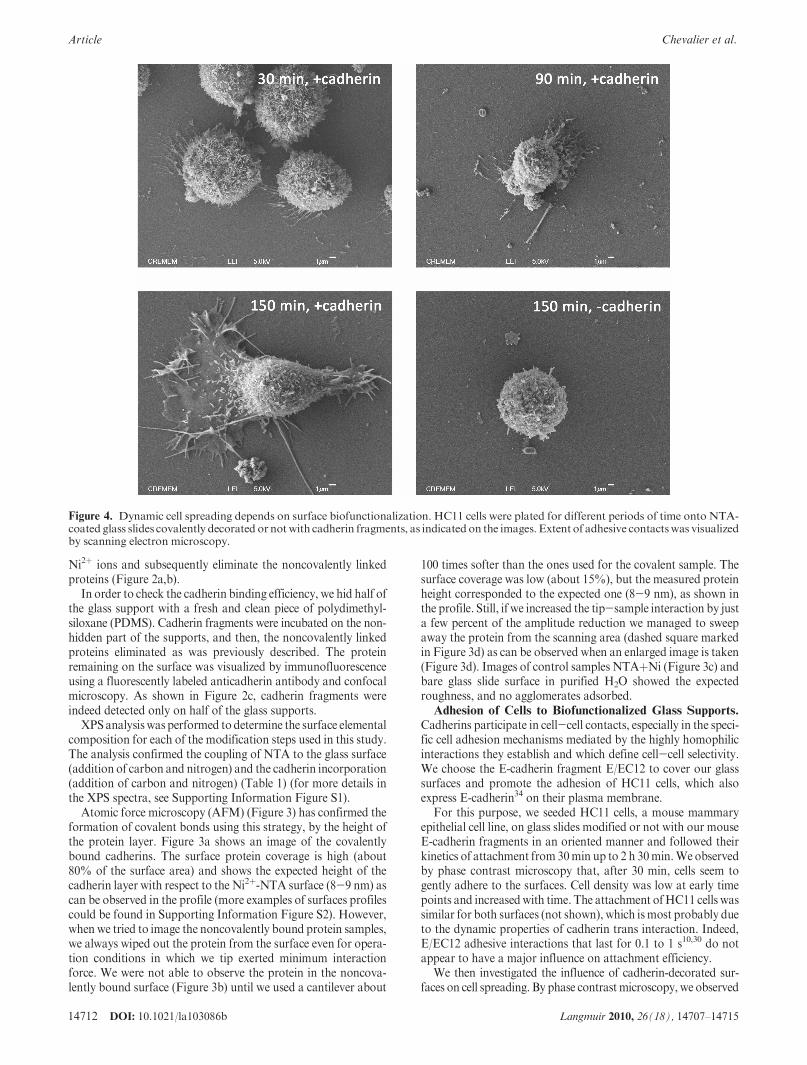
Figure 4. Dynamic cell spreading depends on surface biofunctionalization. HC11 cells were plated for different periods of time onto NTA-coated glass slides covalently decorated ornotwith cadherin fragments, as indicated on the images. Extent of adhesive contactswas visualizedby scanning electron microscopy.

DOI: 10.1021/la103086b 14713Langmuir 2010, 26(18), 14707–14715
Chevalier et al. Article
that cells plated onto cadherin-decorated glass slides were mostlyround at early time points then developed membrane extensionswith time (30% and 38% of cells exhibited extensions after 90 and120min, respectively). These extensions were clearly less numerouson controlNTA-surfaces (6%and 8%of cells exhibited extensionsafter 90 and 120 min, respectively). To gain information aboutthese cell-surface contacts, cell morphology was analyzed using
scanning electron microscopy (SEM). After different incubationtimes, cells were fixed with glutaraldehyde and treated withcontrast agents. As shown in Figure 4, analysis at higher magni-fication revealed that, after 30 min plating on cadherin-coatedsurfaces, cells began to establish contacts with some membraneextensions. After 1 h incubation, although most cells were stillround, we could also observe that some adhered in a stronger
Figure 5. Characterization of cadherin adhesion topography.HC11 cellswere allowed to adhere onNTA-coated (a,b,c) or covalently coatedE-cadherin (d,e,f) coverslips for 1 h 30min in the absence of serum to avoid integrins and growth factor receptor activation. Cells were fixed,permeabilized, and then stained for cadherin (green, a,d), actin (red, b,e), andDNA (blue, c,f in the merge images) and observed by confocallaser scanning microscopy. On cadherin-surfaces, many cells are well-spread and exhibit a large number of membrane tethers involvingcadherin contacts. Most cells still appear spherical onNTA-surfaces (a,b,c). Corresponding orthogonal sections in the x-z and y-z axis areshown alongside, clearly indicating differences in spreading and height of cells on different surfaces. Round cells and spread cells are about13-15 and 6-8 μm height, respectively. Scale bar = 15 μm.

14714 DOI: 10.1021/la103086b Langmuir 2010, 26(18), 14707–14715
Article Chevalier et al.
manner on the surfaces. After 1 h 30 min and 2 h 30 min plating,many cells were well-spread even though some still exhibited around shape. As shown in Figure 4, cells responded to ourcadherin-decorated surfaces, as large membrane extensions werevisible after 2 h 30 min, indicating that cells interact strongly withthe substrate. Cells on control supports without cadherins behavein a different manner. After 2 h 30 min, few cells began to spread,and most of them showed a round shape. Higher magnificationshows that cells did not engage significant membrane extensions toenhance their adhesiveness to the surface.
To further evaluate the functionality of the cadherin fragmentsafter our protocol and analyze the impact of such surface bio-functionalization, we used confocal microscopy. We comparedE-cadherin and actin distribution in HC11 cells adherent onsurfaces covalently decorated with oriented and nonorientedcadherin fragments or NTA-covered surfaces (Figures 5 and 6and Supporting Information Figure S3). We observed after 1 h30min plating similar topographyas described in the SEMexperi-ments: when the cells are cultured on cadherin substrates, manyHC11 cells were well-spread and few still adopted a round shape.On these spread cells, the distribution of actin cytoskeleton wasmost dense at the cell periphery, whereas cadherins were moreuniformly distributed on the cell surface as shown in the merge
image (Figure 5f). On cells attached to cadherin surfaces andexhibiting a round shape, fluorescent labeling highlighted that theactin cytoskeleton was characterized by a circular network offilaments surrounding the nucleus (Figure 5e), together withradial fibers directed toward and ending in the edge of the cell,as already described.47
On substrates with no cadherin functionalization and asobserved by SEM, cells did not spread (Figure 5a,b,c). Nearlyall cells were round with uniform cadherin distribution. Actin didnot form typical cytoskeleton patterns controlled by cadherins asdescribed in Figure 5e.
To better analyze the formation of cadherin-mediated adhesions,we have focused on the ventral cell surface of E-cadherin express-ing cells still in a round shape after 1 h 30 min plating, in contactwith NTA (Figure 6a,b,c), covalent nonoriented (Figure 6d,e,f), orcovalent oriented (Figure 6g,h,i) cadherin-decorated surfaces.E-cadherin exhibited a punctate distribution on all substrates. Nearthe cell edge where new contacts likely form, E-cadherin and actinfilaments (circular and radial) colocalized, essentially on oriented
Figure 6. Effect of surface biofunctionalizationon the recruitment of cadherin and actin at the contact zone.HC11 cells were plated onNTA(a-c), covalently but nonoriented E-cadherin (d-f), or covalently and oriented E-cadherin (g-i) coated surfaces for 1 h 30min. The ventralzone of two round cells was compared for the distribution of cadherin and actin filaments. Panels show for each condition matched three-dimensional projections of 4μmconfocal stacks.Onall types of surfaces, E-cadherin appearedas puncta (a,d,g).However, oriented cadherin-decorated surfaces promoted a peripheral organization and lamellipodia protrusions (g). Interestingly, F-actin exhibited the typical radialorganization driven by cadherin attachments (h) but a diffuse distribution onNTA- and nonoriented cadherin decorated surfaces (b,e). Thisdifference in topography is emphasized in the merge 3D images of the corresponding cells (c,f,i).
(47) Gavard, J.; Lambert, M.; Grosheva, I.; Marthiens, V.; Irinopoulou, T.;Riou, J. F.; Bershadsky, A.; Mege, R. M. Lamellipodium extension and cadherinadhesion: two cell responses to cadherin activation relying on distinct signallingpathways. J. Cell Sci. 2004, 117(Pt 2), 257–70.

DOI: 10.1021/la103086b 14715Langmuir 2010, 26(18), 14707–14715
Chevalier et al. Article
cadherin surfaces. Finally, the topography, visualized by 3D recon-stitution (Figure 6e,f ), clearly showed different actin-cadherindistributions driven by cadherin fragments linked in an orientedfashion onto the surfaces. Such patterns were documented forN-cadherin expressing cell contacts.48
Conclusions
Covalent, stable, and oriented attachment of proteins onsurfaces is a very challenging field of biochemistry and cellbiology. We have designed a fast and inexpensive method forthe immobilization of oligohistidine-taggedproteins by exploitingthe high recognition of this motif to the metal chelator nitrilo-triacetic acid (NTA). Our technology could also be adapted to abroad type of different materials. The use of NTA technologymay be extended to any type of 6�His tagged proteins, and couldbe a great development for biochips, biosensors, substrates for celladhesion, and so forth. Most importantly, this method is compa-tible with a large set of fusion proteins while maintaining theirintegrity, native conformation, and biological function.43 Theprotein immobilization on the surface is controlledwith respect toselectivity: indeed, the orientation of the biomolecules on thesurface is preferentially driven by the position of the histidine tag.With this manuscript, we have also highlighted the importance of
the orientation of adhesion proteins for their incorporation infunctional biomaterials. As has been demonstrated in thiswork, different protein orientations/presentations drive differ-ent cytoskeleton organization, modifying cellular responseagainst them.
Acknowledgment. This work was supported by institutionalfunding from CNRS, as well as by grants from Association pourla Recherche sur le Cancer (subvention libre 3135), R�egionAquitaine, Fondation pour la Recherche M�edicale, la LigueContre le Cancer (Dordogne) CNano GSO (HF), and Pro-gramme Hubert Curien Picasso (HF/JMF), MITYC (Spain)through the projects CTQ2005-07993-C0202/BQU, CTQ2008-03739/PPQ,NAN2004-09125-C07-02, andPET2007_0315.M.L.acknowledges financial support from CSIC. During this work,S.C. received a fellowship from R�egion Aquitaine. J.M.F. thanksARAID and C.C.A. thanks MITYC for financial support.We thank I. Echaniz (INA) for technical support, L. Malicieux,C. Poujol, and P. Legros (PICIN, Universit�e Bordeaux 2) andR. Vall�ee (CRPP) for their help with the confocal microscopy,C. Labrug�ere (ICMCB, Bordeaux) and J. P. Salvetat (CRPP) forXPS measurements and Elisabeth Sellier (CREMEM, Universit�eBordeaux 1) for scanning electron microscopy.
Supporting Information Available: Additional figures asdescribed in text. This material is available free of charge viathe Internet at http://pubs.acs.org.
(48) Lambert, M.; Thoumine, O.; Brevier, J.; Choquet, D.; Riveline, D.; Mege,R. M. Nucleation and growth of cadherin adhesions. Exp. Cell Res. 2007, 313(19),4025–40.