Hyaluronan: from biomimetic to industrial business strategy
22
-
Upload
independent -
Category
Documents
-
view
2 -
download
0
Transcript of Hyaluronan: from biomimetic to industrial business strategy
INFORMATION FOR AUTHORS
Full details of how to submit a manuscript for publication in Natural Product Communications are given in Information for Authors on our Web site
http://www.naturalproduct.us.
Authors may reproduce/republish portions of their published contribution without seeking permission from NPC, provided that any such republication is
accompanied by an acknowledgment (original citation)-Reproduced by permission of Natural Product Communications. Any unauthorized reproduction, transmission or storage may result in either civil or criminal liability.
The publication of each of the articles contained herein is protected by copyright. Except as allowed under national ―fair use‖ laws, copying is not permitted by any means or for any purpose, such as for distribution to any third party (whether by sale, loan, gift, or otherwise); as agent (express or implied) of any third
party; for purposes of advertising or promotion; or to create collective or derivative works. Such permission requests, or other inquiries, should be addressed
to the Natural Product Inc. (NPI). A photocopy license is available from the NPI for institutional subscribers that need to make multiple copies of single articles for internal study or research purposes.
To Subscribe: Natural Product Communications is a journal published monthly. 2011 subscription price: US$1,995 (Print, ISSN# 1934-578X); US$1,995 (Web edition, ISSN# 1555-9475); US$2,495 (Print + single site online); US$595 (Personal online). Orders should be addressed to Subscription Department,
Natural Product Communications, Natural Product Inc., 7963 Anderson Park Lane, Westerville, Ohio 43081, USA. Subscriptions are renewed on an annual
basis. Claims for nonreceipt of issues will be honored if made within three months of publication of the issue. All issues are dispatched by airmail throughout the world, excluding the USA and Canada.
NPC Natural Product Communications
EDITOR-IN-CHIEF
DR. PAWAN K AGRAWAL Natural Product Inc.
7963, Anderson Park Lane,
Westerville, Ohio 43081, USA
EDITORS
PROFESSOR ALESSANDRA BRACA
Dipartimento di Chimica Bioorganicae Biofarmacia,
Universita di Pisa,
via Bonanno 33, 56126 Pisa, Italy
PROFESSOR DEAN GUO
State Key Laboratory of Natural and Biomimetic Drugs,
School of Pharmaceutical Sciences,
Peking University,
Beijing 100083, China [email protected]
PROFESSOR YOSHIHIRO MIMAKI
School of Pharmacy,
Tokyo University of Pharmacy and Life Sciences,
Horinouchi 1432-1, Hachioji, Tokyo 192-0392, Japan
PROFESSOR STEPHEN G. PYNE
Department of Chemistry
University of Wollongong
Wollongong, New South Wales, 2522, Australia
PROFESSOR MANFRED G. REINECKE
Department of Chemistry,
Texas Christian University,
Forts Worth, TX 76129, USA
PROFESSOR WILLIAM N. SETZER
Department of Chemistry
The University of Alabama in Huntsville
Huntsville, AL 35809, USA
PROFESSOR YASUHIRO TEZUKA
Institute of Natural Medicine
Institute of Natural Medicine, University of Toyama,
2630-Sugitani, Toyama 930-0194, Japan
PROFESSOR DAVID E. THURSTON Department of Pharmaceutical and Biological Chemistry,
The School of Pharmacy,
University of London, 29-39 Brunswick Square,
London WC1N 1AX, UK
ADVISORY BOARD
Prof. Berhanu M. Abegaz
Gaborone, Botswana
Prof. Viqar Uddin Ahmad
Karachi, Pakistan
Prof. Øyvind M. Andersen
Bergen, Norway
Prof. Giovanni Appendino
Novara, Italy
Prof. Yoshinori Asakawa
Tokushima, Japan
Prof. Lee Banting
Portsmouth, U.K.
Prof. Julie Banerji
Kolkata, India
Prof. Alejandro F. Barrero
Granada, Spain
Prof. Anna R. Bilia
Florence, Italy
Prof. Maurizio Bruno
Palermo, Italy
Prof. César A. N. Catalán
Tucumán,Argentina
Prof. Josep Coll
Barcelona, Spain
Prof. Geoffrey Cordell
Chicago, IL, USA
Prof. Cristina Gracia-Viguera
Murcia, Spain
Prof. Duvvuru Gunasekar
Tirupati, India
Prof. A.A. Leslie Gunatilaka
Tucson, AZ, USA
Prof. Kurt Hostettmann
Lausanne, Switzerland
Prof. Martin A. Iglesias Arteaga
Mexico, D. F, Mexico
Prof. Jerzy Jaroszewski
Copenhagen, Denmark
Prof. Leopold Jirovetz
Vienna, Austria
Prof. Karsten Krohn
Paderborn, Germany
Prof. Hartmut Laatsch
Gottingen, Germany
Prof. Marie Lacaille-Dubois
Dijon, France
Prof. Shoei-Sheng Lee
Taipei, Taiwan
Prof. Francisco Macias
Cadiz, Spain
Prof. Imre Mathe
Szeged, Hungary
Prof. Joseph Michael
Johannesburg, South Africa
Prof. Ermino Murano
Trieste, Italy
Prof. M. Soledade C. Pedras
Saskatoon, Cnada
Prof. Luc Pieters
Antwerp, Belgium
Prof. Peter Proksch
Düsseldorf, Germany
Prof. Phila Raharivelomanana
Tahiti, French Plynesia
Prof. Monique Simmonds
Richmond, UK
Prof. Valentin Stonik
Vladivostok, Russia
Prof. Winston F. Tinto
Barbados, West Indies
Prof. Karen Valant-Vetschera
Vienna, Austria
Prof. Peter G. Waterman
Lismore, Australia
HONORARY EDITOR
PROFESSOR GERALD BLUNDEN
The School of Pharmacy & Biomedical Sciences,
University of Portsmouth,
Portsmouth, PO1 2DT U.K.
Hyaluronan: From Biomimetic to Industrial Business
Strategy
Erminio Muranoa,b
, Danilo Perina, Riaz Khan
a and Massimo Bergamin
a,b
aPROTOS Research Institute, via Flavia 23/1c/o BIC Incubatori FVG, 34148, Trieste, Italy
bNEALYS srl, via Flavia 23/1c/o BIC Incubatori FVG, 34148, Trieste, Italy
Received: December 20th
, 2010; Accepted: February 2nd
, 2011
Hyaluronan (hyaluronic acid) is a naturally occurring polysaccharide of a linear repeating disaccharide unit consisting of β-(1→4)-linked
D-glucopyranuronic acid and β-(1→3)-linked 2-acetamido-2-deoxy-D-glucopyranose, which is present in extracellular matrices, the synovial
fluid of joints, and scaffolding that comprises cartilage.
In its mechanism of synthesis, its size, and its physico-chemical properties, hyaluronan is unique amongst other glycosaminoglycans. The
network-forming, viscoelastic and its charge characteristics are important to many biochemical properties of living tissues. It is an important
pericellular and cell surface constituent; its interaction with other macromolecules such as proteins, participates in regulating cell behavior
during numerous morphogenic, restorative, and pathological processes in the body. The knowledge of HA in diseases such as various forms
of cancers, arthritis and osteoporosis has led to new impetus in research and development in the preparation of biomaterials for surgical
implants and drug conjugates for targeted delivery.
A concise and focused review on hyaluronan is timely. This review will cover the following important aspects of hyaluronan: (i) biological
functions and synthesis in nature; (ii) current industrial production and potential biosynthetic processes of hyaluronan; (iii) chemical
modifications of hyaluronan leading to products of commercial significance; and (iv) and the global market position and manufacturers of
hyaluronan.
Keywords: Hyaluronan, hyaluronic acid, biosynthesis, tissue engineering, antitumor drugs, crosslinked HA, global markets.
Hyaluronan (also known as hyaluronic acid) (HA) is a
naturally occurring polysaccharide of a linear repeating
disaccharide unit consisting of β-(1→4)-linked D-
glucopyranuronic acid and β-(1→3)-linked 2-acetamido-2-
deoxy-D-glucopyranose, which is present in extracellular
matrices, the synovial fluid of joints, and scaffolding that
comprises cartilage. However, other organisms, such as
bacterial pathogens and virus infected micro algae, are able
to synthesis this polysaccharide. HA was first isolated
from the vitreous body of the eye by Mayer and Palmer [1]
and then found in all vertebrates tissues, from skin to
synovial fluid between joins, from umbilical cord to skin
and cartilage [2-5].
Despite its simplistic structure hyaluronan behaves quite
differently from other glycosaminoglycans in its
mechanism of synthesis, its size, and its physico-chemical
properties. The network-forming, viscoelastic and its
charge characteristics are important to many biochemical
properties of living tissues. It is an important pericellular
and cell surface constituent which, through interaction
with other macromolecules such as proteins, participates in
regulating cell behavior during numerous morphogenic,
restorative and pathological processes in the body. The
knowledge of such biological functions of hyaluronan has
led to the preparation of biomaterials for surgical implants
and drug conjugates for targeted delivery.
The interest in the role of hyaluronan diseases such as
various forms of cancers, arthritis and osteoporosis has led
to new impetus in research and development. Our
laboratory has been involved in the study of chemistry and
physico-chemical properties of hyaluronan with a view to
develop biomaterials and drugs conjugates.
As a general arrangement of the review the subject is
divided into: biological function and synthesis in nature;
industrial production of hyaluronan; chemical modification
of hyaluronan; and market position of hyaluronan.
Biological function and synthesis in nature
Hyaluronan was first isolated from the vitreous body of the
eye by Mayer and Palmer [1] and then found in all
vertebrates tissues, from skin to synovial fluid between
joins, from umbilical cord to skin and cartilage [2-9].
Hyaluronan is a glycosaminoglycan that has a number of
biological functions in vertebrates, being essential for
tissue and organ integrity and function. Hyaluronan
molecules are involved in the activation of signaling
NPC Natural Product Communications 2011
Vol. 6
No. 4
555 - 572
556 Natural Product Communications Vol. 6 (4) 2011 Murano et al.
pathways that control cell proliferation, differentiation,
adhesion and migration [10-20].
The primary structural function of HA is related to its
viscoelastic and hygroscopic properties. It acts as shock-
adsorber and offers lubrication to the joints, creates a
protective and flexible layer between tissues, influence cell
responses during a number of processes, inflammation and
cancer [21-28]. A wide range of different cell responses
are evoked by HA molecules which different molecular
weights [29]. In general, high molecular weight
hyaluronan inhibits cell differentiation and promotes cell
proliferation, being involved in tissue regeneration, wound
healing, epithelial integrity and embryogenesis. Lower
molecular weights HA are anti-angiogenic, immuno-
suppressive and hamper the differentiation by limiting the
cell-cell interactions or ligand access to cell receptors.
Finally, HA oligomers showed angiogenic and pro-
inflammatory activity, increase immune response and are
capable to slow down growth of a number of tumors in
vivo [30-37].
HA mediates his biological function through specific
protein receptors present on the different cell surface,
which include CD44, HARE, RHAMM, LYVE [29,38-43].
In nature, hyaluronan is synthesized by specific
glycosyltransferases (HA synthases or HAse) that
polymerizes the activated donor uridine diphospho- sugars
UDP-glucuronic acid and UDP-N-acetyl-glucosamine into
hyaluronan [44-47]. In nature, hyaluronan was shown to be
synthesized also by a small number of microbial pathogens
such us Pasteurella multocida and the Lancefield group A
and C streptococci among which Streptococcus equi,
the human pathogen Streptococcus pyogenes and the
bovine pathogen Streptococcus uberis. [48-58]. These
microorganisms adopted the strategy to encapsulate their
cells with hyaluronan in order to perfectly disguise from
the host animal defense system and allow the adhesion and
colonization. In Streptococcus and Pasteurella, the HA
capsules represent virulence factors affording to the
bacteria a stealth function against the recognition from the
host immune system [45,59-61]. On the contrary to other
bacterial capsule polysaccharides triggering the antibody
immune response, HA is not immunogenic because it is a
normal component of the host body conferring to these
pathogen bacteria an efficient molecular camouflage.
Moreover, the HA capsule provide a protection against the
reactive oxides released by leukocytes [62] and facilitate the
bacteria migration through epithelial layer into tissues [63].
In Streptococcus equi subsp. zooepidemicus hyaluronic
acid capsular material contributes to adherence properties
of the pathogen possibly conferring to the bacteria the
resistance to phagocytosis by macrophages [64-66].
The importance of the capsule in pharyngeal colonization
and other inflammatory syndromes caused by
Streptococcus pyogenes may reflect the role of hyaluronan
as a specific ligand for attachment of Streptococcus
pyogenes to the hyaluronan-binding protein CD44 on
pharyngeal epithelial cells [67,68].
Pasteurella multocida, a prevalent animal pathogen,
employs a hyaluronan polysaccharide capsule to avoid host
defenses. This pathogen is associated with various diseases
in many animal species; a number of studies suggested that
there is a correlation between the capsule and the virulence
of P. multocida, for example in the pathogenesis of fowl
cholera [52,69-74].
This biomimetic strategy of prokaryotic organisms
efficiently exploits the chemical identity of the bacterial
capsular HA molecules to that of the eukaryotic host
extracellular matrix. Such a biomimetic strategy adopted
by specialized prokaryotic cells to proliferate into an
animal host organism has the same goal as many types of
endogenous cells like tumor cells to invade and proliferate
into normal tissues. In fact, a large number of tumor cells
over express membrane receptors for hyaluronan in order
to more efficiently interact with this major component of
the extracellular matrix to easily spread and proliferate into
the tissues [75,76].
The existence of hyaluronan outside the animals and their
pathogens has been demonstrated in green microalgae
infected by specialized viruses. Chlorovirus is a group of
virus of Phycodnaviridae family that infects the eukaryotic
microalgae Chlorella [77,78]. Chlorovirus dsDNA genome
unusually encodes enzymes involved in sugar metabolism.
The 330-kilobase genome of the chlorovirus PBCV-1
encode other than fructose-6-phosfate aminotransferase
and UDP-glucose dehydrogenase also a HA synthase,
resulting in the production of an HA capsule surrounding
the Chlorella infected cells. However, some chlorovirus
contain both HA synthase and chitin synthase genes and
form both hyaluronan and chitin, an N-acetyl-glucosamine
based polysaccharide found in the crustaceans shell, in the
insect exoskeletons and fungi cell wall [79], on the surface
of their infected cells [80-82].
The function of a HA capsule in chlorovirus infected
chlorella cells is still unknown; however, since the genes
encoding for HA synthesis are transcribed early in
PBCV-1 infection and HA accumulates on the external
surface of the infected chlorella cells, hyaluronan synthesis
might be related to the virus life cycle and the recognition
of infected cells. The distribution of hyaluronan synthases
from vertebrates to bacterial pathogens and algal viruses
makes these fascinating glycosyltransferases an interesting
evolutionary case which can advance the knowledge on the
biological functions of a typical natural molecule like
hyaluronan.
Industrial production of hyaluronan
HA has been traditionally produced by extracting from
rooster combs and bovine vitreous humor The process is
difficult, particularly to ensure the biological safety and to
Review on hyaluronan Natural Product Communications Vol. 6 (4) 2011 557
control the molecular weight of the product [3]. Hence,
since 1980‘s, the commercial production of HA has moved
to the fermentation process using streptococcal bacteria.
The most used strains are S. equi subsp. equi and S. equi
subsp. zooepidemicus that naturally achieve good HA
production rate.
Improved HA producer streptococcal strain has been
obtained by random mutagenesis followed by a serial
selection of the mutant clones. The aim of the strain
selection was mainly oriented to the reduction of the HA-
depolymerising enzyme (hyaluronidase-negative strains)
and other extracellular proteins such as the virulence factor
streptolysins, the exotoxins responsible for the β-hemolysis
(non-hemolytic strains) [83] as well as the production of
high molecular weight HA [84,85]. Several studies were
performed in order to improve the HA yield, molecular
weight and reduce the polydispersity by an accurate
control of the growing conditions. Unfortunately,
Streptococci are lactic acid bacteria that demand expensive
complex growth medium containing yeast or animal
extract, peptone and serum supplemented with an high
glucose content [83,86,87].
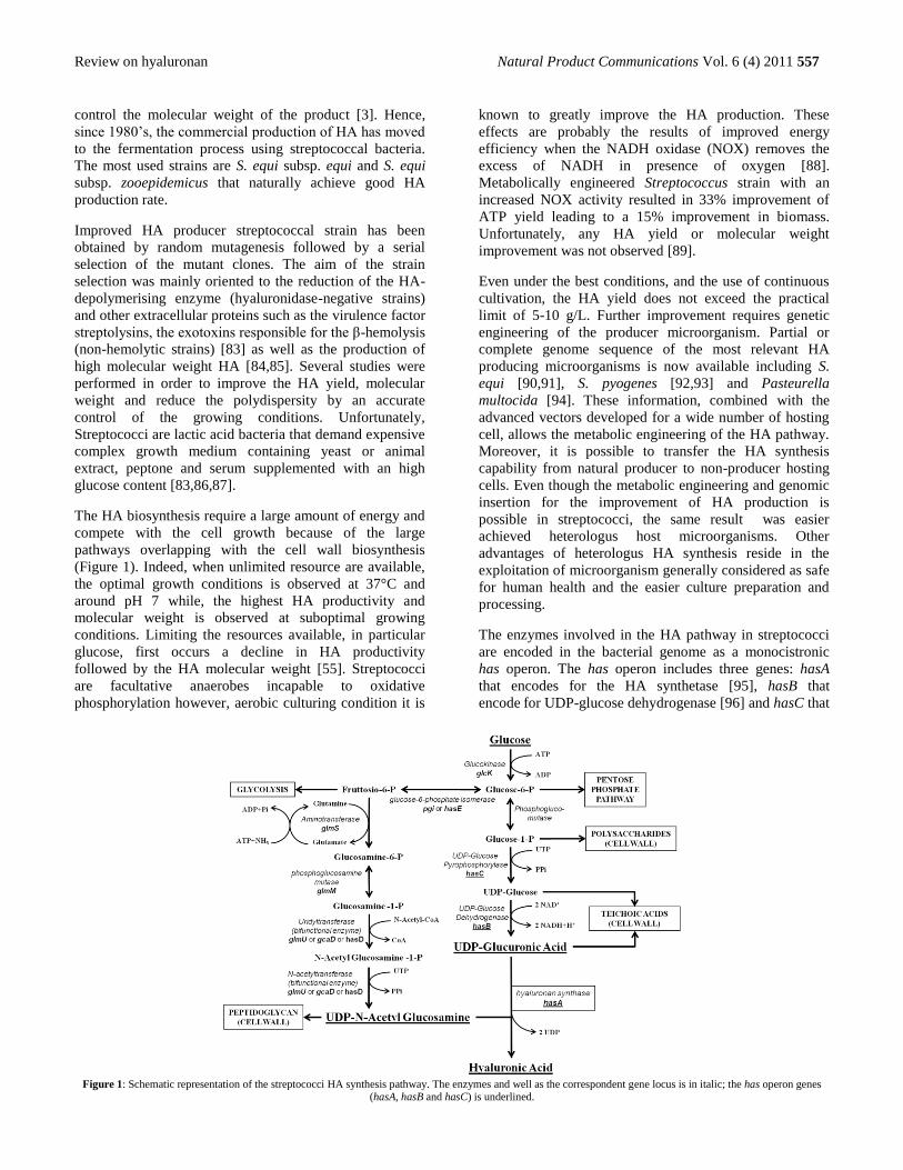
The HA biosynthesis require a large amount of energy and
compete with the cell growth because of the large
pathways overlapping with the cell wall biosynthesis
(Figure 1). Indeed, when unlimited resource are available,
the optimal growth conditions is observed at 37°C and
around pH 7 while, the highest HA productivity and
molecular weight is observed at suboptimal growing
conditions. Limiting the resources available, in particular
glucose, first occurs a decline in HA productivity
followed by the HA molecular weight [55]. Streptococci
are facultative anaerobes incapable to oxidative
phosphorylation however, aerobic culturing condition it is
known to greatly improve the HA production. These
effects are probably the results of improved energy
efficiency when the NADH oxidase (NOX) removes the
excess of NADH in presence of oxygen [88].
Metabolically engineered Streptococcus strain with an
increased NOX activity resulted in 33% improvement of
ATP yield leading to a 15% improvement in biomass.
Unfortunately, any HA yield or molecular weight
improvement was not observed [89].
Even under the best conditions, and the use of continuous
cultivation, the HA yield does not exceed the practical
limit of 5-10 g/L. Further improvement requires genetic
engineering of the producer microorganism. Partial or
complete genome sequence of the most relevant HA
producing microorganisms is now available including S.
equi [90,91], S. pyogenes [92,93] and Pasteurella
multocida [94]. These information, combined with the
advanced vectors developed for a wide number of hosting
cell, allows the metabolic engineering of the HA pathway.
Moreover, it is possible to transfer the HA synthesis
capability from natural producer to non-producer hosting
cells. Even though the metabolic engineering and genomic
insertion for the improvement of HA production is
possible in streptococci, the same result was easier
achieved heterologus host microorganisms. Other
advantages of heterologus HA synthesis reside in the
exploitation of microorganism generally considered as safe
for human health and the easier culture preparation and
processing.
The enzymes involved in the HA pathway in streptococci
are encoded in the bacterial genome as a monocistronic
has operon. The has operon includes three genes: hasA
that encodes for the HA synthetase [95], hasB that
encode for UDP-glucose dehydrogenase [96] and hasC that
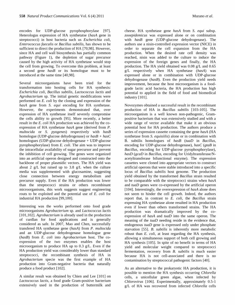
Figure 1: Schematic representation of the streptococci HA synthesis pathway. The enzymes and well as the correspondent gene locus is in italic; the has operon genes
(hasA, hasB and hasC) is underlined.
558 Natural Product Communications Vol. 6 (4) 2011 Murano et al.
encodes for UDP-glucose pyrophosphorylase [97].
Heterologus expression of HA synthetatse (hasA gene in
streptococci) in host bacteria such us Escherichia coli,
Enterococcus faecalis or Bacillus subtilis, has shown to be
sufficient to direct the production of HA [70,98]. However,
since HA and cell wall biosynthesis has partially common
pathway (Figure 1), the depletion of sugar precursor
caused by the high activity of HA synthetase would stop
the cell from growing. To overcome this problem, at least
a second gene hasB or its homologue must to be
introduced at the same time [40,98].
Several microorganisms have been tried for the
transformation into hosting cells for HA synthesis:
Escherichia coli, Bacillus subtilis, Lactococcus lactis and
Agrobacterium sp. The initial genetic modifications were
performed on E. coli by the cloning and expression of the
hasA gene from S. equi encoding for HA synthetase.
However, the experiments demonstrated that, over
expression of HA synthetase itself severely compromise
the cells ability to growth [95]. More recently, a better
result in the E. coli HA production was achieved by the co-
expression of HA synthetase hasA gene (derived from P.
multocida or S. pyogenes) respectively with hasB
homologue (UDP-glucose dehydrogenase) or hasB + hasC
homologues (UDP-glucose dehydrogenase + UDP-glucose
pyrophosphorylase) from E. coli. The aim was to improve
the intracellular availability of sugar precursor and prevent
the inhibition of cell growing. The genes were organized
into an artificial operon designed and constructed onto the
backbone of proper plasmidic vectors. The HA yield was
about 2 g/L but raised up to 3.8 g/L when the culture
media was supplemented with glucosamine, suggesting
close connection between energy metabolism and
precursor supply. Even if the HA production was lower
than the streptococci strains or others recombinant
microorganisms, this work suggests suggest engineering
routs to be exploited and the potential use of E. coli for
industrial HA production [99,100].
Interesting was the works performed onto food grade
microorganisms Agrobacterium sp. and Lactococcus lactis
[101,102]. Agrobacterium is already used in the production
of curdlan for food applications and is generally
considered as safe. In 2007, Mao and Chen, successfully
transfered HA synthetase gene (hasA) from P. multocida
and an UDP-glucose dehydrogenase homologue gene
(hasB) from E. coli into Agrobacterium host. The co-
expression of the two enzymes enables the host
microorganism to produce HA up to 0.3 g/L. Even if the
HA production yield was considerably lower than standard
streptococci, the recombinant synthesis of HA in
Agrobacterium specie was the first example of HA
production into Gram-negative bacteria that naturally
produce a food product [102].
A similar result was obtained by Chien and Lee [101] on
Lactococcus lactis, a food grade Gram-positive bacterium
extensively used in the production of buttermilk and
cheese. HA synthetase gene hasA from S. equi subsp.
zooepidemicus was expressed alone or on combination
with hasB gene (UDP-glucose dehydrogenase). The
authors use a nisin-controlled expression vector (NICE) in
order to separate the cell expansion from the HA
production. When the desired rate cell density was
reached, nisin was added to the culture to induce the
expression of the foreign genes and finally, the HA
production. The HA yield obtained was 0.08 g/L and 0.65
g/L respectively when HA synthetase (hasA) was
expressed alone or in combination with UDP-glucose
dehydrogenase (hasB). Even the production yield needs
improvement, because the host microorganism is a food-
grade lactic acid bacteria, the HA production has high
potential to applied in the field of food and biomedical
industry [101].
Novozymes obtained a successful result in the recombinant
production of HA in Bacillus subtilis [103-105]. The
microorganism is a well known non-pathogenic, Gram-
positive bacterium that was extensively studied and with a
wide range of vector available that make it an obvious
potential host for HA production. The authors produce a
series of expression cassette containing the gene hasA (HA
synthetase from S. equisiilis) alone or in combination with
B. subtilis homologues of hasB (tuaD in Bacillus,
encoding for UDP-glucose dehydrogenase), hasC (gtaB in
Bacillus, encoding for UDP-glucose pyrophosphorylase),
hasD (gcaD in Bacillus, encoding for Uridyltransferase/N-
acetyltransferase bifunctional enzyme). The expression
cassettes were cloned into appropriate vectors to construct
artificial operons that were stable introduced into a specific
locus of Bacillus subtilis host genome. The production
yield obtained by the transformed Bacillus strain resulted
to be comparable with the streptococci strains when hasA
and tuaD genes were co-expressed by the artificial operon
[104]. Interestingly, the overexpression of hasA alone does
not seem to hinder the cell growth. Indeed, the authors
report that, in contrast to E. coli, the Bacillus strain
expressing HA synthetase alone resulted in HA production
even if lower than others transformed strains. The HA
production was dramatically improved by the co-
expression of hasA and tuaD into the same operon. The
meaning of the tuaD needing reside in the evidence that,
endogenous tuaD gene is expressed only under phosphate
starvation [55]. B. subtilis is inherently more metabolic
robust than E. coli, at least regarding the HA synthesis,
allowing a simultaneous support of both cell growing and
HA synthesis [105]. In spite of no benefit in terms of HA
yield and molecular weight compared to streptococci
fermentation, recovery from B. subtilis is much easier
because HA is not cell-associated and there is no
contamination by streptococcal pathogenic factors [40].
As an alternative to the prokaryotic HA production, it is
possible to mention the HA synthesis occurring Chlorella
cells, a unicellular green algae, when infected by
Chlorovirus [106]. Experimentally, approximately 0.5-1
g/L of HA was recovered from infected Chlorella cells
Review on hyaluronan Natural Product Communications Vol. 6 (4) 2011 559
after 4 hours of infection [107]. The yield is lower than typically ones achieved in streptococcal fermentation but further implementation is possible to improve it [40].
Finally, it is interesting the work of Takeo and colleagues [108] on the cloning and expression of mammalian HA synthetase. The authors genetically modify the genome of the fruit fly Drosophila melanogaster, a non-HA-synthesizing animal, by introduction of the single gene encoding mouse HA synthetase. The expression of HA synthetase in vivo resulted in massive HA accumulation in the extracellular space of the Drosophila tissues and caused various morphological defect. The original aim of the work was the development of an in vivo model to study the biosynthetic machinery of the polysaccharides besides, this could represent an alternative route for the HA production in eukaryotic cells [108].
Chemical modification of hyaluronan
The interest in the role of hyaluronic acid in diseases such as various forms of cancers, arthritis and osteoporosis has led to new impetus in research and development. Our laboratory has been involved in the study of chemistry and physico-chemical properties of hyaluronic acid with a view to develop biomaterials and drugs conjugates [109].
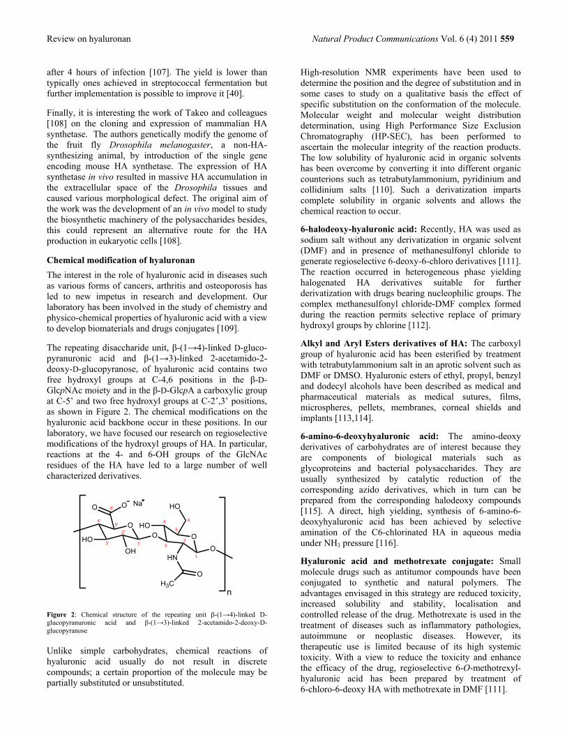
The repeating disaccharide unit, β-(1→4)-linked D-gluco-pyranuronic acid and β-(1→3)-linked 2-acetamido-2-deoxy-D-glucopyranose, of hyaluronic acid contains two free hydroxyl groups at C-4,6 positions in the β-D-GlcpNAc moiety and in the β-D-GlcpA a carboxylic group at C-5’ and two free hydroxyl groups at C-2’,3’ positions, as shown in Figure 2. The chemical modifications on the hyaluronic acid backbone occur in these positions. In our laboratory, we have focused our research on regioselective modifications of the hydroxyl groups of HA. In particular, reactions at the 4- and 6-OH groups of the GlcNAc residues of the HA have led to a large number of well characterized derivatives.
Figure 2: Chemical structure of the repeating unit β-(1→4)-linked D-glucopyranuronic acid and β-(1→3)-linked 2-acetamido-2-deoxy-D-glucopyranose
Unlike simple carbohydrates, chemical reactions of hyaluronic acid usually do not result in discrete compounds; a certain proportion of the molecule may be partially substituted or unsubstituted.
High-resolution NMR experiments have been used to determine the position and the degree of substitution and in some cases to study on a qualitative basis the effect of specific substitution on the conformation of the molecule. Molecular weight and molecular weight distribution determination, using High Performance Size Exclusion Chromatography (HP-SEC), has been performed to ascertain the molecular integrity of the reaction products. The low solubility of hyaluronic acid in organic solvents has been overcome by converting it into different organic counterions such as tetrabutylammonium, pyridinium and collidinium salts [110]. Such a derivatization imparts complete solubility in organic solvents and allows the chemical reaction to occur.
6-halodeoxy-hyaluronic acid: Recently, HA was used as sodium salt without any derivatization in organic solvent (DMF) and in presence of methanesulfonyl chloride to generate regioselective 6-deoxy-6-chloro derivatives [111]. The reaction occurred in heterogeneous phase yielding halogenated HA derivatives suitable for further derivatization with drugs bearing nucleophilic groups. The complex methanesulfonyl chloride-DMF complex formed during the reaction permits selective replace of primary hydroxyl groups by chlorine [112].
Alkyl and Aryl Esters derivatives of HA: The carboxyl group of hyaluronic acid has been esterified by treatment with tetrabutylammonium salt in an aprotic solvent such as DMF or DMSO. Hyaluronic esters of ethyl, propyl, benzyl and dodecyl alcohols have been described as medical and pharmaceutical materials as medical sutures, films, microspheres, pellets, membranes, corneal shields and implants [113,114].
6-amino-6-deoxyhyaluronic acid: The amino-deoxy derivatives of carbohydrates are of interest because they are components of biological materials such as glycoproteins and bacterial polysaccharides. They are usually synthesized by catalytic reduction of the corresponding azido derivatives, which in turn can be prepared from the corresponding halodeoxy compounds [115]. A direct, high yielding, synthesis of 6-amino-6-deoxyhyaluronic acid has been achieved by selective amination of the C6-chlorinated HA in aqueous media under NH3 pressure [116].
Hyaluronic acid and methotrexate conjugate: Small molecule drugs such as antitumor compounds have been conjugated to synthetic and natural polymers. The advantages envisaged in this strategy are reduced toxicity, increased solubility and stability, localisation and controlled release of the drug. Methotrexate is used in the treatment of diseases such as inflammatory pathologies, autoimmune or neoplastic diseases. However, its therapeutic use is limited because of its high systemic toxicity. With a view to reduce the toxicity and enhance the efficacy of the drug, regioselective 6-O-methotrexyl-hyaluronic acid has been prepared by treatment of 6-chloro-6-deoxy HA with methotrexate in DMF [111].
OO O
OO HO
HO
OHN
H3C
HO
OH
O
Na
n
1'
2'
3'
4'5'
6'
1
23
4
5
6

560 Natural Product Communications Vol. 6 (4) 2011 Murano et al.
Hyaluronic acid and taxol conjugate: Taxol (paclitaxel),
an antileukemic and antitumor agent, was first isolated
from the bark of the Pacific yew tree, Taxus bravifolia;
has a limited solubility in water. Hyaluronic acid is over
expressed at sites of tumor and provides a matrix to
facilitate invasion. Hence, to overcome the solubility
problem and to target the tumor cells, a first attempt to
synthesize an hyaluronic acid conjugate of taxol was
performed by Luo et al. by linking the taxol 2‘-OH by
way of succinate ester to adipic dihydrazide-modified HA
(HA-ADH) [117].
Recently, Lee et al conjugated paclitaxel and HA utilizing
a novel solubilization method in a single organic phase.
Hydrophilic HA was completely dissolved in anhydrous
DMSO with addition of poly(ethylene glycol) (PEG) by
forming nano-complexes. Paclitaxel was then chemically
conjugated to HA in the DMSO phase via an ester linkage
without modifying extremely hydrophilic HA [118]. The
conjugate exhibited selective toxicity toward the human
cancer cell lines (breast, colon and ovarian) that are
known to express hyaluronic acid receptors; no toxicity
was noted against a mouse fibroblast cell line at the same
concentrations.
Hyaluronic acid and mitomycin C: Mitomycin C was
linked to the HA molecule by way of an amide
bond between the drug and the carboxyl group of the
D-glucuronic acid moiety. An amide linkage formation is a
dehydration reaction and requires anhydrous systems.
However, as hyaluronic acid is difficult to dissolve in an
anhydrous organic solvent, reaction conditions were
developed to use a water based system [119]. Other
strategies envisage the use of a succinylated HA as active
intermediate for the conjugation with the drug [120].
Hyaluronic acid and camptothecin conjugate: A novel
methodology for making drug conjugates using hyaluronan
as a carrier was recently developed. This strategy involves
a completely regioselective two-step synthesis of 6-amino-
6-deoxyhyaluronan, which is then easily functionalized
making use of a simple succinate linker [116].
Hyaluronic acid and other drugs: A number of
antitumor drugs, such as doxorubicin [121] and
daunorubicin, have been linked at the amino function of
the drug to the carboxylic group of an amino acid or a
peptide spacer arm forming an amide linkage, and then
through the terminal amino group of the amino acid or the
peptide to the carboxyl group of the HA.
Hyaluronic acid and methylprednisolone: Steroid-type
of anti-inflammatory drugs such as methylprednisolone
has proved to be efficacious in reducing symptoms
associated with osteoarthritis of the knee [122]. Alkyl and
aryl esters of HA have been used in the preparations
containing methylprednisolone, both as polymer
matrices in the form of microspheres wherein the drug is
physically incorporated, and as substrates on which
methylprednisiolone is chemically linked [123,124].
Hyaluronic acid and antioxidants: Reactive oxygen
species (ROS) such as superoxide radicals generated by
metabolic processes can depolymerise hyaluronic acid
[125]. It has been demonstrated that many anti-
inflammatory drugs and free radical scavengers are
efficacious in protecting hyaluronic acid depolymerisation
by free radicals [126-128].
Hyaluronic acid and propofol conjugate: 2,6-
Diisopropylphenol (propofol) is an anaesthetic for
intravenous administration whose efficacy as a free radical
scavenger is due to its ability to form stable radicals. Its
effectiveness as a scavenger for hydroxyl radicals
generated by xanthine oxidase has been demonstrated by
assessing in vitro the depolymerisation of hyaluronic acid
in artificial synovial fluid [129]. Low-molecular-mass
hyaluronic acid oligosaccharides have major biological
effects: notably, they appear to play a role in the
recruitment and activation of inflammatory macrophages,
which may result in an exacerbation of the inflammatory
process and tissue damage [130,131]. For this reason,
propofol was proposed as a shield for hyaluronic acid
[124].
Cross-linked Hyaluronic Acid-based materials: Hyaluronic acid is rapidly metabolised in vivo by enzymes
such as hyaluronidase [132] and by free radical oxidations
[131], which limit its use in native form as a biomaterial.
In, addition hyaluronic acid is highly soluble in water. In
order to overcome these limitations, the physico-chemical
properties of hyaluronic acid have been modified using a
variety of cross-linking reagents. These chemical
modifications have led to a number of hyaluronic acid
derivatives with a variety of physical formats such as
liquids of various viscosities, fibers, weaves, sponges and
fleeces. Cross-linked hyaluronic acids with special
properties and structure, such as different degradation rate,
different surface characteristics, different porosities, have
applications as tissue engineering scaffolds for the delivery
of cells, gene transfer, wound healing, post-surgical
adhesion prevention, and implantation of bioactive
compounds in vivo repair sites.
Ossipov et al. developed a new methodology exploiting a
protective group strategy based on initial mild cleavage of
a disulfide bond followed by elimination of the generated
2-thioethoxycarbonyl moiety ultimately affording free
amine-type functionality. The strategy therefore
encompasses a new approach for mild and highly
controlled functionalization of HA with both nucleophilic
and electrophilic chemoselective functionalities with the
emphasis for the subsequent conjugation and in situ
crosslinking [133].
Li et al. synthesized a polymeric nanogel, with tumor
targeting properties and a controllable phototoxicity,
utilizing a low molecular weight-hyaluronic acid
photosensitizer conjugate, resulting in the formulation of
self-organizing nanogels in aqueous solutions [134] while
Review on hyaluronan Natural Product Communications Vol. 6 (4) 2011 561
Kim et al synthesized hyaluronic acid/quantum dots
(QDots) conjugates. The reaction was performed through
amide bond between carboxyl groups of QDots and amine
groups of adipic acid dihydrazide modified HA (HA-
ADH) assessing the possibility of HA derivatives as
target-specific drug delivery carriers for the treatment of
liver diseases [135]. In the field of delivery systems
alkylamino hydrazide hyaluronic acid (HA) derivatives
were prepared to design new biocompatible films able to
release hydrophobic drugs. These films were found to be
more resistant to degradation by hyaluronidase compared
to solutions of unmodified HA at the same concentration
[136]. Bencherif et al developed a nanostructured
hydrogel made of thiolated HA and reacted with
biodegradable POEO300MA-co-PHEMA nanogels
derivatized with acryloyl groups. The resulting scaffold
was obtained through selective atom transfer radical
polymerization (ATRP) and Michael-type addition
reactions, affording a biocompatible matrix for cell and
protein encapsulation in both tissue engineering and drug
delivery applications [137]. Skoza et al exploited the
specificity of hyaluronate lyase to biodegrade HA and
introduce a double bond in the glucuronic acid moiety
followed by the generation of free aldehyde group via
ozonolysis and the subsequent reduction of the generated
ozonide. This gives rise to a new reactive HA substrate to
be derivatized with several molecules (such as biotin,
polymers, or proteins) with application in tissue
engineering and biomaterials production [138].
A strong commercial interest drives the extensive use of
HA hydrogels in cosmetics and tissue engineering. Due to
the frequent use of HA derivatives in aesthetic surgery,
novel, biocompatible, and non-toxic dermal fillers
hyaluronic acid (HA) hydrogels were studied. Yeom et al
successfully developed new hydrogel for tissue
augmentation applications. Instead of using highly
reactive cross-linkers such as divinylsulfone (DVS) [139]
for Hylaform, 1,4-butanediol diglycidyl ether (BDDE) for
Restylane, and 1,2,7,8-diepoxyoctane (DEO) for Puragen,
HA hydrogels were prepared by direct amide bond
formation between the carboxyl groups of HA and
hexamethylenediamine (HMDA) with an optimized
carboxyl group modification for effective tissue
augmentation [140]. Crosslinked hyaluronic acid
hydrogels have been specially designed and synthesized
to promote tissue repair. A range of glycidyl
methacrylate-hyaluronic acid (GMHA) conjugates have
been synthesized and photopolymerized to lead to a
number of crosslinked GMHA hydrogels with different
physical properties such as swelling, mesh size. As
expected, the amount of crosslinking and the degree of
degradation was dependent on the amount of the
conjugated methacrylate; the more crosslinking the less
degradation rate [141,142]. Su et al. developed an
hydrogel formed by oxidated hyaluronic acid (oxi-HA)
cross-linked with adipic acid dihydrazide (ADH). HA was
oxidized by sodium periodate to create aldehyde
functional groups, which could be cross-linked by ADH.
The result was a colorless, transparent and injectable
hydrogel as potential vitreous substitute [143]. Other type
of hydrogel were those made by Oldinsky et al,
characterized by both a hydrogel and a hydrophobic
polymer for biomedical applications. A series of
amphiphilic graft copolymers consisting of HA, a
glycosaminoglycan, and high-density polyethylene
(HDPE), that is, HA-co-HDPE, were fabricated.
Exploiting an esterification reaction between HA and
functionalized HDPE the resulting material was a
semicrystalline, insoluble powder applicable in saline
suspension or molded form, including orthopedic tissue
repair [144].
The extreme versatility of HA is emphazised also by its
employment in material science with carbon nanotubes.
Hybrid hyaluronic acid (HA) hydrogels with single wall
nanotubes (SWNTs) were formed by cross-linking with
divinyl sulfone, producing considerable change in the
morphology of the lyophilized hybrid hydrogels compared
to HA hydrogels itself. As a final result, HA plays a dual
role of matrix and linker for the rigid reinforcing
nanofibers [145]. A similar approach was used by Mendes
et al to produce hydrogels able to be used as potential
biomaterials for the restoration of bone defects [146].
Finally, a current cutting-edge topic is the development of
biocompatible delivery systems to be used as a carrier for
genetic material. Many attempts have been made to
produce the ―perfect‖ carrier, able to overcome all the
issues related to the crossing of the biological barriers
(enzyme degradation, endocytosis, intracellular
trafficking), once administered in the body. Mok et al
produced a Green covalently linked fluorescent protein
(GFP) antisense oligodeoxynucleotide (ODN) / (HA)
conjugate via a reducible disulfide linkage, and the HA-
ODN was complexed with protamine to increase the extent
of cellular uptake and enhance the gene inhibition
efficiency of GFP expression [147,148]. Furthermore, HA
was derivatized exploiting the conjugation with
polyethyleneimine (PEI) chains through the carboxylic
groups to obtain a favorable positively charged
environment for siRNA complexation [149-151]. This
strategy could have potential applications as safe and
effective nonviral carriers for ODN and siRNA nucleic
acid therapeutics
Global markets and manufacturers
The use of hyaluronic acid (HA) has been extensively
investigated in blends with other biopolymer and growth
factors to form matrices to mimic the extracellular matrix
surrounding the living cells. In this section commercially
valuable application of hyaluronic acid as biomaterial will
be described.
The HA market is led by several manufacturers who
compete in different market segments related to their
specific application. Seikagaku Corporation,
manufacturing Artz HA viscosupplementant, is the largest
562 Natural Product Communications Vol. 6 (4) 2011 Murano et al.
company in the global HA market with its dominance in
the Japanese market and its presence in Europe and US.
Among the competitors, Seikagaku is followed by
Genzyme Biosurgery. Q-Med dominates in the field of
dermal filler while the largest competitor in the ophtalmic
viscoelastics market is Advanced Medical Optics (AMO).
Table 1 summarizes the composition of the main
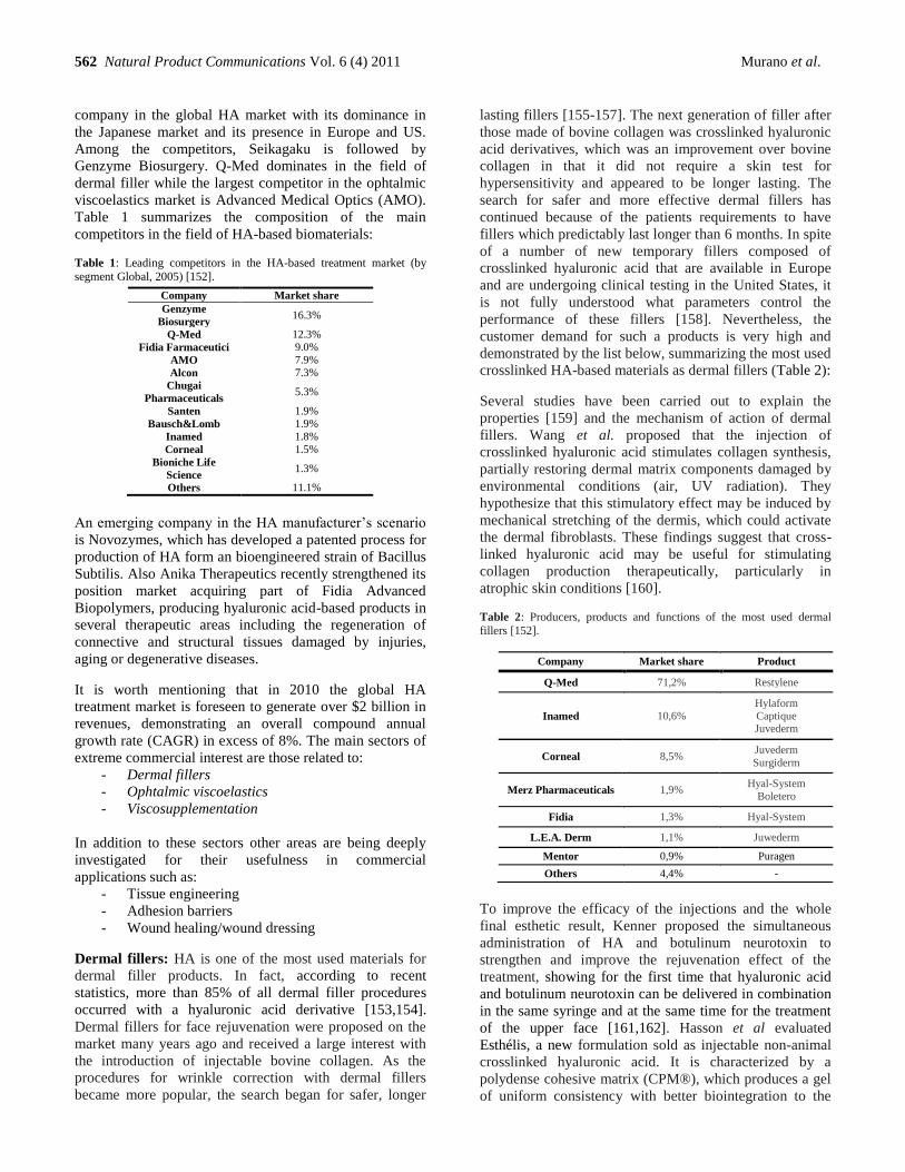
competitors in the field of HA-based biomaterials:
Table 1: Leading competitors in the HA-based treatment market (by
segment Global, 2005) [152].
Company Market share
Genzyme
Biosurgery 16.3%
Q-Med 12.3%
Fidia Farmaceutici 9.0%
AMO 7.9%
Alcon 7.3%
Chugai
Pharmaceuticals 5.3%
Santen 1.9%
Bausch&Lomb 1.9%
Inamed 1.8%
Corneal 1.5%
Bioniche Life
Science 1.3%
Others 11.1%
An emerging company in the HA manufacturer‘s scenario
is Novozymes, which has developed a patented process for
production of HA form an bioengineered strain of Bacillus
Subtilis. Also Anika Therapeutics recently strengthened its
position market acquiring part of Fidia Advanced
Biopolymers, producing hyaluronic acid-based products in
several therapeutic areas including the regeneration of
connective and structural tissues damaged by injuries,
aging or degenerative diseases.
It is worth mentioning that in 2010 the global HA
treatment market is foreseen to generate over $2 billion in
revenues, demonstrating an overall compound annual
growth rate (CAGR) in excess of 8%. The main sectors of
extreme commercial interest are those related to:
- Dermal fillers
- Ophtalmic viscoelastics
- Viscosupplementation
In addition to these sectors other areas are being deeply
investigated for their usefulness in commercial
applications such as:
- Tissue engineering
- Adhesion barriers
- Wound healing/wound dressing
Dermal fillers: HA is one of the most used materials for
dermal filler products. In fact, according to recent
statistics, more than 85% of all dermal filler procedures
occurred with a hyaluronic acid derivative [153,154].
Dermal fillers for face rejuvenation were proposed on the
market many years ago and received a large interest with
the introduction of injectable bovine collagen. As the
procedures for wrinkle correction with dermal fillers
became more popular, the search began for safer, longer
lasting fillers [155-157]. The next generation of filler after
those made of bovine collagen was crosslinked hyaluronic
acid derivatives, which was an improvement over bovine
collagen in that it did not require a skin test for
hypersensitivity and appeared to be longer lasting. The
search for safer and more effective dermal fillers has
continued because of the patients requirements to have
fillers which predictably last longer than 6 months. In spite
of a number of new temporary fillers composed of
crosslinked hyaluronic acid that are available in Europe
and are undergoing clinical testing in the United States, it
is not fully understood what parameters control the
performance of these fillers [158]. Nevertheless, the
customer demand for such a products is very high and
demonstrated by the list below, summarizing the most used
crosslinked HA-based materials as dermal fillers (Table 2):
Several studies have been carried out to explain the
properties [159] and the mechanism of action of dermal
fillers. Wang et al. proposed that the injection of
crosslinked hyaluronic acid stimulates collagen synthesis,
partially restoring dermal matrix components damaged by
environmental conditions (air, UV radiation). They
hypothesize that this stimulatory effect may be induced by
mechanical stretching of the dermis, which could activate
the dermal fibroblasts. These findings suggest that cross-
linked hyaluronic acid may be useful for stimulating
collagen production therapeutically, particularly in
atrophic skin conditions [160]. Table 2: Producers, products and functions of the most used dermal
fillers [152].
To improve the efficacy of the injections and the whole
final esthetic result, Kenner proposed the simultaneous
administration of HA and botulinum neurotoxin to
strengthen and improve the rejuvenation effect of the
treatment, showing for the first time that hyaluronic acid
and botulinum neurotoxin can be delivered in combination
in the same syringe and at the same time for the treatment
of the upper face [161,162]. Hasson et al evaluated
Esthélis, a new formulation sold as injectable non-animal
crosslinked hyaluronic acid. It is characterized by a
polydense cohesive matrix (CPM®), which produces a gel
of uniform consistency with better biointegration to the
Company Market share Product
Q-Med 71,2% Restylene
Inamed 10,6%
Hylaform
Captique Juvederm
Corneal 8,5% Juvederm
Surgiderm
Merz Pharmaceuticals 1,9% Hyal-System
Boletero
Fidia 1,3% Hyal-System
L.E.A. Derm 1,1% Juwederm
Mentor 0,9% Puragen
Others 4,4% -
Review on hyaluronan Natural Product Communications Vol. 6 (4) 2011 563
tissues and a longer duration in the treatment of atrophic
scars [163]. Monheit et al. studied a new type of dermal
filler based on a Dermal gel extra (DGE) crosslinked HA-
based material containing lidocaine, engineered to resist
deformation and degradation. The results obtained on
patients indicate that DGE could provide a comfortable
and cost-effective dermal filler option for clinicians and
patients [164].
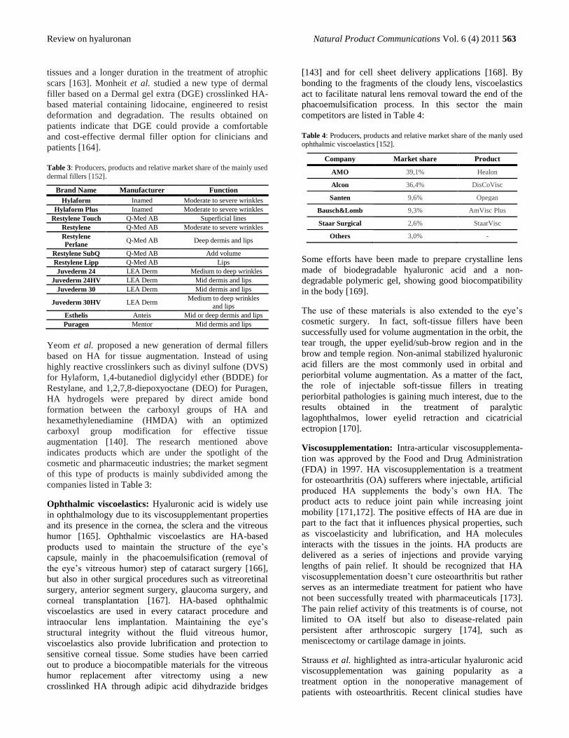
Table 3: Producers, products and relative market share of the mainly used
dermal fillers [152].
Brand Name Manufacturer Function
Hylaform Inamed Moderate to severe wrinkles
Hylaform Plus Inamed Moderate to severe wrinkles
Restylene Touch Q-Med AB Superficial lines
Restylene Q-Med AB Moderate to severe wrinkles
Restylene
Perlane Q-Med AB Deep dermis and lips
Restylene SubQ Q-Med AB Add volume
Restylene Lipp Q-Med AB Lips
Juvederm 24 LEA Derm Medium to deep wrinkles
Juvederm 24HV LEA Derm Mid dermis and lips
Juvederm 30 LEA Derm Mid dermis and lips
Juvederm 30HV LEA Derm Medium to deep wrinkles
and lips
Esthelis Anteis Mid or deep dermis and lips
Puragen Mentor Mid dermis and lips
Yeom et al. proposed a new generation of dermal fillers
based on HA for tissue augmentation. Instead of using
highly reactive crosslinkers such as divinyl sulfone (DVS)
for Hylaform, 1,4-butanediol diglycidyl ether (BDDE) for
Restylane, and 1,2,7,8-diepoxyoctane (DEO) for Puragen,
HA hydrogels were prepared by direct amide bond
formation between the carboxyl groups of HA and
hexamethylenediamine (HMDA) with an optimized
carboxyl group modification for effective tissue
augmentation [140]. The research mentioned above
indicates products which are under the spotlight of the
cosmetic and pharmaceutic industries; the market segment
of this type of products is mainly subdivided among the
companies listed in Table 3:
Ophthalmic viscoelastics: Hyaluronic acid is widely use
in ophthalmology due to its viscosupplementant properties
and its presence in the cornea, the sclera and the vitreous
humor [165]. Ophthalmic viscoelastics are HA-based
products used to maintain the structure of the eye‘s
capsule, mainly in the phacoemulsification (removal of
the eye‘s vitreous humor) step of cataract surgery [166],
but also in other surgical procedures such as vitreoretinal
surgery, anterior segment surgery, glaucoma surgery, and
corneal transplantation [167]. HA-based ophthalmic
viscoelastics are used in every cataract procedure and
intraocular lens implantation. Maintaining the eye‘s
structural integrity without the fluid vitreous humor,
viscoelastics also provide lubrification and protection to
sensitive corneal tissue. Some studies have been carried
out to produce a biocompatible materials for the vitreous
humor replacement after vitrectomy using a new
crosslinked HA through adipic acid dihydrazide bridges
[143] and for cell sheet delivery applications [168]. By
bonding to the fragments of the cloudy lens, viscoelastics
act to facilitate natural lens removal toward the end of the
phacoemulsification process. In this sector the main
competitors are listed in Table 4:
Table 4: Producers, products and relative market share of the manly used
ophthalmic viscoelastics [152].
Company Market share Product
AMO 39,1% Healon
Alcon 36,4% DisCoVisc
Santen 9,6% Opegan
Bausch&Lomb 9,3% AmVisc Plus
Staar Surgical 2,6% StaarVisc
Others 3,0% -
Some efforts have been made to prepare crystalline lens
made of biodegradable hyaluronic acid and a non-
degradable polymeric gel, showing good biocompatibility
in the body [169].
The use of these materials is also extended to the eye‘s
cosmetic surgery. In fact, soft-tissue fillers have been
successfully used for volume augmentation in the orbit, the
tear trough, the upper eyelid/sub-brow region and in the
brow and temple region. Non-animal stabilized hyaluronic
acid fillers are the most commonly used in orbital and
periorbital volume augmentation. As a matter of the fact,
the role of injectable soft-tissue fillers in treating
periorbital pathologies is gaining much interest, due to the
results obtained in the treatment of paralytic
lagophthalmos, lower eyelid retraction and cicatricial
ectropion [170].
Viscosupplementation: Intra-articular viscosupplementa-
tion was approved by the Food and Drug Administration
(FDA) in 1997. HA viscosupplementation is a treatment
for osteoarthritis (OA) sufferers where injectable, artificial
produced HA supplements the body‘s own HA. The
product acts to reduce joint pain while increasing joint
mobility [171,172]. The positive effects of HA are due in
part to the fact that it influences physical properties, such
as viscoelasticity and lubrification, and HA molecules
interacts with the tissues in the joints. HA products are
delivered as a series of injections and provide varying
lengths of pain relief. It should be recognized that HA
viscosupplementation doesn‘t cure osteoarthritis but rather
serves as an intermediate treatment for patient who have
not been successfully treated with pharmaceuticals [173].
The pain relief activity of this treatments is of course, not
limited to OA itself but also to disease-related pain
persistent after arthroscopic surgery [174], such as
meniscectomy or cartilage damage in joints.
Strauss et al. highlighted as intra-articular hyaluronic acid
viscosupplementation was gaining popularity as a
treatment option in the nonoperative management of
patients with osteoarthritis. Recent clinical studies have
564 Natural Product Communications Vol. 6 (4) 2011 Murano et al.
demonstrated that the anti-inflammatory, anabolic, and
chondroprotective actions of hyaluronic acid reduce pain
and improve patient function. With evidence mounting in
support of the efficacy of this treatment modality for
patients with osteoarthritis, its potential use in patient
populations affected by pathologies of weight-bearing joint
such as knee [175] and ankles [176] is deeply investigated.
Bencke et at. showed that the number of patients with
osteoarthritis (OA) is expected to be growing.
Viscosupplementation, in which hyaluronic acid is injected
into the knee joint, has evolved into an important part of
our current therapeutic regimen in addressing the patient
with knee pain due to OA. Although suffering from lack of
an "evidence-based" approach there is a growing body of
data demonstrating the efficacy of HA in decreasing pain
and improving function in patients with knee OA. The
extensive use in clinic of HA has led to the introduction on
the market of various forms of HA, although little data are
available to justify one over the other [177]. Further proofs
about the successful concept of HA as viscosupplementant
were given by Mathieu et al during in vitro studies on
rheological behavior of synovial fluids mixed with linear
and crosslinked HA. They showed as in vitro tests
highlighted the stability of HA over a period of 6 week
without relevant alteration of the rheologic behavior and
no pronounced degradation by catabolytic enzymes [178].
The pharmaceutical companies are very active in this field
since these are high-demand products. The leading
companies in the viscosupplementation field and the
relative HA-based products are summarized in Table 5.
Table 5: Manufacturers, products and share market of the most used
viscosupplementants [152].
Company Market share Product
Seikagaku corporation 35,80% ARTZ
Genzyme Biosurgery 26,10% Synvisc (Hylan G-F20)
Fidia Pharmaceuticals 14,30% Hyalgan
Chugai Pharmaceuticals 8,40% Suvenyl
Bioniche Life Science 2,00% Suplasyn
Others 13,40% -
Tissue engineering: The production of artificial 3D
matrices is nowadays a challenging topic [179],
considering the in vitro tissue growth for surgical
application a strong market need. Several approaches have
been proposed to promote the cell growth on 3D-matrices
made of biocompatible components. Due to a strong
market demand for those kind of artificial tissues research
in regenerative medicine is rapidly expanding to cope with
this new need. Based on the concept of tissue regeneration
triggered by HA-based materials, some products are
commercially available for the treatment of deep dermal
lesions such as non healing ulcers, deep II degree and III
degree burns [180] (HyalograftTM
3D), for autologous skin
replacement (Laserskin®), for autologous fibroblasts and
keratinocytes replacement (Tissuetech) and for wound
healing/dressing, whose use will be discussed later on in
this chapter.
In general, there is nowadays a trend towards supplying
cells with a material to speed up the tissue healing process.
Hydrogel encapsulation made of different biopolymers
provides cells with a three dimensional environment
similar to that experienced in vivo and therefore may allow
the maintenance of normal cellular function in order to
produce tissues similar to those found in the body [181].
Fan et al. synthesized galactosylated hyaluronic acid
(GHA) prepared through the covalent coupling of
lactobionic acid with HA. The derivative was then used to
make highly porous three-dimensional sponges composed
by a blend of chitosan and GHA. The mixture formed a 3D
matrix stabilized by the electrostatic interaction between
the two biopolymers. The matrix was applied to
hepatocytes for liver-specific functions analysis. The
addition of GHA not only improved the wettability and
changed their mechanical properties, but also significantly
influenced the cell attachment ratio. Moreover, liver
functions of the hepatocytes entrapped in the CS/GHA
scaffolds, such as albumin secretion, urea synthesis and
ammonia elimination were improved in comparison with
those in similar scaffolds made of chitosan [182].
Kasahara et al. have developed an original three-
dimensional (3D) scaffold to meet improved
biomechanical and biological requirements. The constructs
were made of chitosan-based hyaluronic acid hybrid
polymer fibers. Previous studies have shown that these
hybrid polymer fibers have superior adhesion of
chondrocytes and the ability to maintain chondrocyte
phenotype. The study aimed at regenerating hyaline-like
cartilage with sufficient mechanical properties combining
a volume-reduced 3D scaffold and a bioreactor system.
The implantation of such a biocomposites was proposed as
a model approach for various cartilaginous lesions [183].
Other studies, such that of Lee et al. focused on the
fabrication of 3D highly porous scaffolds and their
biochemical binding affinity to realize various biomimetic
functionalizations. This was achieved by using a binding
mechanism that incorporated molecules, which strongly
bind to each other. A good example of such a binding
molecule pair was avidin and biotin. In this work, a porous
hybrid scaffolds composed of natural materials such as
collagen and HA was prepared to evaluate morphology,
mechanical strength, in vitro enzymatic degradation and
cytotoxicity of the scaffolds as well as cell growth within
the scaffolds [184]. Other attempts were proposed by Chou
et al., who prepared a glue made of autologous fibrin as a
potential scaffold with very good biocompatibility for
neocartilage formation. However, fibrin glue has been
reported not to provide enough mechanical strength, but
with many growth factors to interfere the tissue growth.
Gelatin/hyaluronic acid/chondroitin-6-sulfate (GHC6S)
tricopolymer sponge was prepared as scaffold for cartilage
tissue engineering and showed very good results in cell
seeding and cell distribution [185]. The results indicated
that the chondrocytes cultured in GHC6S-fibrin glue
would effectively promote extracellular matrix secretion,
inhibiting the degradation. The evidence could support that
Review on hyaluronan Natural Product Communications Vol. 6 (4) 2011 565
GHC6S-fibrin glue would be a promising scaffold for
articular cartilage tissue engineering [186].
Hu and al reported the formation of biocompatible
hydrogels using physically cross-linked biopolymers.
Gellation of silk fibroin (from B. mori silkworm) aqueous
solution was effected by ultrasonication and used to entrap
blended, uncrosslinked, hyaluronic acid (HA) without
chemical cross-linking. This is a novel approach to HA
hydrogel systems, which otherwise require chemical cross-
linking. Further, these systems exploit the beneficial
material and biological properties of both polymers. These
novel non-chemically cross-linked blend hydrogels may be
useful for biomedical applications due to biocompatibility
and the widespread utility of hydrogel systems [187].
Adhesion barriers: Adhesions are a major cause of post-
surgical morbidity and mortality and entail a substantial
medical economic burden. It has been estimated that 90%
of patients undergoing major abdominal surgery and 55%
to 100% of women undergoing pelvic surgery develop
adhesions. These phenomena are tightly linked to two risk
factors: tissue injury and inflammation response. Clinically
significant outcomes associated with adhesions include
chronic pelvic and abdominal pain, bowel obstruction, and
infertility. Consequently, methods for preventing such
adhesions have been the focus of extensive research. Few
barrier devices based on polysaccharides such as
hyaluronic acid or oxidized regenerated cellulose are in
commercial use, since part of them are based on synthetic
polymers (polytetrafluoroethylene) [188,189]. One of the
most used adhesion barrier is Seprafilm®, a composite
made of HA and carboxymethylcellulose employed in a
wide range of applications such as in bowel obstruction in
gynecological malignancies [190], tendons repair [191]
and after myomectomy, which is a procedure to surgically
removed uterine fibroids from the uterus. This procedure
often causes adhesion formation and decreases subsequent
fertility. This was the reason to evaluate the effectiveness
of several antiadhesion barrier materials in preventing
adhesion after myomectomy. Tsuji et al. prospectively
classified 63 women undergoing myomectomy alone into
four groups according to the type of antiadhesion material
used: Hyaluronic acid-carboxymethylcellulose film
(Seprafilm®), Dextran 40 (10% Dextran 40 Low
Injection®), factor 13 with fibrinogen (Beriplast
®) and
control. Seprafilm® was highly effective and was superior
to the other antiadhesion materials tested in preventing
uterine adhesions after myomectomy [192]. Other HA-
based product available on the market is INCERT®-S
(Anika), an anti-adhesion barrier gel for use in spinal
surgeries such as discectomies with a laminectomy or
laminotomy.
Other examples of dangerous adhesions are peritoneal
adhesions. These are among the most frequent cases of
tissue connections that form within the abdominopelvic
cavity following surgery or other injuries. They can cause
major medical complications. For this reason, researchers
are combining barrier devices and pharmacological agents
to be used in the prevention of this type of adhesion
formation. It was hypothesized that an adhesion barrier,
which also delivers anti-adhesion drugs, can address both
physical and physiological causes for adhesion formation.
For this reason an in situ cross-linking hyaluronan
hydrogel (barrier device) was prepared containing the
glucocorticoid receptor agonist budesonide [193]. A
similar approach to prevent peritoneal adhesions was the
application of physical barriers, which can separate the
injured regions during peritoneal healing. The hybrid
cross-linked HA-nanoparticles system appears to be a
biocompatible and highly effective adhesion barrier. This
―hybrid system‖ is able to prevent postoperative peritoneal
adhesions and combine the biocompatibility and ease of
application of in situ cross-linkable hydrogels with the
controlled release features of polymeric nanoparticles
[194]. Furthermore, postoperative peritoneal adhesions can
have serious, potentially lethal consequences.
Pharmacotherapy and barrier devices can reduce adhesion
formation to varying degrees, but their efficacy is limited
by rapid clearance from the peritoneum and lack of
biological activity, respectively. To overcome these
limitations, Yeo et al. have delivered tissue-type
plasminogen activator (tPA), which is deficient in the first
2-3 postoperative days, using a highly cross-linked in situ
forming hyaluronan gel. They demonstrated this
formulation's anti-adhesion activity in animal model that
involved recurrent adhesions [193]. Bioresorbable
membranes composed of hyaluronic acid and
carboxymethylcellulose (HA/CMC) are the most effective
method to prevent intra-abdominal adhesions; however,
their efficacy may be limited to the site of application.
Previous studies have shown that the intraperitoneal
administration of a neurokinin-1 receptor antagonist (NK-
1RA) reduces adhesions; however, the co-administration
of HA/CMC plus an NK-1RA has not been studied. The
co-administration of HA/CMC plus NK-1RA not only
increases the efficacy of the membrane at the site of
application, but also significantly reduces adhesions
formation at distal unprotected sites. This combination
may represent an emerging concept in more effective
adhesion prevention throughout the peritoneum [195].
Zhao et al. prepared a sodium carboxymethylcellulose-
Hyaluronic acid-Carboxymethylchitosan (CMCS) blend
films. The results suggested that the material was held
together by strong molecular interaction and hinted good
compatibility in the blend films. The blend films are
expected to be used as controllable degradation
biomaterials in the field of postsurgical hemostasis and
anti-adhesion [196].
Wound healing and dressing: Successful repair of
wounds and tissues remains a major healthcare and
biomedical challenge in the 21st Century. In particular,
chronic wounds often lead to loss of functional ability,
increased pain and decreased quality of life. Advanced
healing therapies employing biological dressings, skin
substitutes, growth factor-based therapies and synthetic
acellular matrices, all of which aim to correct irregular and
566 Natural Product Communications Vol. 6 (4) 2011 Murano et al.
dysfunctional cellular pathways present in chronic wounds,
are becoming more popular [197]. Commercially available
HA-based products (for healing and dressing) on the
market are Hyalofill-f, Hyalofill-R, Connettivina,
Connettivina Plus, Jaloskin®, Hyalomatrix®. Hyalofill
family products are absorbent, soft and conformable
fibrous fleece (F) or rope (R) composed of HYAFF®, an
ester of hyaluronic acid. Jaloskin® is a transparent film
dressing for the treatment of superficial moderately
exuding wounds. Hyalomatrix® is a bilayered, sterile,
flexible, and conformable wound dressing that acts as an
advanced wound care device.
HA uses in wound healing are strongly justified by its non-
antigenic properties and the number of forms it can be
manufactured, such as gels, creams to sheets of solid
material through lightly woven meshes. Epidermal
engraftment is superior to most of the available
biotechnologies and, as such, the material shows great
promise in both animal and clinical studies of tissue
engineering. Ongoing research works focus on the ability
of the biopolymer to enhance angiogenesis and the
conversion of chronic wounds into acute wounds [198].
Moreover, Gao et al investigated the biological roles of
hyaluronan (HA) fragments in angiogenesis acceleration.
Studies have confirmed that oligosaccharides of HA (o-
HA) are capable of stimulating neovascularization in vitro
and promoting blood flow or angiogenesis in animal
models. However, few laboratories have studied the
function of o-HA as an exogenous treatment in injured
tissue repair in vivo. It is thought that o-HA may lose its
activities when used topically in vivo due to its small size,
which may be absorbed quickly by the surrounding tissues.
A special slow-releasing gel containing a mixture of o-HA
at defined size was prepared and the healing effects, by
topical application to an acute wound model, was studied.
The therapy by o-HA was compared with high molecular
weight HA and the known angiogenesis stimulator, VEGF,
suggesting that o-HA therapy might be useful in acute
wound repair [200]. Ferguson et al worked on HA
fragments together to bioresponsive polymers to enhance
the circulation time and stability of biologically active
proteins and peptides, while reducing their
immunogenicity and toxicity. Recently, crosslinked
material made of HA at different molecular weights
blended with arginine and epidermal growth factor (EGF)
[201] and dextrin-EGF [202] conjugates were proposed,
the latter exploiting the so called Polymer-masked
UnMasked Protein Therapy (PUMPT) concept. The
hypothesis is that conjugation of a biodegradable polymer
to a protein would protect it and mask activity in transit,
while enabling controlled reinstatement of activity at the
target site by triggered degradation of the polymeric
component. It has been developed and shown as potential
modulators of impaired wound healing. In this contest, HA
fragments (Mw 90,000 g/mol) were conjugated to trypsin
as a model enzyme. HA-trypsin conjugates exhibited 52%
greater stability in the presence of elastase, compared to
free trypsin, demonstrating the potential use of HA
conjugates as modulators of tissue repair. Ibrahim et al.
showed that exogenous hyaluronic acid oligomers (o-HA)
stimulate functional endothelialization, though native long-
chain HA was more bioinert and possibly more
biocompatible. Additionally, the author created hydrogels
containing high molecular weight HA (1 × 10⁶ Da) and o-
HA mixtures (HA-o: 0.75-10 kDa) crosslinked with
divinylsulfone (DVS). The greatest endothelial cell (EC)
attachment and proliferation was observed on gels with an
high amount of o-HA. The study showed that the
beneficial EC response to o-HA and biocompatibility of
HA is mostly unaltered by their chemical derivatization
and crosslinking into an hydrogel [203].
References
[1] Meyer K, Palmer JW. (1934) The polysaccharide of the vitreous humor. Journal of Biological Chemistry, 107, 629-634.
[2] Meyer K, Chaffee E. (1941) The mucopolysaccharides of skin. Journal of Biological Chemistry, 138, 491-499. [3] O'Regan M, Martini I, Crescenzi F, De Luca C, Lansing M. (1994) Molecular mechanisms and genetics of hyaluronan
biosynthesis. International Journal of Biological Macromolecules, 16, 283-286. [4] Fraser JR, Laurent TC, Laurent UB. (1997) Hyaluronan: its nature, distribution, functions and turnover. Journal of Internal
Medicine, 242, 27-33.
[5] Nusgens BV. (2010) [Hyaluronic acid and extracellular matrix: a primitive molecule?]. Annales de Dermatologie et de Venereologie, 137 Suppl 1, S3-8.
[6] Laurent TC, Fraser JR. (1992) Hyaluronan. The Journal of the Federation of American Societies for Experimental Biology, 6, 2397-2404.
[7] Poole AR, Kojima T, Yasuda T, Mwale F, Kobayashi M, Laverty S. (2001) Composition and structure of articular cartilage: a
template for tissue repair. Clinical Orthopaedics & Related Research, S26-33. [8] Kogan G, Soltes L, Stern R, Gemeiner P. (2007) Hyaluronic acid: a natural biopolymer with a broad range of biomedical and
industrial applications. Biotechnology Letters 29, 17-25. [9] Manuskiatti W, Maibach HI. (2007) Hyaluronic acid and skin: wound healing and aging. International Journal of Dermatology 35,
539-544 [10] Chen WYJ, Abatangelo G. (1990) Functions of hyaluronan in wound repair. Wound Repair and Rigeneration, 7. [11] Miyake K, Underhill CB, Lesley J, Kincade PW. (1990) Hyaluronate can function as a cell adhesion molecule and CD44
participates in hyaluronate recognition. The Journal of Experimental Medicine, 172, 69-75. [12] Inoue M, Katakami C. (1993) The effect of hyaluronic acid on corneal epithelial cell proliferation. Investigative Opthalmology &
Visual Science, 34, 2313-2315.
Review on hyaluronan Natural Product Communications Vol. 6 (4) 2011 567
[13] Foger N, Marhaba R, Zoller M. (2000) CD44 supports T cell proliferation and apoptosis by apposition of protein kinases. European Journal of Immunology, 30, 2888-2899.
[14] Itano N, Atsumi F, Sawai T, Yamada Y, Miyaishi O, Senga T, Hamaguchi M, Kimata K. (2002) Abnormal accumulation of hyaluronan matrix diminishes contact inhibition of cell growth and promotes cell migration. Proceedings of the National Academy
of Sciences of the United States of America, 99, 3609-3614. [15] Toole BP. (2002) Hyaluronan in morphogenesis. Seminars in Cell & Developmental Biology, 12, 79-87.
[16] Nochi H, Shinomiya T, Tamoto K. (2006) Characterization of hyaluronan-binding proteins on guinea pig polymorphonuclear leukocytes: possible involvement of complement receptor type 3 (CR3, CD11b/CD18) in the hyaluronan-leukocyte interaction. The Journal of Biochemistry, 139, 59-70.
[17] Zhu H, Mitsuhashi N, Klein A, Barsky LW, Weinberg K, Barr ML, Demetriou A, Wu GD. (2006) The role of the hyaluronan receptor CD44 in mesenchymal stem cell migration in the extracellular matrix. Stem Cells, 24, 928-935.
[18] Gao F, Koenitzer JR, Tobolewski JM, Jiang D, Liang J, Noble PW, Oury TD. (2008) Extracellular superoxide dismutase inhibits inflammation by preventing oxidative fragmentation of hyaluronan. The Journal of Biological Chemistry, 283, 6058-6066.
[19] Ghandi NS, Mancera RL. (2008) The structure of glycosaminoglycans and ntheir interactions with proteins. Chemical Biology &
Drug Design, 72, 455-482. [20] Schaefer L, Schaefer RM. (2010) Proteoglycans: from structural compounds to signaling molecules. Cell and Tissue Research, 339,
237-246. [21] Lipponen P, Aaltomaa S, Tammi R, Tammi M, Agren U, Kosma VM. (2001) High stromal hyaluronan level is associated with
poor differentiation and metastasis in prostate cancer. European Journal of Cancer, 37, 849-856. [22] Toole BP, Wight TN, Tammi MI. (2002) Hyaluronan-cell interactions in cancer and vascular disease. The Journal of Biological
Chemistry, 277, 4593-4596.
[23] Misra S, Ghatak S, Zoltan-Jones A, Toole BP. (2003) Regulation of multidrug resistance in cancer cells by hyaluronan. The Journal of Biological Chemistry, 278, 25285-25288.
[24] Hascall VC, Majors AK, De La Motte CA, Evanko SP, Wang A, Drazba JA, Strong SA, Wight TN. (2004) Intracellular hyaluronan: a new frontier for inflammation? The Journal of Biological Chemistry, 1673, 3-12.
[25] Toole BP. (2004) Hyaluronan: from extracellular glue to pericellular cue. Nature Reviews Cancer, 4, 528-539.
[26] Jameson JM, Cauvi G, Sharp LL, Witherden DA, Havran WL. (2005) Gammadelta T cell-induced hyaluronan production by epithelial cells regulates inflammation. The Journal of Experimental Medicine, 201, 1269-1279.
[27] Bharadwaj AG, Rector K, Simpson MA. (2007) Inducible hyaluronan production reveals differential effects on prostate tumor cell growth and tumor angiogenesis. The Journal of Biological Chemistry, 282, 20561-20572.
[28] Twarock S, Tammi MI, Savani RC, Fischer JW. (2010) Hyaluronan stabilizes focal adhesions, filopodia, and the proliferative phenotype in esophageal squamous carcinoma cells. The Journal of Biological Chemistry, 285, 23276-23284.
[29] Stern R, Asari AA, Sugahara KN. (2006) Hyaluronan fragments: an information-rich system. European Journal of Cell Biology,
85, 699-715. [30] Deed R, Rooney P, Kumar P, Norton JD, Smith J, Freemont AJ, Kumar S. (1997) Early-response gene signalling is induced by
angiogenic oligosaccharides of hyaluronan in endothelial cells. Inhibition by non-angiogenic, high-molecular-weight hyaluronan. International Journal of Cancer, 71, 251-256.
[31] Noble PW. (2002) Hyaluronan and its catabolic products in tissue injury and repair. Matrix Biology, 21, 25-29.
[32] Termeer C, Benedix F, Sleeman J, Fieber C, Voith U, Ahrens T, Miyake K, Freudenberg M, Galanos C, Simon JC. (2002) Oligosaccharides of Hyaluronan activate dendritic cells via toll-like receptor 4. The Journal of Experimental Medicine, 195, 99-111.
[33] Trabucchi E, Pallotta S, Morini M, Corsi F, Franceschini R, Casiraghi A, Pravettoni A, Foschi D, Minghetti P. (2002) Low molecular weight hyaluronic acid prevents oxygen free radical damage to granulation tissue during wound healing. International
Journal of Tissue Reactivity, 24, 65-71. [34] Mascarenhas MM, Day RM, Ochoa CD, Choi WI, Yu L, Ouyang B, Garg HG, Hales CA, Quinn DA. (2004) Low molecular
weight hyaluronan from stretched lung enhances interleukin-8 expression. American Journal of Respiratory Cell and Molecular
Biology, 30, 51-60. [35] Powell JD, Horton MR. (2005) Threat matrix: low-molecular-weight hyaluronan (HA) as a danger signal. Immunologic Research,
31, 207-218. [36] Wang CT, Lin YT, Chiang BL, Lin YH, Hou SM. (2006) High molecular weight hyaluronic acid down-regulates the gene
expression of osteoarthritis-associated cytokines and enzymes in fibroblast-like synoviocytes from patients with early osteoarthritis.
Osteoarthritis Cartilage, 14, 1237-1247. [37] Toole BP, Slomiany MG. (2008) Hyaluronan: a constitutive regulator of chemoresistance and malignancy in cancer cells. Seminars
in Cancer Biology, 18, 244-250. [38] Aruffo A, Stamenkovic I, Melnick M, Underhill CB, Seed B. (1990) CD44 is the principal cell surface receptor for hyaluronate.
Cell, 61, 1303-1313. [39] Turley EA, Noble PW, Bourguignon LY. (2002) Signaling properties of hyaluronan receptors. The Journal of Biological
Chemistry, 277, 4589-4592.
[40] Yamada T, Kawasaki T. (2005) Microbial synthesis of hyaluronan and chitin: New approaches. Journal of Bioscience and Bioengineering, 99, 521-528.
[41] Goueffic Y, Guilluy C, Guerin P, Patra P, Pacaud P, Loirand G. (2006) Hyaluronan induces vascular smooth muscle cell migration through RHAMM-mediated PI3K-dependent Rac activation. Cardiovascular Research, 72, 339-348.
[42] Kyosseva SV, Harris EN, Weigel PH. (2008) The hyaluronan receptor for endocytosis mediates hyaluronan-dependent signal
transduction via extracellular signal-regulated kinases. The Journal of Biological Chemistry, 283, 15047-15055. [43] Maxwell CA, McCarthy J, Turley E. (2008) Cell-surface and mitotic-spindle RHAMM: moonlighting or dual oncogenic functions?
Journal of Cell Science, 121, 925-932.
568 Natural Product Communications Vol. 6 (4) 2011 Murano et al.
[44] DeAngelis PL, Papaconstantinou J, Weigel PH. (1993) Isolation of a Streptococcus pyogenes gene locus that directs hyaluronan biosynthesis in acapsular mutants and in heterologous bacteria. Journal of Biological Chemistry, 268, 14568-14571.
[45] DeAngelis PL. (1999) Hyaluronan synthases: fascinating glycosyltransferases from vertebrates, bacterial pathogens, and algal viruses. Cellular and Molecular Life Sciences, 56, 670-682.
[46] Weigel PH, DeAngelis PL. (2007) Hyaluronan synthases: a decade-plus of novel glycosyltransferases. The Journal of Biological Chemistry, 282, 36777-36781.
[47] Kooy FK. (2010) Enzymatic production of hyaluronan oligo- and polysaccharides, University of Wageningen, The Netherlands. PhD Thesis.
[48] Kendall FE, Heidelberger M, Dawson MH. (1937) A serologically inactive polysaccharide elaborated by mucoid strains of Group
A hemolytic Streptococcus. The Journal of Biological Chemistry, 118, 61-69. [49] Carter GR, Annau E. (1953) Isolation of capsular polysaccharides from colonial variants of Pasteurella multocida. American
Journal of Veterinary Research, 14, 475-478. [50] Maclennan AP. (1956) The production of capsules, hyaluronic acid and hyaluronidase by 25 strains of group C streptococci.
Journal of General Microbiology, 15, 485-491.
[51] Boyce JD, Chung JY, Adler B. (2000) Pasteurella multocida capsule: composition, function and genetics. J Biotechnol 83, 153-160 [52] Chung JY, Wilkie I, Boyce JD, Townsend KM, Frost AJ, Ghoddusi M, Adler B. (2001) Role of capsule in the pathogenesis of fowl
cholera caused by Pasteurella multocida serogroup A. Infection and Immunity, 69, 2487-2492. [53] Harrington DJ, Sutcliffe IC, Chanter N. (2002) The molecular basis of Streptococcus equi infection and disease. Microbes and
Infection, 4, 501-510. [54] Ouskova G, Spellerberg B, Prehm P. (2004) Hyaluronan release from Streptococcus pyogenes: export by an ABC transporter.
Glycobiology, 14, 931-938.
[55] Fong Chong B, Blank LM, McLaughlin R, Nielsen LK. (2005) Microbial hyaluronic acid production. Applied Microbiology and Biotechnology, 66, 341-351.
[56] Gao HJ, Du GC, Chen J. (2006) Analysis of metabolic fluxes for hyaluronic acid (HA) production by Streptococcus zooepidemicus. World Journal of Microbiology and Biotechnology, 22, 399-408.
[57] Abbot EL, Smith WD, Siou GP, Chiriboga C, Smith RJ, Wilson JA, Hirst BH, Kehoe MA. (2007) Pili mediate specific adhesion of
Streptococcus pyogenes to human tonsil and skin. Cellular Microbiology, 9, 1822-1833. [58] Voyich JM, Musser JM, DeLeo FR. (2007) Streptococcus pyogenes and human neutrophils: a paradigm for evasion of innate host
defense by bacterial pathogens. Microbes and Infection, 6, 1117-1123. [59] Wessels MR, Moses AE, Goldberg JB, DiCesare TJ. (1991) Hyaluronic acid capsule is a virulence factor for mucoid group A
streptococci. Proceedings of the National Academy of Sciences of the United States of America, 88, 8317-8321. [60] Schmidt KH, Gunther E, Courtney HS. (1996) Expression of both M protein and hyaluronic acid capsule by group A streptococcal
strains results in a high virulence for chicken embryos. Medical Microbiology and Immunology 184, 169-173.
[61] Jing W, DeAngelis PL. (2003) Analysis of the two active sites of the hyaluronan synthase and the chondroitin synthase of Pasteurella multocida. Glycobiology, 13, 661-671.
[62] Cleary PP, Larkin A. (1979) Hyaluronic acid capsule: strategy for oxygen resistance in group A streptococci. Journal of Bacteriology, 140, 1090-1097.
[63] Cywes C, Wessels MR. (2001) Group A Streptococcus tissue invasion by CD44-mediated cell signalling. Nature 414, 648-652
[64] Wayan Teguh Wibawan I, Pasaribu FH, Utama IH, Abdulmawjood A, Lammler C. (2002) The role of hyaluronic acid capsular material of Streptococcus equi subsp. zooepidemicus in mediating adherence to HeLa cells and in resisting phagocytosis. Research
in Veterinary Science, 67, 131-135. [65] Kim SJ, Park SY, Kim CW. (2006) A novel approach to the production of hyaluronic acid by Streptococcus equi subsp.
zooepidemicus. Journal of Microbiology and Biotechnology, 16, 1849-1855. [66] Chen WY, Marcellin E, Hung J, Nielsen LK. (2009) Hyaluronan molecular weight is controlled by UDP-N-acetylglucosamine
concentration in Streptococcus zooepidemicus. The Journal of Biological Chemistry, 284, 18007-18014.
[67] Levin JC, Wessels MR. (1998) Identification of csrR/csrS, a genetic locus that regulates hyaluronic acid capsule synthesis in group A Streptococcus. Molecular Microbiology, 30, 209-219.
[68] Schrager HM, Alberti S, Cywes C, Dougherty GJ, Wessels MR. (1998) Hyaluronic acid capsule modulates M protein-mediated adherence and acts as a ligand for attachment of group A Streptococcus to CD44 on human keratinocytes. Journal of Clinical Investigation, 101, 1708-1716.
[69] Pruimboom IM, Rimler RB, Ackermann MR, Brogden KA. (1996) Capsular hyaluronic acid-mediated adhesion of Pasteurella multocida to turkey air sac macrophages. Avian Diseases, 40, 887-893.
[70] DeAngelis PL, Jing W, Drake RR, Achyuthan AM. (1998) Identification and molecular cloning of a unique hyaluronan synthase from Pasteurella multocida. The Journal of Biological Chemistry, 273, 8454-8458.
[71] DeAngelis PL. (1999) Molecular directionality of polysaccharide polymerization by the Pasteurella multocida hyaluronan synthase. The Journal of Biological Chemistry, 274, 26557-26562
[72] Boyce JD, Adler B. (2000) The capsule is a virulence determinant in the pathogenesis of Pasteurella multocida M1404 (B:2).
Infection and Immunity, 68, 3463-3468. [73] DeAngelis PL, Gunay NS, Toida T, Mao WJ, Linhardt RJ. (2002) Identification of the capsular polysaccharides of Type D and F
Pasteurella multocida as unmodified heparin and chondroitin, respectively. Charbohydrate Research, 337, 1547-1552. [74] Ewers C, Lubke-Becker A, Bethe A, Kiebling S, Filter M, Wieler LH. (2006) Virulence genotype of Pasteurella multocida strains
isolated from different hosts with various disease status. Veterinary Microbiology, 114, 304-317.
[75] Nozoe T, Kohnoe S, Ezaki T, Kabashima A, Maehara Y. (2004) Significance of immunohistochemical over-expression of CD44v6 as an indicator of malignant potential in esophageal squamous cell carcinoma. Journal of cancer research and Clinical Oncology,
130, 334-338.
Review on hyaluronan Natural Product Communications Vol. 6 (4) 2011 569
[76] Udabage L, Brownlee GR, Nilsson SK, Brown TJ. (2005) The over-expression of HAS2, Hyal-2 and CD44 is implicated in the invasiveness of breast cancer. Experimental Cell Research, 310, 205-217.
[77] Van Etten JL. (2003) Unusual life style of giant chlorella viruses. Annual Review of Genetics, 37, 153-195. [78] Fitzgerald LA, Graves MV, Li X, Feldblyum T, Nierman WC, Van Etten JL. (2007) Sequence and annotation of the 369-kb NY-
2A and the 345-kb AR158 viruses that infect Chlorella NC64A. Virology, 358, 472-484. [79] Rinaudo M. (2006) Chitin and chitosan; properties and applications. Progress in Polymer Science, 31, 603-632.
[80] DeAngelis PL, Jing W, Graves MV, Burbank DE, Van Etten JL. (1997) Hyaluronan synthase of chlorella virus PBCV-1. Science 278, 1800-1803
[81] Mohammed Ali AM, Kawasaki T, Yamada T. (2005) Genetic rearrangements on the Chlorovirus genome that switch between
hyaluronan synthesis and chitin synthesis. Virology, 342, 102-110. [82] Wilson WH, Van Etten JL, Allen MJ. (2009) The Phycodnaviridae: the story of how tiny giants rule the world. Current Topics in
Microbiology and Immunology, 328, 1-42. [83] Kim J-H, Yoo S-J, Oh D-K, Kweon Y-G, Park D-W, Lee C-H, Gil G-H. (1996) Selection of a Streptococcus equi mutant and
optimization of culture conditions for the production of high molecular weight hyaluronic acid. Enzyme and Microbial Technology,
19, 440-445. [84] Stangohl S. (2000) Methods and means for the production of hyaluronic acid, USA Patent 6090596
[85] Stangohl S. (2003) Methods and means for the production of hyaluronic acid, USA Patent 6537795 [86] Johns MR, Goh L-T, Oeggerli A. (1994) Effect of pH, agitation and aeration on hyaluronic acid production by Streptococcus
zooepidemicus. Biotechnology Letters, 16, 507-512. [87] Armstrong DC, Johns MR. (1997) Culture Conditions Affect the Molecular Weight Properties of Hyaluronic Acid Produced by
Streptococcus zooepidemicus. Applied and Environmental Microbiology, 63, 2759-2764.
[88] Fong Chong B, Nielsen LK. (2003) Aerobic cultivation of Streptococcus zooepidemicus and the role of NADH oxidase. Biochemical Engineering Journal, 16, 153-162.
[89] Fong Chong B, Nielsen LK. (2003) Amplifying the cellular reduction potential of Streptococcus zooepidemicus. Journal of Biotechnology, 100, 33-41.
[90] Beres SB, Sesso R, Pinto SW, Hoe NP, Porcella SF, Deleo FR, Musser JM. (2008) Genome sequence of a Lancefield group C
Streptococcus zooepidemicus strain causing epidemic nephritis: new information about an old disease. PLoS One, 3, e3026. [91] Holden MT, Heather Z, Paillot R, Steward KF, Webb K, Ainslie F, Jourdan T, Bason NC, Holroyd NE, Mungall K, Quail MA,
Sanders M, Simmonds M, Willey D, Brooks K, Aanensen DM, Spratt BG, Jolley KA, Maiden MC, Kehoe M, Chanter N, Bentley SD, Robinson C, Maskell DJ, Parkhill J, Waller AS. (2009) Genomic evidence for the evolution of Streptococcus equi: host
restriction, increased virulence, and genetic exchange with human pathogens. PLoS Pathogens, 5, e1000346. [92] Ferretti JJ, McShan WM, Ajdic D, Savic DJ, Savic G, Lyon K, Primeaux C, Sezate S, Suvorov AN, Kenton S, Lai HS, Lin SP,
Qian Y, Jia HG, Najar FZ, Ren Q, Zhu H, Song L, White J, Yuan X, Clifton SW, Roe BA, McLaughlin R. (2001) Complete
genome sequence of an M1 strain of Streptococcus pyogenes. Proceedings of the National Academy of Sciences USA, 98, 4658-4663.
[93] Beres SB, Sylva GL, Barbian KD, Lei B, Hoff JS, Mammarella ND, Liu MY, Smoot JC, Porcella SF, Parkins LD, Campbell DS, Smith TM, McCormick JK, Leung DY, Schlievert PM, Musser JM. (2002) Genome sequence of a serotype M3 strain of group A Streptococcus: phage-encoded toxins, the high-virulence phenotype, and clone emergence. Proceedings of the National Academy of
Sciences USA, 99, 10078-10083. [94] May BJ, Zhang Q, Li LL, Paustian ML, Whittam TS, Kapur V. (2001) Complete genomic sequence of Pasteurella multocida,
Pm70. Proceedings of the National Academy of Sciences USA, 98, 3460-3465. [95] DeAngelis PL, Papaconstantinou J, Weigel PH. (1993) Molecular cloning, identification, and sequence of the hyaluronan synthase
gene from group A Streptococcus pyogenes. Journal of Biological Chemistry, 268, 19181-19184. [96] Dougherty BA, van de Rijn I. (1993) Molecular characterization of hasB from an operon required for hyaluronic acid synthesis in
group A streptococci. Demonstration of UDP-glucose dehydrogenase activity. Journal of Biological Chemistry, 268, 7118-7124.
[97] Crater DL, Dougherty BA, van de Rijn I. (1995) Molecular characterization of hasC from an operon required for hyaluronic acid synthesis in group A streptococci. Demonstration of UDP-glucose pyrophosphorylase activity. Journal of Biological Chemistry,
270, 28676-28680. [98] Weigel PH. (2002) Functional characteristics and catalytic mechanisms of the bacterial hyaluronan synthases. IUBMB Life, 54, 201-211. [99] Yu H, Stephanopoulos G. (2008) Metabolic engineering of Escherichia coli for biosynthesis of hyaluronic acid. Metabolic
Engineering, 10, 24-32. [100] Mao Z, Shin HD, Chen R. (2009) A recombinant E. coli bioprocess for hyaluronan synthesis. Applied Microbiology and
Biotechnology, 84, 63-69. [101] Chien LJ, Lee CK. (2007) Hyaluronic acid production by recombinant Lactococcus lactis. Applied Microbiology and
Biotechnology, 77, 339-346. [102] Mao Z, Chen RR. (2007) Recombinant synthesis of hyaluronan by Agrobacterium sp. Biotechnology Progress, 23, 1038-1042 [103] Sloma A, Behr R, Widner W, Tang M, Sternberg D, Brown S. (2003) Methods for producing hyaluronan in a recombinant host
cell. Novozymes Biopolymer, USA Patent WO/2003/054163 [104] Widner B, Behr R, Von Dollen S, Tang M, Heu T, Sloma A, Sternberg D, Deangelis PL, Weigel PH, Brown S. (2005) Hyaluronic
acid production in Bacillus subtilis. Applied and Environmental Microbiology, 71, 3747-3752. [105] Brown SH, Pummill PE. (2008) Recombinant production of hyaluronic acid. Current Pharmaceutical Biotechnology, 9, 239-241 [106] Graves MV, Burbank DE, Roth R, Heuser J, DeAngelis PL, Van Etten JL. (1999) Hyaluronan synthesis in virus PBCV-1-infected
chlorella-like green algae. Virology 257, 15-23 [107] Yamada T. (2004) Method for separating/purifyng hyaluronic acid and/or chitin, JP Patent 2004-283096.
[108] Takeo S, Fujise M, Akiyama T, Habuchi H, Itano N, Matsuo T, Aigaki T, Kimata K, Nakato H. (2004) In vivo hyaluronan synthesis upon expression of the mammalian hyaluronan synthase gene in Drosophila. Journal of Biological Chemistry, 279, 18920-18925.
570 Natural Product Communications Vol. 6 (4) 2011 Murano et al.
[109] Capodilupo AL, Crescenzi V, Francescangeli A, Joudioux R, Leonelli F, Marini Bettolo R, Migneco LM, Quagliariello M, Bellato B, De Luca G, Galbiati E, Renier D, Banzato A, Rosato A. (2005) Hyaluronan Structure, Metabolism, Biological Activities,
Therapeutic Applications, Hyaluronan 2003 Endre A. Balazs and Vincent C. Hascall Editors Matrix Biology Institute (New Jersey) Vol. II, 391-395.
[110] Khan R, Bella J, Konowicz PA, Paoletti S, Vesnaver R, Linda P. (1998) Selective acetylation reactions of hyaluronic acid benzyl ester derivative Carbohydrate Research, 306, 137-146.
[111] Sorbi C, Bergamin M, Bos iS, Dinon F, Aroulmoji V, Khan R, Murano E, Norbedo S. (2009) Synthesis of 6-O-methotrexylhyaluronan as a drug delivery system. Carbohydrate Research, 344, 91-97.
[112] Khan R, Vesnaver R, Stucchi L, Bosco M. (2002) Carboxylated polysaccharides 6-substituted, USA Patent 6482941 B1
[113] Lapcík LJ, Lapcík L, De Smedt S, Demeester J, Chabrecek P. (1998) Hyaluronan: Preparation, Structure, Properties, and Applications. Chemical Reviews, 98, 2663-2684.
[114] Mrácek A, Benesová K, Minarík A, Urban P, Lapcík L. (2007) The diffusion process of sodium hyaluronate (Na-Ha) and Na-Ha-n-alkyl derivatives films swelling. Journal of Biomedical Materials Research Part A 83, 184-190.
[115] Vesnaver R. (1999) New Derivatives of Hyaluronic Acid: Synthesis and Characterisation, University of Trieste, Italy.
[116] Norbedo S, Dinon F, Bergamin M, Bosi S, Aroulmoji V, Khan R, Murano E. (2009) Synthesis of 6-amino-6-deoxyhyaluronan as an intermediate for conjugation with carboxylate-containing compounds: application to hyaluronan-camptothecin conjugates.
Carbohydrate Research, 344, 98-104. [117] Luo Y, Prestwich GD. (1999) Synthesis and Selective Cytotoxicity of a Hyaluronic Acid−Antitumor Bioconjugate. Bioconjugate
Chemistry, 10, 755-763. [118] Lee H, Lee K, Park TG. (2008) Hyaluronic acid-paclitaxel conjugate micelles: synthesis, characterization, and antitumor activity.
Bioconjugate Chemistry, 19, 1319-1325
[119] Akima K, Iwata Y, Matsuo K, Watar N. (1998) Compound for medicinal ingredient and hyaluronic acid and process for producing the same, USA Patent 5733891
[120] Luo Y, Prestwich GD. (2001) Hyaluronic acid-N-hydroxysuccinimide: a useful intermediate for bioconjugation. Bioconjugate Chemistry, 12, 1085-1088.
[121] Luo Y, Bernshaw NJ, Lu ZR, Kopecek J, Prestwich GD. (2002) Targeted delivery of doxorubicin by HPMA copolymer hyaluronan
bioconjugates. Pharmaceutical Research, 19, 396-402. [122] Leardini G, Mattara L, Franceschini M, Perbellini A. (1991) Intra-articular treatment of knee osteoarthritis. A comparative study
between hyaluronic acid and 6-methyl prednisolone acetate, Clinical and Experimental Rheumatology. Clinical and Experimental Rheumatology, 9, 375-381.
[123] Kyyrönen K, Hume L, Benedetti L, Urtti A, Topp E, Stella V. (1991) Dosage Forms From Polymeric Prodrugs: Hydrocortisone Esters of Hyaluronic Acid. International Journal of Pharmaceutics, 3, 41-48
[124] Khan R, Konowicz PA. (1996) Patent WO 96/35721
[125] Soltes L, Mendichi R, Kogan G, Schiller J, Stankovska M, Arnhold J. (2006) Degradative action of reactive oxygen species on hyaluronan. Biomacromolecules, 7, 659-668.
[126] Puig-Parellada P, Planas JM. (1978) Synovial fluid degradation induced by free radicals. In vitro action of several free radical scavengers and anti-inflammatory drugs. Biochemical Pharmacology, 27, 535-537.
[127] Parenti C, Costantino L, Di Bella M. (1990) Heteroarylalkanoic acids with possible antiinflammatory activities. Part 8: The
scavenging of the oxygen-free radicals. Pharmazie, 45, 680-681. [128] Mendoza G, Alvarez AI, Pulido MM, Molina AJ, Merino G, Real R, Fernandes P, Prieto JG. (2007) Inhibitory effects of different
antioxidants on hyaluronan depolymerization. Carbohydrate Research, 342, 96-102. [129] Kvam C, Granese D, Flaibani A, Pollesello P, Paoletti S. (1993) Hyaluronan can be protected from free-radical depolymerisation
by 2,6-diisopropylphenol, a novel radical scavenger. Biochemical and Biophysical Research Communications, 193, 927-933. [130] McKee CM, Penno MB, Cowman M, Burdick MD, Strieter RM, Bao C, Noble PW. (1996) Hyaluronan (HA) fragments induce
chemokine gene expression in alveolar macrophages. The role of HA size and CD44. The Journal of Clinical Investigation, 98,
2403-2413. [131] Berbis P. (2010) [Hyaluronan in inflammation, auto-immunity and cardio-vascular diseases]. Annales de Dermatologie et de
Venereologie, 137 Suppl 1: S40-43. [132] El-Safory NS, Fazary AE, Lee C-K. (2010) Hyaluronidases, a group of glycosidases: Current and future perspectives.
Carbohydrate Polymers, 81, 165-181.
[133] Ossipov DA, Piskounova S, Varghese OP, Hilborn J. (2010) Functionalization of hyaluronic acid with chemoselective groups via a disulfide-based protection strategy for in situ formation of mechanically stable hydrogels. Biomacromolecules, 11, 2247-2254.
[134] Li F, Bae B-c, Na K. (2010) Acetylated Hyaluronic Acid/Photosensitizer Conjugate for the Preparation of Nanogels with Controllable Phototoxicity: Synthesis, Characterization, Autophotoquenching Properties, and in vitro Phototoxicity against HeLa
Cells. Bioconjugate Chemistry, 21, 1312-1320. [135] Kim KS, Hur W, Park SJ, Hong SW, Choi JE, Goh EJ, Yoon SK, Hahn SK. (2010) Bioimaging for targeted delivery of hyaluronic
Acid derivatives to the livers in cirrhotic mice using quantum dots. ACS Nano, 4, 3005-3014.
[136] Kadi S, Cui D, Bayma E, Boudou T, Nicolas C, Glinel K, Picart C, Auzely-Velty R. (2009) Alkylamino hydrazide derivatives of hyaluronic acid: synthesis, characterization in semidilute aqueous solutions, and assembly into thin multilayer films.
Biomacromolecules, 10, 2875-2884. [137] Bencherif SA, Washburn NR, Matyjaszewski K. (2009) Synthesis by AGET ATRP of Degradable Nanogel Precursors for In Situ
Formation of Nanostructured Hyaluronic Acid Hydrogel. Biomacromolecules, 10, 2499-2507.
[138] Ruhela D, Riviere K, Szoka FC. (2006) Efficient Synthesis of an Aldehyde Functionalized Hyaluronic Acid and Its Application in the Preparation of Hyaluronan−Lipid Conjugates. Bioconjugate Chemistry, 17, 1360-1363.
[139] Ilgin P, Avci G, Silan C, Ekici S, Aktas N, Ayyala RS, John VT, Sahiner N. (2010) Colloidal drug carries from (sub)micron hyaluronic acid hydrogel particles with tunable properties for biomedical applications. Carbohydrate Polymers, 82, 997-1003.
Review on hyaluronan Natural Product Communications Vol. 6 (4) 2011 571
[140] Yeom J, Bhang SH, Kim BS, Seo MS, Hwang EJ, Cho IH, Park JK, Hahn SK. (2010) Effect of cross-linking reagents for hyaluronic acid hydrogel dermal fillers on tissue augmentation and regeneration. Bioconjugate Chemistry, 21, 240-247.
[141] Inukai M, Jin Y, Yomota C, Yonese M. (2000) Preparation and characterization of hyaluronate-hydroxyethyl acrylate blend hydrogel for controlled release device. Chemical & Pharmaceutical Bulletin, 48, 850-854.
[142] Leach JB, Bivens KA, Collins CN, Schmidt CE. (2004) Development of photocrosslinkable hyaluronic acid-polyethylene glycol-peptide composite hydrogels for soft tissue engineering. Journal of Biomedical Materials Research Part A 70, 74-82.
[143] Su WY, Chen KH, Chen YC, Lee YH, Tseng CL, Lin FH. (2010) An Injectable Oxidated Hyaluronic Acid/Adipic Acid Dihydrazide Hydrogel as a Vitreous Substitute. Journal of Biomaterials Science, Polymer Edition, in press.
[144] Oldinski RA, Cranson CN, James SP. (2010) Synthesis and characterization of a Hyaluronan-polyethylene copolymer for
biomedical applications. Journal of Biomedical Materials Research Part B: Applied Biomaterials, 94, 441-446. [145] Bhattacharyya S, Guillot S, Dabboue H, Tranchant JF, Salvetat JP. (2008) Carbon nanotubes as structural nanofibers for hyaluronic
acid hydrogel scaffolds. Biomacromolecules, 9, 505-509. [146] Mendes RM, Silva GA, Caliari MV, Silva EE, Ladeira LO, Ferreira AJ. (2010) Effects of single wall carbon nanotubes and its
functionalization with sodium hyaluronate on bone repair. Life Sciences, 87, 215-222.
[147] Mok H, Park JW, Park TG. (2007) Antisense oligodeoxynucleotide-conjugated hyaluronic acid/protamine nanocomplexes for intracellular gene inhibition. Bioconjugate Chemistry, 18, 1483-1489.
[148] Jeong JH, Mok H, Oh YK, Park TG. (2009) siRNA conjugate delivery systems. Bioconjugate Chemistry, 20, 5-14. [149] Jiang G, Park K, Kim J, Kim KS, Oh EJ, Kang H, Han SE, Oh YK, Park TG, Kwang Hahn S. (2008) Hyaluronic acid-
polyethyleneimine conjugate for target specific intracellular delivery of siRNA. Biopolymers, 89, 635-642. [150] Han SE, Kang H, Shim GY, Kim SJ, Choi HG, Kim J, Hahn SK, Oh YK. (2009) Cationic derivatives of biocompatible hyaluronic
acids for delivery of siRNA and antisense oligonucleotides. Journal of Drug Targeting, 17, 123-132.
[151] Jiang G, Park K, Kim J, Kim KS, Hahn SK. (2009) Target specific intracellular delivery of siRNA/PEI-HA complex by receptor mediated endocytosis. Molecular Pharmacology, 6, 727-737.
[152] Millenium Research Group. Global market for hyaluronic acid 2007. [153] Tezel A, Fredrickson GH. (2008) The science of hyaluronic acid dermal fillers. Journal of Cosmetic Laser Therapy, 10, 35-42. [154] Beasley KL, Weiss MA, Weiss RA. (2009) Hyaluronic acid fillers: a comprehensive review. Facial Plastic Surgery, 25, 86-94.
[155] Monheit GD, Coleman KM. (2006) Hyaluronic acid fillers. Dermatologic Therapy, 19, 141-150. [156] Gold MH. (2007) Use of hyaluronic acid fillers for the treatment of the aging face. Journal of Clinical Interventions in Aging, 2,
369-376. [157] Mercer SE, Kleinerman R, Goldenberg G, Emanuel PO. (2010) Histopathologic identification of dermal filler agents. Journal of
Drugs in Dermatology, 9, 1072-1078. [158] Falcone SJ, Berg RA. (2008) Crosslinked hyaluronic acid dermal fillers: a comparison of rheological properties. Journal of
Biomedical Materials Research, Part A 87, 264-271.
[159] Kablik J, Monheit GD, Yu L, Chang G, Gershkovich J. (2009) Comparative physical properties of hyaluronic acid dermal fillers. Dermatologic Surgery, 35 Suppl 1, 302-312.
[160] Wang F, Garza LA, Kang S, Varani J, Orringer JS, Fisher GJ, Voorhees JJ. (2007) In vivo stimulation of de novo collagen production caused by cross-linked hyaluronic acid dermal filler injections in photodamaged human skin. Archives of Dermatology, 143, 155-163.
[161] Benke M, Shaffer B. (2009) Viscosupplementation treatment of arthritis pain. Current Pain Headache Reports, 13, 440-446. [162] Kenner JR. (2010) Hyaluronic acid filler and botulinum Neurotoxin delivered simultaneously in the same syringe for effective and
convenient combination aesthetic rejuvenation therapy. Journal of Drugs in Dermatology, 9, 1135-1138. [163] Hasson A, Romero WA. (2010) Treatment of Facial Atrophic Scars With Esthelis, a Hyaluronic Acid Filler With Polydense
Cohesive Matrix (CPM). Journal of Drugs in Dermatology, 9, 1507-1509. [164] Monheit GD, Baumann LS, Gold MH, Goldberg DJ, Goldman MP, Narins RS, Bachtell N, Garcia E, Kablik J, Gershkovich J,
Burkholder D. (2010) Novel hyaluronic acid dermal filler: dermal gel extra physical properties and clinical outcomes.
Dermatologic Surgery, 36 Suppl 3, 1833-1841. [165] Figueiredo ES, Macedo AC, Figueiredo PF, Figueiredo RS. (2010) [Use of hyaluronic acid in Ophthalmology]. Arquivos
Brasileiros de Oftalmologia, 73, 92-95. [166] Van den Bruel A, Gailly J, Devriese S, Welton N, Shortt A, Vrijens F. (2009) The protective effect of ophthalmic viscoelastic
devices on endothelial cell loss during cataract surgery: a meta-analysis using mixed treatment comparisons. British Journal of
Ophthalmology DOI: 10.1136/bjo.2009.158360. [167] Balazs EA. (2008) Hyaluronan as an ophthalmic viscoelastic device. Current Pharmaceutical Biotechnology, 9, 236-238.
[168] Jui-Yang L, Hui-Kang MD, Hsiao-Yun C, Chi-Chin S, Shu-Jung H, Ya-Ting L, Ging-Ho L. (2010) Ocular Biocompatibility of Carbodiimide Cross-Linked Hyaluronic Acid Hydrogels for Cell Sheet Delivery Carriers. Journal of Biomaterials Science,
Polymer Edition, 21, 359-376. [169] Gwon A, Gruber L. (2010) Engineering the crystalline lens with a biodegradable or non-degradable scaffold. Experimental Eye
Research, 91, 220-228.
[170] Morley AMS, Malhotra R. (2010) Non-surgical Volume Enhancement with Fillers in the Orbit and Periorbital Tissues: Cosmetic and Functional Considerations. Essentials in Ophthalmology, Oculoplastics and Orbit, 221-229.
[171] Migliore A, Giovannangeli F, Bizzi E, Massafra U, Alimonti A, Lagana B, Diamanti Picchianti A, Germano V, Granata M, Piscitelli P. (2010) Viscosupplementation in the management of ankle osteoarthritis: a review. Archives of Orthopaedic and Trauma Surgery DOI: 10.1007/s00402-010-1165-5.
[172] Witteveen AG, Sierevelt IN, Blankevoort L, Kerkhoffs GM, van Dijk CN. (2010) Intra-articular sodium hyaluronate injections in the osteoarthritic ankle joint: effects, safety and dose dependency. Foot and Ankle Surgery, 16, 159-163.
[173] Abate M, Pulcini D, Di Iorio A, Schiavone C. (2010) Viscosupplementation with intra-articular hyaluronic acid for treatment of osteoarthritis in the elderly. Current pharmaceutical design, 16, 631-640.
572 Natural Product Communications Vol. 6 (4) 2011 Murano et al.
[174] Waddell DD, Bert JM. (2010) The use of hyaluronan after arthroscopic surgery of the knee. Arthroscopy, 26, 105-111. [175] Strauss EJ, Hart JA, Miller MD, Altman RD, Rosen JE. (2009) Hyaluronic acid viscosupplementation and osteoarthritis: current
uses and future directions. The American Journal of Sports Medicine, 37, 1636-1644. [176] Sun SF, Chou YJ, Hsu CW, Chen WL. (2009) Hyaluronic acid as a treatment for ankle osteoarthritis. Current Reviews in
Musculoskeletal Medicine, 2, 78-82. [177] Benke M, Shaffer B. (2009) Viscosupplementation treatment of arthritis pain. Current Pain and Headache Reports, 13, 440-446.
[178] Mathieu P, Conrozier T, Vignon E, Rozand Y, Rinaudo M. (2009) Rheologic behavior of osteoarthritic synovial fluid after addition of hyaluronic acid: a pilot study. Clinincal Orthopaedics and Related Research, 467, 3002-3009.
[179] Allison DD, Grande-Allen KJ. (2006) Review. Hyaluronan: a powerful tissue engineering tool. Tissue Engineering, 12, 2131-2140.
[180] Brusselaers N, Pirayesh A, Hoeksema H, Richters CD, Verbelen J, Beele H, Blot SI, Monstrey S. (2010) Skin replacement in burn wounds. Journal of Trauma, 68, 490-501.
[181] Hunt NC, Grover LM. (2010) Cell encapsulation using biopolymer gels for regenerative medicine. Biotechnology Letters, 32, 733-742.
[182] Fan J, Shang Y, Yuan Y, Yang J. (2010) Preparation and characterization of chitosan/galactosylated hyaluronic acid scaffolds for
primary hepatocytes culture. Journal of Material Science: Materials in Medicines, 21, 319-327. [183] Kasahara Y, Iwasaki N, Yamane S, Igarashi T, Majima T, Nonaka S, Harada K, Nishimura S, Minami A. (2008) Development of
mature cartilage constructs using novel three-dimensional porous scaffolds for enhanced repair of osteochondral defects. Journal of Biomedical Materials Research Part A, 86, 127-136.
[184] Lee SJ, Kim SY, Lee YM. (2007) Preparation of porous collagen/hyaluronic acid hybrid scaffolds for biomimetic functionalization through biochemical binding affinity. Journal of Biomedical Materials Research Part B: Applied Biomaterials, 82, 506-518.
[185] Enrione J, Osorio F, López D, Weinstein-Oppenheimer C, Fuentes MA, Ceriani R, Brown DI, Albornoz F, Elizabeth Sánchez E,
Patricio Villalobos P, Somoza RA, Young ME, Acevedo CA. (2010) Characterization of a Gelatin/Chitosan/Hyaluronan scaffold- polymer. Electronic Journal of Biotecnology, 13, 1-11.
[186] Chou CH, Cheng WT, Kuo TF, Sun JS, Lin FH, Tsai JC. (2007) Fibrin glue mixed with gelatin/hyaluronic acid/chondroitin-6-sulfate tri-copolymer for articular cartilage tissue engineering: the results of real-time polymerase chain reaction. Journal of Biomedical Materials Research Part A, 82, 757-767.
[187] Hu X, Lu Q, Sun L, Cebe P, Wang X, Zhang X, Kaplan DL. (2010) Biomaterials from Ultrasonication-Induced Silk Fibroin-Hyaluronic Acid Hydrogels. Biomacromolecules, in press.
[188] Diamond MP, Wexner SD, diZereg GS, Korell M, Zmora O, Van Goor H, Kamar M. (2010) Adhesion prevention and reduction: current status and future recommendations of a multinational interdisciplinary consensus conference. Surgical Innovation, 17, 183-188.
[189] Robertson D, Lefebvre G, Leyland N, Wolfman W, Allaire C, Awadalla A, Best C, Contestabile E, Dunn S, Heywood M, Leroux N, Potestio F, Rittenberg D, Senikas V, Soucy R, Singh S. (2010) Adhesion prevention in gynaecological surgery. Journal of Obstetrics and Gynaecology Canada, 32, 598-608.
[190] Tabata T, Kihira T, Shiozaki T, Tanida K, Kondo E, Nagao K, Okugawa T, Sagawa N. (2010) Efficacy of a sodium hyaluronate-carboxycellulose membrane (seprafilm) for reducing the risk of early postoperative small bowel obstruction in patients with
gynecologic malignancies. International Journal of Gynecological Cancer, 20, 188-193. [191] Yilmaz E, Avci M, Bulut M, Kelestimur H, Karakurt L, Ozercan I. (2010) The Effect of Seprafilm on Adhesion Formation and
Tendon Healing After Flexor Tendon Repair in Chicken. Orthopedics: 164-170. DOI:10.3928/01477447-20100129-16.
[192] Tsuji S, Takahashi K, Yomo H, Fujiwara M, Kita N, Takebayashi K, Miyazaki K, Noda Y. (2005) Effectiveness of antiadhesion barriers in preventing adhesion after myomectomy in patients with uterine leiomyoma. Europena Journal of Obstetrics &
Gynecology an Reproductive Biology, 123, 244-248. [193] Yeo Y, Adil M, Bellas E, Astashkina A, Chaudhary N, Kohane DS. (2007) Prevention of peritoneal adhesions with an in situ cross-
linkable hyaluronan hydrogel delivering budesonide. Journal of Controlled Release, 120, 178-185. [194] Yeo Y, Ito T, Bellas E, Highley CB, Marini R, Kohane DS. (2007) In situ cross-linkable hyaluronan hydrogels containing
polymeric nanoparticles for preventing postsurgical adhesions. Annals of Surgery, 245, 819-824.
[195] Lim R, Stucchi AF, Morrill JM, Reed KL, Lynch R, Becker JM. (2010) The efficacy of a hyaluronate-carboxymethylcellulose bioresorbable membrane that reduces postoperative adhesions is increased by the intra-operative co-administration of a neurokinin
1 receptor antagonist in a rat model. Surgery, 148, 991-999. [196] Zhao XH, He XW, Xie SQ, Yang LS. (2010) Preparation and Properties of Sodium Carboxymethyl Cellulose-Hyaluronic Acid-
Carboxymethyl Chitosan Blend. Applied Mechanics and Materials, 20-23, 1157-1161.
[197] Rizzi SC, Upton Z, Bott K, Dargaville TR. (2010) Recent advances in dermal wound healing: biomedical device approaches. Expert review in Medical Devices, 7, 143-154.
[198] Price RD, Myers S, Leigh IM, Navsaria HA. (2005) The role of hyaluronic acid in wound healing: assessment of clinical evidence. American Journal of Clinical Dermatology, 6, 393-402.
[199] Lin YK, Matsumoto Y, Kuroyanagi Y, Kagawa Y. (2009) A Bilayer Hyaluronic Acid Wound Dressing to Promote Wound Healing in Diabetic Ulcer. Journal of Bioactive and Compatible Polymers, 24, 424-443.
[200] Gao F, Liu Y, He Y, Yang C, Wang Y, Shi X, Wei G. (2010) Hyaluronan oligosaccharides promote excisional wound healing
through enhanced angiogenesis. Matrix Biology, 29, 107-116. [201] Yasuhiro M, Yoshimitsu K. (2010) Development of a Wound Dressing Composed of Hyaluronic Acid Sponge Containing Arginine
and Epidermal Growth Factor. Journal of Biomaterials Science, Polymer Edition, 21, 715-726. [202] Ferguson EL, Alshame AM, Thomas DW. (2010) Evaluation of hyaluronic acid-protein conjugates for polymer masked-unmasked
protein therapy. IInternational Journal of Pharmaceutics 402, 95-102.
[203] Samir I, Qian KK, Anand R. (2010) The impact of hyaluronic acid oligomer content on physical, mechanical, and biologic properties of divinyl sulfone-crosslinked hyaluronic acid hydrogels. Journal of Biomedical Materials Research Part A, 94A,
355-370.

Natural Product Communications
2011 Volume 6, Number 4
Contents
Original Paper Page
Myrtenal, a Controversial Molecule for the Proper Application of the CIP Sequence Rule for Multiple Bonds
L. Gerardo Zepeda, Eleuterio Burgueño-Tapia and Pedro Joseph-Nathan 429
A Facile and Efficient Four-Step Enantioselective Synthesis of (+)-Vernolepin from (+)-Minimolide, the
Major Germacranolide of Mikania minima
Sandra M. Bach, Fernando R. Díaz, Horacio Bach and César A. N. Catalán 433
Use of the Plant Bellardia trixago for the Enantiospecific Synthesis of Odorant Products
Alejandro F. Barrero, M. Mar Herrador, Pilar Arteaga, Alexis Castillo and Alejandro F. Arteaga 439
Botcinolide/Botcinin: Asymmetric Synthesis of the Key Fragments
Jacinto Ramírez-Fernández, José Manuel Botubol, Antonio J. Bustillo, Josefina Aleu, Isidro G. Collado and
Rosario Hernández-Galán 443
C-6 Regioselective Bromination of Methyl Indolyl-3-acetate
Oscar R. Suárez-Castillo, Myriam Meléndez-Rodríguez, Lidia Beiza-Granados, Indira C. Cano-Escudero,
Martha S. Morales-Ríos and Pedro Joseph-Nathan 451
Synthesis of Pyrrolidinoindolines from 2-(2-Oxo-3-indolyl)acetates: Scope and Limitations
Martha S. Morales-Ríos, Ernesto Rivera-Becerril, Daphne E. González-Juárez, Juan Benjamín García-Vázquez,
Joel J. Trujillo-Serrato, Angelina Hernández-Barragán, and Pedro Joseph-Nathan 457
Review/Account
Biomimetic Cyclization of Geraniol Derivatives, a Useful Tool in the Total Synthesis of Bioactive
Monocyclic Terpenoids
Valentina Merlini, Marco Luparia, Alessio Porta, Giuseppe Zanoni and Giovanni Vidari 465
Drimenol: A Versatile Synthon for Compounds with trans-Drimane Skeleton
Manuel Cortés, Virginia Delgado, Claudio Saitz and Veronica Armstrong 477
Recent Advances in the Synthesis of Sesquiterpenolides from Umbelliferae
Francisco M. Guerra, F. Javier Moreno-Dorado, Zacarías D. Jorge and Guillermo M. Massanet 491
Diterpene Lactones with Labdane, Halimane and Clerodane Frameworks
Lúcia Silva, Arlindo C. Gomes and Jesus M. L. Rodilla 497
Stereoselective Synthesis of Five Biologically Active, Naturally Occurring Medium and Large Ring Lactones
J. Alberto Marco and Miguel Carda 505
Enantioselective Synthesis of Alkaloids from Phenylglycinol-Derived Lactams
Mercedes Amat, Núria Llor, Rosa Griera, Maria Pérez and Joan Bosch 515
Aconitum lipo-alkaloids – Semisynthetic Products of the Traditional Medicine
Botond Borcsa, Dezső Csupor, Peter Forgo, Ute Widowitz, Rudolf Bauer and Judit Hohmann 527
Synthesis of Pterocarpans
Leticia Jiménez-González, Carmen Hernández-Cervantes, Míriam Álvarez-Corral, Manuel Muñoz-Dorado and
Ignacio Rodríguez-García 537
Hyaluronan: From Biomimetic to Industrial Business Strategy
Erminio Murano, Danilo Perin, Riaz Khan and Massimo Bergamin 555