Inhibition of hyaluronan synthase-3 decreases subcutaneous colon cancer growth in mice
20
Inhibition of Hyaluronan Synthase-3 Decreases Subcutaneous Colon Cancer Growth by Increasing Apoptosis Brian P. Teng 1,# , Melissa D. Heffler 1,#,* , Eric C. Lai 1 , Ya-Li Zhao 1 , Charles M. LeVea 2 , Vita M. Golubovskaya 1 , and Kelli M. Bullard Dunn 1 1 Department of Surgical Oncology, Roswell Park Cancer Institute and the University at Buffalo/ State University of New York, Buffalo, NY, USA 2 Department of Laboratory Medicine and Pathology, Roswell Park Cancer Institute and the University at Buffalo/State University of New York, Buffalo, NY, USA Abstract Hyaluronan (HA) and hyaluronan synthases (HAS) have been implicated in cancer growth and progression. We previously have shown that HAS3 and HA mediate tumor growth in SW620 colon cancer cells, but the mechanism remains poorly understood. In addition, the effect of HAS3 inhibition on tumor growth with other cells lines has not been explored. We therefore hypothesized that inhibition of HAS3 in highly tumorigenic HCT116 colon cancer cells would decrease tumor growth and that the underlying mechanism would involve altering proliferation and/or apoptosis. HAS3 expression was inhibited by transfection with siRNA; a scrambled sequence served as a control. Stable transfectants were injected into the flanks of nude mice and tumor growth followed for 30 days. Proliferation and apoptosis were then assessed in the harvested tumors. Results were compared using the Students’ t-test and ANOVA where appropriate. siRNA transfection decreased HAS3 expression, protein production, and pericellular HA retention, and decreased in vivo tumor growth. Proliferation was unaffected in the HCT116 tumors, but increased slightly in the SW620 tumors. In contrast, HAS3 inhibition significantly increased apoptosis in all tumors. HAS3 inhibition decreases subcutaneous tumor growth by colon cancer cells and significantly increases apoptosis with less effect on proliferation. These data show that HAS3 and HA mediate colon cancer growth by inhibiting apoptosis. Keywords Colon carcinoma; hyaluronan; hyaluronan synthase; apoptosis proliferation; HCT116; SW620 INTRODUCTION Hyaluronan (HA) is a high molecular weight cell surface glycosaminoglycan that increasingly has been implicated in cancer growth and progression. In human tissues, HA is synthesized by one of three hyaluronan synthases, HAS1, HAS2, or HAS3 [1]. Under normal conditions, HA and HAS are critical for processes requiring cellular proliferation and migration, such as embryogenesis and wound healing [2]. In carcinogenesis, however, © 2011 Bentham Science Publishers Ltd. * Address correspondence to this author at the Roswell Park Cancer Institute, Elm and Carlton Streets, Buffalo, NY 14263 USA; Tel: 716-845-8248; Fax: 716-845-1278; [email protected]. # Equal contribution CONFLICTS OF INTEREST Dr. Vita Golubovskaya is a Co-Founder of CureFAKtor Pharmaceuticals. NIH Public Access Author Manuscript Anticancer Agents Med Chem. Author manuscript; available in PMC 2012 January 25. Published in final edited form as: Anticancer Agents Med Chem. 2011 September 1; 11(7): 620–628. NIH-PA Author Manuscript NIH-PA Author Manuscript NIH-PA Author Manuscript
-
Upload
independent -
Category
Documents
-
view
4 -
download
0
Transcript of Inhibition of hyaluronan synthase-3 decreases subcutaneous colon cancer growth in mice

Inhibition of Hyaluronan Synthase-3 Decreases SubcutaneousColon Cancer Growth by Increasing Apoptosis
Brian P. Teng1,#, Melissa D. Heffler1,#,*, Eric C. Lai1, Ya-Li Zhao1, Charles M. LeVea2, Vita M.Golubovskaya1, and Kelli M. Bullard Dunn1
1Department of Surgical Oncology, Roswell Park Cancer Institute and the University at Buffalo/State University of New York, Buffalo, NY, USA2Department of Laboratory Medicine and Pathology, Roswell Park Cancer Institute and theUniversity at Buffalo/State University of New York, Buffalo, NY, USA
AbstractHyaluronan (HA) and hyaluronan synthases (HAS) have been implicated in cancer growth andprogression. We previously have shown that HAS3 and HA mediate tumor growth in SW620colon cancer cells, but the mechanism remains poorly understood. In addition, the effect of HAS3inhibition on tumor growth with other cells lines has not been explored. We thereforehypothesized that inhibition of HAS3 in highly tumorigenic HCT116 colon cancer cells woulddecrease tumor growth and that the underlying mechanism would involve altering proliferationand/or apoptosis. HAS3 expression was inhibited by transfection with siRNA; a scrambledsequence served as a control. Stable transfectants were injected into the flanks of nude mice andtumor growth followed for 30 days. Proliferation and apoptosis were then assessed in theharvested tumors. Results were compared using the Students’ t-test and ANOVA whereappropriate. siRNA transfection decreased HAS3 expression, protein production, and pericellularHA retention, and decreased in vivo tumor growth. Proliferation was unaffected in the HCT116tumors, but increased slightly in the SW620 tumors. In contrast, HAS3 inhibition significantlyincreased apoptosis in all tumors. HAS3 inhibition decreases subcutaneous tumor growth by coloncancer cells and significantly increases apoptosis with less effect on proliferation. These data showthat HAS3 and HA mediate colon cancer growth by inhibiting apoptosis.
KeywordsColon carcinoma; hyaluronan; hyaluronan synthase; apoptosis proliferation; HCT116; SW620
INTRODUCTIONHyaluronan (HA) is a high molecular weight cell surface glycosaminoglycan thatincreasingly has been implicated in cancer growth and progression. In human tissues, HA issynthesized by one of three hyaluronan synthases, HAS1, HAS2, or HAS3 [1]. Undernormal conditions, HA and HAS are critical for processes requiring cellular proliferationand migration, such as embryogenesis and wound healing [2]. In carcinogenesis, however,
© 2011 Bentham Science Publishers Ltd.*Address correspondence to this author at the Roswell Park Cancer Institute, Elm and Carlton Streets, Buffalo, NY 14263 USA; Tel:716-845-8248; Fax: 716-845-1278; [email protected].#Equal contributionCONFLICTS OF INTERESTDr. Vita Golubovskaya is a Co-Founder of CureFAKtor Pharmaceuticals.
NIH Public AccessAuthor ManuscriptAnticancer Agents Med Chem. Author manuscript; available in PMC 2012 January 25.
Published in final edited form as:Anticancer Agents Med Chem. 2011 September 1; 11(7): 620–628.
NIH
-PA Author Manuscript
NIH
-PA Author Manuscript
NIH
-PA Author Manuscript

these molecules appear to promote processes leading to tumor growth, invasion andmetastasis [3–9]. HAS and HA therefore represent potential targets for studying themechanisms underlying cancer growth and progression.
Colorectal cancer is a leading cause of cancer death in the United States. Nearly 150,000new cases are diagnosed annually and approximately 50,000 patients succumb to thisdisease each year. While some patients with early stage colorectal cancer may be cured bysurgical resection, many patients already have developed distant metastases at the time ofdiagnosis. In addition, among those patients who undergo curative resection, nearly onethird will develop recurrent disease either locally, systemically, or both. Although thegenetic changes associated with the development of colon cancer have been well described,the mechanisms by which colon cancer cells interact with and alter the surroundingextracellular matrix remain incompletely understood. Interestingly, several studies havesuggested a correlation between the presence of HA and poor prognosis in colorectal cancer[10]. Moreover, although HA is not found in normal colonic epithelium, colonicadenocarcinomas stain intensely for HA [11], and intensity of staining correlates withclinical stage [10]. These observations suggest that HA and it’s biosynthetic enzymes, HAS,may play an important role in the progression of this disease, and as such may provideuseful insight into the mechanism(s) of colorectal cancer progression.
We previously have shown that HAS3 is upregulated in metastatic SW620 colon cancercells when compared to cells derived from the primary tumor (SW480). We have furtherdemonstrated that HAS3 and HA mediate processes that contribute to cancer growth andmetastasis. Inhibition of HAS3 decreases anchorage independent growth [4], invasionthrough artificial basement membrane (Matrigel) [5], expression and activity of MMP-7[12], and adhesion to laminin.[6] More recently, we have demonstrated that inhibition ofHAS3 in SW620 cells also inhibits tumor growth in vivo [13]. These data further support amodel of colon cancer growth in which HAS3 expression and HA production are critical fortumor progression. However, the mechanism(s) by which HAS3 and HA affect theseprocesses are not well understood.
Our observation that inhibition of HAS3 decreased the growth of SW620 cells led us tohypothesize that HAS3 inhibition would have a similar effect in an unrelated colon cancercell line. HCT116 is a human colon cancer cell line that is highly tumorigenic in mice andexpresses high levels of HAS3 [14]. In the experiments described herein, we sought todetermine if inhibition of HAS3 in HCT116 cells would decrease tumor growth in a mousemodel. We further sought to investigate the mechanism underlying this effect andhypothesized that HAS3 affects tumor growth by altering proliferation and/or apoptosis.
MATERIALS AND METHODSCell Culture and Reagents
HCT116 cells were purchased from ATCC and maintained in McCoy’s 5A medium + 10%fetal bovine serum (37°C, in 5% CO2). Cells at passages 4 through 8 were used for allexperiments.
Inhibition of HAS3 expression and HA production—HAS3 expression was inhibitedby transfection with HAS3 siRNA. Custom HAS3 siRNA oligonucleotides were obtainedfrom Ambion (Austin, TX) and utilized as previously described [13]. A scrambled sequencewas used as a negative control. The sequences were inserted into pSuper puro plasmids(Oligoengine, Seattle, WA) following the manufacturer’s protocol, then transfected usingFugeneHD (Roche, Indianapolis, IN). Clonal selection was used to identify colonies that
Teng et al. Page 2
Anticancer Agents Med Chem. Author manuscript; available in PMC 2012 January 25.
NIH
-PA Author Manuscript
NIH
-PA Author Manuscript
NIH
-PA Author Manuscript

expressed low levels of HAS3. Stable transfectants were maintained in mediumsupplemented with puromycin (0.25 ug/ml).
Determination of HAS Expression and HA ProductionHAS3 isozyme expression was assessed by semi-quantitative RT-PCR as previouslydescribed [4]. Poly(A)+ RNA was isolated from subconfluent cells using the OligotexmRNA isolation kit (Qiagen, Valencia, CA) and quantified by Ribogreen fluorescence(Molecular Probes, Eugene, OR). mRNA templates (50 ng) were reverse transcribed with anoligo(dT) primer using the Superscript II first strand cDNA synthesis kit (Invitrogen,Carlsbad, CA). A PCR oligonucleotide specific for HAS3 was utilized as previouslydescribed [3]. Glyceraldehyde-3-phosphate dehydrogenase (GAPDH) was amplified withevery reaction (Invitrogen). Cycling conditions were as follows: 1 minute initialdenaturation at 95°C; 30 cycles of 30 seconds denaturation at 95°C, 30 seconds annealing at60°C, and 30 seconds of extension at 72°C; 5 minutes final extension at 72°C. Reactionproducts (5µl/reaction) were electrophoresed on a 2% agarose gel, stained with ethidiumbromide, and digitally photographed. To determine relative expression, digital images wereintegrated using Molecular Analyst software (NIH, Bethesda, MD) and band intensitiesnormalized to the corresponding GAPDH band.
After confirming that siRNA inhibition of HAS3 decreased mRNA expression, Western blotanalysis on whole cell lysates was used to assess protein production. Subconfluent cellswere washed with PBS and incubated in lysis buffer (50mM Tris-HCl [pH 7.5], 150mMsodium chloride, 1% Triton X-100, 0.5% sodium deoxycholate, 0.1% SDS, 10% glycerolwith protease inhibitors [10µg/ml leupeptin, 10µg/ml PMSF, 1µg/ml aprotinin, 1mMsodium vanate and 5mM sodium fluoride]) for 30 minutes at 4°C. Protein samples (50ug)were separated by electrophoresis and transferred onto a Hybond-ECL nitrocellulosemembrane (Amersham Biosciences, Pascataway, NJ) then blocked for one hour in 1% BSA+ 0.01% Tween-20. Membranes were then incubated with primary anti-HAS3 (H-64) rabbitpolyclonal antibody (Santa Cruz Biotechnologies, Santa Cruz, CA), washed, then incubatedwith a peroxidase-conjugated donkey anti-rabbit secondary antibody (GE Healthcare UKLimited, Piscataway, NJ) + 3% BSA in Tris-buffered saline. Immunoblots were washed anddeveloped with Western Blot Luminol Reagent (Santa Cruz Biotechnologies, Santa Cruz,CA). The membranes were then blocked and incubated overnight monoclonal anti-beta-actinmouse antibody to serve as a loading control (Sigma-Aldrich, St. Louis, MO), thenincubated with peroxidase-conjugated sheep anti-mouse secondary antibody (GE HealthcareUK Limited, Piscataway, NJ), and developed as described.
Immunocytochemistry was then used to detect HAS3 protein expression in cultured cells.Circular cover slips were placed in each well of a 24-well plate, and then seeded with 1×10⁵
cells per well. After 24 hours, the cells were washed and fixed in 0.5ml of 3.7%formaldehyde. After washing, the plates were placed on ice with 0.5ml of permeabilizingsolution, incubated for 3 minutes, and then washed. Cells were then blocked for 20 minutesat room temperature with 10 µL of 25% normal goat serum. Cells were then incubated withprimary anti-HAS3 antibody (1:200; Invitrogen, Carlsbad, CA) for 30 minutes, washed, thenincubated with Alexa Fluor 594 goat anti-rabbit secondary antibody (1:200) for 30 minutesunder dark conditions. Cells were counterstained with a FITC conjugated phalloidin (1:25;Molecular Probes, Eugene, OR) to label actin. The coverslips were then transferred to a slideand examined using a Zeiss Observer.A1 microscope at 100X magnification.
To determine if inhibition of HAS3 resulted in a decrease in pericellular HA matrixretention, a particle exclusion assay (PEA) was performed.[12] Cells were cultured in 48well plates, then treated with either sodium acetate in phenol red-free MEM+0.1% BSA orStreptomyces hyaluronidase to degrade HA (20 min. at 37° C). Medium was removed and
Teng et al. Page 3
Anticancer Agents Med Chem. Author manuscript; available in PMC 2012 January 25.
NIH
-PA Author Manuscript
NIH
-PA Author Manuscript
NIH
-PA Author Manuscript

the cells were incubated with 2mg/ml aggrecan in MEM/0.1% BSA for 90 min. at 37° C.The aggrecan solution was removed and 1×10⁸ glutaraldehyde-fixed sheep red blood cells(RBCs; Accurate Chemical and Scientific Corp., Westbury, NY) in PBS/1% BSA wasadded, allowed to settle for 15 minutes, and viewed and photographed using phase-contrastmicroscopy at 400X magnification. The HA matrix appeared as a halo surrounding each cellfrom which the RBCs were excluded. The absence of the halo around cells pretreated withhyaluronidase demonstrated that the halos consisted of HA. To quantify matrix retention,outlines of matrices and cellular boundaries from 20 randomly selected representative cellswere traced and relative areas calculated using NIH ImageJ software (Bethesda, MD). HAmatrix thickness was reported as the ratio of matrix area to cell area (matrix: cell ratio = 1indicates no matrix).
Colon Cancer Cell Growth In VivoMale BALB/c nude mice (National Cancer Institute, Bethesda, MD) were housed in laminarair-flow cabinets and all procedures were performed aseptically in a laminar air-flow hoodwith approval of and according to Animal Care and Use Committee guidelines.
Tumors cells (7×10⁶) were suspended in serum free medium, and injected subcutaneouslyinto the flanks of the mice (one injection per mouse; HAS3 silenced group N = 24, HAS3scrambled group N= 16). Tumor diameters were measured with digital calipers and volumescalculated. The animals were euthanized by CO2 inhalation after 30 days. Tumors weredissected free of surrounding tissues, measured, and weighed.
To assess whether proliferation and/or apoptosis were affected by HAS3 inhibition,immunohistochemistry was used to detect either ki-67 (proliferation) or caspase-3(apoptosis). Tumors from the HCT116 cell line (N= 24 in the HAS3 silenced group, N= 16in the HAS3 scrambled group) and the previously harvested tumors from the SW620 cellline (N= 20 in the HAS3 silenced group, N=17 in the HAS3 scrambled group) were studied[13]. Tumor tissue was fixed in 10% buffered formalin and processed using routinepathologic techniques. Slides were deparaffinized in Xylene and rehydrated using gradedalcohols. Endogenous peroxidase was quenched with aqueous 3% H2O2 for 10 minutes.Antigen retrieval was performed in the microwave for 10 min. in pH 6.0 citrate buffer.Slides were then loaded on the DAKO autostainer and the following program used forstaining: Casein 0.03% in PBS/T block for 30 minutes, followed by incubation with primaryantibody for one hour (ki-67 Lab Vision rabbit monoclonal 1µg/ml or caspase-3 CellSignaling #9661 rabbit polyclonal 0.33µg/ml). An isotype-matched control (0.33 µg/mlrabbit IgG) was used as a negative control. A wash was followed by incubation with goatanti-rabbit biotinylated secondary antibody and ABC reagent (Vector Universal kit). DABchromagen was applied for 5 minutes and the slides counterstained with hematoxylin.Assessment was performed by a pathologist who was blinded to the study condition (CL).Proliferation was assessed in the ki-67 stained slides by a standard pathologic gradingsystem for staining intensity; slides were graded from 1 (low) to 4 (high). Apoptotic indexwas calculated based upon caspase-3 staining; apoptotic index was defined as the ratio ofpositively stained cells (apoptotic) to total cells.
Histologic Analysis & HA StainingTumors were fixed in 10% buffered formalin, paraffin embedded, sectioned at 4µmintervals, and stained with hematoxylin and eosin for routine histologic analysis. To assessthe presence of HA in the tumors, sections were stained with colloidal iron, which bindsHA, as previously described [15]. To confirm the specificity of staining, a second set ofslides were pretreated with hyaluronidase to digest HA. Briefly, slides were incubated at37°C for 1 hour in either buffer solution (pH 6.0; 0.1 M monobasic potassium phosphate,
Teng et al. Page 4
Anticancer Agents Med Chem. Author manuscript; available in PMC 2012 January 25.
NIH
-PA Author Manuscript
NIH
-PA Author Manuscript
NIH
-PA Author Manuscript

and 0.1M dibasic sodium phosphate) or hyaluronidase digestion solution (4 mg/ml testicularhyaluronidase + buffer solution; Sigma Aldrich, St. Louis, MS). Slides were rinsed in tapwater for 5 minutes and then in 12% acetic acid for 3 minutes, then incubated in colloidaliron solution (20mL Mullers Colloidal Iron solution, 15mL ddH2O, 5mL glacial acetic acid;Poly Scientific R&D Corp., Bay Shore, NY) for 1 hour. Samples were then rinsed four timesin 12% acetic acid solution, and placed in hydrochloric acid/potassium ferrocyonide solution(50mL 5% potassium ferrocyonide, 50ml 5% hydrochloric acid) for 20 minutes. Thedifference in intensity of staining between non-digested and hyaluronidase digested sectionsrepresented HA within the tumors.
All experiments were performed at least in triplicate. Statistical analysis was performedusing GraphPad Prism version 5.00. The results were compared using Student’s t-test andANOVA where appropriate.
RESULTSsiRNA Transfection Inhibits HAS3 Expression
Our previous work demonstrated that HAS3 inhibition decreased cellular growth in vitro intwo closely related colon cancer cell lines, SW480 (isolated from a primary tumor) andSW620 (isolated from a lymph node metastasis in the same patient).[4] Subsequently, wehave shown that inhibition of HAS3 in SW620 cells decreases subcutaneous tumor growthin mice [13]. However, this effect has not yet been shown in colon cancer cells that areunrelated to SW480 and SW620, nor has a mechanism been fully described. We thereforeset out to determine if HAS3 inhibition in highly tumorigenic HCT116 cells would decreasegrowth and affect proliferation and/or apoptosis. We silenced HAS3 using an siRNAtransfection strategy. Unmanipulated (control) HCT-116 cells expressed high levels ofHAS3 mRNA (data not shown). Similarly, control cells transfected with a scrambledsequence of the HAS3 siRNA expressed high levels of HAS3. Inhibition of HAS3 in thesecells decreased HAS3 band intensity by 70% Fig. (1). Western blot analysis confirmed thatthe decrease in HAS3 mRNA translated into a decrease in HAS3 protein in whole celllysates Fig. (2A) and decreased HAS3 immunocytochemical staining in cultured cells Fig.(2B).
siRNA Transfection Inhibits HA ProductionA particle exclusion assay was used to confirm that inhibition of HAS3 expression resultedin decreased pericellular HA retention. The HAS3 silenced cells had smaller pericellular HAmatrices than did the HAS3 scrambled cells mean matrix to cell ratio 1.160+/−0.1022 vs.1.409+/−0.2516, p<0.0001; Fig. (3). These data confirmed that our siRNA HAS3 silencingstrategy successfully inhibited HAS3 expression and HA production.
Inhibition of HAS3 Expression Decreases In Vivo Tumor GrowthAfter stable transfection with either HAS3 siRNA or the HAS3 scrambled sequence, cellswere injected into the flanks of nude mice. Individual mouse weights were not recorded, butthere was no qualitative difference in size between the mice bearing the HAS3 silencedtumors and controls. The HAS3 silenced cells produced significantly smaller tumors that didthe HAS3 scrambled cells. By day 10, there was a statistically significant difference intumor volume between the two cell lines Fig. (4A). Final mean tumor mass was significantlylower in the HAS3 silenced group compared to the HAS3 scrambled group 0.62+/−0.37 gvs. 0.93+/−0.53 g, p=0.014; Fig. (4B), as was mean tumor volume 588+/−276 mm3 vs.1199+/−543 mm3 p=0.006; Fig. (4C). Qualitatively, HAS3 silenced tumors stained lessintensely for HA than did HAS3 scrambled tumors Fig. (5). No attempt was made toquantify differences in HA staining.
Teng et al. Page 5
Anticancer Agents Med Chem. Author manuscript; available in PMC 2012 January 25.
NIH
-PA Author Manuscript
NIH
-PA Author Manuscript
NIH
-PA Author Manuscript

Inhibition of HAS3 Expression Increases ApoptosisInhibition of tumor growth may result from decreased proliferation, increased apoptosis, orsome combination of these two processes, among others. We therefore sought to determineif either of these processes was affected by HAS3 inhibition in both the HCT116 derivedtumors and in the previously harvested SW620 derived tumors. In the HCT116 derivedtumors, proliferation was unaffected by HAS3 silencing Fig. (6A); however, in the SW620derived tumors, HAS3 silencing resulted in a slight increase in proliferation Fig. (6B).Apoptosis, on the other hand, was more dramatically affected. HAS3 silencing increasedapoptotic indices in the HCT116 derived tumors by approximately 25% and increasedapoptotic indices in the SW620 derived tumors by greater than 50% Fig. (6C). Takentogether, these data suggest that inhibition of HAS3 decreases colon cancer growth in vivoprimarily by increasing apoptosis.
DISCUSSIONHA and its biosynthetic enzymes, hyaluronan synthases (HAS) are ubiquitous in normalhuman tissues and play critical roles in tissues undergoing remodeling and rapid cellularturnover. In cancer, however, these properties may promote cancer cell growth, invasion andmetastasis. HA increasingly has been implicated in the growth and progression of a varietyof malignancies including colon cancer.[10] Similarly, differential expression of HASisozymes also affects tumor growth in several cancers [3, 16, 17]. Our previous work hasdemonstrated that HAS3 in particular may be critical for colon cancer progression. Wefound that HAS3 was upregulated in metastatic SW620 cells compared to primary SW480cells, and that HA and HAS3 mediate anchorage independent growth, invasion of artificialbasement membrane, and adhesion to laminin [4–6]. We subsequently demonstrated thatinhibition of HAS3 in SW620 cells reliably decreased subcutaneous tumor growth in amouse model [12]. These observations suggested that HA and HAS3 mediate importantprocesses that promote colon cancer cell growth, and as such, may represent new diagnosticand/or therapeutic targets. The mechanism by which HA and HAS3 promote tumor growth,however, is incompletely understood.
HCT116 is a highly tumorigenic human colon cancer cell line that expresses high levels ofHAS3. Ghatak, et al., have shown that HA mediates HCT116 anchorage independentgrowth in vitro [18], therefore we sought to determine if inhibition of HAS3 in these cellswould decrease tumor growth in vivo. Moreover, we sought to elucidate the mechanismunderlying HA and HAS3 mediated growth in these tumors. Increased tumor growth mayresult from increased cellular proliferation or decreased apoptosis, among other processes.Our previous work suggested that HAS3 inhibition did not affect either viability orproliferation in vitro [4]; therefore, in addition to proliferation, we studied apoptosis bycomparing caspase-3 staining. Inhibition of HAS3 in HCT116 cells successfully inhibitedHAS3 expression and HA production, and resulted in a decrease in subcutaneous tumorgrowth by nearly 50%. Moreover, the HAS3 silenced tumors demonstrated a markedincrease in apoptotic index without any appreciable affect on proliferation.
Like the HCT116 derived tumors, the previously harvested SW620 tumors alsodemonstrated a significant increase in apoptosis in the HAS3 silenced tumors, but themagnitude of change was much greater in the SW620 cell line. In addition, in contrast to ourprevious in vitro work, the SW620 derived tumors demonstrated a slight, but statisticallysignificant, increase in proliferation. Although these results seem contradictory, the balancebetween proliferation and apoptosis is likely to determine the overall effect on tumorgrowth. It is possible, therefore, that the greater than 50% increase in apoptotic index inthese tumors was abrogated to some degree by the increase in proliferation. Nevertheless,the overall effect of HAS3 inhibition was a decrease in tumor weight and volume[13],
Teng et al. Page 6
Anticancer Agents Med Chem. Author manuscript; available in PMC 2012 January 25.
NIH
-PA Author Manuscript
NIH
-PA Author Manuscript
NIH
-PA Author Manuscript

suggesting that the proapoptotic effect was of greater biologic importance than theproliferative effect. Finally, because apoptotic indices were very low in all tumors (5% orless), small changes in the percent of apoptotic cells may have resulted in significantbiologic differences. These data suggest that HAS3 and HA mediate tumor growth in vivo intwo unrelated colon cancer cell lines and do so primarily by inhibiting apoptosis.Nevertheless, the association between apoptosis and proliferation is likely to be complex,and our ongoing work is focused on better delineating the relationship between HAS3, HAand these processes.
HA and HAS have been implicated in both cellular proliferation and in apoptosis, andeffects have been variably reported. Although we have demonstrated that inhibition ofHAS3 results in increased apoptosis, the affects of HA and HAS inhibition on this processare inconsistent. HA previously has been shown to inhibit apoptosis in human chondrocytes[19], epithelial corneal cells[20], prostate cancer cells[21], and breast cancer cells [22].However, inhibition of HAS in melanoma cells by 4-Methylumbelliferone (4-MU) affectsproliferation, but not apoptosis.[23] In contrast, Lokeshwar and colleagues report that 4-MUincreases apoptosis in prostate cancer cells [21]. Proliferation data are also variable. Ourdata suggests that while HAS3 silencing does not affect proliferation in HCT116 cells, itdoes increase proliferation in SW620 cells. Torre, et al., have similarly shown thatexogenous HA increases proliferation and prevents chemotherapy-induced cell death in headand neck cancer cells [24]. In contrast, inhibition of HAS2 and/or HAS3 by either shRNA or4-MU decreases proliferation in esophageal squamous cell carcinoma cells [25]. In addition,the contribution of other HAS isozymes is variable. HAS1, for example, seems to play aprimary role in embryogenesis, but is not expressed in either SW620 or HCT116 coloncancer cells. Similarly, HAS2, though present, is found in colon cancer cells to a lesserextent than HAS3, and although HAS2 inhibition impedes in vitro cancer growth, this effectis less significant than that seen with HAS3 inhibition [5]. Clearly, the cellular effects of HAand HAS are complex and the effect on cancer growth and progression will likely dependupon the balance between proapoptotic and proliferative forces.
The downstream mechanism(s) by which HA and HAS3 affect apoptosis are not welldefined, however interaction with any of several cell surface HA receptors is likely to play arole. In addition to being retained at the cell surface by the HAS isozymes, HA binds to andactivates several protein receptors. HA binds to CD44[26], the receptor for hyaluronanmediated motility (RHAMM)[27], lymphatic vessel endothelial receptor 1[28], layilin[29],toll-like receptors 2 and 4[30], the HA receptor for endocytosis (HARE)[31], and stabilin[32]. Of these, CD44 and RHAMM are the best studied and best described in cancer. CD44has long been recognized in human cancers. Overexpression of CD44 and several of itssplice variants has been detected in colorectal adenomas, carcinomas, and in advanced coloncancer [33, 34]. Increasingly, RHAMM also has been implicated in cancer growth andprogression, and overexpression of RHAMM predicts poor prognosis in colon and rectalcancers [35–37]. Finally, co-expression of both CD44 and RHAMM recently has beenreported to predict poor prognosis in diffuse large B-cell lymphomas and there is increasingevidence that these two receptors may function synergistically [38]. These lines of evidencesuggest that HA interaction with CD44, RHAMM, or both is likely to mediate thetumorigenic effects of this molecule.
We previously have shown that HA mediates invasion through artificial basementmembrane (Matrigel) via interaction with CD44 in SW620 cells [5]. Similarly, HCT116cells are known to express CD44 and disruption of the HA-CD44 interaction inhibits cellgrowth [18]. CD44 activation is linked to downstream signaling cascades including theRAF/MEK/MAPK, PI3K/AKT, Rho kinase, and FAK pathways that are known to affectapoptosis, cell survival and proliferation [24–26]. HA-CD44 interaction has been shown to
Teng et al. Page 7
Anticancer Agents Med Chem. Author manuscript; available in PMC 2012 January 25.
NIH
-PA Author Manuscript
NIH
-PA Author Manuscript
NIH
-PA Author Manuscript

decrease apoptosis in breast tumor cells [22] and human myeloid cells [39]. Misra andcolleagues have further demonstrated that HA-CD44 interactions mediate cell survival andproliferation via cyclooxygenase-2 activation and that inhibition of COX-2 activity inducescaspase-3 activity [40]. Interestingly, inhibition of CD44 expression in HT-29 colon cancercells has been shown to increase susceptibility to etoposide-induced apoptosis, furthersupporting the contention that HA-CD44 interaction may decrease apoptosis [41].Therefore, it is possible that HA-CD44 interaction is responsible for inhibiting apoptosis inHCT116 and SW620 colon cancer cells. Our previous work suggested that HA-CD44interaction was critical for invasion, and experiments are underway to assess whether thisinteraction underlies tumor growth in vivo.
HCT116 cells also express RHAMM [42] and we have detected RHAMM mRNA in SW620cells (unpublished data). Like CD44, this receptor has been linked to a variety of cellularprocesses including apoptosis and proliferation. Recently, RHAMM has been shown toinfluence cell motility and mitosis by activating the MEK1/ERK1 and 2 pathway [43–45].RHAMM also activates FAK and supports proliferation and migration of esophageal cancercells [25]. Therefore, it is equally possible that the affect of HA on apoptosis is mediated viaRHAMM activation with or without CD44 activation.
In addition to interactions with CD44 and RHAMM, the biologic effects of HA and HAS3depend upon a variety of factors. For example, the differential effect of HA on differenttumor types may also be influenced by the length of the HA polymer. We have shown that54kD exogenous HA restores anchorage independent growth of colon cancer cells in vitrowhile 220kD HA does not.[4, 18]. In addition, although an increase in HA production in avariety of cancer cell lines has been shown to increase growth, small HA oligosaccharidesinhibit cancer cell growth [18]. Recently, Alaniz and colleagues demonstrated that lowmolecular weight HA inhibits tumor cell proliferation by decreasing proliferation [46]. Incontrast, high molecular weight HA appears to decrease apoptosis in a human epithelial cellline [20]. Sheehan, et al., have shown that high molecular weight HA induces apoptosis anddecreases proliferation in macrophages, a result similar to our observation in the HCT116cells [47]. Because HAS isozymes synthesize and retain different length polymers [48], it ispossible that our selective inhibition of HAS3 resulted in a decrease in a specific HApolymer that inhibits apoptosis. Further investigation will be necessary to determine themechanism underlying this activity.
Similarly, whether HA is freely secreted or retained at the cell surface may impact itsbiological activity. Inhibition of cellular production consistently has been shown to decreasetumor cell growth by our group and others, however the addition of exogenous HA has amore variable effect. While exogenous HA increases anchorage independent growth bycolon cancer cells in vitro, the addition of HA in the form of a hyaluronancarboxymethylcellulose membrane decreased intraperitoneal tumor growth in vivo [49].Interestingly, the combination of hyaluronan and irinotecan has been shown to increaseprogression free survival in patients with metastatic colorectal cancer in a randomized phaseII trial [50]. Whether these differential effects result from differences in receptor binding,polymer length and/or degradation, or some other process remains to be determined.
In summary, we have shown that inhibition of HAS3 in HCT116 human colon cancer cellsdecreases HA production and significantly decreases tumor growth in vivo. We have furtherdemonstrated that HAS3 inhibition in both HCT116 derived tumors and SW620 derivedtumors increases apoptosis. These data support a model of tumor progression in which HAand HAS3 mediate colon cancer growth and metastasis by inhibiting apoptosis. As such,HAS3 and HA may provide potential targets for diagnosis and treatment of patients withcolorectal cancer.
Teng et al. Page 8
Anticancer Agents Med Chem. Author manuscript; available in PMC 2012 January 25.
NIH
-PA Author Manuscript
NIH
-PA Author Manuscript
NIH
-PA Author Manuscript

AcknowledgmentsWe thank Mary Vaughan, Jian Tan, and Elizabeth Brese in the Department of Pathology for their help inpreparation of the tumor samples and with immunohistochemistry. We would also like to thank ChristopherJohnson for technical assistance with immunocytochemical staining. This work was supported in part by theAmerican Society of Colon and Rectal Surgeons Foundation General Surgery Resident Research Initiation Grants(BPT, MDH), and the Susan G. Komen for the Cure Breast Cancer Foundation Grant (VG).
This work was supported in part by the American Society of Colon and Rectal Surgeons Foundation GeneralSurgery Resident Research Initiation Grants (BT and MH), and the Susan G. Komen for the Cure Breast CancerFoundation Grant (VG).
REFERENCES1. Watanabe K, Yamaguchi Y. Molecular identification of a putative human hyaluronan synthase. J.
Biol. Chem. 1996; 271:22945–22948. [PubMed: 8798477]2. Menzel E, Farr C. Hyaluronidase and its substrate hyaluronan: biochemistry, biological activities
and therapeutic uses. Cancer Letters. 1998; 131:3–11. [PubMed: 9839614]3. Simpson M, Wilson C, Furcht L, Spicer A, Oegema T, McCarthy J. Manipulation of hyaluronan
synthase expression in prostate adenocarcinoma cells alters pericellular matrix retention andadhesion to bone marrow endothelial cells. J. Biol. Chem. 2002; 277(12):10050–10057. [PubMed:11790779]
4. Bullard K, Kim H-R, Wheeler M, Wilson C, Neudauer C, Simpson M, McCarthy J. Hyaluronansynthase-3 is upregulated in metastatic colon carcinoma cells and manipulation of expression altersmatrix retention and cellular growth. Int. J. Cancer. 2003; 107(5):739–746. [PubMed: 14566823]
5. Kim H-R, Wheeler M, Wilson C, Iida J, Eng D, Simpson M, McCarthy J, Bullard K. Hyaluronanfacilitates invasion of colon carcinoma cells in vitro via interaction with CD44. Cancer Research.2004; 64:4569–4576. [PubMed: 15231668]
6. Laurich C, Wheeler M, Iida J, Neudauer C, McCarthy J, Bullard K. Hyaluronan mediates adhesionof metastatic colon carcinoma cells. J. Surg. Res. 2004; 122:70–74. [PubMed: 15522317]
7. Kosaki R, Watanabe K, Yamaguchi Y. Overproduction of hyaluronan by expression of thehyaluronan synthase Has2 enhances anchorage independent growth and tumorigenecity. CancerResearch. 1999; 59:1141–1145. [PubMed: 10070975]
8. Itano N, Fukiko A, Sawai T, Yamada Y, Miyaishi O, Senga T, Hamaguchi M, Kimata K. Abnormalaccumulation of hyaluronan matrix diminishes contact inhibition of cell growth and promotes cellmigration. Proc. Natl. Acad. Sci. USA. 2002; 99(6):3609–3614. [PubMed: 11891291]
9. Itano N, Sawai T, Miyaishi O, Kimata K. Relationship between hyaluronan production andmetastatic potential of mouse mammary carcinoma cells. Cancer Research. 1999; 59:2499–2504.[PubMed: 10344764]
10. Ropponen K, Tammi M, Parkkinen J, Eskelinen M, Tammi R, Lipponen P, Agren U, Alhava E,Kosma V-M. Tumor cell-associated hyaluronan as an unfavorable prognostic factor in colorectalcancer. Cancer Research. 1998; 58:342–347. [PubMed: 9443415]
11. Wang C, Tammi M, Guo H, Tammi R. Hyaluronan distribution in the normal epithelium ofesophagus, stomach, and colon and their cancers. Am. J. Pathol. 1996; 148:1861–1869. [PubMed:8669472]
12. Bullard Dunn K, Lee P, Wilson C, Iida J, Wasiluk K, Wheeler M, McCarthy J. Inhibition ofHyaluronan synthase-3 decreases MMP-7 expression and activity. Surgery. 2009; 145:322–329.[PubMed: 19231585]
13. Lai E, Singh R, Teng B, Zhao Y-L, Sharratt E, Howell G, Rajput A, Bullard Dunn K. Inhibition ofhyaluronan synthase-3 decreases subcutaneous colon cancer growth in mice. Dis. Colon Rectum.2010; 53:475–482. [PubMed: 20305449]
14. Rajput A, Dominguez San Martin I, Rose R, Beko A, Levea C, Sharratt E, Mazurchuk R, HoffmanRM, Brattain MG, Wang J. Characterization of HCT116 human colon cancer cells in an orthotopicmodel. J. Surg. Res. 2008; 147(2):276–281. [PubMed: 17961596]
15. Manual of Histologic Staining Methods of the Armed Forces Institute of Pathology. 3rd ed.. NewYork: McGraw-Hill Book Co.; 1968.
Teng et al. Page 9
Anticancer Agents Med Chem. Author manuscript; available in PMC 2012 January 25.
NIH
-PA Author Manuscript
NIH
-PA Author Manuscript
NIH
-PA Author Manuscript

16. Simpson M, Wilson C, McCarthy J. Inhibition of prostate tumor cell hyaluronan synthesis impairssubcutaneous growth and vascularization in immunocompromised mice. Am. J. Pathol. 2002;161(3):849–857. [PubMed: 12213713]
17. Auvinen P, Tammi R, Parkkinen J, Tammi M, Agren U, Johanddon R, Hirvikoski P, Eskelinen M,Kosma V-M. Hyaluronan in peritumoral stroma and malignant cells associates with breast cancerspreading and predicts survival. Am. J. Pathol. 2000; 156:529–536. [PubMed: 10666382]
18. Ghatak S, Misra S, Toole B. Hyaluronan oligosaccharides inhibit anchorage-independent growth oftumor cells by suppressing the phosphoinosotide 3-kinase/Akt cell survival pathway. J. Biol.Chem. 2002; 277(41):38013–38020. [PubMed: 12145277]
19. Peng H, Zhou JL, Liu SQ, Hu QJ, Ming JH, Qiu B. Hyaluronic acid inhibits nitric oxide-inducedapoptosis and dedifferentiation of articular chondrocytes in vitro. Inflamm. Res. 59(7):519–530.[PubMed: 20077126]
20. Pauloin T, Dutot M, Joly F, Warnet JM, Rat P. High molecular weight hyaluronan decreases UVB-induced apoptosis and inflammation in human epithelial corneal cells. Molecular vision. 2009;15:577–583. [PubMed: 19325714]
21. Lokeshwar VB, Lopez LE, Munoz D, Chi A, Shirodkar SP, Lokeshwar SD, Escudero DO, Dhir N,Altman N. Anti-tumor activity of hyaluronic acid synthesis inhibitor 4-methylumbelliferone inprostate cancer cells. Cancer Research. 70(7):2613–26123. [PubMed: 20332231]
22. Bourguignon LY, Spevak CC, Wong G, Xia W, Gilad E. Hyaluronan-CD44 interaction withprotein kinase C(epsilon) promotes oncogenic signaling by the stem cell marker Nanog and theProduction of microRNA-21, leading to down-regulation of the tumor suppressor protein PDCD4,anti-apoptosis, and chemotherapy resistance in breast tumor cells. J. Biol. Chem. 2009; 284(39):26533–26546. [PubMed: 19633292]
23. Edward M, Quinn JA, Pasonen-Seppanen SM, McCann BA, Tammi RH. 4-Methylumbelliferoneinhibits tumour cell growth and the activation of stromal hyaluronan synthesis by melanoma cell-derived factors. Br. J. Dermatol. 2010; 162(6):1224–1232. [PubMed: 20163414]
24. Torre C, Wang SJ, Xia W, Bourguignon LY. Reduction of hyaluronan-CD44-mediated growth,migration, and cisplatin resistance in head and neck cancer due to inhibition of Rho kinase andPI-3 kinase signaling. Arch. Otolaryngol.--Head & Neck Surg. 136(5):493–501. [PubMed:20479382]
25. Twarock S, Tammi MI, Savani RC, Fischer JW. Hyaluronan stabilizes focal adhesions, filopodiaand the proliferative phenotype in esophageal squamous carcinoma cells. J. Biol. Chem. 2010;285(30):23276–23284. [PubMed: 20463012]
26. Toole BP. Hyaluronan-CD44 Interactions in Cancer: Paradoxes and Possibilities. Clin. Cancer Res.2009; 15(24):7462–7468. [PubMed: 20008845]
27. Assmann V, Gillett C, Poulsom R, Ryder K, Hart I, Hanby A. The pattern of expression of themicrotubule-binding protein RHAMM/IHABP in mammary carcinoma suggests a role in theinvasive behaviour of tumour cells. J. Pathol. 2001; 195(2):191–196. [PubMed: 11592098]
28. Banerji S, Ni J, Wang S, Clasper S, Su J, Tammi R, Jones M, Jackson D. LYVE-1, a newhomologue of the CD44 glycoprotein, is a lymph-specific receptor for hyaluronan. J. Cell Biol.1999; 144(4):789–801. [PubMed: 10037799]
29. Bono P, Rubin K, Higgins J, Hynes R. Layilin, a novel integral membrane protein, is a hyaluronanreceptor. Mol Biol Cell. 2001; 12(4):891–900. [PubMed: 11294894]
30. Turley E, Noble P, Bourguignon L. Signaling properties of hyaluronan receptors. J. Biol. Chem.2002; 277:4589–4592. [PubMed: 11717317]
31. Weigel J, Weigel P. Characterization of the recombinant rat 175-kDa hyaluronan receptor forendocytosis (HARE). J. Biol. Chem. 2003; 278(44):42802–42811. [PubMed: 12933790]
32. Politz O, Gratchev A, McCourt P, Schledzewski K, Guillot P, Johansson S, Svineng G, Franke P,Kannicht C, Kzhyshkowska J, Longati P, Velten F, Johansson S, Goerdt S. Stabilin-1 and -2constitute a novel family of fasciclin-like hyaluronan receptor homologues. Biochem. J. 2002;362(Pt. 2):144–164.
33. Kopp R, Fichter M, Schalhorn G, Danescu J, Classen S. Frequent expression of the high molecular,673-bp CD44v3,v8-10 variant in colorectal adenomas and carcinomas. Inter. J. Molecul. Med.2009; 24(5):677–683.
Teng et al. Page 10
Anticancer Agents Med Chem. Author manuscript; available in PMC 2012 January 25.
NIH
-PA Author Manuscript
NIH
-PA Author Manuscript
NIH
-PA Author Manuscript

34. Yamada Y, Itano N, Narimatsu H, Kudo T, Hirohashi S, Ochiai A, Niimi A, Ueda M, Kimata K.Receptor for hyaluronan-mediated motility and CD44 expression in colon cancer assessed byquantitative analysis using real-time reverse transcriptase-polymerase chain reaction. Jpn. J.Cancer Res. 1999; 90(9):987–992. [PubMed: 10551329]
35. Zlobec I, Baker K, Terracciano L, Peter S, Degen L, Beglinger C, Lugli A. Two-marker proteinprofile predicts poor prognosis in patients with early rectal cancer. Br. J. Cancer. 2008; 99(10):1712–1717. [PubMed: 18985041]
36. Lugli A, Zlobec I, Gunthert U, Minoo P, Baker K, Tornillo L, Terracciano L, Jass JR.Overexpression of the receptor for hyaluronic acid mediated motility is an independent adverseprognostic factor in colorectal cancer. Mod. Pathol. 2006; 19(10):1302–1309. [PubMed:16763611]
37. Zlobec I, Baker K, Terracciano LM, Lugli A. RHAMM, p21 combined phenotype identifiesmicrosatellite instability-high colorectal cancers with a highly adverse prognosis. Clin. CancerRes. 2008; 14(12):3798–3806. [PubMed: 18559599]
38. Nagel S, Hirschmann P, Dirnhofer S, Gunthert U, Tzankov A. Coexpression of CD44 variantisoforms and receptor for hyaluronic acid-mediated motility (RHAMM, CD168) is an InternationalPrognostic Index and C-MYC gene status-independent predictor of poor outcome in diffuse largeB-cell lymphomas. Exper. Hematol. 38(1):38–45. [PubMed: 19857547]
39. Onoda M, Nakaseko C, Yokota A, Saito Y. Ligation of CD44 with low-molecular-weighthyaluronan and a monoclonal antibody leads to inhibition of drug-induced apoptosis in a humanmyeloid cell line. Hematology (Amsterdam, Netherlands). 2009; 14(4):213–219.
40. Misra S, Obeid LM, Hannun YA, Minamisawa S, Berger FG, Markwald RR, Toole BP, Ghatak S.Hyaluronan constitutively regulates activation of COX-2-mediated cell survival activity inintestinal epithelial and colon carcinoma cells. J. Biol. Chem. 2008; 283(21):14335–14344.[PubMed: 18326857]
41. Subramaniam V, Vincent IR, Gilakjan M, Jothy S. Suppression of human colon cancer tumors innude mice by siRNA CD44 gene therapy. Exper. Molecul. Pathol. 2007; 83(3):332–340.
42. Sohr S, Engeland K. RHAMM is differentially expressed in the cell cycle and downregulated bythe tumor suppressor p53. Cell cycle (Georgetown, Tex. 2008; 7(21):3448–3460.
43. Tolg C, Hamilton SR, Nakrieko KA, Kooshesh F, Walton P, McCarthy JB, Bissell MJ, Turley EA.Rhamm−/ − fibroblasts are defective in CD44-mediated ERK1,2 motogenic signaling, leading todefective skin wound repair. J. Cell Biol. 2006; 175(6):1017–1028. [PubMed: 17158951]
44. Hamilton SR, Fard SF, Paiwand FF, Tolg C, Veiseh M, Wang C, McCarthy JB, Bissell MJ,Koropatnick J, Turley EA. The hyaluronan receptors CD44 and Rhamm (CD168) form complexeswith ERK1,2 that sustain high basal motility in breast cancer cells. J. Biol. Chem. 2007; 282(22):16667–16680. [PubMed: 17392272]
45. Tolg C, Hamilton SJ, Morningsar L, Zhang J, Esguerra KV, Telmer PG, Luyt LG, Harrison R,McCarthy JB, Turley EA. RHAMM promotes interphase microtubule instability and mitoticspindle integrity through MEK1/ERK1-2 activity. J. Biol. Chem. 2010; 285:26461–26474.[PubMed: 20558733]
46. Alaniz L, Rizzo M, Malvicini M, Jaunarena J, Avella D, Atorrasagasti C, Aquino JB, Garcia M,Matar P, Silva M, Mazzolini G. Low molecular weight hyaluronan inhibits colorectal carcinomagrowth by decreasing tumor cell proliferation and stimulating immune response. Cancer Lett.2009; 278(1):9–16. [PubMed: 19185418]
47. Sheehan KM, DeLott LB, West RA, Bonnema JD, DeHeer DH. Hyaluronic acid of high molecularweight inhibits proliferation and induces cell death in U937 macrophage cells. Life Sciences.2004; 75(26):3087–3102. [PubMed: 15488890]
48. Itano N, Sawai T, Yoshida M, Lenas P, Yamada Y, Imagawa M, Shinomura T, Hamaguchi M,Yoshida Y, Ohnuki Y, Miyauchi S, Spicer A, McDonald J, Kimata K. Three isoforms ofmammalian hyaluronan synthases have distinct enzymatic properties. J. Biol. Chem. 1999;274:25085–25092. [PubMed: 10455188]
49. Lee P, Windsperger A, Wilson C, McCarthy J, Wasiluk K, Rothenberger D, Bullard Dunn K. Theeffect of a hyaluronanmethylcellulose membrane versus polyglactin 910 mesh on intraabdominaltumor formation in mice. Dis. Colon Rectum. 2008; 51(9):1403–1407. [PubMed: 18418651]
Teng et al. Page 11
Anticancer Agents Med Chem. Author manuscript; available in PMC 2012 January 25.
NIH
-PA Author Manuscript
NIH
-PA Author Manuscript
NIH
-PA Author Manuscript

50. Gibbs P, Clingan PR, Ganju V, Strickland AH, Wong SS, Tebbutt NC, Underhill CR, Fox RM,Clavant SP, Leung J, Pho M, Brown TJ. Hyaluronan-Irinotecan improves progression-freesurvival in 5-fluorouracil refractory patients with metastatic colorectal cancer: a randomized phaseII trial. Cancer Chemother. Pharmacol. 2011; 67(1):153–163. [PubMed: 20333384]
Teng et al. Page 12
Anticancer Agents Med Chem. Author manuscript; available in PMC 2012 January 25.
NIH
-PA Author Manuscript
NIH
-PA Author Manuscript
NIH
-PA Author Manuscript

Fig. (1).HAS3 inhibition by siRNA transfection decreased HAS3 expression in HCT116 cells. A.HAS3 expression was detected in transfected cells by semi-quantitative RT-PCR. Poly(A)+RNA was isolated from subconfluent cells and quantified by Ribogreen fluorescence. EachmRNA template (50 ng) was reverse transcribed with an oligo(dT) primer using theSuperscript II first strand cDNA synthesis kit. A PCR oligonucleotide specific for HAS3was used as previously described and amplified using the cycling conditions described in“Materials and Methods” [3]. GAPDH was amplified with every reaction to standardizeconditions. B. HAS3 band intensity was decreased by approximately 70% by HAS3silencing. Band intensity was calculated by comparing the HAS3 bands to the correspondingGAPDH band.
Teng et al. Page 13
Anticancer Agents Med Chem. Author manuscript; available in PMC 2012 January 25.
NIH
-PA Author Manuscript
NIH
-PA Author Manuscript
NIH
-PA Author Manuscript

Fig. (2).A. HAS3 inhibition decreased HAS3 protein production in HCT116 cells. Both HCT116control and HAS3 scrambled cell lysates demonstrated intense HAS3 protein bands,whereas the HAS3 band was undetectable in the HAS3 silenced lysates. HAS3 protein wasdetected by Western blot analysis. Subconfluent cells were and incubated in lysis buffer for60 minutes on ice. Protein samples (20ug) were separated by electrophoresis and transferredHybond-ECL nitrocellulose membrane (Amersham Biosciences, Pascataway, NJ). Themembrane was blocked overnight, incubated with primary antibody, washed, incubated witha peroxidase-conjugated secondary antibody, washed, and developed with achemiluminescent detection kit. B. HAS3 inhibition decreased HAS3 protein detection by
Teng et al. Page 14
Anticancer Agents Med Chem. Author manuscript; available in PMC 2012 January 25.
NIH
-PA Author Manuscript
NIH
-PA Author Manuscript
NIH
-PA Author Manuscript

immunocytochemistry in cell culture. HAS3 (red fluorescence) was easily detectable in boththe HCT116 control cells and the HAS3 scrambled cells, but was minimally detectable inthe HAS3 silenced cells. Beta-actin control (green fluorescence) was similar among all celllines. Cells in 24-well plates were fixed and then permeabilized, blocked, incubated withprimary antibody, washed, incubated with secondary antibody and FITC conjugatedphalloidin, then viewed using a Zeiss Observer. A1 microscope at 100× magnification.
Teng et al. Page 15
Anticancer Agents Med Chem. Author manuscript; available in PMC 2012 January 25.
NIH
-PA Author Manuscript
NIH
-PA Author Manuscript
NIH
-PA Author Manuscript
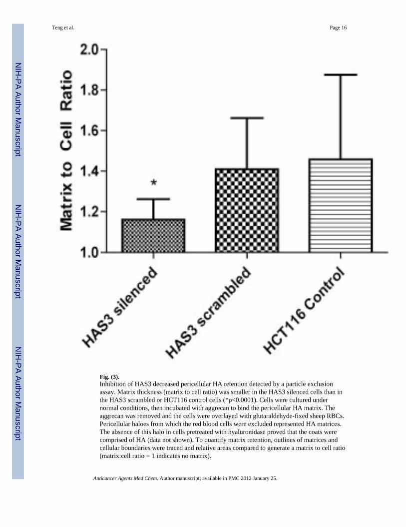
Fig. (3).Inhibition of HAS3 decreased pericellular HA retention detected by a particle exclusionassay. Matrix thickness (matrix to cell ratio) was smaller in the HAS3 silenced cells than inthe HAS3 scrambled or HCT116 control cells (*p<0.0001). Cells were cultured undernormal conditions, then incubated with aggrecan to bind the pericellular HA matrix. Theaggrecan was removed and the cells were overlayed with glutaraldehyde-fixed sheep RBCs.Pericellular haloes from which the red blood cells were excluded represented HA matrices.The absence of this halo in cells pretreated with hyaluronidase proved that the coats werecomprised of HA (data not shown). To quantify matrix retention, outlines of matrices andcellular boundaries were traced and relative areas compared to generate a matrix to cell ratio(matrix:cell ratio = 1 indicates no matrix).
Teng et al. Page 16
Anticancer Agents Med Chem. Author manuscript; available in PMC 2012 January 25.
NIH
-PA Author Manuscript
NIH
-PA Author Manuscript
NIH
-PA Author Manuscript

Fig. (4).Inhibition of HAS3 decreased subcutaneous tumor growth in mice. Tumors cells (7×10⁶)were injected subcutaneously into the flanks of the nude mice (HAS3 silenced N = 24,HAS3 scrambled N= 16). Tumor diameters were measured with digital calipers and volumescalculated. After sacrifice, the tumors were measured and weighed. A. After 10 days, tumorvolumes were significantly decreased in the HAS3 silenced tumors and this differencepersisted for the length of the experiment (*p<0.05). B. Tumor mass was decreased byapproximately 30% (p=0.0136), and C. tumor volume was decreased by greater than 50%(p=0.0063).
Teng et al. Page 17
Anticancer Agents Med Chem. Author manuscript; available in PMC 2012 January 25.
NIH
-PA Author Manuscript
NIH
-PA Author Manuscript
NIH
-PA Author Manuscript

Fig. (5).Tumors grown from HAS3 silenced cells stain less intensely with colloidal iron than dotumors grown with HAS3 scrambled (control) cells. Digestion with hyaluronidase decreasedintensity of staining in both sets of tumors, but the qualitative difference was greater in theHAS3 scrambled tumors compared to the HAS3 silenced tumors. This observation confirmsthat HAS3 inhibition resulted in decreased tumoral HA in vivo. (A) HAS3 silenced tumorstained with colloidal iron (blue pigment represents HA); (B) HAS3 silenced tumorpretreated with hyaluronidase then stained with colloidal iron; (C) HAS3 scrambled stainedwith colloidal iron; (D) HAS3 scrambled tumor pretreated with hyaluronidase then stainedwith colloidal iron.
Teng et al. Page 18
Anticancer Agents Med Chem. Author manuscript; available in PMC 2012 January 25.
NIH
-PA Author Manuscript
NIH
-PA Author Manuscript
NIH
-PA Author Manuscript

Fig. (6).Inhibition of HAS3 in both HCT 116 and SW620 derived tumors increased apoptosis.Apoptosis was assessed using caspase-3 immunostaining and proliferation assessed usingki-67 immunostaining. Slides were deparaffinized, endogenous peroxidase quenched, andantigen retrieval performed by microwave heating. Slides were then incubated with theappropriate primary antibody, washed, incubated with a biotinylated secondary antibody,and developed using an ABC reagent and DAB chromagen. An isotype matched primaryantibody was used as a control. A. HCT116 derived tumors display greater caspase-3staining (brown pigment) in the HAS3 silenced tumors than in the HAS3 scrambled tumors.Ki-67 staining (brown pigment) is similar in both groups. B. Proliferation was quantified by
Teng et al. Page 19
Anticancer Agents Med Chem. Author manuscript; available in PMC 2012 January 25.
NIH
-PA Author Manuscript
NIH
-PA Author Manuscript
NIH
-PA Author Manuscript

assessing ki-67 staining intensity (low = 1; high = 4). HAS3 inhibition had no effect onproliferation in the HCT116 derived tumors, but slightly increased proliferation in theSW620 derived tumors (p=0.0255). C. Apoptotic index was calculated by comparing totalapoptotic cells to total cells. Apoptotic index was significantly increased by HAS3 inhibitionin both sets of tumors (*p<0.0001; ** p=0.0252). Isotype matched controls for both ki-67and caspase-3 staining showed no staining (data not shown).
Teng et al. Page 20
Anticancer Agents Med Chem. Author manuscript; available in PMC 2012 January 25.
NIH
-PA Author Manuscript
NIH
-PA Author Manuscript
NIH
-PA Author Manuscript