Hyaluronan Aggregation in Different Human Organs - MDPI
12
Citation: Stecco, A.; Cowman, M.; Pirri, N.; Raghavan, P.; Pirri, C. Densification: Hyaluronan Aggregation in Different Human Organs. Bioengineering 2022, 9, 159. https://doi.org/10.3390/ bioengineering9040159 Academic Editor: Susan Sharfstein Received: 2 March 2022 Accepted: 4 April 2022 Published: 5 April 2022 Publisher’s Note: MDPI stays neutral with regard to jurisdictional claims in published maps and institutional affil- iations. Copyright: © 2022 by the authors. Licensee MDPI, Basel, Switzerland. This article is an open access article distributed under the terms and conditions of the Creative Commons Attribution (CC BY) license (https:// creativecommons.org/licenses/by/ 4.0/). bioengineering Review Densification: Hyaluronan Aggregation in Different Human Organs Antonio Stecco 1 , Mary Cowman 2 , Nina Pirri 3 , Preeti Raghavan 4 and Carmelo Pirri 5, * 1 Rusk Rehabilitation, New York University School of Medicine, New York, NY 10016, USA; [email protected] 2 Department of Biomedical Engineering, New York University Tandon School of Engineering, New York, NY 10016, USA; [email protected] 3 Department of Medicine—DIMED, School of Radiology, Radiology Institute, University of Padua, 35122 Padova, Italy; [email protected] 4 Department of Physical Medicine and Rehabilitation and Neurology, Johns Hopkins University School of Medicine, Baltimore, MD 21205, USA; [email protected] 5 Department of Neurosciences, Institute of Human Anatomy, University of Padova, 35121 Padova, Italy * Correspondence: [email protected] Abstract: Hyaluronan (HA) has complex biological roles that have catalyzed clinical interest in several fields of medicine. In this narrative review, we provide an overview of HA aggregation, also called densification, in human organs. The literature suggests that HA aggregation can occur in the liver, eye, lung, kidney, blood vessel, muscle, fascia, skin, pancreatic cancer and malignant melanoma. In all these organs, aggregation of HA leads to an increase in extracellular matrix viscosity, causing stiffness and organ dysfunction. Fibrosis, in some of these organs, may also occur as a direct consequence of densification in the long term. Specific imaging evaluation, such dynamic ultrasonography, elasto- sonography, elasto-MRI and T1ρ MRI can permit early diagnosis to enable the clinician to organize the treatment plan and avoid further progression of the pathology and dysfunction. Keywords: densification; hyaluronan; aggregation; liver; kidney; fascia; blood vessels; eye; skin; lung; muscle 1. Introduction The importance of hyaluronan (HA) at the cellular level is well known. HA can be produced by many cell types (fibroblasts, stellate cells, cancer associated fibroblasts, fasciacytes) [1], although mesenchymal cells are believed to be the predominant source of HA. Indeed, a burst of HA synthesis occurs just prior to mitosis, enabling some cells to become dissociated from neighboring cells and to lose the adhesion from their surrounding extracellular matrix (ECM) in preparation for division [2]. However, its role at the level of the organ and organism level has been less studied [3]. HA originally thought to be a mere “space-filler” in the ECM is now known to have important roles both structurally and as a signaling molecule [4]. HA fragments are able to stimulate several cell surface receptors, such as CD44, RHAMM, LYVE-1, HARE, layilin and Toll-4, acting as signaling pathways for gene expression regulation [5]. Furthermore, other receptors as TSHR [6], molecules as leukoregulin [7] and emmprin [8], and conditions as hypoxia [9], can stimulate the synthesis of hyaluronan [10,11]. HA fragments can promote tumor cell growth through CD44-dependent tyrosine kinase signal in tumor cells [5,12–14]. HA has complex biological roles that have catalyzed clinical interest in several fields of medicine, such as ophthalmic surgery, articular pathologies, cutaneous repair, skin remodeling, vascular prosthesis, adipose tissue engineering, nerve reconstruction and cancer therapy [15,16]. HA is present in many strains of bacteria and in all vertebrates, in which it is localized in the embryonic tissues and soft connective tissue ECM [15]. Bioengineering 2022, 9, 159. https://doi.org/10.3390/bioengineering9040159 https://www.mdpi.com/journal/bioengineering
-
Upload
khangminh22 -
Category
Documents
-
view
1 -
download
0
Transcript of Hyaluronan Aggregation in Different Human Organs - MDPI
�����������������
Citation: Stecco, A.; Cowman, M.;
Pirri, N.; Raghavan, P.; Pirri, C.
Densification: Hyaluronan
Aggregation in Different Human
Organs. Bioengineering 2022, 9, 159.
https://doi.org/10.3390/
bioengineering9040159
Academic Editor: Susan Sharfstein
Received: 2 March 2022
Accepted: 4 April 2022
Published: 5 April 2022
Publisher’s Note: MDPI stays neutral
with regard to jurisdictional claims in
published maps and institutional affil-
iations.
Copyright: © 2022 by the authors.
Licensee MDPI, Basel, Switzerland.
This article is an open access article
distributed under the terms and
conditions of the Creative Commons
Attribution (CC BY) license (https://
creativecommons.org/licenses/by/
4.0/).
bioengineering
Review
Densification: Hyaluronan Aggregation in DifferentHuman OrgansAntonio Stecco 1, Mary Cowman 2, Nina Pirri 3, Preeti Raghavan 4 and Carmelo Pirri 5,*
1 Rusk Rehabilitation, New York University School of Medicine, New York, NY 10016, USA;[email protected]
2 Department of Biomedical Engineering, New York University Tandon School of Engineering,New York, NY 10016, USA; [email protected]
3 Department of Medicine—DIMED, School of Radiology, Radiology Institute, University of Padua,35122 Padova, Italy; [email protected]
4 Department of Physical Medicine and Rehabilitation and Neurology, Johns Hopkins University School ofMedicine, Baltimore, MD 21205, USA; [email protected]
5 Department of Neurosciences, Institute of Human Anatomy, University of Padova, 35121 Padova, Italy* Correspondence: [email protected]
Abstract: Hyaluronan (HA) has complex biological roles that have catalyzed clinical interest in severalfields of medicine. In this narrative review, we provide an overview of HA aggregation, also calleddensification, in human organs. The literature suggests that HA aggregation can occur in the liver,eye, lung, kidney, blood vessel, muscle, fascia, skin, pancreatic cancer and malignant melanoma. In allthese organs, aggregation of HA leads to an increase in extracellular matrix viscosity, causing stiffnessand organ dysfunction. Fibrosis, in some of these organs, may also occur as a direct consequence ofdensification in the long term. Specific imaging evaluation, such dynamic ultrasonography, elasto-sonography, elasto-MRI and T1ρ MRI can permit early diagnosis to enable the clinician to organizethe treatment plan and avoid further progression of the pathology and dysfunction.
Keywords: densification; hyaluronan; aggregation; liver; kidney; fascia; blood vessels; eye; skin;lung; muscle
1. Introduction
The importance of hyaluronan (HA) at the cellular level is well known. HA canbe produced by many cell types (fibroblasts, stellate cells, cancer associated fibroblasts,fasciacytes) [1], although mesenchymal cells are believed to be the predominant source ofHA. Indeed, a burst of HA synthesis occurs just prior to mitosis, enabling some cells tobecome dissociated from neighboring cells and to lose the adhesion from their surroundingextracellular matrix (ECM) in preparation for division [2]. However, its role at the levelof the organ and organism level has been less studied [3]. HA originally thought to be amere “space-filler” in the ECM is now known to have important roles both structurallyand as a signaling molecule [4]. HA fragments are able to stimulate several cell surfacereceptors, such as CD44, RHAMM, LYVE-1, HARE, layilin and Toll-4, acting as signalingpathways for gene expression regulation [5]. Furthermore, other receptors as TSHR [6],molecules as leukoregulin [7] and emmprin [8], and conditions as hypoxia [9], can stimulatethe synthesis of hyaluronan [10,11]. HA fragments can promote tumor cell growth throughCD44-dependent tyrosine kinase signal in tumor cells [5,12–14]. HA has complex biologicalroles that have catalyzed clinical interest in several fields of medicine, such as ophthalmicsurgery, articular pathologies, cutaneous repair, skin remodeling, vascular prosthesis,adipose tissue engineering, nerve reconstruction and cancer therapy [15,16]. HA is presentin many strains of bacteria and in all vertebrates, in which it is localized in the embryonictissues and soft connective tissue ECM [15].
Bioengineering 2022, 9, 159. https://doi.org/10.3390/bioengineering9040159 https://www.mdpi.com/journal/bioengineering

Bioengineering 2022, 9, 159 2 of 12
HA is the simplest of the glycosaminoglycans (GAGs) composed of a linear non-sulfated polymer of up to 20,000 repeats of its disaccharide unit D-glucuronic acid andN-acetyl-D-glucosamine [17]. Due to the carboxyl groups in the molecule, HA is negativelycharged and highly hydrophilic, being able to hold water molecules up to 1000-fold of itsmolecular weight [17]. HA is produced by HA synthases (HAS1–3) in the plasma membraneof different cells (fibroblasts, stellate cells, cancer associated fibroblasts, fasciacytes), forthen be secreted out of the cell membrane [17]. HAS1 and HAS2 are both able to produceHA of 2–4 × 10⁶ Da, but HAS2 is the main HA synthase in the skin [17]; while HAS3produces HA of 0.4–2.5 × 10⁵ Da [4]. The turnover of HA occurs rapidly and its half-lifevaries from 12–24 h in the skin to a few minutes in the bloodstream [18]. The different HAmolecular sizes can display different and sometimes opposing biological actions. In fact,the equilibrium between HA synthesis and degradation has a key role in determining themolecular weight (MW) of HA and, consequently, its properties [19,20]. Therefore, HAhas a key role not only physiologically, such as in ECM hydration and regulation of tissuehomeostasis and resistance to compressive forces, but also in pathological conditions [15].At high molecular weights, HA is able to form a viscous network, and by interacting withmany proteoglycans, such as aggrecan, gives rise to molecular composites that occupy ahuge volume and are responsible for the gel state of the matrix. These large complexes ofHA and HA-binding proteoglycans crosslink with other matrix proteins, such as collagen,and result in the formation of super-molecular structures which increase tissue stiffness [21].Excessive accumulation of HA in a spread surface can dramatically increase its viscosityand alter its lubricating properties [22]. At high concentrations, HA behaves similar toa non-Newtonian fluid and becomes more viscous [23], because the HA chains entangle,contributing to the hydrodynamic properties of the solution. Hyper-viscous ECM increasesthe passive resistance to movement, and can reduce force transmission during movement,with the consequent stiffness [24]. Stecco et al. identified this phenomenon in fascial tissuedefining it as “densification”: a high viscous state of ECM due to HA super-aggregationwith a decreased water binding capacity [25]. The purpose of this narrative review is tooutline the mechanisms underlying increase of the tissue stiffness with HA accumulationand aggregation, identify which organs are affected and the clinical consequences in eachof them.
2. Materials and Methods
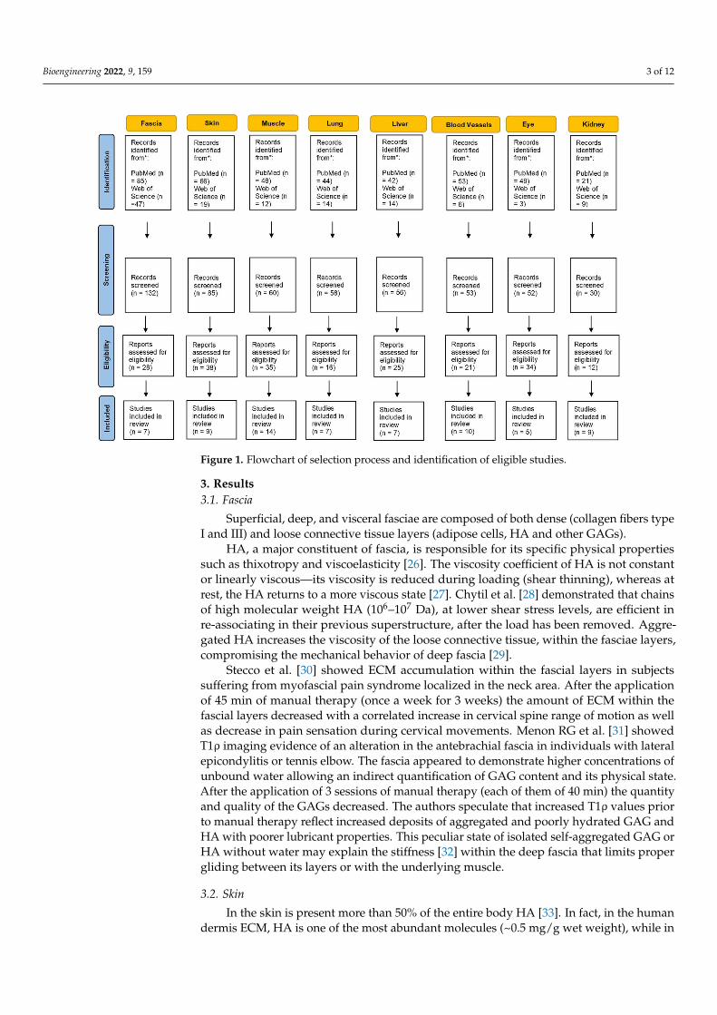
A substantial literature search was conducted to review the literature from 1960through October 2021. Two electronic databases, i.e., PubMed and Web of Science wereused to identify all relevant English publications without any category restriction. TheMeSH keywords used were: “hyaluronan aggregation”, “hyaluronic acid aggregation”,“hyaluronan densification” and “stiffness”. The search was extended through the refer-ence lists of the recruited texts. Relevant secondary references were also captured. Theauthors looked for papers discussing hyaluronan aggregation and stiffness in differentextra-articular anatomical structures. Different organs and tissues were found includingliver, kidney, eyes, skin, blood vessels, lung, muscle and fascia. Two reviewers (CP and NP)independently selected the articles by reading the titles and abstracts. A third reviewer (AS)finalized the selection in case of disagreement. After the selection, each title/abstract/fulltext was independently evaluated by each of the authors. The following data were recorded;any discrepancies were resolved by agreement among the authors. The flowchart of thestudy is shown in Figure 1.
Bioengineering 2022, 9, 159 3 of 12Bioengineering 2022, 8, x FOR PEER REVIEW 3 of 12
Figure 1. Flowchart of selection process and identification of eligible studies.
3. Results 3.1. Fascia
Superficial, deep, and visceral fasciae are composed of both dense (collagen fibers type I and III) and loose connective tissue layers (adipose cells, HA and other GAGs).
HA, a major constituent of fascia, is responsible for its specific physical properties such as thixotropy and viscoelasticity [26]. The viscosity coefficient of HA is not constant or linearly viscous—its viscosity is reduced during loading (shear thinning), whereas at rest, the HA returns to a more viscous state [27]. Chytil et al. [28] demonstrated that chains of high molecular weight HA (106–107 Da), at lower shear stress levels, are efficient in re-associating in their previous superstructure, after the load has been removed. Aggregated HA increases the viscosity of the loose connective tissue, within the fasciae layers, compromising the mechanical behavior of deep fascia [29].
Stecco et al [30] showed ECM accumulation within the fascial layers in subjects suffering from myofascial pain syndrome localized in the neck area. After the application of 45 minutes of manual therapy (once a week for 3 weeks) the amount of ECM within the fascial layers decreased with a correlated increase in cervical spine range of motion as well as decrease in pain sensation during cervical movements. Menon RG et al [31] showed T1ρ imaging evidence of an alteration in the antebrachial fascia in individuals with lateral epicondylitis or tennis elbow. The fascia appeared to demonstrate higher concentrations of unbound water allowing an indirect quantification of GAG content and its physical state. After the application of 3 sessions of manual therapy (each of them of 40 minutes) the quantity and quality of the GAGs decreased. The authors speculate that increased T1ρ values prior to manual therapy reflect increased deposits of aggregated and poorly hydrated GAG and HA with poorer lubricant properties. This peculiar state of isolated self-aggregated GAG or HA without water may explain the stiffness [32] within the deep fascia that limits proper gliding between its layers or with the underlying muscle.
Figure 1. Flowchart of selection process and identification of eligible studies.
3. Results3.1. Fascia
Superficial, deep, and visceral fasciae are composed of both dense (collagen fibers typeI and III) and loose connective tissue layers (adipose cells, HA and other GAGs).
HA, a major constituent of fascia, is responsible for its specific physical propertiessuch as thixotropy and viscoelasticity [26]. The viscosity coefficient of HA is not constantor linearly viscous—its viscosity is reduced during loading (shear thinning), whereas atrest, the HA returns to a more viscous state [27]. Chytil et al. [28] demonstrated that chainsof high molecular weight HA (106–107 Da), at lower shear stress levels, are efficient inre-associating in their previous superstructure, after the load has been removed. Aggre-gated HA increases the viscosity of the loose connective tissue, within the fasciae layers,compromising the mechanical behavior of deep fascia [29].
Stecco et al. [30] showed ECM accumulation within the fascial layers in subjectssuffering from myofascial pain syndrome localized in the neck area. After the applicationof 45 min of manual therapy (once a week for 3 weeks) the amount of ECM within thefascial layers decreased with a correlated increase in cervical spine range of motion as wellas decrease in pain sensation during cervical movements. Menon RG et al. [31] showedT1ρ imaging evidence of an alteration in the antebrachial fascia in individuals with lateralepicondylitis or tennis elbow. The fascia appeared to demonstrate higher concentrations ofunbound water allowing an indirect quantification of GAG content and its physical state.After the application of 3 sessions of manual therapy (each of them of 40 min) the quantityand quality of the GAGs decreased. The authors speculate that increased T1ρ values priorto manual therapy reflect increased deposits of aggregated and poorly hydrated GAG andHA with poorer lubricant properties. This peculiar state of isolated self-aggregated GAG orHA without water may explain the stiffness [32] within the deep fascia that limits propergliding between its layers or with the underlying muscle.
3.2. Skin
In the skin is present more than 50% of the entire body HA [33]. In fact, in the humandermis ECM, HA is one of the most abundant molecules (~0.5 mg/g wet weight), while in
Bioengineering 2022, 9, 159 4 of 12
the epidermis is ~0.1 mg/g [33]. It is also known that in the dermis and in the epidermisthe HA turnover rate are, respectively, a half-life of <1 day and 2–4 h [17]. The combinationof hyaluronidase (HYAL) 1 and 2 mediates this rapid clearance [17].
Since HA exits in both free [34] and tissue receptor-bound [35] forms, HA crosslinkingwith matrix proteins determines the formation of supermolecular structures increasing tis-sue stiffness [4]. Older skin shows a higher association of HA with other tissue proteins [36],resulting in reduced free HA [36]. The same authors reported that HA concentration is 4.3and 6.7 times higher, respectively, in adults and senescent material, compared to fetal skin.This suggest that, with increasing age, HA became progressively more tightly bound [36].Even in photoaged skin, as maybe result of abnormal accumulation, an increase in dermalHA was observed that [21].
3.3. Muscle
HA acts as a lubricant within the endomysium, perimysium, and epimysium to aidthe sliding of muscle fibers during movement [37], and its layer thickness is continuouslyremodeled in response to mechanical stimuli. Histological studies using animal modelshave shown that joint immobilization leads to the accumulation of hyaluronan in theskeletal muscle [38]. Following a stroke, catabolic pathways are activated in muscles thatenhance muscle wasting and atrophy leading to ECM volume increase relative to musclefibers [39]. An imbalance ECM turnover, as ensues of muscle paralysis and immobility,can result in increasing HA production and/or reduced degradation. Accumulation ofHA in the ECM can increase its fluid viscosity causing lack of gliding between musclefibers, reduced force transmission and consequently increased resistance or stiffness duringattempted movement [40].
This phenomenon can also explain why immobility reduces range of motion, as notedin the ankles and feet when taking the first few steps out of bed in the morning [41].Herda et al. [42] reported that, following two minutes of dynamic stretching, passive resis-tive torque and passive stiffness decreased. This indicated modifications in the viscoelasticproperties of the muscular-tendon unit. Similarly, Nordez et al. [43] have reported that,during cyclic stretching protocols, viscosity plays a major role in passive stiffness changes.Other authors explained how an increase in temperature, not only reduce the musclesviscous resistance [44], but also the passive resistive torque and the muscular-tendon unitstiffness [45]. A recent case series proved the efficacy of intramuscular hyaluronidaseinjections for reducing muscle stiffness, and increasing passive and active range of motionin the post-stroke arm, with over three months lasting effects [40]. The same authors havedemonstrated the presence of high concentrations of HA in muscles affected by centralneurological injury using T1ρ MRI mapping which is sensitive to the chemical exchangeof large macromolecules such as hyaluronan with protons in bulk water [46]. Prolongedimmobility also initiates a complex pathologic pathway resulting in fibrogenesis [47]. Infact, an alteration in the structure and function of the muscle, defined as contracture, can bedue to an excess of ECM HA that was then replaced by collagen, leading to permanent andirreversible thickening of the endomysium and perimysium [48].
3.4. Lung
Nettelbladt et al. [49] found that normal lung tissue shows positive staining for HAin the basal membrane and in the submucosal tissue of bronchi and bronchioles and theadventitia of arteries and veins. In contrast, in the areas treated with bleomycin, the alveolidemonstrated increased accumulation of HA with positive HA staining of the alveolarinterstitial tissue in injured areas with markedly thickened alveolar septa. HA in the normalrat lung is also bordered to peribronchial and perivascular spaces, while it is not visible inthe alveolar tissue [49]. Hence it was suggested that the accumulation of HA has a role inalveolar interstitial edema. In the early phase of bleomycin-induced lung injury, there is aconsiderable but transient accumulation of HA in the alveolar space that provides a matrixfor later deposition of denser structures such as collagen. These results were confirmed by
Bioengineering 2022, 9, 159 5 of 12
Juul et al. [50] who saw, as response to fibrogenic stimulus in lung exposed to bleomycininjury, an increased deposition of HA. In previous studies of the process of healing indamaged tissue, it has been documented that an early accumulation of HA precedes scarformation [51].
Bronchoalveolar lavage studies in humans have demonstrated highly increased lavagefluid HA concentrations in interstitial diseases, extrinsic alveolitis [52], idiopathic lungfibrosis, and adult respiratory distress syndrome [53]. In addition, Collum SD et al. [54]revealed that hyaluronan fragments resulted in increased human pulmonary artery smoothmuscle cell stiffness and proliferation, demonstrating that elevated hyaluronan is a patho-logical process that modulated pulmonary hypertension associated with lung fibrosis thatcan be treated by inhibiting hyaluronan synthesis.
3.5. Liver
HA is a major component of the extracellular matrix of the liver [55]. In the fibroticliver the quantity of ECM can be up to eight-fold higher than that of the normal liver [56].Fibrosis involves histological and molecular re-arrangement of various types of collagens,proteoglycans, glycoproteins and HA [57]. For this reason, HA level is one of the bestpredictors of liver fibrosis and is directly linked to modifications in ECM turnover duringfibrogenesis [58]. It was proven a continuous hepatic stellate cell (HSC) activation, andtherefore increased HA synthesis during chronic liver inflammation [59]. As consequenceof liver cell injury, together with HA level serum rising, a transformation of stellate cellsinto myofibroblasts occur, releasing various ECM components as elastin, collagens, glyco-proteins, and proteoglycans [59]. The same Authors proved that HSC activation is the resultof an im balance between ECM synthesis and degradation [59]. Sakakibara et al. reportedhyaluronic acid and dermatan sulfate accumulation in the liver parenchymal cells after10 days incubation [60]. Similar results in vivo were found by Koizumi T et al. [61] whoshow how chronic hepatic damage gave rise to an increase of HA and dermatan sulfatewith time as fibrogenesis advanced.
The liver is also the primary organ responsible for HA degradation [59]. Both theincreased synthesis and decreased clearance during fibrogenesis increases HA concentra-tion [62], making HA a candidate for measuring liver fibrosis.
According to Nobili et al. [63], serum HA > 1200 ng/mL makes the absence of fibrosisunlikely (7%, 95% CI: 1% to 14%), while serum HA > 2100 ng/mL makes significant fibrosisvery likely (89%, 95% CI: 75% to 100%). In several others studies, HA turned out to be thebest class I biomarker of fibrosis having an area under curve (AUC) of 0.97, a sensitivity of86–100%, and specificity of about 88% in investigations of cirrhosis due to non-alcoholic fattyliver disease [64] and other etiologies [65]. In addition, Kaneda et al. [66] have selected HA asa predictive marker for severe fibrosis, defining 42 ng/mL of HA serum the 100% predictivecut-off value. This value is also associated with an optimal combination of sensitivity (100%,95% confidence interval [CI] 90–100%) and specificity (89%, 95% CI: 80–94%).
3.6. Blood Vessels
HA is one of the most prevalent GAGs in the vascular extracellular matrix. AbnormalHA accumulation within blood vessel walls is prominent in most vascular pathologicalconditions such as atherosclerosis and restenosis [67,68]. HA is up-regulated in areas ofvascular injury and increases the migration and proliferation of vascular smooth musclecells (VSMCs), which are key events in the progression of atherosclerosis and vascularnarrowing [69]. VSMCs, in their natural state, are located in the tunica media, but they mi-grate to the tunica intima during the early stages of atherosclerosis [70]. Once in the intima,these VSMCs begin manufacturing large amounts of ECM components, including HA [71]that form intercellular “cable-like” structures in the ECM [72]. Interestingly, studies havedemonstrated that these cable-like structures are involved in the early proinflammatorystage of atherosclerosis [73].
Bioengineering 2022, 9, 159 6 of 12
Increased aggregation of HA and versican, in vivo, tends to increase the swellingpressure of the ECM. If it will not be counteracted by fibrillar matrix components, it wouldpredispose the artery to stenosis [74]. Links, that stabilize the association between versicanand HA, may aid in the versican retention in normal and diseased arterial tissue [74].
In human re-stenotic lesions, HA appears to be concentrated in the intima and adven-titia [75]. Evidence of HA accumulation in the intima of injured and aging arterial vesselsis demonstrated in both human and animal studies [76]. Older rats were shown to have anincreased ratio of hyaluronidase to HA at baseline and at all time points post injury [76],likely representing a role for HA and hyaluronidase in the aging vessel. Sadowitz et al. [77]showed that, in the two processes most responsible for vessel renarrowing such intimalhyperplasia and wound contracture, HA has an integral and early role in the developmentand structure of re-stenotic lesions after vascular instrumentation. These observations areconsistent with increased vascular stiffness induced by HA accumulation in the aorta seenby Lorentzen et al. [78]. Interestingly, in this study, increased proliferation of vascularsmooth muscle cells was also associated with increased HA accumulation.
3.7. Eyes
HA makes up 96% of the total GAG in the vitreous humor [79]. High molecular massHA at high concentrations possesses viscoelastic and exclusion properties [80]. Since HAsugars bind and organize water molecules, this proteoglycan-collagen interaction results ingel formation [81], directly contributing to the supramolecular organization of the vitreousand its homeostasis and biomechanics [82]. Authors found the enormous potential of HAand other binding partners, with N-terminal and C-terminal regions of proteoglycans (suchas versican), to form macromolecular complexes which play a crucial role in the structuralstability and functionality of the vitreous, due to their important biological properties” [83].
Narayanan and Kuppermann [84] reported the use hyaluronidases for pharmacologicvitreolysis avoiding the complications of surgery and anesthesia-related. Dissolution ofthe HA and collagen complex results in decreased viscosity of the extracellular matrix [85].HA can also act much similar to an ion-exchange resin in which an electrostatic interactionforms the basis for various HA properties, including its influence upon osmotic pressureand ion transport within the vitreous [86].
3.8. Kidney
Over the years, HA has emerged as an important molecule in nephrological and uro-logical studies involving ECM organization, inflammation, tissue regeneration, and viralsensing [86]. In a healthy human kidney, very little HA is expressed, but it is accumulatedin the cortical interstitium during progressive kidney disease. This increase of HA con-centration stimulates the recruitment of monocytes into the interstitial space, potentiatingthe interstitial inflammation [87]. In several kidney diseases, elevated levels of HA inrenal tissue were reported in both rat models (ischemia-reperfusion injury) and humandiseases (diabetic nephropathy, renal transplant rejection and kidney stone formation) [3].Selbi et al. [88] have demonstrated that renal proximal tubular epithelial cells surroundthemselves in vitro with HA in an organized pericellular matrix or ’coat’. This is associatedwith cell migration, pericellular HA cable-like structures formation, epithelial cell trans-differentiation, ref. [89] and facilitation of fibrotic response in the context of progressiverenal disease.
It was noted that papillary mesenchymal cells stimulate HA synthesis after an obstruc-tion, rising the interstitial HA levels [90]. HA can form gel-like matrices that are negativelycharged and able to bind crystals. This is particularly relevant in obstructive uropathy [86].Furthermore, HA is present in kidney stones in fractions that are in disproportion to HAurinary levels [91].
Han et al. [92] found, in chronic kidney disease (CKD) of animal model, an increasedexpression of HA. This accumulation of HA, in the renal cortex and sclerotic vessels in CKD,was in contract with the regular HA presence, only in the renal medulla under physiologic
Bioengineering 2022, 9, 159 7 of 12
conditions. In CKD, HA accumulation was associated with an increasing α-SMA andconsequently a pro-inflammatory and fibrogenic milieu [93].
4. Discussion
HA is a major component of the extracellular matrix surrounding migrating andproliferating cells and therefore, it is an important component of healing or regeneratingtissue. One molecule of HA may contain as many as 20,000 repeating disaccharide unitsin a linear array. Extensive intrachain hydrogen bonding, mutual repulsion between thenegatively charged carboxylate groups, and the beta linkages between the sugar residueslend HA an inherent stiffness [94].
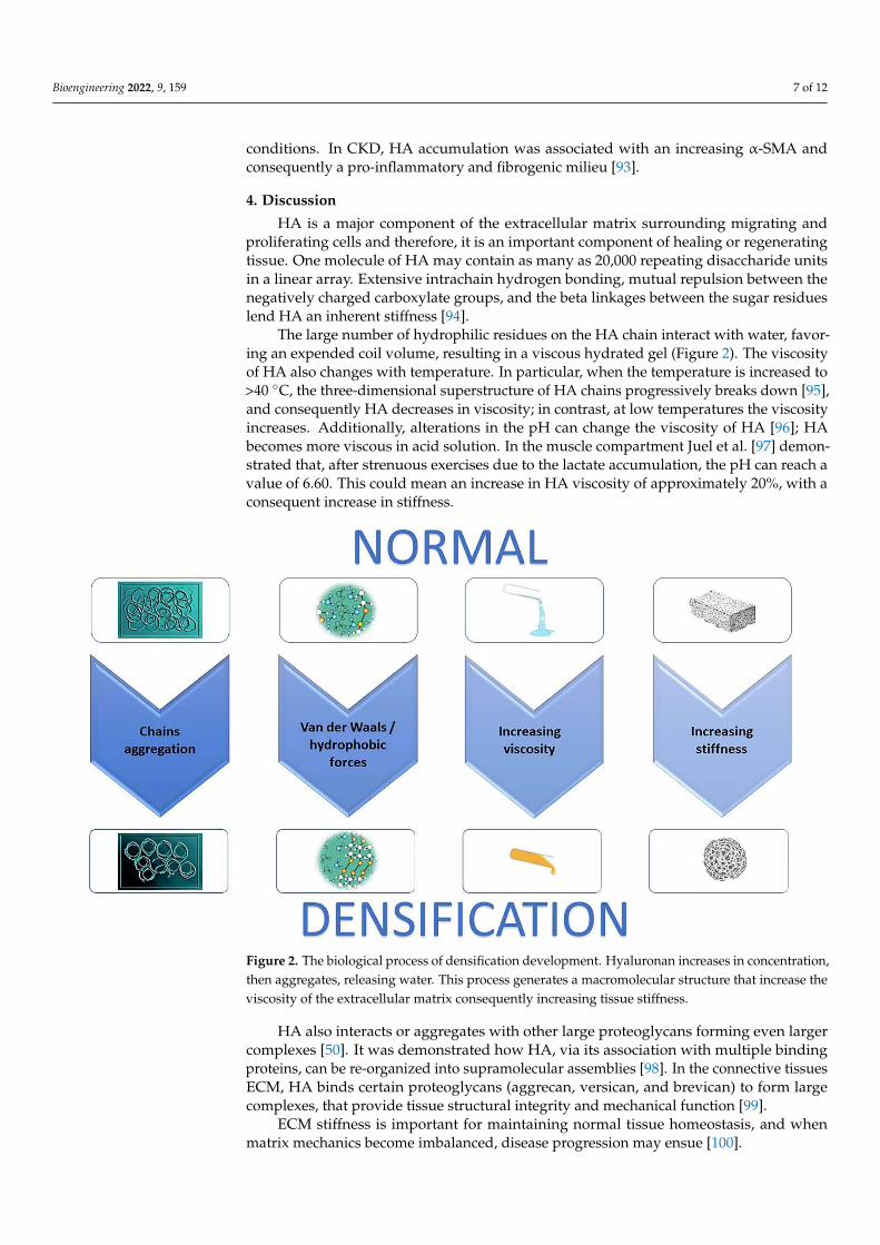
The large number of hydrophilic residues on the HA chain interact with water, favor-ing an expended coil volume, resulting in a viscous hydrated gel (Figure 2). The viscosityof HA also changes with temperature. In particular, when the temperature is increased to>40 ◦C, the three-dimensional superstructure of HA chains progressively breaks down [95],and consequently HA decreases in viscosity; in contrast, at low temperatures the viscosityincreases. Additionally, alterations in the pH can change the viscosity of HA [96]; HAbecomes more viscous in acid solution. In the muscle compartment Juel et al. [97] demon-strated that, after strenuous exercises due to the lactate accumulation, the pH can reach avalue of 6.60. This could mean an increase in HA viscosity of approximately 20%, with aconsequent increase in stiffness.
Bioengineering 2022, 8, x FOR PEER REVIEW 7 of 12
negatively charged and able to bind crystals. This is particularly relevant in obstructive uropathy [86]. Furthermore, HA is present in kidney stones in fractions that are in dispro-portion to HA urinary levels [91].
Han et al. [92] found, in chronic kidney disease (CKD) of animal model, an increased expression of HA. This accumulation of HA, in the renal cortex and sclerotic vessels in CKD, was in contract with the regular HA presence, only in the renal medulla under phys-iologic conditions. In CKD, HA accumulation was associated with an increasing α-SMA and consequently a pro-inflammatory and fibrogenic milieu [93].
4. Discussion HA is a major component of the extracellular matrix surrounding migrating and pro-
liferating cells and therefore, it is an important component of healing or regenerating tis-sue. One molecule of HA may contain as many as 20,000 repeating disaccharide units in a linear array. Extensive intrachain hydrogen bonding, mutual repulsion between the neg-atively charged carboxylate groups, and the beta linkages between the sugar residues lend HA an inherent stiffness [94].
The large number of hydrophilic residues on the HA chain interact with water, fa-voring an expended coil volume, resulting in a viscous hydrated gel (Figure 2). The vis-cosity of HA also changes with temperature. In particular, when the temperature is in-creased to >40 °C, the three-dimensional superstructure of HA chains progressively breaks down [95], and consequently HA decreases in viscosity; in contrast, at low temper-atures the viscosity increases. Additionally, alterations in the pH can change the viscosity of HA [96]; HA becomes more viscous in acid solution. In the muscle compartment Juel et al. [97] demonstrated that, after strenuous exercises due to the lactate accumulation, the pH can reach a value of 6.60. This could mean an increase in HA viscosity of approxi-mately 20%, with a consequent increase in stiffness.
Figure 2. The biological process of densification development. Hyaluronan increases in concentra-tion, then aggregates, releasing water. This process generates a macromolecular structure that in-crease the viscosity of the extracellular matrix consequently increasing tissue stiffness.
Figure 2. The biological process of densification development. Hyaluronan increases in concentration,then aggregates, releasing water. This process generates a macromolecular structure that increase theviscosity of the extracellular matrix consequently increasing tissue stiffness.
HA also interacts or aggregates with other large proteoglycans forming even largercomplexes [50]. It was demonstrated how HA, via its association with multiple bindingproteins, can be re-organized into supramolecular assemblies [98]. In the connective tissuesECM, HA binds certain proteoglycans (aggrecan, versican, and brevican) to form largecomplexes, that provide tissue structural integrity and mechanical function [99].
ECM stiffness is important for maintaining normal tissue homeostasis, and whenmatrix mechanics become imbalanced, disease progression may ensue [100].
Bioengineering 2022, 9, 159 8 of 12
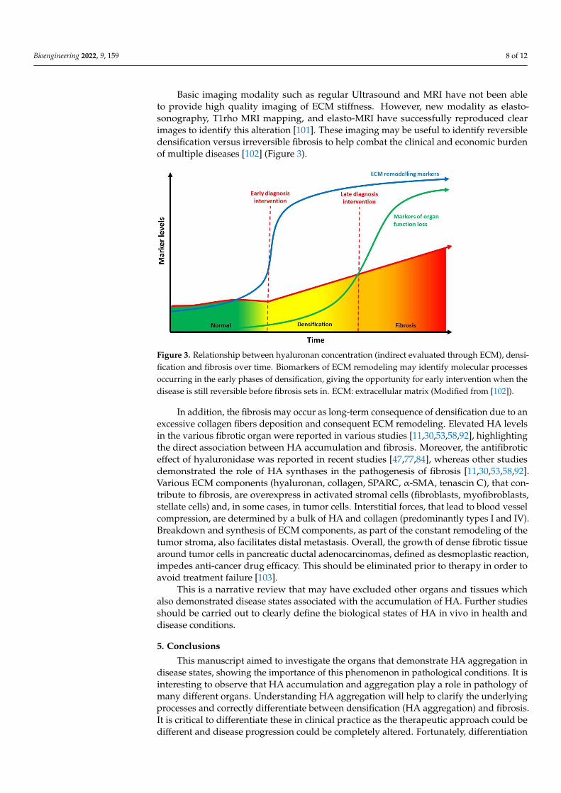
Basic imaging modality such as regular Ultrasound and MRI have not been ableto provide high quality imaging of ECM stiffness. However, new modality as elasto-sonography, T1rho MRI mapping, and elasto-MRI have successfully reproduced clearimages to identify this alteration [101]. These imaging may be useful to identify reversibledensification versus irreversible fibrosis to help combat the clinical and economic burdenof multiple diseases [102] (Figure 3).
Bioengineering 2022, 8, x FOR PEER REVIEW 8 of 12
HA also interacts or aggregates with other large proteoglycans forming even larger complexes [50]. It was demonstrated how HA, via its association with multiple binding proteins, can be re-organized into supramolecular assemblies [98]. In the connective tis-sues ECM, HA binds certain proteoglycans (aggrecan, versican, and brevican) to form large complexes, that provide tissue structural integrity and mechanical function [99].
ECM stiffness is important for maintaining normal tissue homeostasis, and when ma-trix mechanics become imbalanced, disease progression may ensue [100].
Basic imaging modality such as regular Ultrasound and MRI have not been able to provide high quality imaging of ECM stiffness. However, new modality as elasto-sonog-raphy, T1rho MRI mapping, and elasto-MRI have successfully reproduced clear images to identify this alteration [101]. These imaging may be useful to identify reversible densi-fication versus irreversible fibrosis to help combat the clinical and economic burden of multiple diseases [102] (Figure 3).
Figure 3. Relationship between hyaluronan concentration (indirect evaluated through ECM), den-sification and fibrosis over time. Biomarkers of ECM remodeling may identify molecular processes occurring in the early phases of densification, giving the opportunity for early intervention when the disease is still reversible before fibrosis sets in. ECM: extracellular matrix (Modified from [102]).
In addition, the fibrosis may occur as long-term consequence of densification due to an excessive collagen fibers deposition and consequent ECM remodeling. Elevated HA levels in the various fibrotic organ were reported in various studies [11,30,53,58,92], high-lighting the direct association between HA accumulation and fibrosis. Moreover, the an-tifibrotic effect of hyaluronidase was reported in recent studies [47,77,84], whereas other studies demonstrated the role of HA synthases in the pathogenesis of fibrosis [11,30,53,58,92]. Various ECM components (hyaluronan, collagen, SPARC, α-SMA, tenas-cin C), that contribute to fibrosis, are overexpress in activated stromal cells (fibroblasts, myofibroblasts, stellate cells) and, in some cases, in tumor cells. Interstitial forces, that lead to blood vessel compression, are determined by a bulk of HA and collagen (predomi-nantly types I and IV). Breakdown and synthesis of ECM components, as part of the con-stant remodeling of the tumor stroma, also facilitates distal metastasis. Overall, the growth of dense fibrotic tissue around tumor cells in pancreatic ductal adenocarcinomas, defined as desmoplastic reaction, impedes anti-cancer drug efficacy. This should be eliminated prior to therapy in order to avoid treatment failure [103].
This is a narrative review that may have excluded other organs and tissues which also demonstrated disease states associated with the accumulation of HA. Further studies should be carried out to clearly define the biological states of HA in vivo in health and disease conditions.
Figure 3. Relationship between hyaluronan concentration (indirect evaluated through ECM), densi-fication and fibrosis over time. Biomarkers of ECM remodeling may identify molecular processesoccurring in the early phases of densification, giving the opportunity for early intervention when thedisease is still reversible before fibrosis sets in. ECM: extracellular matrix (Modified from [102]).
In addition, the fibrosis may occur as long-term consequence of densification due to anexcessive collagen fibers deposition and consequent ECM remodeling. Elevated HA levelsin the various fibrotic organ were reported in various studies [11,30,53,58,92], highlightingthe direct association between HA accumulation and fibrosis. Moreover, the antifibroticeffect of hyaluronidase was reported in recent studies [47,77,84], whereas other studiesdemonstrated the role of HA synthases in the pathogenesis of fibrosis [11,30,53,58,92].Various ECM components (hyaluronan, collagen, SPARC, α-SMA, tenascin C), that con-tribute to fibrosis, are overexpress in activated stromal cells (fibroblasts, myofibroblasts,stellate cells) and, in some cases, in tumor cells. Interstitial forces, that lead to blood vesselcompression, are determined by a bulk of HA and collagen (predominantly types I and IV).Breakdown and synthesis of ECM components, as part of the constant remodeling of thetumor stroma, also facilitates distal metastasis. Overall, the growth of dense fibrotic tissuearound tumor cells in pancreatic ductal adenocarcinomas, defined as desmoplastic reaction,impedes anti-cancer drug efficacy. This should be eliminated prior to therapy in order toavoid treatment failure [103].
This is a narrative review that may have excluded other organs and tissues whichalso demonstrated disease states associated with the accumulation of HA. Further studiesshould be carried out to clearly define the biological states of HA in vivo in health anddisease conditions.
5. Conclusions
This manuscript aimed to investigate the organs that demonstrate HA aggregation indisease states, showing the importance of this phenomenon in pathological conditions. It isinteresting to observe that HA accumulation and aggregation play a role in pathology ofmany different organs. Understanding HA aggregation will help to clarify the underlyingprocesses and correctly differentiate between densification (HA aggregation) and fibrosis.It is critical to differentiate these in clinical practice as the therapeutic approach could bedifferent and disease progression could be completely altered. Fortunately, differentiation
Bioengineering 2022, 9, 159 9 of 12
is possible through imaging. The therapeutic window to treat a patient presenting densifi-cation, before fibrosis is established, is quite wide, enabling prevention to an irreversiblestate, which is always the best.
Author Contributions: Conceptualization, A.S. and C.P.; methodology, A.S., N.P. and C.P.; software,C.P.; validation, A.S., M.C., N.P., P.R. and C.P.; formal analysis, A.S., M.C., N.P., P.R. and C.P.;investigation, A.S.; N.P. and C.P.; data curation, A.S.; N.P. and C.P.; writing—original draft preparation,A.S. and C.P.; writing—review and editing, A.S., M.C., N.P., P.R. and C.P.; visualization, A.S., M.C.,N.P., P.R. and C.P.; supervision, A.S. and C.P.; project administration, A.S. and C.P. All authors haveread and agreed to the published version of the manuscript.
Funding: This research received no external funding.
Institutional Review Board Statement: Not applicable.
Informed Consent Statement: Not applicable.
Data Availability Statement: The data presented in this study are available on request from thecorresponding author.
Conflicts of Interest: The authors declare no conflict of interest.
References1. Jiang, D.; Liang, J.; Noble, P.W. Hyaluronan in tissue injury and repair. Annu. Rev. Cell Dev. Biol. 2007, 23, 435–461. [CrossRef]
[PubMed]2. Hascall, V.C.; Majors, A.K.; De La Motte, C.A.; Evanko, S.P.; Wang, A.; Drazba, J.A.; Wight, T.N. Intracellular hyaluronan: A new
frontier for inflammation? Biochim. Et Biophys. Acta (BBA)-Gen. Subj. 2004, 1673, 3–12. [CrossRef] [PubMed]3. Stridh, S.; Palm, F.; Hansell, P. Renal interstitial hyaluronan: Functional aspects during normal and pathological conditions. Am. J.
Physiol. Regul. Integr. Comp. Physiol. 2012, 302, R1235–R1249. [CrossRef] [PubMed]4. Lee, J.; Spicer, A. Hyaluronan: A multifunctional, megaDalton, stealth molecule. Curr. Opin. Cell Biol. 2000, 12, 581. [CrossRef]5. Savani, R.C.; Delisser, H.M. Hyaluronan and its Receptors in Lung Health and Disease. In Proteoglycans in Lung Disease; CRC
Press: Boca Raton, FL, USA, 2002; pp. 102–135.6. Zhang, L.; Bowen, T.; Grennan-Jones, F.; Paddon, C.; Giles, P.; Webber, J.; Ludgate, M. Thyrotropin receptor activation increases
hyaluronan production in preadipocyte fibroblasts: Contributory role in hyaluronan accumulation in thyroid dysfunction. J. Biol.Chem. 2009, 284, 26447–26455. [CrossRef]
7. Smith, T.J.; Wang, H.S.; Evans, C.H. Leukoregulin is a potent inducer of hyaluronan synthesis in cultured human orbital fibroblasts.Am. J. Physiol.-Cell Physiol. 1995, 268, C382–C388. [CrossRef]
8. Marieb, E.A.; Zoltan-Jones, A.; Li, R.; Misra, S.; Ghatak, S.; Cao, J.; Toole, B.P. Emmprin promotes anchorage-independent growthin human mammary carcinoma cells by stimulating hyaluronan production. Cancer Res. 2004, 64, 1229–1232. [CrossRef]
9. Gao, F.; Okunieff, P.; Han, Z.; Ding, I.; Wang, L.; Liu, W.; Zhang, L. Hypoxia-Induced Alterations in Hyaluronan andHyaluronidase. In Oxygen Transport to Tissue XXVI; Springer: Boston, MA, USA, 2005; pp. 249–256.
10. David-Raoudi, M.; Deschrevel, B.; Leclercq, S.; Galéra, P.; Boumediene, K.; Pujol, J.P. Chondroitin sulfate increases hyaluronanproduction by human synoviocytes through differential regulation of hyaluronan synthases: Role of p38 and Akt. Arthritis Rheum.Off. J. Am. Coll. Rheumatol. 2009, 60, 760–770. [CrossRef]
11. Ohtani, T.; Memezawa, A.I.; Okuyama, R.; Sayo, T.; Sugiyama, Y.; Inoue, S.; Aiba, S. Increased hyaluronan production anddecreased E-cadherin expression by cytokine-stimulated keratinocytes lead to spongiosis formation. J. Investig. Dermatol. 2009,129, 1412–1420. [CrossRef]
12. Toole, B.P.; Hascall, V.C. Hyaluronan and tumor growth. Am. J. Pathol. 2002, 161, 745. [CrossRef]13. Li, Y.; Heldin, P. Hyaluronan production increases the malignant properties of mesothelioma cells. Br. J. Cancer 2001, 85, 600–607.
[CrossRef] [PubMed]14. Zoltan-Jones, A.; Huang, L.; Ghatak, S.; Toole, B.P. Elevated hyaluronan production induces mesenchymal and transformed
properties in epithelial cells. J. Biol. Chem. 2003, 278, 45801–45810. [CrossRef] [PubMed]15. Abatangelo, G.; Vindigni, V.; Avruscio, G.; Pandis, L.; Brun, P. Hyaluronic Acid: Redefining Its Role. Cells 2020, 9, 1743. [CrossRef]
[PubMed]16. Misra, S.; Ghatak, S.; Zoltan-Jones, A.; Toole, B.P. Regulation of multidrug resistance in cancer cells by hyaluronan. J. Biol. Chem.
2003, 278, 25285–25288. [CrossRef]17. Anderegg, U.; Simon, J.C.; Averbeck, M. More than just a filler—The role of hyaluronan for skin homeostasis. Exp. Dermatol. 2014,
23, 295–303. [CrossRef]18. Fraser, J.R.; Laurent, T.C.; Laurent, U.B. Hyaluronan: Its nature, distribution, functions and turnover. J. Intern. Med. 1997,
242, 27–33. [CrossRef]
Bioengineering 2022, 9, 159 10 of 12
19. Tavianatou, A.G.; Caon, I.; Franchi, M.; Piperigkou, Z.; Galesso, D.; Karamanos, N.K. Hyaluronan: Molecular size-dependentsignaling and biological functions in inflammation and cancer. FEBS J. 2019, 286, 2883–2908. [CrossRef]
20. Tavianatou, A.G.; Piperigkou, Z.; Barbera, C.; Beninatto, R.; Masola, V.; Caon, I.; Onisto, M.; Franchi, M.; Galesso, D.;Karamanos, N.K. Molecular size-dependent specificity of hyaluronan on functional properties, morphology and matrix composi-tion of mammary cancer cells. Matrix Biol. Plus 2019, 3, 100008. [CrossRef]
21. Lee, D.H.; Oh, J.H.; Chung, J.H. Glycosaminoglycan and proteoglycan in skin aging. J. Dermatol. Sci. 2016, 83, 174–181. [CrossRef]22. Matteini, P.; Dei, L.; Carretti, E.; Volpi, N.; Goti, A.; Pini, R. Structural Behavior of Highly Concentrated Hyaluronan. Biomacro-
molecules 2009, 10, 1516–1522. [CrossRef]23. Pavan, P.G.; Stecco, A.; Stern, R.; Stecco, C. Painful connections: Densification versus fibrosis of fascia. Curr. Pain Headache Rep.
2014, 18, 441. [CrossRef] [PubMed]24. Amir, A.; Kim, S.; Stecco, A.; Jankowski, M.P.; Raghavan, P. Hyaluronan homeostasis and its role in pain and muscle stiffness.
PM&R 2022. [CrossRef]25. Stecco, A.; Gesi, M.; Stecco, C.; Stern, R. Fascial components of the myofascial pain syndrome. Curr. Pain Headache Rep. 2013,
17, 352. [CrossRef]26. Stecco, C.; Stern, R.; Porzionato, A.; Macchi, V.; Masiero, S.; Stecco, A.; De Caro, R. Hyaluronan within fascia in the etiology of
myofascial pain. Surg. Radiol. Anat. 2011, 33, 891–896. [CrossRef] [PubMed]27. Dintenfass, L. Lubrication in synovial joints: A theoretical analysis: A rheological approach to the problems of joint movements
and joint lubrication. JBJS 1963, 45, 1241–1256. [CrossRef]28. Chytil, M.; Strand, S.; Christensen, B.E.; Pekar, M. Calorimetric and light scattering study of interactions and macromolecular
properties of native and hydrophobically modified hyaluronan. Carbohydr. Polym. 2010, 81, 855–863. [CrossRef]29. Yucesoy, C.A.; Baan, G.C.; Huijing, P.A. Substantial inter-antagonistic epimuscular myofascial force transmission occurs in the rat
between the deep flexor muscles and the muscles of the anterior crural and peroneal compartments. J. Electromyogr. Kinesiol. 2010,20, 118–126. [CrossRef]
30. Stecco, A.; Meneghini, A.; Stern, R.; Stecco, C.; Imamura, M. Ultrasonography in myofascial neck pain: Randomized clinical trialfor diagnosis and follow-up. Surg. Radiol. Anat. 2014, 36, 243–253. [CrossRef]
31. Menon, R.G.; Oswald, S.F.; Raghavan, P.; Regatte, R.R.; Stecco, A. T1ρ-Mapping for Musculoskeletal Pain Diagnosis: Case Seriesof Variation of Water Bound Glycosaminoglycans Quantification before and after Fascial Manipulation® in Subjects with ElbowPain. Int. J. Environ. Res. Public Health 2020, 17, 708. [CrossRef]
32. Luomala, T.; Pihlman, M.; Heiskanen, J.; Stecco, C. Case study: Could ultrasound and elastography visualized densified areasinside the deep fascia? J. Bodyw. Mov. Ther. 2014, 18, 462–468. [CrossRef]
33. Stern, R.; Maibach, H.I. Hyaluronan in skin: Aspects of aging and its pharmacologic modulation. Clin. Dermatol. 2008, 26, 106–122.[CrossRef] [PubMed]
34. Laurent, U.B.G.; Laurent, T.C. On the origin of hyaluronate in blood. Biochem. Int. 1981, 2, 195–199.35. Yoneda, M.; Suzuki, S.; Kimata, K. Hyaluronic acid associated with the surfaces of cultured fibroblasts is linked to a serum-derived
85-kDa protein. J. Biol. Chem. 1990, 265, 5247–5257. [CrossRef]36. Meyer, L.J.; Stern, R. Age-dependent changes of hyaluronan in human skin. J. Invest. Dermatol. 1994, 102, 385–389. [CrossRef]
[PubMed]37. Piehl-Aulin, K.; Laurent, C.; Engstrom-Laurent, A.; Hellstrom, S.; Henriksson, J. Hyaluronan in human skeletal muscle of lower
extremity: Concentration, distribution, and effect of exercise. J. Appl. Physiol. 1991, 71, 2493–2498. [CrossRef]38. Okita, M.; Yoshimura, T.; Nakano, J.; Motomura, M.; Eguchi, K. Effects of reduced joint mobility on sarcomere length, collagen
fibril arrangement in the endomysium, and hyaluronan in rat soleus muscle. J. Muscle Res. Cell Motil. 2004, 25, 159–166. [CrossRef]39. Springer, J.; Schust, S.; Peske, K.; Tschirner, A.; Rex, A.; Engel, O.; Scherbakov, N.; Meisel, A.; von Haehling, S.;
Boschmann, M.; et al. Catabolic signaling and muscle wasting after acute ischemic stroke in mice: Indication for a stroke-specificsarcopenia. Stroke 2014, 45, 3675–3683. [CrossRef]
40. Raghavan, P.; Lu, Y.; Mirchandani, M.; Stecco, A. Human Recombinant Hyaluronidase Injections for Upper Limb Muscle Stiffnessin Individuals with Cerebral Injury: A Case Series. EBioMedicine 2016, 9, 306–313. [CrossRef]
41. Konrad, A.; Stafilidis, S.; Tilp, M. Effects of acute static, ballistic, and PNF stretching exercise on the muscle and tendon tissueproperties. Scand. J. Med. Sci. Sports 2016, 27, 1070–1080. [CrossRef]
42. Herda, T.J.; Cramer, J.T.; Ryan, E.D.; McHugh, M.P.; Stout, J.R. Acute effects of static versus dynamic stretching on isometricpeak torque, electromyography, and mechanomyography of the biceps femoris muscle. J. Strength Cond. Res. 2008, 22, 809–817.[CrossRef]
43. Nordez, A.; McNair, P.J.; Casari, P.; Cornu, C. Static and cyclic stretching: Their different effects on the passive torque-angle curve.J. Sci. Med. Sport. 2010, 13, 156–160. [CrossRef] [PubMed]
44. Bishop, D.J. Warm up I: Potential mechanisms and the effects of passive warm up on exercise performance. Sports Med. 2003, 33,439–454. [CrossRef] [PubMed]
45. Buchthal, F.; Kaiser, E.; Knappeis, G.G. Elasticity, Viscosity and Plasticity in the Cross Striated Muscle Fibre. Acta Physiol. Scand.2008, 8, 16–37. [CrossRef]
46. Menon, R.G.; Raghavan, P.; Regatte, R.R. Quantifying muscle glycosaminoglycan levels in patients with post-stroke musclestiffness using T1ρ MRI. Sci. Rep. 2019, 9, 14513.
Bioengineering 2022, 9, 159 11 of 12
47. Lampi, M.C.; Reinhart-King, C.A. Targeting extracellular matrix stiffness to attenuate disease: From molecular mechanisms toclinical trials. Sci. Transl. Med. 2018, 10, eaao0475. [CrossRef]
48. Lieber, R.L.; Steinman, S.; Barash, I.A.; Chambers, H. Structural and functional changes in spastic skeletal muscle. Muscle Nerve2004, 29, 615–627. [CrossRef]
49. Nettelbladt, O.; Hallgren, R. Hyaluronan (hyaluronic acid) in bronchoalveolar lavage fluid during the development of bleomycin-induced alveolitis in the rat. Am. Rev. Respir. Dis. 1989, 140, 1028–1032. [CrossRef]
50. Juul, S.E.; Kinsella, M.G.; Jackson, J.C.; Truog, W.E.; Standaert, T.A.; Hodson, W.A. Changes in hyaluronan deposition duringearly respiratory distress syndrome in premature monkeys. Pediatr. Res. 1994, 35, 238–243. [CrossRef]
51. Toole, B.P. Glycosaminoglycans in Morphogenesis. In Cellular Biology of Extracellular Matrix; Hay, E.D., Ed.; Plenum PublishingCorporation: New York, NY, USA, 1981; pp. 251–294.
52. Bjermer, L.; Engström-Laurent, A.; Thunell, M.; Hällgren, R. Hyaluronic acid in bronchoalveolar lavage fluid in patients withsarcoidosis: Relationship to lavage mast cells. Thorax 1987, 42, 933–938. [CrossRef]
53. Cui, Z.; Liao, J.; Cheong, N.; Longoria, C.; Cao, G.; DeLisser, H.M.; Savani, R.C. The Receptor for Hyaluronan-Mediated Motility(CD168) promotes inflammation and fibrosis after acute lung injury. Matrix Biol. 2019, 78–79, 255–271. [CrossRef]
54. Collum, S.D.; Chen, N.Y.; Hernandez, A.M.; Hanmandlu, A.; Sweeney, H.; Mertens, T.C.J.; Weng, T.; Luo, F.; Molina, J.G.;Davies, J.; et al. Inhibition of hyaluronan synthesis attenuates pulmonary hypertension associated with lung fibrosis. Br. J.Pharmacol. 2017, 174, 3284–3301. [CrossRef] [PubMed]
55. Arenson, B.M.; Bissell, D.M. Glycosaminoglycan, proteoglycan, and hepatic fibrosis. Gastroenterology 1987, 92, 536–538. [CrossRef]56. Wells, R.G. Cellular sources of extracellular matrix in hepatic fibrosis. Clin. Liver Dis. 2008, 12, 759–768. [CrossRef] [PubMed]57. Gressner, O.A.; Weiskirchen, R.; Gressner, A.M. Evolving concepts of liver fibrogenesis provide new diagnostic and therapeutic
options. Comp. Hepatol. 2007, 6, 7. [CrossRef] [PubMed]58. Guha, I.N.; Parkes, J.; Roderick, P.; Chattopadhyay, D.; Cross, R.; Harris, S.; Kaye, P.; Burt, A.D.; Ryder, S.D.; Aithal, G.P.; et al.
Noninvasive markers of fibrosis in nonalcoholic fatty liver disease: Validating the European liver fibrosis panel and exploringsimple markers. Hepatology 2008, 47, 455–460. [CrossRef]
59. Hansen, J.F.; Christiansen, K.M.; Staugaard, B.; Moessner, B.K.; Lillevang, S.; Krag, A.; Christensen, P.B. Combining liver stiffnesswith hyaluronic acid provides superior prognostic performance in chronic hepatitis C. PLoS ONE 2019, 14, e0212036. [CrossRef]
60. Sakakibara, K.; Umeda, M.; Saito, S.; Nagase, S. Production of collagen and acidic glycosaminoglycans by an epithelial liver cellclone in culture. Exp. Cell Res. 1977, 110, 159–165. [CrossRef]
61. Koizumi, T.; Nakamura, N.; Abe, H. Changes in acid mucopolysaccharide in the liver in hepatic fibrosis. Biochim. Et Biophys. Acta(BBA)-Gen. Subj. 1967, 148, 749–756. [CrossRef]
62. Neuman, M.G.; Cohen, L.B.; Nanau, R.M. Hyaluronic acid as a non-invasive biomarker of liver fibrosis. Clin. Biochem. 2016,49, 302–315. [CrossRef]
63. Nobili, V.; Alisi, A.; Torre, G.; De Vito, R.; Pietrobattista, A.; Morino, G.; De Ville De Goyet, J.; Bedogni, G.; Pinzani, M. Hyaluronicacid predicts hepatic fibrosis in children with nonalcoholic fatty liver disease. Transl. Res. 2010, 156, 229–234. [CrossRef]
64. Lydatakis, H.; Hager, I.P.; Kostadelou, E.; Mpousmpoulas, S.; Pappas, S.; Diamantis, I. Non-invasive markers to predict the liverfibrosis in nonalcoholic fatty liver disease. Liver Int. 2006, 26, 864–871. [CrossRef] [PubMed]
65. Guechot, J.; Laudat, A.; Loria, A.; Serfaty, L.; Poupon, R.; Giboudeau, J. Diagnostic accuracy of hyaluronan and type III procollagenamino terminal peptide serum assays as markers of liver fibrosis in chronic viral hepatitis C evaluated by ROC curve analysis.Clin. Chem. 1996, 42, 558–563. [CrossRef] [PubMed]
66. Kaneda, H.; Hashimoto, E.; Yatsuji, S.; Tokushige, K.; Shiratori, K. Hyaluronic acid levels can predict severe fibrosis and plateletcounts can predict cirrhosis in patients with nonalcoholic fatty liver disease. J. Gastroenterol. Hepatol. 2006, 21, 1459–1465.[CrossRef] [PubMed]
67. Caon, I.; Bartolini, B.; Moretto, P.; Parnigoni, A.; Caravà, E.; Vitale, D.L.; Alaniz, L.; Viola, M.; Karousou, E.; De Luca, G.; et al.Sirtuin 1 reduces hyaluronan synthase 2 expression by inhibiting nuclear translocation of NF-κB and expression of the long-noncoding RNA HAS2-AS1. J. Biol. Chem. 2020, 295, 3485–3496. [CrossRef] [PubMed]
68. Lennon, F.E.; Singleton, P.A. Hyaluronan regulation of vascular integrity. Am. J. Cardiovasc. Dis. 2011, 1, 200.69. Bot, P.; Hoefer, I.; Piek, J.; Pasterkamp, G. Hyaluronic acid: Targeting immune modulatory components of the extracellular matrix
in atherosclerosis. Curr. Med. Chem. 2008, 15, 786. [CrossRef]70. Papakonstantinou, E.; Roth, M.; Block, L.; Mirtsou-Fidani, V.; Argiriadis, P.; Karakiulakis, G. The differential distribution of
hyaluronic acid in the layers of human atheromatic aortas is associated with vascular smooth muscle cell proliferation andmigration. Atherosclerosis 1998, 138, 79. [CrossRef]
71. Evanko, S.; Angello, J.; Wight, T. Formation of hyaluronan-and versican-rich pericellular matrix is required for proliferation andmigration of vascular smooth muscle cells. Arterioscler. Thromb. Vasc. Biol. 1999, 19, 1004. [CrossRef]
72. Wilkinson, T.; Bressler, S.; Evanko, S.; Braun, K.R.; Wight, T.N. Overexpression of hyaluronan synthases alters vascular smoothmuscle cell phenotype and promotes monocyte adhesion. J. Cell. Physiol. 2006, 206, 378. [CrossRef]
73. de la Motte, C.A.; Hascall, V.; Drazba, J.; Bandyopadhyay, S.K.; Strong, S.A. Mononuclear leukocytes bind to specific hyaluronanstructures on colon mucosal smooth muscle cells treated with polyinosinic acid: Polycytidylic Acid. Am. J. Pathol. 2003, 163, 121.[CrossRef]
Bioengineering 2022, 9, 159 12 of 12
74. Evanko, S.P.; Johnson, P.Y.; Braun, K.R.; Underhill, C.B.; Dudhia, J.; Wight, T.N. Platelet-derived growth factor stimulates theformation of versican-hyaluronan aggregates and pericellular matrix expansion in arterial smooth muscle cells. Arch. Biochem.Biophys. 2001, 394, 29–38. [CrossRef] [PubMed]
75. Riessen, R.; Wight, T.; Pastore, C.; Henley, C.; Isner, J.M. Distribution of hyaluronan during extracellular matrix remodeling inhuman restenotic arteries and balloon-injured rat carotid arteries. Circulation 1996, 93, 1141. [CrossRef] [PubMed]
76. Chajara, A.; Delpech, B.; Courel, M.; Leroy, M.; Basuyau, J.P.; Lévesque, H. Effect of aging on neointima formation and hyaluronan,hyaluronidase, and hyaluronectin production in injured rat aorta. Atherosclerosis 1998, 138, 53. [CrossRef]
77. Sadowitz, B.; Seymour, K.; Gahtan, V.; Maier, K.G. The role of hyaluronic acid in atherosclerosis and intimal hyperplasia. J. Surg.Res. 2012, 173, e63–e72. [CrossRef]
78. Lorentzen, K.A.; Chai, S.; Chen, H.; Danielsen, C.C.; Simonsen, U.; Wogensen, L. Mechanisms involved in extracellular matrixremodeling and arterial stiffness induced by hyaluronan accumulation. Atherosclerosis 2016, 244, 195–203. [CrossRef]
79. Peng, Y.; Yu, Y.; Lin, L.; Liu, X.; Zhang, X.; Wang, P.; Hoffman, P.; Kim, S.Y.; Zhang, F.; Linhardt, R.J. Glycosaminoglycans frombovine eye vitreous humour and interaction with collagen type II. Glycoconj. J. 2018, 35, 119–128. [CrossRef]
80. Chakrabarti, B.; Park, J.W. Glycosaminoglycans: Structure and interaction. CRC Crit. Rev. Biochem. 1980, 8, 225–313. [CrossRef]81. Stepp, M.A.; Menko, A.S. Immune responses to injury and their links to eye disease. Transl. Res. 2021, 236, 52–71. [CrossRef]82. Theocharis, D.A.; Skandalis, S.S.; Noulas, A.V.; Papageorgakopoulou, N.; Theocharis, A.D.; Karamanos, N.K. Hyaluronan and
chondroitin sulfate proteoglycans in the supramolecular organization of the mammalian vitreous body. Connect. Tissue Res. 2008,49, 124–128. [CrossRef]
83. Narayanan, R.; Kuppermann, B.D. Hyaluronidase for pharmacologic vitreolysis. Dev. Ophthalmol. 2009, 44, 20–25.84. Comper, W.D.; Laurent, T.C. Physiological function of connective tissue polysaccharides. Physiol. Rev. 1978, 58, 255–315.
[CrossRef] [PubMed]85. Oh, J.H.; Kim, Y.K.; Jung, J.Y.; Shin, J.E.; Kim, K.H.; Cho, K.H.; Eun, H.C.; Chung, J.H. Intrinsic aging- and photoaging dependent
level changes of glycosaminoglycans and their correlation with water content in human skin. J. Dermatol. Sci. 2011, 62, 192–201.[CrossRef] [PubMed]
86. Kaul, A.; Singampalli, K.L.; Parikh, U.M.; Yu, L.; Keswani, S.G.; Wang, X. Hyaluronan, a double-edged sword in kidney diseases.Pediatr. Nephrol. 2021, 37, 735–744. [CrossRef] [PubMed]
87. Eddy, A.A. Molecular basis of renal fibrosis. Pediatr. Nephrol. 2000, 15, 290–301. [CrossRef] [PubMed]88. Selbi, W.; de la Motte, C.A.; Hascall, V.C.; Day, A.J.; Bowen, T.; Phillips, A.O. Characterization of hyaluronan cable structure and
function in renal proximal tubular epithelial cells. Kidney Int. 2006, 70, 1287–1295. [CrossRef]89. Yang, J.; Liu, Y. Dissection of key events in tubular epithelial to myofibroblast transition and its implications in renal interstitial
fibrosis. Am. J. Pathol. 2001, 159, 1465–1475. [CrossRef]90. Johnsson, C.; Hällgren, R.; Wahlberg, J.; Tufveson, G. Renal accumulation and distribution of hyaluronan after ureteral obstruction.
Scand. J. Urol. Nephrol. 1997, 31, 327–331. [CrossRef]91. Morse, R.M.; Resnick, M.I. A new approach to the study of urinary macromolecules as a participant in calcium oxalate crystalliza-
tion. J. Urol. 1988, 139, 869–873. [CrossRef]92. Han, D.H.; Song, H.K.; Lee, S.Y.; Song, J.H.; Piao, S.G.; Yoon, H.E.; Ghee, J.Y.; Yoon, H.J.; Kim, J.; Yang, C.W. Upregulation of
hyaluronan and its binding receptors in an experimental model of chronic cyclosporine nephropathy. Nephrology 2010, 15, 216–224.[CrossRef]
93. Hansell, P.; Göransson, V.; Odlind, C.; Gerdin, B.; Hällgren, R. Hyaluronan content in the kidney in different states of bodyhydration. Kidney Int. 2000, 58, 2061–2068. [CrossRef]
94. Heatley, F.; Scott, J.E. A water molecule participates in the secondary structure of hyaluronan. Biochem. J. 1988, 254, 489–493.[CrossRef] [PubMed]
95. Tømmeraas, K.; Melander, C. Kinetics of Hyaluronan Hydrolysis in Acidic Solution at Various pH Values. Biomacromolecules 2008,9, 1535–1540. [CrossRef] [PubMed]
96. Gatej, I.; Popa, M.; Rinaudo, M. Role of the pH on Hyaluronan Behavior in Aqueous Solution. Biomacromolecules 2005, 6, 61–67.[CrossRef] [PubMed]
97. Juel, C.; Klarskov, C.; Nielsen, J.J.; Krustrup, P.; Mohr, M.; Bangsbo, J. Effect of high-intensity intermittent training on lactate andH+ release from human skeletal muscle. Am. J. Physiol. Endocrinol. Metab. 2004, 286, E245–E251. [CrossRef]
98. Day, A.J.; Prestwich, G.D. Hyaluronan-binding proteins: Tying up the giant. J. Biol. Chem. 2002, 277, 4585–4588. [CrossRef]99. Wight, T.N. Versican: A versatile extracellular matrix proteoglycan in cell biology. Curr. Opin. Cell Biol. 2002, 14, 617–623.
[CrossRef]100. Handorf, A.M.; Zhou, Y.; Halanski, M.A.; Li, W.J. Tissue stiffness dictates development, homeostasis, and disease progression.
Organogenesis 2015, 11, 1–15. [CrossRef]101. Bae, Y.H.; Mui, K.L.; Hsu, B.Y.; Liu, S.L.; Cretu, A.; Razinia, Z.; Xu, T.; Puré, E.; Assoian, R.K. A FAK-Cas-Rac-lamellipodin
signaling module transduces extracellular matrix stiffness into mechanosensitive cell cycling. Sci. Signal. 2014, 7, ra57. [CrossRef]102. Genovese, F.; Manresa, A.A.; Leeming, D.J.; Karsdal, M.A.; Boor, P. The extracellular matrix in the kidney: A source of novel
non-invasive biomarkers of kidney fibrosis? Fibrogenesis Tissue Repair 2014, 7, 4. [CrossRef]103. Ebelt, N.D.; Zamloot, V.; Manuel, E.R. Targeting desmoplasia in pancreatic cancer as an essential first step to effective therapy.
Oncotarget 2020, 11, 3486–3488. [CrossRef]