Structural characterization of inclusion complexes between cyanidin-3-O-glucoside and...
9
Carbohydrate Polymers 102 (2014) 269–277 Contents lists available at ScienceDirect Carbohydrate Polymers jo ur nal homep age: www.elsevier.com/locate/carbpol Structural characterization of inclusion complexes between cyanidin-3-O-glucoside and -cyclodextrin Ana Fernandes a , Galya Ivanova b , Natércia F. Brás b , Nuno Mateus a , Maria J. Ramos b , Maria Rangel c , Victor de Freitas a,∗ a Centro de Investigac ¸ ão em Química (CIQ), Departamento de Química e Bioquímica, Faculdade de Ciências, Universidade do Porto, Rua do Campo Alegre, 687, 4169-007 Porto, Portugal b REQUIMTE, Departamento de Química e Bioquímica, Faculdade de Ciências, Universidade do Porto, Rua do Campo Alegre, s/n, 4169-007 Porto, Portugal c REQUIMTE, Instituto de Ciências Biomédicas de Abel Salazar, Universidade do Porto, Rua Jorge Viterbo Ferreira, 228, 4050-313 Porto, Portugal a r t i c l e i n f o Article history: Received 7 June 2013 Received in revised form 6 November 2013 Accepted 26 November 2013 Available online 4 December 2013 Keywords: Anthocyanins -Cyclodextrin DOSY Molecular Dynamics simulations NMR ROESY a b s t r a c t Inclusion complex formation between the multiple equilibrium forms of cyanidin-3-O-glucoside (cy3glc) and -cyclodextrin (-CD) was investigated using a combined approach of NMR spectroscopy and Molec- ular Dynamics simulation. Diffusion ordered NMR spectroscopy (DOSY) and study of nuclear Overhauser effects (NOE) were used to determine the selective intermolecular interactions and structure of these complexes in aqueous solution. The observed chemical shift displacements of resonance signals of protons from the interior of -CD cavity and protons belonging to the hemiketal (B) and cis-chalcone forms (Cc), the diffusion measure- ments using DOSY and the NOE studies have anticipated the formation of an inclusion complex between these two forms and -CD. The analysis of the NMR spectral data has shown no evidence of internal interaction between -CD and the flavylium cation (AH + ) or trans-chalcone (Ct) forms of cy3glc. The hemiketal formed a 1:1 inclusion complex with -cyclodextrin in which the pyranic C ring is deeply included inside the -CD cavity while B ring lies on the plane of the wider rim of -CD. The structure of the complexes was also clarified through a theoretical approach by Molecular Dynamics simulation. © 2013 Elsevier Ltd. All rights reserved. 1. Introduction Cyclodextrins (CD) are cyclic oligosaccharides consisting of six or more (1 →4 )--d-glucopyranose units, possessing the remark- able ability of forming inclusion complexes with a variety of organic molecules (Scheme 1) (Rymdén, Carlfors, & Stilbs, 1983; Thaning et al., 2008). Due to their unique structural features, these molecules are invaluable in a variety of industrial applica- tions namely in the Food Industry, where they have been used for controlling flavor release, masking odors and tastes, stabilizing emulsions, controlling or masking color, and protecting ingredients from oxidation and from thermal decomposition (Del Valle, 2004; Szejtli, 2003). In recent years, replacing synthetic dyes by natural colorants has become a major issue for the Food Industry since the use of synthetic additives is becoming less popular among the consumers as they easily associate these colorants to toxic issues. Anthocyanins are potentially very interesting as natural food colorants due to their ability to impart vibrant colors. These ∗ Corresponding author. Tel.: +351 220402558; fax: +351 220402659. E-mail address: [email protected] (V. de Freitas). polyphenolic compounds provide colors ranging from salmon-pink to red and violet, they are water-soluble pigments which simpli- fies their incorporation in aqueous food systems and do not pose any particular problems in terms of food safety (Torskangerpoll & Andersen, 2005). On the contrary, these compounds are anti- oxidant and were described to have some putative beneficial health effects (Clifford, 2000; He & Giusti, 2010). However, the application of anthocyanins as food colorants requires improvement of their chemical stability since they are very unstable and susceptible to be transformed in food matrices dur- ing storage (Ersus & Yurdagel, 2007). The fact that anthocyanins co-exist in aqueous solution in a complex network of equilibrium species depending on pH is an important factor related to the color displayed by these compounds. In very acidic aqueous solutions, the red flavylium cation (AH + ) is predominant. As pH increases, water addition on flavylium cation gives rise to the colorless hemiketal (B). This specie is in fast equilibrium with some yellow cis-chalcone (Cc). Finally, the slowest process is the cis-trans isom- erization to lead to a small mole fraction of yellow trans-chalcone at equilibrium (Ct). Concurrently, deprotonation of the flavylium cation also occurs, leading to blue-purple quinoidal bases (A) (Brouillard & Delaporte, 1977; Brouillard & Dubois, 1977; Brouillard & Lang, 1990) (Scheme 1). For anthocyanins, the quinoidal bases are 0144-8617/$ – see front matter © 2013 Elsevier Ltd. All rights reserved. http://dx.doi.org/10.1016/j.carbpol.2013.11.037
-
Upload
independent -
Category
Documents
-
view
2 -
download
0
Transcript of Structural characterization of inclusion complexes between cyanidin-3-O-glucoside and...

Sc
AMa
6b
c
a
ARRAA
KA�DMNR
1
oaoTttfefS
hsa
c
0h
Carbohydrate Polymers 102 (2014) 269– 277
Contents lists available at ScienceDirect
Carbohydrate Polymers
jo ur nal homep age: www.elsev ier .com/ locate /carbpol
tructural characterization of inclusion complexes betweenyanidin-3-O-glucoside and �-cyclodextrin
na Fernandesa, Galya Ivanovab, Natércia F. Brásb, Nuno Mateusa, Maria J. Ramosb,aria Rangelc, Victor de Freitasa,∗
Centro de Investigac ão em Química (CIQ), Departamento de Química e Bioquímica, Faculdade de Ciências, Universidade do Porto, Rua do Campo Alegre,87, 4169-007 Porto, PortugalREQUIMTE, Departamento de Química e Bioquímica, Faculdade de Ciências, Universidade do Porto, Rua do Campo Alegre, s/n, 4169-007 Porto, PortugalREQUIMTE, Instituto de Ciências Biomédicas de Abel Salazar, Universidade do Porto, Rua Jorge Viterbo Ferreira, 228, 4050-313 Porto, Portugal
r t i c l e i n f o
rticle history:eceived 7 June 2013eceived in revised form 6 November 2013ccepted 26 November 2013vailable online 4 December 2013
eywords:nthocyanins
a b s t r a c t
Inclusion complex formation between the multiple equilibrium forms of cyanidin-3-O-glucoside (cy3glc)and �-cyclodextrin (�-CD) was investigated using a combined approach of NMR spectroscopy and Molec-ular Dynamics simulation. Diffusion ordered NMR spectroscopy (DOSY) and study of nuclear Overhausereffects (NOE) were used to determine the selective intermolecular interactions and structure of thesecomplexes in aqueous solution.
The observed chemical shift displacements of resonance signals of protons from the interior of �-CDcavity and protons belonging to the hemiketal (B) and cis-chalcone forms (Cc), the diffusion measure-
-CyclodextrinOSYolecular Dynamics simulationsMROESY
ments using DOSY and the NOE studies have anticipated the formation of an inclusion complex betweenthese two forms and �-CD. The analysis of the NMR spectral data has shown no evidence of internalinteraction between �-CD and the flavylium cation (AH+) or trans-chalcone (Ct) forms of cy3glc.
The hemiketal formed a 1:1 inclusion complex with �-cyclodextrin in which the pyranic C ring is deeplyincluded inside the �-CD cavity while B ring lies on the plane of the wider rim of �-CD. The structure ofthe complexes was also clarified through a theoretical approach by Molecular Dynamics simulation.
. Introduction
Cyclodextrins (CD) are cyclic oligosaccharides consisting of sixr more (1 →4 )-�-d-glucopyranose units, possessing the remark-ble ability of forming inclusion complexes with a variety ofrganic molecules (Scheme 1) (Rymdén, Carlfors, & Stilbs, 1983;haning et al., 2008). Due to their unique structural features,hese molecules are invaluable in a variety of industrial applica-ions namely in the Food Industry, where they have been usedor controlling flavor release, masking odors and tastes, stabilizingmulsions, controlling or masking color, and protecting ingredientsrom oxidation and from thermal decomposition (Del Valle, 2004;zejtli, 2003).
In recent years, replacing synthetic dyes by natural colorantsas become a major issue for the Food Industry since the use ofynthetic additives is becoming less popular among the consumers
s they easily associate these colorants to toxic issues.Anthocyanins are potentially very interesting as natural foodolorants due to their ability to impart vibrant colors. These
∗ Corresponding author. Tel.: +351 220402558; fax: +351 220402659.E-mail address: [email protected] (V. de Freitas).
144-8617/$ – see front matter © 2013 Elsevier Ltd. All rights reserved.ttp://dx.doi.org/10.1016/j.carbpol.2013.11.037
© 2013 Elsevier Ltd. All rights reserved.
polyphenolic compounds provide colors ranging from salmon-pinkto red and violet, they are water-soluble pigments which simpli-fies their incorporation in aqueous food systems and do not poseany particular problems in terms of food safety (Torskangerpoll& Andersen, 2005). On the contrary, these compounds are anti-oxidant and were described to have some putative beneficial healtheffects (Clifford, 2000; He & Giusti, 2010).
However, the application of anthocyanins as food colorantsrequires improvement of their chemical stability since they are veryunstable and susceptible to be transformed in food matrices dur-ing storage (Ersus & Yurdagel, 2007). The fact that anthocyaninsco-exist in aqueous solution in a complex network of equilibriumspecies depending on pH is an important factor related to the colordisplayed by these compounds. In very acidic aqueous solutions,the red flavylium cation (AH+) is predominant. As pH increases,water addition on flavylium cation gives rise to the colorlesshemiketal (B). This specie is in fast equilibrium with some yellowcis-chalcone (Cc). Finally, the slowest process is the cis-trans isom-erization to lead to a small mole fraction of yellow trans-chalcone
at equilibrium (Ct). Concurrently, deprotonation of the flavyliumcation also occurs, leading to blue-purple quinoidal bases (A)(Brouillard & Delaporte, 1977; Brouillard & Dubois, 1977; Brouillard& Lang, 1990) (Scheme 1). For anthocyanins, the quinoidal bases are
270 A. Fernandes et al. / Carbohydrate Polymers 102 (2014) 269– 277
O
O-Glc
OH
OH
OH
OH
O
O-Glc
OH
O
OH
OH
O
O-Glc
OH
OHOH
OH
OH
OH
OHOH
O-Glc
O
OH
OH
OH
OH
OH
OH OHO-Glc
O
O
O
OH
OH
HOCH2
O OH
OH
HOCH2 O
O
O
OH
OH
HOCH2
O
O
OH
OHHOCH
2
O
O
O
OHOH
HOCH2
O
OHOH
HOCH2
O
OH
OH
HOCH2
O
+Kh
Ka KtKi
4
6
8
2'
5'6'
Quino idal base (A) Flavy lium cat ion (AH+ ) Hemiketal (B) Cis -chalc one (Cc) Tran s-cha lcone (Ct )
12
3
4
5
-cyclodex trin
coside
naP
ai1LTitt1rf(aS1
cattFD
2
2
pgfC2
Scheme 1. Network of the equilibrium forms of cyanidin-3-O-glu
ot the most stable specie at the equilibrium and they tend to dis-ppear to form the most stable hemiketal (B) form (Pina, Melo, Laia,arola, & Lima, 2012).
The formation of inclusion complexes between cyclodextrinsnd anthocyanins was found to improve the anthocyanin stabil-ty and prevent losses during storage (Chandra, Nair, & Iezzoni,993; Howard, Brownmiller, Prior, & Mauromoustakos, 2013;ewis, Walker, & Lancaster, 1995; Mourtzinos et al., 2008; Tamura,akada, Yamagami, & Shiomi, 1998). However, in several stud-es an anti-copigmentation effect was described, resulting fromhe better inclusion of the colorless forms into the cyclodex-rin cavity (Dangles, Wigand, & Brouillard, 1992; Lewis et al.,995; Yamada, Komiya, & Akaki, 1980). Some preliminary kineticesults have also demonstrated the preferable inclusion of theseorms, established on the increase of the hydration constantKh) (Fernandes, Sousa, Azevedo, Mateus, & de Freitas, 2013)nd on the isomerization observed rate constant (kobs) (Dangles,toeckel, Wigand, & Brouillard, 1992; Dangles, Wigand, et al.,992).
Therefore, a correct knowledge of the factors that govern antho-yanin’s color stability seems to be decisive in terms of a putativepplication in food matrices. The aim of the present work was to dis-inguish the different anthocyanin (cy3glc) colorless forms by theirendency for associating with �-cyclodextrin in aqueous solution.or this purpose, a combination of NMR experiments (ROESY andOSY) and a theoretical approach has been undertaken.
. Experimental part
.1. Materials
�-Cyclodextrin (�-CD) and deuterium oxide (D2O) (99.9%) wereurchased from Sigma–Aldrich® (Madrid, Spain). Cyanidin-3-O-
lucoside (cy3glc) was extracted and purified in the laboratoryrom blackberries (Rubus fruticosus) by semipreparative HPLC with18 reversed phase column, as described elsewhere (Azevedo et al.,010).at different pH values and chemical structure of �-cyclodextrin.
2.2. Sample preparation
Solutions of cyanidin-3-O-glucoside at different concentrations(8.6 mM, 5.2 mM, 2.7 mM, 2.1 mM and 1.4 mM) and �-cyclodextrin(8.6 mM) were prepared in 99.9% D2O at room temperature and pDwas adjusted to 3.9 by the addition of NaOD (Popov, Rönkkömäki,& Lajunen, 2006). For the study of the inclusion complexes forma-tion 1.4 mM of cyanidin-3-O-glucoside was dissolved in 99.9% D2Oand appropriate amounts of �-cyclodextrin were added (1.2 mM;1.4 mM; 2.2 mM; 3.4 mM and 4.5 mM). pD was also adjusted to 3.9.Samples were allowed to equilibrate for at least 8 h before the NMRstudy.
2.3. Uv–vis absorption spectra
Absorption spectra of cyanidin-3-O-glucoside solution (1.4 mM)and absorption spectra of samples of cyanidin-3-O-glucoside and�-cyclodextrin at different concentrations were obtained in anUV–vis spectrophotometer (Thermo Scientific Evolution Array UV-vis spectrophotometer) fitted with a thermostated 1.0 cm pathlength cuvette device at 25 ◦C.
2.4. NMR spectroscopy
All NMR experiments were recorded on a Bruker Avance III600 HD spectrometer, operating at 600.13 MHz for protons and150.92 MHz for carbon, equipped with 5 mm CryoProbe Prodigyand pulse gradient units, capable of producing magnetic fieldpulsed gradients in the z-direction of 50 G/cm. The NMR measure-ments have been done with standard BRUKER pulse sequences, indeuterium oxide (D2O), at 300 K and at pD 3.9. 1H NMR experi-ments were performed with water suppression using excitationsculpting with gradients (Hwang & Shaka, 1995), acquisition time1.36 s, relaxation delay 2 s, 128 or 256 transients of a spectral width
of 10,000 Hz were collected into 32 K time domain points. Typicalmeasuring conditions for the 1H/1H and 2D spectra (COSY, TOCSY,ROESY, NOESY) recorded in phase sensitive mode and with watersuppression were: relaxation delay 2 s, 64–128 scans, a total 2 K
ate Po
dowupw32
mdDisfletse1t(gdtccst1
2
fifgtIecwwc1atawuop&uwwawstsabuu
A. Fernandes et al. / Carbohydr
ata points in F2 and 256 data points in F1 over a spectral widthf 10,000 Hz. TOCSY spectra were recorded MLEV-17 sequenceith a mixing time of 80 ms. ROESY experiments were carried outsing a mixing time of 100, 200, 400, 500, 700 and 800 ms in thehase-sensitive mode. 2D 1H/13C HSQC and HMBC experimentsere carried out with a spectral width of ca 10,000 Hz for 1H and
2,000 Hz for 13C, relaxation delay 1.5 s, Fourier transform (FT) size K × 1 K.
Proton registered diffusion-ordered NMR (1H DOSY) experi-ents were acquired using the bipolar longitudinal eddy current
elay (BPPLED – Bipolar Pulsed Field Gradient Longitudinal Eddyelay) pulse sequence (Wu, Chen, & Johnson, 1995). The exper-
mental conditions (5 mm NMR tubes, amount of the solute andolvent, a temperature of 300 K, no sample rotation and high airow of 535 L/h to avoid convections in the samples) for all DOSYxperiments were kept constant. Before the NMR experiments, theemperature was equilibrated and maintained at 300 K, as mea-ured using the spectrometer thermocouple system. Typically, inach experiment a number of 16 spectra of 32 K data points and28 or 256 scans were collected, with values for the duration ofhe magnetic field pulse gradients (ı) of 2.4 ms, diffusion times�) of 120 ms and an eddy current delay set to 5 ms. The pulseradient (g) was incremented from 2 to 98% of the maximum gra-ient strength in a linear ramp. The spectra were processed withhe Bruker Topspin software package (version 3.2). The diffusionoefficients (D) were obtained by measuring the signal intensity ofharacteristic resonances of the species presented in the samplestudied and were all scaled to the absolute D value obtained for ace-one (0.001 M; 1.69 × 10⁻⁹ m2 s−1, calculated standard deviation of.15 × 10−3) used as an internal reference.
.5. Molecular Dynamics simulation
The initial structure of �-cyclodextrin (�-CD) was obtainedrom its complex with cyclo/maltodextrin-binding protein (pdbd: 2ZYN at 1.7 A resolution) (Matsumoto et al., 2009) retrievedrom the protein databank. Given the absence of cyanidin-3-O-lucoside (cy3glc) structure in this database, the initial geometry ofhis compound was built with the GaussView software (Gaussiannc., Pittsburgh, PA). To calculate the optimized geometries andlectronic properties for the subsequent parameterization of thisompound, the Gaussian 09 suite of programs (Frisch et al., 2009)as used to perform restricted Hartree–Fock calculations (RHF),ith the 6-31G(d) basis set. Atomic charges were further recal-
ulated using RESP algorithm (Bayly, Cieplak, Cornell, & Kollman,993). This methodology was chosen for its consistency with thatdopted for the parameterization process in AMBER 10.0 simula-ion package (Case et al., 2008). The geometry optimizations and
Molecular Dynamics (MD) simulation on cy3glc:�-CD complexere performed with the parameterization adopted in AMBER 10.0,sing the GAFF force field, the general Amber force field for smallrganic molecules, and the Glycam2004 force field (Glycam-04arameters) for carbohydrates (Basma, Sundara, Calgan, Vernali,
Woods, 2001; Kirschner & Woods, 2001a, 2001b). In this sim-lation, an explicit solvation model with pre-equilibrated TIP3Pater molecules was used, filling a truncated octahedral boxith a minimum distance of 12 A between the box faces and
ny atom of the cy3glc:�-CD complex. The size of this systemas 6004 atoms. The complex geometry was minimized in two
tages: first both compounds were kept fixed and only the posi-ion of the water molecules was minimized. Afterwards the fullystem was minimized. Subsequently, an MD simulation of 100 ps
t constant volume and temperature, and considering periodicoundaries conditions was run, followed by 30 ns of MD sim-lation with NPT ensemble, in which Langevin dynamics wassed (collision frequency of 1.0 ps−1) to control the temperaturelymers 102 (2014) 269– 277 271
at 303.15 K (Izaguirre, Catarello, Wozniak, & Skeel, 2001). Thesesimulations were carried out using the Sander module, the bondlengths involving hydrogen atoms were constrained using theSHAKE algorithm, and the equations of motion were integratedwith a 2 fs time step using the Verlet leapfrog algorithm (Ryckaert,Ciccotti, & Berendsen, 1977). The Particle-Mesh Ewald (PME)method (Essmann et al., 1995) was used to include the long-rangeinteractions, and the nonbonded interactions were truncated witha 10 A cutoff. The MD trajectory was saved every 2 ps and the MDresults were analyzed with the PTRAJ module of AMBER 10.0 (Caseet al., 2008).
3. Results and discussion
The ability of cyclodextrins to form an inclusion complex witha guest molecule is a function of two key factors. The first con-cerns the steric requirements and depends on the relative size ofthe cyclodextrin to the size and to the physical–chemical prop-erties of the molecule to include. The second critical requirementcorresponds to the thermodynamic interactions between the dif-ferent components of the system (Del Valle, 2004). For a complexto be formed there must be a favorable net energetic driving forcethat pulls the guest into the cyclodextrins and a variety of non-covalent forces such as van der Waals forces, hydrogen bonds andother forces are responsible for the formation of a stable complex(Bourvellec, 2003).
In the present work, the ability for inclusion complex forma-tion between the different equilibrium forms of cy3glc and �-CDin aqueous solution and the structure of these inclusion complexeswere studied by NMR spectroscopy and theoretical calculations.pD 3.9 was selected to perform this study since at this pD valueapproximately 75% of the anthocyanin corresponds to the hemike-tal form with the remaining 25% corresponding to the flavyliumcation, to the cis and trans-chalcone and to the quinoidal base form(Pina et al., 2012).
3.1. NMR spectral characterization of cy3glc
The detailed knowledge of the structural and spectral charac-teristics of anthocyanins is of primary significance prior to theinvestigation of the molecular structure of inclusion complexeswith �-cyclodextrin. It is well known that anthocyanins can co-exist in aqueous solution in equilibrium between five speciesdepending on pH (Scheme 1). Additionally, the relative speciespopulation is affected by the possible self-association of antho-cyanins as a result of the vertical stacking caused by hydrophobicinteraction between aromatic rings (Dangles, Stoeckel, et al.,1992; Hoshino, Matsumoto, Harada, & Goto, 1982; Houbiers, Lima,Mac anita, & Santos, 1998; Leydet et al., 2012).
Therefore, a preliminary NMR spectral characterization ofcy3glc in aqueous solution, at pD 3.9 and different concentrationswas performed. The assignment of the resonance signals in 1H NMRspectra of anthocyanin was based on the analysis of one and twodimensional (COSY, TOCSY, HSQC, HMBC) NMR spectra. The NMRdata are consistent with the data already published (Houbiers et al.,1998; Jordheim, Fossen, & Andersen, 2006; Santos et al., 1993) andthe assignments of the chemical shifts and coupling constants ofprotons H2′, H6′, H5′ and H4 in the different equilibrium formsare presented in Table 1. The resonance signals of protons H6and H8 in the A ring disappeared from the spectrum a few hoursafter sample preparation due to protons exchange with deuterium
(Houbiers et al., 1998; Ishizu, Hirata, Yamamoto, & Harano, 2006).The hemiketal can be easily identified not only because it is themajor specie at this pH value but also because of the existenceof twin peaks related to the presence of the two epimers (R/S) on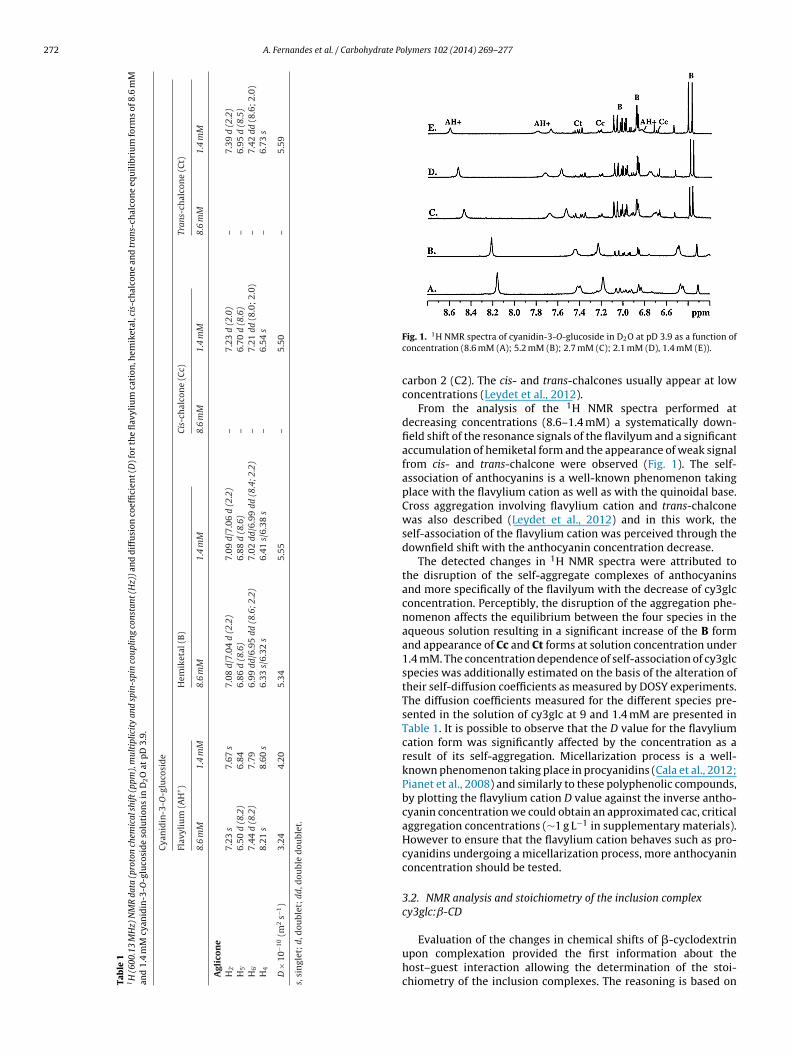
272 A. Fernandes et al. / Carbohydrate Polymers 102 (2014) 269– 277
Tab
le
11H
(600
.13
MH
z)
NM
R
data
(pro
ton
chem
ical
shif
t (pp
m),
mul
tipl
icit
y
and
spin
-spi
n
coup
ling
cons
tant
(Hz)
) an
d
dif
fusi
on
coef
fici
ent (
D) f
or
the
flav
yliu
m
cati
on, h
emik
etal
, cis
-ch
alco
ne
and
tran
s-ch
alco
ne
equ
ilib
riu
m
form
s
of
8.6
mM
and
1.4
mM
cyan
idin
-3-O
-glu
cosi
de
solu
tion
s
in
D2O
at
pD
3.9.
Cya
nid
in-3
-O-g
luco
sid
e
Flav
yliu
m
(AH
+)
Hem
iket
al
(B)
Cis-
chal
con
e
(Cc)
Tran
s-ch
alco
ne
(Ct)
8.6
mM
1.4
mM
8.6
mM
1.4
mM
8.6
mM
1.4
mM
8.6
mM
1.4
mM
Agl
ico
ne
H2′
7.23
s7.
67
s7.
08
d/7.
04
d
(2.2
)7.
09
d/7.
06
d
(2.2
)–
7.23
d
(2.0
)–
7.39
d
(2.2
)H
5′6.
50
d
(8.2
)
6.84
6.86
d
(8.6
)
6.88
d
(8.6
)
–
6.70
d
(8.6
)
– 6.
95
d
(8.5
)H
6′7.
44
d
(8.2
)7.
796.
99
dd/6
.95
dd
(8.6
; 2.2
)7.
02
dd/6
.99
dd
(8.4
; 2.2
)–
7.21
dd
(8.0
;
2.0)
–
7.42
dd
(8.6
;
2.0)
H4
8.21
s
8.60
s
6.33
s/6.
32
s
6.41
s/6.
38
s
–
6.54
s
–
6.73
s
D
×
10−1
0(m
2s−1
)
3.24
4.20
5.34
5.55
–
5.50
–
5.59
s,
sin
glet
;
d,
dou
blet
;
dd, d
oubl
e
dou
blet
.
Fig. 1. 1H NMR spectra of cyanidin-3-O-glucoside in D2O at pD 3.9 as a function ofconcentration (8.6 mM (A); 5.2 mM (B); 2.7 mM (C); 2.1 mM (D), 1.4 mM (E)).
carbon 2 (C2). The cis- and trans-chalcones usually appear at lowconcentrations (Leydet et al., 2012).
From the analysis of the 1H NMR spectra performed atdecreasing concentrations (8.6–1.4 mM) a systematically down-field shift of the resonance signals of the flavilyum and a significantaccumulation of hemiketal form and the appearance of weak signalfrom cis- and trans-chalcone were observed (Fig. 1). The self-association of anthocyanins is a well-known phenomenon takingplace with the flavylium cation as well as with the quinoidal base.Cross aggregation involving flavylium cation and trans-chalconewas also described (Leydet et al., 2012) and in this work, theself-association of the flavylium cation was perceived through thedownfield shift with the anthocyanin concentration decrease.
The detected changes in 1H NMR spectra were attributed tothe disruption of the self-aggregate complexes of anthocyaninsand more specifically of the flavilyum with the decrease of cy3glcconcentration. Perceptibly, the disruption of the aggregation phe-nomenon affects the equilibrium between the four species in theaqueous solution resulting in a significant increase of the B formand appearance of Cc and Ct forms at solution concentration under1.4 mM. The concentration dependence of self-association of cy3glcspecies was additionally estimated on the basis of the alteration oftheir self-diffusion coefficients as measured by DOSY experiments.The diffusion coefficients measured for the different species pre-sented in the solution of cy3glc at 9 and 1.4 mM are presented inTable 1. It is possible to observe that the D value for the flavyliumcation form was significantly affected by the concentration as aresult of its self-aggregation. Micellarization process is a well-known phenomenon taking place in procyanidins (Cala et al., 2012;Pianet et al., 2008) and similarly to these polyphenolic compounds,by plotting the flavylium cation D value against the inverse antho-cyanin concentration we could obtain an approximated cac, criticalaggregation concentrations (∼1 g L−1 in supplementary materials).However to ensure that the flavylium cation behaves such as pro-cyanidins undergoing a micellarization process, more anthocyaninconcentration should be tested.
3.2. NMR analysis and stoichiometry of the inclusion complexcy3glc:ˇ-CD
Evaluation of the changes in chemical shifts of �-cyclodextrinupon complexation provided the first information about thehost–guest interaction allowing the determination of the stoi-chiometry of the inclusion complexes. The reasoning is based on

A. Fernandes et al. / Carbohydrate Po
Fig. 2. 1H NMR spectra of 1.4 mM cyanidin-3-O-glucoside solution in D2O at pD 3.9(a
tcnt2
tstoosuwaiKat
pAoson(tdst(Trewes
in the behavior of the anthocyanin equilibrium forms, cy3glc:�-
A) and 1H NMR spectra of cyanidin-3-O-glucoside with increasing ratios of �-CDfter reaching equilibrium (1:0 (A); 1:0.8 (B); 1:1.6 (C); 1:2.4 (D); 1:3.2 (E)).
he expectation that if the guest molecule is imbedded in the CDavity, the screening environment should be sensed by magneticuclei inside the �-CD cavity (H3 and H5) but not by outside pro-ons (H1, H2 and H4) (Polyakov, Leshina, Hand, Petrenko, & Kispert,004).
In order to minimize the effect of the self-aggregation ofhe anthocyanin, the lowest solution concentration (1.4 mM) waselected to perform the subsequent NMR study of the interac-ion between cy3glc and �-CD and for structural characterizationf cy3glc:�-CD complex (Fig. 2). 1H NMR spectra with high res-lution were recorded for cy3glc:�-CD in D2O at pD 3.9. Thepectra are dominated by the intense resonance signals of �-CD andpfield shifts of the resonance signals of H3 and H5 �-CD’s protonsere detected. This behavior may result mainly from the magnetic
nisotropic shielding by the aromatic rings in cy3glc molecules,ncluded within the cavity of �-CD (Ishizu et al., 2006; Ishizu,ajitani, Tsutsumi, Yamamoto, & Harano, 2008). Only minor alter-tions in the chemical shift of protons H1, H2 and H4 which lie onhe outer surface of the cavity was observed.
In the 1H NMR spectra of cy3glc:�-CD, signals belonging to therotons of AH+, B, Cc and Ct forms of cy3glc were clearly observed.dditionally, it was found that the addition of �-CD to the aque-us solution of cy3glc immediately induced the appearance of newet of resonance signals identified as BCD (Fig. 2B–E). The intensityf these signals increased while the intensity of the resonance sig-als of the B form decreased with time. On the basis of 1D and 2DTOCSY, HMBC) NMR analysis, these new signals were attributed tohe hemiketal anthocyanin form complexed with �-CD (BCD). Wellefined long range correlations were observed in the 1H/13C HMBCpectra between characteristic signals of the BCD, such as betweenhe protons at ı 7·16 ppm (H2′), ı 6·98 ppm (H6′) and ı 6·76 ppmH4) with carbon C2 at 102.3 ppm, characteristic of hemiketal form.he 1H signals of BCD were found to be downfield shifted withespect to the spectra of B before the addition of �-CD. The twopimeric forms could not be distinguishable after complexation,
hich could be a result of a preferable inclusion of one of the twopimers into �-CD’s cavity. Indeed, the use of cyclodextrins for theeparation of isomers was already been reported in the literature
lymers 102 (2014) 269– 277 273
(Ishizu et al., 2008; Upadhyay & Kumar, 2009). Also, for the protonssignals of Cc a downfield shifted were observed. Interconversionbetween cis- and trans-chalcone is in slow exchange (in the NMRtime scale) while the chemical exchange between the hemiketaland the cis-chalcone is faster (Pina, 1998; Pina et al., 2012). Bearingthis, the chemical shifts changes observed for the cis-chalcone couldresult from the faster chemical exchange. However, based on theNOE interaction observed between this equilibrium form and �-CDand on the alteration of the D value, we assume that the chemicalshift changes may be a result from the interaction with �-CD’s inter-nal protons, namely a steric compression effect (Wood, Hruska, &Saenger, 1977) instead of chemical exchange. Only minor chemicalshifts changes were detected in 1H NMR spectra of cy3glc:�-CD forthe protons and Ct forms.
For the evaluation of the stoichiometry of the inclusion com-plexes cy3glc:�-CD, Job’ continuous variation technique wasapplied (Job, 1928). For this purpose, solutions with differentcy3glc:�-CD molar ratios were prepared and the data were plot-ted in the form XH�ı versus XH, where XH is the mole fraction of�-CD. The Job’s plot showed a maximum at r = 0.5 (where r = [�-CD]/([�-CD] + [Cy3glc])), indicating that the complexes possesseda 1:1 stoichiometry.
Moreover, when performing the NMR titration it was noticedthat the addition of �-CD affected principally the hydration reac-tion, mainly disturbing the normalized molar proportions betweenthe flavylium cation and the hydrated hemiketal. The other equilib-rium forms, cis- and trans-chalcones were almost unaffected withthe increase of �-CD (Fig. 3A). These preliminary results suggestthe preference inclusion of the hemiketal into �-CD with this com-pound catalyzing the transformation of the flavylium cation intothe hemiketal, a phenomenon leading to the shifting of the pigmenthydration equilibrium toward the formation of more hemiketalwith the addition of more �-CD. The normalized molar proportionof the various equilibrium forms were determined by integrationof signals in the 1H NMR spectra. Concurrently, during the sameexperiments with the addition of �-cyclodextrin a clear change inthe color of the anthocyanin solution was observed. �-cyclodextrininduces a strong decrease in the anthocyanin visible absorptionband and this decrease was greater with higher concentrations of �-CD added (Fig. 3B). This behavior had already been described in theliterature (Dangles, Stoeckel, et al., 1992; Dangles, Wigand, et al.,1992; Fernandes et al., 2013) and suggests a preferential interactionbetween the anthocyanin hemiketal form and �-CD.
3.3. Diffusion ordered NMR spectroscopy
Diffusion ordered NMR spectroscopy (DOSY) is a well-established technique for characterizing the structure anddynamics of complex systems. The self-diffusion coefficients ofthese systems reveal structural characteristics such as molecularsize, weight and shape (Brand, Cabrita, & Berger, 2005; Cohen,Avram, & Frish, 2005). This methodology relies on the determina-tion of the diffusion coefficients in the absence and presence of hostmolecules (�-cyclodextrin in this study), thus providing informa-tion about the size and evidence on the magnitude of the bindingconstants (Avram & Cohen, 2002; Fielding, 2000).
Typically, cy3glc is approximately one half of the average size of�-CD. In these DOSY studies it was intended not only in confirmingthe interactions between cy3glc and �-CD but also distinguishingthe affinity of the existing anthocyanin equilibrium species at pD3.9 for inclusion complex formation with �-CD. Since a modifica-tion in a solution composition was expected to induce changes
CD samples containing cy3glc to �-CD ratio of 1:0.8, 1:1.6, 1:2.4and 1:3.2 were studied. The diffusion coefficients of �-CD and theexisting equilibrium forms of cy3glc in cy3glc:�-CD samples were

274 A. Fernandes et al. / Carbohydrate Polymers 102 (2014) 269– 277
F chalco� isibled
dapapoittcfa
tlthfdwosTatcscicsi
aat
TDh3
D
ig. 3. Normalized molar proportion of the flavylium cation (AH+), hemiketal (B), cis--CD ratios, determined by integration of signals in the 1H NMR spectra (A). UV–vifferent concentrations (B).
etermined and compared with the corresponding values of cy3glcnd �-CD alone. The diffusion measurements using DOSY wereerformed under identical experimental conditions taking specialttention on the concentration, pH and temperature of the sam-les. The diffusion coefficients (D) of cy3glc equilibrium forms werebtained by measuring the signal intensity at more than one placen the spectra and those for �-CD from the resonance signals ofhe anomeric protons. To account for changes in the samples solu-ion viscosity or drift on the experimental conditions, the diffusionoefficient values were all scaled to the absolute D value obtainedor acetone (0.001 M; 1.69 × 10⁻⁹ m2 s−1, calculated standard devi-tion of 1.15 × 10⁻³) used as an internal reference.
In the absence of �-CD the D value of B, Cc and Ct was foundo be very similar to 5.5 × 10⁻¹⁰ m2 s−1 (Table 2). The significantlyower D value observed for AH+ was attributed to the tendency ofhis equilibrium form to self-aggregate resulting on a specie withigher hydrodynamic diameter. Concerning the results obtained
or the inclusion complexes cy3glc:�-CD, diverse behavior of theifferent anthocyanin equilibrium forms in the presence of �-CDas observed. The diffusion coefficients measured for the new set
f signals detected in cy3glc:�-CD (BCD) form was found to beimilar to the corresponding values of �-CD (4.0 × 10⁻¹⁰ m2 s−1).his is an indication of intermolecular interactions and complex-tion between these two species was hence assumed. Likewise,he decrease of the diffusion coefficient of cis-chalcone (Cc) iny3glc:�-CD samples, to values close to that of �-CD reveal a pos-ible interaction between these two species. However, only smallhanges in the self-diffusion of trans-chalcone (Ct) was observedn the presence of �-CD. This fact and the registered absence ofhemical shift displacements of Ct resonance signals in the 1H NMRpectra of cy3glc:�-CD strongly suggest a lack of intermolecularnteractions between and �-CD.
The flavylium cation diffusion coefficient was almost unchanged
nd lower than �-CD’s during the experiments until the thirdnthocyanin:�-CD ratio was achieved. At this ratio only traces ofhe AH+ equilibrium form was detected, not allowing the correct Dable 2iffusion coefficients (D) obtained for �-cyclodextrin and for flavylium cation,emiketal, cis-chalcone and trans-chalcone equilibrium forms of 1.4 mM cyanidin--O-glucoside solutions with increasing amounts of �-CD in D2O at pD 3.9.
Ratio cy3glc:ˇ-CD D × 10−10 (m2 s−1)
AH+ B Cc Ct BCD
1:0.0 4.2 5.4 5.5 5.6 0.01:0.8 4.2 5.5 4.0 5.4 3.91:1.6 a 5.7 3.9 5.5 3.81:2.4 a 5.8 4.0 5.5 3.81:3.2 a 5.7 4.0 5.5 3.9
�-CD × 10−10 (m2 s−1) = 4.0.a Undetected.
ne (Cc), trans-chalcone (Ct) and hemiketal complexed (BCD) as a function of different spectra of cyanidin-3-O-glucoside alone and in the presence of �-cyclodextrin at
determination (Table 2). The results from the analysis of DOSY spec-tra of the inclusion complex cy3glc:�-CD indicate that only two ofthe anthocyanin equilibrium forms, B and Cc, showed preferencefor association and affinity for intermolecular complex formationwith �-CD. These results are in agreement with the observed chem-ical shifts alterations registered for protons of B, Cc and �-CD in 1HNMR spectra of cy3glc:�-CD. The observed changes in the chemicalshifts and self-diffusion change for B and Cc suggest an inclusion ofthese species into the cavity of �-CD.
3.4. Putative structure of the inclusion complex of cy3glc withˇ-CD
Beside the chemical shifts changes and changes of diffusionconstants initiated by complexation with CD, changes of NOE for thespecies in the solution serve as a measure of intermolecular bindingand inclusion complex formation. Further reliable information onthe inclusion complex formation can be obtained by the evaluationof the intermolecular proximity of protons in the guest species tothe H3 and H5 protons, located inside of the cyclodextrin cavity bymeasurements of NOE. Additionally, information can be extractedfor the preferred structural arrangement of the guest moleculesin the cyclodextrin cavity in the process of inclusion complexesformation.
Therefore, structural investigations based on NOE studies wereperformed to further determine the behavior and mode of interac-tion of the different forms of anthocyanin co-existing in aqueoussolution at pD 3.9 with �-CD and to define the structure of thecy3glc:�-CD complex. The NOE relies on different correlation times(�c) of free and bound molecules and the observation of inter-molecular NOE between molecules in solution is a strong indicationfor intermolecular association processes. Rotating frame nuclearOverhauser spectroscopy (ROESY) was applied to study solutionscontaining equimolar amounts of �-CD and cy3glc in D2O. A num-ber of ROESY experiments using a mixing time in the range between100 and 800 ms were recorded with the aim to discriminate theNOE originating from the bound state of cy3glc with �-CD (inter-molecular NOE) and NOE of the non-bounded species in the solution(intramolecular NOE) (Meyer & Peters, 2003). In the ROESY spec-trum, the cross-peak connecting two proton resonances indicatesthat those proton nuclei are in close proximity in the ground stateof the molecule (Ishizu et al., 2006). The intensity of the rotatingframe nuclear Overhauser effect (ROE) was estimated from the inte-gral volume of the cross-peak and was classified as strong, medium,weak or very weak (Ishizu et al., 2008).
The analysis of 2D ROESY spectra of cy3glc:�-CD acquired at
different mixing times showed correlations between protons ofcy3glc’s hemiketal form complexed with �-CD and H3 and H5 pro-tons lying on the inner surface of �-CD (Fig. 4). The H3 protons of�-CD shares strong intermolecular ROE correlations with protons
A. Fernandes et al. / Carbohydrate Polymers 102 (2014) 269– 277 275
Fig. 4. 2D ROESY (600 MHz) spectra of cy3glc:�-CD (1:1) acquired in D2O (pD 3.9)with a mixing time of 500 ms, showing characteristic inter- and intra- (expandedsn
HlpCoibHColif(e
bloHCehctaitc
acit
o
Table 3Distance values obtained for the closest contacts between the hydrogen atoms ofcy3glc and �-CD compounds.
Atom of cy3glc Atom of �-CD Distance (Å)
H4 H3-Glc7 and H5-Glc7 3.29 ± 1.02 and 3.22 ± 0.82H6 – –H8 H3-Glc3 and H5-Glc3 3.60 ± 1.24 and 2.93 ± 0.93H2′ H3-Glc5 4.87 ± 1.47
to a solvent probe and indicates which atoms are most exposed to
pectral area) molecular NOE interactions in the complex. The assignment of reso-ances of BCD and �-CD is included.
4 from C-ring and H6′ from ring B, medium intermolecular corre-ations with proton H2′ and weak intermolecular correlations withroton H5′ (also from B ring) of the hemiketal form BCD. H5 of �-D shares strong ROE correlations with protons H4 (Fig. 4). Thesebservations strongly suggest that the cy3glc’s C-ring is deeplyncluded in the �-CD cavity. No NOE interactions were detectedetween protons from the anthocyanin BCD with the outer protons2 and H4. All together, these data suggest that probably the A- and-ring are included from the wide secondary hydroxyl group sidef the �-CD cavity being closer to the narrow rim, whilst the B-ringies on the plane of the wider rim of �-CD. This type of molecularnclusion had already been described for other polyphenols, namelyor (−)-epicatechin gallate (also presenting a catechol B ring) and−)-epigallocatechin gallate (presenting a pyrogallol B ring) (Ishizut al., 2006, 2008).
In this spectrum no dipole–dipole interactions were registeredetween protons of the flavylium cation and trans-chalcone equi-
ibrium forms and �-CD. However, small but detectible NOE wasbserved between characteristic protons of cis-chalcone and �-CD’s3. The structural similarity of the two equilibrium forms BCD andc was assumed to be a reason for their similar behavior in a pres-nce of CD. The presence of a tetrahedral carbon atom in the neutralemiketal makes it much less rigid then the almost planar flavyliumation and probably allow a closer fit to the macrocyclic cavity. Inhe cis-chalcone the presence of an open chain probably also allows
better molecular inclusion in the hydrophobic cyclodextrin cav-ty. Additionally, for the charged and highly polar flavylium cationhe relative desolvation of the guest molecule when it enters theyclodextrin cavity is probably more endothermic.
In the 2D ROESY spectra recorded at longer mixing time (500nd 800 ms) weak intramolecular interactions between the antho-yanin protons were observed besides the intermolecular dipolenteraction between BCD and Cc with the interior �-CD cavity pro-
ons.Further details of the solution structure of the complex werebtained from Molecular Dynamics simulation.
H5′ H3-Glc5 4.66 ± 1.77H6′ H3-Glc5 4.56 ± 1.65
3.5. Structure of the inclusion complex cy3glc-ˇ-CD by MolecularDynamics simulation studies
To better understand the inclusion of the hemiketal cy3glcform in the hydrophobic �-CD’s internal cavity, computationalstudies were additionally carried out. An MD simulation of 30 nswas performed for this complex, which allowed the respec-tive conformational space to be sampled. The last 20 ns of thisMD simulation were used for the subsequent structural analysis.The root–mean–square deviation (RMSD) value obtained for thiscompound was 2.92 ± 0.62 A. This small value reveals higher equil-ibration and stability of this complex during the MD simulation.Fig. 5 shows the closest structure to the average geometry of cy3glc-�-CD complex, which is in good agreement with the data obtainedin the NMR studies. In order to measure the movement of the cy3glcmolecule relative to the average structure over the whole simula-tion, root–mean–square fluctuation (RMSF) values by atom werealso calculated. Analysing these values it was noticed higher RMSFvalues for the main hydroxyl groups of the glucose unit and theHO-C3′ group of the B ring, as well as for the H5′ and H6′ atomswhich reveal a higher degree of flexibility during the MD sim-ulation. The dynamic behavior of these atoms agrees with theirpreviously referred close contact with solvent molecules.
To clarify the binding and inclusion mode of cy3glc:�-CD com-plex, the distances between all H atoms of A-, B- and C-rings of thehemiketal and the H1, H2, H3, H4 and H5 atoms of each glucoseunit from the �-CD were also calculated during the MD simulation.Table 3 shows the average distances of the closest contacts betweenthe hydrogen atoms of cy3glc and the �-CD molecules. As seen inthe Molecular Dynamics simulation, the binding and inclusion ofcy3glc in the cavity of �-CD are ensured by the establishment ofhydrophobic interactions between the polyphenolic and glucoserings, as well as hydrogen bonds with the hydrophilic hydroxylpolar groups of both compounds. All these crucial interactions con-tribute to the formation and stabilization of this complex. As seenin Fig. 5, the A- and C-nucleus of cy3glc provide the major contri-bution to the binding driving force, followed by the B-ring, whilstits glucose residue seems to perturb the cy3glc’s inclusion in the �-CD and is facing the solvent. Similarly to the present NMR studies,these results also reveal a higher proximity between the H4, H2′,H5′ and H6′ atoms of cy3glc and the internal H3 and H5 atoms ofseveral glucose units, in particular the Glc3, Glc5 and Glc7 units. Itwas also obtained large distances to the outer H1, H2 and H4 atomsof �-CD, and it is noteworthy that the H6 at the A-ring of cy3glc isvery distant to the hydrogen atoms of �-CD and directed to the sol-vent molecules during the whole simulation (data not shown). Tobetter characterize this cy3glc:�-CD inclusion mode, the solvent-accessible surface area (SASA) values for some nonpolar H atomsof both cy3glc (H4, H6, H8, H2′, H5′ and H6′) and �-CD (H1, H2, H3,H4 and H5) molecules during the MD simulation were calculated.The SASA defines the surface area of a group/atom that is accessible
the solvent. Table 4 shows the obtained SASA values with standarddeviations for each atom. As expected, the outer H1, H2 and H4atoms of �-CD have shown higher values. On the other hand, the

276 A. Fernandes et al. / Carbohydrate Polymers 102 (2014) 269– 277
Fig. 5. Structure of cy3glc:�-CD complex in front-view (A) and side-view (B). The �-CD issticks and colored in gray. All H3 and H5 protons of each glucose unit from �-CD are coorange (H4, H8, H2′ , H5′ and H6′) and green (H6).
Table 4SASA values obtained for several hydrogen atoms of cy3glc and �-CD molecules.
Atom SASA (Å2)
H1-Glc of �-CD 74.55 ± 4.89H2-Glc of �-CD 108.42 ± 3.85H3-Glc of �-CD 2.03 ± 3.37H4-Glc of �-CD 44.90 ± 3.98H5-Glc of �-CD 0.20 ± 0.56H4 of cy3glc 0.03 ± 0.32H6 of cy3glc 7.82 ± 2.83H8 of cy3glc 0.05 ± 0.28H2′ of cy3glc 2.26 ± 2.63
′
iatfaStamoit
4
stcsftbpcb
Bayly, C. I., Cieplak, P., Cornell, W. D., & Kollman, P. A. (1993). A well-behaved elec-
H5 of cy3glc 5.34 ± 3.43H6′ of cy3glc 2.61 ± 3.54
nternal H3 and H5 atoms present smaller values of SASA, whichgrees with the hydrophobic environment of �-CD’s cavity. Rela-ively to the cy3glc molecule, the higher SASA value was obtainedor the H6 atom (7.82 ± 2.83 A2), which indicates that this specifictom is highly exposed to the solvent. On the other hand, minimalASA values were obtained for H4, H8, H2′, H5′ and H6′. Accordingo the SASA values obtained, a higher protection of H4, H8, H2′, H5′
nd H6′ atoms was noticed due to the interaction with the �-CDolecule. Therefore, the hydrophilic environment around the H6
f cy3glc point out by this data demonstrates that this atom is prox-mal to solvent water molecules and is directed to the exterior ofhe �-CD cavity.
. Conclusion
Nuclear Magnetic Resonance analysis and Molecular Dynamicsimulation performed in this study have clearly proved the selec-ive inclusion of the anthocyanin hemiketal form into the �-CDavity. This assumption was based on the anthocyanin chemicalhift alteration after complexation with �-CD, decrease on the dif-usion coefficient of the hemiketal equilibrium form to values closeo the corresponding values of �-CD and on the observed NOE effectetween this equilibrium form with CD’s interior protons. The com-
utational data support and clarify the successful inclusion of they3glc molecule inside the hydrophobic cavity of �-CD proposedy the present experimental studies.represented with surface and colored by atom type, the cy3glc is represented withlored in blue, while the nonpolar hydrogen atoms from the cy3glc are colored in
The results achieved offer a contribution to the elucidation ofthe formation and the structure of inclusion complexes betweenthe equilibrium network of anthocyanins forms and cyclodextrinsincreasing the knowledge regarding the phenomena that drive thestabilization of these natural colorants.
Acknowledgments
This work received financial support from FEDER fundsthrough COMPETE, POPH/FSE, QREN and FCT (Fundac ãopara a Ciência e Tecnologia) from Portugal by one PhDscholarship (SFRH/BD/65350/2009) and through projectsPTDC/QUI/67915/2006, PTDC/QUI/122916/2010 and PEst-C/QUI/UI0081/2011. The Bruker Avance III 600 HD spectrometerwas purchased under the framework of QREN, through projectNORTE-07-0162-FEDER-000048, and is part of the Portuguese NMRNetwork created with support of the Portuguese Foundation forScience and Technology through contract REDE/1517/RMN/2005,with funds from POCI 2010 (FEDER) and (FCT). To all financingsources the authors are greatly indebted.
Appendix A. Supplementary data
Supplementary data associated with this article can befound, in the online version, at http://dx.doi.org/10.1016/j.carbpol.2013.11.037.
References
Avram, L., & Cohen, Y. (2002). Complexation in pseudorotaxanes based on�-cyclodextrin and different �,�-diaminoalkanes by NMR diffusion measure-ments. Journal of Organic Chemistry, 67(8), 2639–2644.
Azevedo, J., Fernandes, I., Faria, A., Oliveira, J., Fernandes, A., de Freitas, V., et al.(2010). Antioxidant properties of anthocyanidins, anthocyanidin-3-glucosidesand respective portisins. Food Chemistry, 119(2), 518–523.
Basma, M., Sundara, S., Calgan, D., Vernali, T., & Woods, R. J. (2001). Solvatedensemble averaging in the calculation of partial atomic charges. Journal of Com-putational Chemistry, 22(11), 1125–1137.
trostatic potential based method using charge restraints for deriving atomiccharges – The resp model. Journal of Physical Chemistry, 97(40), 10269–10280.
Bourvellec, C. (2003). Association entre les procyanidols et les polymères pariétaux depommes: quantification et conséquences. Universite de Rennes I: (Vol. Docteur).

ate Po
B
B
B
B
C
C
C
C
C
D
D
D
E
E
F
F
F
GH
H
H
H
H
I
I
I
J
A. Fernandes et al. / Carbohydr
rand, T., Cabrita, E. J., & Berger, S. (2005). Intermolecular interaction as inves-tigated by NOE and diffusion studies. Progress in Nuclear Magnetic ResonanceSpectroscopy, 46(4), 159–196.
rouillard, R., & Delaporte, B. (1977). Chemistry of anthocyanin pigments. 2. Kineticand thermodynamic study of proton transfer, hydration, and tautomeric reac-tions of malvidin 3-glucoside. Journal of the American Chemical Society, 99(26),8461–8468.
rouillard, R., & Dubois, J. E. (1977). Mechanism of the structural transformationsof anthocyanins in acidic media. Journal of the American Chemical Society, 99(5),1359–1364.
rouillard, R., & Lang, J. (1990). The hemiacetal-cis-chalcone equilibrium of malvin,a natural anthocyanin. Canadian Journal of Chemistry, 68, 755.
ala, O., Dufourc, E. J., Fouquet, E., Manigand, C., Laguerre, M., & Pianet, I. (2012). Thecolloidal state of tannins impacts the nature of their interaction with proteins:The case of salivary proline-rich protein/procyanidins binding. Langmuir, 28(50),17410–17418.
ase, D. A., Darden, T. A., Cheatham, I. T. E., Simmerling, C. L., Wang, J., Duke, R. E.,et al. (2008). AMBER 10. San Francisco: University of California.
handra, A., Nair, M. G., & Iezzoni, A. F. (1993). Isolation and stabilization of antho-cyanins from tart cherries (Prunus cerasus L.). Journal of Agricultural and FoodChemistry, 41(7), 1062–1065.
lifford, M. N. (2000). Anthocyanins – Nature, occurrence and dietary burden. Journalof the Science of Food and Agriculture, 80(7), 1063–1072.
ohen, Y., Avram, L., & Frish, L. (2005). Diffusion NMR spectroscopy in supramolecu-lar and combinatorial chemistry: An old parameter—New Insights. AngewandteChemie International Edition, 44(4), 520–554.
angles, O., Stoeckel, C., Wigand, M. C., & Brouillard, R. (1992). Two very distincttypes of anthocyanin complexation: Copigmentation and inclusion. TetrahedronLetters, 33(36), 5227–5230.
angles, O., Wigand, M. C., & Brouillard, R. (1992). Anthocyanin anti-copigmenteffect. Phytochemistry, 31(11), 3811–3812.
el Valle, E. M. M. (2004). Cyclodextrins and their uses: A review. Process Biochem-istry, 39(9), 1033–1046.
rsus, S., & Yurdagel, U. (2007). Microencapsulation of anthocyanin pigments ofblack carrot (Daucuscarota L.) by spray drier. Journal of Food Engineering, 80(3),805–812.
ssmann, U., Perera, L., Berkowitz, M. L., Darden, T., Lee, H., & Pedersen, L. G. (1995).A smooth particle mesh Ewald method. Journal of Chemical Physics, 103(19),8577–8593.
ernandes, A., Sousa, A., Azevedo, J., Mateus, N., & de Freitas, V. (2013). Effect ofcyclodextrins on the thermodynamic and kinetic properties of cyanidin-3-O-glucoside. Food Research International, 51(2), 748–755.
ielding, L. (2000). Determination of association constants (Ka) from solution NMRdata. Tetrahedron, 56, 6151–6170.
risch, M. J., Trucks, G. W., Schlegel, H. B., Scuseria, G. E., Robb, M. A., Cheeseman, J.R., et al. (2009). Gaussian 09.
aussian Inc., C.O. P., Bldg. 6, Pittsburgh, PA 15106, USA.e, J. A., & Giusti, M. M. (2010). Anthocyanins: Natural colorants with health-
promoting properties. In M. P. Doyle, & T. R. Klaenhammer (Eds.), Annual reviewof food science and technology (Vol. 1) (pp. 163–187). Palo Alto: Annual Reviews.
oshino, T., Matsumoto, U., Harada, N., & Goto, T. (1982). Evidence for the self-association of anthocyanins IV. PMR spectroscopic evidence for the verticalstacking of anthocyanin molecules. Tetrahedron Letters, 23(4), 433–436.
oubiers, C., Lima, J. C., Mac anita, A. L., & Santos, H. (1998). Color stabilization of mal-vidin 3-glucoside: Self-aggregation of the flavylium cation and copigmentationwith the Z-chalcone form. Journal of Physical Chemistry B, 102(18), 3578–3585.
oward, L. R., Brownmiller, C., Prior, R. L., & Mauromoustakos, A. (2013). Improvedstability of chokeberry juice anthocyanins by �-cyclodextrin addition and refrig-eration. Journal of Agricultural and Food Chemistry, 61(3), 693–699.
wang, T. L., & Shaka, A. J. (1995). Water suppression that works. Excitation sculpt-ing using arbitrary wave-forms and pulsed-field gradients. Journal of MagneticResonance, Series A, 112(2), 275–279.
shizu, T., Hirata, C., Yamamoto, H., & Harano, K. (2006). Structure and intramolecularflexibility of �-cyclodextrin complex with (−)-epigallocatechin gallate in aque-ous solvent. Magnetic Resonance in Chemistry, 44(8), 776–783.
shizu, T., Kajitani, S., Tsutsumi, H., Yamamoto, H., & Harano, K. (2008).Diastereomeric difference of inclusion modes between (−)-epicatechin gallate,(−)-epigallocatechin gallate and (+)-gallocatechin gallate, with �-cyclodextrin
in aqueous solvent. Magnetic Resonance in Chemistry, 46(5), 448–456.zaguirre, J. A., Catarello, D. P., Wozniak, J. M., & Skeel, R. D. (2001). Langevin stabi-lization of molecular dynamics. Journal of Chemical Physics, 114(5), 2090–2098.
ob, P. (1928). Formation and stability of inorganic complexes in solution. Annali diChimica, 9, 113–203.
lymers 102 (2014) 269– 277 277
Jordheim, M., Fossen, T., & Andersen, Ø. M. (2006). Characterization of hemiacetalforms of anthocyanidin 3-O-�-glycopyranosides. Journal of Agricultural and FoodChemistry, 54(25), 9340–9346.
Kirschner, K. N., & Woods, R. J. (2001a). Quantum mechanical study of the nonbondedforces in water–methanol complexes. Journal of Physical Chemistry A, 105(16),4150–4155.
Kirschner, K. N., & Woods, R. J. (2001b). Solvent interactions determine carbohydrateconformation. Proceedings of the National Academy of Sciences of the United Statesof America, 98(19), 10541–10545.
Lewis, C. E., Walker, J. R. L., & Lancaster, J. E. (1995). Effect of polysaccharides on thecolour of anthocyanins. Food Chemistry, 54(3), 315–319.
Leydet, Y., Gavara, R., Petrov, V., Diniz, A. M., Parola, A. J., Lima, J. C., et al. (2012).The effect of self-aggregation on the determination of the kinetic and ther-modynamic constants of the network of chemical reactions in 3-glucosideanthocyanins. Phytochemistry, 83, 125–135.
Matsumoto, N., Yamada, M., Kurakata, Y., Yoshida, H., Kamitori, S., Nishikawa, A.,et al. (2009). Crystal structures of open and closed forms of cyclo/maltodextrin-binding protein. FEBS Journal, 276(11), 3008–3019.
Meyer, B., & Peters, T. (2003). NMR spectroscopy techniques for screening and iden-tifying ligand binding to protein receptors. Angewandte Chemie InternationalEdition, 42(8), 864–890.
Mourtzinos, I., Makris, D. P., Yannakopoulou, K., Kalogeropoulos, N., Michali, I., &Karathanos, V. T. (2008). Thermal stability of anthocyanin extract of Hibiscussabdariffa L. in the presence of �-cyclodextrin. Journal of Agricultural and FoodChemistry, 56(21), 10303–10310.
Pianet, I., André, Y., Ducasse, M. A., Tarascou, I., Lartigue, J. C., Pinaud, N., et al. (2008).Modeling procyanidin self-association processes and understanding their micel-lar organization: A study by diffusion NMR and molecular mechanics. Langmuir,24(19), 11027–11035.
Pina, F. (1998). Thermodynamics and kinetics of flavylium salts. Malvidin revisited.Journal of the Chemical Society, Faraday Transactions, 94(15), 2109–2116.
Pina, F., Melo, M. J., Laia, C. A. T., Parola, A. J., & Lima, J. C. (2012). Chemistry and appli-cations of flavylium compounds: A handful of colours. Chemical Society Reviews,41(2), 869–908.
Polyakov, N. E., Leshina, T. V., Hand, E. O., Petrenko, A., & Kispert, L. D.(2004). �-Ionone cyclodextrins inclusion complexes: 1H NMR study and pho-tolysis. Journal of Photochemistry and Photobiology A: Chemistry, 161(2/3),261–267.
Popov, K., Rönkkömäki, H., & Lajunen, L. H. J. (2006). Guidelines for NMR measure-ments for determination of high and low pKa values (IUPAC Technical Report).Pure and Applied Chemistry, 78(3), 663–675.
Ryckaert, J. P., Ciccotti, G., & Berendsen, H. J. C. (1977). Numerical-integrationof cartesian equations of motion of a system with constraints –Molecular-dynamics of N-alkanes. Journal of Computational Physics, 23(3),327–341.
Rymdén, R., Carlfors, J., & Stilbs, P. (1983). Substrate binding to cyclodextrins inaqueous solution: A multicomponent self-diffusion study. Journal of inclusionphenomena, 1(2), 159–167.
Santos, H., Turner, D. L., Lima, J. C., Figueiredo, P., Pina, F. S., & Mac anita, A. L. (1993).Elucidation of the multiple equilibria of malvin in aqueous solution by one- andtwo-dimensional NMR. Phytochemistry, 33(5), 1227–1232.
Szejtli, J. (2003). Cyclodextrins. In P. Tomasik (Ed.), Chemical and functional propertiesof food saccharides. Cracow: CRC Press.
Tamura, H., Takada, M., et al. (1998). The Color Stability and Antioxidative Activ-ity of an Anthocyanin and b-Cyclodextrin Complex. Functional Foods for DiseasePrevention I, 701, 157–171.
Thaning, J., Stevensson, B., Ostervall, J., Naidoo, K. J., Widmalm, G., & Maliniak, A.(2008). NMR studies of molecular conformations in �-cyclodextrin. Journal ofPhysical Chemistry B, 112(29), 8434–8436.
Torskangerpoll, K., & Andersen, Ø. M. (2005). Colour stability of antho-cyanins in aqueous solutions at various pH values. Food Chemistry, 89(3),427–440.
Upadhyay, S. K., & Kumar, G. (2009). NMR and molecular modelling studies on theinteraction of fluconazole with �-cyclodextrin. Chemistry Central Journal, 3, 9.
Wood, D. J., Hruska, F. E., & Saenger, W. (1977). Proton NMR study of the inclusion ofaromatic molecules in �-cyclodextrin. Journal of the American Chemical Society,99(6), 1735–1740.
Wu, D. H., Chen, A. D., & Johnson, C. S. (1995). An improved diffusion-ordered spec-
troscopy experiment incorporating bipolar-gradient pulses. Journal of MagneticResonance, Series A, 115(2), 260–264.Yamada, T., Komiya, T., & Akaki, M. (1980). Formation of an inclusion complexof anthocyanin with cyclodextrin. Agricultural and Biological Chemistry, 44(6),1411–1413.