Using ipsilateral motor signals in the unaffected cerebral hemisphere as a signal platform for...
23
Using Ipsilateral Motor Signals in the Unaffected Cerebral Hemisphere as a Signal Platform for Brain Computer Interfaces in Hemiplegic Stroke Survivors David T. Bundy 1 , Mark Wronkiewicz 1 , Mohit Sharma 1 , Daniel W. Moran 1,2 , Maurizio Corbetta 2 , and Eric C. Leuthardt, MD 1,2,3,4 1 Department of Biomedical Engineering, Washington University in St. Louis, Campus Box 8057, 660 South Euclid, St Louis, MO 63130 2 Department of Anatomy and Neurobiology, University in St. Louis, School of Medicine, St Louis, MO 3 Department of Neurological Surgery, Washington University in St. Louis, School of Medicine, Campus Box 8057, 660 South Euclid, St Louis, MO 63130 4 Center for Innovation in Neuroscience and Technology, Washington University School of Medicine, St. Louis, MO Abstract Objective—Brain computer interface (BCI) systems have emerged as a method to restore function and enhance communication in motor impaired patients. To date, this has been primarily applied to patients who have a compromised motor outflow due to spinal cord dysfunction, but an intact and functioning cerebral cortex. The cortical physiology associated with movement of the contralateral limb has typically been the signal substrate that has been used as a control signal. While this is an ideal control platform in patients with an intact motor cortex, these signals are lost after a hemispheric stroke. Thus, a different control signal is needed that could provide control capability for a patient with a hemiparetic limb. Previous studies have shown that there is a distinct cortical physiology associated with ipsilateral, or same sided, limb movements. Thus far, it was unknown whether stroke survivors could intentionally and effectively modulate this ipsilateral motor activity from their unaffected hemisphere. Therefore, this study seeks to evaluate whether stroke survivors could effectively utilize ipsilateral motor activity from their unaffected hemisphere to achieve this BCI control. Approach—To investigate this possibility, electroencephalographic (EEG) signals were recorded from four chronic hemispheric stroke patients as they performed (or attempted to perform) real and imagined hand tasks using either their affected or unaffected hand. Following performance of the screening task, the ability of patients to utilize a BCI system was investigated during on-line control of a 1-dimensional control task. Main Results—Significant ipsilateral motor signals (associated with movement intentions of the affected hand) in the unaffected hemisphere, which were found to be distinct from rest and contralateral signals, were identified and subsequently used for a simple online BCI control task. We demonstrate here for the first time that EEG signals from the unaffected hemisphere, associated with overt and imagined movements of the affected hand, can enable stroke survivors Corresponding Author: E.C. Leuthardt, M.D., Department of Neurosurgery, Washington University in St. Louis, School of Medicine, Campus Box 8057, 660 South Euclid, St Louis, MO 63130. Phone: (314) 362 8012, FAX: (314) 362-2107, [email protected]. Disclosures: ECL and DWM have stock ownership in the company Neurolutions NIH Public Access Author Manuscript J Neural Eng. Author manuscript; available in PMC 2013 June 01. Published in final edited form as: J Neural Eng. 2012 June ; 9(3): 036011. doi:10.1088/1741-2560/9/3/036011. NIH-PA Author Manuscript NIH-PA Author Manuscript NIH-PA Author Manuscript
-
Upload
independent -
Category
Documents
-
view
1 -
download
0
Transcript of Using ipsilateral motor signals in the unaffected cerebral hemisphere as a signal platform for...

Using Ipsilateral Motor Signals in the Unaffected CerebralHemisphere as a Signal Platform for Brain Computer Interfacesin Hemiplegic Stroke Survivors
David T. Bundy1, Mark Wronkiewicz1, Mohit Sharma1, Daniel W. Moran1,2, MaurizioCorbetta2, and Eric C. Leuthardt, MD1,2,3,4
1Department of Biomedical Engineering, Washington University in St. Louis, Campus Box 8057,660 South Euclid, St Louis, MO 631302Department of Anatomy and Neurobiology, University in St. Louis, School of Medicine, St Louis,MO3Department of Neurological Surgery, Washington University in St. Louis, School of Medicine,Campus Box 8057, 660 South Euclid, St Louis, MO 631304Center for Innovation in Neuroscience and Technology, Washington University School ofMedicine, St. Louis, MO
AbstractObjective—Brain computer interface (BCI) systems have emerged as a method to restorefunction and enhance communication in motor impaired patients. To date, this has been primarilyapplied to patients who have a compromised motor outflow due to spinal cord dysfunction, but anintact and functioning cerebral cortex. The cortical physiology associated with movement of thecontralateral limb has typically been the signal substrate that has been used as a control signal.While this is an ideal control platform in patients with an intact motor cortex, these signals are lostafter a hemispheric stroke. Thus, a different control signal is needed that could provide controlcapability for a patient with a hemiparetic limb. Previous studies have shown that there is adistinct cortical physiology associated with ipsilateral, or same sided, limb movements. Thus far, itwas unknown whether stroke survivors could intentionally and effectively modulate this ipsilateralmotor activity from their unaffected hemisphere. Therefore, this study seeks to evaluate whetherstroke survivors could effectively utilize ipsilateral motor activity from their unaffectedhemisphere to achieve this BCI control.
Approach—To investigate this possibility, electroencephalographic (EEG) signals were recordedfrom four chronic hemispheric stroke patients as they performed (or attempted to perform) realand imagined hand tasks using either their affected or unaffected hand. Following performance ofthe screening task, the ability of patients to utilize a BCI system was investigated during on-linecontrol of a 1-dimensional control task.
Main Results—Significant ipsilateral motor signals (associated with movement intentions of theaffected hand) in the unaffected hemisphere, which were found to be distinct from rest andcontralateral signals, were identified and subsequently used for a simple online BCI control task.We demonstrate here for the first time that EEG signals from the unaffected hemisphere,associated with overt and imagined movements of the affected hand, can enable stroke survivors
Corresponding Author: E.C. Leuthardt, M.D., Department of Neurosurgery, Washington University in St. Louis, School of Medicine,Campus Box 8057, 660 South Euclid, St Louis, MO 63130. Phone: (314) 362 8012, FAX: (314) 362-2107,[email protected].
Disclosures: ECL and DWM have stock ownership in the company Neurolutions
NIH Public AccessAuthor ManuscriptJ Neural Eng. Author manuscript; available in PMC 2013 June 01.
Published in final edited form as:J Neural Eng. 2012 June ; 9(3): 036011. doi:10.1088/1741-2560/9/3/036011.
NIH
-PA Author Manuscript
NIH
-PA Author Manuscript
NIH
-PA Author Manuscript
to control a one-dimensional computer cursor rapidly and accurately. This ipsilateral motoractivity enabled users to achieve final target accuracies between 68 and 91% within 15 minutes.
Significance—These findings suggest that ipsilateral motor activity from the unaffectedhemisphere in stroke survivors could provide a physiological substrate for BCI operation that canbe further developed as a long-term assistive device or potentially provide a novel tool forrehabilitation.
KeywordsElectroencephalography; EEG; Ipsilateral; Motor; Brain Computer Interface; Neuroprosthetics;Stroke; Hemiplegia; BCI
1. IntroductionCurrently a challenge in the treatment of stroke survivors is the rehabilitation of chronicallylost motor functions. Several studies describing hemiparesis in chronic stroke survivorsdemonstrate that motor recovery plateaus 3 months post-stroke (Duncan, Goldstein et al.1992; Jorgensen, Nakayama et al. 1995; Lloyd-Jones, Adams et al. 2009). A potential novelapproach for the restoration of function and improving the quality of life of these patientscould be the use of a brain computer interface (BCI). These systems use signals recordedfrom the central nervous system as a control signal for operating a computer or other device.Restoring function could be accomplished either through controlling an assistive deviceindependent of the unaffected hand, or through paired BCI control and peripheralstimulation to induce functional recovery through endogenous plasticity. Thus far,substantial research has shown that information from motor cortex contralateral to anintended limb encodes useful information about motor intent and can be used to controlmultiple degree-of-freedom BCI systems with a variety of recording modalities (Taylor,Tillery et al. 2002; Leuthardt, Schalk et al. 2004; Wolpaw and McFarland 2004; Hochberg,Serruya et al. 2006; Schalk, Miller et al. 2008; Velliste, Perel et al. 2008; Rouse and Moran2009). While these physiologic signals are useful in controlling BCI systems designed formotor impaired patients with intact cortex (Pfurtscheller, Guger et al. 2000; Taylor, Tilleryet al. 2002; Wolpaw and McFarland 2004; Kubler, Nijboer et al. 2005; Hochberg, Serruya etal. 2006), a different cortical signal would be necessary in hemiplegic stroke survivors thatsuffer damage to primary motor cortex contralateral to the affected limb. This is importantboth in a traditional BCI device (which enables brain-derived control of an assistivemachine) and also in potentially encouraging functional rehabilitation (to facilitateendogenous recovery of limb function). Taken together, there is a substantive need todevelop new methods for restoring function in chronic hemiplegic stroke survivors, whichmay be accomplished through utilizing novel cortical control signals in conjunction with aBCI system.
Recent work by Wisneski et al. has demonstrated a separable and distinct corticalphysiology associated with ipsilateral hand movements (i.e. movements on the same side asthe respective hemisphere) that can be distinguished from cortical signals associated withmovement contralateral to a given hemisphere (2008). Electrocorticographic (ECoG) signalswere recorded while subjects engaged in specific ipsilateral or contralateral hand motortasks. Ipsilateral hand movements were associated with electrophysiological changes thatoccurred in lower frequency spectra (average 37.5Hz), at distinct anatomic locations (mostnotably in premotor cortex), and earlier (by 160 ms) than changes associated withcontralateral hand movements. Given that these cortical changes occurred earlier and werelocalized preferentially in premotor cortex compared to those associated with contralateralmovements, the authors postulated that ipsilateral cortex is more associated with motorplanning than its execution. Furthermore, while rehabilitation from stroke has traditionally
Bundy et al. Page 2
J Neural Eng. Author manuscript; available in PMC 2013 June 01.
NIH
-PA Author Manuscript
NIH
-PA Author Manuscript
NIH
-PA Author Manuscript
been viewed as a “perilesional awakening” of cortex (Weiller, Chollet et al. 1992; Tecchio,Zappasodi et al. 2006), recent studies in stroke survivors have shown that the potential forrecovery is inversely correlated to corticospinal tract damage (Carter, Patel et al. 2011).While in general outcome is better when perilesional activity produces a more normalpattern of contralateral activation is restored after stroke (Ward, Brown et al. 2003; Ward,Brown et al. 2003), this often may not occur because of the severity of the injury to thecorticospinal tract or cortex. Furthermore, ipsilateral activity has been shown to play a rolein the planning of arm movements (Schaefer, Haaland et al. 2009; Schaefer, Haaland et al.2009), and activity from the ipsilateral unaffected hemisphere has been shown to increasewith increases in functional outcome in some patients (Cramer, Nelles et al. 1997; Tecchio,Zappasodi et al. 2006). Therefore, the unaffected hemisphere would provide an alternativepathway, allowing it to play a compensatory role in motor control in people with severelesions. Additionally, it has been shown in hemiplegic stroke survivors that ipsilateral motoractivity is independent of contralateral motor activity and that affected and unaffected limbmovements can be discriminated from neural activity in a single hemisphere (Cramer, Market al. 2002). Taken together, these indicate that 1) there is a separable physiology associatedwith actively planning and executing ipsilateral hand movements and 2) that this physiologyappears to be involved in the functional reorganization of unaffected cortex and representsan alternative pathway that may facilitate some level of recovery in patients with largecortical lesions or lesions transecting the corticospinal tract.
In the past, a few case studies have demonstrated the use of BCI systems utilizingperilesional cortex contralateral to the affected limb in individual stroke survivors (Buch,Weber et al. 2008; Daly, Cheng et al. 2009; Broetz, Braun et al. 2010). This project,however, sought to develop a wholly new approach by creating a contralesional BCI instroke survivors. In this study, we examined whether the physiology associated withipsilateral hand movements could be used as a control signal for a BCI in hemispheric strokepatients (Figure 1). Because brain signals were found to be optimal below 40 Hz and locatedin more prefrontal regions, we hypothesized that these signals would be accessible withelectroencephalography (EEG) and provide sufficient information to control a simpledevice. We demonstrate for the first time that this ipsilateral cortical physiology can beeffectively used to control a cursor in a one-dimensional control task. These findings supportthe feasibility of using brain signals from the unaffected hemisphere as a signal platform inthe setting of unilateral stroke for potential functional restoration. This approach isespecially salient in dense hemiplegics for whom there is an absence of rehabilitationoptions or alternatives because they have minimal functional capacity to participate incurrent rehabilitation paradigms.
2. Methods2.1 Subjects
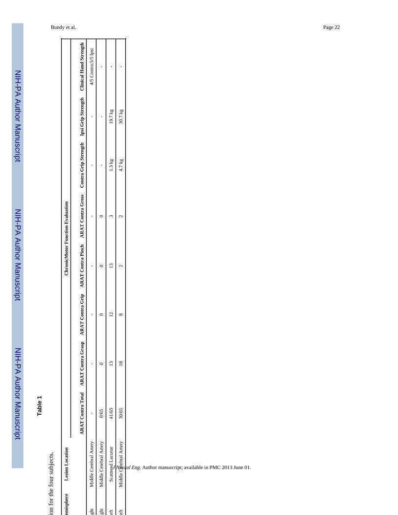
This study utilized four chronic first time (17–53 months post-stroke) hemispheric strokesurvivors (age 48–61). Exclusion criteria included prior strokes. Additionally strokes thatresulted in dementia, inattention, or aphasia, which would prevent subjects from performingthe required cognitive tasks, were also excluded. The study was approved by the HumanResearch Protection Organization of the Washington University Medical Center. Prior toinclusion in the study, subjects provided their written informed consent. Subjects wereenrolled from a previous study (Carter, Astafiev et al. 2010; Carter, Patel et al. 2011), whichprovided data on lesion localization and chronic functional evaluation. Prior to enrollment inthe study, lesion locations and functional motor evaluations were considered from over 40potential subjects. The 4 subjects utilized were selected considering the exclusion criteria aswell as the fact that more severely impaired patients represent the population more likely tobenefit from BCI applications. Before this study, subjects had no prior training on the use of
Bundy et al. Page 3
J Neural Eng. Author manuscript; available in PMC 2013 June 01.
NIH
-PA Author Manuscript
NIH
-PA Author Manuscript
NIH
-PA Author Manuscript
a BCI system. Demographic and clinical information for each of the four subjects is shownin Table 1.
2.2 Lesion SegmentationSegmentation of stroke lesions was performed as described in Carter et al. using T1-weighted MP-RAGE and T2-weighted spin echo images (Carter, Astafiev et al. 2010).Voxels were categorized into air, cerebrospinal fluid (CSF), gray matter, and white matterby an unsupervised fuzzy class means-based segmentation. Expert judgment was used todetermine the boundary between CSF and lesioned parenchyma. Figure 2 shows the locationand extent of lesions in each subject.
2.3 EEG RecordingsIn all subjects, EEG was recorded from 33 (Patients 1 and 2) or 45 (Patients 3 and 4) scalplocations over frontal and parietal regions within the 10–20 system of electrode locations.Recording locations were channel positions AF5, AF3, AFZ, AF4, AF6, F5, F3, F1, FZ, F2,F4, F6, FC5, FC3, FC1, FCZ, FC2, FC4, FC6, C5, C3, C1, CZ, C2, C4, C6, CP5, CP3, CP1,CPZ, CP2, CP4, CP6 (all patients) and P5, P3, P1, PZ, P2, P4, P6, PO5, PO3, POZ, PO4,PO6 (Patients 3 and 4 only). Recordings were digitized using 16-channel digital amplifiers(g.USBamp, g.tec, Austria). The left and right ear lobes were used as the ground andreference respectively. Signals were spatially filtered using a bipolar derivation to enhancethe spatial specificity of recordings. Recordings were sampled at 256 Hz (subjects 1 and 2)or 512 Hz (subjects 3 and 4) and were high-pass filtered at 0.1 Hz prior to analysis. A Dellcomputer running the BCI2000 software platform was used to acquire, process, and store theEEG data for real-time stimulus presentation and time-locked acquisition and analysis ofbrain signals (Schalk, McFarland et al. 2004).
2.4 Control Feature ScreeningInitially, subjects underwent screening to identify features of cortical activity to be used insubsequent closed-loop BCI control experiments. This procedure involved an experiment inwhich EEG signals were recorded while the subject performed overt or imagined self-timed,self-selected finger-tapping movements of the right or left hand in isolation from theopposite hand, or rested. Cues for the rest and finger movement conditions were presentedas words (‘Right’, ‘Left’) and a fixation cross (Rest) on a computer screen that was placedapproximately 75cm in front of the subject. For the overt movement condition, subjects withresidual function in their affected hand (Patient 1) were instructed to perform overtmovements of both hands, while those with less function in the affected hand wereinstructed to perform overt movements of the unaffected hand and intended movements ofthe affected hand. In a second screening task, all subjects were instructed to performimagined movements of both the affected and unaffected hands. Cues were presented in arandom order with each stimulus presented for a period of 2.5s. Subjects were instructed toperform the specified action for the duration of the stimulus presentation. In subjects withchronic hemiplegia preventing individual finger movements of the affected hand (subjects 2,3, and 4) the subjects were instructed to overtly move their unaffected hand and imaginesimilar movements of the affected hand during the respective stimulus periods. All subjectsperformed the overt movement task initially while being observed for successful taskperformance, followed by performance of the imagined movement task.
EEG data collected during the experiment were converted from the time domain to thefrequency domain using the maximum entropy method for autoregressive spectralestimation (Marple 1987). Power spectra were estimated in 2 Hz bins ranging from 1 to 55Hz. Candidate features were identified by calculating the signed coefficient of determination(r2) between the ‘rest’ interval spectral power levels and the affected hand movement
Bundy et al. Page 4
J Neural Eng. Author manuscript; available in PMC 2013 June 01.
NIH
-PA Author Manuscript
NIH
-PA Author Manuscript
NIH
-PA Author Manuscript
spectral power levels. EEG features in particular electrodes and frequency bands with thegreatest percentage of their variance explained by the task (i.e. the highest r2 values), werechosen as candidate control features for closed-loop BCI experiments. Selection ofcandidate control features was also further constrained to contain only electrodes over theunaffected hemisphere during movement or imagined movement of the affected hand.Where possible, candidate features were selected to discriminate affected hand movementfrom rest as well as affected hand movement from unaffected hand movement.
2.5 Closed-loop BCI EvaluationAfter determining candidate EEG control features associated with intended movement of theaffected hand using the screening procedure described previously, subjects participated inclosed-loop BCI control evaluation (see Figure 3). For this evaluation, the subjects’objective was to perform intended movements of the affected hand in order to hit a targetwith a cursor. The target was presented on either the right or left side of a screen and thecursor moved along a single dimension. Several control scenarios were tested; (1) overtmovement of the affected hand versus rest (Patient 1), (2) intended movement of theaffected hand versus rest (Patients 2, 3, and 4), and (3) imagined movement of the affectedhand versus imagined movement of the unaffected hand (Patients 1 and 2). The variousclosed-loop control conditions used depended upon the discriminability of control featuresas well as patient attention and fatigue. The velocity of the cursor was calculated from thereal-time EEG features through the BCI2000 software package. The change in power frombaseline of the selected EEG features (i.e. the power in the selected frequency bin(s) at theselected electrode location(s) over the unaffected hemisphere) were weighted and summedto allow the patient to control the cursor. As all of the features identified for these subjectswere power decreases, they were weighted negatively so the task-related score increasedwith task-related power decreases. The feature power levels were translated into the cursorscore through BCI2000 (Schalk, McFarland et al. 2004). In order to normalize the weightedand summed power levels, the normalizer was trained using several trials in each targetdirection in which the subject attempted to perform the selected control task (i.e. affectedhand movement, resting) in order to control the cursor. The mean of the weighted andsummated features was calculated after the training period (1–2 minutes) and was used tonormalize the scores to have zero mean. The normalized score was then used to control thecursor velocity. The velocity of the cursor was updated every 40 ms based upon spectraestimated from an autoregressive method using data acquired over the previous 280 ms.Subjects performed consecutive trials in which they attempted to move the cursor to thepresented target. Each trial began with the presentation of a target randomly selected to be ateither the right or left side of the screen. After a 1 s delay, the cursor appeared in the middleof the screen with its motion in the horizontal dimension controlled by the subject’s EEGsignals. The subject was instructed to begin performing the particular hand movement taskor rest condition to move the cursor to the selected target as soon as the target appeared onthe screen. Each trial was assessed as a success (cursor hit the selected target), or failure(cursor hit the opposite target or time ran out before success occurred, 6–10s). Trials weregrouped into runs of 2 min with rest periods of approximately 1 min in between runs. Theaccuracy was assessed as the number of successful trials divided by the total number of trialsat the end of each run. The development of BCI control over time was assessed bycomparing the accuracy at the end of each consecutive run after training with a particulartask and associated EEG control features. Because each trial did not have to result in a targetbeing hit, chance was not necessarily 50%. As described previously by Leuthardt et. al.,chance performance was determined by running multiple runs of control trials usingGaussian white noise signals yielding a mean chance performance of 46.2% (2.7% SD)(Leuthardt, Gaona et al. 2011). Subjects performed between 85 and 246 control trials.
Bundy et al. Page 5
J Neural Eng. Author manuscript; available in PMC 2013 June 01.
NIH
-PA Author Manuscript
NIH
-PA Author Manuscript
NIH
-PA Author Manuscript

3. ResultsEach subject demonstrated cortical activations in the unaffected hemisphere associated withintended movement of the affected hand. Figure 4 displays that for each subject there was acortical activation ipsilateral to the side of intended movement of the affected hand (withinthe unaffected hemisphere). Notably, consistent with Wisneski el al., when compared tocortical activations in the unaffected hemisphere associated with movement intentions of theunaffected hand (i.e. contralateral movements) there were notable differences in frequencyspectra and anatomic locations (Wisneski, Anderson et al. 2008). Figure 5 illustrates thisspectral distinction in cortical activity within a single exemplar subject (patient 3). In thissubject, the topography at 18 Hz is broad and fairly similar between ipsilateral andcontralateral movement, while there are distinct differences in topography at the higher beta(26 Hz) and low gamma (>30 Hz) frequencies. Most notably there is a more extensiveactivation in the prefrontal region (in the left unaffected hemisphere) that demonstrates a 26Hz power modulation with ipsilateral right hand movement that is not present withcontralateral left hand movement. Moreover, when examining all subjects, various locationsand EEG frequencies separated intended affected hand movements both from rest and fromunaffected hand movements (Figure 6). The locations that optimally separated ipsilateralfrom contralateral intentions in the unaffected hemisphere were located both over traditionalsensorimotor regions, as well as more anterior areas associated with premotor planning.Furthermore, qualitatively it was observed that subjects with more severe motorimpairments demonstrate a more anterior shift in the activations within the unaffectedhemisphere associated with intended movement of the ipsilateral affected hand. This shift inipsilateral activity is similar to the anterior and ventral shift in ipsilateral activity shown byCramer et al. with fMRI (Cramer, Finklestein et al. 1999).
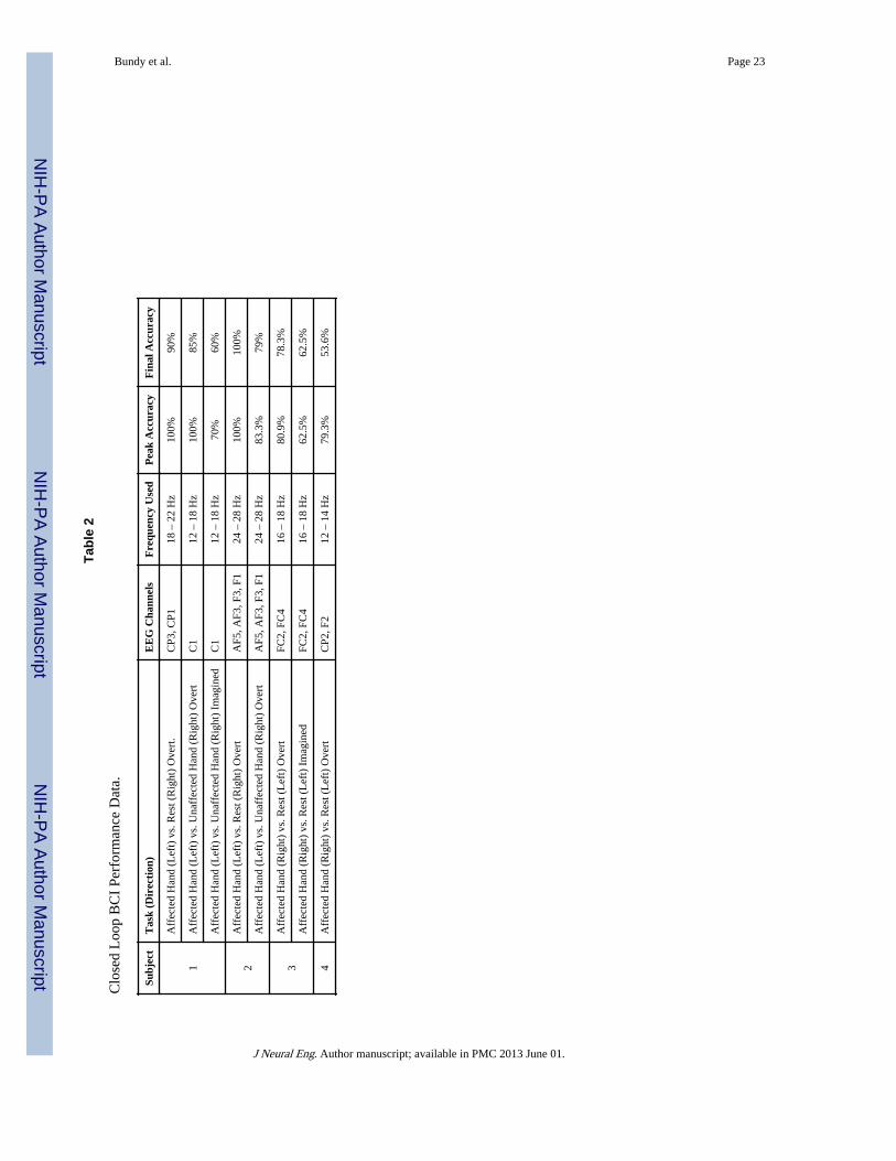
Select features were then identified for subsequent online BCI operation. Figure 7 illustratesthe significant (p<0.05) activations differentiating movements of the affected hand from rest,the unaffected hand from rest, and the affected hand from the unaffected hand at frequenciesutilized for subsequent BCI control by each patient. The time course of each subject’sperformance during closed-loop BCI control is shown in Figure 8. Peak target accuracies forall subjects were all between 62.5% and 100% and final accuracies for all subjects werebetween 53.6% and 100% and therefore were all above chance (46.2%, 2.7% SD). This levelof performance was achieved in BCI control experiments with durations ranging from 6 to10 min. Table 2 summarizes the activities utilized for BCI control, the EEG features used,and the peak and final BCI accuracy achieved by the subject.
The EEG recordings from the closed-loop control experiments were also examined post-hocto compare the cortical activations associated with the screening task to those associatedwith the closed-loop task. The activations in the selected EEG control frequency for theaffected hand movement versus rest conditions are shown for both the screening and controlexperiments in Figure 9. As can be seen, there is good correspondence between thetopographies of the activations during the control and screening task, indicating that thesuccessful BCI control performance was achieved through performance of the intendedmotor task and not a spurious or alternative strategy. Furthermore, correlations between thetopography of activations in the selected control frequency between screening and controltasks (patient 1: R=0.82, p<0.0001; patient 2: R=0.6108, p=0.0002; patient 3: R=0.7098,p<0.0001; patient 4: R=−0.0194; P=0.8994) were highly significant in 3 of the 4 subjects.The discrepancy in patient 4 can be attributed to the patient tiring towards the end of thesession, likely leading to changes to cortical activations due to decreases in attention to thetask. This decrease in attention and different neural activity would also explain the fact thatpatient 4 demonstrated poorer peak and final BCI control accuracies than the other 3subjects.
Bundy et al. Page 6
J Neural Eng. Author manuscript; available in PMC 2013 June 01.
NIH
-PA Author Manuscript
NIH
-PA Author Manuscript
NIH
-PA Author Manuscript

4. DiscussionThis paper presents an important demonstration for the potential that hemispheric strokesurvivors could use the unaffected side of their brain to potentially restore their unilateralmotor deficit through the use of a BCI. By identifying cortical signals associated withipsilateral hand movements, an EEG-based BCI can capture the brain’s intention to move aparetic hand. This can be accomplished irrespective of their actual capacity to execute amotor movement due to their injured primary motor cortex and/or descending white mattertracts. In this study, we demonstrate that it is possible to detect in EEG over the unaffectedcortex, real and imagined intentions to move the stroke affected hand and, for the first time,show that these unaffected hemisphere signals can be used for simple brain-derived controlof a device. Subjects achieved BCI control with peak accuracy rates between 62.5% and100% rapidly, often with only a 30 minute screening task and less than 15 minutes oftraining. Importantly, these ipsilateral motor signals were distinct in anatomic location andspectral content from the cortical physiology associated with contralateral movements. Thisseparable physiology and its ease of use for a BCI, provides an important first step towardsusing neuroprosthetic systems for a currently large and underserved patient population withmotor disability. Given that stroke is the most common neurological disorder, affecting795,000 patients per year in the U.S. alone (Lloyd-Jones, Adams et al. 2009), these findingscan substantially extend the potential clinical impact of neuroprosthetic approaches.
This work was unique in its focus on using neural activity in the unaffected hemisphererelated to intended movement of the ipsilateral limb for controlling a BCI system. Whileother studies have investigated the use of control signals from perilesional cortical areas(Buch, Weber et al. 2008; Daly, Cheng et al. 2009; Broetz, Braun et al. 2010), this is the firststudy to focus exclusively on ipsilateral motor activity from the unaffected hemisphere afterstroke . This is important because BCI systems are likely to be most clinically relevant tostroke survivors with a chronic unrecovered hemiplegic motor deficit. Because strokesurvivors with any residual motor functions are likely to be candidates for currentrehabilitation methodologies (Takahashi, Der-Yeghiaian et al. 2008; Wolf, Thompson et al.2010), BCI systems are likely to be used either by the most severely motor-impaired patientswho had an absence of any function from the onset (limiting their rehabilitation options), orhave failed to recover function after extensive therapy. Often these patient populations arelikely to have significant cortical and/or subcortical lesions that transect the corticospinaltract descending to contralateral motor pathways (Binkofski, Seitz et al. 1996; Schaechter,Perdue et al. 2008; Carter, Patel et al. 2011). Because of these significant lesions, thesepatients are likely to have atypical neural activity in the affected hemisphere during intendedmovement of the contralateral limb when compared with normal controls (Calford andTweedale 1990). By using ipsilateral motor intentions in the undamaged hemisphere for aBCI control signal, the impact of the stroke with regards to the quality of the control signalwill be minimized. Taken together, there are likely two ways in which this methodologycould be applied, either as a chronic BCI system for long-term assistive device control, or asBCI-assisted rehabilitation tool.
As in traditional BCI systems, stroke survivors could control an artificial device to aid indaily tasks or manipulate the hemiplegic limb. Building on work by Wisneski et al., whoshowed that motor intact patients could use ipsilateral motor signals for BCI control, thisstudy extended those findings by demonstrating that stroke survivors can intentionallymodulate similar ipsilateral motor signals from their unaffected hemisphere to control a BCIsystem. Several of the subjects that were studied in this study had fairly significant motorimpairments from their strokes (Subjects 2, 3, and 4). Importantly, these subjects generallyhad shifts of the ipsilateral motor activity in their unaffected hemispheres to locationsanterior to traditional sensorimotor cortices. This finding is in line with other results that
Bundy et al. Page 7
J Neural Eng. Author manuscript; available in PMC 2013 June 01.
NIH
-PA Author Manuscript
NIH
-PA Author Manuscript
NIH
-PA Author Manuscript
have demonstrated changes in motor activity ipsilateral to the unaffected hemisphere(Cramer, Nelles et al. 1997; Green, Bialy et al. 1999; Tecchio, Zappasodi et al. 2006). It isimportant to note that the ipsilateral control signals were used to control a cursor on a screenas a proof of concept. This control could be easily extended to a simple one-dimensionalgrasping hand orthotic that could facilitate activities of daily living (Lauer, Peckham et al.1999).
With regards to the choice of signal platform for utilizing an assistive BCI system afterstroke, there are several considerations to take into account. In order to implement atraditional assistive BCI system, it will be important to scale the BCI control to a greaternumber of degrees of freedom. Currently, non-invasive BCI systems have produced at most3 degrees-of-freedom in online task performance (McFarland, Sarnacki et al. 2010). Thislevel of control required a large amount of training and utilized signals from both corticalhemispheres and midline electrodes that were related to movements of the feet and bothhands. Given the broad cortical activations observed in EEG recordings, the necessity ofdeveloping BCI systems independent of the normal operation of the unaffected hand forcompletion of bimanual tasks, and attentional limitations to the amount of BCI training timein some patients, it is doubtful that EEG would allow for discernment of grasping andkinematic hand movements necessary for scaling an ipsilateral BCI system to a highernumber of degrees of freedom in stroke survivors. Therefore, more complex control mayrequire more invasive approaches such as electrocorticography (ECoG). To date, morecomplex movement kinematics of both hand and finger movements have been shown to bediscernable using either macroscale or microscale ECoG signals (Wisneski, Anderson et al.2008; Zanos, Miller et al. 2008; Leuthardt, Freudenberg et al. 2009; Scherer, Zanos et al.2009). Additionally, ECoG has allowed for off-line decoding of 2D joystick movementsfrom ipsilateral cortex (Ganguly, Secundo et al. 2009; Sharma, Gaona et al. 2009). As workcontinues to develop BCI systems for chronic stroke survivors, it will be important to gain abetter understanding of the specific cortical dynamics associated with ipsilateral motoractivity after stroke and the optimal signal platform that provides the highest benefit relativeto the clinical risk of implementation.
In addition to providing a means for long-term assistive device control after stroke, BCIsystems may provide a novel rehabilitation tool. The choice of motor signals from theunaffected hemisphere in this study has particular relevance to the potential for using BCIsystems as a rehabilitation methodology. A number of previous studies have demonstratedchanges in ipsilateral motor activity from the unaffected hemisphere after stroke. Functionalimaging of chronic stroke survivors has shown increases in the ipsilateral motor activationsof the unaffected hemisphere after recovery from stroke when compared to normal controls(Weiller, Chollet et al. 1992; Weiller, Ramsay et al. 1993; Cramer, Nelles et al. 1997;Nelles, Spiekramann et al. 1999; Tecchio, Zappasodi et al. 2006) as well as increases inipsilateral, contralesional activity after constraint induced movement therapy (CIMT) (Levy,Nichols et al. 2001; Schaechter, Kraft et al. 2002). Furthermore, inhibitory transcranialmagnetic stimulation in the unaffected premotor cortex slowed the reaction times foraffected hand movements in stroke survivors (Johansen-Berg, Rushworth et al. 2002). Whilethese results seem to indicate that ipsilateral activity from the unaffected hemisphere plays afacilitating role in recovery of motor function after stroke, there are also a number of studiesthat have shown potentially contradictory findings regarding increases in ipsilateral,contralesional motor activity after stroke. In particular, low ipsilateral TMS thresholds wereassociated with poor recovery after stroke (Turton, Wroe et al. 1996; Netz, Lammers et al.1997) and decreases in ipsilateral activity from the unaffected hemisphere correlated withlongitudinal and cross-sectional studies of recovery (Ward, Brown et al. 2003; Ward, Brownet al. 2003). It is important to note that several of the studies described above demonstratedboth perilesional changes in activity, as well as ipsilateral activity from the unaffected
Bundy et al. Page 8
J Neural Eng. Author manuscript; available in PMC 2013 June 01.
NIH
-PA Author Manuscript
NIH
-PA Author Manuscript
NIH
-PA Author Manuscript
hemisphere after recovery, indicating that both cortical areas may lead to recovery (Green,Bialy et al. 1999; Levy, Nichols et al. 2001). Furthermore, another study revealed thatpatients who recover completely showed few changes to the location of their contralesionalmotor activity, while patients that recovered incompletely showed better recovery withincreased ipsilateral activity in the unaffected hemisphere (Tecchio, Zappasodi et al. 2006).This makes sense when one considers that corticospinal tract damage has been shown to behighly correlated with motor impairment after stroke (Fries, Danek et al. 1993; Carter, Patelet al. 2011). While patients with some residual motor function will most likely rehabilitatethrough reorganization of residual motor pathways, those for whom it is totally obliteratedwill likely need to develop new cortical and subcortical pathways (i.e. contralesional/ipsilateral motor pathways) for a more limited recovery. Thus, the patients that are mostlikely to be candidates for a BCI-based therapy, are those who have substantial damage totheir corticospinal tract, requiring that an alternative pathway be utilized for functionalrehabilitation to take place. Because of this, the results described in this paper demonstratingthat stroke survivors can utilize ipsilateral motor activity from their unaffected hemisphereto control a BCI system represent a significant step in the development of a BCI system forencouraging rehabilitation after stroke. Moreover, in the setting of rehabilitation where theBCI is only needed transiently and the goal is to augment cortical plasticity, lower degreesof freedom for a less invasive option may be an ideal tradeoff in this clinical context.
While this work represents an exciting demonstration of the possibilities for stroke survivorsto achieve increased function through controlling BCI systems with neural activity in theirunaffected hemisphere, there are several limitations and future considerations. First, thework represents only a limited number of patients and while one patient (Patient 1), hadsome recovery, most of the patients were selected for participation because they had moresignificant motor impairments, making them more representative of the patient populationmost likely to benefit from BCI applications. Because of this it is unknown how well theseresults will generalize to the broader population of stroke survivors. However, thepopulation included patients with lesions of both hemispheres as well as both cortical andsubcortical infarcts (see Table 1), indicating that BCI systems can be utilized by patientswith various lesion types and locations. Furthermore, because BCI systems will most likelybe applied to the most significantly impaired patients, these subjects do represent theintended clinical population. Second, it is impossible to ensure that the subject’s BCI controlis truly achieved through the screened motor imagery task, particularly in cases in whichsubjects have no visible motor control. In this study however, the similarity between neuralactivity during the screening and control tasks (see Figure 9) provide evidence that thesubject is indeed performing the imagery task indicated. Additionally, a BCI system needs tofunction independently of the unaffected hand to allow for completion of bimanual tasks.Because of differences in the attentional requirements of attempting to move the unaffectedhand, it is difficult to truly assess the independence of the ipsilateral, contralesional motorsignals and the traditional contralateral, contralesional motor signals during bimanual tasks,however, the ability of two subjects (Patients 1 and 2) to achieve on-line control withalternating movements of the affected and unaffected hands provides evidence thatipsilateral, contralesional motor activity is independent from unaffected hand movement inthis patient population. Finally, it is important to note that the observed increase inipsilateral, contralesional activations after stroke may represent increases in the attentionalrequirements of attempting to move the affected hand (Johansen-Berg, Rushworth et al.2002). Regardless of whether this represents increased attention as has been postulated fromprevious imaging literature or enhancements in motor planning, once this signal is identifiedand engaged by the user, its utilization becomes of importance to assistive technologies, orto functional reorganization of the cortex and its neural output for a novel purpose. In thefuture, it will be important to explicitly test the changes in neural activity in the unaffectedhemisphere as stroke survivors use BCI systems for longer time periods.
Bundy et al. Page 9
J Neural Eng. Author manuscript; available in PMC 2013 June 01.
NIH
-PA Author Manuscript
NIH
-PA Author Manuscript
NIH
-PA Author Manuscript
In summary, these results move the applications of BCI forward to potentially benefit thelarge number of motor-impaired hemispheric stroke survivors. The study shows in particularthat hemispheric stroke patients can volitionally control signals from the unaffectedhemisphere for device operation. This specific use of the contralesional hemisphere mayprovide a novel neuroprosthetic approach for increasing function in the more impairedstroke populations whose rehabilitation options are currently limited.
AcknowledgmentsThis publication was made possible by Grant Number UL1 RR024992 and TL1 RR024995 from the NIH-NationalCenter for Research Resources (NCRR) and grant numbers 1R0100085606 and (NICHD) RO1 HD061117-05A2.We would also like to thank the patients who participated in this study. Without their interest and effort thisresearch would not be possible.
ReferencesBinkofski F, Seitz RJ, Arnold S, Classen J, Benecke R, Freund HJ. Thalamic metbolism and
corticospinal tract integrity determine motor recovery in stroke. Ann Neurol. 1996; 39(4):460–470.[PubMed: 8619524]
Broetz D, Braun C, Weber C, Soekadar SR, Caria A, Birbaumer N. Combination of brain-computerinterface training and goal-directed physical therapy in chronic stroke: a case report. NeurorehabilNeural Repair. 2010; 24(7):674–679. [PubMed: 20519741]
Buch E, Weber C, Cohen LG, Braun C, Dimyan MA, Ard T, Mellinger J, Caria A, Soekadar S,Fourkas A, Birbaumer N. Think to move: a neuromagnetic brain-computer interface (BCI) systemfor chronic stroke. Stroke. 2008; 39(3):910–917. [PubMed: 18258825]
Calford MB, Tweedale R. Interhemispheric transfer of plasticity in the cerebral cortex. Science. 1990;249(4970):805–807. [PubMed: 2389146]
Carter AR, Astafiev SV, Lang CE, Connor LT, Rengachary J, Strube MJ, Pope DL, Shulman GL,Corbetta M. Resting interhemispheric functional magnetic resonance imaging connectivity predictsperformance after stroke. Ann Neurol. 2010; 67(3):365–375. [PubMed: 20373348]
Carter AR, Patel KR, Astafiev SV, Snyder AZ, Rengachary J, Strube MJ, Pope A, Shimony JS, LangCE, Shulman GL, Corbetta M. Upstream Dysfunction of Somatomotor Functional ConnectivityAfter Corticospinal Damage in Stroke. Neurorehabil Neural Repair. 2011
Cramer SC, Finklestein SP, Schaechter JD, Bush G, Rosen BR. Activation of distinct motor cortexregions during ipsilateral and contralateral finger movements. J Neurophysiol. 1999; 81(1):383–387. [PubMed: 9914297]
Cramer SC, Mark A, Barquist K, Nhan H, Stegbauer KC, Price R, Bell K, Odderson IR, Esselman P,Maravilla KR. Motor cortex activation is preserved in patients with chronic hemiplegic stroke. AnnNeurol. 2002; 52(5):607–616. [PubMed: 12402258]
Cramer SC, Nelles G, Benson RR, Kaplan JD, Parker RA, Kwong KK, Kennedy DN, Finklestein SP,Rosen BR. A functional MRI study of subjects recovered from hemiparetic stroke. Stroke. 1997;28(12):2518–2527. [PubMed: 9412643]
Daly JJ, Cheng R, Rogers J, Litinas K, Hrovat K, Dohring M. Feasibility of a New Application ofNoninvasive Brain Computer Interface (BCI): A Case Study of Training for Recovery ofVolitional Motor Control After Stroke. J Neurol Phys Ther. 2009; 33(4):203–211. [PubMed:20208465]
Duncan PW, Goldstein LB, Matchar D, Divine GW, Feussner J. Measurement of motor recovery afterstroke. Outcome assessment and sample size requirements. Stroke. 1992; 23(8):1084–1089.[PubMed: 1636182]
Fries W, Danek A, Scheidtmann K, Hamburger C. Motor recovery following capsular stroke. Role ofdescending pathways from multiple motor areas. Brain. 1993; 116(Pt 2):369–382. [PubMed:8461971]
Ganguly K, Secundo L, Ranade G, Orsborn A, Chang EF, Dimitrov DF, Wallis JD, Barbaro NM,Knight RT, Carmena JM. Cortical representation of ipsilateral arm movements in monkey andman. J Neurosci. 2009; 29(41):12948–12956. [PubMed: 19828809]
Bundy et al. Page 10
J Neural Eng. Author manuscript; available in PMC 2013 June 01.
NIH
-PA Author Manuscript
NIH
-PA Author Manuscript
NIH
-PA Author Manuscript
Green JB, Bialy Y, Sora E, Ricamato A. High-resolution EEG in poststroke hemiparesis can identifyipsilateral generators during motor tasks. Stroke. 1999; 30(12):2659–2665. [PubMed: 10582993]
Hochberg LR, Serruya MD, Friehs GM, Mukand JA, Saleh M, Caplan AH, Branner A, Chen D, PennRD, Donoghue JP. Neuronal ensemble control of prosthetic devices by a human with tetraplegia.Nature. 2006; 442(7099):164–171. [PubMed: 16838014]
Johansen-Berg H, Rushworth MF, Bogdanovic MD, Kischka U, Wimalaratna S, Matthews PM. Therole of ipsilateral premotor cortex in hand movement after stroke. Proc Natl Acad Sci U S A.2002; 99(22):14518–14523. [PubMed: 12376621]
Jorgensen HS, Nakayama H, Raaschou HO, Vive-Larsen J, Stoier M, Olsen TS. Outcome and timecourse of recovery in stroke. Part II: Time course of recovery. The Copenhagen Stroke Study.Arch Phys Med Rehabil. 1995; 76(5):406–412. [PubMed: 7741609]
Kubler A, Nijboer F, Mellinger J, Vaughan TM, Pawelzik H, Schalk G, McFarland DJ, Birbaumer N,Wolpaw JR. Patients with ALS can use sensorimotor rhythms to operate a brain-computerinterface. Neurology. 2005; 64(10):1775–1777. [PubMed: 15911809]
Lauer RT, Peckham PH, Kilgore KL. EEG-based control of a hand grasp neuroprosthesis.Neuroreport. 1999; 10(8):1767–1771. [PubMed: 10501572]
Leuthardt EC, Freudenberg Z, Bundy D, Roland J. Microscale recording from human motor cortex:implications for minimally invasive electrocorticographic brain-computer interfaces. NeurosurgFocus. 2009; 27(1):E10. [PubMed: 19569885]
Leuthardt EC, Gaona C, Sharma M, Szrama N, Roland J, Freudenberg Z, Solis J, Breshears J, SchalkG. Using the electrocorticographic speech network to control a brain-computer interface inhumans. J Neural Eng. 2011; 8(3):036004. [PubMed: 21471638]
Leuthardt EC, Schalk G, Wolpaw JR, Ojemann JG, Moran DW. A brain-computer interface usingelectrocorticographic signals in humans. J Neural Eng. 2004; 1(2):63–71. [PubMed: 15876624]
Levy CE, Nichols DS, Schmalbrock PM, Keller P, Chakeres DW. Functional MRI evidence of corticalreorganization in upper-limb stroke hemiplegia treated with constraint-induced movement therapy.Am J Phys Med Rehabil. 2001; 80(1):4–12. [PubMed: 11138954]
Lloyd-Jones D, Adams R, Carnethon M, De Simone G, Ferguson TB, Flegal K, Ford E, Furie K, GoA, Greenlund K, Haase N, Hailpern S, Ho M, Howard V, Kissela B, Kittner S, Lackland D,Lisabeth L, Marelli A, McDermott M, Meigs J, Mozaffarian D, Nichol G, O’Donnell C, Roger V,Rosamond W, Sacco R, Sorlie P, Stafford R, Steinberger J, Thom T, Wasserthiel-Smoller S, WongN, Wylie-Rosett J, Hong Y. Heart disease and stroke statistics--2009 update: a report from theAmerican Heart Association Statistics Committee and Stroke Statistics Subcommittee. Circulation.2009; 119(3):e21–181. [PubMed: 19075105]
Marple, L. Digital spectral analysis with applications. Upper Saddle River, NJ: Prentice-Hall; 1987.
McFarland DJ, Sarnacki WA, Wolpaw JR. Electroencephalographic (EEG) control of three-dimensional movement. J Neural Eng. 2010; 7(3):036007. [PubMed: 20460690]
Nelles G, Spiekramann G, Jueptner M, Leonhardt G, Muller S, Gerhard H, Diener HC. Evolution offunctional reorganization in hemiplegic stroke: a serial positron emission tomographic activationstudy. Ann Neurol. 1999; 46(6):901–909. [PubMed: 10589543]
Netz J, Lammers T, Homberg V. Reorganization of motor output in the non-affected hemisphere afterstroke. Brain. 1997; 120(Pt 9):1579–1586. [PubMed: 9313641]
Pfurtscheller G, Guger C, Muller G, Krausz G, Neuper C. Brain oscillations control hand orthosis in atetraplegic. Neurosci Lett. 2000; 292(3):211–214. [PubMed: 11018314]
Rouse AG, Moran DW. Neural adaptation of epidural electrocorticographic (EECoG) signals duringclosed-loop brain computer interface (BCI) tasks. Conf Proc IEEE Eng Med Biol Soc. 2009:5514–5517. [PubMed: 19964124]
Schaechter JD, Kraft E, Hilliard TS, Dijkhuizen RM, Benner T, Finklestein SP, Rosen BR, CramerSC. Motor recovery and cortical reorganization after constraint-induced movement therapy instroke patients: a preliminary study. Neurorehabil Neural Repair. 2002; 16(4):326–338. [PubMed:12462764]
Schaechter JD, Perdue KL, Wang R. Structural damage to the corticospinal tract correlates withbilateral sensorimotor cortex reorganization in stroke patients. Neuroimage. 2008; 39(3):1370–1382. [PubMed: 18024157]
Bundy et al. Page 11
J Neural Eng. Author manuscript; available in PMC 2013 June 01.
NIH
-PA Author Manuscript
NIH
-PA Author Manuscript
NIH
-PA Author Manuscript
Schaefer SY, Haaland KY, Sainburg RL. Dissociation of initial trajectory and final position errorsduring visuomotor adaptation following unilateral stroke. Brain Res. 2009; 1298:78–91. [PubMed:19728993]
Schaefer SY, Haaland KY, Sainburg RL. Hemispheric specialization and functional impact ofipsilesional deficits in movement coordination and accuracy. Neuropsychologia. 2009; 47(13):2953–2966. [PubMed: 19573544]
Schalk G, McFarland DJ, Hinterberger T, Birbaumer N, Wolpaw JR. BCI2000: a general-purposebrain-computer interface (BCI) system. IEEE Trans Biomed Eng. 2004; 51(6):1034–1043.[PubMed: 15188875]
Schalk G, Miller KJ, Anderson NR, Wilson JA, Smyth MD, Ojemann JG, Moran DW, Wolpaw JR,Leuthardt EC. Two-dimensional movement control using electrocorticographic signals in humans.J Neural Eng. 2008; 5(1):75–84. [PubMed: 18310813]
Scherer R, Zanos SP, Miller KJ, Rao RP, Ojemann JG. Classification of contralateral and ipsilateralfinger movements for electrocorticographic brain-computer interfaces. Neurosurg Focus. 2009;27(1):E12. [PubMed: 19569887]
Sharma M, Gaona C, Roland J, Anderson N, Freudenberg Z, Leuthardt EC. Ipsilateral directionalencoding of joystick movements in human cortex. Conf Proc IEEE Eng Med Biol Soc.2009:5502–5505. [PubMed: 19965051]
Takahashi CD, Der-Yeghiaian L, Le V, Motiwala RR, Cramer SC. Robot-based hand motor therapyafter stroke. Brain. 2008; 131(Pt 2):425–437. [PubMed: 18156154]
Taylor DM, Tillery SI, Schwartz AB. Direct cortical control of 3D neuroprosthetic devices. Science.2002; 296(5574):1829–1832. [PubMed: 12052948]
Tecchio F, Zappasodi F, Tombini M, Oliviero A, Pasqualetti P, Vernieri F, Ercolani M, Pizzella V,Rossini PM. Brain plasticity in recovery from stroke: an MEG assessment. Neuroimage. 2006;32(3):1326–1334. [PubMed: 16806985]
Turton A, Wroe S, Trepte N, Fraser C, Lemon RN. Contralateral and ipsilateral EMG responses totranscranial magnetic stimulation during recovery of arm and hand function after stroke.Electroencephalogr Clin Neurophysiol. 1996; 101(4):316–328. [PubMed: 8761041]
Velliste M, Perel S, Spalding MC, Whitford AS, Schwartz AB. Cortical control of a prosthetic arm forself-feeding. Nature. 2008; 453(7198):1098–1101. [PubMed: 18509337]
Ward NS, Brown MM, Thompson AJ, Frackowiak RS. Neural correlates of motor recovery afterstroke: a longitudinal fMRI study. Brain. 2003; 126(Pt 11):2476–2496. [PubMed: 12937084]
Ward NS, Brown MM, Thompson AJ, Frackowiak RS. Neural correlates of outcome after stroke: across-sectional fMRI study. Brain. 2003; 126(Pt 6):14305–1448.
Weiller C, Chollet F, Friston KJ, Wise RJ, Frackowiak RS. Functional reorganization of the brain inrecovery from striatocapsular infarction in man. Ann Neurol. 1992; 31(5):463–472. [PubMed:1596081]
Weiller C, Ramsay SC, Wise RJ, Friston KJ, Frackowiak RS. Individual patterns of functionalreorganization in the human cerebral cortex after capsular infarction. Ann Neurol. 1993; 33(2):181–189. [PubMed: 8434880]
Wisneski KJ, Anderson N, Schalk G, Smyth M, Moran D, Leuthardt EC. Unique cortical physiologyassociated with ipsilateral hand movements and neuroprosthetic implications. Stroke. 2008;39(12):3351–3359. [PubMed: 18927456]
Wolf SL, Thompson PA, Winstein CJ, Miller JP, Blanton SR, Nichols-Larsen DS, Morris DM,Uswatte G, Taub E, Light KE, Sawaki L. The EXCITE stroke trial: comparing early and delayedconstraint-induced movement therapy. Stroke. 2010; 41(10):2309–2315. [PubMed: 20814005]
Wolpaw JR, McFarland DJ. Control of a two-dimensional movement signal by a noninvasive brain-computer interface in humans. Proc Natl Acad Sci U S A. 2004; 101(51):17849–17854. [PubMed:15585584]
Zanos S, Miller KJ, Ojemann JG. Electrocorticographic spectral changes associated with ipsilateralindividual finger and whole hand movement. Conf Proc IEEE Eng Med Biol Soc. 2008;2008:5939–5942. [PubMed: 19164072]
Bundy et al. Page 12
J Neural Eng. Author manuscript; available in PMC 2013 June 01.
NIH
-PA Author Manuscript
NIH
-PA Author Manuscript
NIH
-PA Author Manuscript
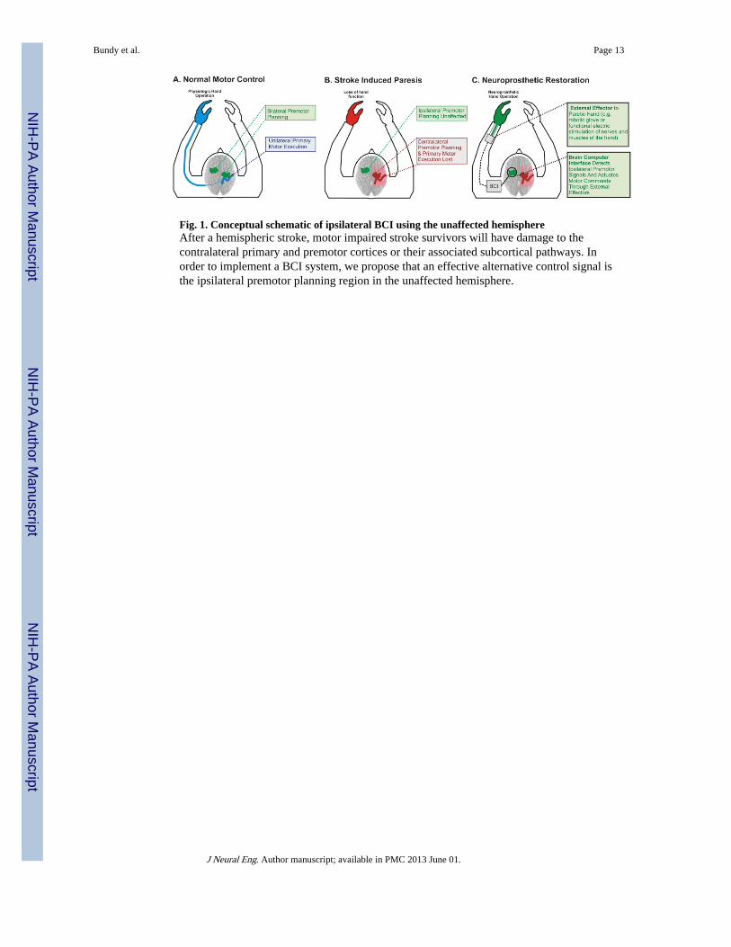
Fig. 1. Conceptual schematic of ipsilateral BCI using the unaffected hemisphereAfter a hemispheric stroke, motor impaired stroke survivors will have damage to thecontralateral primary and premotor cortices or their associated subcortical pathways. Inorder to implement a BCI system, we propose that an effective alternative control signal isthe ipsilateral premotor planning region in the unaffected hemisphere.
Bundy et al. Page 13
J Neural Eng. Author manuscript; available in PMC 2013 June 01.
NIH
-PA Author Manuscript
NIH
-PA Author Manuscript
NIH
-PA Author Manuscript

Fig. 2. Lesion characteristicsLesion locations segmented from T1-weighted MP-RAGE and T2-weighted spin echoimages. Selected axial slices show upper, intermediate, and lower areas of the lesion (Left-on-left orientation).
Bundy et al. Page 14
J Neural Eng. Author manuscript; available in PMC 2013 June 01.
NIH
-PA Author Manuscript
NIH
-PA Author Manuscript
NIH
-PA Author Manuscript
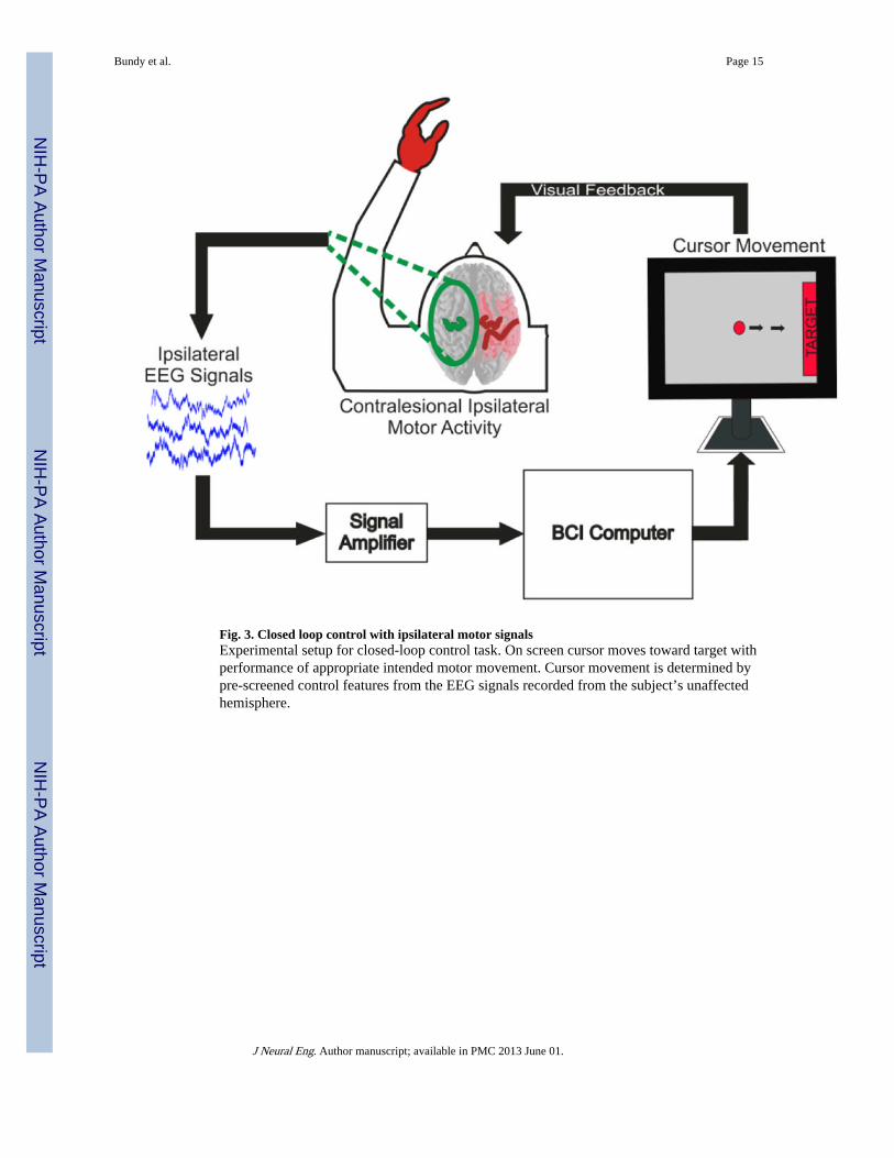
Fig. 3. Closed loop control with ipsilateral motor signalsExperimental setup for closed-loop control task. On screen cursor moves toward target withperformance of appropriate intended motor movement. Cursor movement is determined bypre-screened control features from the EEG signals recorded from the subject’s unaffectedhemisphere.
Bundy et al. Page 15
J Neural Eng. Author manuscript; available in PMC 2013 June 01.
NIH
-PA Author Manuscript
NIH
-PA Author Manuscript
NIH
-PA Author Manuscript

Fig. 4. Topography of screening task activationsTopographical maps of the maximum coefficient-of-determination of significant (p<0.05)event-related power decreases between 0 Hz and 50 Hz for affected hand movement vs. restconditions in each patient. As power decreases cause more negative signed r2 values, morenegative areas in the topographic activations represent increased neural activity. Green andred highlighting on the topographic plots illustrates the unaffected and affected hemispherein each patient respectively. Feature plots demonstrate significant ipsilateral coefficient-of-determination values from the unaffected hemisphere across the frequency spectrum,illustrating that the cortical activations are observed across the frequency spectrum, butparticularly in the mu (8–12 Hz) and beta (12–30 Hz) bands. Channels displayed on thefeature plots correspond to electrode positions AF5, AF3, AFZ, F5, F3, F1, FZ, FC5, FC3,FC1, FCZ, C5, C3, C1, CZ, CP5, CP3, CP1, CPZ (Patients 1 and 2) and AFZ, AF4, AF6,FZ, F2, F4, F6, FCZ, FC2, FC4, FC6, CZ, C2, C4, C6, CPZ, CP2, CP4, CP6, PZ, P2, P4,P6, POZ, PO4, PO6 (Patients 3 and 4).
Bundy et al. Page 16
J Neural Eng. Author manuscript; available in PMC 2013 June 01.
NIH
-PA Author Manuscript
NIH
-PA Author Manuscript
NIH
-PA Author Manuscript
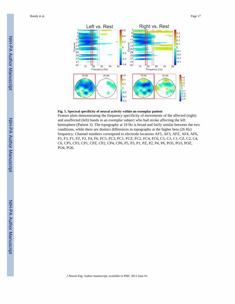
Fig. 5. Spectral specificity of neural activity within an exemplar patientFeature plots demonstrating the frequency specificity of movements of the affected (right)and unaffected (left) hands in an exemplar subject who had stroke affecting the lefthemisphere (Patient 3). The topography at 18 Hz is broad and fairly similar between the twoconditions, while there are distinct differences in topography at the higher beta (26 Hz)frequency. Channel numbers correspond to electrode locations AF5, AF3, AFZ, AF4, AF6,F5, F3, F1, FZ, F2, F4, F6, FC5, FC3, FC1, FCZ, FC2, FC4, FC6, C5, C3, C1, CZ, C2, C4,C6, CP5, CP3, CP1, CPZ, CP2, CP4, CP6, P5, P3, P1, PZ, P2, P4, P6, PO5, PO3, POZ,PO4, PO6.
Bundy et al. Page 17
J Neural Eng. Author manuscript; available in PMC 2013 June 01.
NIH
-PA Author Manuscript
NIH
-PA Author Manuscript
NIH
-PA Author Manuscript

Fig. 6. Spectral specificity differentiates movement conditions across subjectsFeature plots demonstrate significant (p<0.05) r2 values differentiating affected handmovements from rest (left plots) and affected hand movement from unaffected handmovements (right plots) based upon electrodes located over the unaffected hemisphere. Allfour patients show significant decreases in power related to intended movement of theipsilateral, unaffected hand. Additionally, 3 of the 4 patients have unique spatial and spectralactivations differentiating affected hand and unaffected hand movements within theunaffected hemisphere either through decreases in power in the alpha (9–12Hz) or Beta (12–30Hz) bands (Patients 1 and 2) or increases in gamma band (>30 Hz) power (Patient 3).Channels displayed correspond to electrode positions AF5, AF3, AFZ, F5, F3, F1, FZ, FC5,FC3, FC1, FCZ, C5, C3, C1, CZ, CP5, CP3, CP1, CPZ (Patients 1 and 2) and AFZ, AF4,AF6, FZ, F2, F4, F6, FCZ, FC2, FC4, FC6, CZ, C2, C4, C6, CPZ, CP2, CP4, CP6, PZ, P2,P4, P6, POZ, PO4, PO6 (Patients 3 and 4).
Bundy et al. Page 18
J Neural Eng. Author manuscript; available in PMC 2013 June 01.
NIH
-PA Author Manuscript
NIH
-PA Author Manuscript
NIH
-PA Author Manuscript

Fig. 7. Topography of screening task activationsTopographical maps of significant (p<0.05) coefficient of determination values from themotor screening task in all patients. The stroke injured hemisphere and affected hand areaare labeled in red. Frequency bands shown correspond to those utilized in each patient’srespective closed-loop BCI control experiments. All subjects demonstrated significantactivations related to intended movements of their affected hand in their unaffectedhemisphere (highlighted within the red boxes).
Bundy et al. Page 19
J Neural Eng. Author manuscript; available in PMC 2013 June 01.
NIH
-PA Author Manuscript
NIH
-PA Author Manuscript
NIH
-PA Author Manuscript

Fig. 8. Learning curves for BCI control tasksAll subjects achieved peak accuracies greater than 62.5% with only 6 to 10 minutes oftraining time.
Bundy et al. Page 20
J Neural Eng. Author manuscript; available in PMC 2013 June 01.
NIH
-PA Author Manuscript
NIH
-PA Author Manuscript
NIH
-PA Author Manuscript

Fig. 9. Comparison of neural activity during screening and BCI controlSignificant (p<0.05) coefficient of determination values for affected hand vs. rest conditionsduring motor screening and control tasks. The stroke injured hemisphere and affected handarea labeled in red. Selected frequencies were utilized for overt/intended affected hand vs.rest control in each patient. Patients exhibit similar topographies of activations, indicatingthat patients likely utilized the screened motor activity to achieve BCI control. Correlationvalues between topographies of the screening and control task were (patient 1: R=0.82,p<0.0001; patient 2: R=0.6108, p=0.0002; patient 3: R=0.7098, p<0.0001; patient 4: R=−0.0194; p=0.8994).
Bundy et al. Page 21
J Neural Eng. Author manuscript; available in PMC 2013 June 01.
NIH
-PA Author Manuscript
NIH
-PA Author Manuscript
NIH
-PA Author Manuscript
NIH
-PA Author Manuscript
NIH
-PA Author Manuscript
NIH
-PA Author Manuscript
Bundy et al. Page 22
Tabl
e 1
Dem
ogra
phic
and
clin
ical
info
rmat
ion
for
the
four
sub
ject
s.
Pat
ient
Age
Sex
Tim
efr
omst
roke
(mos
.)
Les
ion
Hem
isph
ere
Les
ion
Loc
atio
nC
hron
icM
otor
Fun
ctio
n E
valu
atio
n
AR
AT
Con
tra
Tot
alA
RA
T C
ontr
a G
rasp
AR
AT
Con
tra
Gri
pA
RA
T C
ontr
a P
inch
AR
AT
Con
tra
Gro
ssC
ontr
a G
rip
Stre
ngth
Ipsi
Gri
p St
reng
thC
linic
al H
and
Stre
ngth
148
M53
Rig
htM
iddl
e C
ereb
ral A
rter
y-
--
--
--
4/5
Con
tra;
5/5
Ipsi
265
F17
Rig
htM
iddl
e C
ereb
ral A
rter
y0/
650
00
0-
--
354
F26
Lef
tSc
atte
red
Lac
unar
41/6
513
1213
31.
3 kg
19.7
kg
-
461
M20
Lef
tM
iddl
e C
ereb
ral A
rter
y30
/65
188
22
4.7
kg30
.7 k
g-
J Neural Eng. Author manuscript; available in PMC 2013 June 01.
NIH
-PA Author Manuscript
NIH
-PA Author Manuscript
NIH
-PA Author Manuscript
Bundy et al. Page 23
Tabl
e 2
Clo
sed
Loo
p B
CI
Perf
orm
ance
Dat
a.
Subj
ect
Tas
k (D
irec
tion
)E
EG
Cha
nnel
sF
requ
ency
Use
dP
eak
Acc
urac
yF
inal
Acc
urac
y
1
Aff
ecte
d H
and
(Lef
t) v
s. R
est (
Rig
ht)
Ove
rt.
CP3
, CP1
18 –
22
Hz
100%
90%
Aff
ecte
d H
and
(Lef
t) v
s. U
naff
ecte
d H
and
(Rig
ht)
Ove
rtC
112
– 1
8 H
z10
0%85
%
Aff
ecte
d H
and
(Lef
t) v
s. U
naff
ecte
d H
and
(Rig
ht)
Imag
ined
C1
12 –
18
Hz
70%
60%
2A
ffec
ted
Han
d (L
eft)
vs.
Res
t (R
ight
) O
vert
AF5
, AF3
, F3,
F1
24 –
28
Hz
100%
100%
Aff
ecte
d H
and
(Lef
t) v
s. U
naff
ecte
d H
and
(Rig
ht)
Ove
rtA
F5, A
F3, F
3, F
124
– 2
8 H
z83
.3%
79%
3A
ffec
ted
Han
d (R
ight
) vs
. Res
t (L
eft)
Ove
rtFC
2, F
C4
16 –
18
Hz
80.9
%78
.3%
Aff
ecte
d H
and
(Rig
ht)
vs. R
est (
Lef
t) I
mag
ined
FC2,
FC
416
– 1
8 H
z62
.5%
62.5
%
4A
ffec
ted
Han
d (R
ight
) vs
. Res
t (L
eft)
Ove
rtC
P2, F
212
– 1
4 H
z79
.3%
53.6
%
J Neural Eng. Author manuscript; available in PMC 2013 June 01.