Morphologic Variants of Familial Arrhythmogenic Right Ventricular Dysplasia/Cardiomyopathy
Upload
khangminh22Category
view
6download
0
Arch Clin Med Case Rep 2019; 3 (6): 534-543 DOI: 10.26502/acmcr.96550131
Archives of Clinical and Medical Case Reports 534
Case Report
Familial Hemiplegic Migraine: A New Gene in an Italian Family
Pietro Palumbo1#
, Orazio Palumbo1#*
, Francesca Felicia Operto
2, Stefano Castellana
3, Ester Di
Muro1,4
, Maria Pia Leone1, Tommaso Biagini
3, Grazia Pastorino
2, Tommaso Mazza
3, Massimo
Carella1*
, Giangennaro Coppola2
#Both the authors contributed equally
1Division of Medical Genetics, Fondazione IRCCS Casa Sollievo della Sofferenza, Viale Padre Pio, 71013 San
Giovanni Rotondo (FG), Italy 2Child and Adolescent Neuropsychiatry, Department of Medicine, Surgery and Odontoiatry, University of Salerno,
Largo d’Ippocrate 1, 84100 Salerno, Italy 3Bioinformatics Unit, Fondazione IRCCS Casa Sollievo della Sofferenza, Viale Padre Pio, 71013 San Giovanni
Rotondo (FG), Italy 4Department of Cellular Biotechnologies and Hematology, University of Roma “La Sapienza”, Roma, Italy
*Corresponding Authors: Dr. Orazio Palumbo, Division of Medical Genetics, Fondazione IRCCS Casa Sollievo
della Sofferenza, Viale Padre Pio, 71013 San Giovanni Rotondo (FG), Italy, Tel: +390882416350; Fax: +390882
411616; E-mail: [email protected]
Dr. Massimo Carella, Division of Medical Genetics, Fondazione IRCCS Casa Sollievo della Sofferenza, Viale Padre
Pio, 71013 San Giovanni Rotondo (FG), Italy, E-mail: [email protected]
Received: 06 August 2019; Accepted: 30 August 2019; Published: 25 November 2019
Abstract
Hemiplegic migraine (HM) is a rare form of migraine characterized by severe attacks of unilateral and pulsating
headache (associated with photophobia, phonophobia or nausea). To date, mutations in three different genes,
ATP1A2, CACNA1A and SCN1A, are known to cause familial hemiplegic migraine (FHM) or sporadic hemiplegic
migraine (SHM) phenotype, while mutations in ATP1A3 were recently associated to alternating hemiplegia of
childhood (AHC). Here we report an Italian family who presented with familial hemiplegic migraine, carrier of an
heterozygous ATP1A4 mutations c.1798 C>T, predicted to cause the p. (Pro600Ser) amino acid substitution, which
segregated with the disease phenotype in four members. The variant, identified by using whole exome sequencing
combined with Sanger sequencing, was predicted to be disease-causing by prediction software and to affect protein
stability by thermodynamic study. To our knowledge, this is the first report of a mutation of ATP1A4 gene,
associated to FHM, thus useful to further expand our understanding of the molecular etiology of HM. Furthermore,

Arch Clin Med Case Rep 2019; 3 (6): 534-543 DOI: 10.26502/acmcr.96550131
Archives of Clinical and Medical Case Reports 535
our evidence may suggest to consider ATP1A4 in addition to ATP1A2, ATP1A3, CACNA1A and SCN1A, in genetic
screening of patients affected by HM.
Keywords: ATP1A4; Whole exome sequencing; Hemiplegic migraine
Abbreviations: HM: hemiplegic migraine; FHM: familiar hemiplegic migraine; SHM: sporadic hemiplegic
migraine; AHC: alternating hemiplegia of childhood; WES: whole exome sequencing; EKG: Electrocardiography
SVT: supraventricular tachycardia; VT: ventricular tachycardia; MRI: Magnetic Resonance Imaging; EEG:
electroencephalography; ACMG: American college of medical genetics
1. Introduction
Hemiplegic migraine (HM) is a rare form of migraine characterized by severe attacks of unilateral and pulsating
headaches (associated with photophobia, phonophobia or nausea). Headaches are often preceded by neurological
symptoms of aura (visual, sensory, speech and hemiparesis disorders) which can be very prolonged and severe in
some patients [1]. HM represents a clinically heterogeneous disorder, which may present additional manifestation
such as permanent ataxia, infantile convulsions, epileptic seizures, cerebral edema, and coma after minor head
trauma and intellectual disability. In addition, HM may arise as: i) familial hemiplegic migraine (FHM) when at
least one first- or second-degree patients’ relative is affected; ii) sporadic hemiplegic migraine (SHM), with the
same clinical manifestation of FHM, but no relative presents the disease [2]. Another clinical entity related to HM is
the alternating hemiplegia of childhood (AHC), a complex syndrome characterized by episodes of hemiplegia
alternating in laterally, paroxysmal disorders, permanent neurological dysfunction, onset of the neurological
symptoms before 18 months of age [3].
It is known that the primary cause of familial hemiplegic migraine (and common migraine forms) resides in the
brain, but the mechanisms of primary brain dysfunction at the cell and neuronal circuitry are not clearly understood
[4]. From a molecular point of view, HM has been described as a monogenic, genetically heterogeneous
channelopathy disorder, with four causative genes to date known, CACNA1A (FHM) [5], ATP1A2 (FHM) [6],
SCN1A (FHM) [7] and ATP1A3 (AHC) [8].
Despite their role in individual families with several affected family members, mutations in these genes do not seem
clarify the majority of cases of HM in the general population, suggesting additional causal or contributing
variants/genes for HM. A few other genes have been suggested, but their causality has not been confirmed [9]. In
this paper we describe a missense variant in ATP1A4 identified by Whole Exome Sequencing (WES) combined to
Sanger sequencing in four members of an Italian non-consanguineous family, affected by FHM, useful to expand
our knowledge about the pathogenesis of this condition.
2. Case Presentation
The pedigree of the family described in this report is shown in Figure 1.

Arch Clin Med Case Rep 2019; 3 (6): 534-543 DOI: 10.26502/acmcr.96550131
Archives of Clinical and Medical Case Reports 536
Figure 1: Pedigree of the family. Filled symbols represent affected individuals.
2.1 I.2: female, 76 years of age
Soon after the first pregnancy at the age of 23 years, onset of bilateral headache with an average duration of about 1-
2 hours and variable intensity, not always requiring drug therapy. Migraine attacks were preceded by a visual aura
lasting about 20 minutes, mainly triggered by sleep deprivation, poor food intake and psychophysical stress. In the
last months, headache became frequent (about twice a day, every other day) and severe, associated with visual aura
and followed by short lasting limb weakness. Headache was promptly stopped by ibuprofen 600 mg as single dose.
Last 24-h holter EKG disclosed 421 supraventricular extrasystolic beats (SVEBs) with twelve couples and 9 short
runs.
2.2 II.2: female, 50 years of age
Since the age of 6 years, migraine attacks recurring mainly on the right side with ipsilateral hemianopsia or
scintillating scotomas, and subsequent paresthesia at the right upper limb, then at the right side of the tongue and
face, and at the ipsilateral lower limb. Each episode lasted at least 2 hours, followed by vomiting, photo- and
phonophobia. Confusion and weakness persisted until the next day. An early 600 mg single dose of ibuprofen
usually controlled symptoms. In adolescent age about 3-4 extremely severe episodes per year were reported. In the
last 20 years (after the first pregnancy), about 1 episode per year until the age of 49, when episodes increased in
frequency and intensity. A recent 24-h holter EKG disclosed 7 single SVEBs. Past clinical history: reported
diagnosis of psoriatic arthritis, fibromyalgia and severe urolithiasis.
2.3 II.4: female, 46 years of age
Headache onset was at about 12 years of age, with flashing lights in the right eye lasting about half an hour followed
by paresthesias and hyposthenia at the right upper limb and ipsilateral labial angle. The migraineous attacks were
described as intense, pulsating and disabling. Ketoprofen single dose could stop the progression of symptoms if
taken at the onset of the headache. Initially, the frequency of the episodes was 3-4 per year, falling to 1/year until 41
years of age. More recently, sporadic short lasting episodes of hemiplegic migraine tend to recur. A recent 24-h
holter EKG recording detected 39 monomorphic isolated ventricular exstrasystolic beats (VEBs), and 18 single
SVEBs. Past medical history showed a Sjögren Syndrome and mild urolithiasis.

Arch Clin Med Case Rep 2019; 3 (6): 534-543 DOI: 10.26502/acmcr.96550131
Archives of Clinical and Medical Case Reports 537
2.4 III.2: male, proband, 14 years of age
The child was born at term, after an uneventful pregnancy and normal delivery. Newborn body-weight was 3100 g.
Psychomotor development and school performances were in the normal range. At the age of 9 years and 4 months,
the child manifested upon awakening his first episode of headache, consisting of frontal pulsating pain lasting one
hour and a half, associated with central flashing scotomas, paresthesias at the right side, with dysarthria lasting about
30 minutes and subsequent hyposthenia at the same body side. After resolution of these symptoms, the patient made
a full recovery. No nausea or vomiting were reported. On the same day he performed an EEG recording and a brain
CT scan, both of which were normal. About 20 episodes with overlapping disabling symptoms recurred in the
following two months, preventing him from going to school.
Considering the clinical and family history, a Familial Hemiplegic Migraine (FHM) was then suspected, for which a
high resolution SNP-Array analysis performed in the patient and his parents showed no pathological deletions or
duplications. In addition, Sanger sequencing of the main genes, so far associated with this condition, including
CACNA1A, ATP1A2, SCN1A, PRRT2, SLC1A4, SLC4A4, did not disclose any significant mutation. In the last 12
months (age 14 years) headache attacks decreased to 1-2 per month, lasting 2-3 hours each. They were associated
with hemiparesis and right amaurosis and responded to ibuprofen therapy. A holter EKG recording performed at the
age of 14 years showed no VY, 1 SVT, and no other significant alterations.
3. Material and Methods
3.1 Whole exome sequencing (WES)
Peripheral blood samples were taken from the patient and their parents, and genomic DNA was isolated by using
Bio Robot EZ1 (Quiagen, Solna, Sweden). The quality of DNA was tested on 1% electrophorese agarose gel, and
the concentrations was quantified with Nanodrop 2000 C spectrophotometer (Thermo Fisher Scientific, Waltham,
MA, USA). The molecular testing carried out in this report is based on routine clinical care and local Ethics
committee approval was not requested. Proband’s DNA was analyzed by whole exome sequencing (WES) by using
SureSelect Human Clinical Research Exome (Agilent Technologies) following manufacturer instructions. This is a
combined shearing free transposase-based library prep and target enrichment solution, which enables comprehensive
coverage of the entire exome. This system enables a specific mapping of reads to targets for deep coverage of targets
protein coding regions from RefSeq, GENCODE, CCDS, and UCSC known Genes, with excellent overall exonic
coverage and increased coverage of HGMD, OMIM, ClinVar, and ACMG targets. Sequencing was performed on a
NextSeq 500 system (Illumina Inc.) by using the High Output flow cells (300 cycles), with a minimum expected
coverage depth of 70x. The average coverage obtained was 107x. Putative pathogenic variant was confirmed by
Sanger sequencing in the proband’s and parents’ DNA. PCR products were sequenced by using BigDye Terminator
v1.1 sequencing kit (Applied Biosystems) and ABI Prism 3100 Genetic Analyzer (Thermo Fisher Scientific).
3.2 Bioinformatics analysis
The generated paired-end reads were initially checked for their quality with the FastQC tool [Andrews S. (2010).
FastQC: a quality control tool for high throughput sequence data. Available online at:
Arch Clin Med Case Rep 2019; 3 (6): 534-543 DOI: 10.26502/acmcr.96550131
Archives of Clinical and Medical Case Reports 538
http://www.bioinformatics.babraham.ac.uk/projects/fastqc] and then mapped to the hg19 reference genome sequence
by means of Bowtie. Depth of coverage statistics for the target regions were calculated by TEQC ver. 3.47. Variants
were called by means of the HaplotypeCaller tool of GATK ver. 3.58 [10] and were annotated with ANNOVAR
[11], using RefSeq gene and transcript annotations (updated to Dec 2016). Variants were found in dbSNP ver. 150
[12], ExAC ver. 0.3 [13], and Exome Variant Server (http://evs.gs.washington.edu/EVS, accessed at December
2016) [14], HRC [15], Kaviar [16] and ClinVar [17]. Missense variants were further annotated by querying the
dbNSFP ver. 3.2 resources and retrieving pre-computed pathogenicity predictions and evolutionary conservation
measures [18].
The stability of the ATP1A4 protein upon mutation was investigated thermodynamically through the FoldX16
algorithm (see Results). This study was conducted on the wild-type protein model obtained from the Protein Data
Bank [19]. It was mutated in-silico through UCSC Chimera [20], yielding a second protein model. Both were
minimized, namely all the side chains were slightly moved in order to reduce the Van der Waals’ clashes, before
being analyzed by FoldX. The standalone version of FoldX is downloadable from http://foldx.crg.es. It was run with
standard parameters and used to compute the total energy values of the wild-type and mutated models of ATP1A4.
These values were used to predict the overall protein stability, with and without mutations.
4. Results
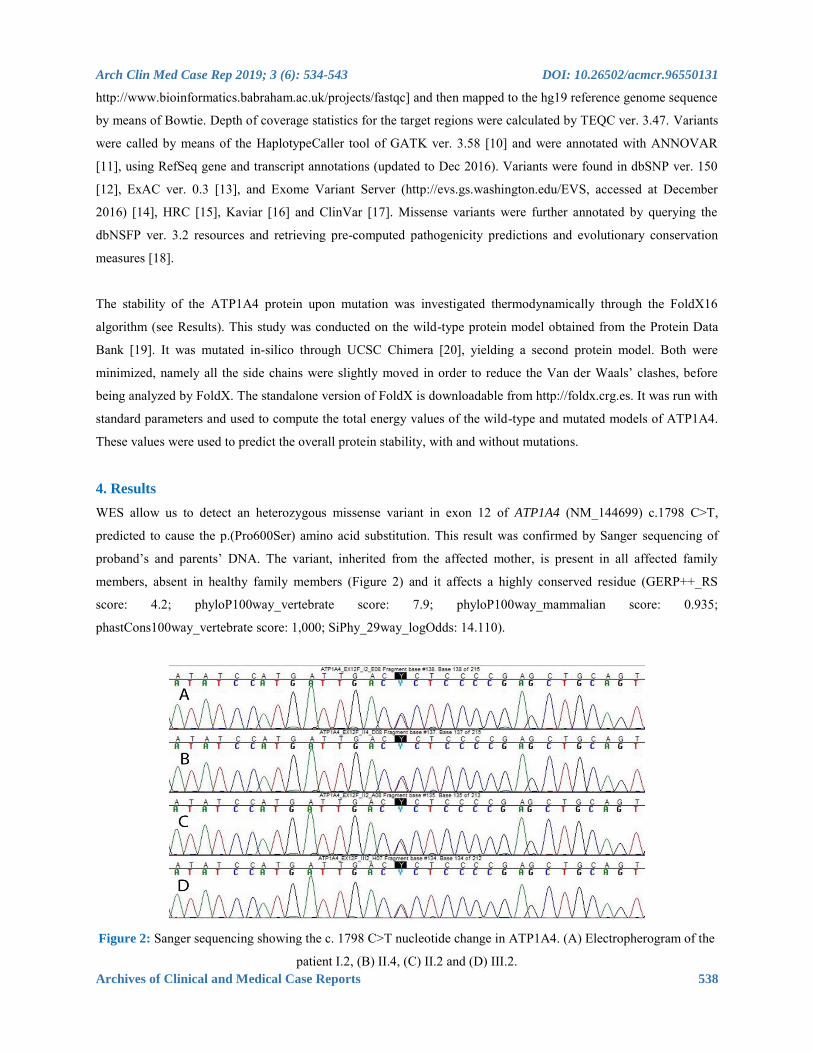
WES allow us to detect an heterozygous missense variant in exon 12 of ATP1A4 (NM_144699) c.1798 C>T,
predicted to cause the p.(Pro600Ser) amino acid substitution. This result was confirmed by Sanger sequencing of
proband’s and parents’ DNA. The variant, inherited from the affected mother, is present in all affected family
members, absent in healthy family members (Figure 2) and it affects a highly conserved residue (GERP++_RS
score: 4.2; phyloP100way_vertebrate score: 7.9; phyloP100way_mammalian score: 0.935;
phastCons100way_vertebrate score: 1,000; SiPhy_29way_logOdds: 14.110).
Figure 2: Sanger sequencing showing the c. 1798 C>T nucleotide change in ATP1A4. (A) Electropherogram of the
patient I.2, (B) II.4, (C) II.2 and (D) III.2.

Arch Clin Med Case Rep 2019; 3 (6): 534-543 DOI: 10.26502/acmcr.96550131
Archives of Clinical and Medical Case Reports 539
Furthermore, the variant is located in the ATP1A4 topological domain and it is predicted to be harmful by several
pathogenicity predictor software, including SIFT, PolyPhen2, LRT, CADD, DANN, FatHmm. The variant has been
reported in dbSNP (rs142338502), and is quite rare (MAF of 0.001 reported in genomAD and 1000Genomes, 0.002
in ExAC). The free energy calculations for this variant were ΔGmut=44.88 kcal/mol and ΔGwt=42.79 kcal/mol,
from which ΔΔG = ΔGmut - ΔGwt = +2.09 kcal/mol. The difference in free energy resulted to be positive and in the
range to classify the p.(Pro600Ser) variant as highly destabilizing. The localization of the variant is depicted in
Figure 3.
Figure 3: Location of the Pro600Ser substitution in the ATP1A4 protein / Ribbon diagram of ATP1A4, with
Pro600Ser colored in green.
5. Discussion
In recent years, increasing evidences showed that HM tends to run in families, suggesting that genetic determinants
play a significant role in the disease. It has been shown that at least 50% of migraineurs have a parent affected by a
similar condition and a familial liability has been confirmed in several studies [21]. Although familial does not
necessarily mean genetic, epidemiological evidence seems to indicate a close gene–environment interaction. To
date, four genes, two encoding for ion channels and two encoding an ATP excharger, have been found to be
responsible for several forms of HM [22, 23]. CACNA1A (OMIM # 601011) was the first gene associated to FHM,
and to date more than 70 causative variants have been identified. CACNA1A encodes the α1 subunit of neuronal
CaV2.1 (P/ Q-type) voltage-gated calcium channels that are widely expressed throughout the central nervous system
[24]. Variants in this channel causes excessive release of the neurotransmitter glutamate, thus altering physiologic
conditions [25]. The second gene encoding for a ion channel is SCN1A (OMIM # 182389), which encodes the α1
subunit of neuronal NaV1.1 voltage-gated sodium channels and is a well-known epilepsy gene, with more than 100
truncating and missense variants associated with epilepsy [26]. The first gene encoding an ATP exchanger and
associated to HM is ATP1A2 (OMIM # 182340), which encodes the α2 subunit of sodium-potassium pump. To date
more than 40 variants involving ATP1A2 are known to cause FHM both without other clinical manifestation (pure
FHM) [6] or in addition to other symptoms such as AHC [27], epilepsy [28], intellectual disability [29]. ATP1A2
have also been associated with non-hemiplegic migraine and common migraine [30], providing a strong evidence of
the wide genetic and clinical heterogeneity of these conditions. The second gene encoding an ATP excharger and

Arch Clin Med Case Rep 2019; 3 (6): 534-543 DOI: 10.26502/acmcr.96550131
Archives of Clinical and Medical Case Reports 540
associated, as ATP1A2, to different neurological conditions such as AHC, rapid-onset Dystonia Parkinsonism (RDP)
and CAPOS syndrome (cerebellar ataxia, areflexia, pes cavus, optic atrophy, and sensorineural hearing loss), is
ATP1A3 (OMIM #182350) which encodes the α3 subunit of sodium-potassium pump [12]. We describe the first
Italian family to date reported in medical literature, with four members affected by FHM in which a novel putative
pathogenic variant in ATP1A4 gene has been identified and segregates in all affected members. To the best of our
knowledge, this is the first time the ATP1A4 gene has been associated to this clinical condition, and for this reason
useful to expand our knowledge regarding the molecular mechanisms underlying HM.
The protein encoded by ATP1A4 gene belongs to the family of P-type cation transport ATPases, and to the
subfamily of Na+/K+ -ATPases. Na+/K+ -ATPase is an integral membrane protein responsible for establishing and
maintaining the electrochemical gradients of Na and K ions across the plasma membrane, by the active transport of
three sodium ions for two potassium ions across cell membranes [31]. These gradients are essential for
osmoregulation, for sodium-coupled transport of a variety of organic and inorganic molecules, and for the electrical
excitability of nerve and muscle. This enzyme is composed of two subunits, a large catalytic subunit (alpha) and a
smaller glycoprotein subunit (beta). The catalytic subunit (alpha) of Na+/K+ -ATPase is encoded by multiple genes,
and ATP1A4gene encodes an α4 subunit, highly expressed in testis, but also in the brain and skeletal muscle [32],
showing the properties to associate different beta subunits in different tissues [33]. Although, to date, ATP1A4 was
never associated to neurological condition, it encodes for one of the component of the catalytic subunit of the
Na+/K+ -ATPase, as ATP1A2 and ATP1A3 genes, already known to be causative of FHM, AHC and other
neurologic conditions. Thus, due to these functional evidences, the contribution of ATP1A4 alteration in the onset of
HM could be likely. In the family described here, composed by eight people among three generations, all affected
members (four) carries the variation in ATP1A4. Even if the detected variant presents a MAF higher than expected
for a monogenic disorder, this evidence is not uncommon for conditions which did not affect reproductive fitness of
carriers people, such as for example in hypertrophic cardiomyopathy.
From a clinical point of view, they presented paresthesias at the right side and an increasing frequency of HM during
the years. The age of onset is similar for II2 (6 years old), II4 (12 years old), III2 (9 years old), such as the duration
of 1.5-2 hours, and for all patients headache decrease with Ibuprofen/Ketoprofen assumption. Other clinical
manifestations are quite different, such as flashing lights in the right eye (II4) and central flashing scotomas (III2),
but this is a frequent evidence in pathologies with high clinical heterogeneity. Understanding the primary brain
dysfunction involved in hemiplegic migraine is essential in order to develop more specific and effective drugs for
the control of clinical manifestation, and to identify new drug targets, so increasing knowledge of genetic
contribution to these diseases is crucial.
6. Conclusions In conclusion, we described for the first time a putative pathogenic variant of ATP1A4, identified in a family
affected of FHM. We suggest, in order to increase the chance to identify the molecular cause of the disease, to

Arch Clin Med Case Rep 2019; 3 (6): 534-543 DOI: 10.26502/acmcr.96550131
Archives of Clinical and Medical Case Reports 541
screen the sequence of ATP1A4 in addition to CACNA1A, SCN1A, ATP1A2 and ATP1A3in patients affected by HM,
or to sequence ATP1A4 in all patients negative to previous genetic screening of other candidate genes.
Acknowledgments We thank the family for their cooperation.
Conflict of Interest The authors declare that the research was conducted in the absence of any commercial or financial relationships that
could be construed as a potential conflict of interest.
Author Contributions
PP, OP, MC and GC conceived the study, designed experiments and wrote the manuscript. GC, FFO and GP
collected the patients’ clinical information. SC, TB and TM performed the analysis of TRS data and modeling the
impact of the reported variant on the three-dimensional structure of the protein. EDM and MPL performed the
molecular analysis. PP, OP, EDM and MPL interpreted the results. All authors reviewed and approved the final
manuscript.
Funding
This study was supported by a grant of the Italian Ministry of Health (Ricerca Corrente 2019) to Massimo Carella.
Consent for Publication This study was carried out following the recommendations of the Declaration of Helsinki and the Italian law for
biomedical experimentation with written informed consent from all subjects.
References
1. Headache Classification Committee of the International Headache Society (IHS). The international
classification of headache disorders, 3rd edition (beta version). Cephalalgia 38 (2018) 1-211.
2. Russell MB, Ducros A. Sporadic and familial hemiplegic migraine: pathophysiological mechanisms,
clinical characteristics, diagnosis, and management. Lancet Neurol 10 (2011): 457-470.
3. Panagiotakaki E, De Grandis E, Stagnaro M, et al. Clinical profile of patients with ATP1A3 mutations in
Alternating Hemiplegia of Childhood-a study of 155 patients. Orphanet J Rare Dis. 10 (2015): 123.
4. Zarcone D, Corbetta S. Shared mechanisms of epilepsy, migraine and affective disorders. Neurol Sci 38
(2017): 73-76.
5. Ophoff RA, Terwindt GM, Vergouwe MN, et al. Familial hemiplegic migraine and episodic ataxia type-2
are caused by mutations in the Ca2+ channel gene CACNL1A4. Cell 87 (1996): 543-552.
6. De Fusco M, Marconi R, Silvestri L, et al. Haploinsufficiency of ATP1A2 encoding the Na+/K+ pump
alpha2 subunit associated with familial hemiplegic migraine type 2. Nat Genet 33 (2003): 192-196.

Arch Clin Med Case Rep 2019; 3 (6): 534-543 DOI: 10.26502/acmcr.96550131
Archives of Clinical and Medical Case Reports 542
7. Dichgans M, Freilinger T, Eckstein G, et al. Mutation in the neuronal voltage-gated sodium channel
SCN1A in familial hemiplegic migraine. Lancet 366 (2005): 371-377.
8. Carecchio M, Zorzi G, Ragona F, et al. ATP1A3-related disorders: An update. Eur J Paediatr Neurol 22
(2017): 257-263.
9. Ferrari MD, Klever RR, Terwindt GM, et al. Migraine pathophysiology: lessons from mouse models and
human genetics. Lancet Neurol 14 (2015): 65-80.
10. McKenna A, Hanna M, Banks E, et al. The Genome Analysis Toolkit: a MapReduce framework for
analyzing next-generation DNA sequencing data. Genome Res 20 (2010): 1297-1303.
11. Wang K, Li M, Hakonarson H. ANNOVAR: Functional annotation of genetic variants from next-
generation sequencing data. Nucleic Acids Res 38 (2010): e164.
12. Sherry ST, Ward MH, Kholodov M, et al. dbSNP: the NCBI database of genetic variation. Nucleic Acids
Res 29 (2001): 308-311.
13. Exome Aggregation Consortium. Analysis of protein-coding genetic variation in 60,706 humans. BioRxiv
(2015). doi: http://dx.doi.org/10.1101/030338
14. Exome Variant Server, NHLBI GO Exome Sequencing Project (ESP), Seattle, WA (URL:
http://evs.gs.washington.edu/EVS/)
15. McCarthy S, Das S, Kretzschmar W, et al. Haplotype Reference Consortium. A reference panel of 64,976
haplotypes for genotype imputation. Nat Genet 48 (2016): 1279-1283.
16. Glusman G, Caballero J, Mauldin DE, et al. KAVIAR: an accessible system for testing SNV novelty.
Bioinformatics 27 (2011): 3216-3217.
17. Landrum MJ, Lee JM, Riley GR, et al. ClinVar: public archive of relationships among sequence variation
and human phenotype. Nucleic Acids Res 42 (2014): D980-D985.
18. Liu X, Wu C, Li C, et al. dbNSFP v3.0: A One-Stop Database of Functional Predictions and Annotations
for Human Non-synonymous and Splice Site SNVs. Hum Mutat 37 (2016): 235-241.
19. Berman HM, Battistuz T, Bhat TN, et al. The Protein Data Bank. Acta Crystallografica D58 (2002): 899-
907.
20. Pettersen EF, Goddard TD, Huang CC, et al. UCSF Chimera, A Visualization System for Exploratory
Research and Analysis. J Comput Chem 25 (2004): 1605-1612.
21. Schu¨rks M. Genetics of migraine in the age of genome wide association studies. J Headache Pain 13
(2012): 1-9.
22. Hiekkala ME, Vuola P, Artto V, et al. The contribution of CACNA1A, ATP1A2 and SCN1A mutations in
hemiplegic migraine: A clinical and genetic study in Finnish migraine families. Cephalalgia 38 (2018):
1849-1863.
23. Carecchio M, Zorzi G, Ragona F, et al. ATP1A3-related disorders: An update. Eur J Paediatr Neurol 22
(2018): 257-263.
24. Westenbroek RE, Sakurai T, Elliott EM, et al. Immunochemical identification and subcellular distribution
of the alpha 1A subunits of brain calcium channels. J Neurosci 15 (1995): 6403-6418.

Arch Clin Med Case Rep 2019; 3 (6): 534-543 DOI: 10.26502/acmcr.96550131
Archives of Clinical and Medical Case Reports 543
25. van den Maagdenberg AM, Pietrobon D, Pizzorusso T, et al. A Cacna1a knockin migraine mouse model
with increased susceptibility to cortical spreading depression. Neuron 41 (2004): 701-710.
26. Vahedi K, Depienne C, Le Fort D, et al. Elicited repetitive daily blindness: a new phenotype associated
with hemiplegic migraine and SCN1A mutations. Neurology 72 (2009): 1178-1183.
27. Swoboda KJ, Kanavakis E, Xaidara A, et al. Alternating hemiplegia of childhood or familial hemiplegic
migraine? A novel ATP1A2 mutation. Ann Neurol 55 (2004): 884-887.
28. Jurkat-Rott K, Freilinger T, Dreier JP, et al. Variability of familial hemiplegic migraine with novel A1A2
Na(+)/K(+)-ATPase variants. Neurology 62 (2004): 1857-1861.
29. Vanmolkot KR, Stroink H, Koenderink JB, et al. Severe episodic neurological deficits and permanent
mental retardation in a child with a novel FHM2 ATP1A2 mutation. Ann Neurol 59 (2006): 310-314.
30. Todt U, Dichgans M, Jurkat-Rott K, et al. Rare missense variants in ATP1A2 in families with clustering of
common forms of migraine. Hum Mutat 26 (2005): 315-321.
31. Woo AL, James PF, Lingrel JB. Characterization of the fourth alpha isoform of the Na,K-ATPase. J
Membr Biol 169 (1999): 39-44.
32. Lingrel J, Moseley A, Dostanic I, et al. Functional roles of the alpha isoforms of the Na,K-ATPase. Ann N
Y Acad Sci 986 (2003): 354-359.
33. Sanchez G, Nguyen AN, Timmerberg B, et al. The Na,K-ATPase alpha4 isoform from humans has distinct
enzymatic properties and is important for sperm motility. Mol Hum Reprod 12 (2006): 565-576.
This article is an open access article distributed under the terms and conditions of the
Creative Commons Attribution (CC-BY) license 4.0
Citation: Palumbo P, Palumbo O, Operto FF, Castellana S, Muro ED, Leone MP, Biagini T, Pastorino G,
Mazza T, Carella M, Coppola G. Familial Hemiplegic Migraine: A New Gene in an Italian Family.
Archives of Clinical and Medical Case Reports 3 (2019): 534-543.
Copyright © 2022 FDOKUMEN