Simultaneous retinal and cortical visually evoked electrophysiological responses in between migraine...
12
Original Article Simultaneous retinal and cortical visually evoked electrophysiological responses in between migraine attacks Bao N Nguyen, Allison M McKendrick and Algis J Vingrys Abstract Purpose: People with migraine often report aversion to flickering lights and show abnormal results on behavioural tasks that require the processing of temporal visual information. Studies have reported that the cortically evoked electro- physiological response to a flickering visual stimulus is abnormal; however, none have considered whether there is an underlying pre-cortical abnormality. In this cross-sectional study, we consider whether people with migraine have retinal and cortical electrophysiological abnormalities to flickering stimuli. Methods: Monocular transient (1 Hz) and steady-state (8.3 Hz) pattern reversal electroretinograms (PERGs) and pattern visual evoked responses (PVERs) were measured simultaneously in 45 people with migraine (26 without aura, 19 with aura) and 30 non-headache controls at a time between migraine attacks. Results: PERG amplitude and timing did not differ significantly between groups. Transient PVER amplitude was significantly reduced (28%) in the migraine with aura group compared to the controls F(2,72) ¼ 3.6, p ¼ 0.03). Both migraine groups showed significant reductions (32%, 39%) in steady-state PVER amplitude relative to controls (F(2,70) ¼ 4.3, p ¼ 0.02). Conclusions: This study finds normal retinal processing of flickering stimuli in the presence of abnormal cortical function between migraine attacks. Keywords Migraine, temporal processing, contrast processing, visual evoked potentials, electroretinogram Date received: 2 April 2012; revised: 30 May 2012; accepted: 14 June 2012 Introduction Most studies of visual processing in people with migraine focus on cortical processing, as migraine is a disorder of the central nervous system (1). Abnormal performance on cortically processed behavioural tasks has been well documented between migraine attacks (2–8). Such tasks rely on normal contrast processing, which begins in the retina, then the lateral geniculate nucleus, before further processing in the primary visual cortex (V1) (9). It is important to ensure that differences attributed to cortical processing do not reflect reduced inputs from earlier, pre-cortical stages of the visual pathway. Several lines of research suggest non-cortical dys- function in migraine. Between attacks, people with migraine show abnormal performance on behavioural tasks designed to assess pre-cortical function (8,10–12). Visual field deficits associated with migraine can be non-homonymous and unilateral (13–19) and some- times described as ‘glaucomatous’ because of their loca- lised and arcuate pattern (13,17). It has been suggested that such deficits may arise from peripheral vascular causes (17,20), with indirect evidence being an associ- ation between migraine and peripheral vasospastic ten- dencies (20), including transient retinal blood flow changes (21). Moreover, migraine is a recognised risk Department of Optometry and Vision Sciences, The University of Melbourne, Australia Corresponding author: Allison M McKendrick, Department of Optometry and Vision Sciences, The University of Melbourne, Parkville VIC 3010 Australia. Email: [email protected] Cephalalgia 0(0) 1–12 ! International Headache Society 2012 Reprints and permissions: sagepub.co.uk/journalsPermissions.nav DOI: 10.1177/0333102412453953 cep.sagepub.com
-
Upload
independent -
Category
Documents
-
view
1 -
download
0
Transcript of Simultaneous retinal and cortical visually evoked electrophysiological responses in between migraine...
Original Article
Simultaneous retinal and cortical visuallyevoked electrophysiological responses inbetween migraine attacks
Bao N Nguyen, Allison M McKendrick and Algis J Vingrys
Abstract
Purpose: People with migraine often report aversion to flickering lights and show abnormal results on behavioural tasks
that require the processing of temporal visual information. Studies have reported that the cortically evoked electro-
physiological response to a flickering visual stimulus is abnormal; however, none have considered whether there is an
underlying pre-cortical abnormality. In this cross-sectional study, we consider whether people with migraine have retinal
and cortical electrophysiological abnormalities to flickering stimuli.
Methods: Monocular transient (1 Hz) and steady-state (8.3 Hz) pattern reversal electroretinograms (PERGs) and pattern
visual evoked responses (PVERs) were measured simultaneously in 45 people with migraine (26 without aura, 19 with
aura) and 30 non-headache controls at a time between migraine attacks.
Results: PERG amplitude and timing did not differ significantly between groups. Transient PVER amplitude was significantly
reduced (28%) in the migraine with aura group compared to the controls F(2,72)¼ 3.6, p¼ 0.03). Both migraine groups
showed significant reductions (32%, 39%) in steady-state PVER amplitude relative to controls (F(2,70)¼ 4.3, p¼ 0.02).
Conclusions: This study finds normal retinal processing of flickering stimuli in the presence of abnormal cortical function
between migraine attacks.
Keywords
Migraine, temporal processing, contrast processing, visual evoked potentials, electroretinogram
Date received: 2 April 2012; revised: 30 May 2012; accepted: 14 June 2012
Introduction
Most studies of visual processing in people withmigraine focus on cortical processing, as migraine is adisorder of the central nervous system (1). Abnormalperformance on cortically processed behavioural taskshas been well documented between migraine attacks(2–8). Such tasks rely on normal contrast processing,which begins in the retina, then the lateral geniculatenucleus, before further processing in the primary visualcortex (V1) (9). It is important to ensure that differencesattributed to cortical processing do not reflect reducedinputs from earlier, pre-cortical stages of the visualpathway.
Several lines of research suggest non-cortical dys-function in migraine. Between attacks, people withmigraine show abnormal performance on behaviouraltasks designed to assess pre-cortical function (8,10–12).
Visual field deficits associated with migraine can benon-homonymous and unilateral (13–19) and some-times described as ‘glaucomatous’ because of their loca-lised and arcuate pattern (13,17). It has been suggestedthat such deficits may arise from peripheral vascularcauses (17,20), with indirect evidence being an associ-ation between migraine and peripheral vasospastic ten-dencies (20), including transient retinal blood flowchanges (21). Moreover, migraine is a recognised risk
Department of Optometry and Vision Sciences, The University of
Melbourne, Australia
Corresponding author:
Allison M McKendrick, Department of Optometry and Vision Sciences,
The University of Melbourne, Parkville VIC 3010 Australia.
Email: [email protected]
Cephalalgia
0(0) 1–12
! International Headache Society 2012
Reprints and permissions:
sagepub.co.uk/journalsPermissions.nav
DOI: 10.1177/0333102412453953
cep.sagepub.com

factor for visual field progression in normal tensionglaucoma (22) – a form of glaucoma associated witha vascular aetiology (20) – and has been associatedwith retinal nerve fibre layer loss (23).
One method of comparing retinal to cortical func-tion in the same individual is by concurrent electro-physiological measurement of neuronal activity in theeye and brain. Pattern electrophysiology invokes activ-ity of contrast-dependent neurones. The pattern elec-troretinogram (PERG) assesses the integrity of retinalganglion cells, whereas the function of the visual path-way, including V1, can be assessed with the patternvisual evoked response (PVER) (24). The patternedstimulus can be contrast-reversed at different temporalfrequencies. The standard electrophysiological responseis a transient response (<3Hz), where the response iscomplete before the next contrast reversal. Transientrecording allows visualisation of a single waveformcharacterised by identifiable components. At highertemporal frequencies (�4Hz), the successive waveformsoverlap and a steady-state response is generated.Steady-state electrophysiological recording is presumedto measure similar mechanisms as behavioural meas-ures of temporal processing (flicker). Flickering andmoving stimuli have consistently identified differencesbetween migraine and non-headache groups, (2,3,5–8,11,14,19,25,26) including tests of pre-cortical function(10,16).
Previous electrophysiological studies during theinterictal period in migraine have yielded varying out-comes (27,28). Many studies report anomalous PVERresults, but most do not consider whether the retina isnormal. Only two studies have noted that the PERGwas normal in the presence of abnormal cortical func-tion, both measured using transient stimulation (29,30).However, despite studies that have found abnormalcortical responses to steady-state stimulation (31–33),the steady-state retinal response has not previouslybeen measured in migraine.
The purpose of this study was to measure both tran-sient and steady-state retinal and cortical responses inthe same individuals with migraine on the same day.This will bring together a range of techniques that havebeen used previously in isolation to build up a morecomplete picture of how the electrophysiologicalresponse is different between migraine attacks. Wehypothesise that a concomitant retinal and corticalabnormality would suggest that the cortical involve-ment arises, at least partly, downstream from retinaldysfunction. To our knowledge, this is the first studyto measure steady-state and transient electrophysio-logical responses at the retina and cortex simultan-eously, which will reveal whether the corticalabnormalities are explained by aberrant function earlierin the visual pathway.
Methods
Participants
Participants in this cross-sectional study were recruitedvia an advertisement within the University ofMelbourne community, as well as from a database ofprevious study participants, from June 2010 toSeptember 2011. Thirty non-headache controls (aged19–46 years, mean � standard deviation [SD]: 26� 7),26 people with migraine without aura (MO, aged 20–41years, mean� SD: 28� 6) and 19 people with migrainewith aura (MA, aged 19–43 years, mean� SD: 33� 6)were tested at the Department of Optometry and VisionSciences at the University of Melbourne. On average,the MA group was older than the control and MOgroups (F(2,72)¼ 6.88, p¼ 0.002). The proportion ofmales to females was not significantly different betweeneach group (controls 9:21, MO: 4:22, MA: 5:14).
All participants had best corrected visual acuity of6/7.5 or better and refractive errors of less than �5.00D sphere and �2.00 D astigmatism. Participants werefree from systemic disease and medications known toaffect visual function or neurological state, includingprophylactic migraine medications, and had normalfindings in a comprehensive eye examination (slitlampbiomicroscopy, applanation tonometry, ophthalmos-copy and optic nerve head imaging). All participantswere classified as within normal limits using theMoorfield’s regression analysis (MRA) tool of theHeidelberg retinal tomograph (Heidelberg Engineering,Heidelberg, Germany). TheMRA has excellent specificity(>85%) for cross-sectional diagnoses of open-angle glau-coma (34).
Control participants had never experienced amigraine and were free from regular headaches (lessthan four in the past year). All migraine participantsmet the criteria of the International Headache Societyfor either MA or MO (35) and were tested at least sevendays after a migraine attack to minimise residual effectsof medications and the possible influence of transientpost-migraine fatigue or nausea. Participants completeda custom headache questionnaire and the MigraineDisability Assessment Score (MIDAS) to determine avariety of headache characteristics, summarised inFigure 1. The MIDAS questionnaire is a validated toolthat scores the impact of headaches on tasks of dailyliving over the past three months (36). MIDAS scoresare interpreted as minimal (grade 1, score 0–5), mild(grade 2, score 6–10), moderate (grade 3, score 11–20)or severe disability (grade 4, score 21þ). Six of the 30control participants (20%) reported that only one familymember suffered from migraines (e.g. mother, sister,grandmother), whereas 33 of the 45 participants withmigraine (73%) had at least one other family member
2 Cephalalgia 0(0)
who suffered frommigraines (e.g. parent, sibling, grand-parent, aunt, cousin).
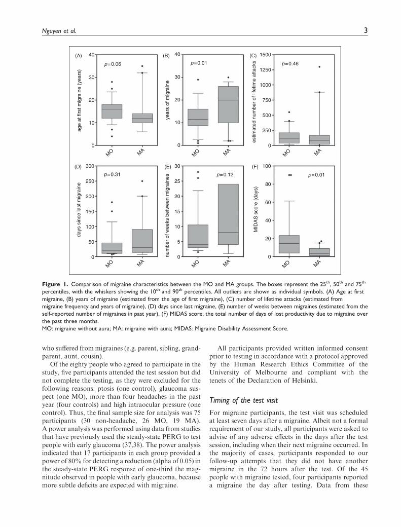
Of the eighty people who agreed to participate in thestudy, five participants attended the test session but didnot complete the testing, as they were excluded for thefollowing reasons: ptosis (one control), glaucoma sus-pect (one MO), more than four headaches in the pastyear (four controls) and high intraocular pressure (onecontrol). Thus, the final sample size for analysis was 75participants (30 non-headache, 26 MO, 19 MA).A power analysis was performed using data from studiesthat have previously used the steady-state PERG to testpeople with early glaucoma (37,38). The power analysisindicated that 17 participants in each group provided apower of 80% for detecting a reduction (alpha of 0.05) inthe steady-state PERG response of one-third the mag-nitude observed in people with early glaucoma, becausemore subtle deficits are expected with migraine.
All participants provided written informed consentprior to testing in accordance with a protocol approvedby the Human Research Ethics Committee of theUniversity of Melbourne and compliant with thetenets of the Declaration of Helsinki.
Timing of the test visit
For migraine participants, the test visit was scheduledat least seven days after a migraine. Albeit not a formalrequirement of our study, all participants were asked toadvise of any adverse effects in the days after the testsession, including when their next migraine occurred. Inthe majority of cases, participants responded to ourfollow-up attempts that they did not have anothermigraine in the 72 hours after the test. Of the 45people with migraine tested, four participants reporteda migraine the day after testing. Data from these
40(A) (B) (C)
(D) (E) (F)
30
20
10
age
at fi
rst m
igra
ine
(yea
rs)
year
s of
mig
rain
e
estim
ated
num
ber
of li
fetim
e at
tack
sM
IDA
S s
core
(da
ys)
num
ber
of w
eeks
bet
wee
n m
igra
ines
days
sin
ce la
st m
igra
ine
0
40
30
20
10
0
1250
1500
1000
500
750
250
p=0.46p=0.01
p=0.01p=0.12p=0.31
p=0.06
0
100
80
40
60
20
0
25
30
20
10
15
5
0
250
300
200
100
150
50
0
MO MA
MO MA
MO MA
MO MA
MO MA
MO MA
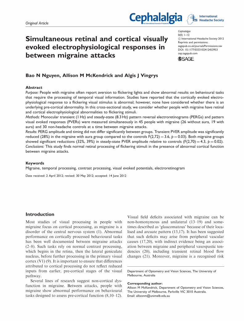
Figure 1. Comparison of migraine characteristics between the MO and MA groups. The boxes represent the 25th, 50th and 75th
percentiles, with the whiskers showing the 10th and 90th percentiles. All outliers are shown as individual symbols. (A) Age at first
migraine, (B) years of migraine (estimated from the age of first migraine), (C) number of lifetime attacks (estimated from
migraine frequency and years of migraine), (D) days since last migraine, (E) number of weeks between migraines (estimated from the
self-reported number of migraines in past year), (F) MIDAS score, the total number of days of lost productivity due to migraine over
the past three months.
MO: migraine without aura; MA: migraine with aura; MIDAS: Migraine Disability Assessment Score.
Nguyen et al. 3

participants are represented as unfilled symbols (seeFigure 4).
Pattern electrophysiology
The PERG and PVER were recorded simultaneouslyfrom each eye according to ISCEV (InternationalSociety for Clinical Electrophysiology of Vision) stand-ards (39,40) using the Espion system (Diagnosys LLC,Cambridge, UK). A black-and-white square-wavecheckerboard stimulus was presented on a gamma-cor-rected Sony G520 21-inch CRT monitor (100Hz,1024� 768 pixels). The checkerboard subtended 31�
at a viewing distance of 50 cm, and consisted of 0.8�
checks of 96% contrast and mean luminance of52 cd/m2. The contrast of the checkerboard was coun-terphased at 1Hz (transient) and 8.3Hz (steady-state).In addition, the transient response to 0.25� checks wasmeasured to confirm the absence of optical problemsthat may affect the response to finer checks (39). Thesteady-state response to 16� checks was recorded toenable determination of the PERG ratio (41)—a sensi-tive measure of PERG dysfunction. The different spa-tiotemporal frequencies were presented in randomorder and counterbalanced between participants.
A 0.5�-diameter red square provided a central fixationtarget.
PERG responses were recorded with corneal DTLelectrodes near the lower limbus and referenced to theipsilateral canthus with silver-silver chloride electrodes.The PVER was recorded with gold cup electrodes at Oz,OR and OL, and referenced to Fz with the commonground at Cz (Figure 2). Electrode impedance waskept below 5 k�. Signals were amplified, bandpass-fil-tered (1.25–100Hz) and digitised (1000Hz) to 16-bitresolution. Blink artefacts where signals exceeded�100 mV were automatically rejected. As people withmigraine can find high-contrast patterns aversive (42),each stimulus presentation was limited to approxi-mately 20 seconds, corresponding to 25 sweeps, fol-lowed by a brief break. A total of 200 signals wereaveraged for each stimulus condition of differing spa-tiotemporal frequency. To confirm intrasessional repro-ducibility, we computed an index of responseconsistency (39,40), the coefficient of variation (COV)between two consecutive partial averages of 100sweeps. Typically, the COV of amplitude for thePERG (transient 9.1%, steady-state 8.1%) and PVER(transient 10.7%, steady-state 9.8%) were comparableto those reported in the literature (data not shown)
inactive PVERelectrode
inactive PERGelectrode
active PERG electrode
20%
30%
40%
10%nasion
inion
groundelectrode
active PVERelectrode
Fz
Oz
Cz
OL
10%
(A)
(B) OZ OR
Figure 2. Electrode locations for simultaneous standard PVER/PERG recording. (A) For the PVER, scalp electrodes are placed in
proportion to the size of the head in accordance with the International 10-20 system, which divides the distance between the two
bony landmarks, the nasion and inion, into 10% and 20% proportions. The active PVER electrode is located along the vertical midline at
OZ and the inactive reference electrode at FZ. For the PERG, the active electrode is a DTL carbon-fibre electrode placed along the
lower eyelid margin. The inactive PERG electrode is a skin electrode placed at the cheekbone (left eye shown only). The common
ground electrode is placed at CZ, midway between the nasion and inion on the midline. (B) The right and left lateral PVER electrodes
are placed at OR and OL, respectively.
PVER: pattern visual evoked response; PERG: pattern reversal electroretinogram.
4 Cephalalgia 0(0)

(43,44). A typical recording session lasted approxi-mately 35 minutes.
Analysis was performed offline using MicrosoftExcel (Microsoft, Redmond, WA, USA). Peak-to-peak amplitudes and peak times for the transientPERG P50 and N95 and PVER P100 and N135were measured according to ISCEV standards(39,40). Retinocortical time was calculated as theinterlatency period from the PERG P50 to PVERP100, as a measure of signal transmission timebetween retina and cortex (45). Steady-state wave-forms were analysed in the frequency domain afterdiscrete Fourier transformation of resampled wave-forms into a binary series (Figure 3). Because everychange from black to white represents a single changein contrast, the largest contrast-evoked response is at
the contrast-reversal frequency, which occurs at twicethe stimulation frequency (second harmonic 2F,16.7Hz). Steady-state responses were excluded if the2F amplitude was not significantly different from noise(p> 0.05), where noise was estimated from the averageamplitude at the two neighbouring frequencies (14.6and 18.8Hz) (46). Phase was calculated from the arc-tangent of the ratio of the real-to-imaginary compo-nents of the discrete Fourier analysis. Because therecan be an infinite set of phases separated by 2pradians with the same arctangent, the value modulowas returned (p rads). The phase convention was suchthat increasing phase corresponded to a delay in thesignal. PVER interhemispheric asymmetry was definedas the absolute percentage amplitude differencebetween the response at OR and OL (29) (Figure 2).
Transient PVER(A)
(B) (C)
P100 amplitude
Transient PERG Steady state
DFT
31°
B
50 cm
A
P50 amplitude
N135 amplitude
N95 amplitude
2F amplitude
N75
N35
0 50 100 150 200 250
0 50 100 150 200 250
Time (ms) Frequency (Hz)
0 10 20 30 40 50 60 70
Figure 3. Schematic of PERG/PVER recording setup and representative waveforms. (A) The transient PVER waveform is charac-
terised by a prominent positive peak at 100 ms (vertical dotted line). The P100 amplitude is measured as the peak-to-peak distance
from the first negative trough at 75 ms (N75) to the top of the positive peak. The N135 amplitude is taken from the P100 to the
second negative trough (vertical dashed line). (B) The transient PERG waveform is characterised by a prominent positive peak at 50 ms
(P50, vertical dotted line) and a negative trough (N95, vertical dashed line). Peak-to-peak P50 and N95 amplitudes are taken from N35
to P50, and P50 to N95, respectively. (C) Steady-state waveforms are resampled to give 512 data points and converted into the
frequency domain by discrete Fourier transformation in order to measure the amplitude at the second harmonic (2 F). Noise is
derived from the average amplitudes at the two neighbouring frequencies, 14.6 and 18.8 Hz (vertical dotted lines).
PVER: pattern visual evoked response; PERG: pattern reversal electroretinogram.
Nguyen et al. 5

A PERG ratio was computed by dividing the steady-state amplitudes to 0.8� and 16� checks (41).
Statistical analysis
Statistical analysis was performed using SPSS version17.0 (SPSS Inc., Chicago, IL, USA). Age was entered asa covariate in all the analyses. Comparisons betweencontrol and migraine groups were performed using arepeated measures analysis of co-variance (RM-ANCOVA). Comparisons between migraine groupswere performed using student t-tests or non-parametricMann-Whitney rank sum tests where the data was non-Gaussian (Kolmogorov-Smirnov test). A p< 0.05 wasconsidered significant for all statistical evaluations. Asthere was no difference between the left and right eyesfor any measures, the right eye response is representedin all figures.
Results
Migraine characteristics
Figure 1 shows boxplots of the migraine characteristics.The MA group had a longer duration of migraine his-tory (Mann-Whitney rank sum test, p¼ 0.01), whereasthe MO group reported significantly greater impact oftheir migraines on daily activities, as reflected in thehigher MIDAS questionnaire scores (Mann-Whitneyrank sum test, p¼ 0.01).
Was there evidence for retinal dysfunction inpeople with migraine?
Table 1 is a summary of the PERG findings. An RM-ANCOVA (within factors: eye and component;between factor: group) was performed separately for
30
25
20
15
10
5
0
12
10
8
6
4
2
0control
Transient PVER P100 amplitude Steady-state PVER amplitude
p=0.02p=0.02
p=0.03
(A) (B)
p=0.01
MO MA
PV
ER
am
plitu
de (
mV)
PV
ER
am
plitu
de (
mV)
control MO MA
Figure 4. Distributions of the (A) transient PVER P100 amplitude and (B) steady-state PVER 2F amplitude in the control, MO and
MA groups. The four participants who happened to be tested the day before a migraine are shown as unfilled symbols. Error bars
indicate the mean� SEM.
PVER: pattern visual evoked response; SEM: standard error of the mean.
Table 1. Summary of PERG parameters (mean� SD).a
Parameter Controls MO MA p value
Transient P50 amplitude (mV) 6.57� 1.83 6.60� 2.08 7.17� 2.16 0.46
N95 amplitude (mV) 10.3� 2.52 10.6� 3.07 10.6� 2.75 0.81
P50 time (ms) 51� 2 51� 3 51� 2 0.26
N95 time (ms) 104� 5 105� 5 104� 5 0.37
Steady-state 2F amplitude (mV) 3.52� 0.818 3.25� 1.10 3.43� 1.02 0.66
2F phase (p rads) 1.4� 0.8 1.6� 0.6 1.4� 0.7 0.99
PERG ratio 1.2� 0.2 1.3� 0.4 1.3� 0.3 0.23
ap values represent the main effect of group (repeated measures analysis of co-variance [RM-ANCOVA]).
PERG: pattern reversal electroretinogram; SD: standard deviation; MO: migraine without aura; MA: migraine with aura.
6 Cephalalgia 0(0)

transient PERG amplitudes and peak times, where thecomponents of interest were the P50 and N95. Therewas no significant difference between the groups for anyof the transient measures (p> 0.05). For the 2F ampli-tude, phase and PERG ratio, separate RM-ANCOVA(within factor: eye; between factor: group) analysesshowed that the groups did not differ for any of thesteady-state measures (p> 0.05).
Was there evidence for cortical dysfunction inpeople with migraine?
Figure 4 shows the group mean amplitudes for the tran-sient PVER. An RM-ANCOVA (within factors: eyeand component; between factor: group) showed a sig-nificant difference between groups, where the compo-nents of interest were the P100 and N135(F(2,71)¼ 4.21, p¼ 0.02). Both the control and MOgroups had significantly higher P100 amplitudes thanthe MA group (post-hoc Bonferroni multiple compari-sons, p¼ 0.02), but there was no difference between thecontrol and MO groups. The difference between thegroups was more apparent for the P100 (significantinteraction between group and component:F(2,72)¼ 3.58, p¼ 0.03). To calculate the relative per-centage reduction in amplitude, each individual’s P100amplitude was normalised to the average control P100amplitude. The MA group amplitude was, on average,reduced by 28%. As a control for optical factors, wemeasured the transient PVER to smaller (0.25�) checksas per ISCEV standards (39), for which the P100 amp-litude did not differ between groups (data not shown).Table 2 is a summary of all of the timing measures,which did not differ between groups (RM-ANCOVAwithin factor: eye; between factor: group).
Figure 4 shows the group mean steady-state PVERamplitudes. The steady-state response of one MA par-ticipant was excluded from analysis, as the signal wasnot significantly different to noise. Relative to controls,the steady-state PVER amplitudes of the migrainegroups were reduced (RM-ANCOVA: within factor:eye; between factor: group; F(2,70)¼ 4.293, p¼ 0.02).
Both migraine groups were similarly affected (post-hocBonferroni multiple comparisons MO: p¼ 0.02, MA:p¼ 0.01). When normalised to the mean control 2Famplitude, the MO group and the MA group amplitudewere reduced, on average, by 32% and 39%, respect-ively. There was no significant difference in phasebetween groups (Table 2).
Because an increased interhemispheric asymmetry inPVER has been reported previously in some migrainestudies (29,47–50), we recorded the PVER at the rightand left hemispheres, and calculated an absolute amp-litude asymmetry. For the steady-state PVER, the lat-eral responses of three controls and four MAparticipants were excluded because at least oneresponse was not significantly different from noise.Separate RM-ANCOVA (within factor: eye; betweenfactor: groups) analyses showed that there was no dif-ference in percentage asymmetry for either transient(controls: 16.2� 11.4%; MO: 21.2� 15.5%; MA:22.8� 19.6%) or steady-state responses (controls:18.2� 13.2%; MO: 24.4� 17.3%; MA: 19.4� 9.3%).Given that the PVER amplitude can be altered in thepre-attack period (within 72 hours of a migraine) (51),we excluded the data from the four individuals whoreported a migraine the day after the test session(unfilled symbols in Figure 4). The results of the statis-tical analyses were unchanged.
Did the steady-state response reveal greaterdysfunction than the transient response?
To compare steady-state to transient responses betweengroups, we analysed the P100 and 2F PVER amplitudesin a combined RM-ANCOVA (within factors: eye andtest type; between factor: group), where test type wastransient or steady-state (Figure 4). The PVER ampli-tude differed between the groups (F(2,70)¼ 3.53,p¼ 0.04). Post-hoc Bonferroni multiple comparisonsrevealed that the PVER amplitudes of the MA groupwere reduced for both test types relative to the MO(p¼ 0.05) and control groups (p¼ 0.01), whereas thePVER amplitudes of the MO and control groups
Table 2. Summary of PVER timing parameters (mean� SD).a
Parameter Controls MO MA p value
Transient N75 time (ms) 73� 4 71� 5 71� 5 0.41
P100 time (ms) 102� 5 101� 4 101� 5 0.62
N135 time (ms) 146� 16 146� 8 145� 15 0.85
Retinocortical time (ms) 51� 4 51� 4 50� 5 0.91
Steady-state 2F phase (p rads) 0.7� 0.4 0.5� 0.2 0.5� 0.2 0.11
ap values represent the main effect of group (repeated measures analysis of co-variance [RM-ANCOVA]).
PVER: pattern visual evoked response; SD: standard deviation; MO: migraine without aura; MA: migraine with aura.
Nguyen et al. 7

were similar. The interaction between test type (transi-ent vs. steady-state) and group approached significance(F(2,70)¼ 2.91, p¼ 0.06). Effect sizes (Cohen’s d) weredetermined to enable comparison of the magnitude ofPVER deficit across both transient and steady-staterecording, taking into consideration measurementvariability. The effect size (d) represents the differencebetween the control and MA groups in numbers ofstandard deviations, which was calculated as
d ¼ ð�m � �cÞ=�pooled
where
�pooled ¼ffiffiffiffiffiffiffiffiffiffiffiffiffiffiffiffiffiffiffiffiffiffiffiffiffiffiffi½ð�2m þ �
2c Þ=2�
q
and �m and �c are the MA and control group meanPVER amplitudes, and �m and �c are the standard devi-ations. The effect sizes for the magnitude of the reduc-tion in PVER amplitude in the MA group were largeyet similar for the two test types (steady-state response:d¼ 0.92; transient response: d¼ 0.86).
Relationship with migraine features
Steady-state PVER amplitudes were reduced in bothmigraine groups relative to controls. To explore thepossibility that the reduced cortical response wasrelated to a migraine feature other than the presenceor absence of aura, Spearman’s rank correlation coef-ficients were determined for the entire migraine cohort.There were no significant correlations between PVERamplitude and any of the reported migraine character-istics (age at first migraine, duration of migraine his-tory, estimated number of lifetime attacks, days sincelast migraine, weeks between migraines and days of lostproductivity in past three months).
Discussion
This study aimed to determine whether people withmigraine have abnormal retinal and/or cortical electro-physiological responses between attacks, and includedboth steady-state and transient protocols. The steady-state response is the response to rapidly alternating sti-muli, whereas the transient response is seen to low tem-poral frequencies. Note that the terminology ‘sustained’and ‘transient’ are often used to mean the opposite,outside of electrophysiology. A concomitant PERGand PVER abnormality would suggest that the abnor-mal cortical response arises, at least partly, downstreamfrom retinal dysfunction. There was no evidence forsuch an effect within our data. The exact site of the
abnormal cortical response is unclear, as the PVERreflects the patency of input from LGN to V1, andincludes the input of lateral connections within V1, aswell as feedback connections to V1 from higher corticalareas. Our findings are consistent with previous studieswhere the transient PERG was normal but the PVERwas abnormal (29,30).
A novel component of this study was the addition ofsteady-state recording to the standard clinical protocolof transient recording, given the vast number of behav-ioural studies that report abnormal visual function withflickering or moving stimuli between migraine attacks(2,3,5–8,10,11,14,16,18,19,25,26). Steady-state PERGmeasures are more sensitive than transient measuresin identifying early retinal ganglion cell dysfunction(41). In our study, both transient and steady-statePERG measures were normal. Given behavioural evi-dence for pre-cortical dysfunction (8,10–12), our find-ings do not necessarily preclude the presence of someretinal involvement in migraine. Rather, the standardclinical protocols employed in this study do not identifyabnormalities in people with migraine. This resultmight arise if the test is insensitive to the type of dys-function possibly present in migraine. Unlike glau-coma, migraine is not a predominant and globalretinal ganglion cell dysfunction, for which the PERGis a sensitive indicator (24). If pre-cortical dysfunctionin migraine is of a more subtle or localised nature, itmay not be detected by our standard, full-field PERGparadigm. Further investigations may make use ofmultifocal ERG and VER techniques that enable highspatial resolution and have the potential to providemore information about the presence of localisedvisual field deficits in people with migraine (13–19).
For the transient PVER, the only abnormal findingwas a reduced P100 amplitude within the MA group.Most PVER studies report increased P100 amplitude orno difference at all (27,28). There are difficulties recon-ciling electrophysiological studies in migraine due tovast differences in experimental protocols. If we com-pare our results to those obtained under similar stimu-lus conditions (1Hz), one study reported a 17%reduction in P100 amplitude in a pooled migrainegroup (52), while another reported a 36% reductionin P100 amplitude in people with at least 30 years ofmigraines with aura (53). In contrast, other studiesreport increased amplitude in specific migraine groups(left-sided hemicrania (54), MO and short-durationMA of less than 10 years (30)) or normal performance(32,49). For the steady-state PVER, we found reducedamplitudes in both migraine groups. In contrast, previ-ous studies have in general reported higher steady-stateamplitudes, although the test protocols varied consid-erably (31–33). Shibata and colleagues observed a sig-nificantly increased second harmonic amplitude in both
8 Cephalalgia 0(0)

MO and MA groups relative to controls at 5Hz but not10Hz (31). The difference was evident for the gratingpattern of 0.5 cyc/deg spatial frequency (equivalent to1.4� checks) and not for the smaller patterns tested (2cyc/deg¼ 0.4�). Marrelli et al. tested children withmigraine and found higher amplitudes for the first har-monic response only (32). The presence of a significantfirst harmonic, however, may indicate technical prob-lems (40), as the greatest response is expected to occurat the contrast-reversal frequency (second harmonic).The protocol used by Diener et al. (33) is most similarto ours (1� checks at 8.33Hz), but the signals were notanalysed in the frequency domain and therefore aredifficult to compare to our findings.
One possible interpretation of our data is thatreduced PVER amplitudes reflect a low cortical pre-activation level (55), which has been proposed to arisefrom lower levels of central neuromodulators (e.g. sero-tonin (56)). Indirect evidence for a reduced pre-activa-tion level in migraine in visual electrophysiology studiesis the initial reduction in PVER amplitude relative tocontrols in the first block of averaged responses, typic-ally obtained within the first minute of recording(57–59) or when two paired-pulse responses are mea-sured in close succession (60). Parallel findings ofreduced somatosensory evoked early high-frequencyoscillations also imply decreased cortical activation inboth MA and MO (61). With increasing stimulation,the cortical response is augmented up to a maximum(or ‘ceiling’), at which point the response is reduced(habituation) in non-headache controls (for a review,see Coppola and colleagues (62)). A low pre-activationstate may be protective against cortical hyperexcitabil-ity, the presumed anomalous neural state in migraine,by allowing for a large range of activity before the ‘ceil-ing’ is reached (55). Deficient habituation is the mostconsistent electrophysiological finding across multiplesensory modalities in migraine (62) and is typicallydemonstrated using continuous stimulation for atleast two minutes. As we did not wish to study habitu-ation, the stimuli in our study were presented for ashort time (approximately 20 seconds) with briefbreaks between each presentation, which is typical ofclinical PVER recordings. Most PVER studies thathave not specifically explored habituation fail to specifywhether the recordings are made continuously. Withoutbreaks between presentations, the increased PVERamplitude often reported may result from the averagingof progressively larger amplitudes over time due toabnormal habituation in migraine (55).
Alternatively, changes in PVER amplitude mightreflect anomalies in the balance between intracorticalinhibition and excitation, in particular manifesting asa difference in perceptual centre-surround suppression.Recently, Battista and colleagues demonstrated
enhanced centre-surround suppression in migraine(2,3), leading to a greater reduction in the perceivedcontrast of a drifting central grating patch in the pres-ence of a higher contrast surround of the same spatio-temporal frequency, phase and orientation. Ourextended checkerboard stimulus may have induced asimilar centre-surround perceptual effect, which, ifenhanced in our migraine group would result inincreased suppression of the central stimulus and a rela-tively more depressed PVER response relative tocontrols.
The reduction in PVER might alternatively reflectstructural abnormalities or metabolic disadvantage,possibly as a cumulative result of repetitive ischaemiawith migraine attacks (53). The use of advanced neu-roimaging techniques to rule out cortical lesions asso-ciated with migraine (1) falls outside the scope of thisstudy. An indirect way to infer whether repeatedmigraine episodes are associated with cumulativeinjury is by correlating years of migraine or attack fre-quency with the PVER, which in this study was notsignificant (data not shown). However, retrospectivereporting may not be accurate, and migraine frequencycan vary over a lifetime. To determine whether anabnormally reduced PVER is a result of repeatedmigraine attacks requires longitudinal investigation. Itis also unknown whether damage needs to be cumula-tive, as one migraine might cause lasting damage.
It is worth considering whether our results areexplicable by non-visual mechanisms. Prolonged testingcan induce drowsiness, thereby reducing the VER amp-litude (24). Although attention was not specifically con-trolled, we have no reason to suspect that the peoplewith migraine were more drowsy or fatigued than ourcontrols. The examiner frequently asked participants toindicate whether the fixation target was clear, and thestimuli were interleaved with regular breaks.Participants were tested at least seven days aftermigraine offset to minimise migraine-related fatigue.There is also no evidence for greater accommodativefatigue in the migraine group, as their responses tothe finer checks (0.25�) were no different to controls(data not shown). An alternate possibility is greateraversion to the checkerboard stimulus in the migrainegroup, as has been previously reported (26,42). Greateraversion may decrease the ability to voluntarily attendto the target and therefore reduce the PVER amplitude(24). We did not formally measure aversion; however,the examiner informally asked for participant feedbackregarding the testing. None of the participants reporteddiscomfort during the recordings or needed to abort thetesting due to heightened aversion or an impendingmigraine attack. It is worth noting that our participantsself-selected to be involved in a study with extensivevisual testing; hence, people with strong aversion to
Nguyen et al. 9

particular visual stimuli may have been less likely tovolunteer. Our participant sample may not be represen-tative of a broader clinical group of people withmigraine, who may have more severe events, chronicmigraines, or be taking prophylactic medications.
In this study, the MO and MA groups differed intheir transient cortical response. The lack of a findingof reduced transient PVER amplitude in the MO groupimplies that their deficit might show temporal tuning,by only being present at higher temporal frequencystimulation (Figure 4). This is consistent with the tem-poral tuning found in one MO participant byMcKendrick et al. using behavioural methods (16).However, this study also reported greater deficits inflicker contrast processing to higher temporal frequen-cies for MA subjects. It is still unclear whether adifference in temporal processing exists, given thenon-significant interaction between group and temporalfrequency in this study.
Alternatively, the two migraine subtype groups dif-fered in age; however, the effect of age on PVER amp-litude is insignificant across the age range of ourmigraine population (19–43 years) (63). Furthermore,age was not identified as a significant co-factor contri-buting to a between group difference for any of ourstatistical analyses. The MA group also reported alonger migraine history. A previous study of 47 MAand 37 MO participants reported a significant correl-ation between disease duration and reduced PVERamplitude (53); however, that study comprised 14 par-ticipants (15%) with at least a 30-year history of MA.Only one MA participant in our study had experiencedmore than 30 years of migraine, so similar correlationscannot be performed. People who experience aura areat slightly greater risk of subclinical brain lesion,stroke, cardiovascular disease and other ischaemic vas-cular events than are those who experience MO (for areview, see Schwedt and Dodick (1) and Bigal andLipton (64)). This suggests that the presence of auramay influence a person’s susceptibility to adverse vas-cular effects, although the absolute risk is only margin-ally higher (65). A larger population study that includesneuroimaging measures would be required to investi-gate the presence of brain lesions and the effect onthe PVER in people with migraine.
In summary, this study extends our previous know-ledge of cortically evoked responses between migraineevents, by concurrently measuring retinal responses toboth steady-state and transient stimulation. We con-firm previous reports of abnormal cortically evokedresponses between migraine attacks. However, theoverlap between the migraine and control groups indi-cates that these tests are unlikely to be of use as clinicalmarkers of the disease. PVER amplitude reduction wasevident in both migraine subtypes at high temporal
frequencies using standard clinical protocols andcould not be explained by impairments in the eye orthe transmission along the visual pathway. Furtherresearch is required to ascertain the underlying mech-anism for reduced PVER amplitude to identify the cor-responding physiological (structural, vascular and/ormetabolic) differences in the migraine brain.
Funding
This work was supported by the National Health andMedical Research Council (grant number 509208) and
Australian Research Council (grant number FT0990930) toauthor AMM.
References
1. Schwedt TJ and Dodick DW. Advanced neuroimaging ofmigraine. Lancet Neurol 2009; 8: 560–568.
2. Battista J, Badcock DR and McKendrick AM. Migraineincreases centre-surround suppression for drifting visualstimuli. PLoS One 2011; 6: e18211.
3. Battista J, Badcock DR and McKendrick AM. Center-surround visual motion processing in migraine. InvestOphthalmol Vis Sci 2010; 51: 6070–6076.
4. Tibber MS, Guedes A and Shepherd AJ. Orientation dis-
crimination and contrast detection thresholds in migrainefor cardinal and oblique angles. Invest Ophthalmol Vis Sci2006; 47: 5599–5604.
5. Ditchfield JA, McKendrick AM and Badcock DR. Pro-cessing of global form and motion in migraineurs. VisionRes 2006; 46: 141–148.
6. Antal A, Temme J, Nitsche MA, et al. Altered motionperception in migraineurs: evidence for interictal corticalhyperexcitability. Cephalalgia 2005; 25: 788–794.
7. McKendrick AM and Badcock DR. Motion processing
deficits in migraine. Cephalalgia 2004; 24: 363–372.8. McKendrick AM, Vingrys AJ, Badcock DR, et al. Visual
dysfunction between migraine events. Invest Ophthalmol
Vis Sci 2001; 42: 626–633.9. Kaplan E, Lee BB and Shapley R. New views of pri-
mate retinal function. In: Osborne N and Chader G
(eds) Progress in retinal research. Oxford: Pergamon,1990, pp.273–336.
10. Coleston DM, Chronicle E, Ruddock KH, et al. Precor-
tical dysfunction of spatial and temporal visual process-ing in migraine. J Neurol Neurosurg Psychiatry 1994; 57:1208–1211.
11. McKendrick AM and Badcock DR. Contrast-processing
dysfunction in both magnocellular and parvocellularpathways in migraineurs with or without aura. InvestOphthalmol Vis Sci 2003; 44: 442–428.
12. McKendrick AM and Sampson GP. Low spatialfrequency contrast sensitivity deficits in migraine arenot visual pathway selective. Cephalalgia 2009; 29:
539–549.13. Lewis RA, Vijayan N, Watson C, et al. Visual field loss in
migraine. Ophthalmology 1989; 96: 321–326.
10 Cephalalgia 0(0)

14. Drummond PD and Anderson M. Visual field loss afterattacks of migraine with aura. Cephalalgia 1992; 12:349–352.
15. De Natale RD, Polimeni D, Narbone MC, et al. Visualfield defects in migraine patients. In: Mills R (ed.)Perimetry update 1992/93: proceedings of the XthInternational Perimetric Society meeting. Amsterdam:
Kugler Publications, 1993, pp.283–284.16. McKendrick AM, Vingrys AJ, Badcock DR, et al. Visual
field losses in subjects with migraine headaches. Invest
Ophthalmol Vis Sci 2000; 41: 1239–1247.17. Comoglu S, Yarangumeli A, Koz OG, et al. Glaucoma-
tous visual field defects in patients with migraine.
J Neurol 2003; 250: 201–206.18. McKendrick AM and Badcock DR. Decreased visual
field sensitivity measured 1 day, then 1 week, after
migraine. Invest Ophthalmol Vis Sci 2004; 45: 106–170.19. McKendrick AM and Badcock DR. An analysis of the
factors associated with visual field deficits measured withflickering stimuli in-between migraine. Cephalalgia 2004;
24: 389–397.20. Flammer J, Pache M and Resink T. Vasospasm, its role
in the pathogenesis of diseases with particular reference
to the eye. Prog Retin Eye Res 2001; 20: 319–349.21. Killer HE, Forrer A and Flammer J. Retinal vasospasm
during an attack of migraine. Retina 2003; 23: 253–254.
22. Drance S, Anderson DR and Schulzer M. Risk factorsfor progression of visual field abnormalities in normal-tension glaucoma. Am J Ophthalmol 2001; 131: 699–708.
23. Martinez A, Proupim N and Sanchez M. Retinal nerve
fibre layer thickness measurements using optical coher-ence tomography in migraine patients. Br J Ophthalmol2008; 92: 1069–1075.
24. Principles and Practice of Clinical Electrophysiology ofVision. 2nd edn. Cambridge, MA: MIT Press, 2006.
25. Shepherd AJ. Local and global motion after-effects are
both enhanced in migraine, and the underlying mechan-isms differ across cortical areas. Brain 2006; 129:1833–1843.
26. Karanovic O, Thabet M, Wilson HR, et al. Detection anddiscrimination of flicker contrast in migraine. Cephalalgia2011; 31: 723–736.
27. Magis D, Ambrosini A, Bendtsen L, et al. Evaluation and
proposal for optimalization of neurophysiological tests inmigraine: part 1—electrophysiological tests. Cephalalgia2007; 27: 1323–1338.
28. Ambrosini A, de Noordhout AM, Sandor PS, et al. Elec-trophysiological studies in migraine: a comprehensivereview of their interest and limitations. Cephalalgia
2003; 23(Suppl 1): 13–31.29. Shibata K, Osawa M and Iwata M. Simultaneous record-
ing of pattern reversal electroretinograms and visualevoked potentials in migraine. Cephalalgia 1997; 17:
742–747.30. Khalil NM. Investigations of visual function in migraine by
visual evoked potentials and visual psychophysical tests.
PhD Thesis, University of London, 1991.31. Shibata K, Yamane K, Otuka K, et al. Abnormal visual
processing in migraine with aura: a study of steady-state
visual evoked potentials. J Neurol Sci 2008; 271: 119–126.
32. Marrelli A, Tozzi E, Porto C, et al. Spectral analysis ofvisual potentials evoked by pattern-reversal checkerboardin juvenile patients with headache. Headache 2001; 41:
792–797.33. Diener HC, Scholz E, Dichgans J, et al. Central effects of
drugs used in migraine prophylaxis evaluated by visualevoked potentials. Ann Neurol 1989; 25: 125–130.
34. Wollstein G, Garway-Heath DF and Hitchings RA.Identification of early glaucoma cases with the scanninglaser ophthalmoscope. Ophthalmology 1998; 105:
1557–1563.35. Headache Classification Subcommittee of the
International Headache Society. The international classi-
fication of headache disorders. 2nd edn. Cephalalgia2004; 24(Suppl 1): 9–160.
36. Lipton RB, Stewart WF, Sawyer J, et al. Clinical utility
of an instrument assessing migraine disability: theMigraine Disability Assessment (MIDAS) questionnaire.Headache 2001; 41: 854–861.
37. Bach M and Speidel-Fiaux A. Pattern electroretinogram
in glaucoma and ocular hypertension. Doc Ophthalmol1989; 73: 173–181.
38. Trick GL. Retinal potentials in patients with primary
open-angle glaucoma: physiological evidence for tem-poral frequency tuning deficits. Invest Ophthalmol VisSci 1985; 26: 1750–1758.
39. Odom JV, Bach M, Brigell M, et al. ISCEV standard forclinical visual evoked potentials. Doc Ophthalmol 2010;120: 111–119.
40. Holder GE, Brigell MG, Hawlina M, et al. ISCEV stand-
ard for clinical pattern electroretinography—2007update. Doc Ophthalmol 2007; 114: 111–116.
41. Bach M and Hoffmann MB. Update on the pattern elec-
troretinogram in glaucoma. Optom Vis Sci 2008; 85:386–395.
42. Marcus DA and Soso MJ. Migraine and stripe-induced
visual discomfort. Arch Neurol 1989; 46: 1129–1132.43. Porciatti V and Ventura LM. Normative data for a user-
friendly paradigm for pattern electroretinogram record-
ing. Ophthalmology 2004; 111: 161–168.44. Otto T and Bach M. Retest variability and diurnal effects
in the pattern electroretinogram. Documenta Ophthalmol1996; 92: 311–323.
45. Kaufman D and Celesia GG. Simultaneous recording ofpattern electroretinogram and visual evoked responses inneuro-ophthalmologic disorders. Neurology 1985; 35:
644–651.46. Meigen T and Bach M. On the statistical significance
of electrophysiological steady-state responses. Doc
Ophthalmol 1999; 98: 207–232.47. Shibata K, Osawa M and Iwata M. Pattern reversal
visual evoked potentials in migraine with aura andmigraine aura without headache. Cephalalgia 1998; 18:
319–323.48. Coppola G, Parisi V, Fiermonte G, et al. Asymmetric
distribution of visual evoked potentials in patients with
migraine with aura during the interictal phase. Eur JOphthalmol 2007; 17: 828–835.
49. Logi F, Bonfiglio L, Orlandi G, et al. Asymmetric scalp
distribution of pattern visual evoked potentials during
Nguyen et al. 11

interictal phases in migraine. Acta Neurol Scand 2001;104: 301–307.
50. Tagliati M, Sabbadini M, Bernardi G, et al. Multichannel
visual evoked potentials in migraine. ElectroencephalogrClin Neurophysiol 1995; 96: 1–5.
51. Sand T, Zhitniy N, White LR, et al. Visual evoked poten-tial latency, amplitude and habituation in migraine: a
longitudinal study. Clin Neurophysiol 2008; 119:1020–1027.
52. Boylu E, Domac FM, Kocer A, et al. Visual evoked
potential abnormalities in migraine patients.Electromyogr Clin Neurophysiol 2010; 50: 303–308.
53. Khalil NM, Legg NJ and Anderson DJ. Long term
decline of P100 amplitude in migraine with aura.J Neurol Neurosurg Psychiatry 2000; 69: 507–511.
54. Kennard C, Gawel M, Rudolph Nde M, et al. Visual
evoked potentials in migraine subjects. Res Clin StudHeadache 1978; 6: 73–80.
55. Schoenen J, Wang W, Albert A, et al. Potentiationinstead of habituation characterizes visual evoked poten-
tials in migraine patients between attacks. Eur J Neurol1995; 2: 115–122.
56. Sicuteri F. Headache as possible expression of deficiency
of brain 5-hydroxytryptamine (central denervation super-sensitivity). Headache 1972; 12: 69–72.
57. Afra J, Cecchini AP, De Pasqua V, et al. Visual evoked
potentials during long periods of pattern-reversal stimu-lation in migraine. Brain 1998; 121(Pt 2): 233–241.
58. Coppola G, Curra A, Sava SL, et al. Changes invisual-evoked potential habituation induced by hyper-ventilation in migraine. J Headache Pain 2010; 11:
497–503.59. Coppola G, Cremers J, Gerard P, et al. Effects of light
deprivation on visual evoked potentials in migraine with-out aura. BMC Neurol 2011; 11: 91.
60. Hoeffken O, Stude P, Lenz M, et al. Visual paired-pulse stimulation reveals enhanced visual cortex excit-ability in migraineurs. Eur J Neurosci 2009; 30:
714–720.61. Coppola G, Vandenheede M, Di Clemente L, et al.
Somatosensory evoked high-frequency oscillations
reflecting thalamo-cortical activity are decreased inmigraine patients between attacks. Brain 2005; 128:98–103.
62. Coppola G, Pierelli F and Schoenen J. Habituation andmigraine. Neurobiol Learn Mem 2009; 92: 249–259.
63. Celesia GG, Kaufman D and Cone S. Effects of age andsex on pattern electroretinograms and visual evoked
potentials. Electroencephalogr Clin Neurophysiol 1987;68: 161–171.
64. Bigal ME and Lipton RB. The epidemiology, burden,
and comorbidities of migraine. Neurol Clin 2009; 27:321–334.
65. Schurks M, Rist PM, Bigal ME, et al. Migraine and car-
diovascular disease: systematic review and meta-analysis.BMJ 2009; 339: b3914.
12 Cephalalgia 0(0)
All in-text references underlined in blue are linked to publications on ResearchGate, letting you access and read them immediately.