Multi-Modal Exercise Training and Protein-Pacing Enhances ...
Upload
independentCategory
view
0download
0
Post-treatment with amphetamine enhances reinnervation of theipsilateral side cortex in stroke rats
Hua-Shan Liu2, Hui Shen1, Brandon K. Harvey1, Priscila Castillo1, Hanbing Lu2, YihongYang2,#, and Yun Wang1,#,*1 Neural Protection and Regeneration Section, National Institute on Drug Abuse, Baltimore, MD2 Magnetic Resonance Imaging and Spectroscopy Section, National Institute on Drug Abuse,Baltimore, MD
AbstractAmphetamine (AM) treatment has been shown to alter behavioral recovery after ischemia causedby embolism, permanent unilateral occlusion of the common carotid and middle cerebral arteries,or unilateral sensorimotor cortex ablation in rats. However, the behavioral results are inconsistentpossibly due to difficulty controlling the size of the lesion before treatment. There is also evidencethat AM promotes neuroregeneration in the cortex contralateral to the infarction; however theeffects of AM in the ipsilateral cortex remain unclear. The purpose of this study was to employT2-weighted imaging (T2WI) to establish controlled criteria for AM treatment and to examineneuroregenerative effects in both cortices after stroke. Adult rats were anesthetized, and the rightmiddle cerebral artery was ligated for 90 min to generate lesions in the ipislateral cortex. Animalswere separated into two equal treatment groups (AM or saline) according to the size of infarction,measured by T2WI at 2 days after stroke. AM or saline was administered to stroke rats every thirdday starting on day 3 for four weeks. AM treatment significantly reduced neurological deficits, asmeasured by body asymmetry and Bederson’s score. T2WI and diffusion tensor imaging (DTI)were used to examine the size of infarction and axonal reinnervation, respectively, before andfollowing treatment on days 2, 10 and 25 after stroke. AM treatment reduced the volume of tissueloss on days 10 and 25. A significant increase in fractional anisotropy ratio was found in theipislateral cortex after repeated AM administration, suggesting a possible increase in axonaloutgrowth in the lesioned side cortex. Western analysis indicated that AM significantly increasedthe expression of synaptophysin ipsilaterally and neurofilament bilaterally. AM also enhancedmatrix metalloproteinase (MMP) enzymatic activity, determined by MMP zymography in thelesioned side cortex. qRT-PCR was used to examine the expression of trophic factors after the 1st
and 2nd doses of AM or saline injection. The expression of BDNF, but not BMP7 or CART, wassignificantly enhanced by AM in the lesioned side cortex. In conclusion, post-stroke treatmentwith AM facilitates behavioral recovery, which is associated with an increase in fractionalanisotropy activity, enhanced fiber growth in tractography, synaptogenesis, upregulation ofBDNF, and MMP activity mainly in the lesioned cortex. Our data suggest that the ipsilateralcortex may be the major target of action in stroke brain after AM treatment.
*Correspondence to: Yun Wang, Neural Protection and Regeneration Section, Intramural Research Program, National institute onDrug Abuse, Baltimore, 21224., [email protected].#Yihong Yang and Yun Wang contributed equally to this work as senior authors. Hua-Shan Liu and Hui Shen contributed equally tothis work.Publisher's Disclaimer: This is a PDF file of an unedited manuscript that has been accepted for publication. As a service to ourcustomers we are providing this early version of the manuscript. The manuscript will undergo copyediting, typesetting, and review ofthe resulting proof before it is published in its final citable form. Please note that during the production process errors may bediscovered which could affect the content, and all legal disclaimers that apply to the journal pertain.
NIH Public AccessAuthor ManuscriptNeuroimage. Author manuscript; available in PMC 2012 May 1.
Published in final edited form as:Neuroimage. 2011 May 1; 56(1): 280–289. doi:10.1016/j.neuroimage.2011.02.049.
NIH
-PA Author Manuscript
NIH
-PA Author Manuscript
NIH
-PA Author Manuscript

INTRODUCTIONDuring stroke, an ischemic brain area receives inadequate blood and oxygen supply, causingapoptosis, infarction, and loss of brain function. Much research is being conducted toidentify and optimize pharmacological treatments for stroke, with a focus on limiting theextent of neuronal injury and on enhancing recovery post-stroke. However, most treatments,such as the use of thrombolytic agents, are limited by a narrow therapeutic time window.
Both clinical and pre-clinical studies have indicated that administration of a high dose ofamphetamine (AM) is associated with ischemic brain injury. AM or its analogs facilitateischemic damage in rodent or human brain. We have also reported that administration ofmethamphetamine, at high doses (40 mg/kg), augments ischemic injury by inhibiting theexpression of protective neurotrophic factors and by up-regulating apoptotic markers in thebrain (Shen et al., 2008; Wang et al., 2001). These data suggest that high dose AM or itsanalogs potentiate ischemic injury in brain.
AM, at lower doses, has differential responses after ischemic brain injury. The behavioralresponses after post-stroke AM treatment vary depending upon the model of stroke used andsize of the lesion. For example, AM (2 mg/kg), given every third day after small lesions(<10 mm3 per rat), produced by focal injection of endothelin-1, facilitated recovery ofskilled forelimb use on the paw-reach or foot-fault tests (Adkins and Jones, 2005; Gilmouret al., 2005). On the other hand, AM failed to improve the qualitative aspects of reachingmovements in animals with small lesions induced by devascularization of blood vessels inthe forelimb area of the motor cortex (Alaverdashvili et al., 2007).
The beneficial effects of AM have also been reported in stroke animals with relatively largelesions. Treatment with AM after stroke, induced by injecting emboli into the internalcarotid artery (Rasmussen et al., 2006) or aspiration of sensorimotor cortex (Ramic et al.,2006), improved cognitive performance in rats. A more recent study also demonstrated thatAM enhanced long-term improvement in forelimb motor function in stroke rats afterpermanent unilateral occlusion of the common carotid and MCA (Papadopoulos et al.,2009). However, in all these studies, volume and location of infarction were not monitoredbefore treatment; animals were randomly assigned to receive either AM or vehicle.Although the size of lesion was found to be not significantly different between AM- andvehicle- treated animals at 10 weeks post- MCAo, after excluding the animals withsubcortical lesion and/or no lesion in the brain forelimb region (Papadopoulos et al., 2009)or 28 days after stroke (Rasmussen et al., 2006), it is still not clear if chronic AM treatmentaltered the size of lesion since the infarction volume was not known before treatment. Themarked variability of infarction was also demonstrated in another study where the locationof infarction extended to striatum and/or cortex (Rasmussen et al., 2006). Moreover, AM isan indirect dopaminergic agonist, AM-mediated behavioral improvement may be influencedby the dopaminergic innervation of striatum. In this study, we used a transient distal MCAligation technique to generate unilateral lesions mainly in the temporoparietal cortex, whichhas less dopaminergic innervation in rats. To reduce variation in the size of the lesion,animals were grouped according to the size of infarction using T2-weighted imaging (T2WI)before treatment. T2WI can delineate ischemic lesions as a hyperintense area with hightissue contrast without image distortion artifacts.
Another confounding factor in evaluating behavioral recovery after repeated AMadministration in stroke animals is that AM itself alters behavioral responses. Acuteadministration of AM releases dopamine from presynaptic terminals and augmentslocomotor behaviors. Repeated administration of AM induces behavioral sensitization (Tienet al., 2007). On the other hand, high doses of AM analogs deplete dopamine and lesions
Liu et al. Page 2
Neuroimage. Author manuscript; available in PMC 2012 May 1.
NIH
-PA Author Manuscript
NIH
-PA Author Manuscript
NIH
-PA Author Manuscript

presynaptic terminals (Chou et al., 2008). To reduce pharmacological ambiguity by AM instroke animals, neurological symptoms were scored using an elevated body asymmetry test(Borlongan et al., 1998) and Bederson’s neurological test (Bederson et al., 1986) afterrepeated AM administration. For comparison, we also used activity chambers to examinelocomotor activity (Luo et al., 2009; Shen and Wang, 2010; Tomac et al., 1999), which canbe influenced by AM itself (Tien et al., 2007).
Up to now, there have been no reports of any MRI study to examine structural changes afterAM treatment in stroke animals. Diffusion tensor imaging (DTI) has been widely used fornoninvasive investigation of white matter structure and plasticity (Basser and Pierpaoli,1996; Le et al., 2001). Fractional anisotropy (FA), an index derived from the diffusiontensor, can be used to quantify the diffusion of water in axonal fibers, reflecting themicrostructure and integrity of white matter. The orientation and density of axonal fibers canbe visualized by tractography, based on similarities of the shape and orientation of thediffusion ellipsoids among neighboring voxels (Basser et al., 2000). In this study, FA andtractography were used to examine time-dependent changes in plasticity of white matterstructures following AM treatment in stroke animals.
The mechanism of AM –mediated regeneration has not been thoroughly investigated. AMenhanced cortico-efferent projections from the opposite, non-damaged motor cortex to thedeafferented basilar pontine nuclei after sensorimotor cortex lesions in rats (Ramic et al.,2006). This response was further potentiated if combined with physical rehabilitation.Similarly, administration of AM combined with focused activity and enriched environmentincreased axonal sprouting into deafferentated subcortical areas from contralateral projectionneurons in rats with permanent MCA and unilateral common carotid occlusion(Papadopoulos et al., 2009). These data suggest that AM activates neuronal sprouting fromthe contralateral cortex. Since anatomical tracers were injected only into the contralateralcortex in these studies, the role of AM on neural regeneration on the lesioned side cortex hasnot been fully investigated.
We, and others, have shown that AM regulates expression of certain genes in dopaminergicand non-dopaminergic cells. For example, AM increases the expression of brain-derivedneurotrophic factor (BDNF) (Meredith et al., 2002) and cocaine- and amphetamine-regulated transcript (CART) in striatum, hippocampus, and cortex (Douglass et al., 1995).Administration of CART peptide increases BDNF expression in brain (Wu et al., 2006).Since CART and BDNF have protective or trophic activity on CNS neurons (Jia et al., 2008;Xu et al., 2006), it is possible that AM-mediated regeneration may involve upregulation ofBDNF, CART, or other trophic factors.
In this study, we used MRI to evaluate the changes in brain tissue integrity in a wellcontrolled distal MCAo model of stroke in rats. All animals underwent MRI examinations tomeasure the size of lesion and to evaluate the changes in brain tissue integrity before andafter AM or vehicle treatment. Our data suggest that a potent trophic response was inducedby AM in the ipsilateral cortex after distal MCAo.
MATERIALS AND METHODSSurgery and Drug Administration
Adult male Sprague-Dawley rats, purchased from Charles River Laboratories Inc., werehoused in an enriched environment by providing a toy (nylabone) or crinkle paper in theirhome cages with a 12 hour dark (6pm to 6 am) and 12 hour light (6 am to 6 pm) cycleapproved by the NIDA ACUC. Rats were anesthetized with chloral hydrate (0.4 g/kg, i.p.).The right MCA was ligated with a 10-O suture and common carotids were clamped
Liu et al. Page 3
Neuroimage. Author manuscript; available in PMC 2012 May 1.
NIH
-PA Author Manuscript
NIH
-PA Author Manuscript
NIH
-PA Author Manuscript

bilaterally by nontraumatic arterial clips to generate focal infarction in the cerebral cortex.The ligature and clips were then removed after 90-min ischemia to allow reperfusion. Thevolume of infarction was measured at 2 days after MCA occlusion (MCAo) by T2-weightedimaging (T2WI, see below). The inclusion criterion was set at a lesion volume of 100 mm3,measured by T2WI, on the 2nd day after MCAo. Three rats with lesion volume (LV) lessthan 100 mm3 were excluded from this study. Animals were then separated into 2 groupswith similar lesion sizes (Fig 1), and were treated with AM (2 mg/kg, i.p., lesioned volume =195.33 ± 14.09 mm3) or saline (lesioned volume= 191.34 ± 13.89 mm3) every third day forfour weeks. Lesion volume was not different between these two groups before treatment (p=0.869, t test). No animal died during surgery or during post-stroke drug treatment.
Magnetic Resonance Imaging (MRI)—Rats underwent T2WI and DTI on day 2, 10and 25 after stroke onset under isoflurane anesthesia (3% for induction and 1.8% formaintenance) in air/O2 (80:20). MRI experiments were performed on a Brucker Biospin 9.4T animal MRI scanner (Bruker Medizintechnik, Karlsruhe, Germany) equipped with anactively shielded gradient coil. The inner diameter of the gradient coil was 0.12 m, and themaximum gradient strength was 400 mT/m. A birdcage coil driven in linear mode was usedfor radiofrequency excitation, and a single-turn circular surface coil (2 cm in diameter) wasused for signal reception (Lu et al., 2010). The animals were secured on a custom madeholder equipped with a nose cone for administration of anesthetic gases and ear bars tominimize head motion. Rectal temperature was maintained at 37.5±0.5°C with a feedback-controlled, water-circulated heating pad. ROI analyses in all MR images were analyzed byan experienced researcher blinded to the treatment information while tracing the regions ofinterest. Intraobserver reliability was assessed by calculating 1-way random intraclasscorrelation coefficients with p value less than 0.001 for LV and FA measurements.
T2WI were acquired using a Rapid Acquisition with Relaxation Enhancement (RARE)sequence with the following parameters: field of view = 3.2×3.2 cm2, matrix size = 192×192(zero-filled to 256×256), repetition time (TR) = 2750 ms, echo time (TE) = 13.3 ms, and 23slices with 1 mm thickness. DTI were acquired using a single-shot echo-planar imaging(EPI) sequence. Scan parameters were: 19 slices with 1mm thickness, FOV = 3.2×3.2 cm2,matrix size = 96×96, TR = 9500 ms and TE =38 ms. Thirty diffusion-weighted images withdiffusion gradients along independent orientations (b=1000 s/mm2) and 1 baseline image(b=0) were acquired for each slice, and the acquisition was repeated three times to improvesignal-to-noise ratio. The total acquisition time was approximately 40 minutes. Geometricdistortions in EPI were corrected using the Phase Labeling for Additional CoordinateEncoding (PLACE) scheme (Xiang and Ye, 2007). This was done by acquiring an additionalset of EPI data with the phase-encoding blip offset by −1 and 0. This dataset was used toderive pixel-wise displacement maps, which were subsequently applied to correct for imagesacquired with the same EPI parameters. Diffusion tensors and FA were analyzed, and fiber-tract maps were created using the “dTV II” diffusion tensor imaging software developed byImage Computing and Analysis Laboratory, Dept. Radiology, Univ. Tokyo Hospital, Japan(http://www.ut-radiology.umin.jp/people/masutani/dTV.htm; (Masutani et al., 2003). Foreach voxel, the diffusion tensor elements were diagonalized to obtain three eigenvectors (e1,e2 and e3) and their corresponding three eigenvalues (λ1, λ2 and λ3). The eigenvectorassociated with the largest eigenvalue was referred to as the primary eigenvector indicatingthe maximum diffusion, whereas the eigenvector with the minimum eigenvalue wasassociated with the direction with the least diffusion (Basser and Pierpaoli, 1996). Thedegree of diffusion anisotropy was quantified by the fractional anisotropy (FA) (Masutani etal., 2003), which is defined as:
Liu et al. Page 4
Neuroimage. Author manuscript; available in PMC 2012 May 1.
NIH
-PA Author Manuscript
NIH
-PA Author Manuscript
NIH
-PA Author Manuscript
where λM = (λ1 +λ2+λ3)/3.
The direction of the principal eigenvector of the diffusion tensor, was used to represent themajor orientation of the axonal fiber bundle in the image voxel. The tracking algorithmmoved along the principal vectors of the diffusion tensors that have high anisotropy values.Tracking lines were propagated until the line encountered a voxel with an FA value less than0.18 or the tracking length was larger than 160 steps. Tractographies were displayed withcolors of a directional code: red for left-right, blue for cranial caudal, and green for dorsal-ventral directions.
Ischemic injury size was determined from the hyper-intensity region in the T2-weightedimages. An area containing ischemic tissue and peri-lesioned boundaries was first manuallytraced and mirrored onto the contralateral side in each slice. Voxels in the lesion area withimage intensities higher than the mean + 2 standard deviations of the intensity in themirrored contralateral area were defined as the infarct zone (Chan et al., 2009). The volumeof infarction in each animal was obtained from the product of the slice thickness (1mm) andthe sum of lesion areas in all slices. To reduce variability among animals, the change inlesion (relative lesion volume or rLV) on days 10 and 25 post-MCAo was analyzed bycomparison to the volume of infarction on day 2 in each animal.
For FA analysis, a region-of-interest (ROI) was selected within the peri-lesioned region inthe ipsilateral white matter area (external capsule, coronal slices located approximately from1.64 to −5.63 mm relative to bregma) based on its relatively clear gray/white matterboundaries (Fig. 2A). The voxel size for DTI was 0.33 × 0.33 × 1 mm3. An additional ROIwas placed in the area in the contralateral white matter for comparison.
For tractography, analysis of the tracked fibers was performed using fiber density, whichwas calculated as the total number of successfully tracked lines divided by the seeding area(i.e. fibers/voxel). The seeding area was selected in the lesion area of the imaged slice withthe maximal lesion size shown in the FA maps, which includes both the ischemic tissues andthe lesion boundary with elevated FA values. The fiber density measured from thecontralateral side with a similar size of seeding area was used as a reference for comparison.
Behavior—Three behavioral tests were used to examine recovery after treatment:
a. Body asymmetry was analyzed using an elevated body swing test (Borlongan andHida, 1998). Rats were examined for lateral movements/turning when their bodieswere suspended 20 cm above the testing table by lifting their tails. The frequencyof initial turning of the head or upper body contralateral to the ischemic side wascounted in 20 consecutive trials. The maximum impairment in body asymmetry instroke animals is 20 contralateral turns/20 trials. In normal rats, the average bodyasymmetry is 10 contralateral turns/20 trials (i.e. the animals turn in each directionwith equal frequency).
b. Neurological deficits were also evaluated using Bederson’s score (Bederson et al.1986). In a postural reflex test, rats were examined for the degree of abnormalposture when suspended by 20–30 cm above the testing table. They were scoredaccording to the following criteria.
Liu et al. Page 5
Neuroimage. Author manuscript; available in PMC 2012 May 1.
NIH
-PA Author Manuscript
NIH
-PA Author Manuscript
NIH
-PA Author Manuscript

0 Rats extend both forelimbs straight. No observable deficit.
1 Rats keep the one forelimb to the breast and extend the other forelimbstraight.
2 Rats show decreased resistance to lateral push in addition to behavior inscore 1 without circling.
3 Rats twist the upper half of their body in addition to behavior in score 2.
c. Locomotor activity: Each animal was individually placed in a 42×42×31 cmactivity monitor for 24 hours (Omnitech Electronic Inc, Columbus, OH) one dayafter the 2nd and 4th doses of AM injection. Water and food were provided in thechambers. The monitor contained 16 horizontal and 8 vertical infrared sensorsspaced 2.5 cm apart. Locomotor activity was calculated using the number ofinfrared beams broken by the animals.
Tissue harvestingThe brains were sliced coronally at 2-mm intervals. We have previously demonstrated thatthe largest lesion, as examined by TTC staining, occurred in the 3rd and 4th slices from therostral end (Airavaara et al., 2009; Wang et al., 2005). These slices were collected forWestern, MMP and Rt-PCR analysis.
Western BlottingBrain tissues (right and left cortex) were homogenized in RIPA buffer containing 50mMTris HCl pH 7.4, 1% NP40, 0.25% deoxycholic acid, 150 mM sodium chloride, 1 mMEDTA and 1% protease inhibitor (Sigma). Lysates were centrifuged at 14000 × g for 5 minat 4°C and the supernatant was collected. Total protein concentration in each sample wasdetermined by DC assay (Biorad) using bovine serum albumin (BSA) as a standard curve.Lysates were separated by 4–20% Tris-HCl gel (Bio-Rad); proteins were transferred topolyvinyldifluoride membranes (Biorad). After pre-blocking in Odyssey blocking buffer(Li-Cor, Lincoln, NE, USA), membranes were incubated at 4°C with the primary antibodyto synaptophysin (1:5000, Millipore), mouse anti-neurofilament (1:500, Chemicon), orrabbit anti-actin (1:500) overnight, then incubated for 1 h at room temperature in Donkey-anti-rabbit IR-800nm or Donkey anti-mouse IR-700nm secondary antibodies (1:2500,Rockland). Membranes were scanned using an Odyssey Infrared Imager (Li-Cor).Fluorescence density was measured using image analysis.
Gelatin Zymography for MMP activityBrain tissues were homogenized in lysis buffer containing 1% TritonX-100 and 1% proteaseinhibitor cocktail (Sigma). Samples were electrophoresed on Novex 10% Gymogram(Gelatin) Gel (Invitrogen). Gels were renatured overnight at room temperature, incubated indeveloping buffer at 37°C overnight, and then stained with Coomassie brilliant blue R-250for 30 min at room temperature.
Gelatinolytic activity representing MMP-2 was observed as a clear band of digested gelatinagainst a blue background of undegraded gelatin (Sariahmetoglu et al., 2007). Images wereacquired using a UVP imaging system and the pixel density of MMP bands quantified usingImage J.
Expression of BDNF, BMP7 and CARTCortex from the lesioned and contralateral hemispheres, as well as from non-stroke ratscorresponding to the MCAo region, were dissected and frozen in isopentane on dry ice.
Liu et al. Page 6
Neuroimage. Author manuscript; available in PMC 2012 May 1.
NIH
-PA Author Manuscript
NIH
-PA Author Manuscript
NIH
-PA Author Manuscript

Tissue was stored at −80°C until RNA isolation. Tissue was homogenized (Polytron,Kinematica) in Trizol reagent (Invitrogen) containing 5 ug/ml linear polycrylamide(GenElute) and RNA extracted according to the manufacturer. RNA was DNAse treated andcleaned using RNeasy MinElute Cleanup Kit (Qiagen). One μg of RNA was reversetranscribed using iScript Select cDNA Synthesis Kit (Bio-Rad) in 20 μl reactions. ThecDNA samples were diluted 1:5 with nuclease-free water and analyzed by qRt-PCR usingTaqman primers and probes as follows.
Ube2i: 5′GCCACCACTGTTTCATCCAAA3′ (fwd),
5′FAM-CGTGTATCCTTCTGGCACAGTGTGC-BH1 3′ (probe),
5′GCCGCCAGTCCTTGTCTTC3′ (rev);
BMP7: 5′TCTCTCACTGCCCACCTTGG3′ (fwd),
5′FAM-ACAGACCAAGCACCTCTCCTGAGCCTTC-BH1 3′(probe),
5′TGAAAGTATGCTGCTTATCAACCAC3′ (rev);
CART: 5′CGAGCCCTGGACATCTACTC3′ (fwd),
5′FAM-TGCCGTGGATGATGCGTCCC-BH1 3′(probe),
5′CCTTGGCAGCTCCTTCTCAT3′ (rev) and
BDNF sequences were as previously described (Gomez-Pinilla et al., 2002).
Results of the qRt-PCR were calculated using the 2-ΔΔCT method (Schmittgen and Livak,2008). Briefly, the threshold cycle (Ct) of CART, BMP7 and BDNF was normalized to theCt of the reference gene, Ube2i, for each sample, which was used to determine fold-changesin mRNA expression between left and right hemispheres in the various treatment groups.
Statistical AnalysisAll data were expressed as means ± SEM. Data were analyzed using Student’s t test (for LV,tractography, Bederson’s neurological test, Western, and zymography analysis) or two wayANOVA and post-hoc Newman-Keuls tests (for rLV, FA, body asymmetry, and RTPCRanalysis). All analyses were done using SigmaStat software ver 3.0.
ResultsMRI
A total of 32 stroke rats were used for the MRI study. At 2 days after MCAo, an increase inintensity of T2-weighted images was found in cortex, ipsilateral to the lesioned side, in allanimals. Based on the volume of infarction, animals were evenly distributed into twotreatment groups, AM and veh (Fig 1A, 1B). Thus, lesion volume (LV) was not differentbetween these two groups before treatment (AM group: 195.33 ± 14.09 mm3; Veh group:191.34 ± 13.89 mm3, p= 0.869, t test). The size of lesion was further examined after AMtreatment (days 10 and 25 after MCAo). Relative lesion volume (rLV) of each animal ondays 10 and 25 was calculated as the sum of the lesion area across all slices, divided by thelesion volume measured at the 2nd day after stroke. Using a two-way ANOVA, we found atime (p < 0.0001) and treatment (p< 0.0001) dependent reduction in rLV. There is asignificant interaction between treatment and follow-up time points (p< 0.0001). Post hocanalysis showed that AM significantly reduced rLV on 10 and 25 days after stroke (p =0.0003 and p = 0.0002 for 10 and 25 days, respectively, post hoc test, Fig 1C). Typical T2-weighted images from one animal treated with AM or veh are shown in Fig 1D1 and Fig1D2.
Liu et al. Page 7
Neuroimage. Author manuscript; available in PMC 2012 May 1.
NIH
-PA Author Manuscript
NIH
-PA Author Manuscript
NIH
-PA Author Manuscript

The calculated FA maps in normal rat brain regions are consistent with myelin-stainedhistological images which can be obtained from the public Brainmaps database (websitewww.brainmaps.org). Using a two way ANOVA, we found that FA values in thecontralateral white matter area (Fig 2A) were not significantly (p>0.8) altered before AMtreatment on day 2 (veh: 0.75 ± 0.44 vs. AM: 0.74 ± 0.58) as well as after AM treatment onday 10 (veh: 0.73 ± 0.43 vs. AM: 0.75 ± 0.20) and day 25 (veh: 0.75 ± 0.22 vs. AM: 0.74 ±0.31). FA on the lesioned side hemisphere was normalized by comparison with thecontralateral white matter in the same slice (Fig 2A). FA was significantly enhanced by AMtreatment (p < 0.0001) or follow-up time (p < 0.0001, 2-Way ANOVA). There was asignificant interaction between treatment and follow-up time (p < 0.0001). AM significantlyincreased FA on days 10 and 25 days after stroke (p = 0.0064 and p = 0.001 for 10 and 25days, respectively, post hoc test, Fig 2B). Typical FA maps from each group are shown inFig 2C and 2D.
Axonal fibers were measured by tractography on day 25 after MCAo. There was an increasein fiber growth in AM –treated (upper panels, Fig 3), as compared to veh –treated (lowerpanels, Fig 3), animals. Fiber density in tractography was further analyzed by comparingnumber of fibers per seeding area. No significant difference in fiber density was foundbetween AM and Veh groups (AM: 0.99 ± 0.001 vs. veh: 0.99 ± 0.005, P = 0.67, two-sample t-test, Fig 3) in the contralateral area. AM treatment significantly enhanced densityof fibers in the lesion side cortex in stroke brains (AM: 0.78 ± 0.03 vs. veh: 0.67 ± 0.04, P =0.02, two-sample t-test, Fig 3).
BehaviorThree tests were used to examine behavioral recovery before and after treatment. Using anelevated body asymmetry test, we found that all stroke animals (n=16) developed prominentbody asymmetry by biased movement of the upper body to the side contralateral to thelesion in 20 trials on day 2 after MCAo (Fig 4A). Post-treatment with AM (n=8)significantly reduced the frequency of body asymmetry in stroke rats starting from day 9after MCAo (*p<0.05, 2-Way ANOVA+Post-hoc Newman Keuls test, Fig 4A). Bederson’sneurological tests were carried out on day 3 (before treatment) and on day 12 (aftertreatment). Bederson’s score was significantly reduced by AM treatment (p<0.05, Fig 4B, ttest). 24-hour locomotor activity was examined on days 7 and 13 after treatment (i.e. 1 dayafter the 2nd and 4th doses of AM). No significant difference in horizontal activity, totaldistance travelled, and movement time were found between animals treated with AM andsaline (p>0.05, t test).
Expression of Neurofilament and SynaptophysinThe immunoreactivity of neurofilament (NF) and synaptophysin was examined by Westernblotting in 16 rats on day 27 after MCAo (or 2 days after the 3rd MRI exam). AM treatmentsignificantly increased NF protein, normalized to actin, in the ipsilateral (p= 0.036, t-test)and contralateral cortices (p = 0.021, t-test; Fig 5A). These data suggest that post-stroketreatment with AM increases the levels of NF protein in cerebral cortex.
Compared to saline treatment, AM increased synaptophysin protein, normalized to actin, inthe lesioned-side cortex (Fig 5B). The significant increase in synaptophysin expression wasfound in the lesioned (p<0.001, t test, Fig 5B), but not the contralateral (p=0.554, t test),cortex after AM treatment. Our data suggest that post-stroke treatment with AM increasessynaptophysin protein levels in the ipsilateral cortex.
Liu et al. Page 8
Neuroimage. Author manuscript; available in PMC 2012 May 1.
NIH
-PA Author Manuscript
NIH
-PA Author Manuscript
NIH
-PA Author Manuscript
Gelatinolytic activityMMP-2 activity was examined using gelatin zymography. Cortical tissue was harvested at 4weeks after AM or saline treatment. No detectable MMP2 activity was found in control non-stroke animals (Fig 6A). Similar to previous reports, MMP-2 enzymatic activity wasenhanced in the ischemic side cortex. Treatment with AM, compared to vehicle, furtherenhanced MMP-2 activity (Fig 6A). MMP-2 band intensity was quantified with ImageJanalysis. AM significantly increased gelatinolyic activity in the ipsilateral cortex of strokerats (t-test, p=0.05, Fig 6B).
Expression of BDNF, BMP7, and CARTA total of 31 stroke and 10 non-stroke animals were used to examine the expression ofBDNF, BMP7, and CART within one day after the 1st and 2nd AM or saline injections.Since there was no lesion in the non-stroke animals, data from the left and right cortices inthese animals were combined. There was a significant difference in BDNF mRNAexpression in the lesioned side cortex (F2,42=10.721, p<0.001, two way ANOVA). Post-hocNewman Keuls analysis indicates that BDNF mRNA was significantly reduced after MCAo(p<0.001, non-stroke vs. saline+stroke). Treatment with AM significantly increased BDNFexpression in the ischemic cortex (p=0.030, saline+stroke vs. AM+stroke, Fig 7A1). Nodifference was found in the contralateral cortex (F2,42=0.584, p=0.562, Fig 7A2). Similar toour previous reports (Chang et al., 2003), we found that ischemia enhances BMP7 mRNAexpression in the ischemic cortex (F2,43=21.866, p<0.001, two way ANOVA, Fig 7B1). Nodifference was found in the stroke animals treated with AM or vehicle (p=0.767, Newman-Keuls test, Fig 7C, D). CART mRNA expression in the lesioned side cortex wassignificantly reduced after MCAo (p<0.001, F2,42=0.7.707, Fig 7C1). However, AMtreatment did not alter CART expression in the lesioned (p=0.241) and non-lesioned sidecortices (p=0.316, Fig 7C2).
DiscussionIn this study, we used T2WI and a transient MCA ligation model to generate consistentunilateral ischemic brain lesion in the cortex in rats. Post-stroke treatment with AM reducedbody asymmetry and neurological scores while increasing FA, measured by DTI. AMupregulated expression of BDNF mRNA as well as the axonal proteins synaptophysin andneurofilament. Collectively, our data indicates that post- treatment with low doses of AMprovides beneficial outcomes after the onset of stroke.
We used MRI to evaluate the structural changes in peri-lesioned areas non-invasively at 2,10 and 25 days after stroke. We found the lesion volume, measured by T2WI, was notdifferent before AM or vehicle treatment on day 2 but was significantly reduced by AM ondays 10 and 25 after stroke. We postulate that the reduction in cerebral infarction wasmediated by endogenous reinnveration in the lesioned cortex, which is further supported byDTI.
FA has been used to monitor the maturation of white matter (Provenzale et al., 2007) and thedegree of myelination in human brain developmental studies (Lobel et al., 2009), as well aswhite matter damage in stroke patients or experimental stroke animals (Jiang et al., 2010;Watanabe et al., 2001). There are limited studies using FA to examine reorganization ofwhite matter in brain repair (Jiang et al., 2010). In correspondence with previousimmunohistochemical studies, in which sprouting of fibers from surviving neurons andformation of new synapses were found in peri-infarct cortical zones (Dijkhuizen et al., 2001;Kawamata et al., 1997), we demonstrated an increase in FA ratio in the peri-lesioned area instroke rats. Our results are also in close agreement with recent studies showing that axonal
Liu et al. Page 9
Neuroimage. Author manuscript; available in PMC 2012 May 1.
NIH
-PA Author Manuscript
NIH
-PA Author Manuscript
NIH
-PA Author Manuscript

projections were reorganized and overall orientation of the axonal projection in DTI wasparallel to the lesion boundary with a significant increase in FA values in stroke animalsafter neurorestroative treatments (Ding et al., 2008; Jiang et al., 2010; Li et al., 2009). AMtreatment further and time-dependently enhanced FA in the lesioned cortex. Using fibertractography, the increase in fiber growth was found mainly in the ipsilateral cortex. Takentogether, these MRI data suggest that AM enhances neuronal connectivity in peri-lesionedtissue, which may contribute to the recovery of neurological function after ischemicstroke(Granziera et al., 2007).
Post-stroke treatment with AM has been shown to induce recovery of certain skilledforelimb behaviors in animals (Adkins and Jones, 2005; Gilmour et al., 2005). However,AM failed to improve the qualitative aspects of reaching movements after ischemic braininjury (Alaverdashvili et al., 2007). The inconsistency of behavioral recovery may beattributed to the size of infarction, striatal vs. cortical lesion, and others as reportedpreviously. In this study, we controlled the size of lesion by MRI to ensure there was nodifference in the infarct volume between the two groups before starting treatment. We alsolimited the lesion to the ipsilateral cortex by using a distal MCAo model to generateinfarction. We found that neurological indices, such as body asymmetry (Borlongan et al.,1998) and Bederson’s neurological scores (Bederson et al., 1986), were reduced by AM.However, AM did not alter 24-hour locomotor behavior, including horizontal and verticalactivity, in stroke animals. These data suggest that AM induced some, but not all, behavioralimprovements. It has been long known that AM can alter locomotor behavior by releasing ordepleting DA from nerve terminals (Chou et al., 2008). Repeated administration of AMfacilitates locomotor sensitization (Tien et al., 2007). It is likely that the lack of behavioralrecovery in locomotor activity may be confounded by AM actions on dopaminergic neurons.In contrast, the change in neurological score, as examined by the elevated body asymmetryand Bederson’s neurological tests, has been successfully used to evaluate behavioralrecovery after cortical stroke (Airavaara et al., 2010; Luo et al., 2009; Shen and Wang,2010) and, thus, maybe a more reliable method to study effects of repeated AMadministration.
Previous studies have indicated that AM treatment, combined with focused activity andenriched environment or exercise, improves AM-induced behavioral recovery and increasesaxonal sprouting from contralateral projection neurons as measured by injection of theanterograde tracer BDA into the contralateral sensorimotor cortex in stroke rats(Papadopoulos et al., 2009; Rasmussen et al., 2006), suggesting AM can induce sproutingfrom the contralateral hemisphere. In the current study, all animals were housed in anenriched environment without providing further rehabilitation or exercise. An increase inneurofilament expression was found in both the ipsilateral and the contralateral cortex afterrepeated AM treatment. These data suggest that AM -mediated behavioral recovery involvesaxonal regeneration from cortex. Interestingly, the synaptic marker synaptophysin (Calhounet al., 1996) and BDNF mRNA were upregulated only in the lesioned side cortex in AM-treated stroke rats. Together with increase in FA and reduction of lesion volume in theipsilateral cortex, these data suggest that treatment with AM alone induced regenerationmainly in the lesioned side cortex. The potentiation of AM –mediated behavioral responsesand axonal sprouting in contralateral cortex after physical therapy or exercise (Brown et al.,2004; Rasmussen et al., 2006) may involve other factors, because exercise by itself increasesBDNF expression in the contralateral hemisphere in stroke animals (Kim et al., 2005).
The increase in synaptophysin and FA ratios is accompanied by increased BDNF mRNAexpression in the lesioned cortex. This response is selective because BMP7, a trophic factorwith neuroregenerative properties in stroke animals (Chang et al., 2003; Chou et al., 2006),and CART, which regulates BDNF expression (Wu et al., 2006), were not altered by AM.
Liu et al. Page 10
Neuroimage. Author manuscript; available in PMC 2012 May 1.
NIH
-PA Author Manuscript
NIH
-PA Author Manuscript
NIH
-PA Author Manuscript

BDNF can regulate dendritic branching in cortex (McAllister et al., 1995; McAllister et al.,1996). Transplantation of bone marrow cells, together with BDNF, enhanced celldifferentiation and improved motor recovery in stroke rats (Chen et al., 2000). Takentogether, these data suggest that AM selectively upregulates BDNF expression in lesionedcortex and may contribute to axonal differentiation in this region.
Our data also suggest that AM treatment increases MMP-2 activity in the ipsilateral cortex.MMPs, a group of enzymes which control proteolysis of extracellular matrix, have multipleroles after brain ischemia (Cunningham et al., 2005). At an early stage after reperfusion,increases in MMP lead to cleavage of PARP-1 and apoptosis. MMP inhibitors attenuated theearly increases of oxidized DNA (Yang et al., 2010). On the other hand, MMP increasesnerve sprouting (Saygili et al., 2010) and promotes reinnervation of the lesioned area in therecovery stage after stroke (Lee et al., 2006). In this study, we found MMP-2 activity wasenhanced in the lesioned side cortex at 4 weeks after stroke during recovery, which isconsistent with a role in promoting reinnervation in the lesioned cortex. The increase inMMP-2 activity and BDNF expression may facilitate neural sprouting in the lesioned cortex.
In conclusion, our data indicate that post-treatment with low-dose AM facilitates functionalbehavioral recovery, which may be mediated by reinnervation and upregulation of BDNF inthe lesioned side cortex of the stroke brain.
AcknowledgmentsThis study was supported by National Institute on Drug Abuse, Intramural Research Program.
List of abbreviation
AM amphetamine
BDNF brain-derived neurotrophic factor
BMP7 Bone morphogenetic protein 7
BSA bovine serum albumin
CART cocaine- and amphetamine-regulated transcript
DTI Diffusion tensor imaging
EPI echo-planar imaging
FA fractional anisotropy
MCA middle cerebral artery
MCAo middle cerebral artery occlusion
MMP matrix metalloproteinase
MRI Magnetic resonance imaging
PLACE Phase Labeling for Additional Coordinate Encoding
RARE Rapid Acquisition with Relaxation Enhancement
rLV Relative lesion volume
ROI region-of-interest
T2WI T2-weighted imaging
TE echo time
Liu et al. Page 11
Neuroimage. Author manuscript; available in PMC 2012 May 1.
NIH
-PA Author Manuscript
NIH
-PA Author Manuscript
NIH
-PA Author Manuscript

TR repetition time
Reference ListAdkins DL, Jones TA. D-amphetamine enhances skilled reaching after ischemic cortical lesions in
rats. Neurosci Lett. 2005; 380:214–218. [PubMed: 15862888]Airavaara M, Chiocco MJ, Howard DB, Zuchowski KL, Peranen J, Liu C, Fang S, Hoffer BJ, Wang
Y, Harvey BK. Widespread cortical expression of MANF by AAV serotype 7: localization andprotection against ischemic brain injury. Exp Neurol. 2010; 225:104–113. [PubMed: 20685313]
Airavaara M, Shen H, Kuo CC, Peranen J, Saarma M, Hoffer BJ, Wang Y. Mesencephalic astrocyte-derived neurotrophic factor (MANF) reduces ischemic brain injury and promotes behavioralrecovery in rats. J Comp Neurol. 2009; 515:116–124. [PubMed: 19399876]
Alaverdashvili M, Lim DH, Whishaw IQ. No improvement by amphetamine on learned non-use,attempts, success or movement in skilled reaching by the rat after motor cortex stroke. Eur JNeurosci. 2007; 25:3442–3452. [PubMed: 17553013]
Basser PJ, Pajevic S, Pierpaoli C, Duda J, Aldroubi A. In vivo fiber tractography using DT-MRI data.Magn Reson Med. 2000; 44:625–632. [PubMed: 11025519]
Basser PJ, Pierpaoli C. Microstructural and physiological features of tissues elucidated by quantitative-diffusion-tensor MRI. J Magn Reson B. 1996; 111:209–219. [PubMed: 8661285]
Bederson JB, Pitts LH, Tsuji M, Nishimura MC, Davis RL, Bartkowski H. Rat middle cerebral arteryocclusion: evaluation of the model and development of a neurologic examination. Stroke. 1986;17:472–476. [PubMed: 3715945]
Borlongan CV, Hida H, Nishino H. Early assessment of motor dysfunctions aids in successfulocclusion of the middle cerebral artery. Neuroreport. 1998; 9:3615–3621. [PubMed: 9858369]
Brown AW, Bjelke B, Fuxe K. Motor response to amphetamine treatment, task-specific training, andlimited motor experience in a postacute animal stroke model. Exp Neurol. 2004; 190:102–108.[PubMed: 15473984]
Calhoun ME, Jucker M, Martin LJ, Thinakaran G, Price DL, Mouton PR. Comparative evaluation ofsynaptophysin-based methods for quantification of synapses. J Neurocytol. 1996; 25:821–828.[PubMed: 9023727]
Chan KC, Khong PL, Lau HF, Cheung PT, Wu EX. Late measures of microstructural alterations insevere neonatal hypoxic-ischemic encephalopathy by MR diffusion tensor imaging. Int J DevNeurosci. 2009; 27:607–615. [PubMed: 19505567]
Chang CF, Lin SZ, Chiang YH, Morales M, Chou J, Lein P, Chen HL, Hoffer BJ, Wang Y.Intravenous admininstration of Bone morphogenetic protein-7 after ischemia improves motorfunction in stroke rats. Stroke. 2003; 34:558–564. [PubMed: 12574575]
Chen J, Li Y, Chopp M. Intracerebral transplantation of bone marrow with BDNF after MCAo in rat.Neuropharmacology. 2000; 39:711–716. [PubMed: 10699437]
Chou J, Harvey BK, Chang CF, Shen H, Morales M, Wang Y. Neuroregenerative effects of BMP7after stroke in rats. J Neurol Sci. 2006; 240:21–29. [PubMed: 16236321]
Chou J, Luo Y, Kuo CC, Powers K, Shen H, Harvey BK, Hoffer BJ, Wang Y. Bone morphogeneticprotein-7 reduces toxicity induced by high doses of methamphetamine in rodents. Neuroscience.2008; 151:92–103. [PubMed: 18082966]
Cunningham LA, Wetzel M, Rosenberg GA. Multiple roles for MMPs and TIMPs in cerebralischemia. Glia. 2005; 50:329–339. [PubMed: 15846802]
Dijkhuizen RM, Ren J, Mandeville JB, Wu O, Ozdag FM, Moskowitz MA, Rosen BR, Finklestein SP.Functional magnetic resonance imaging of reorganization in rat brain after stroke. Proc Natl AcadSci U S A. 2001; 98:12766–12771. [PubMed: 11606760]
Ding G, Jiang Q, Li L, Zhang L, Zhang ZG, Ledbetter KA, Panda S, Davarani SP, Athiraman H, Li Q,Ewing JR, Chopp M. Magnetic resonance imaging investigation of axonal remodeling andangiogenesis after embolic stroke in sildenafil-treated rats. J Cereb Blood Flow Metab. 2008;28:1440–1448. [PubMed: 18418368]
Liu et al. Page 12
Neuroimage. Author manuscript; available in PMC 2012 May 1.
NIH
-PA Author Manuscript
NIH
-PA Author Manuscript
NIH
-PA Author Manuscript
Douglass J, McKinzie AA, Couceyro P. PCR differential display identifies a rat brain mRNA that istranscriptionally regulated by cocaine and amphetamine. J Neurosci. 1995; 15:2471–2481.[PubMed: 7891182]
Gilmour G, Iversen SD, O’Neill MF, O’Neill MJ, Ward MA, Bannerman DM. Amphetaminepromotes task-dependent recovery following focal cortical ischaemic lesions in the rat. BehavBrain Res. 2005; 165:98–109. [PubMed: 16105695]
Gomez-Pinilla F, Ying Z, Roy RR, Molteni R, Edgerton VR. Voluntary exercise induces a BDNF-mediated mechanism that promotes neuroplasticity. J Neurophysiol. 2002; 88:2187–2195.[PubMed: 12424260]
Granziera C, D’Arceuil H, Zai L, Magistretti PJ, Sorensen AG, de Crespigny AJ. Long-termmonitoring of post-stroke plasticity after transient cerebral ischemia in mice using in vivo and exvivo diffusion tensor MRI. Open Neuroimag J. 2007; 1:10–17. [PubMed: 19018310]
Jia J, Chen X, Zhu W, Luo Y, Hua Z, Xu Y. CART protects brain from damage through ERKactivation in ischemic stroke. Neuropeptides. 2008; 42:653–661. [PubMed: 18644622]
Jiang Q, Zhang ZG, Chopp M. MRI evaluation of white matter recovery after brain injury. Stroke.2010; 41:S112–S113. [PubMed: 20876482]
Kawamata T, Dietrich WD, Schallert T, Gotts JE, Cocke RR, Benowitz LI, Finklestein SP.Intracisternal basic fibroblast growth factor enhances functional recovery and up-regulates theexpression of a molecular marker of neuronal sprouting following focal cerebral infarction. ProcNatl Acad Sci U S A. 1997; 94:8179–8184. [PubMed: 9223335]
Kim MW, Bang MS, Han TR, Ko YJ, Yoon BW, Kim JH, Kang LM, Lee KM, Kim MH. Exerciseincreased BDNF and trkB in the contralateral hemisphere of the ischemic rat brain. Brain Res.2005; 1052:16–21. [PubMed: 16054599]
Le BD, Mangin JF, Poupon C, Clark CA, Pappata S, Molko N, Chabriat H. Diffusion tensor imaging:concepts and applications. J Magn Reson Imaging. 2001; 13:534–546. [PubMed: 11276097]
Lee SR, Kim HY, Rogowska J, Zhao BQ, Bhide P, Parent JM, Lo EH. Involvement of matrixmetalloproteinase in neuroblast cell migration from the subventricular zone after stroke. JNeurosci. 2006; 26:3491–3495. [PubMed: 16571756]
Li L, Jiang Q, Ding G, Zhang L, Zhang ZG, Li Q, Panda S, Kapke A, Lu M, Ewing JR, Chopp M.MRI identification of white matter reorganization enhanced by erythropoietin treatment in a ratmodel of focal ischemia. Stroke. 2009; 40:936–941. [PubMed: 19150870]
Lobel U, Sedlacik J, Gullmar D, Kaiser WA, Reichenbach JR, Mentzel HJ. Diffusion tensor imaging:the normal evolution of ADC, RA, FA, and eigenvalues studied in multiple anatomical regions ofthe brain. Neuroradiology. 2009; 51:253–263. [PubMed: 19132355]
Lu H, Scholl CA, Zuo Y, Demny S, Rea W, Stein EA, Yang Y. Registering and analyzing rat fMRIdata in the stereotaxic framework by exploiting intrinsic anatomical features. Magn ResonImaging. 2010; 28:146–152. [PubMed: 19608368]
Luo Y, Kuo CC, Shen H, Chou J, Greig NH, Hoffer BJ, Wang Y. Delayed treatment with a p53inhibitor enhances recovery in stroke brain. Ann Neurol. 2009; 65:520–530. [PubMed: 19475672]
Masutani Y, Aoki S, Abe O, Hayashi N, Otomo K. MR diffusion tensor imaging: recent advance andnew techniques for diffusion tensor visualization. Eur J Radiol. 2003; 46:53–66. [PubMed:12648802]
McAllister AK, Katz LC, Lo DC. Neurotrophin regulation of cortical dendritic growth requiresactivity. Neuron. 1996; 17:1057–1064. [PubMed: 8982155]
McAllister AK, Lo DC, Katz LC. Neurotrophins regulate dendritic growth in developing visual cortex.Neuron. 1995; 15:791–803. [PubMed: 7576629]
Meredith GE, Callen S, Scheuer DA. Brain-derived neurotrophic factor expression is increased in therat amygdala, piriform cortex and hypothalamus following repeated amphetamine administration.Brain Res. 2002; 949:218–227. [PubMed: 12213320]
Papadopoulos CM, Tsai SY, Guillen V, Ortega J, Kartje GL, Wolf WA. Motor recovery and axonalplasticity with short-term amphetamine after stroke. Stroke. 2009; 40:294–302. [PubMed:19038917]
Liu et al. Page 13
Neuroimage. Author manuscript; available in PMC 2012 May 1.
NIH
-PA Author Manuscript
NIH
-PA Author Manuscript
NIH
-PA Author Manuscript
Provenzale JM, Liang L, DeLong D, White LE. Diffusion tensor imaging assessment of brain whitematter maturation during the first postnatal year. AJR Am J Roentgenol. 2007; 189:476–486.[PubMed: 17646476]
Ramic M, Emerick AJ, Bollnow MR, O’Brien TE, Tsai SY, Kartje GL. Axonal plasticity is associatedwith motor recovery following amphetamine treatment combined with rehabilitation after braininjury in the adult rat. Brain Res. 2006; 1111:176–186. [PubMed: 16920088]
Rasmussen RS, Overgaard K, Hildebrandt-Eriksen ES, Boysen G. D-amphetamine improves cognitivedeficits and physical therapy promotes fine motor rehabilitation in a rat embolic stroke model.Acta Neurol Scand. 2006; 113:189–198. [PubMed: 16441250]
Sariahmetoglu M, Crawford BD, Leon H, Sawicka J, Li L, Ballermann BJ, Holmes C, Berthiaume LG,Holt A, Sawicki G, Schulz R. Regulation of matrix metalloproteinase-2 (MMP-2) activity byphosphorylation. FASEB J. 2007; 21:2486–2495. [PubMed: 17435175]
Saygili E, Schauerte P, Pekassa M, Saygili E, Rackauskas G, Schwinger RH, Weis J, Weber C, MarxN, Rana OR. Sympathetic Neurons Express and Secrete MMP-2 and MT1-MMP to Control NerveSprouting via Pro-NGF Conversion. Cell Mol Neurobiol. 2010
Schmittgen TD, Livak KJ. Analyzing real-time PCR data by the comparative C(T) method. Nat Protoc.2008; 3:1101–1108. [PubMed: 18546601]
Shen H, Luo Y, Kuo CC, Wang Y. BMP7 reduces synergistic injury induced by methamphetamineand ischemia in mouse brain. Neurosci Lett. 2008; 442:15–18. [PubMed: 18598737]
Shen H, Wang Y. Correlation of locomotor activity and brain infarction in rats with transient focalischemia. J Neurosci Methods. 2010; 182:150–154. [PubMed: 19917312]
Tien LT, Ho IK, Loh HH, Ma T. Role of mu-opioid receptor in modulation of preproenkephalinmRNA expression and opioid and dopamine receptor binding in methamphetamine-sensitizedmice. J Neurosci Res. 2007; 85:673–680. [PubMed: 17139685]
Tomac AC, Grinberg A, Huang SP, Nosrat C, Wang Y, Borlongan CV, Lin SZ, Chiang YH, Olson L,Westphal H, Hoffer BJ. GFR alpha 1 availability regulates GDNF signaling: Evidence from micecarrying one or two mutated alleles. Neuroscience. 1999; 95:1011–1023. [PubMed: 10682708]
Wang Y, Chang CF, Chou J, Chen HL, Deng X, Harvey BK, Cadet JL, Bickford PL. Dietarysupplementation with blueberries, spinach, or spirulina reduces ischemic brain damage. ExpNeurol. 2005; 193:75–84. [PubMed: 15817266]
Wang Y, Hayashi T, Chang CF, Chiang YH, Tsao LI, Su TP, Borlongan CV, Lin SZ.Methamphetamine potentiates ischemia/reperfusion insults after transient middle cerebral arteryligation. Stroke. 2001; 32:775–782. [PubMed: 11239201]
Watanabe T, Honda Y, Fujii Y, Koyama M, Matsuzawa H, Tanaka R. Three-dimensional anisotropycontrast magnetic resonance axonography to predict the prognosis for motor function in patientssuffering from stroke. J Neurosurg. 2001; 94:955–960. [PubMed: 11409525]
Wu B, Hu S, Yang M, Pan H, Zhu S. CART peptide promotes the survival of hippocampal neurons byupregulating brain-derived neurotrophic factor. Biochem Biophys Res Commun. 2006; 347:656–661. [PubMed: 16842741]
Xiang QS, Ye FQ. Correction for geometric distortion and N/2 ghosting in EPI by phase labeling foradditional coordinate encoding (PLACE). Magn Reson Med. 2007; 57:731–741. [PubMed:17390358]
Xu Y, Zhang W, Klaus J, Young J, Koerner I, Sheldahl LC, Hurn PD, Martinez-Murillo F, AlkayedNJ. Role of cocaine- and amphetamine-regulated transcript in estradiol-mediated neuroprotection.Proc Natl Acad Sci USA. 2006; 103:14489–14494. [PubMed: 16971488]
Yang Y, Candelario-Jalil E, Thompson JF, Cuadrado E, Estrada EY, Rosell A, Montaner J, RosenbergGA. Increased intranuclear matrix metalloproteinase activity in neurons interferes with oxidativeDNA repair in focal cerebral ischemia. J Neurochem. 2010; 112:134–149. [PubMed: 19840223]
Liu et al. Page 14
Neuroimage. Author manuscript; available in PMC 2012 May 1.
NIH
-PA Author Manuscript
NIH
-PA Author Manuscript
NIH
-PA Author Manuscript
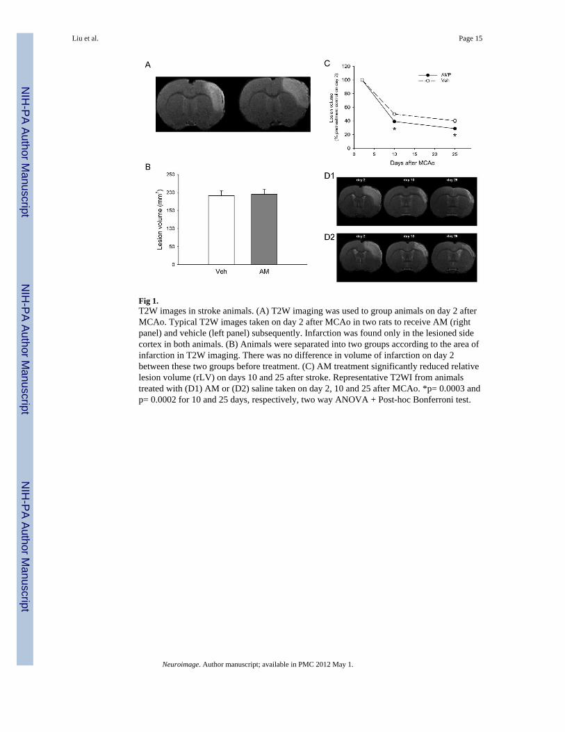
Fig 1.T2W images in stroke animals. (A) T2W imaging was used to group animals on day 2 afterMCAo. Typical T2W images taken on day 2 after MCAo in two rats to receive AM (rightpanel) and vehicle (left panel) subsequently. Infarction was found only in the lesioned sidecortex in both animals. (B) Animals were separated into two groups according to the area ofinfarction in T2W imaging. There was no difference in volume of infarction on day 2between these two groups before treatment. (C) AM treatment significantly reduced relativelesion volume (rLV) on days 10 and 25 after stroke. Representative T2WI from animalstreated with (D1) AM or (D2) saline taken on day 2, 10 and 25 after MCAo. *p= 0.0003 andp= 0.0002 for 10 and 25 days, respectively, two way ANOVA + Post-hoc Bonferroni test.
Liu et al. Page 15
Neuroimage. Author manuscript; available in PMC 2012 May 1.
NIH
-PA Author Manuscript
NIH
-PA Author Manuscript
NIH
-PA Author Manuscript
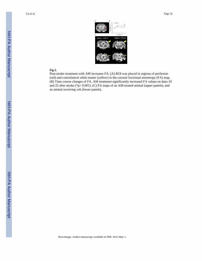
Fig 2.Post-stroke treatment with AM increases FA. (A) ROI was placed in regions of perilesion(red) and contralateral white matter (yellow) in the coronal fractional anisotropy (FA) map.(B) Time course changes of FA. AM treatment significantly increased FA values on days 10and 25 after stroke (*p< 0.001). (C) FA maps of an AM-treated animal (upper panels), andan animal receiving veh (lower panels).
Liu et al. Page 16
Neuroimage. Author manuscript; available in PMC 2012 May 1.
NIH
-PA Author Manuscript
NIH
-PA Author Manuscript
NIH
-PA Author Manuscript
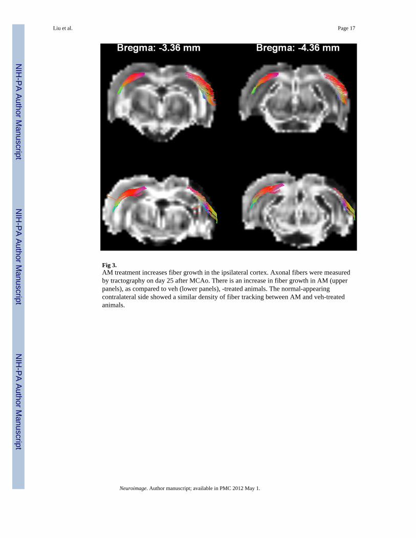
Fig 3.AM treatment increases fiber growth in the ipsilateral cortex. Axonal fibers were measuredby tractography on day 25 after MCAo. There is an increase in fiber growth in AM (upperpanels), as compared to veh (lower panels), -treated animals. The normal-appearingcontralateral side showed a similar density of fiber tracking between AM and veh-treatedanimals.
Liu et al. Page 17
Neuroimage. Author manuscript; available in PMC 2012 May 1.
NIH
-PA Author Manuscript
NIH
-PA Author Manuscript
NIH
-PA Author Manuscript
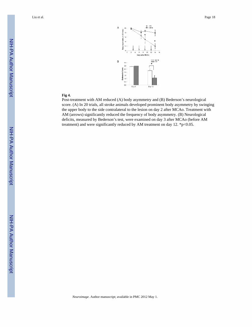
Fig 4.Post-treatment with AM reduced (A) body asymmetry and (B) Bederson’s neurologicalscore. (A) In 20 trials, all stroke animals developed prominent body asymmetry by swingingthe upper body to the side contralateral to the lesion on day 2 after MCAo. Treatment withAM (arrows) significantly reduced the frequency of body asymmetry. (B) Neurologicaldeficits, measured by Bederson’s test, were examined on day 3 after MCAo (before AMtreatment) and were significantly reduced by AM treatment on day 12. *p<0.05.
Liu et al. Page 18
Neuroimage. Author manuscript; available in PMC 2012 May 1.
NIH
-PA Author Manuscript
NIH
-PA Author Manuscript
NIH
-PA Author Manuscript
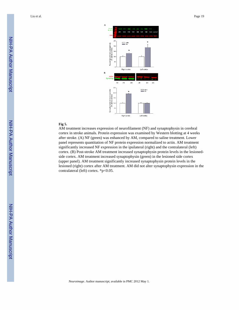
Fig 5.AM treatment increases expression of neurofilament (NF) and synaptophysin in cerebralcortex in stroke animals. Protein expression was examined by Western blotting at 4 weeksafter stroke. (A) NF (green) was enhanced by AM, compared to saline treatment. Lowerpanel represents quantitation of NF protein expression normalized to actin. AM treatmentsignificantly increased NF expression in the ipsilateral (right) and the contralateral (left)cortex. (B) Post-stroke AM treatment increased synaptophysin protein levels in the lesioned-side cortex. AM treatment increased synaptophysin (green) in the lesioned side cortex(upper panel). AM treatment significantly increased synaptophysin protein levels in thelesioned (right) cortex after AM treatment. AM did not alter synaptophysin expression in thecontralateral (left) cortex. *p<0.05.
Liu et al. Page 19
Neuroimage. Author manuscript; available in PMC 2012 May 1.
NIH
-PA Author Manuscript
NIH
-PA Author Manuscript
NIH
-PA Author Manuscript
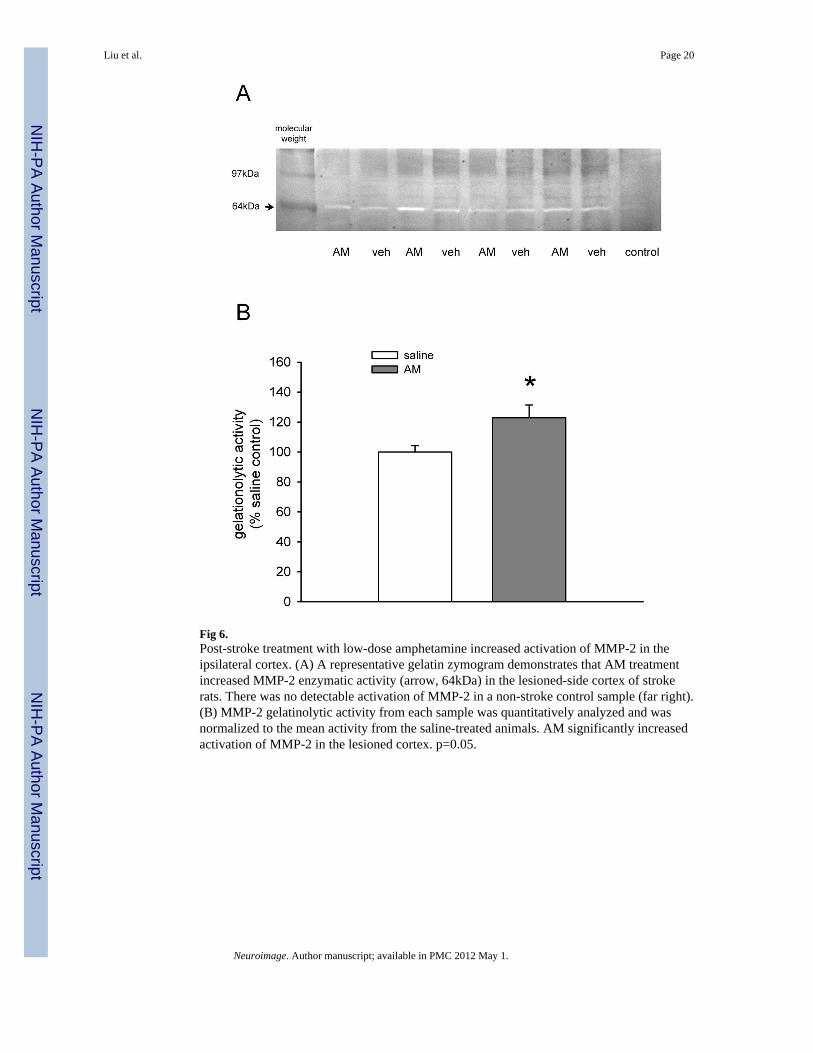
Fig 6.Post-stroke treatment with low-dose amphetamine increased activation of MMP-2 in theipsilateral cortex. (A) A representative gelatin zymogram demonstrates that AM treatmentincreased MMP-2 enzymatic activity (arrow, 64kDa) in the lesioned-side cortex of strokerats. There was no detectable activation of MMP-2 in a non-stroke control sample (far right).(B) MMP-2 gelatinolytic activity from each sample was quantitatively analyzed and wasnormalized to the mean activity from the saline-treated animals. AM significantly increasedactivation of MMP-2 in the lesioned cortex. p=0.05.
Liu et al. Page 20
Neuroimage. Author manuscript; available in PMC 2012 May 1.
NIH
-PA Author Manuscript
NIH
-PA Author Manuscript
NIH
-PA Author Manuscript

Fig 7.Post-stroke treatment with AM increased BDNF mRNA expression in the lesioned sidecortex. Brain samples were collected within one day after the 1st or 2nd dose of AM orsaline. All data were normalized to the non-stroke controls. (A1) In the ipsilateral cortex,BDNF mRNA activity was significantly reduced after MCAo. Treatment with AMsignificantly increased BDNF expression. (A2) BDNF mRNA levels were not affected byAM in the contralateral side cortex. (B) Ischemia enhanced BMP7 mRNA expression in thelesioned side cortex (B1). AM did not alter BMP7 mRNA expression on either side. (C)CART mRNA expression in the lesioned side cortex was significantly reduced after MCAo(C1). AM treatment did not alter CART expression in the lesioned (C1) and non-lesionedside cortices (C2). *p <0.05, 2-way ANOVA.
Liu et al. Page 21
Neuroimage. Author manuscript; available in PMC 2012 May 1.
NIH
-PA Author Manuscript
NIH
-PA Author Manuscript
NIH
-PA Author Manuscript
Copyright © 2022 FDOKUMEN