USING e-ANNOTATION TOOLS FOR ELECTRONIC PROOF ...
51
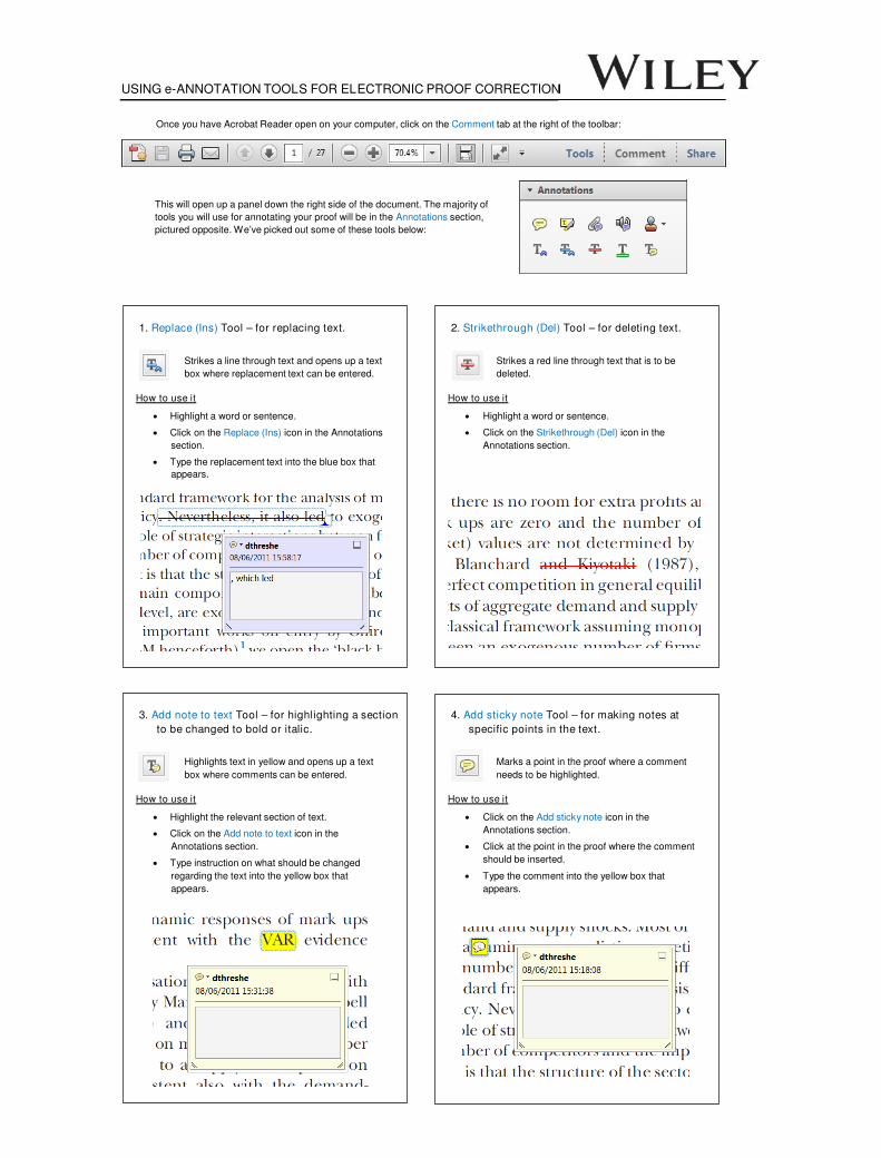
USING e-ANNOTATION TOOLS FOR ELECTRONIC PROOF CORRECTION Once you have Acrobat Reader open on your computer, click on the Comment tab at the right of the toolbar: This will open up a panel down the right side of the document. The majority of tools you will use for annotating your proof will be in the Annotations section, pictured opposite. We’ve picked out some of these tools below: 1. Replace (Ins) Tool – for replacing text. Strikes a line through text and opens up a text box where replacement text can be entered. How to use it ‚ Highlight a word or sentence. ‚ Click on the Replace (Ins) icon in the Annotations section. ‚ Type the replacement text into the blue box that appears. 2. Strikethrough (Del) Tool – for deleting text. Strikes a red line through text that is to be deleted. How to use it ‚ Highlight a word or sentence. ‚ Click on the Strikethrough (Del) icon in the Annotations section. 3. Add note to text Tool – for highlighting a section to be changed to bold or italic. Highlights text in yellow and opens up a text box where comments can be entered. How to use it ‚ Highlight the relevant section of text. ‚ Click on the Add note to text icon in the Annotations section. ‚ Type instruction on what should be changed regarding the text into the yellow box that appears. 4. Add sticky note Tool – for making notes at specific points in the text. Marks a point in the proof where a comment needs to be highlighted. How to use it ‚ Click on the Add sticky note icon in the Annotations section. ‚ Click at the point in the proof where the comment should be inserted. ‚ Type the comment into the yellow box that appears.
-
Upload
khangminh22 -
Category
Documents
-
view
1 -
download
0
Transcript of USING e-ANNOTATION TOOLS FOR ELECTRONIC PROOF ...
USING e-ANNOTATION TOOLS FOR ELECTRONIC PROOF CORRECTION
Once you have Acrobat Reader open on your computer, click on the Comment tab at the right of the toolbar:
This will open up a panel down the right side of the document. The majority of
tools you will use for annotating your proof will be in the Annotations section,
pictured opposite. We’ve picked out some of these tools below:
1. Replace (Ins) Tool – for replacing text.
Strikes a line through text and opens up a text
box where replacement text can be entered.
How to use it
‚ Highlight a word or sentence.
‚ Click on the Replace (Ins) icon in the Annotations
section.
‚ Type the replacement text into the blue box that
appears.
2. Strikethrough (Del) Tool – for deleting text.
Strikes a red line through text that is to be
deleted.
How to use it
‚ Highlight a word or sentence.
‚ Click on the Strikethrough (Del) icon in the
Annotations section.
3. Add note to text Tool – for highlighting a section
to be changed to bold or italic.
Highlights text in yellow and opens up a text
box where comments can be entered.
How to use it
‚ Highlight the relevant section of text.
‚ Click on the Add note to text icon in the
Annotations section.
‚ Type instruction on what should be changed
regarding the text into the yellow box that
appears.
4. Add sticky note Tool – for making notes at
specific points in the text.
Marks a point in the proof where a comment
needs to be highlighted.
How to use it
‚ Click on the Add sticky note icon in the
Annotations section.
‚ Click at the point in the proof where the comment
should be inserted.
‚ Type the comment into the yellow box that
appears.

USING e-ANNOTATION TOOLS FOR ELECTRONIC PROOF CORRECTION
5. Attach File Tool – for inserting large amounts of
text or replacement figures.
Inserts an icon linking to the attached file in the
appropriate place in the text.
How to use it
‚ Click on the Attach File icon in the Annotations
section.
‚ Click on the proof to where you’d like the attached
file to be linked.
‚ Select the file to be attached from your computer
or network.
‚ Select the colour and type of icon that will appear
in the proof. Click OK.
6. Drawing Markups Tools – for drawing
shapes, lines and freeform annotations on
proofs and commenting on these marks.
Allows shapes, lines and freeform annotations to be
drawn on proofs and for comment to be made on
these marks.
How to use it
" Click on one of the shapes in the Drawing Markups
section.
" Click on the proof at the relevant point and draw the
selected shape with the cursor.
" To add a comment to the drawn shape, move the
cursor over the shape until an arrowhead appears.
" Double click on the shape and type any text in the
red box that appears.

R EV I EW AR T I C L E
Ribosomally encoded antibacterial proteins and peptides from
Pseudomonas1; 21; 2
Maarten G.K. Ghequire & Ren�e De Mot
Centre of Microbial and Plant Genetics, University of Leuven, Heverlee, Belgium
Correspondence: Ren�e De Mot, Centre of
Microbial and Plant Genetics, University of
Leuven, Kasteelpark Arenberg 20, B-3001
Heverlee, Belgium. Tel.: +32 16 329 681;
fax: +32 16 321 966;
e-mail: [email protected]
Received 26 February 2014; revised 5 May
2014; accepted 16 May 2014.
DOI: 10.1111/1574-6976.12079
Editor: Alain Filloux
Keywords
bacteriocin; pyocin; tailocin; T6SS; CDI; Rhs;
lectin; microcin3 .
Abstract
Members of the Pseudomonas genus produce diverse secondary metabolites
affecting other bacteria, fungi or predating nematodes and protozoa but are
also equipped with the capacity to secrete different types of ribosomally
encoded toxic peptides and proteins, ranging from small microcins to large tai-
locins. Studies with the human pathogen Pseudomonas aeruginosa have revealed
that effector proteins of type VI secretion systems are part of the antibacterial
armamentarium deployed by pseudomonads. A novel class of antibacterial pro-
teins with structural similarity to plant lectins was discovered by studying
antagonism among plant-associated Pseudomonas strains. A genomic perspec-
tive on pseudomonad bacteriocinogeny shows that the modular architecture of
S-pyocins of P. aeruginosa is retained in a large diversified group of bacterioc-
ins, most of which target DNA or RNA. Similar modularity is present in as
yet poorly characterized Rhs (recombination hot spot) proteins and CDI
(contact-dependent inhibition) proteins. Well-delimited domains for receptor
recognition or cytotoxicity enable the design of chimeric toxins with novel
functionalities, which has been applied successfully for S- and R-pyocins. Little
is known regarding how these antibacterials are released and ultimately reach
their targets. Other remaining issues concern the identification of environmen-
tal triggers activating these systems and assessment of their ecological impact
in niches populated by pseudomonads.
Scope of the review
Pseudomonas strains are able to indwell very diverse niches,
ranging from terrestrial and aquatic environments to tis-
sues of eukaryotic hosts. Population of such environments
involves a struggle for living space and organic nutrients
with a plethora of other microorganisms. Pseudomonads
display a highly versatile metabolism and several secondary
metabolites affecting other bacteria and fungi have been
identified, such as 2,4-diacetylphloroglucinol, phenazines,
pyrrolnitrin, pyoluteorin, and lipopeptides (Gross & Loper,
2009). While such antibiotics mainly affect phylogenetically
distant rivals, only few secondary metabolites are toxic to
fellow pseudomonads that are attracted to common niches
(Li et al., 2011). The ribosomal machinery, on the other
hand, enables biosynthesis of very diverse bacterial peptides
and proteins, collectively designated bacteriocins, that are
only deleterious to members of a certain bacterial species
or a subset of phylogenetically close relatives of the
producer that itself carries an immunity protein-based
system preventing self-intoxication. The study of such
protein antibiotics has regained attention as a possible
way to minimize collateral damage to nontarget microbiota
and as a potential source of novel molecular targets to
alleviate problems with multiresistance to available antibi-
otics.
Among Gram-negative bacteria, the colicins produced
by enterobacteria are by far the best studied group of
such narrow-spectrum antagonistic proteins, and a wealth
of information is available on the molecular mechanisms
involved in the different stages of their killing action
(reviewed by Cascales et al., 2007). Several bacteriocins of
pseudomonads share basic characteristics with colicins
and insights from colicin biology have been instrumental
to identify novel so-called pyocins with similar cytotoxici-
ties. Studies on interactions among Gram-negative bacte-
rial relatives have disclosed in recent years that certain
features of colicins and pyocins, such as the modular
1
2
3
4
5
6
7
8
9
10
11
12
13
14
15
16
17
18
19
20
21
22
23
24
25
26
27
28
29
30
31
32
33
34
35
36
37
38
39
40
41
42
43
44
45
46
47
48
49
50
51
52
53
FEMS Microbiol Rev && (2014) 1–46 ª 2014 Federation of European Microbiological Societies.
Published by John Wiley & Sons Ltd. All rights reserved
F E M S R E 1 2 0 7 9 Dispatch: 17.6.14 CE: Kathiravan D.
Journal Code Manuscript No. No. of pages: 46 PE: Kiruthika

nature of toxin/immunity pairs, are equally retained in
substrates of Type V and Type VI systems that mediate
antagonism (Braun & Patzer, 2013). In 2002, the review
by Michel-Briand and Baysse was essentially confined
to colicin-like and phage-like pyocins of Pseudomonas
aeruginosa, as the only pseudomonad bacteriocins suffi-
ciently characterized at that time. Here, we review the
current knowledge of these systems and describe novel
types of bacteriocins that have been identified in the last
decade. This served as a basis to provide an overview of
the bacteriocinogenic potential of the Pseudomonas genus
by scrutiny of available (draft) genome sequences.
S-type pyocins
Soluble or S-type pyocins are protease- and heat-sensitive,
chromosome-encoded bacteriocins from P. aeruginosa
that are able to kill cells from the same species. These an-
tibacterials are secreted as binary protein complexes con-
sisting of a large protein that harbors the killing function
and a smaller immunity protein that remains tightly
bound to the cytotoxic domain of the former. The physi-
cal association of the toxin with its cognate immunity
protein, reflected in the clustering of their structural
genes, ensures that the producer strain is not harmed
before the bacteriocin is released (Michel-Briand & Bay-
sse, 2002).
To date, several S-type pyocins have been described
and characterized: pyocins S1 (Ito et al., 1970), S2 (Ohka-
wa et al., 1973), AP41 (Holloway et al., 1973; Sano & Ka-
geyama, 1981), S3 (Duport et al., 1995), S4 (Elfarash
et al., 2012) and S5 (Ling et al., 2010). Pyocin Sa (Govan,
1986) turned out to be identical to pyocin S2 (Denayer
et al., 2007). A bacteriocin nearly identical to pyocin S1
but equipped with a different type of killing and immu-
nity function was designated pyocin S6 (Dingemans et al.,
2013). Whereas all these proteins share identical parts or
display significant local sequence homology, this is not
the case for pyocin PaeM, which is produced without a
known immunity partner (Barreteau et al., 2009).
Although the study of S-type pyocins has mainly
focused on their occurrence and action in the human
opportunistic pathogen P. aeruginosa, genes encoding
structurally related proteins are found in other Pseudomo-
nas species as well, but functional characterization of the
latter is lacking (Parret & De Mot, 2002).
Domain architecture of S-type pyocins
To kill a target cell, a S-type pyocin would first bind to a
specific receptor located on the outer membrane of the
bacterial cells and it would then be further translocated
to exert its inhibitory function. The modular organization
design of the distinct domains correlates well with these
multiple steps of mode of action (Michel-Briand &
Baysse, 2002). In most S-type bacteriocins, the amino-ter-
minal domain of the large protein bears the receptor-
binding function and its carboxy-terminal part engenders
the lethal effect (Sano et al., 1993b; Parret & De Mot,
2002). In pyocins with a cytoplasmic target, they are con-
nected by a domain that mediates translocation (corre-
sponding to Pfam domain PF06958; Pyocin_S). An
additional polypeptide segment of unknown function can
be positioned between the receptor-binding and translo-
cation domains, but this part is dispensable for killing, as
observed for pyocins S2 and AP41 (Sano et al., 1993a).
The modular composition of S-type pyocins is also
underlined by the fact that functional bacteriocins can be
constructed by joining domains from different pyocins.
Examples include combined domains of pyocins S1 and
AP41 (Sano et al., 1993a) and a pyocin S5/S2 chimer
(Elfarash et al., 2014). In addition, active pyocin/colicin
hybrids with domains from pyocin S3 and colicin E3
(Gupta et al., 2013; see Potential applications for S-type
pyocins) and with domains from pyocins S1 or S2 and
colicins E2 or E3 have been engineered (Kageyama et al.,
1996). Pyocin S5 and PaeM that do not require transloca-
tion to the cytoplasm for killing, exhibit a different
domain architecture with the translocation domain pre-
ceding the receptor-binding domains (Barreteau et al.,
2009; Elfarash et al., 2014). Pyocin size varies signifi-
cantly, ranging from 289 amino acids for PaeM to 777
amino acids for AP41 (Table 1). A schematic representa-
tion of the gene and domain organization of character-
ized and selected predicted S-type pyocins in
Pseudomonas spp. is shown in Fig. 1.
Biological properties and structure of S-type
pyocins
Different S-pyocin-killing domains have been described in
Table 1. Pyocin S1, S2, S3, and AP41 display DNase
activity (Seo & Galloway, 1990; Sano, 1993; Sano et al.,
1993b; Duport et al., 1995), while pyocin S4 harbors a
tRNase (Elfarash et al., 2012) and pyocin S6 a rRNase
(Fig. 1; Dingemans et al., 2013). Pyocin DNase domains
typically bear a conserved HNH-endonuclease motif, as
observed in pyocins S1, S2, and AP41 (Parret & De Mot,
2002). This motif constitutes the core of the catalytic site
of the endonuclease and can chelate a single metal ion,
required for hydrolysis of the dsDNA strand. These
HNH-nucleases have also been detected in colicins (Pap-
adakos et al., 2012). Pyocin S3 on the contrary does not
contain this HNH-motif and its lethal domain lacks
sequence homology with the corresponding domains of
other DNase pyocins (Parret & De Mot, 2002). Enzymatic
FEMS Microbiol Rev && (2014) 1–46ª 2014 Federation of European Microbiological Societies.
Published by John Wiley & Sons Ltd. All rights reserved
2 M.G.K. Ghequire & R. De Mot
1
2
3
4
5
6
7
8
9
10
11
12
13
14
15
16
17
18
19
20
21
22
23
24
25
26
27
28
29
30
31
32
33
34
35
36
37
38
39
40
41
42
43
44
45
46
47
48
49
50
51
52
53

activity has not been detected for pyocin S5; instead, this
pyocin is known to kill a target bacterium via pore for-
mation, resulting in membrane damage and leakage of
intracellular compounds (Fig. 1; Ling et al., 2010).
A pyocin with yet a different cytotoxic activity was
identified in P. aeruginosa strains JJ692 and DET08
encoded within their exoU-carrying genomic islands. This
enzyme, PaeM, displays homology with colicin M, a lipid
II-degrading bacteriocin from Escherichia coli. It does not
exert its enzymatic activity in the cytoplasm; instead, it is
active in the periplasm where it blocks peptidoglycan syn-
thesis (Fig. 1; Barreteau et al., 2009). Unlike the lytic
activity of colicin M on sensitive E. coli cells, PaeM only
exhibits a bacteriostatic effect on its target cells (Barreteau
et al., 2009). For the plant pathogen, Pseudomonas syrin-
gae pv. tomato DC3000 and the wheat rhizosphere isolate
Pseudomonas fluorescens Q8r1–96, recombinant homologs
(PsyM and PflM, respectively) were shown to be func-
tional toxins as well, however, displaying lower in vitro
phosphodiesterase activity. Of fourteen P. aeruginosa
strains tested, only two strains were susceptible to PaeM
and one of these indicators also was inhibited by PflM.
No other target strain was identified among 40 strains
representing the three species, pointing to a quite narrow
activity spectrum for this type of pseudomonad bacterio-
cin, as compared to S-type pyocins. In a subsequent inde-
pendent study, activity of the DC3000 recombinant
enzyme, there denoted syringacin M, was shown against
two P. syringae strains belonging to the pathovars syringae
(LMG 5084) and lachrymans (LMG 5456), while other
Pseudomonas species were not affected. The same narrow
activity spectrum was exhibited by a close homolog puri-
fied from mitomycin-induced culture supernatant of
P. syringae pv. syringae LMG 1247 (Grinter et al., 2012b).
Currently, structures of only two pyocins have been
solved: PaeM from P. aeruginosa and syringacin M from
P. syringae (Fig. 2). These proteins share structural simi-
larities with colicin M. Their crystal structures consist of
a short disordered amino-terminal translocation domain,
followed by a central globular a-helical receptor-binding
domain and a cytotoxic domain incorporating a half
b-barrel fold, characteristic for these bacteriocins (Zeth
et al., 2008) but adopting a modified active site architec-
ture. A Mg2+ ion was identified in the active site of PaeM
in line with the dependence of colicin M activity on this
cation (Barreteau et al., 2012). However, in crystallized
syringacin M, this site was occupied by Ca2+ that sup-
ports catalytic activity similarly to Mg2+ (Grinter et al.,
2012b). Barreteau and coworkers (Barreteau et al., 2012)
showed that in vitro catalytic activity of the isolated cyto-
toxic domain was enhanced, suggesting that interdomain
interactions dampen the enzymatic activity which is sup-
posed to be fully released upon cellular entry and interac-
tion with its substrate. Although the syringacin M receptor-
binding domains lacks discernible sequence homology to
the equivalent colicin M region, they adopt a very similar
fold, presumably as a result of diversifying selection
(Grinter et al., 2012b) rather than recombination (Zeth
Table 1. Characteristics of Pseudomonas aeruginosa pyocins
Pyocin Strain Mode of action
Toxin Immunity protein
ReceptorAmino acids Domain(s)* Amino acids Domain†
AP41 PAF41-2 DNase (HNH) 777 PF06958; PF12639 90 PF01320 ?
M1 JJ692 Lipid II degradation 289 PF14859 – – ?
S1 NIH-H DNase (HNH) 618 PF06958; PF12639 87 PF01320 ?‡
S2 PAO1 DNase (HNH) 689 PF03515; PF06958; PF12639 87 PF01320 FpvAI
S3 P12 DNase 766 PF06958 153 – FpvAII
S4 PAO1 tRNase 764 PF03515; PF06958; PF12106 112 PF11480 FpvAI
S5 PAO1 Pore-forming 498 PF01024 108 PF03526 FptA
S6 CF-PA39 rRNase 571 PF06958; PF09000 77 – ?
M4 BL01 Lipid II degradation 342 PF14859 – – ?
S7 BWHPSA018 rRNase 642 PF06958; PF09000 77 – ?
S8 VRFPA07 DNase (HNH) 772 PF06958; PF12639 85 PF01320 ?
S9 BL04 DNase (HNH) 421 PF06958; PF12639 84 PF01320 ?
S10 PABL056 DNase 517 PF05488; PF06958 157 – ?
S11 LESB58 tRNase 662 PF03515; PF06958; PF11429 90 PF09204 ?
S12 PA7 tRNase 740 PF06958; PF11429 90 PF09204 ?
Proteins with demonstrated bacteriocin activity and representatives of predicted novel pyocin types are shown in bold and italic font, respectively.
*Pfam domains are ordered from amino- to carboxy-terminus: PF01024, Colicin; PF03515, Cloacin; PF05488, PAAR_motif; PF06958, Pyocin_S;
PF09000, Cytotoxic; PF11429, Colicin_D; PF12016, Colicin_C; PF12639, Colicin-DNase; PF14859, Colicin_M. The carboxy-terminal domain confer-
ring cytotoxic activity is underlined.†Pfam domains: PF01320, Colicin_Pyocin; PF03526, ImmE1; PF09204, Colicin_immun; PF11480, ImmE5.‡The receptor of pyocin S1 in unknown, but is FpvA independent (Denayer et al., 2007).
FEMS Microbiol Rev && (2014) 1–46 ª 2014 Federation of European Microbiological Societies.
Published by John Wiley & Sons Ltd. All rights reserved
Protein-mediated warfare among pseudomonads 3
1
2
3
4
5
6
7
8
9
10
11
12
13
14
15
16
17
18
19
20
21
22
23
24
25
26
27
28
29
30
31
32
33
34
35
36
37
38
39
40
41
42
43
44
45
46
47
48
49
50
51
52
53

Fig. 1. 7Gene organization of pseudomonad bacteriocins. Arrows illustrate representative coding regions and are grouped according to known
or predicted toxic activity. The DNA regions encoding the respective bacteriotoxic domains are highlighted in color as specified in the inset box.
The coding region(s) of the respective immunity protein(s) located downstream of the toxin gene, if present, are shown in a fainter shade of
the same color. The Pfam accession numbers of killing and immunity domains are specified in Table 1. The Pyocin_S domain is shown in gray.
The additionally detected toxin/immunity domains in putative bacteriocins are marked as Pore-2 (PF01024/PF03857) and tRNase-2 (PF11429/
PF09204). Additional colors indicate conservation of an amino-terminal domain between different bateriotoxic proteins and noncolored parts
denote a lack of significant similarity. Genomic DNA regions with clustered bacteriocin genes and multiple bacteriocin operons within a single
strain are differentiated with labels (a) through (d). Names of functionally characterized bacteriocins are highlighted with red font. Proposed
names for new types of pyocins with a single toxic domain (blue) or hybrid forms with two toxic domains (green) are also shown in color. The
crossed region represents a pseudogene. The scale bar represents a DNA region of 500 bp. Abbreviations used for species names: Paer,
Pseudomonas aeruginosa; Pchl, Pseudomonas chlororaphis; Pent, Pseudomonas entomophila; Pflu, Pseudomonas fluorescens; Pman,
Pseudomonas mandelii; Pple, Pseudomonas plecoglossicida; Ppro, Pseudomonas protegens; Pput, Pseudomonas putida; Pseu, Pseudomonas sp.,
Psyn, Pseudomonas synxantha; Psyr, Pseudomonas syringae; Ptol, Pseudomonas tolaasii. Other abbreviations: PyoS, pyocin S; Rhs, protein with
Rhs domain; cdiBAI, CDI system operon; mcbABCDEFG, B-type microcin operon. An overview of representative tailocin gene clusters is
presented in Fig. 6.
LOW
RESOLUTIO
NCOLOR
FIG
FEMS Microbiol Rev && (2014) 1–46ª 2014 Federation of European Microbiological Societies.
Published by John Wiley & Sons Ltd. All rights reserved
4 M.G.K. Ghequire & R. De Mot
1
2
3
4
5
6
7
8
9
10
11
12
13
14
15
16
17
18
19
20
21
22
23
24
25
26
27
28
29
30
31
32
33
34
35
36
37
38
39
40
41
42
43
44
45
46
47
48
49
50
51
52
53

et al., 2008). Taking into account that these colicin M-
like proteins form a distinct group of pyocins as com-
pared to other modular S-type bacteriocins, we propose
pyocin M to refer to this Pseudomonas protein family.
Extracellular release of S-pyocin/immunity
protein complexes
S-type pyocins are released as bacteriocin/immunity pro-
tein complexes at equimolar ratio. The size of the immu-
nity proteins of S-type pyocins ranges from 77 to 153
amino acids, much smaller than the cognate killer pro-
teins (Table 1). Their coexpression is crucial as they tran-
siently inhibit the lethal function of the pyocin (Sano &
Kageyama, 1981; Seo & Galloway, 1990; Sano et al.,
1993b; Duport et al., 1995; Rasouliha et al., 2013).
Immunity proteins from S1, S2 and AP41 share homol-
ogy as they shield homologous HNH nuclease domains.
Pyocin S3 is associated with a different type of immunity
protein, in line with its classification as a different type of
DNase toxin (Parret & De Mot, 2002). Protection is pro-
vided by interaction of the amino-terminal end of the
immunity protein and the carboxy-terminal cytotoxic
part of the killer protein (Sano et al., 1993b; Kageyama
et al., 1996). Bacteriocin-producing cells may need to
neutralize pyocin produced by other clonemates. It was
found that this so-called soaking effect causes a reduced
fitness of the pyocin producer strain (Inglis et al., 2013).
To ensure a swift ‘trapping’ of a corresponding killing
domain, immunity genes of nuclease-type of bacteriocins
are located immediately downstream of the pyocin genes
and transcribed as an operon (Fig. 1). Their Shine–Dal-
garno boxes are located within the lethal part of the pyo-
cin gene (Sano et al., 1993b). Interestingly, the presence
of ‘orphan’ immunity genes, encoding proteins that
complement a matching S-type pyocin toxicity domain
but that are not preceded by toxin genes, confers protec-
tion to sensitive strains. This way, invading pyocins
may be captured, impeding their action in target cells
(Denayer et al., 2007; Elfarash et al., 2012; Rasouliha
et al., 2013; Elfarash et al., 2014). Notably, the immunity
gene of the pore-forming pyocin S5 is transcribed in the
opposite direction of the toxin gene (Fig. 1; Stover et al.,
2000).
The mechanism behind self-immunity of pseudomo-
nads producing colicin M-like bacteriocins is currently
not known. In E. coli, this is ensured by a coexpressed
immunity protein (Cmi) anchored in the cytoplasmic
membrane facing the periplasm (Cascales et al., 2007).
Genes encoding homologs of Cmi or the structurally
related YebF protein (G�erard et al., 2011; Us�on et al.,
2012) are not found in Pseudomonas genomes.
After synthesis, the pyocin complexes are released from
the producers, without the need of a (cleavable) signal
sequence. Colicins take advantage of a lysis protein,
encoded nearby the colicin gene, whereas S-type pyocins
do not have these available (Michel-Briand & Baysse,
2002). It was suggested that S-pyocins may take advan-
tage of the lytic systems enabling the secretion of phage
tail-like bacteriocins (see Genetic determinants of R-type
pyocins; Nakayama et al., 2000). However, several S-pyo-
cin-encoding Pseudomonas strains lack phage tail-like
bacteriocin clusters and their accompagnied release cassettes
(see In silico analysis of tailocins in other Pseudomonas
species).
S-type pyocin receptors and translocation
The observation that several S-type pyocins kill target
cells much more efficiently under iron-poor conditions
led to the idea that these bacteriocins take advantage of
iron-regulated receptors for cell entry (Ohkawa et al.,
1980; Sano et al., 1993b; Duport et al., 1995; Elfarash
et al., 2012, 2014). When iron is limiting, bacteria will
express outer-membrane proteins that promote its
uptake. One important strategy used by P. aeruginosa to
(a) (b)
Fig. 2. Structures of lipid II-degrading M-type
pyocins PaeM (a, PDB 4G75) and syringacin M
(b, PDB 4FZM). Ribbon structures are shown in
a transparent surface model. Translocation
domains are colored purple, receptor-binding
domains orange and catalytic domains blue.
The amino-terminal translocation domain of
syringacin M is unstructured.
COLOR
FEMS Microbiol Rev && (2014) 1–46 ª 2014 Federation of European Microbiological Societies.
Published by John Wiley & Sons Ltd. All rights reserved
Protein-mediated warfare among pseudomonads 5
1
2
3
4
5
6
7
8
9
10
11
12
13
14
15
16
17
18
19
20
21
22
23
24
25
26
27
28
29
30
31
32
33
34
35
36
37
38
39
40
41
42
43
44
45
46
47
48
49
50
51
52
53

enhance iron uptake is the secretion of the siderophores
pyoverdine and pyochelin, low-molecular-weight Fe3+-
chelating molecules. Three pyoverdine types can be dis-
tinguished, classified based on their oligopeptide side
chains and each recognized by a specific outer-membrane
receptor. After iron binding, siderophores are taken up by
the iron-regulated outer-membrane proteins (IROMPs).
Energy for this process and siderophore recycling is trans-
duced by the cytoplasmic membrane protein TonB
(Cornelis & Dingemans, 2013).
Mutagenesis experiments provided unequivocal evi-
dence that several S-type pyocins indeed use IROMPs as
a receptor (Table 1). Pyocin S2 and S4 take advantage of
the type I ferripyoverdine receptor (FpvAI; Ohkawa et al.,
1980; Smith et al., 1992; Denayer et al., 2007; Elfarash
et al., 2012), whereas pyocin S3 uses the type II ferripyov-
erdine receptor (FpvAII; Baysse et al., 1999). The
pore-forming pyocin S5 hijacks the FptA ferripyochelin
receptor to inhibit sensitive strains (Elfarash et al., 2014).
As the latter receptor is widespread among P. aeruginosa
strains, the percentage of sensitive strains in a representa-
tive test panel is higher than usual. The receptors of
pyocins S1 and S6 have not yet been identified, but sensi-
tivity seems independent of the ferripyoverdine receptor
produced (Denayer et al., 2007; Dingemans et al., 2013).
Following contact with these receptors, pyocins are
translocated across the outer membrane. The exact mech-
anism remains unknown though the use of the ferrisider-
ophore receptors suggests that S-type pyocins are
translocated in a similar way as the pyoverdines and py-
ochelin, energized by the TonB system (Cornelis &
Dingemans, 2013). Several colicins are translocated via
the TonB machinery as well, equally taking advantage of
outer-membrane receptors for siderophores, such as lin-
ear catecholate transporter Cir, iron-enterobactin trans-
porter FepA and ferrichrome-iron receptor FhuA (Jakes
& Cramer, 2012).
Pyocin AP41 seems to enter cells via another receptor
and uptake mechanism, although iron was found to play
a major role as well. Different mutants tolerant to pyocin
AP41 (tol phenotype) were isolated (Holloway et al.,
1973), revealing involvement of a cluster of seven genes,
organized in three operons (orf1-tolQRA, tolB and oprL-
orf2) and regulated by iron availability and growth phase
(Duan et al., 2000). The orf1 gene is nonessential, while
mutants in tolQ and tolA could not be obtained, probably
because gene inactivation would result in a lethal pheno-
type (Wei et al., 2009). Nevertheless, introduction of the
tolQRA genes in a tol mutant is able to restore killing by
AP41 (also called AR41), indicating that the Tol proteins
are involved in pyocin uptake (Dennis et al., 1996). High
iron concentration (FeCl3) causes a reduced expression of
the complete locus, due to the presence of a Fur-repressor
(Lafontaine & Sokol, 1998; Duan et al., 2000). In con-
trast, RegA functions as a positive regulator under iron-
restricted conditions (Duan et al., 2000). Study of the
expression profile of orf1-tolQRA demonstrated that this
operon contains one constitutive promoter in front of
orf1, and one iron-regulated promoter located within orf1
(Wei et al., 2009). The oprL gene encodes a 18 kDa
outer-membrane peptidoglycan-associated lipoprotein
required for a normal cellular morphology and is located
downstream of tolB. An oprL knockout mutant is viable
but is very sensitive to osmotic pressure, underlining its
role in cell envelope integrity (Lim et al., 1997). More
recently, the growth phase-dependent regulation of the
tol-oprL locus was found to be controlled by quorum
sensing (QS), dependent on N-acyl homoserine lactone
(AHL). When residing in a stationary phase, bacteria will
not opt for a pronounced expression of these genes as
they are required for growth and cell division, explaining
the downregulation of the locus (Wei et al., 2009).
Although the receptor of pyocin AP41 is not known, the
proposed uptake route via the Tol machinery is in line
with observations made for colicins. In Tol-energized
uptake of several colicins, the primary outer-membrane
receptors (such as the TonB-dependent vitamin B12 trans-
porter BtuB) and the outer-membrane proteins actually
translocating the bacteriocins (such as the porin OmpF)
are not involved in siderophore-mediated iron uptake
(Jakes & Cramer, 2012).
Potential applications for S-type pyocins
The S-type pyocins may be valuable tools in future thera-
peutic applications. This is illustrated by the potent activ-
ity of pyocin S2 against P. aeruginosa biofilms. Tested
against different clinical isolates, this bacteriocin kills both
mucoid and nonmucoid strains with similar efficiency
when these are growing in a biofilm. Moreover, survival
rates of biofilms treated with tobramycin or aztreonam at
equal concentrations are considerably higher (> 100-fold).
In vivo pyocin S2 activity was validated in a P. aerugin-
osa-infected Galleria mellonella caterpillar model (Smith
et al., 2012).
From synthetic biology, engineered E. coli strains were
developed, able to sense the presence of P. aeruginosa cells
and subsequently killing them, both in planktonic and bio-
film growth conditions. In a first approach, E. coli was
equipped with a pyocin S5 gene, under the control of a
luxR promoter (Saeidi et al., 2011). The latter is activated
after binding of a LasR/3-oxo-C12-AHL complex. In the
construct, constitutive expression of lasR gene was driven
by a tetR promoter. Hence, production of 3-oxo-C12-AHL,
a native AHL from P. aeruginosa, can trigger biosynthesis
of pyocin S5, concomitant with the accumulation of
FEMS Microbiol Rev && (2014) 1–46ª 2014 Federation of European Microbiological Societies.
Published by John Wiley & Sons Ltd. All rights reserved
6 M.G.K. Ghequire & R. De Mot
1
2
3
4
5
6
7
8
9
10
11
12
13
14
15
16
17
18
19
20
21
22
23
24
25
26
27
28
29
30
31
32
33
34
35
36
37
38
39
40
41
42
43
44
45
46
47
48
49
50
51
52
53

colicin E7 lysis protein, put under control of the same Plaspromoter. When reaching a threshold intracellular concen-
tration of E7 lysis protein, E. coli cells will burst, causing
the release of the pyocin into the environment and killing
P. aeruginosa (Saeidi et al., 2011). In a similar system,
E. coli sentinels were armed with a chimeric pyocin, con-
stituted with the receptor and translocation domain of
pyocin S3 and the killing and immunity domain of colicin
E3, under the control of the Plas promoter (Gupta et al.,
2013). The target detection module provides LasR, specifi-
cally interacting with the P. aeruginosa autoinducer. A
Plas-regulated gfp gene was included for fluorescence-based
monitoring of the binding of 3-oxo-C12-AHL to the regu-
latory protein. To enable secretion rather than suicidal
release of the chimeric toxin, the flagellar secretion tag
FlgM was introduced. On semi-solid medium, the E. coli
producer, insensitive to its own engineered toxin, inhibited
the growth of co-cultured P. aeruginosa. In principle, the
modular design of these sensitive sense and destroy sys-
tems allows to virtually target any pathogen of interest,
provided that an appropriate bacteriocin gene is known.
Especially, when sentinels are equipped with multiple tox-
ins, targeting different cellular receptors, resistance of a
bacterial target can be overcome.
In silico analysis reveals novel S-typepyocins in Pseudomonas genomes
So far most of the work on S-type pyocins has been
focused on P. aeruginosa, relying on the functional char-
acterization of growth-inhibitory proteins detected in su-
pernatants or in agar. In silico analysis previously
suggested that these antibacterials may be far more wide-
spread among pseudomonads (Parret & De Mot, 2002).
In the past few years, decreasing costs of sequencing led
to an exponential growth in available (draft) genome
sequence data. In this section, we highlight the distribu-
tion and diversity of S-type pyocinogenic genes in
P. aeruginosa and other pseudomonads. This analysis
revealed the existence of several novel S-type pyocin genes
in Pseudomonas genomes.
HNH DNase pyocins
DNA degradation by pyocin S1, S2, and AP41 is medi-
ated by their carboxy-terminal nuclease domain (HNH
family; Pfam domain PF12639, SMART domain
SM00507). Pyocins S1 and S2 have virtually identical
DNase domains (apart from a single conservative Val-Ile
substitution), but they share only c. 60% amino acid
sequence identity with the corresponding part of pyocin
AP41. This is reflected in the homology of their immu-
nity proteins: those of pyocin S1 and pyocin S2 differ by
only one amino acid but they share only c. 44% identity
with the one of pyocin AP41. Current genome sequence
data suggest a prevalence of genes encoding orthologs of
pyocin S1 (23 strains) or pyocin AP41 (17 strains) over
those encoding pyocin S2 proteins (11 strains; Supporting
information, Table S1). AP41 and S1 pyocinogeny are
combined in seven strains, significantly more than the
pyocin AP41-S2 combination (two strains), whereas no
strain carries both pyocin S1 and S2 genes. Probably, the
occurrence of pyocin AP41 on a transposable element
(TnAP41) has promoted its acquisition by various
P. aeruginosa strains (Sano & Kageyama, 1993).
Pseudomonas aeruginosa genome analyses uncovered
two additional HNH subtypes present in a few strains
only. In three pyocin S2-positive strains (BL09, BL13 and
BL20) and strain VRFPA07, the pyocin AP41 amino-ter-
minal domain is fused to a distinct DNase domain that
shares only c. 63% amino acid sequence identity with py-
ocins S1, S2, and AP41 (c. 44% for the associated immu-
nity protein). For this third HNH-type protein, the
designation pyocin S8 is proposed (Fig. 1, Table 1). A
fourth divergent HNH subtype is found in strains
BWHPSA026 (presence also combined with pyocin S2),
BL04, HB15, and VRFPA01. Its DNase domain bears
more similarity to the previous type (60% amino acid
identity vs. only 48% for pyocins S1, S2, and AP41), but
it contains a different, much smaller amino-terminal
domain (c. 200/350 residues less than in pyocins S1/
AP41). This fourth type HNH pyocin is designated pyo-
cin S9 here (Fig. 1, Table 1). The difference from the
other P. aeruginosa pyocin systems is again reflected in a
divergent cognate immunity protein.
Overall, pyocin S9 of P. aeruginosa is actually more
similar (c. 50% amino acid identity) to a number of
putative bacteriocins encoded in the genomes of strains
related to the P. fluorescens clade such as Pseudomonas
chlororaphis subsp. aureofaciens 30–84 (Loper et al.,
2012). These are part of a large group of c. 70 different
putative bacteriocins that were retrieved through a search
of pseudomonad genomic sequences for gene product
pairs consisting of a protein with a HNH DNase-like
carboxy-terminal domain and its apparent self-immunity-
conferring partner. Phylogenetic analysis of a representa-
tive subset of these predicted bacteriocins visualizes the
broad diversity collectively represented by these DNase
domains composed of about 135 amino acids (Fig. 3).
The distribution of most subclusters is not confined to
particular species, as already noted for the functionally
characterized and predicted bacteriocins occurring in
P. aeruginosa. Even more sequence divergence is present
among the corresponding immunity proteins that com-
prise on average about 85–90 amino acids (Fig. S1). Also,
the amino-terminal domains of the killer proteins are
FEMS Microbiol Rev && (2014) 1–46 ª 2014 Federation of European Microbiological Societies.
Published by John Wiley & Sons Ltd. All rights reserved
Protein-mediated warfare among pseudomonads 7
1
2
3
4
5
6
7
8
9
10
11
12
13
14
15
16
17
18
19
20
21
22
23
24
25
26
27
28
29
30
31
32
33
34
35
36
37
38
39
40
41
42
43
44
45
46
47
48
49
50
51
52
53

very diversified in sequence and length, ranging from
c. 270 amino acids (for the majority of them) to 780 resi-
dues for P. fluorescens Q2–87, a wheat rhizosphere isolate
with take-all biocontrol activity (Fig. 1; Loper et al.,
2012). Remarkably, the latter proteins harbor two Pyo-
cin_S domains.
In some strains (e.g. Pseudomonas sp. GM25, Pseudomonas
pseudoalcaligenes KF707), the first, probably, cognate
immunity gene is followed by a second immunity gene
that might protect against related DNase bacteriocins pro-
duced by rival pseudomonads. The capacity of a particular
strain to encode multiple functionally related HNH-type
pyocins seems not to be uncommon among pseudomonad
species (Table S1). Most prominent in this respect is
P. fluorescens Pf0-1 with four such systems, of which two
are organized as a tandem of toxin/immunity gene pairs.
The cluster of one of the other systems is expanded with
an additional immunity gene. The P. chlororaphis subsp.
aurantiaca PB-St2 genome is equipped with the potential
to encode three DNase bacteriocins of this type. In
addition to the three respective immunity genes, this
strain contains an extra immunity gene at a fourth
genomic location that lacks an appropriate DNase partner
(Fig. 1).
A dual DNase-based bacteriocinogenic capacity is also
found in P. syringae pv. syringae B728a (Figs 1 and 3).
The predicted toxins are of similar sizes (c. 650 amino
acids) but lack pronounced sequence similarity, which
also applies to the respective immunity proteins. Relatives
of one of these systems occur in other P. syringae strains
(e.g. P. syringae pv. actinidiae M302091, P. syringae pv.
tomato DC3000), while the second one has a much
more restricted distribution in this phytopathogenic spe-
cies. In general, strains originating from soil and plant
Fig. 3. Phylogenetic analysis of cytotoxic domains in representative pseudomonad bacteriocins of the HNH DNase pyocin family. The ML
phylogenetic tree of HNH-related DNase domains is rooted with the corresponding Escherichia coli colicin E9 domain (blue). Multiple predicted
bacteriocins occurring in a particular strain are marked by extensions (a) through (d). For Pseudomonas aeruginosa, the functionally characterized
members are shown in red and the newly proposed pyocins types S8 and S9 are marked in green. Internal sequences present in dual-domain
proteins with a carboxy-terminal domain of the same family [extension (S1)] or in combination with a carboxy-terminal domain related to pyocin
S3 [extension (S3)] are highlighted in pink and orange, respectively. For the former type, the corresponding carboxy-terminal domains are shown
in the same color. Abbreviations used for species names can be retrieved in the legend of Fig. 1. Additional abbreviations: Pfra, Pseudomonas
fragi; Ppse, Pseudomonas pseudoalcaligenes; Ppsy, Pseudomonas psychrophila. The scale bar represents 0.3 substitutions per site. Bootstrap
values (percentage of 100 replicates) are shown at the branches.
COLOR
FEMS Microbiol Rev && (2014) 1–46ª 2014 Federation of European Microbiological Societies.
Published by John Wiley & Sons Ltd. All rights reserved
8 M.G.K. Ghequire & R. De Mot
1
2
3
4
5
6
7
8
9
10
11
12
13
14
15
16
17
18
19
20
21
22
23
24
25
26
27
28
29
30
31
32
33
34
35
36
37
38
39
40
41
42
43
44
45
46
47
48
49
50
51
52
53

environments, including some strains with biocontrol
capacities such as P. fluorescens Q2-87 and SS101, Pseudo-
monas synxantha BG33R, and P. chlororaphis subsp. au-
reofaciens 30–84 (Loper et al., 2012), are well represented
in the pseudomonad collection displaying this kind of
bacteriocinogeny. This is also apparent from the frequent
occurrence of this antagonistic property in several iso-
lates that were sequenced as part of a microbiome study
of the poplar rhizosphere and endosphere (Brown et al.,
2012). Of 21 isolates, 11 strains (Pseudomonas sp. GM18,
GM21, GM25, GM33, GM50, GM60, GM67, GM78,
GM79, GM80 and GM102) are potentially able to pro-
duce one or two DNA-targeting bacteriocins of the HNH
family.
Non-HNH DNase pyocins
The DNase domain of P. aeruginosa pyocin S3 lacks
homology to the cytotoxic part of members of the HNH
DNase pyocin family. Genome querying revealed gene
pairs encoding putative bacteriocins of this type in at
least 23 P. aeruginosa strains in addition to strain P12
(Table S1). Close orthologs are encoded by some strains
such as the highly virulent P. aeruginosa UCBPP-PA14
(Lee et al., 2006), whereas also more diverged homologs
occur, for instance in the melon rhizosphere isolate
P. aeruginosa M18 (Wu et al., 2011a). A putative novel
representative of this bacteriocin group, only showing sig-
nificant homology within the carboxy-terminal nuclease
domain, is apparently encoded by the blood stream iso-
late P. aeruginosa PABL056 (Ozer et al., 2012) and is des-
ignated pyocin S10 (Fig. 1, Table 1). Notably, these
strains lack the capacity to also produce a HNH-type of
bacteriocin, with few exceptions: genes encoding pyocin
S3 and the newly identified pyocin S9 are both present in
strains BL04 and HB15, whereas the combination of py-
ocins S3 and AP41 genes only is found in strain M9A1.
Phylogenetic analysis of the pyocin S3-like cytotoxic
domains derived from predicted bacteriocin genes present
in a rather limited number of strains from other pseudo-
monad species, visualizes the extensive sequence diversity
evolved for this module (Fig. S2). Compared with the S3
prototype from P. aeruginosa P12, the level of amino acid
identity ranges from 73% (P. aeruginosa M18) down to
38% (Pseudomonas putida GB-1). The amino-terminal
domains of these putative bacteriocins also vary markedly
in size (between c. 300 and 750 amino acids). The degree
of sequence conservation between the cognate immunity
proteins is much lower compared with the respective
DNase domains (Fig. S3). For instance, the modules of
P. aeruginosa strains P12 and M18 share only 40% amino
acid sequence identity, and alignment of the strain P12
and P. putida GB-1 sequences reveals only borderline
similarity (24% identity). Contrary to the observed prefer-
ence of P. aeruginosa strains for deploying only one of
both DNase-type bacteriocins, combined HNH/S3-type
bacteriocinogenic potential is more frequent in other
pseudomonad species. Among nine different S3-like sys-
tems identified outside the P. aeruginosa species, three
were harbored by strains equally carrying genes for a mem-
ber of the HNH family: P. chlororaphis subsp. aureofaciens
30–84 and P. fluorescens SS101, two wheat isolates with
biological control potential for fungal diseases (Loper
et al., 2012), and the river isolate Pseudomonas sp. M1, of
interest for its capacity to degrade recalcitrant organic
compounds (Soares-Castro & Santos, 2013). Moreover, a
novel type of hybrid bacteriocin protomer in which the
regular carboxy-terminal pyocin S3-like module is com-
bined with a centrally located HNH domain, is found in
four additional strains (discussed in section Putative novel
pyocins with a tandem DNase architecture).
In most of these strains, the characteristic genetic orga-
nization for nuclease bacteriocins is conserved, with a
toxin gene immediately followed at its 30-end by the cog-
nate immunity gene. As already pointed out for the HNH
family, in some S3 pyocinogens, the putative self-immu-
nity gene is part of an expanded immunity locus, com-
posed of additional immunity genes of the same type in a
tandem organization (Fig. 1). Notable examples of
expanded pyocin S3-related clusters are found in P. chlo-
roraphis subsp. aureofaciens 30–84 and P. fluorescens
SS101. In strain 30–84 a cluster of three such immunity
genes is present. Their sequence similarity hints to a com-
mon origin and/or duplication events (Fig. S3). Even four
immunity homologs are clustered in P. fluorescens SS101,
but the second gene appears to be inactive due to a
frameshift in the coding region (Fig. 1). The sequence
similarity among these SS101 immunity gene products is
quite low. The similarity of the fourth gene to a self-
immunity gene present in another plant-associated pseu-
domonad (P. syringae pv. avellanae ISPaVe037; O’Brien
et al., 2012) suggests its acquisition by horizontal transfer
rather than gene duplication (Fig. S3).
Some pseudomonads that lack an identifiable pyocin
S3-type nuclease gene, do carry a S3-type immunity gene
(Fig. S3). Such chromosomal orphan is present in the
biocontrol strain Pseudomonas protegens Pf-5 isolated
from soil (Loper et al., 2012), while distinct but mutually
related plasmid-borne genes are found in another biocon-
trol strain originating from pear phyllosphere (on plasmid
pA506 of P. fluorescens A506; Stockwell et al., 2013) and
in a cave isolate (plasmid pMP-R124 of P. fluorescens
R124; Barton et al., 2013). Highly conserved orthologs of
a related orphan gene designated bip, encoding a putative
bacteriocin–immunity protein and residing on plasmid
pPsv48B of the phytopathogen Pseudomonas savastanoi
FEMS Microbiol Rev && (2014) 1–46 ª 2014 Federation of European Microbiological Societies.
Published by John Wiley & Sons Ltd. All rights reserved
Protein-mediated warfare among pseudomonads 9
1
2
3
4
5
6
7
8
9
10
11
12
13
14
15
16
17
18
19
20
21
22
23
24
25
26
27
28
29
30
31
32
33
34
35
36
37
38
39
40
41
42
43
44
45
46
47
48
49
50
51
52
53

NCPPB 3335 (Bardaji et al., 2011), are located on plas-
mid pPSR1 of P. syringae pv. syringae A2 (Sundin et al.,
2004) and also occur in the genome of several other path-
ovars [pv. glycinea B076 (Qi et al., 2011); pv. maculicola
ES4326 (Schreiber et al., 2012); pv. theae ICMP 3923]. In
these plasmids, all members of the pT23A family, the bac-
teriocin/immunity gene is part of a small cargo region
located between two backbone regions (replication/stable
maintenance and MOBP6-type conjugative transfer). This
strategic positioning suggests that such bacteriocin/immu-
nity genes can spread by conjugative plasmid transfer and
could fulfill an ecological role in competition among
pseudomonads or, potentially, other pyocin S3-related
DNase bacteriocin producers occupying similar environ-
ments. Production of one such bacteriocin, carocin S1, by
the soft rot-causing c-proteobacterial phytopathogen
Pectobacterium carotovorum was reported (Chuang et al.,
2007).
Putative novel pyocins with a tandem DNase
architecture
Six predicted S1-type pyocins (Pseudomonas mandelii
36MFCvi1.1, Pseudomonas spp. 45MFCol3.1, GM33,
GM50, GM79, and GM102) and four putative S3-types
pyocins (P. fluorescens NCIMB 11674, Pseudomonas
plecoglossicida NB2011, P. putida GB-1, and CSV86) have
an unusual domain composition. Between the ‘regular’
carboxy-terminal and amino-terminal regions, they
contain a similar extra segment of c. 250 amino acids,
consisting of a pyocin-diagnostic domain (Pyocin_S;
PF06958) followed by a pyocin S1-like cytotoxic domain
(Colicin-DNase; PF12639; Fig. 1). Hence, these putative
bacteriocins display a tandem DNase architecture. The
extra internal nuclease domains constitute a phylogenetic
cluster that is related to – but distinct from – the HNH
DNase domains as present in the pyocin S1–S2–AP41
family (Fig. 3). These hybrid pyocins are tentatively desig-
nated pyocin H1 (with S1–S1 organization) and pyocin
H2 (with S1–S3 organization).
These 10 atypical pyocin-like genes encode proteins
with length ranged from 632 amino acids (strain NB2011)
to 872 amino acids (strain CSV86) and are found in four
different genomic contexts but, remarkably, their cognate
immunity gene is consistently followed by a gene encoding
a putative immunity-like protein with a peculiar domain
configuration. The latter is composed of a carboxy-termi-
nal Pyocin_S domain (however not connected to any
cytotoxic domain) and an amino-terminal domain quite
similar to pyocin S1-type immunity protein, connected
by a sequence of around 265 amino acids (Fig. 1). The
different genomic locations are somehow reflected in
the sequence similarity of these architecturally similar
proteins. Five of them show orthology (> 80% amino acid
identity for rhizosphere isolates 36MFCvi1.1, 45MFCol3.1,
GM50, GM79, and GM102), but their homology to the
other proteins is moderate to low (from c. 50% for strains
GM33 and NCIMB 11764 to < 30% for strains CSV86,
GB-1, and NB2011). Notably, this strain set encompasses
not only several rhizosphere isolates (including GM33)
but also strains originating from quite diverse environ-
ments: the cyanide-utilizing river mud isolate P. fluores-
cens NCIMB 11764 (Vilo et al., 2012), the manganese-
oxidizing freshwater isolate P. putida GB-1 (Wu et al.,
2011b), the naphthalene degrader P. putida CSV86 iso-
lated from soil (Phale et al., 2013), and the fish pathogen
P. plecoglossicida NB2011 (Mao et al., 2013).
Despite the considerable divergence across these strains,
which seems not to be dependent on the nature of the
carboxy-terminal DNase domain already present (pyocin
S1- or pyocin S3-like), the three-gene synteny is con-
served. Furthermore, the quite similar cluster topology of
the amino-terminal immunity domains vs. the upstream-
encoded immunity proteins (Figs S1 and S3), strongly
hints to a functional module in which accessory toxin/
immunity pairs coevolved in separate strains. The unprec-
edented domain architecture of such chimeric immunity
protein suggests that this may be required for self-protec-
tion against the expanded cytotoxic capacity engendered
by the supplemental DNase activity. If so, such dual-
activity toxin might be produced as a trimeric complex
with both of its immunity protomers.
tRNase pyocins
Pyocin S4-like tRNases
In addition to P. aeruginosa PAO1, the colicin E5-like
pyocin S4 gene pair is found in about 24 currently avail-
able genomic sequences of this species, a number quite
comparable to the occurrence of pyocin S3 family mem-
bers. In both cases, only few of the immunity genes,
although as highly conserved as the upstream toxin gene,
are actually annotated. The pyocins S3 and S4 distribu-
tion profiles among P. aeruginosa strains show however
little overlap (Table S1). Strains showing this dual
pyocinogenic capacity are UCBPP-PA14, BL16, and
BWHPSA027. Conversely, nearly, half of the pyocin S4-
positive strains combine this feature with genes encoding
either pyocin S2 (as found in strain PAO1) or pyocin S1,
but no co-occurrence with pyocin AP41 is noted. Highly
in contrast to the other S-type pyocin families, this tRN-
ase toxin gene pair is rare among other pseudomonad
species. Moreover, the single currently identifiable homol-
ogous system, found in P. fluorescens NCIMB 11764,
displays only moderate amino acid similarity for the
FEMS Microbiol Rev && (2014) 1–46ª 2014 Federation of European Microbiological Societies.
Published by John Wiley & Sons Ltd. All rights reserved
10 M.G.K. Ghequire & R. De Mot
1
2
3
4
5
6
7
8
9
10
11
12
13
14
15
16
17
18
19
20
21
22
23
24
25
26
27
28
29
30
31
32
33
34
35
36
37
38
39
40
41
42
43
44
45
46
47
48
49
50
51
52
53

cytotoxic domain and for the immunity protein (54%
and 32% amino acid identity, respectively), and the diver-
gent amino-terminal part of c. 300 amino acids is less
than half the size of the equivalent pyocin S4 region
(Fig. 1). A comparable level of similarity is present in a
putative orphan immunity protein that is encoded in a 8-
kb genomic region of unknown function, carrying some
Rhs-like proteins (see Rhs elements as mediators of inter-
cellular competition) and apparently inserted between py-
overdine biosynthetic gene clusters of P. fluorescens F113
(Redondo-Nieto et al., 2013).
Pyocins carrying ColD-like tRNase domains
Quite a number of P. aeruginosa strains carry a gene cod-
ing for a bacteriocin with a second type of tRNase
domain (Colicin_D; Pfam PF11429; Fig. S4). Their car-
boxy-terminal domain shows significant similarity to the
cytotoxic domain of E. coli colicin D (Cascales et al.,
2007), Klebsiella oxytoca klebicin D (Chavan et al., 2005),
and Pe. carotovorum carocin S2 (Chan et al., 2011). Coli-
cin D cleaves anticodon loops of at least three of the four
tRNAArg, in contrast to the tRNAse from colicin E5 (same
type as present in pyocin S4) that prefers tRNAAsn,
tRNAAsp, tRNAHis, and tRNATyr (Papadakos et al., 2012).
The biological activity of this novel type of P. aeruginosa
bacteriocin, currently found in about 35 strains, has not
yet been investigated. In P. aeruginosa, this family harbors
two different subtypes (pyocins S11 and S12, Table 1)
that carry similar tRNase modules and immunity proteins
(c. 65% and 54% amino acid identity, respectively) but
differ in their amino-terminal domains (Fig. S5). For
pyocin S11, it closely resembles the equivalent part of the
HNH DNase pyocin S2, whereas pyocin S12 shares this
domain with the non-HNH DNase pyocin S3 (Fig. 1).
These similarities suggest that pyocins S11 and S12 recog-
nize receptors on susceptible cells similar to those tar-
geted by pyocins S2 and S3, respectively. The
considerable sequence divergence of the cytotoxic
domains, compared with colicin D and also between py-
ocins S11 and S12, suggests that their tRNA specificities
may differ.
The S11-type is the most abundant, occurring in
about 30 strains, including P. aeruginosa LESB58 (Liver-
pool Epidemic Strain B58; Winstanley et al., 2009). The
multiresistant strain PA7 (Roy et al., 2010) and a few
other isolates (BL01, BL21, S54485, X13273) encode the
S12 type of pyocin. The misleading annotation of these
proteins as pyocins S2 (strain LESB58) and S3 (strain
PA7) reflects a common problem with reliable auto-
matic annotation of such modular bacteriocins in which
the cytotoxic moiety occupies only a small part of the
toxin.
A number of strains display dual tRNase bacteriocinog-
eny mediated by pyocins S4 and S11 (strains 2192,
BWHPSA009, BWHPSA028, SCV20265, WC55), but none
of these isolates has the capacity to additionally produce
a DNase pyocin (Table S1). Combination of colicin D-
like tRNase and pyocin AP41-mediated DNase activities
is however found in at least two strains, either containing
a LESB58 ortholog (strain BL07) or a PA7 ortholog
(strain BL01). Remarkably, not only pyocin S4-type tRN-
ase-mediated antagonistic potential is very rare in pseu-
domonad species other than P. aeruginosa. Also, the
colicin D-type of tRNase is largely confined to this single
species.
Pore-forming pyocins
Nearly, 20 P. aeruginosa strains, including UCBPP-PA14,
possess highly conserved orthologs of the pyocin S5 genes
of strain PAO1, making it one of the smaller group of py-
ocins in this species. Most of the strains also contain a
pyocin system based on DNA degradation (pyocins S1,
S2, or S3) and/or tRNA degradation. For the latter cate-
gory, pyocin S4 co-occurrence seems to be favored as it is
combined with pyocin S5 in more than half of the strains,
while pyocin S12 genes are absent in the identified S5
pyocinogens (Table S1).
The inspection of pseudomonad genomes other than
P. aeruginosa reveals additional bacteriocins with car-
boxy-terminal domains and cognate immunity proteins
that display significant homology with the pore-forming
domain and immunity protein of pyocin S5 (c. 50% and
30–35% amino acid identity, respectively; Fig. 1). This
sequence conservation contrasts with the diversity of their
amino-terminal domains which range in length from
c. 190 residues (P. fluorescens 2–92) to c. 375 residues
(Pseudomonas sp. GM60) and bear little sequence similar-
ity among each other, apart from those encoded by
P. fluorescens strains SBW25, A506, and – to a lesser
extent – S12 (Fig. 1).
rRNase pyocins
Until recently, a counterpart of the 16S rRNase bacterioc-
ins colicin E3 and cloacin DF13 was not characterized in
a pseudomonad strain. A candidate gene cluster, identi-
fied in the epidemic cystic fibrosis clone CF-PA39, was
shown to encode a new type of P. aeruginosa bacteriocin,
designated pyocin S6 (Dingemans et al., 2013). Pyocin S6
is nearly identical to pyocin S1, except that its carboxy-
terminal domain conferring DNase activity is replaced
with a colicin E3-like module, likely to mediate killing
by breakdown of ribosomal RNA (Fig. 1). The pyocin
S6 operon is also present in an incorrectly annotated
FEMS Microbiol Rev && (2014) 1–46 ª 2014 Federation of European Microbiological Societies.
Published by John Wiley & Sons Ltd. All rights reserved
Protein-mediated warfare among pseudomonads 11
1
2
3
4
5
6
7
8
9
10
11
12
13
14
15
16
17
18
19
20
21
22
23
24
25
26
27
28
29
30
31
32
33
34
35
36
37
38
39
40
41
42
43
44
45
46
47
48
49
50
51
52
53

genomic stretch of strain PA45 (Segata et al., 2013).
Inspection of Pseudomonas genomes revealed yet another
colicin E3-like gene pair, but in this case, the rRNase
domain is fused to an amino-terminal domain nearly
identical to the equivalent part of pyocin S2. This addi-
tional pyocin system, designated here pyocin S7
(Table 1), is harbored by two other P. aeruginosa strains,
JD312 (genes not annotated; Dettman et al., 2013) and
BWHPSA018 (Fig. 1). The corresponding immunity pro-
teins of pyocin S6 and pyocin S7 are identical.
Compared with the other pyocin families, the ribo-
somal RNA-targeting system appears to have by far the
narrowest distribution among P. aeruginosa isolates. This
is in stark contrast with the predicted abundance of this
type of toxin family in other pseudomonads. Using the
amino acid sequences of the colicin E3 catalytic domain
and of the cognate immunity protein as queries, a large
number of gene tandems (c. 60) potentially encoding sev-
eral novel rRNase bacteriocin/immunity pairs emerged.
Phylogenetic analysis of the cytotoxicity-mediating
domain sequences (covering about 95 residues) of a rep-
resentative subset highlights their pronounced and spe-
cies-independent diversity, although two main branches
can be distinguished (Fig. 4). This dichotomy is even
more pronounced for the respective immunity proteins
(Fig. S6). In the subset, most related to the colicin E3
immunity protein the diagnostic Pfam PF03513 domain
is detected. This is not the case for the subset with some-
what shorter immunity proteins, including those of
P. aeruginosa pyocins S6 and S7.
Some strains carry two rRNase bacteriocin operons at
unlinked genomic locations. These pairs of cytotoxic
domains and immunity proteins exhibit significant
sequence relatedness for some strains (clustering in one
main branch), but others exhibit only remote sequence
similarity (present in different main branches). The pro-
nounced phylogenetic dichotomy suggests that the latter
systems may not share a common ancestor. The respec-
tive toxin pairs of a particular strain also carry rather
divergent amino-terminal domains with amino acid iden-
tities ranging from c. 60% for P. chlororaphis O6 to only
35% for P. chlororaphis subsp. aureofaciens 30–84. The
majority of the pyocin S6-related bacteriocins consist of
about 400 amino acids, but the size varies between 642
amino acids for P. aeruginosa strains with the pyocin S2-
type domain and only 280 amino acids for Pseudomonas
sp. Ag1. Similar to the amino-terminal domain sharing
between pyocins S6 and S1, and between pyocins S7 and
S2, three P. chlororaphis strains encode a protein with a
90% identical amino-terminal domain (Fig. 1) but linked
to a different cytotoxic domain. Pseudomonas chlororaphis
O6 [Pchl O6 (b)] carries a rRNase domain, whereas
P. chlororaphis subsp. aurantiaca PB-St2 and P. chlorora-
phis subsp. aureofaciens 30–84 bear a HNH domain [Pchl
Fig. 4. 8Phylogenetic analysis of the cytotoxic
domains in representative pseudomonad
bacteriocins of the novel pyocin S6 family.
Based on alignment of the carboxy-terminal
domains, a ML phylogenetic tree was
constructed (rooted with the Escherichia coli
colicin E3 rRNase domain; blue). Two
predicted rRNase bacteriocins in a particular
strain are discriminated by extensions (a) and
(b). The Pseudomonas aeruginosa members
(red and green) and an Rhs protein (pink) are
highlighted. The two main groups of rRNase
cytotoxic domains are indicated (groups 1 and
2). Abbreviations used for species names:
Pmor, Pseudomonas moraviensis; Ppoa,
Pseudomonas poae (others as in the legend of
Figs 1 and 3). The scale bar represents 0.3
substitutions per site. Bootstrap values
(percentage of 100 replicates) are shown at
the branches.
LOW
RESOLUTIO
NCOLOR
FIG
FEMS Microbiol Rev && (2014) 1–46ª 2014 Federation of European Microbiological Societies.
Published by John Wiley & Sons Ltd. All rights reserved
12 M.G.K. Ghequire & R. De Mot
1
2
3
4
5
6
7
8
9
10
11
12
13
14
15
16
17
18
19
20
21
22
23
24
25
26
27
28
29
30
31
32
33
34
35
36
37
38
39
40
41
42
43
44
45
46
47
48
49
50
51
52
53

PB-St2 (b)]. For the latter two proteins, the amino-termi-
nal domains are better conserved than the DNase mod-
ules (only c. 60% id).
Also, for S6-type pyocins, expanded immunity gene
clusters have been assembled in some strains. For
instance, Pseudomonas tolaasii 6264 and Pseudomonas sp.
GM18 (a cluster, Fig. 4) carry, just downstream of their
self-immunity gene, respectively, one and two quite dif-
ferent immunity genes (Fig. 1). The quite low overall
sequence conservation between the pyocin S6 immunity
proteins, along with the deviating codon usage and their
small size (75–95 amino acids), hampers their identifica-
tion and genomic annotation. Manual inspection on the
other hand is facilitated by the consistent tight operon-
like linkage with the cognate rRNase gene, similar to the
other nucleic acid-degrading systems.
The rRNase-type bacteriocinogenic capacity seems to
be common in populations of nonpathogenic pseudomo-
nads isolated from soil and plant environments, including
several biocontrol-active pseudomonads: P. fluorescens
strains A506, F113, and SBW25, P. chlororaphis strains
30–84 and O6, Pseudomonas brassicacearum Q8r1-96,
P. synxantha BG33R, and Pseudomonas poae RE*1-1-14
(Fig. 1; Loper et al., 2012; M€uller et al., 2013; Redondo-
Nieto et al., 2013). This property is also present in several
poplar rhizosphere and endosphere isolates (Brown et al.,
2012): five strains included in the phylogenetic compari-
son (GM18, GM25, GM50, GM79, GM102), and addi-
tionally, five strains not shown in the comparative
analysis (GM17, GM21, GM30, GM41, GM48). Thus, in
this sample, about half of the 21 strains sequenced is
potentially able to produce one or two bacteriocins with
this killing activity. Six of these strains (underlined) also
display HNH-type DNA-targeting bacteriocinogenic
potential, suggesting a relatively wide distribution and
probable significant ecological role of such complemen-
tary nuclease-dependent antagonism in plant root envi-
ronments. Also, one middle-sized Rhs protein (417
amino acids) equipped with a rRNase module and cog-
nate immunity protein was identified in the nicotine-
degrading soil isolate P. putida S16 (Yu et al., 2011),
indicating that such nuclease module can also be shared
by different types of bacteriotoxins (Fig. 1).
Remarkably, the prominent phyllosphere inhabitant
and plant pathogen P. syringae seems not to make use of
this type of antagonistic proteins. Given the observation
that pyocin S6 is also very rare among P. aeruginosa iso-
lates, its presence may provide little competitive advan-
tage during colonization or infection of their eukaryotic
hosts. Alternatively, it may actually represent a recent
acquisition of soilborne pseudomonad origin. It should be
pointed out that a considerable number of P. aeruginosa
isolates lacking the pyocin S6 toxin gene, do carry a close
homolog of the pyocin S6 immunity gene. For instance,
in CF-isolate C7447m (Yin et al., 2013), this orphan is
located downstream of the pyocin S2 gene pair of this
strain. It is preceded by a small unannotated ORF that
would encode a pyocin S6-type of toxic domain, which is
reminiscent of the minimal toxin-CT/immunity modules
(Poole et al., 2011). It was suggested that such gene pairs
might contribute to immunity but, by retaining the toxin
warhead sequence, could also serve as a reservoir for
future assembly of novel antagonistic capacities.
Lipid II-degrading bacteriocins of the pyocin M
family
Screening of pseudomonad genomes using the pyocin M
amino acid sequences from PaeM (pyocin M1), syringa-
cin M (pyocin M2), and PflM (pyocin M3) as queries
revealed several candidate lipid II-targeting bacteriocins
(Fig. 1, Table S1). An identical ortholog of PaeM is
found in a limited number of P. aeruginosa isolates (e.g.
strains 6077, 39016, BL08, BL14, BL17, E2, JJ692, MH27,
U2504), which reflects the distribution of the 80-kb geno-
mic island carrying the corresponding gene (exa13) in
addition to the virulence factor exoU (Kulasekara et al.,
2006). In some other P. aeruginosa strains (e.g. BL01,
BL03, BWHPSA008, JD332), a distantly related colM-like
gene was identified (encoding pyocin M4, Table 1), adja-
cent to the conserved arc operon for anaerobic arginine
catabolism (Verhoogt et al., 1992). Genes encoding close
syringacin M homologs have integrated at different loca-
tions in P. syringae pathovar genomes. Most of the pro-
teins are composed of nearly identical killing domains
(> 95% amino acid identity; Fig. 5) and very similar
translocation and receptor domains (> 90% AA-identity).
However, some of these strains (e.g. P. syringae pv. mors-
prunorum M302280 and P. syringae pv. theae ICMP3923)
carry genes coding for a second, only distantly related
and less abundant colicin M-like protein, a variant of
which is also found in P. syringae pv. aceris M302273
(Fig. S7).
Considerable diversification of this type of bacteriocin-
ogenic potential is apparent among the few other strains
identified, mostly nonpathogenic isolates from plants.
Their enzymatic domains share < 45% amino acid iden-
tity with syringacin M or PaeM (Fig. 5). The amino-ter-
minal domains are of three different types showing no
significant sequence similarities among them. They are
either (distantly) related to PaeM (strains GM21, 6264,
BG33R, DF41, NFM4421) or to syringacin M (all P. sy-
ringae strains), with a third group consisting of putative
M-type pyocins from P. fluorescens HK44 (Chauhan
et al., 2011) and Pseudomonas sp. GM33, GM49, GM55,
and NZ011 (Fig. S7). The amino-terminal region of
FEMS Microbiol Rev && (2014) 1–46 ª 2014 Federation of European Microbiological Societies.
Published by John Wiley & Sons Ltd. All rights reserved
Protein-mediated warfare among pseudomonads 13
1
2
3
4
5
6
7
8
9
10
11
12
13
14
15
16
17
18
19
20
21
22
23
24
25
26
27
28
29
30
31
32
33
34
35
36
37
38
39
40
41
42
43
44
45
46
47
48
49
50
51
52
53

P. aeruginosa pyocin M4 represents a fourth type. How-
ever, such lack of discernable sequence similarity does not
exclude a similar 3D architecture. In this bacteriocin fam-
ily, a very similar killing domain can be combined with
sequence-unrelated moieties, as exemplified by colicin M,
syringacin M and pectocin M from Pe. carotovorum
(Fig. 5). For the latter protein, it will also be of interest
to see how the amino-terminal domain, which shows
striking similarity to plant [2Fe–2S] ferredoxins (Grinter
et al., 2012a), is folded and integrated with the enzymatic
domain.
The domain flexibility of these bacteriocins is further
evident from a protein, encoded by the nematicidal strain
P. synxantha BG33R that displays an unprecedented
domain combination. This putative colicin M-like bacte-
riocin is fused at its carboxy-terminus with an extra
domain that bears similarity (c. 35% amino acid identity)
to pore-forming modules as found in colicin N (Vetter
et al., 1998). The presence of a downstream convergent
immunity gene of the same category (GenBank Accession
Number EIK72461) suggests that this may represent a
toxin capable of interfering with both peptidoglycan
assembly and cytoplasmic membrane integrity. This
hybrid pyocin carrying two functionally unrelated inhibi-
tory modules is designated pyocin H3 (Fig. 1).
In a number of strains, the M-type pyocins are encoded
by cargo genes on prophages. This is the case for the related
prophages that occupy the trpE–trpG intergenic regions in
P. syringae pv. tomato DC3000 (pyocin M2) and P. syrin-
gae pv. syringae 642 (pyocin M2 ortholog), as well for
the same prophage inserted between mutS and cinA in
P. fluorescens Q8r1-96 (pyocin M3) and P. brassicacearum
subsp. brassicacearum NF4421 (pyocin M3 ortholog).
However, acquisition of the gene encoding a pyocin M3
homolog (65% amino acid identity) in another P. brassica-
cearum strain (DF41) apparently occurred independently
of this type of mobile element.
Tailocins: phage tail-like bacteriocins
Phage tail-like bacteriocins represent a second class of
antibacterial defence weapons, present in many proteo-
bacterial genera. In Pseudomonas, two major classes of
phage tail-like particles have been described, viz. R-type
and F-type pyocins (Michel-Briand & Baysse, 2002). The
more general term ‘tailocins’ has been proposed to mark
the widespread occurrence of these antibacterials (Gill &
Young, 2011).
R-type pyocins
R-type pyocins have mainly been studied in P. aeruginosa
and display activity against other strains of this species.
Multiple strains producing R pyocins have been described
and they are classified according to their target spectrum.
Currently, five subgroups have been characterized, named
Fig. 5. Phylogenetic analysis of cytotoxic
domains present in representative colicin
M-like pseudomonad bacteriocins (red),
Escherichia coli colicin M (blue) and
Pectobacterium carotovorum pectocin M
(orange). In pyocin H3 (pink), the
peptidoglycan-degrading pyocin M domain is
linked to an extra pore-forming colicin N-like
domain at its carboxy-terminus. Abbreviations
used for Pseudomonas species names can be
retrieved in the legend of Fig. 1. The scale bar
represents 0.4 substitutions per site. Bootstrap
values (percentage of 100 replicates) are
shown at the branches.
COLOR
FEMS Microbiol Rev && (2014) 1–46ª 2014 Federation of European Microbiological Societies.
Published by John Wiley & Sons Ltd. All rights reserved
14 M.G.K. Ghequire & R. De Mot
1
2
3
4
5
6
7
8
9
10
11
12
13
14
15
16
17
18
19
20
21
22
23
24
25
26
27
28
29
30
31
32
33
34
35
36
37
38
39
40
41
42
43
44
45
46
47
48
49
50
51
52
53

R1–R5 (Michel-Briand & Baysse, 2002; Leiman & Shneid-
er, 2012). Some additional R-type pyocins, not assigned
to one of these groups, include C9 (Higerd et al., 1967),
pyocin 21 (Govan, 1974a), and 430c (Govan, 1974b).
Structure and mode of action of R-type pyocins
R-type pyocins closely resemble T-even bacteriophage
tails and appear as rigid and non-flexuous particles. Elec-
tron microscopy study suggests that they are built from a
double hollow cylinder, consisting of a rigid inner core
and a contractible outer sheath, 1200 �A long and 150 �A
in outer diameter. The surface of the extended sheath
contains different sets of striations, originating from the
helically structured six subunit-containing annuli. A base-
plate is attached to the sheath and serves as a docking
point for the six tail fibers functioning as an anchor for
attachment to target cells (Ishii et al., 1965; Higerd et al.,
1969; Takeda & Kageyama, 1975). The specific antigens
defining target strain specificity are located at the distal
portion of the fibers and categorize the pyocins in one of
the five classes (R1–R5; Ohsumi et al., 1980; Kumazaki &
Ishii, 1982).
Upon contact, the sheath contracts and the pyocin core
is inserted into the cell envelope (Higerd et al., 1969; Go-
van, 1974a; Shinomiya et al., 1975; Uratani, 1982). The
pyocin forms a channel through the inner membrane that
causes depolarization of the membrane (Uratani & Hosh-
ino, 1984) and effects arrest of protein and nucleic acid
synthesis (Kaziro & Tanaka, 1965; Ohsumi et al., 1980),
ultimately resulting in cell death. This killing is very effi-
cient as attachment of only a single particle may result in
cell death (Kageyama et al., 1964).
Receptor of R-type pyocins
The receptor of R-pyocin tail fibers is mainly constituted
of lipopolysaccharide. It was observed that upon addition
of purified lipopolysaccharide, survival of cells to such
pyocin was higher (Ikeda & Egami, 1969; Govan,
1974a, b; Meadow & Wells, 1978; Kumazaki et al., 1982).
More specifically, the carbohydrate moieties of the lipo-
polysaccharide are part of the receptor sites. Using
defined lipopolysaccharide mutants, a L-rhamnose residue
and two distinct D-glucose residues in the lipopolysaccha-
ride outer core were found to participate in the receptor
site of pyocin R1, R2, and R5, respectively (K€ohler et al.,
2010). For pyocin R3, the absence of a terminal D-glucose
residue in the lipopolysaccharide core confers resistance
to R-pyocin killing (Kocincova & Lam, 2013).
Interestingly, R pyocins from P. aeruginosa also target
strains of other bacterial genera, such as Campylobacter
sp. (Blackwell et al., 1982), Haemophilus ducreyi (Cam-
pagnari et al., 1994), Haemophilus influenzae (Phillips
et al., 1990), Neisseria gonorrhoeae (Morse et al., 1976;
Blackwell et al., 1979), and Neisseria meningitidis (Black-
well & Law, 1981), suggesting that these bacteria share
common lipopolysaccharide receptors with P. aeruginosa
(Connelly & Allen, 1983; Filiatrault et al., 2001).
Genetic determinants of R-type pyocins
The genetic locus of pyocin R2 was taken into focus in
P. aeruginosa PAO1 (Nakayama et al., 2000). Located
between trpE and trpG, the pyocin R2 cluster is followed
downstream by a pyocin F2 locus, separated from the lat-
ter by a lysis gene cassette (Fig. 6). The 16-ORF gene
cluster (prf10–prf23, encoding PA0614–PA0627) displays
similarities to phage P2, 186, and ΦCTX. Moreover, the
gene order of the P2 homologs is quite conserved, with
minimal rearrangements. This allows assignment of func-
tions to these different ORFs as being involved in the
assembly and the formation of the core and sheath, the
tail fibers, and the baseplate. The R2 locus does not bear
genes involved in head formation, replication and integra-
tion, despite its common ancestry with P2 phages, sug-
gesting an independent evolution. Hence, R-type pyocins
should be considered evolutionarily highly specialized
phage tails, rather than defective phages (Nakayama et al.,
2000). The common ancestry of R-type pyocins with bac-
teriophages is underlined by immunological cross-reactiv-
ity with bacteriophage PS17 (Shinomiya, 1984; Shinomiya
& Ina, 1989).
Overall, the DNA sequences of the R-pyocin genomic
regions are well conserved. The highest sequence diver-
gence is noted for the tail fiber genes (Fig. 7). In R-pyo-
cin-mediated killing, the tail fiber protein TfpH (Prf15/
PA0620 in pyocin R2 of strain PAO1) is a major determi-
nant for specific recognition of a target strain. Reflecting
this diversifying role, the gene pair encoding TfpH and
its cognate assembly chaperone (Prf16/PA0621 in pyocin
R2 of strain PAO1) show the least conservation among
pyocin R genes. At this point, five subtypes (R1 through
R5) of Prf15 are known (Michel-Briand & Baysse, 2002;
Williams et al., 2008). Although conferring different host
ranges, the amino acid sequences of Prf15-R3 and Prf15-
R4 are nearly identical to Prf15-R2 (strain PAO1), but
Prf15-R1 (for instance of strain LESB58; K€ohler et al.,
2010) and Prf15-R5 (for instance of strain PABL056; Ozer
et al., 2012) have quite different carboxy-terminal
sequences. When considering these three main biochemi-
cal types for amino acid sequence-based comparison, a
prevalence of the R2/R3/R4-types (26 strains) and R5-
type (21 strains) compared with the less frequently occur-
ring R1-type (12 strains) becomes apparent (Table S1). In
P. aeruginosa PA7, a distinct putative tail fiber protein is
FEMS Microbiol Rev && (2014) 1–46 ª 2014 Federation of European Microbiological Societies.
Published by John Wiley & Sons Ltd. All rights reserved
Protein-mediated warfare among pseudomonads 15
1
2
3
4
5
6
7
8
9
10
11
12
13
14
15
16
17
18
19
20
21
22
23
24
25
26
27
28
29
30
31
32
33
34
35
36
37
38
39
40
41
42
43
44
45
46
47
48
49
50
51
52
53

encoded that is much smaller (c. 400 amino acids) and
lacks significant amino acid similarity apart from a con-
served amino-terminal domain of unknown function
(DUF3751; Pfam PF12571; Fig. 6). Currently, no ortho-
logs of this additional type (designated here as R6) and
the corresponding chaperone are known in other strains.
R-type pyocin retargeting and potential
applications
Taking into account that R-type pyocins display very spe-
cific and potent bactericidal activities, these antibacterials
may be considered for use as chemotherapeutic agents.
Moreover, R- and F-pyocins are protease-resistant, which
is not the case for S pyocins. The efficacy of R-type pyoc-
ins has been validated in P. aeruginosa-infected mice by
intraperitoneal and intravenous administration (Merrikin
& Terry, 1972; Haas et al., 1974; Scholl & Martin, 2008;
Ritchie et al., 2011). The rather narrow antibacterial spec-
trum of R-type pyocins is a main disadvantage however
and may compromise their clinical usefulness in the end.
As mentioned in section Genetic determinants of
R-type pyocins, target strain specificity of R pyocins is
mainly determined by the tail fibers, encoded by prf15.
Fig. 6. 9Synteny of individual and fused R- and F-type pyocin gene clusters in pseudomonads. The gene organizations are compared with the R2/
F2 genes of Pseudomonas aeruginosa PAO1. Homologous genes are represented in the same color, with color coding based on the functions
assigned to the genes in strain PAO1. In addition, putative pyocin and cargo genes absent from the P. aeruginosa systems are highlighted in
distinct colors. The upper box visualizes the extent of nucleotide sequence conservation (fully conserved, bright green; poorly conserved, red)
among aligned dual R/F clusters of the eight different combinations types discussed in the text. The more divergent combined R/F cluster of
strain P. aeruginosa PA7 is shown for comparison. In addition to genes discussed in the text (prf9, prf20), the tail fiber protein-encoding genes of
strain PAO1 are numbered for the R pyocin (prf15) and F pyocin (prf38, prf41; Nakayama et al., 2000). As a representative for the highly similar
gene clusters encoding R1- through R5-type pyocins, the genomic region of the mono-R strain LESB58 is shown. The genetic backbone of a F1-
and F2-pyocinogenic strain is illustrated with the coding regions of strains M18 and BL02. Putative pyocin R-like gene organizations of type R-a
are shown for Pseudomonas fluorescens strains F113, Pf29Arp and SBW25, Pseudomonas putida W619, and Pseudomonas sp. TKP (not included
similar clusters: P. fluorescens Q2–87; Pseudomonas spp. Ag1, GM60, and GM67). The R-b type of organization is illustrated for P. fluorescens
A506, BBc6R8, and SS101 and for Pseudomonas poae RE*1-1-14 (not included similar clusters: Pseudomonas sp. CF150 (99% identity to the
A506 sequence) and Pseudomonas sp. FH4). The pyocin genes are all located in the trpE–trpG intergenic regions of P. aeruginosa strains and
between mutS and cinA of other pseudomonads, with the respective regulatory genes proximal to trpE or mutS. The scale bar represents a
genomic region of 2 kb.
LOW
RESOLUTIO
NCOLOR
FIG
FEMS Microbiol Rev && (2014) 1–46ª 2014 Federation of European Microbiological Societies.
Published by John Wiley & Sons Ltd. All rights reserved
16 M.G.K. Ghequire & R. De Mot
1
2
3
4
5
6
7
8
9
10
11
12
13
14
15
16
17
18
19
20
21
22
23
24
25
26
27
28
29
30
31
32
33
34
35
36
37
38
39
40
41
42
43
44
45
46
47
48
49
50
51
52
53

Interestingly, the exchange of prf15 of pyocin R2 with
the corresponding tail fiber genes of other R-type pyoc-
ins results in functional bacteriocins with the target spec-
trum of the tail fiber ‘donor’ (Williams et al., 2008).
Taking this idea further, the antibacterial spectrum of R2
can also be diversified by replacement with the tail fiber
of a P. aeruginosa-specific bacteriophage, as demon-
strated for PS17. Engineered pyocins targeting E. coli and
Yersinia pestis strains were created by fusing the con-
served R2 amino-terminal region-encoding portion of
prf15 to the variable carboxy-terminal parts of the phage
tail fiber genes. To ensure proper assembly and optimal
activity of the engineered pyocins, coexpression of the
cognate chaperone protein Prf16 is recommended (Wil-
liams et al., 2008). Several artificial pyocins carrying chi-
meric tail fiber genes were engineered. Targets include a
number of food-borne pathogens, such as E. coli O157:
H7 (Scholl et al., 2009; Ritchie et al., 2011) and E. coli
O104:H4 (Scholl et al., 2012). Useful candidate bacterio-
phage tail spike proteins for introduction in the R-type
pyocins may be selected after genome screening and
identification of lysogenic bacteriophages in the corre-
sponding bacterial genomes of interest (Scholl et al.,
2012).
An additional advantage of these engineered highly spe-
cific bacteriocin particles is that their potent activity does
not induce Shiga toxin (Stx) production (Scholl et al.,
2009). There is clinical evidence that antibiotic therapies
of Stx-producing E. coli may lead to an enhanced risk of
hemolytic-uremic syndrome (Wong et al., 2000). The
potential usefulness of these engineered pyocins against
E. coli O157:H7 has been demonstrated in a rabbit model
of diarrheal disease by orogastric administration (Ritchie
et al., 2011) and in the decontamination of beef surfaces
(Scholl et al., 2009). This way, R-type pyocins may serve
as an engineering platform for the production of differ-
ent, highly specific antimicrobials targeting virtually any
pathogen of interest without disrupting the normal host
microbiota.
F-type pyocins
A second type of P. aeruginosa phage tail-like bacteriocins
are the F pyocins. These particles do not exhibit the typi-
cal sheath-core structure of R pyocins. Several F-type py-
ocins have been reported: pyocin 28 (Takeya et al., 1967),
430f (Govan, 1974b), F1 and F2 (Kuroda & Kageyama,
1979) and F3 (Kuroda & Kageyama, 1981). Their produc-
tion is often accompanied by a coexpressed R-type pyocin
or bacteriophage (Govan, 1974b; Kuroda & Kageyama,
1979).
F-pyocin structure
Electron microscopy study demonstrated that F-type py-
ocins appear as flexuous, noncontractile rods, with an
Fig. 7. Phylogenetic analysis of tail fiber
proteins encoded by R-type pyocin genes of
Pseudomonas aeruginosa and other
pseudomonads. The major R-types are
represented by sequences from P. aeruginosa
strains LESB58 (R1), PAO1 (R2; nearly identical
to R3 and R4), PABL056 (R5), and PA7 (new
type R6), all colored red. Abbreviations used
for species names can be retrieved in the
legend of Fig. 1. The scale bar represents 0.4
substitutions per site. Bootstrap values
(percentage of 100 replicates) are shown at
the branches.
COLOR
FEMS Microbiol Rev && (2014) 1–46 ª 2014 Federation of European Microbiological Societies.
Published by John Wiley & Sons Ltd. All rights reserved
Protein-mediated warfare among pseudomonads 17
1
2
3
4
5
6
7
8
9
10
11
12
13
14
15
16
17
18
19
20
21
22
23
24
25
26
27
28
29
30
31
32
33
34
35
36
37
38
39
40
41
42
43
44
45
46
47
48
49
50
51
52
53

estimated length of 106 nm and width of 10 nm. Con-
trary to the pyocin length that may not be uniform, the
width is fixed with regular striations demarcating the
individual rings or annuli, 23 in total, constituted by a
19.5-kDa protein as main subunit protein. One end of
the F-type pyocin is square, while the other tapers to a
point from which a fiber complex originates, allowing the
binding to a receptor cell. The latter structure is com-
posed of several long and short filaments, with an esti-
mated length of 43 nm (Takeya et al., 1969; Kuroda &
Kageyama, 1979; Kuroda et al., 1979; Kuroda & Kagey-
ama, 1981). These fibers are the main determinant of
specificity and, consequently, are antigen specific (Kuroda
& Kageyama, 1981; Kuroda & Kagiyama, 1983). Kinetic
analysis of killing suggests that F pyocins act by a single-
hit process, similar to R-type pyocins (Kuroda & Kagey-
ama, 1979; Kuroda et al., 1979).
Genetic organization of F-type pyocins
The genetic determinants of pyocin F2 have also been
studied in detail in P. aeruginosa PAO1 (Nakayama et al.,
2000). Genetic analysis of this pyocin revealed a 16-ORF
gene cluster (prf28–prf43, encoding PA0632–PA0648),
downstream of a pyocin R2 gene cluster and separated
from the latter by a lysis gene cassette (Fig. 6). Genetic
similarities and synteny with k phage suggest that several
ORFs are involved in rod formation, guaranteeing overall
integrity and proper length of the tail structure. Addi-
tional downstream-located genes are involved in the for-
mation of the fiber-like filaments. They are organized as
two similar three-gene cassettes, both of which are
thought to be functional. Pseudomonas aeruginosa PML14
equally produces pyocin F2 and harbors a similar gene
cluster with minimal changes but is not accompanied by
a R-pyocin gene cluster (Nakayama et al., 2000; Michel-
Briand & Baysse, 2002). The pyocin F1 cluster lacks such
seemingly duplicated 30-region (Nakayama et al., 2000)
and this is the case as well for all other non-F2-type pyo-
cin clusters identified in P. aeruginosa genomes (see
Genomic backbones of pseudomonad tailocins). Despite
the apparent common ancestry with phage k, F-type py-
ocins should also be regarded as evolutionarily highly
specialized phage tails, similar to R-type pyocins (Nakay-
ama et al., 2000). Analogous to what is observed for R-
type pyocins and related phages, anti-F sera may neutral-
ize phages as well, such as KF1 (Kuroda & Kagiyama,
1983; Kuroda et al., 1983).
The highest sequence divergence among F-type pyocins
is displayed by the tail fiber proteins (Fig. 6), as equally
observed for the R-type pyocin fibers. The same trend is
apparent for the two tail fibers within the F2 module of
PAO1 [Prf38 (PA0643) and Prf41 (PA0646) (Nakayama
et al., 2000)]. The amino acid sequence of the pyocin F3
tail fiber protein (strain PAF41, also producing pyocin
AP41) is not known (Kuroda & Kagiyama, 1983). To this
extent, homologs of Prf15 (c. 700 amino acids) and of
Prf38/Prf41 (ranging in length from c. 350 to 460 resi-
dues) are instrumental for sequence-based differentiation
of R- and F-type pyocins, respectively.
Phylogenetic analysis of predicted tail fiber proteins
encoded by the F-pyocin regions of P. aeruginosa strains
indicates the existence of additional (sub)types (Fig. S8).
These protomers share a conserved amino-terminal
region of about 140 amino acids, but the remaining part
(ranging in length from 210 to 340 residues) has diverged
strongly. The PA14 protein is clearly related to the F2-
type tail fiber protein Prf41 (89% amino acid identity)
and the M18 protein is most similar to the F1-type pro-
tein Prf41 (66%). Two additional types represented by
the more distant homologs of strain PA7 and strain DK2
cannot be particularly linked to one of the known F-
types. As the tail fiber proteins are major determinants of
receptor-recognition specificity, this suggests that the
PA7- and DK2-encoded pyocins adopt the general F-type
tailocin architecture but likely bind to receptors different
from those recognized on cells susceptible to either pyoc-
ins F1, F2, or F3. About half of the 61 F-type pyocin
clusters inspected belong to the F2 type, followed in
number by the DK2 type (12 orthologs). The F1 type
appears much less abundant (six strains), comparable in
frequency of occurrence to the M18 and PA14 proteins
(Table S1).
Regulation of pyocin expression in
P. aeruginosa
The expression of R-type, F-type, and S-type pyocins is
regulated by a common mechanism in P. aeruginosa. Reg-
ulatory sequences called P-boxes, often presented in
repeats, are located in noncoding upstream regions of the
pyocin genes and clusters, and serve as a binding site for
the PrtN activator (Matsui et al., 1993). These P-box ele-
ments share no similarity with the SOS boxes of the coli-
cin genes from E. coli, and hence, the LexA repressor is
not involved. Similar to colicins, however, pyocin produc-
tion was found inducible upon DNA-damaging treatment
by UV light or mitomycin C, and appeared dependent on
recA (Sano & Kageyama, 1993; Sano et al., 1993b). Under
stress conditions, an activated RecA will cleave the repres-
sor protein PrtR. This will cause liberation of the pro-
moter region of prtN, and hence, the expression of this
gene. PrtN activator is able to bind the P-boxes which
will ultimately initiate pyocin expression (Fig. 8). prtR
and prtN are both small proteins encoded by genes
located upstream of the R2/F2 locus in P. aeruginosa
FEMS Microbiol Rev && (2014) 1–46ª 2014 Federation of European Microbiological Societies.
Published by John Wiley & Sons Ltd. All rights reserved
18 M.G.K. Ghequire & R. De Mot
1
2
3
4
5
6
7
8
9
10
11
12
13
14
15
16
17
18
19
20
21
22
23
24
25
26
27
28
29
30
31
32
33
34
35
36
37
38
39
40
41
42
43
44
45
46
47
48
49
50
51
52
53

PAO1, in opposite direction (Fig. 6). PrtR shares remark-
able similarities with the cI repressor from phage Φ80
(Matsui et al., 1993). Together with prtN, PrtR also
inhibits expression of ptrB. The latter gene encodes a
small protein that is involved in repression of exsA, a
master activator of the type III secretion system. PtrB is
not involved in pyocin expression, however (Wu & Jin,
2005). This P-box regulatory system has been detected in
several P. aeruginosa pyocins, viz. S1, S2, AP41, R2, and
F2 (Shinomiya et al., 1983; Sano & Kageyama, 1987; Mat-
sui et al., 1993). Pyocin S5 expression on the contrary
seems not to be subjected to this regulatory scheme as
there are no P-box elements in its 50 region (Parret & De
Mot, 2000). Chang et al. (2005) found that H2O2-induced
oxidative stress upregulates transcription of pyocin S5. In
support of this, chromatin immunoprecipitation of the
region upstream of the pyocin S5 immunity gene showed
it to be bound by OxyR, a H2O2-responsive transactivator
(Wei et al., 2012). Upon treatment with the antibiotic
ciprofloxacin, the R2/F2 locus displays a strong upregula-
tion (Brazas & Hancock, 2005), contrary to treatments
with ceftazidime (Blazquez et al., 2006). Recently it was
found that reactive oxygen species released by neutroph-
ils, H2O2 stress and ciprofloxacin treatment cause their
effects in a second way, via upregulation of the PrtR
mRNA level. PrtR overexpression represses the endoge-
nous prtR promoter activity, suggesting an autorepressive
mechanism (Sun et al., 2014). Similar to the pyocins of
PAO1, PaeM production from P. aeruginosa JJ692 is up-
regulated upon treatment with ciprofloxacin (Barreteau
et al., 2012).
Several other studies hinted to additional environmen-
tal factors influencing pyocin expression, mostly based on
results obtained in model strain P. aeruginosa PAO1. The
exact role of these responses and a possible connection
with the RecA/PrtR/PrtN system remain poorly under-
stood to date. Transcriptomic and phenotypic analyses
indicated that pyocin production is enhanced when
P. aeruginosa is growing in a biofilm, both under aerobic
and anaerobic conditions. In addition, co-culture with a
pyocin-sensitive isolate indicates that this expression is of
major importance in population dynamics (Waite & Cur-
tis, 2009). Similarly, lowered oxygen concentrations,
mimicking growth conditions of P. aeruginosa in a cystic
fibrosis lung, upregulated pyocin S2 expression (Alvarez-
Ortega & Harwood, 2007). Extracytoplasmic function
sigma factor PA4896 controls the expression of pyocins
R2, F2, and S5, but not S2. Dependence on a surface sig-
naling system may be rationalized by cells trying to
acquire siderophores from other bacteria simultaneously
trying to kill them (Llamas et al., 2008). Under denitrify-
ing conditions, P. aeruginosa PAO1 forms nitrogen oxide
as an intermediate. The latter induces the SOS response,
activating pyocin expression, and R pyocin proteins were
found to accumulate in shedded membrane vesicles (Toy-
ofuku et al., 2013). Also, F pyocin protomers, as well as
pyocins S2 and S5, have been identified in P. aeruginosa
PAO1 membrane vesicles (Choi et al., 2011). These vesi-
cles play a role as vehicles mediating interbacterial com-
petition and may also contain murein hydrolases and
antibacterial quinolones (Tashiro et al., 2012).
Genomic backbones of pseudomonad tailocins
In silico analysis of tailocins in P. aeruginosa
The trpE–trpGDC intergenic sequence represents a hot
spot for integration of the R- and F-type of pyocins in
P. aeruginosa (Nakayama et al., 2000). By PCR scanning,
strains were identified in which the prototypical organiza-
tion of P. aeruginosa, with an apparent fusion of R2-F2
type of pyocin genes, was conserved but also other
(a)
(b)
Fig. 8. 10Model of pyocin regulation in
Pseudomonas aeruginosa. (a) In noninducing
conditions, expression of pyocin genes is
repressed indirectly by PtrR. (b) After DNA-
damaging treatment (thunderbolt), RecA is
activated (glowing and discolored) and
activates autocleavage of PrtR. Consequently,
PrtR can no longer inhibit transcription of
prtN, encoding the pyocin transcriptional
activator PrtN. SOS boxes and P boxes are
represented by small purple and black boxes
respectively. Agents causing DNA damage
include UV-light, mitomycin C, and nitrogen
oxide.
LOW
RESOLUTIO
NCOLOR
FIG
FEMS Microbiol Rev && (2014) 1–46 ª 2014 Federation of European Microbiological Societies.
Published by John Wiley & Sons Ltd. All rights reserved
Protein-mediated warfare among pseudomonads 19
1
2
3
4
5
6
7
8
9
10
11
12
13
14
15
16
17
18
19
20
21
22
23
24
25
26
27
28
29
30
31
32
33
34
35
36
37
38
39
40
41
42
43
44
45
46
47
48
49
50
51
52
53

configurations were proposed, in which either an R- or
F-type or both types were absent. Current full and draft
genomic sequence data disclose a more comprehensive
picture of the particular gene organization in this region.
Inspection of the fully sequenced trpE–trpG genomic
regions of 91 P. aeruginosa strains showed a quite similar
number of ‘mono-tailocin’ strains, pyocinogenic for either
R (29) or F (31), as compared to ‘duo-tailocin’ strains,
pyocinogenic for both R and F (30). A fourth type of
trpE–trpG intergenic organization is found in the multi-
drug-resistant blood isolate VRFPA06, in which a 30.3-kb
DNA sequence with several coding regions of unknown
function is positioned between the end of a pyocin R2
cluster and the trpG gene (Murugan et al., 2014). The
presence of a bacteriophage P4-like integrase gene flank-
ing the R-pyocin lysis cassette genes suggests a mobile
nature for this inserted DNA region (Kung et al., 2010).
The different evolutionary relatedness of R- and F-type
pyocins, to phage P2 and phage k, respectively (Nakay-
ama et al., 2000), suggests that the various types of
co-integrated R/F clusters were assembled from separate
pre-existing R- and F-type pyocins. In the various cluster
combinations, production of both types of toxic particles
by a particular strain relies on a shared conserved regula-
tory system to trigger expression and on a common lysis
cassette for their release from producer cells (Fig. 6).
Such dual R/F pyocin producers are not rare. The R2/F2
co-integrated pyocin region of strain PAO1, studied as a
model system for both pyocin types, occurs in at least 10
other strains. In addition, alternative combinations of dif-
ferent R/F hybrid pairs exist, although occurring less fre-
quently (Table S1). In the two strains encoding an F-PA7
ortholog, the genes are clustered with an R2 pyocin
region (strain BL04) or with the PA7-specific R6 genes.
Other variations on the theme of hybrid pyocin assem-
blages are the tandems R2/F-PA14 (strains PA14, BL17
and BWHPSA028) and R2/F-M18 (strain BWHPSA026)
in R2-pyocinogenic isolates, and the paired clusters R1/F2
(strains BL18, BL25, MTB-1, and VRFPA07) and R1/F1
(strains PAK, CF5, and MA8.1) in R1-pyocinogenic
strains. Apparently, also F1- and R5-types are compatible
as such co-integrated clusters occupy the trpE–trpG inter-
genic regions of strains BL21, SS4485, and X13273.
In silico analysis of tailocins in other
Pseudomonas species
Production of a phage-like bacteriocin from a pseudomo-
nad species other than P. aeruginosa was reported only
recently (Fischer et al., 2012). The activity of wheat rhizo-
sphere isolate P. fluorescens SF4c, killing P. fluorescens
CTR212, was attributed to a high-molecular mass bacte-
riocin whose production was upregulated by exposure of
the producer to UV light or mitomycin C. An inactive
mutant was obtained by disrupting a gene predicted to be
involved in tail formation (equivalent of Prf20/PA0625 in
P. aeruginosa PAO1). By comparison with homologs pres-
ent in some other P. fluorescens strains, this gene was pre-
sumed to reside in the intergenic region between mutS
and cinA, a hotspot for the integration of prophages
(Mavrodi et al., 2009; Loper et al., 2012). Sequence analy-
sis of the mutS- and cinA-flanking regions revealed the
presence of a prtR and lysis gene homolog, respectively,
pointing to a potential R-type pyocin gene cluster. This
genomic organization appears to be conserved in the
genomic sequences of a number of other isolates, mostly
originating from plant or soil environments: P. fluorescens
strains A506, Q2-87, SS101, SBW25 (Loper et al., 2012),
BBc6R8 (Deveau et al., 2014), F113 (Redondo-Nieto
et al., 2013) and Pf29Arp (Marchi et al., 2013), P. poae
RE*1-1-14 (M€uller et al., 2013), P. putida W619 (Wu
et al., 2011b), and Pseudomonas spp. Ag1 (Alvarez et al.,
2012), GM60, GM67 (Brown et al., 2012), TKP (Ohtsubo
et al., 2014) and CF150 (McTee et al., 2013).
The overall synteny with the P. aeruginosa pyocin R
clusters suggests that these genes compose the genetic
backbone for production of R-type tailocins. However, a
number of striking differences are apparent. Of the
P. aeruginosa regulatory genes, only a prtR homolog is
present and it is positioned in opposite orientation, con-
vergent with the other pyocin genes. Although well con-
served among these pseudomonads themselves, these PrtR
proteins share < 40% amino acid identity with P. aeru-
ginosa. Together with the absence of PrtN and PtrB, this
divergence suggests that the regulatory mechanisms trig-
gering expression deviate from P. aeruginosa (Wu & Jin,
2005).
A significant difference is also apparent in the respec-
tive lysis cassettes. The first two genes, encoding a holin
(Prf9/PA0614) and a chitinase-like lytic enzyme (Prf24/
PA0629; Pfam PF00182), are conserved but the additional
genes are of different origin. The P. aeruginosa proteins
Prf25 (PA0630) and Prf26 (PA0631) are homologous to
the lambdoid P. aeruginosa phage D3 proteins Orf32 and
Orf33 (Kropinski, 2000; Canchaya et al., 2003). Their
functions seem to fulfilled by two small proteins encoded
by a gene pair located in different reading frames within
the same nucleotide stretch (Fig. 6). These proteins are
homologous to the spanins required for lysis by phage k,
the integral cytoplasmic protein Rz (Pfam PF03245) and
the outer-membrane lipoprotein Rz1 (Pfam PF06085;
Summer et al., 2007; Berry et al., 2012). Due to this unu-
sual gene organization, only few of the Rz1-encoding
genes within these R-pyocin clusters are annotated (like-
wise for strain SF4c). Remarkably, these spanin genes are
absent from the lysis cassette of P. fluorescens A506 and
FEMS Microbiol Rev && (2014) 1–46ª 2014 Federation of European Microbiological Societies.
Published by John Wiley & Sons Ltd. All rights reserved
20 M.G.K. Ghequire & R. De Mot
1
2
3
4
5
6
7
8
9
10
11
12
13
14
15
16
17
18
19
20
21
22
23
24
25
26
27
28
29
30
31
32
33
34
35
36
37
38
39
40
41
42
43
44
45
46
47
48
49
50
51
52
53

Pseudomonas sp. CF150. Like the overall clusters, also the
Rz and Rz1 sequences have diverged considerably among
the different strains. For instance, amino acid sequence
identity among the seven P. fluorescens strains ranges
from 53% to 73% for Rz and from 61% to 83% for Rz1.
Another sequence stretch lacking synteny with P. aeru-
ginosa corresponds to the region between the PAO1 genes
encoding Prf18 (PA0623; tail tube protein) and Prf20
(PA0625; tail length determinant). Instead, significant
homology (35% amino acid identity) is found with a pro-
tein encoded by a similarly positioned gene (orf30) of
temperate Vibrio phage VP882 (Lan et al., 2009). Also,
the tail fiber region of this plasmid-like prophage (in par-
ticular tail fiber protein Orf21 with c. 60% amino acid
identity) shares homology with three tail protein genes in
a subset of these strains (P. fluorescens strains A506,
BBc6R8, FH4, SS101, and P. poae RE*1-1-14). However,
all the other strains carry the corresponding phage P2-
related genes, like all P. aeruginosa strains (Fig. 6).
The alignment also indicates that additional tailocin-
associated cargo genes have been acquired by some of
these strains. The P. fluorescens SS101 region upstream of
the cinA-proximal lysis genes is loaded with a homolog of
the P. aeruginosa gene encoding AmpDh3, a peptidogly-
can peptidase involved in cell wall remodeling (Lee et al.,
2013). Genes located between mutS and prtR encode a
putative toxin of the Zeta family (inhibiting peptidogly-
can synthesis; Mutschler & Meinhart, 2011) and of the
YafO family (inhibiting protein synthesis; Zhang et al.,
2009) in P. fluorescens strains BBc6R8 and SS101, respec-
tively. The mutS–prtR intergenic sequences are the most
variable parts of these clusters and expanded considerably
in P. fluorescens SS101 (by 5.1 kb) and P. fluorescens
A506 (by 6.7 kb). In the latter strain, this stretch codes
for putative metabolic enzymes (a carboxylase, 2-hydroxy-
acid dehydrogenase, and methyltransferase).
Type VI secretion systems
One more complex competition-mediating machinery is
the type VI secretion system (T6SS), of which multiple
variants with differentiated functions may coexist in a
particular strain. Originally identified in Vibrio cholerae
(Pukatzki et al., 2006), this system was later found to be
widespread among Gram-negative bacteria, including
pseudomonads. Remarkably, the T6SS not only aims at
bacteria but also at eukaryotic cells. Targeted microorgan-
isms may be from the same or a different genus. Growth
inhibition by the T6SS requires cell-to-cell contact and is
mediated by a syringe-like apparatus that allows the
transfer of toxic effector molecules. Contrary to the con-
tact-dependent inhibition (CDI) system (see Contact-
dependent inhibition), these toxins are not covalently
coupled to their ‘carriers’. Similar to S-type pyocins and
CDI effectors, the inhibitory molecules of the T6SS
require immunity proteins to prevent self-inhibition
(B€onemann et al., 2010; Silverman et al., 2012; Ho et al.,
2014; Russell et al., 2014).
Type VI secretion (T6S) has been linked to a variety of
processes, including conjugation, biofilm formation, viru-
lence, quorum-sensing regulation and persistence in
chronic infections, but these topics are not within the
scope of this review.
Structural features
From a structural point of view, T6SSs share many simi-
larities with phage-type infection systems. The T6SS con-
sists of an inverted phage-derived syringe-like apparatus
embedded in a cell envelope-spanning membrane-associ-
ated assembly. The core of the T6S machinery consists of
a minimal set of 13 proteins that are required to obtain a
functional architecture, most of them predicted to have a
cytoplasmic localization (Filloux et al., 2008; Pukatzki
et al., 2009; B€onemann et al., 2010; Cascales & Cambillau,
2012; Silverman et al., 2012; Coulthurst, 2013; Ho et al.,
2014; Russell et al., 2014).
To date, the global T6SS ultrastructure has not been
determined, although several individual T6SS components
have been characterized. T6SS landmarks include an
AAA+ Clp-like ATPase (ClpB/ClpV), an IcmF (intracellu-
lar multiplication protein F) homolog, and a regulatory
FHA (forkhead-associated) domain protein. The phage
tail-like structure consists of Hcp (hemolysin-coregulated
protein), VgrG (valine/glycine repeat protein G), TssB
(type six secretion B) and TssC (Cascales & Cambillau,
2012; Silverman et al., 2012; Ho et al., 2014). The first
two proteins are secreted by the T6S machinery and share
structural homology with tail tube protein gp19 and tail
spike proteins gp5/gp27 from bacteriophage T4, respec-
tively (Kanamaru, 2009; Leiman et al., 2009). Hcp1
adopts a hexameric-ring structure and is the major con-
stituent of the tube (Mougous et al., 2006), while VgrG
appears as a trimer at the needle tip, allowing membrane
breaching and puncturing of the bacterial envelope (Puk-
atzki et al., 2007; Hachani et al., 2011). Proteins from the
PAAR repeat superfamily are present at the VgrG spike
and form a sharp conical extension and are involved in
attachment of effector domains for translocation into tar-
get cells (Shneider et al., 2013). Notably, an amino-termi-
nal PAAR domain is present in the predicted DNase
pyocin S10 (Table 1). Hcp and VgrG require a functional
T6S apparatus for their release into the extracellular med-
ium, to form the nanosyringe. TssB and TssC (HsiB/
HsiC) form a Hcp tube-surrounding dynamic sheath
that is able to undergo repeated cycles of extension and
FEMS Microbiol Rev && (2014) 1–46 ª 2014 Federation of European Microbiological Societies.
Published by John Wiley & Sons Ltd. All rights reserved
Protein-mediated warfare among pseudomonads 21
1
2
3
4
5
6
7
8
9
10
11
12
13
14
15
16
17
18
19
20
21
22
23
24
25
26
27
28
29
30
31
32
33
34
35
36
37
38
39
40
41
42
43
44
45
46
47
48
49
50
51
52
53

contraction, meanwhile remaining attached to the inner
membrane (Basler et al., 2012; Lossi et al., 2013; Zoued
et al., 2013). Energy necessary to drive the secretion of
Hcp is thought to be provided by the aforementioned
ClpV that disassembles the contracted T6SS sheath struc-
ture (Basler & Mekalanos, 2012). Several other compo-
nents participating in the process have been identified but
their exact contribution in the syringe dynamics often
remains unclear. TssE (or HsiF) is a gp25-like component
(Lossi et al., 2011), while TagJ interacts with TssB, possi-
bly by modulating its incorporation into the T6SS (Lossi
et al., 2012).
The inhibitory effect caused by the T6SS originates
from its ability to deliver deleterious effector molecules
into the cytoplasm or periplasm of target cells (Hood
et al., 2010; Russell et al., 2013). Recently, it was revealed
that Hcp bears a key role in this process, as the central
pore of the Hcp ring is able to temporarily bind the effec-
tors. Consequently, Hcp not only acts as a structural tail
tube protein, but also functions as a chaperone exporting
the T6SS toxins (Silverman et al., 2013). Furthermore,
the observation that several effectors may not be Hcp sta-
bilized, but be VgrG associated instead hints to a second
pathway for T6S effector export (Whitney et al., 2014).
Multiplicity of T6SSs in pseudomonads
The genome of P. aeruginosa PAO1 encodes three T6SS
loci, termed Hcp secretion island I (HSI-I), HSI-II and
HSI-III, each set up by 15–20 genes. Of these, HSI-I has
been studied in detail and currently serves as a model sys-
tem for the study of the syringe dynamics, effector mole-
cules and the physiological role of T6SSs. H1-T6SS,
located on HSI-I, secretes at least six toxic effector mole-
cules, Tse1 to Tse6 (Type VI secretion exported 1-2-3-4-
5-6) into bacterial target cells (see next section). The
other T6SSs, that is, H2-T6SS and H3-T6SS, export no-
noverlapping sets of effector molecules (Bleves et al.,
2010; Hood et al., 2010; Russell et al., 2014). Next to
P. aeruginosa, bioinformatic analysis revealed the presence
of putative T6SS gene clusters in numerous other Pseudo-
monas species. Functional characterization of these sys-
tems is lacking in most cases, however. Genomic
similarities and phylogenetic divergence of T6SS loci sug-
gest an independent evolution (Sarris & Scoulica, 2011).
Consequently, it is often difficult to dissect the exact roles
of these T6SSs and to estimate whether they would be
involved in interbacterial inhibition processes at all.
In silico analysis of the genomes of six P. syringae path-
ovars indicated the presence of one to two T6SS gene
clusters, with P. syringae pv. syringae DC3000 carrying
two clusters. Putative effector orthologs were identified as
well, though their number was strain dependent (Sarris
et al., 2010). These results support the previous observa-
tion that several P. syringae strains carry T6SS core genes
(Shrivastava & Mande, 2008). Two distinct T6SS genomic
islands were found in genome sequence drafts of several
Pseudomonas cannabina pv. alisalensis strains, one orthol-
ogous to the T6SS-II cluster of strain DC3000, while the
other is more closely related to T6SS-I from P. aeruginosa
PAO1. The absence of orthologs of Tse effectors in these
strains hints to significant differences in the biological
function of these systems (Sarris et al., 2013). Two T6SS
islands were equally found in Pseudomonas mendocina,
homologous to HSI-I and HSI-II from PAO1, while
Pseudomonas entomophila carries one HSI-II-like locus
(Sarris & Scoulica, 2011). Genome mining of several
P. fluorescens genomes indicated that these strains carry
one to three T6SS clusters, encompassing four types in
total, three of these being similar to the P. aeruginosa loci
(Loper et al., 2012). Model strain P. protegens Pf-5
equally carries a cluster similar to HSI-I (Hassan et al.,
2010; Loper et al., 2012). In a broader genomic survey,
comparison of 34 pseudomonad genomes indicated that
virtually all Pseudomonas strains carry at least one T6SS
locus. Clusters can be classified in five main phylogenetic
groups based on comparison of 11 core genes. Specific
sets of additional genes, associated with vgrG genes, may
play a role in secretion or could be new T6S effectors
(Barret et al., 2011).
Functionality of a P. syringae T6SS has been demon-
strated for strain DC3000, where Hcp2 is involved in
competition of its producer with enterobacteria and
yeasts. On the contrary, Hcp2 does not contribute to vir-
ulence or colonization of tomato or Arabidopsis plants
(Haapalainen et al., 2012). Biocontrol strain P. fluorescens
Pf29Arp, that is closely related to the P. brassicacearum-
like subgroup, carries four T6SS clusters. In the presence
of necrotized wheat roots, infected with the pathogenic
fungus Gaeumannomyces graminis var. tritici, expression
of two clpV genes is significantly upregulated, but not for
the other two clpV genes, although these are expressed as
well. It is unclear however whether the upregulation of
two T6SS clusters is linked with the biocontrol function
of Pf29Arp (Marchi et al., 2013). Pseudomonas fluorescens
MFE01 carries at least two hcp genes. Mutation of hcp2
results in reduced or abolished antagonism against a
number of pseudomonads (Decoin et al., 2014).
Toxic effectors and immunity proteins of T6SSs
T6S-targeted bacterial cells are killed by inhibitory effectors,
transferred via the tail-like apparatus and subsequently inter-
fering with essential processes. As mentioned before, these
proteins are guided to the T6S apparatus upon binding to
the Hcp chaperone. To prevent self-inhibition of the
FEMS Microbiol Rev && (2014) 1–46ª 2014 Federation of European Microbiological Societies.
Published by John Wiley & Sons Ltd. All rights reserved
22 M.G.K. Ghequire & R. De Mot
1
2
3
4
5
6
7
8
9
10
11
12
13
14
15
16
17
18
19
20
21
22
23
24
25
26
27
28
29
30
31
32
33
34
35
36
37
38
39
40
41
42
43
44
45
46
47
48
49
50
51
52
53

producer, immunity proteins are available that may form
complexes with their cognate partners. Genes encoding
such inhibitor proteins are typically located immediately
downstream of the effector protein genes and are required
for producing dedicated E–I (effector/immunity) pairs (Rus-
sell et al., 2014).
Secretome analysis of the H1-T6SS of P. aeruginosa
PAO1 revealed three substrates that are secreted under
tight posttranslational control, Tse1-2-3 (Hood et al.,
2010). Recently, it was found that three more effectors
are associated with H1-T6SS, hence called Tse4, Tse5, and
Tse6 (Whitney et al., 2014). Tse1 and Tse3 (Fig. 9) are
targeted to the periplasm, both acting as peptidoglycan-
hydrolyzing enzymes (Russell et al., 2011). Their mecha-
nism of action is quite different, however. Tse1 belongs
to the superfamily of NlpC/P60 endopeptidases and
cleaves the c-D-glutamyl-L-meso-diaminopimelic acid of
peptidoglycan in target cells (Russell et al., 2011; Benz
et al., 2012; Chou et al., 2012; Ding et al., 2012; Zhang
et al., 2012b). In addition to Tse1, three other amidase
effector groups, able to cleave within the peptide stem of
peptidoglycan and its cross-links, have been identified via
a heuristic approach. The preferred cleavage sites of these
Type VI amidase effector (Tae) proteins vary, however,
suggesting that effector production may depend on the
peptidoglycan structure of the targeted organisms (Russell
et al., 2012). Next to Tae1 (or Tse1), two additional Tae
structures have been solved. Tae3 in complex with Tai3
(Type VI amidase immunity, Fig. 9) from Ralstonia pick-
ettii 12D occurs as as a heterohexamer, composed of two
Tae3 proteins and two Tai3 heterodimers (Dong et al.,
2013), while Tae4/Tai4 from Salmonella Typhimurium
LT2 and Enterobacter cloacae ATCC13047 (Fig. 9) occur
as a heterotetramer of two Tae4 molecules and a Tai4
dimer (Benz et al., 2013; Zhang et al., 2013). Tse3 that
anchors to the target membrane in a calcium-dependent
way (Lu et al., 2014), adopts a goose-type lysozyme-like
structure and acts as a muramidase that cleaves the b-1,4
bond between N-acetylmuramic acid (MurNAc) and N-
acetylglucosamine (GlcNAc) in peptidoglycan (Li et al.,
2013b; Lu et al., 2013; Wang et al., 2013). In P. protegens
Pf-5, a peptidoglycan glycoside hydrolase effector not or-
thologous to Tse3 was identified via an informatics search
and characterized. This effector protein is involved in
inhibition of P. putida KT2440 (Whitney et al., 2013).
Immunity to the Tse1 and Tse3 effectors is provided by
Tsi1 (Type VI secretion immunity 1) and Tsi3, respec-
tively, present in the periplasm of the producer (Russell
et al., 2011; Benz et al., 2012; Ding et al., 2012; Shang
et al., 2012). Tse2, protected by a Tsi2 dimer, causes qui-
escence but not death, in recipient cells by an unknown
mechanism, possibly by exerting nuclease activity. As Tsi2
acts as a cytoplasmic neutralizer, Tse2 is thought to exert
its function in the cytoplasm of the target cells (Hood
et al., 2010; Li et al., 2012; Zou et al., 2012). Tsi1-3 are
not exported and reside in the periplasm of the producer,
while Tsi2 is complexed with Tse2 and dissociates prior
to or during the secretion process (Hood et al., 2010;
Russell et al., 2011; Li et al., 2012). Less is known about
effectors Tse4, Tse5, and Tse6, protected by Tsi4-5-6,
respectively, mainly due to a lack of homology with pre-
viously characterized proteins (Whitney et al., 2014). Tse4
is Hcp stabilized, similar to Tse1, Tse2, and Tse3, and
acts in the periplasm via an unknown mechanism. Ho-
mologs of this effector/immunity pair are also encoded in
the genomes of several other pseudomonads, such as
P. brassicacearum NFM421, P. fluorescens F113, and
P. protegens Pf-5, but their functionality remains to be
demonstrated. Tse5 and Tse6 are PAAR repeat-containing
proteins interacting with a cognate VgrG, but not with
Hcp, for host export. Tse5 exerts its toxic function in the
periplasm, in contrast to Tse6 that acts as a potent
(a) (b) (c) (d)
Fig. 9. Structures of T6SS effectors/immunity protein complexes. Effector proteins are colored wheat (surface), immunity proteins cyan (ribbons
with transparent surface). Residues involved in toxin/immunity protein interactions are colored red. (a) Tse1–Tsi1 from strain PAO1 (PDB 4FQB),
(b) Tse3–Tsi3 from strain PAO1 (PDB 4M5F), (c) Tae3 in complex with a Tai3 heterodimer as part of a heterohexamer (not shown) from Ralstonia
pickettii 12D (PDB 4HZ9), (d) heterotetramer of two Tae4 proteins and a Tai4 dimer from Enterobacter cloacae ATCC13047 (PDB 4HFK).
COLOR
FEMS Microbiol Rev && (2014) 1–46 ª 2014 Federation of European Microbiological Societies.
Published by John Wiley & Sons Ltd. All rights reserved
Protein-mediated warfare among pseudomonads 23
1
2
3
4
5
6
7
8
9
10
11
12
13
14
15
16
17
18
19
20
21
22
23
24
25
26
27
28
29
30
31
32
33
34
35
36
37
38
39
40
41
42
43
44
45
46
47
48
49
50
51
52
53

cytoplasmic toxin. Inhibitory domains of Tse5 and Tse6
are located at their carboxy-terminal termini, in the latter
case carrying a putative Toxin 61 domain. The presence
of Rhs/YD-repeats in Tse5 suggests that its secretion pro-
ceeds via the T6S pathway (see Rhs elements as mediators
of intercellular competition).
As substrates of P. aeruginosa PAO1 H2-T6SS, a super-
family of phospholipase effectors was identified. These
enzymes probably target phospholipids that are accessible
from the periplasm, as associated genes encoding for
immunity partners are predicted periplasmic proteins.
Antibacterial activity of the lipase effector PldA (or Tle5,
Type VI lipase effector) is effectuated by the degradation
of phosphatidylethanolamine, the major phospholipid in
bacterial membranes. Immunity to Tle5 is granted by Tli5
(Russell et al., 2013). In addition, the H2-T6SS is also
required for bacterial internalization into epithelial cells
and plays a role in worm virulence (Sana et al., 2012). In
contrast to H1-T6SS and H2-T6SS, no role in interbacte-
rial competitition could yet be attributed to H3-T6SS, a
system that also contributes to virulence in a worm
model (Sana et al., 2013). In P. aeruginosa PA14, H2-
T6SS and H3-T6SS can compensate for one another in
mouse virulence (Lesic et al., 2009).
Regulation of T6SSs in Pseudomonas
Transcriptional regulation
In P. aeruginosa PAO1, the three T6SS loci are regulated
in a different way. The latter is in line with their distinct
evolutionary origins and the different roles these systems
may fulfill. Cluster-dependent regulation also suggests
that these systems act in completely different contexts
(Bingle et al., 2008). Transcription of T6SSs depends on a
myriad of factors, including environmental, QS and other
bacteria-derived signals (Silverman et al., 2012). These
topics will be briefly summarized, but the reader is
referred to more specialized reviews for HSI regulation
(Miyata et al., 2013).
QS was found to play a major role in HSI gene clus-
ter regulation. It represses HSI-I, but activates the co-
regulated HSI-II and HSI-III (Lesic et al., 2009; Sana
et al., 2012; Sana et al., 2013). The RpoN r54 factor
causes different effects on the T6SS operons. RpoN acti-
vates the expression of one of the H3-T6SS operons
(left), but represses the other (right) and H2-T6SS
(Sana et al., 2013), this negative regulation being rather
atypical for T6SSs (Bernard et al., 2011). In addition,
the Sfa3 enhancer-binding protein of H3-T6SS has no
effect on RpoN-mediated transcription, while the repres-
sion of H2-T6SS by RpoN is Sfa2-dependent (Sana
et al., 2013). Next to this, HSI-I is regulated by two-
component systems and small RNAs (sRNAs) (Good-
man et al., 2004; Mougous et al., 2006). Mutation of
the suhB gene causes an upregulation of GacA and the
downstream sRNAs, RsmY, and RsmZ, which triggers
T6SS in P. aeruginosa PAK (Li et al., 2013a). Similarly,
GacA was found to upregulate expression of different
T6SSs in the rhizobacterium P. aeruginosa M18 (Wei
et al., 2013). Mutation of the sensor RetS causes Hcp1
expression in PAK via enhanced c-di-GMP signaling
(Moscoso et al., 2011). RetS forms a heterodimer with
GacS, hereby preventing the autophosphorylation of the
latter. Consequently, phosphotransfer-dependent activa-
tion of GacA cannot occur (Brencic et al., 2009). Next
to RetS, sRNA production via GacA is also controlled
by HptB via a complex regulatory cascade. Interestingly,
HptB only regulates rsmY expression (Bordi et al.,
2010). Furthermore, HSI-II is also regulated by iron. In
its promoter region, two putative Fur boxes are present,
causing negative regulation by iron (Sana et al., 2012).
Interestingly, the H1-T6SS is assembled in response to
foreign T6SS-mediated attack from hostile bacteria (Bas-
ler et al., 2013). This counterattack measure is also
adopted upon gene transfer from Type IV secretion sys-
tems (Ho et al., 2013).
A number of other conditions have been associated with
altered T6SS levels, though they are often the result of indi-
rect effects. Subinhibitory concentrations of kanamycin
induce the expression of H1-T6SS in P. aeruginosa PAK.
This is only observed when a functional Gac/Rsm pathway
is present (Jones et al., 2013). In the host-adapted, algi-
nate-overproducing P. aeruginosa 2192, the presence of
human respiratory mucus triggers expression of the HSI-I
gene cluster (Cattoir et al., 2013). In P. syringae B728a, but
not in strain DC3000, expression of T6SS can be induced
by an osmotic upshift (Freeman et al., 2013). In P. fluores-
cens Pf0-1, a soil-induced gene product predicted to be a
component of a T6SS (Pfl01_5595) was shown important
in arid soil persistence, underlining the role of microbial
interactions in natural environments (Varivarn et al.,
2013).
Posttranslational regulation
At posttranslational level, H1-T6SS functioning is strictly
modulated by the phosphorylation state of Fha1. This is
controlled by the transmembrane serine-threonine Hanks-
type kinase PpkA. When Fha1 is phosphorylated, Hcp1
secretion will be triggered. The PP2C-type phosphatase
PppA antagonizes the phosphorylation of Fha1 (Mougous
et al., 2006). PpkA contains autophosphorylation sites
and its kinase activity is positively regulated by TagR
(Hsu et al., 2009). In addition to this, three other PpkA/
PppA checkpoint regulators, TagT–TagS–TagQ, were
FEMS Microbiol Rev && (2014) 1–46ª 2014 Federation of European Microbiological Societies.
Published by John Wiley & Sons Ltd. All rights reserved
24 M.G.K. Ghequire & R. De Mot
1
2
3
4
5
6
7
8
9
10
11
12
13
14
15
16
17
18
19
20
21
22
23
24
25
26
27
28
29
30
31
32
33
34
35
36
37
38
39
40
41
42
43
44
45
46
47
48
49
50
51
52
53

found, with TagTS forming an ATP-binding cassette
(ABC) transporter complex. Study of their cellular locali-
zation suggests that these four proteins participate in a
trans-membrane signaling pathway (Casabona et al.,
2013). TagF functions as a posttranslational repressor,
acting independently from the threonine phosphorylation
pathway (Silverman et al., 2011).
Contact-dependent inhibition
CDI represents another strategy of bacteria to protect
themselves from rival bacteria. CDI+ bacteria own a vari-
ety of toxins that allow growth inhibition of neighboring
competitors via direct cell-to-cell contact. Similar to S-
type pyocins and effectors of T6SSs, producers need to
temporarily inactivate inhibitory domains by forming
CDI toxin/immunity protein pairs. CDI systems belong
to the type V two-partner secretion systems (TPS; Aoki
et al., 2011; Ruhe et al., 2013a; Hayes et al., 2014). CDI
has been well studied in E. coli and Burkholderia, but the
exact nature of this inhibitory system in Pseudomonas
remains poorly understood.
Similar to T6SS-mediated antagonism, CDI is thought
to play a role beyond bacterial competition. In the model
strain Burkholderia thailandensis E264, CDI proteins were
associated with autoaggregation and adherence to an abi-
otic surface (Anderson et al., 2012) and with biofilm for-
mation (Garcia et al., 2013). Bacteria may use CDI in
cooperative behaviors to build communities and to pre-
vent non-self bacteria from entering a biofilm community
(Garcia et al., 2013).
Genetic organization of CDI systems
CDI mediated by the CdiB/CdiA system was initially
observed in E. coli isolate EC93. The antibacterial effect is
caused by expression of the cdiBAI gene cluster, which is
sufficient to confer the CDI+ inhibitor phenotype to
E. coli K-12 cells (Aoki et al., 2005). CdiB and CdiA are
members of the TpsB and TpsA families of TPS proteins,
respectively. TpsB is an outer-membrane b-barrel protein
that facilitates the export of TpsA across the outer mem-
brane. The latter is a very large hemagglutinin-repeat pro-
tein that extends far from the cell surface (Mazar &
Cotter, 2007). The growth-inhibitory effect of CdiA is
caused by a toxin domain, located at its C-terminus,
called CdiA-CT. Overall, CdiA homologs share large
regions of sequence homology, but their carboxy-terminal
regions (c. 300 AA) diverge abruptly after a common
VENN peptide motif (PF04829), suggesting that many
different toxins (CdiA-CTs) in CDI+ strains exist. These
findings also imply that CDI+ bacteria exploit a common
secretion mechanism (Aoki et al., 2010).
In Burkholderia, the CDI operon organization is differ-
ent, constituted as bcpAIOB (Burkholderia CDI proteins
A, I, O and B). The additional small protein BcpO was
predicted to be a lipoprotein (Anderson et al., 2012).
Sequence divergence of the Burkholderia cytotoxic
domains is observed after a different conserved Nx(Q/E)
LYN motif (Anderson et al., 2012; Nikolakakis et al.,
2012).
Receptor binding of CdiA proteins
CdiA initially interacts with the BamA receptor of CDI-
sensitive E. coli cells (Aoki et al., 2008). BamA is an essen-
tial, highly conserved protein participating in the b-barrel
assembly machinery (BAM complex), required for the
appropriate integration of proteins into the outer mem-
brane (Webb et al., 2012). Replacement of bamA alleles
with the E. coli MC4100 bamA, sensitizes other enterobac-
teria to the E. coli EC93 system CDIEC93. Vice versa, several
heterologous bamA genes confer CDIEC93 resistance on
E. coli. Sequence variation among enterobacterial BamA
orthologs seems concentrated to three surface-exposed
loops. Scrutiny of these stretches showed that the
CdiAEC93-binding epitope is defined by the combination
of two of these loops (loops 6 and 7). Though BamA
sequence variation is quite high, only a limited number of
loop sets seem to exist, suggesting that BamAE. coli and
CdiAEC93 fulfill a key role in self-nonself discrimination
(Ruhe et al., 2013b). Using CdiA-specific antibodies it was
found that CdiA from E. coli 536 is deposited onto the
surface of target bacteria. The CdiA-CT is translocated into
target cells, while the amino-terminal region stays at the
surface, suggesting cleavage prior to translocation (Webb
et al., 2013). One cleavage site is located at the amino-ter-
minus, likely removing the signal peptide, while (at least)
two other cleavage sites are situated within the carboxy-
terminal portion of CdiA (Aoki et al., 2005). Furthermore,
AcrB was identified as a potential CdiA downstream target
(Aoki et al., 2008). Located in the inner membrane, this
protein is part of a distinct multicomponent machine,
exporting small molecules into the extracellular environ-
ment, hereby acting as a multidrug resistance pump (Nika-
ido & Pag�es, 2012). At this point it is still unclear however
how the CdiA effector would exploit AcrB to deliver its
toxin domain (Aoki et al., 2008).
Interestingly, knocking out additional proteins of the
Bam and Acr multi-component machines does not affect
CDI. This suggests that inhibition resulting from interac-
tions between (parts of) CdiA with BamA or AcrB origi-
nate from novel, import-related functions of the latter
proteins (Aoki et al., 2008). Key interaction partners for
binding of CdiAs in other genera than E. coli remain
undisclosed at this moment.
FEMS Microbiol Rev && (2014) 1–46 ª 2014 Federation of European Microbiological Societies.
Published by John Wiley & Sons Ltd. All rights reserved
Protein-mediated warfare among pseudomonads 25
1
2
3
4
5
6
7
8
9
10
11
12
13
14
15
16
17
18
19
20
21
22
23
24
25
26
27
28
29
30
31
32
33
34
35
36
37
38
39
40
41
42
43
44
45
46
47
48
49
50
51
52
53

Biological activity of CdiA-CTs
The toxicity function of a CdiA is located at its C-termi-
nus, therefore termed CdiA-CT. This small domain is
bound and inactivated by a cognate immunity protein,
called CdiI, protecting the CDI+ cells from auto-inhibi-
tion. Sequence heterogeneity suggests a wide range of
toxic activities being displayed by CdiA-CTs (Aoki et al.,
2010; Nikolakakis et al., 2012). The carboxy-terminal
domain of CdiA3937-2 from Dickeya dadantii 3937 shares
35% identity with the pyocin S3 nuclease domain and
was confirmed to exert DNase activity. On the contrary,
the toxic domain of E. coli 536 cleaves tRNA (Aoki et al.,
2010). In Burkholderia pseudomallei ten different CDI
sequence types were identified based on polymorphisms
within the cdiA-CT/cdiI coding regions. Three of these
were found to display tRNase activity, based on their sim-
ilarities with the cytotoxic domain of colicin E5 (Nik-
olakakis et al., 2012). HecA (or CdiAEC16) from
D. dadantii EC16 carries a carboxy-terminal domain that
resembles the rRNase from colicin E3 (Walker et al.,
2004). Crystal structures of the CdiA-CT/CdiI complexes
of a Zn2+-dependent DNase from E. coli EC869 and the
tRNase from B. pseudomallei 1026b were solved, showing
that each immunity proteins binds and inactivates its
cognate toxin in a unique way (Morse et al., 2012). Bio-
informatic analysis predicts that several CDI toxins are
nucleases, adenosine deaminases, ADP-ribosyl cyclases or
metallopeptidases. Despite low sequence homology
between CdiA-CTs, their structures may be quite similar
in the end (Zhang et al., 2011).
Interestingly, certain CdiA-CT/CdiI pairs are found in
diverse bacterial clades. This suggests that cdiA-CT/cdiI
gene pairs can be horizontally exchanged between bacte-
ria. The modular nature of CdiAs is underlined by the
observation that chimeric CdiAs are functional (Aoki
et al., 2010). Additional cdiA-CT/cdiI gene pairs are often
located downstream of the main cdi cluster. These toxin/
immunity proteins pairs are called orphan modules as
they seem displaced from their full-length CdiAs. As such
these orphans do not encode functional CdiAs, though
they may provide a reservoir for CdiA-CT/CdiI variation.
They often contain insertion sequence elements and
transposon-related genes, pointing to horizontal gene
transfer as well (Poole et al., 2011).
CdiI as a protector from auto-inhibition
The cdiI gene is tightly linked to cdiA and encodes a
small immunity protein that protects CDI+ cells from
auto-inhibition (Aoki et al., 2005). Structures of two
different CdiA-CT/CdiI complexes indicate that the inter-
action between both partners causes an occlusion of the
active site, though the way this is achieved may be some-
how different (Morse et al., 2012). The presence of a
functional CdiI is sufficient to protect the strain from au-
toinhibition (Aoki et al., 2009). CdiI immunity proteins
are specific for their cognate CdiA-CTs but may display a
high level of variability. Therefore, proper immunity will
only be provided if a suitable antitoxin partner is present.
Consequently CDI concerns a network of toxin/immunity
protein pairs, each having the potential to mediate inter-
strain competition (Aoki et al., 2010; Nikolakakis et al.,
2012).
In D. dadantii EC16, a CDI immunity protein called
VirA has been associated with plant virulence (Rojas
et al., 2004). This effect could be explained in D. dadantii
3937, where a virA mutant is outcompeted when grown
together with the wild-type strain on chicory (Aoki et al.,
2010). Such mutant is viable under laboratory conditions
but on plants expression of the associated toxin gene is
induced, resulting in cell death due to the lack of protec-
tion by the cognate immunity protein.
Putative CDI systems of pseudomonads
The presence of diagnostic structural motifs (hemagglutti-
nin repeats; Pfam families PF05594 and PF13332), secre-
tory signature motifs (hemagglutination activity domain,
PF05860) and carboxy-terminal border region (VENN
motif, PF04829), along with well-conserved tripartite
organization assists in recognizing potential CDI gene
clusters in pseudomonads (Table 2, Table S2). These
genomic regions are often poorly annotated due to the
polymorphic nature of the CdiA and CdiI proteins and
the low level – or even lack – of discernable sequence
homology for the relatively short CdiA-CT domains and
the small-sized the CdiI proteins (Poole et al., 2011).
In addition to D. dadantii, some P. syringae strains, as
well as a few other phytopathogenic bacteria, are
equipped with a gene cluster for a putative CDI system
with DNase activity related to pyocin S3: P. syringae pv.
theae ICMP 3923 (partially sequenced CdiA of > 1383
amino acids), Xanthomonas axonopodis pv. citri 306
(CdiA of 4753 amino acids; da Silva et al., 2002), and
Pantoea ananatis LMG 5342 (CdiA of 3728 amino acids;
De Maayer et al., 2012). Sequence comparison of the cor-
responding predicted activity domains of CdiA and of the
CdiI proteins (all in the range of c. 140–145 amino acids)
supports an evolutionary relationship with the DNase and
immunity modules of S3-type pyocins (Figs S2 and S3).
A close homolog of the ICMP 3923 CdiA protein is
encoded by the P. syringae pv. syringae SM genome (full-
length protein of 6173 amino acids; GenBank Accession
Number EPF67127), but the pyocin S3-like carboxy-ter-
minal sequence is replaced by a domain of unknown
FEMS Microbiol Rev && (2014) 1–46ª 2014 Federation of European Microbiological Societies.
Published by John Wiley & Sons Ltd. All rights reserved
26 M.G.K. Ghequire & R. De Mot
1
2
3
4
5
6
7
8
9
10
11
12
13
14
15
16
17
18
19
20
21
22
23
24
25
26
27
28
29
30
31
32
33
34
35
36
37
38
39
40
41
42
43
44
45
46
47
48
49
50
51
52
53

function, illustrating the polymorphic nature of such tox-
ins. In the unannotated region downstream of P. syringae
SM cdiA not only the putative cognate immunity gene
is present, but also an orphan cdiA-CT/cdiI module.
This gene pair encodes a small CdiA-like protein (only
155 amino acids) and immunity protein nearly identical
in sequence to the CdiA-CT/immunity protein pair of
P. syringae ICMP 3923 (94% and 93%, respectively).
Another example of apparent recruitment of a CdiA-
CT/immunity module from a bacteriocin family is found
for a putative CDI system of P. fluorescens FH5 (Rhodes
et al., 2013; Fig. 1). Phylogenetic analysis of the CT
domain assigns these CdiA/CdiI components (5688/90
amino acids) to the colicin D family, suggesting that this
CDI system involves degradation of tRNA in target cells
(Figs S4 and S5). The considerable sequence divergence
from other colicin D members, including the novel pyoc-
ins S11 and S12, hints to probable differences in tRNA
substrate specificities. In addition, a few orphan immu-
nity proteins encoded by genes apparently lacking an
upstream-located cognate tRNase toxin gene were identi-
fied in P. syringae pv. tomato strains T1 (conserved but
not annotated in at least three other strains of this path-
ovar), in some other P. syringae isolates of unspecified
pathotype (UB246, USA011), in the mushroom pathogen
Pseudomonas gingeri NCPPB 3146, and in rhizosphere
isolate P. fluorescens Q8r1-96 (Fig. S5). Two of these
immunity genes are located in the polymorphic immunity
regions downstream of an intact CDI system (P. fluorescens
Q8r1–96) or conserved Rhs system (P. syringae USA011;
see Rhs elements as mediators of intercellular competi-
tion), both of unknown cytotoxic type, while others are
still associated with sequence remnants of a CDI system
(P. syringae pv. tomato T1) or putative S-type pyocin
(P. gingeri NCPPB 3146).
The lack of functional data for CDI-like proteins of
pseudomonads does not preclude that they may be of
importance for interactions between Pseudomonas strains
and other bacteria. Bioinformatic analysis of polymorphic
cytotoxins has revealed the existence of a multitude of
toxin modules and immunity proteins, in particular sys-
tems that affect nucleic acids (Zhang et al., 2011,
2012a, b). Inspection of pseudomonad genomes for gene
clusters encoding CdiA systems with a discernable type of
toxin and immunity domain (based on Pfam and SMART
toxin domains) reveals a considerable number of such
putative strain-specific toxins, suggesting they also play a
significant role in the social life of pseudomonads
(Table 2). In addition to the pyocin-like modules target-
ing DNA or RNA already described, 11 different types of
CdiA proteins were identified, all of them targeting
nucleic acids. Many of these belong to recently defined
DNase subfamilies of the HNH superfamily (Zhang et al.,
2012a). Additional CdiA proteins are encoded by pseudo-
monad genomes, but their carboxy-terminal part and
immunity partner protein lack a match to a defined pro-
tein family domain. This is for instance the case for CdiA
proteins from P. aeruginosa PA7 (3563 amino acids) and
Table 2. Predicted CDI toxins with known CT domains and corresponding immunity proteins in Pseudomonas genomes
CDI protein Immunity protein*
StrainsName† Size‡ CT domain§ Name† Size‡ Domain§
Burkholderia-like deaminase 5207 PF14424 Imm2 117 PF14426 P. aeruginosa 2192
Burkholderia-like tRNAse 3213–3563 – – 130 – P. aeruginosa PA7, Pseudomonas sp. M1
ColD-like tRNase 5688 PF11429 Colicin immunity 90 PF9204 P. fluorescens FH5
Endo U nuclease 5627–5644 PF14436 – 84–122 – P. aeruginosa PAO1, P. aeruginosa
SCV20265
HNH-AHH nuclease 3074–3092 PF14412 – 135–204 – P. brassicacearum subsp. brassicacearum
NFM421, P. fluorescens F113,
P. fluorescens Q8r1–96
HNHc nuclease 2776 SM000507 DUF2465 156 PF14085 P. fluorescens NZ052
HNH-HHH nuclease 4058 PF15637 – 143 – P. fluorescens SBW25
HNH-LHH nuclease 4310 PF14411 SUKH–5 139 PF14567 Pseudomonas sp. VLB120
MafB19 deaminase 3484–3871 PF14437 – 129–133 – P. aeruginosa BL08, P. aeruginosa
VRFPA08
Pyocin S3-like DNase > 1383 – – 141 – P. syringae pv. theaeICMP 3923
Restriction endonuclease fold7 2772–4196 PF15469 – 248–250 – P. fluorescens SBW25, P. fluorescens
SS101, P. fulva12-X
GenBank Accession Numbers are provided in Table S2.
*Product of gene located immediately downstream of toxin gene.†Based on domain present in CT module; –, not assigned.‡Number of amino acids or size range of different proteins.§Pfam (PF) or SMART (SM); –, not defined.
FEMS Microbiol Rev && (2014) 1–46 ª 2014 Federation of European Microbiological Societies.
Published by John Wiley & Sons Ltd. All rights reserved
Protein-mediated warfare among pseudomonads 27
1
2
3
4
5
6
7
8
9
10
11
12
13
14
15
16
17
18
19
20
21
22
23
24
25
26
27
28
29
30
31
32
33
34
35
36
37
38
39
40
41
42
43
44
45
46
47
48
49
50
51
52
53

Pseudomonas sp. M1 (3213 amino acids) that show low
but significant homology to a novel type of tRNase
domain identified in B. pseudomallei 1026b (Table 2;
Fig. S9; Morse et al., 2012; Nikolakakis et al., 2012).
Rhs elements as mediators ofintercellular competition
Rearrangement hotspot (rhs) elements were initially iden-
tified in E. coli as genomic sites that promote recombina-
tion, based on their role in chromosome duplications
(Lin et al., 1984). Later, these elements were detected in
numerous other bacterial genera, including Pseudomonas.
From a structural point of view, Rhs proteins share
similarities with CdiA proteins, though the mechanism
required for the secretion of the former seems to be
different (Hayes et al., 2014). Similar to T6SSs, Rhs pro-
teins have been associated with both bacterial/bacterial
and bacterial/host interactions.
Structural features of Rhs proteins
Rhs proteins are large and typically comprise c. 1500 resi-
dues in Gram-negative bacteria. They are composed of a
central repeat region constituted from tyrosine/aspartate
(YD) repeat units (PF05593), unrelated to the hemagglu-
tinin repeats from CdiA. This large domain is encoded by
a GC-rich rhs region and is called the core. The number
of YD repeats may vary, but overall this region displays a
high degree of intra- and interspecies sequence conserva-
tion (Jackson et al., 2009). In front of the core, at the
amino-terminus, a clade-specific domain is located. The
carboxy-terminal domains of Rhs proteins are termed
‘tips’ and are separated from the core by a highly con-
served 61-amino acid region. In Enterobacteriaceae, the
latter is typified by a PXXXXDPXGL sequence at the end.
The Rhs carboxy-terminal domains, referred to as Rhs-
CTs, are encoded by GC-poor sequences and several of
them share features with the toxin modules of CdiAs
(Jackson et al., 2009). Comparative genomics indicated
that the Rhs core domains and variable carboxy-terminal
modules are evolutionarily decoupled, hinting that rhs
diversity originates from recombination events (Jackson
et al., 2009; Zhang et al., 2011). Interestingly, the toxin
domain-encoding 30-ends are also followed by small genes
encoding self-immunity factors, called RhsI proteins, rec-
ognizing specifically their cognate toxin partners, similar
to CdiA-CdiI and pyocin-type bacteriocin/immunity pairs
(Poole et al., 2011; Koskiniemi et al., 2013). Furthermore,
rhs loci are frequently followed by orphan modules, simi-
lar to the cdiA-CT/cdiI toxin/immunity gene pairs (see
Biological activity of CdiA-CTs; Poole et al., 2011; Zhang
et al., 2011). Generation of functional chimeric genes
through recombination between such orphan rhs-CT and
the main rhs can provide a competitive advantage, as was
recently demonstrated in Salmonella Typhimurium LT2
populations subjected to experimental evolution in liquid
culture and within a mouse host (Koskiniemi et al.,
2014). Recent work indicates that Tse5, a Rhs protein
from P. aeruginosa PAO1, requires the T6SS because it is
dependent on ClpV/VgrG4 for its secretion (see Toxic ef-
fectors and immunity proteins of T6SSs in Pseudomonas;
Whitney et al., 2014). Similar to this, RhsA and RhsB
from D. dadantii 3937 have been associated with VgrGs
for proper export (Koskiniemi et al., 2013).
Inhibitory activities of Rhs systems
The inhibitory function of Rhs proteins has been demon-
strated in numerous studies, both in interbacterial and in
bacterial/host interactions. Expression of the carboxy-ter-
minal fragment of rhsA from E. coli initiates a toxic cellu-
lar effect. This effect could be impeded by the
coexpression of a downstream gene (Vlazny & Hill,
1995), apparently functioning as an antitoxin module.
Subsequently, Aggarwal and Lee demonstrated that the
RhsA function interferes with transcription and transla-
tion, though the exact mechanism of its action remains
unknown (Aggarwal & Lee, 2011). In P. savastanoi
pv. savastanoi ITM317, an rhs-like genetic element was
found to confer bacteriocin-like activity against multiple
strains of the same pathovar. A highly homologous YD-
repeat protein is also encoded by P. syringae pv. syringae
B728a, though the respective Rhs-CTs share no discern-
able similarities. Analysis of the molecular size of the
P. savastanoi inhibitor indicates that this Rhs protein is
proteolytically cleaved (Sisto et al., 2010), reminiscent of
the processing observed for CdiA proteins (see Receptor
binding of CdiA proteins). Two Rhs proteins from D. da-
dantii 3937 and the distantly related cell wall-associated
protein A (WapA) from Bacillus subtilis 168 carry poly-
morphic carboxy-terminal toxin domains that are accom-
panied by neutralizing immunity proteins (RhsI and
WapI, respectively; Poole et al., 2011; Koskiniemi et al.,
2013). These Rhs proteins are deployed to sustain growth
inhibition of neighboring cells. The D. dadantii proteins
harbor DNase domains, whereas WapA acts by tRNase
activity on tRNA2Arg. Interestingly, the secretion route of
these proteins seems to be different. WapA appears to fol-
low the general secretory pathway, directed by the pres-
ence of a cleavable Sec-secretion signal sequence, whereas
the rhs genes from Dickeya are linked to hcp and vgrG
genes, pointing toward T6SS-mediated translocation (see
Structural features of Rhs proteins). As the T6S apparatus
typically transports smaller effectors, cleavage of the Rhs
protein may be required at some point (Koskiniemi et al.,
FEMS Microbiol Rev && (2014) 1–46ª 2014 Federation of European Microbiological Societies.
Published by John Wiley & Sons Ltd. All rights reserved
28 M.G.K. Ghequire & R. De Mot
1
2
3
4
5
6
7
8
9
10
11
12
13
14
15
16
17
18
19
20
21
22
23
24
25
26
27
28
29
30
31
32
33
34
35
36
37
38
39
40
41
42
43
44
45
46
47
48
49
50
51
52
53

2013). Similar to Dickeya, a rhs element accompanied by
a hcp and a vgrG gene, was found in P. aeruginosa PA14.
There was no evidence, however, that this Rhs protein
was an effector of the associated T6SS (Jones et al.,
2014).
In the enterohemorrhagic strain E. coli O26:H�, it was
found that intestinal colonization of calves is disturbed in
an rhsA insertion mutant (van Diemen et al., 2005). The
presence of a rhs gene in an endosymbiotic bacterium of
aphids was associated with increased survival following
attack by parasitic wasps (Degnan & Moran, 2008).
XadM, a Rhs family protein identified in Xanthomonas
oryzae pv. oryzae BXO43, with several homologs in other
Xanthomonas and Burkholderia strains, is required for
attachment, biofilm formation and virulence to rice. Its
carboxy-terminus displays similarity to a domain of
unknown function (NLPC_P60) detected in several lipo-
proteins (Pradhan et al., 2012). In P. aeruginosa PSE9,
the rhsT gene located on the genomic island PAGI-9 of
P. aeruginosa PSE9 encodes a virulence protein that is
delivered to eukaryotic cells and activates the inflamma-
some (Kung et al., 2012). The presence of two conserved
histidines in the toxic domain of RhsT suggests that this
protein acts as a RNase A-like metal-independent nucle-
ase (Zhang et al., 2012a). These different studies under-
line the diverse role of Rhs proteins as secreted toxins
but also add to the role of these systems in bacterial
pathogenesis.
Rhs polymorphic domains in pseudomonads
Functional data on Rhs proteins from pseudomonads is
still limited at this point. Consequently, their role in in-
terbacterial competition processes is hard to predict. Sim-
ilar to CdiA proteins, a multitude of cytotoxic domains
appears to be encoded by these Rhs elements, primarily
affecting nucleic acids (Zhang et al., 2011, 2012a). Scru-
tiny of pseudomonad genomes for gene clusters encoding
Rhs systems with a known type of toxin and immunity
domain (Pfam and SMART databases) indicates the pres-
ence of a large array of strain-specific toxin modules
(Table 3). Although it cannot be excluded that some of
the Rhs proteins (also) play a role in interaction of pseu-
domonads with a eukaryotic host (as demonstrated for
the virulence factor RhsT of P. aeruginosa PSE9, Kung
et al., 2012), most likely their primary function lies in
competition with other bacteria. In addition to the pyo-
cin-like modules targeting DNA or RNA and putative
nuclease-active CDI systems already described, 20 differ-
ent types of Rhs proteins were identified, nearly all of
them targeting nucleic acids (Table 3, Table S2). Many of
these belong to recently defined DNase families of the
HNH superfamily (Zhang et al., 2012a). Although the rhs
loci are often incompletely annotated, it is clear that
much more Rhs proteins are encoded by pseudomonad
genomes. However, their highly diverse carboxy-terminal
domain and immunity partner protein often cannot be
assigned to a particular protein family.
Pseudomonas fluorescens NCIMB 11764 carries a Rhs
protein with a carboxy-terminal Toxin 61 domain, the
latter homologous to the P. aeruginosa Tse6-CT domain
(Whitney et al., 2014). It appears that some of the toxin
modules of Rhs proteins share a common ancestry with
certain S-type pyocins. A HNH domain predicted to
confer DNase activity is present in a Rhs protein of
P. syringae pv. syringae SM but its immunity partner
lacks discernable homology with those of the HNH-type
pyocins (Table 3). The RhsB protein of another phyto-
pathogen, D. dadantii 3937, is equipped with a similar
carboxy-terminal module/immunity protein combination
and mediates T6SS-dependent intercellular competition
relying on its capacity to degrade target cell DNA (Kos-
kiniemi et al., 2013). A Rhs protein bearing a pyocin S6/
S7-related rRNase domain and cognate immunity protein
was identified in P. putida S16 (Yu et al., 2011). By one
of the rhs loci of P. entomophila L48 (Vodovar et al.,
2006), a colicin D-like Rhs protein/immunity pair (puta-
tive tRNase activity) is encoded (Table 3; Fig. 1). The
L48 Rhs cytotoxic domain is most similar to the CdiA-
CT domain of P. fluorescens FH5, but quite distant from
the tRNase modules of pyocins S11 and S12, suggesting
a different ancestry for the enzymatic modules present in
these pyocins, on the one hand, and those incorporated
in the polymorphic toxins, on the other hand (Fig. S5).
The colicin D-type of immunity gene was also detected
in P. syringae USA011, located in the polymorphic
region downstream of the genes encoding a conserved
Rhs system of unknown cytotoxic type. Apparently,
acquiring and maintaining individual immunity genes
may contribute to protection against toxins from
competitors.
Lectin-like bacteriocins
The first lectin-like bacteriocin was identified in banana
rhizosphere isolate P. putida BW11M1 as a secreted heat-
and protease-sensitive chromosome-encoded antibacterial
protein, able to kill other plant-associated Pseudomonas
strains (Parret et al., 2003). Similar bacteriocins were char-
acterized in P. protegens (Parret et al., 2005), P. syringae
(Ghequire et al., 2012b) and P. aeruginosa (McCaughey
et al., 2014), though sequence homology proved to be low.
Contrary to S-type pyocins, lectin-like bacteriocins do not
bear a known cytotoxic domain, nor do they require an
immunity protein to prevent autoinhibition (Parret et al.,
2003).
FEMS Microbiol Rev && (2014) 1–46 ª 2014 Federation of European Microbiological Societies.
Published by John Wiley & Sons Ltd. All rights reserved
Protein-mediated warfare among pseudomonads 29
1
2
3
4
5
6
7
8
9
10
11
12
13
14
15
16
17
18
19
20
21
22
23
24
25
26
27
28
29
30
31
32
33
34
35
36
37
38
39
40
41
42
43
44
45
46
47
48
49
50
51
52
53

Structure and domain organization of lectin-
like bacteriocins
The first identified member of this novel family of
antibacterial proteins was called LlpA, abbreviation for
lectin-like putidacin A and referring to its remarkable
sequence homology with monocot mannose-binding lec-
tins (MMBLs) from plants (Parret et al., 2003). In the lat-
ter organisms, these carbohydrate-binding proteins have
been associated with a number of antimicrobial activities,
such as antifungal, antiviral, or nematicidal action,
though no antibacterial plant MMBL has been identified
at this point (Ghequire et al., 2012a). The amino acid
sequence of LlpA suggests the presence of a tandem of
such MMBL domains, followed by a short, highly variable
carboxy-terminal extension that does not occur in plant
MMBLs (Parret et al., 2003; Van Damme et al., 2008).
Monocot mannose-binding lectin domains typically adopt
Table 3. Predicted Rhs toxins with known CT domains and corresponding immunity proteins in Pseudomonas genomes
Rhs protein RhsI*
StrainsName† Size‡ CT domain§ Name† Size‡ Domain§
ColD-like tRNase 1502 PF11429 Colicin immunity 96 PF9204 P. entomophila L48
ColE3-like 16S rRNase 417 PF09000 – 84 – P. putida S16
DYW deaminase 410 – Imm5 193 PF14423 P. syringae pv. glycinea B076,
P. syringae pv. phaseolicola 1448a
HNH-AHH nuclease 1107–1400 PF14412 – 176–199 – P. plecoglossicida NB2011, P. putida
DOT-T1E, P. putida F1, P. putida ND6,
P. syringae BRIP39023
HNHc nuclease 881 SM000507 – 158 – P. syringae pv. syringae SM
HNH-EHHH nuclease 1569–1603 PF15657 Imm30 130–134 PF15549 P. fluorescens SBW25, P. fluorescens
SS101, P. poae RE*1-1-14, P. synxantha
BGR33, P. syringae ICMP 18806
HNH-GH-E nuclease 1399 PF14410 YjcQ–like 108 PF09639 P. syringae BRIP34881
HNH-GHH nuclease 1484 PF15636 SUKH–1 199 PF09346 Pseudomonas sp. TKP
HNH-GHH2 nuclease 1404 PF15635 – 188 – P. putida S16, P. monteilii SB3078
HNH-HHH nuclease 1385–1392 PF15637 – 153–158 – P. putida KT2440, P. syringae
pv. syringae SM
HNH-LHH nuclease 484–688 PF14411 SUKH–6 156 PF14568 P. putida DOT-T1E, P. syringae BRIP34881
HNH-SHH nuclease 1408–1530 PF15652 SUKH–1 161–197 PF09346 P. putida KT2440, P. plecoglossicida NB2011
HNH-WHH nuclease 1386–1472 PF14414 SUKH–1 136–157 PF09346 P. brassicacearum subsp. brassicacearum
NFM421, P. fluorescens F113,
P. fluorescens Q2–87, P. poae RE*1-1-14,
P. putida W619
HYD1 ADP-ribosyltransferase 1534–1537 PF15633 – 97–212 – P. poae RE*1-1-14, Pseudomonas sp. TKP
NS endonuclease 1407–1599 PF13930 DUF1963 220–222 PF09234 P. chlororaphis subsp.aureofaciens 30–84,
Pseudomonas sp. GM41
NTF2 fold 97–150 PF15655 P. syringae BRIP34881, P. syringae
pv. glycinea B076, P. syringae
pv. syringae B64, Pseudomonas sp. GM55,
Pseudomonas sp. VLB120
ParB-like nuclease 1511–1566 PF02195 DUF260 126–136 PF03195 P. entomophila L48, P. fluorescens A506,
Pseudomonas sp. CFT9
Pput2613-deaminase 866–1560 PF14427 Imm7 127–131 PF15588 P. entomophila L48, P. putida ND6
Toxin 61 1631 PF15538 – 90 – P. fluorescens NCIMB 11764
URI nuclease 898–1562 PF15653 DUF2384 93–95 PF09722 P. fluorescens SBW25,
P. mandelii 36MFCvi1.1
Imm25 135–144 – P. fluorescens BBc6R8, Pseudomonas sp.
Ag1, Pseudomonas sp. VLB120
YwqJ-like deaminase 1561 PF14431 – 120 – Pseudomonas sp. GM80
GenBank Accession Numbers are provided in Table S2.
*Product of gene located immediately downstream of toxin gene.†Based on domain present in CT module; –, not assigned.‡Number of amino acids or size range of different proteins.§Pfam (PF) or SMART (SM); –, not defined.
FEMS Microbiol Rev && (2014) 1–46ª 2014 Federation of European Microbiological Societies.
Published by John Wiley & Sons Ltd. All rights reserved
30 M.G.K. Ghequire & R. De Mot
1
2
3
4
5
6
7
8
9
10
11
12
13
14
15
16
17
18
19
20
21
22
23
24
25
26
27
28
29
30
31
32
33
34
35
36
37
38
39
40
41
42
43
44
45
46
47
48
49
50
51
52
53

a b-prism fold, stabilized by a conserved tryptophan
triad, and harbor three potential carbohydrate-binding
pockets, each set up by a conserved QxDxNxVxY
sequence motif (with x, any amino acid). However, motif
degeneracy suggests that several of these sites may be
inactive in LlpAs (Ghequire et al., 2012a; McCaughey
et al., 2014). The size of the MMBL domain is c. 12 kDa,
resulting in a rather narrow molecular weight range of
28–32 kDa for LlpAs, contrary to S-type pyocins where
considerable size variation is observed (Table 1).
The crystal structure of the prototype LlpA from
P. putida BW11M1 revealed a rigid, tightly intercon-
nected, tandem MMBL structure sharing unequivocal
similarities with plant MMBLs (Fig. 10; Parret et al.,
2004; Ghequire et al., 2013a). More recently, the structure
of pyocin L1, a distant LlpA homolog from P. aeruginosa
C1433 sharing c. 27% amino acid identity, was eluci-
dated, displaying a similar lectin architecture but appar-
ently more closely related to dimeric plant lectins due to
a different relative orientation of the MMBL modules
(McCaughey et al., 2014). Loyal to their protein family,
these LlpAs were found to act as genuine lectins though
binding to mannose and oligomannosides is rather weak
(Ghequire et al., 2013a; McCaughey et al., 2014), hinting
to another preferred carbohydrate ligand. Recently it was
found that these tandem MMBLs bind D-rhamnose with
higher affinity (McCaughey et al., 2014). Consistent with
this, binding to CPA, the P. aeruginosa common polysac-
charide antigen that is predominantly composed of
D-rhamnosyl residues (Lam et al., 2011), was demon-
strated. Furthermore, lipopolysaccharide mutants in CPA
biosynthesis genes were shown to be almost insensitive to
pyocin L1. Further support for D-rhamnose-containing
lipopolysaccharide acting as a receptor for these MMBL
lectins stems from the capacity of P. putida LlpA to bind
lipopolysaccharide of sensitive P. syringae cells (McCaug-
hey et al., 2014). The ability to bind carbohydrates could
only be attributed to the second, carboxy-terminal MMBL
domain. The construction of P. putida–P. protegens chi-
meric LlpA forms revealed that the amino-terminal
MMBL module hosts the target specificity function (Ghe-
quire et al., 2013a).
Occurrence of MMBL bacteriocins
Originally identified in a P. putida rhizosphere isolate,
whole-genome sequencing projects have revealed a num-
ber of putative LlpAs in other Pseudomonas strains. The
occurrence of llpA sequences seems biased toward plant-
and soil-associated isolates, and it is tempting to speculate
about plant-to-bacterium transfer of the ancestral pro-
karyotic genes (Ghequire et al., 2012a; Loper et al., 2012).
Next to LlpA from P. putida BW11M1, narrow-spectrum
antibacterial activity was also assigned to two proteins in
biocontrol strain P. protegens Pf-5, called LlpA1 and
LlpA2 (Fig. 1), with near identical amino acid sequences
and indistinguishable target spectrum (Parret et al.,
2005). More recently, a P. syringae LlpA (Ghequire et al.,
2012b) and a P. aeruginosa-specific LlpA (Fig. 1; McC-
aughey et al., 2014) were demonstrated to act as func-
tional bacteriocins as well. One peculiar characteristic of
these LlpAs is their overlapping activity spectrum: a strain
(a) (b)
Fig. 10. Ribbon structures of lectin-like
bacteriocins Pseudomonas putida LlpA (a, PDB
4GC2) and Pseudomonas aeruginosa pyocin
L1 (b, PDB 4LED). Oligomannoside (GlcNAcb
(1–2)Mana(1–3)[GlcNAcb(1–2)Mana(1–6)]Man)
in complex with LlpA and D-rhamnose residues
in complex with pyocin L1 are shown as
spheres bound to the respective carboxy-
terminal MMBL domains.
COLOR
FEMS Microbiol Rev && (2014) 1–46 ª 2014 Federation of European Microbiological Societies.
Published by John Wiley & Sons Ltd. All rights reserved
Protein-mediated warfare among pseudomonads 31
1
2
3
4
5
6
7
8
9
10
11
12
13
14
15
16
17
18
19
20
21
22
23
24
25
26
27
28
29
30
31
32
33
34
35
36
37
38
39
40
41
42
43
44
45
46
47
48
49
50
51
52
53

may be sensitive to more than one LlpA, even if sequence
homology between the amino-terminal domains of the
LlpAs involved is barely present. Tandem-MMBL bacte-
riocins were also retrieved in two Xanthomonas pathovars
(Ghequire et al., 2012b) and in Burkholderia cenocepacia
(Ghequire et al., 2013b), equally displaying genus-specific
activity.
The sequence divergence among currently known LlpA
proteins is visualized in Fig. S10. The most distant Pseu-
domonas sequences share only 27% amino acid sequence
identity, mostly focused around the MMBL motifs and
the MMBL-stabilizing tryptophan triads. No obvious cor-
relation is apparent between the LlpA sequence-based
phylogeny and taxonomic relatedness of the respective
producers, suggesting LlpAs to act as highly diversified
niche-specific molecular weapons. Considering the limited
number of identified llpA genes, it appears that LlpA
production is relatively rare among pseudomonads, com-
pared to the abundance of pyocin-like bacteriocins. The
llpA clusters from recently sequenced P. aeruginosa
genomes extend the occurrence of these antimicrobials to
a human pathogenic pseudomonad. At this point, it
remains unclear how MMBL bacteriocins are released by
their producer strains as Pseudomonas LlpAs are typically
not preceded by a cleavable signal sequence. Exception to
this are the proteins encoded by one of the P. aeruginosa
clusters, apparently following a different Sec-dependent
route (Fig. S10).
Phylogenetic analysis of individual MMBL modules
unveiled a clustering of the amino-terminal MMBL
domains, separate from clustered branches with carboxy-
terminal domains (Ghequire et al., 2012a). The latter is
consistent with the domain-function segregation demon-
strated for P. putida–P. protegens LlpA chimers (Ghequire
et al., 2013a). Higher sequence conservation of the car-
boxy-terminal MMBL may be imposed by binding to a
common lipopolysaccharide moiety. Conversely, the
amino-terminal domains evolved rather independently to
define diversified target spectra.
Genome mining of MMBL domains among pseudomo-
nads also revealed a second type of putative lectin-like
bacteriocin (Ghequire et al., 2012a), which we call LlpB
(Fig. 1). Consisting of only a single MMBL domain, these
LlpBs equally carry a poorly conserved carboxy-terminal
extension. Initial assays with recombinantly produced
LlpBs indicate that these proteins are functional bacte-
riocins as well (Ghequire, 2013).
Lectin-like bacteriocins as a cargo in
Pseudomonas prophages
Similar to pyocin M2 and some of its homologs, a num-
ber of lectin-like bacteriocins are also encoded by cargo
genes of putative prophages. In P. syringae pv. syringae
642, this is the case for both a pyocin M2 and a LlpA
homolog, albeit in different prophages and unlinked
genomic locations. A similar prophage carrying a llpA-like
gene is present in the Pseudomonas sp. GM80 genome,
whereas its counterpart in P. fluorescens A506 has taken
up an llpB gene (Fig. 1). In Pseudomonas sp. GM80, a
second distinct llpA gene is equally located on a prophage
that has inserted in the mutS-cinA intergenic region and
the latter configuration is also found in P. chlororaphis
subsp. aureofaciens 30–84 for yet another llpA gene. Like
strain GM80, P. protegens Pf-5 also displays a dual LlpA
bacteriocinogeny (Parret et al., 2005). LlpA1 of this strain
is encoded by a cargo gene on prophage-1, equally inte-
grated between the mutS and cinA genes, whereas its
llpA2 gene is located in the defective prophage-4 region
(Mavrodi et al., 2009). The llpA2 gene is also present in
P. protegens CHA0, a strain closely related to Pf-5 (Jous-
set et al., 2014), but only a short prophage-4 remnant
remains, apparently due to a c. 10-kb deletion in the
llpA2 upstream region. The llpA1 gene is lacking in this
strain. Actually, its mutS–cinA hotspot has been tar-
geted by the same type of llpA-carrying phage as present
in Pseudomonas sp. GM80 and P. chlororaphis subsp.
aureofaciens 30–84, however, without bringing an llpA
cargo. The high sequence similarity between LlpA1 and
LlpA2 of P. protegens Pf-5, exhibiting indistinguishable
activity spectra (Parret et al., 2005), suggests that llpA1
may have been acquired more recently and is maintained
to enable further divergence of the both bacteriocin genes
toward a broader antagonistic spectrum.
Regulation of LlpA synthesis
At this point, only limited information is available on the
regulation of llpA genes. Constitutive expression has
been observed for the LlpAs of P. putida BW11M1 and
P. protegens Pf-5 (Parret et al., 2003, 2005), and – similar
to S-type pyocins – LlpABW11M1 production is enhanced
upon exposure to UV light (Parret et al., 2003). For the
P. putida bacteriocin, reduced production was detected
upon recA and spoT inactivation, similar to S-type pyocins
(see Regulation of pyocin expression in P. aeruginosa).
Conversely, a recJ mutant exhibited an overexpression
phenotype (de los Santos et al., 2005).
Thiazole-/oxazole-modified microcins
Microcin B-like bacteriocins of P. syringae
In contrast to their capacity to produce very diverse sec-
ondary metabolites (Gross & Loper, 2009), pseudomonads
are not endowed with elaborate tools for post-translational
FEMS Microbiol Rev && (2014) 1–46ª 2014 Federation of European Microbiological Societies.
Published by John Wiley & Sons Ltd. All rights reserved
32 M.G.K. Ghequire & R. De Mot
1
2
3
4
5
6
7
8
9
10
11
12
13
14
15
16
17
18
19
20
21
22
23
24
25
26
27
28
29
30
31
32
33
34
35
36
37
38
39
40
41
42
43
44
45
46
47
48
49
50
51
52
53

modification of ribosomally synthesized peptides (Arnison
et al., 2013). Recently, the genetic capacity to produce
B-type microcins was however demonstrated for strains
of the P. syringae pathovars glycinea and aesculi by gen-
ome exploration with genes of the E. coli microcin B17
(Ec-McB) operon mcbABCDEFG as queries (Metelev et al.,
2013). The DNA gyrase inhibitor microcin B17 is derived
from the ribosomally synthesized McbA precursor by
extensive post-translational processing (reviewed by,
Duquesne et al., 2007; Severinov et al., 2007). The McbA
leader peptide drives substrate binding by the McbBCD
complex that converts selected serine and cysteine residues
to azoles or thiazoles by consecutive McbCD-mediated
ATP-dependent cyclodehydration (Dunbar et al., 2012)
and McbB-catalyzed dehydrogenation. The unmodified
leader peptide is proteolytically removed by conserved
enzymes (TldD, TldE) prior to export by the dedicated
ABC transporter McbEF. Both this transporter and McbG
contribute to self-immunity of the microcin B-producing
E. coli cells.
In two P. syringae strains, an equivalent mcbABCDEFG
operon was identified, but production of a microcin-like
compound was not detected, albeit active mcb transcrip-
tion was demonstrated (Metelev et al., 2013). How-
ever, two biologically active gyrase-targeting microcins,
Ps-McB1 (minor product) and Ps-McB2 (major product
with the five McbA carboxy-terminal amino acids lack-
ing), were produced by E. coli carrying the P. syringae
mcb gene cluster. Two mcb-lacking P. syringae strains
and P. aeruginosa PAO1 were found susceptible to both
Ps-McBs. While these pseudomonads are not susceptible
to Ec-McB, P. syringae microcin exhibits anti-E. coli activ-
ity. By using chimeric mcbA genes, Metelev et al. (2013)
identified a centrally located unmodified tri-glycine moi-
ety as a key determinant for activity against the pseudo-
monads, apparently required for uptake by a specific, as
yet unidentified transporter.
Microcinogenic potential of pseudomonads
Scrutiny of currently available genome sequences of pseu-
domonads indicates that several strains of different spe-
cies are potential producers of microcin B-like
bacteriocins, as inferred from the presence of an equiva-
lent mcbABCDEF cluster, however, consistently lacking
mcbG (Fig. 1). The mcb clusters, which are often incom-
pletely annotated, are not located in a particular genomic
region of these strains. The azole-triazole pattern in
mature Ec-McB and Ps-McB peptides is essentially the
same, but more diversity among putative B-type microc-
ins from other Pseudomonas strains is potentially present
in the deduced McbA amino acid sequences (Fig. 11). On
both sides of the conserved central glycine triplet, the
presence of potential modification sites allows for patterns
different from those of E. coli and P. syringae microcins.
The sequence divergence is also apparent for the respec-
tive leader sequences. This trend is reflected in the phylo-
genetic relationship among the cognate synthetase and
transporter proteins, indicating that, unexpectedly, the
P. syringae microcin assembly system is actually more
similar to the E. coli machinery (including the presence
of the McbG protein) in comparison to the putative Mcb
proteins in other pseudomonads. No equivalent mcb gene
cluster was found in one of the many available P. aeru-
ginosa genome sequences. It appears that microcin pro-
duction is relatively rare among pseudomonads compared
with the abundance of pyocin-like bacteriocins.
General discussion
Current knowledge on pseudomonad bacteriocins is lar-
gely based on early study carried out with P. aeruginosa
to characterize the antagonistic behavior observed among
strains of this pathogenic species. When this aspect of
pseudomonad sociomicrobiology was reviewed by
Fig. 11. 11Microcin B-like bacteriocins in pseudomonads. The ML phylogenetic tree (left) is inferred from multiple alignment of concatenated
Pseudomonas and Escherichia coli McbBCDEF amino acid sequences. Cleavage sites (orange X) of the leader sequences in the E. coli MccB17
precursor and the Pseudomonas syringae Ps-Mcb peptides, shown in the multiple alignment of McbA sequences (right), are indicated. Sites with
amino acid pairs or triplets subject to (potential) posttranslational modifications are highlighted in color: G-C (thiazole; green), G-S (oxazole;
blue), G-C-S (fused thiazole-oxazole; purple), G-S-C (fused oxazole-thiazole; red). The pentapeptide lacking in Ps-McB2 is boxed. The central G-G-
G motif of Ps-Mcb is shown on a gray background.LOW
RESOLUTIO
NCOLOR
FIG
FEMS Microbiol Rev && (2014) 1–46 ª 2014 Federation of European Microbiological Societies.
Published by John Wiley & Sons Ltd. All rights reserved
Protein-mediated warfare among pseudomonads 33
1
2
3
4
5
6
7
8
9
10
11
12
13
14
15
16
17
18
19
20
21
22
23
24
25
26
27
28
29
30
31
32
33
34
35
36
37
38
39
40
41
42
43
44
45
46
47
48
49
50
51
52
53

Michel–Briand and Baysse (Michel-Briand & Baysse,
2002), two main different types of DNA-degrading
S-type pyocins and two main sorts of phage tail-like
particles (R and F pyocins) had been identified. Subse-
quently, cytotoxic activities that were initially discovered
in colicins from E. coli, targeting RNA or disrupting cell
envelope integrity, were also assigned to S-type pyocins.
However, insights in the biology of these pseudomonad,
bacteriocins is lagging behind the colicin field, in partic-
ular with respect to structural characterization which is
instrumental to elucidate how these proteins reach their
target. Some recent additions to the armamentarium of
pseudomonads are B-type microcins and lectin-like bac-
teriocins, the latter not (yet) known to be produced by
enterobacteria.
Inspection of currently available pseudomonad genome
sequences provides a glimpse inside the bacteriocin pro-
files of these bacteria and highlights the strain-specific
nature of bacteriocinogeny. Even within the P. aeruginosa
species, considered to be a phylogenetically well-defined
entity, few strains display the same bacteriocinogenic pro-
file. This is illustrated in Fig. 12 in which the known bac-
teriocins of the model strain P. aeruginosa PAO1 are
compared with 52 different bacteriocin gene combina-
tions predicted for individual strains. In P. aeruginosa,
the systems acting on nucleic acids (DNA or RNA) pre-
dominate together with the widespread tailocins. The lat-
ter class of bacteriocins, evolutionary related to phages, is
relatively rare among other Pseudomonas species and is
confined to two subtypes of R-like pyocins. These pre-
dicted tailocin systems differ from their P. aeruginosa
counterparts not only in the host specificity determinants
and lysis cassettes used but also in the regulatory mod-
ules. Furthermore, these mobile units are integrated at a
different but specific genomic location and seem apt to
expansion with cargo genes. Certain bacteriocin genes are
also loaded onto prophages, as noted for some members
of the pyocin M and LlpA families. Such hitchhiking is
reminiscent of prophage-carried eukaryote-targeting tox-
ins such as Stx and Dtx (diphtheria toxin) that can assist
in defense of their bacterial-host population against bac-
teria-predating amoebae (Arnold & Koudelka, 2014). The
assembly and fine structure of pseudomonad tailocins is
an unexplored field and application of a structural biol-
ogy approach, as currently used to elucidate the intrica-
cies of the T6S toxin delivery system – also sharing
features with certain phages – is likely to provide new
insights in the mode of action of this killing machinery.
Categorization of S-type pyocins based on their car-
boxy-terminal cytotoxic domains further highlights their
dynamic modular architecture. It was already described in
P. aeruginosa that the same receptor-recognition domain
can be fused to a different HNH-type DNase domain
present in pyocins S1 and S2. Genomic analysis shows
that certain receptor-recognition domains are also polyva-
lently employable with toxin modules of a different nat-
ure (Fig. 1). Such domain sharing occurs between a
HNH DNase and a rRNAse in both P. aeruginosa and
P. chlororaphis, and between a non-HNH DNase and a
tRNAse among P. aeruginosa strains. Combinations of
a HNH DNase, a non-HNH DNase or a rRNase with a
particular receptor-recognition domain are found in
different P. aeruginosa strains. Proof-of-principle that this
modularity can be exploited to engineer bacteriocins with
novel target specificities or equipped with a different type
of warhead has already been delivered. The functionality
of certain pyocin–colicin chimers shows that this mimicry
of Nature’s recombinational strategy is not constrained
by genus limits. A conceptually similar strategy can
even be applied to tailocins by transposing insights from
phage biology to these complex bacteriocins, enabling the
construction of inter-generic tailocin hybrids with new
functionalities by exchange of tail fiber genes.
Some novel features not associated with P. aeruginosa
bacteriocins emerge from the genomic perspective on
bacteriocinogeny broadened to other members of the
Pseudomonas genus. This highlights the huge genetic
diversity contributed by other pseudomonad species, being
most prominent for strains originating from plant and soil
environments (Fig. 13). Microcin-encoding gene clusters
were not yet identified in P. aeruginosa but are present in
several other species. Although nucleic acids are equally
the preferred target in species other than P. aeruginosa,
the widespread occurrence and broad diversity of pre-
dicted rRNase bacteriocin genes, encoding a novel type of
cytotoxic activity only recently described for P. aeruginosa
and present in only few of its strains, is striking. Certain
strains dispose of an expanded offensive arsenal composed
of multiple bacteriocins, in particular enzymes of the
HNH DNase and rRNase types. On the other hand, strains
tend to enlarge their defensive capacity against intruding
toxins from competitors by expanded immunity loci or
acquisition of orphan immunity genes. Of particular inter-
est are three different types of hybrid genes that encode
putative bacteriocins with two different cytotoxic modules
integrated in a single polypeptide. Conceivably, such dual
mode of action may enhance the efficacy of a bacteriocin-
mediated strike, as tolerance of the attacked to two differ-
ent cytotoxic agents is less likely.
Whereas little is known how various types of pyocins are
secreted, studies with P. aeruginosa have contributed sub-
stantially to recognition of the important role played by
T6S-translocated effector molecules in antagonistic interac-
tions with rival bacteria. Though not yet well understood
in pseudomonads, conceptually similar strategies, making
use of T5S-translocated CDI-mediating substances and of
FEMS Microbiol Rev && (2014) 1–46ª 2014 Federation of European Microbiological Societies.
Published by John Wiley & Sons Ltd. All rights reserved
34 M.G.K. Ghequire & R. De Mot
1
2
3
4
5
6
7
8
9
10
11
12
13
14
15
16
17
18
19
20
21
22
23
24
25
26
27
28
29
30
31
32
33
34
35
36
37
38
39
40
41
42
43
44
45
46
47
48
49
50
51
52
53

Rhs protein release, appear to be at hand for pseudomo-
nads. The modular nature of pyocins is reflected in the
general architecture of CdiA and Rhs proteins. Apparently,
these three classes of toxins share certain types of cytotoxic
modules, along with cognate self-protective immunity
proteins. Whereas most predicted pyocin gene products
can be assigned to a known group of pyocins or colicins,
the equally carboxy-terminally located cytotoxic modules
seem to have diversified to a much larger extent in CdiA
proteins and Rhs proteins, warranting their classification as
polymorphic toxins. With only few of such toxin modules
being functionally characterized and many escaping
sequence homology-based familial assignment, it is clear
that a considerable part of the ribosomally encoded armory
remains to be uncovered. Nevertheless, it seems that
the use of the nucleic acid-targeting strategy, dominating
S-type pyocin action, is also deployed frequently with these
polymorphic toxins.
The functionality of many of the bacteriocin genes
described in this review, including divergent members of
known groups as well as representatives of novel groups,
requires experimental validation. Often, expression of
bacteriocin genes in a natural host cannot be provoked
in laboratory conditions. Hence, identification of the
environmental factors triggering production of bacterioc-
ins is another imperative toward assessment of their
ecological role in bacterial rivalry and of their impact on
populations of related bacteria favoring similar niches.
Fig. 12. 12Bacteriocin profiles of Pseudomonas
aeruginosa strains. The bacteriocin
complement of strain PAO1 is compared to
the predicted bacteriocin profiles of
representative strains carrying genes or gene
clusters for at least two different types of
bacteriocins. The presence of a particular type
of bacteriocin gene or gene cluster, classified
by assigned/predicted toxic domain or mode
of action, is indicated by colored dots.
LOW
RESOLUTIO
NCOLOR
FIG
FEMS Microbiol Rev && (2014) 1–46 ª 2014 Federation of European Microbiological Societies.
Published by John Wiley & Sons Ltd. All rights reserved
Protein-mediated warfare among pseudomonads 35
1
2
3
4
5
6
7
8
9
10
11
12
13
14
15
16
17
18
19
20
21
22
23
24
25
26
27
28
29
30
31
32
33
34
35
36
37
38
39
40
41
42
43
44
45
46
47
48
49
50
51
52
53

The stimuli that activate bacteriocin-mediated warfare
may differ among particular niches, such as plant rhizo-
spheres colonized by saprophytic bacteria or eukaryotic
host tissues invaded by pathogenic, symbiotic, or endo-
phytic bacteria.
Acknowledgements
M.G.K.G. is the recipient of a postdoctoral fellowship
(PDM13/146) from the research council of the KU Leu-
ven. Part of this work was financially supported by FWO
Vlaanderen (Research project G.0393.09 to R.D.M.). The
authors wish to thank Pierre Cornelis for providing the
pyocin S6-encoding sequence prior to publication. The
authors acknowledge valuable comments from the anony-
mous reviewers.
References
Aggarwal K & Lee KH (2011) Overexpression of cloned RhsA
sequences perturbs the cellular translational machinery in
Escherichia coli. J Bacteriol 193: 4869–4880.
Alvarez C, Kukutla P, Jiang J, Yu W & Xu J (2012) Draft
genome sequence of Pseudomonas sp. strain Ag1, isolated
from the midgut of the malaria mosquito Anopheles
gambiae. J Bacteriol 194: 5449.
Alvarez-Ortega C & Harwood CS (2007) Responses of
Pseudomonas aeruginosa to low oxygen indicate that growth
Fig. 13. 13Bacteriocin profiles of pseudomonads
other than Pseudomonas aeruginosa. The
predicted bacteriocin complement of
representative strains carrying genes or gene
clusters for at least two different bacteriocins
are shown. The presence of a particular type
of bacteriocin gene or gene cluster is indicated
by a colored dot. Multiple dots of the same
color reflect the potential to produce two to
four bacteriocins of a particular toxin type.
The pyocin classification is based on similarity
of the putative toxic domain or mode of
action to P. aeruginosa bacteriocins: DNase-1
(pyocins S1, S2, AP41, S8, and S9); DNase-2
(pyocins S3 and S10); tRNase (pyocin S4);
pore-forming toxin (pyocin S5); rRNase (pyocin
S6 and S7). In addition, strains encoding a
putative Rhs or CDI protein with a toxic
domain similar to pyocins are included
(triangle or diamond symbol). Proteins with a
rRNase domain phylogenetically clustering with
the toxic domain of P. aeruginosa pyocin S6/
S7 (light orange dots) are distinguished from
those constituting a separate cluster (darker
orange dots). Putative lipid II-targeting toxins
distinct from Pseudomonas syringae syringacin
M (PyoM2; light purple dots) are marked with
a different color (darker purple dots). A
different shade of green denotes the two
main types of R-pyocin-like gene clusters (see
Fig. 6). Gene clusters encoding TOMM-type
microcins are similar to the P. syringae pv.
glycinea B076 mcb operon. The eye symbols
denote hybrid forms with two toxic domains
(H1–H3; discussed in the text).
LOW
RESOLUTIO
NCOLOR
FIG
FEMS Microbiol Rev && (2014) 1–46ª 2014 Federation of European Microbiological Societies.
Published by John Wiley & Sons Ltd. All rights reserved
36 M.G.K. Ghequire & R. De Mot
1
2
3
4
5
6
7
8
9
10
11
12
13
14
15
16
17
18
19
20
21
22
23
24
25
26
27
28
29
30
31
32
33
34
35
36
37
38
39
40
41
42
43
44
45
46
47
48
49
50
51
52
53

in the cystic fibrosis lung is by aerobic respiration. Mol
Microbiol 65: 153–165.
Anderson MS, Garcia EC & Cotter PA (2012) The
Burkholderia bcpAIOB genes define unique classes of
two-partner secretion and contact dependent growth
inhibition systems. PLoS Genet 8: e1002877.
Aoki SK, Pamma R, Hernday AD, Bickham JE, Braaten BA &
Low DA (2005) Contact-dependent inhibition of growth in
Escherichia coli. Science 309: 1245–1248.
Aoki SK, Malinverni JC, Jacoby K et al. (2008)
Contact-dependent growth inhibition requires the essential
outer membrane protein BamA (YaeT) as the receptor and
the inner membrane transport protein AcrB. Mol Microbiol
70: 323–340.
Aoki SK, Webb JS, Braaten BA & Low DA (2009)
Contact-dependent growth inhibition causes reversible
metabolic downregulation in Escherichia coli. J Bacteriol 191:
1777–1786.
Aoki SK, Diner EJ, de Roodenbeke CT et al. (2010) A
widespread family of polymorphic contact-dependent toxin
delivery systems in bacteria. Nature 468: 439–442.
Aoki SK, Poole SJ, Hayes CS & Low DA (2011) Toxin on a
stick: modular CDI toxin delivery systems play roles in
bacterial competition. Virulence 2: 356–359.
Arnison PG, Bibb MJ, Bierbaum G et al. (2013) Ribosomally
synthesized and post-translationally modified peptide
natural products: overview and recommendations for a
universal nomenclature. Nat Prod Rep 30: 108–160.
Arnold JW & Koudelka GB (2014) The Trojan Horse of the
microbiological arms race: phage-encoded toxins as a
defence against eukaryotic predators. Environ Microbiol 16:
454–466.
Bardaji L, Perez-Martinez I, Rodriguez-Moreno L,
Rodriguez-Palenzuela P, Sundin GW, Ramos C & Murillo J
(2011) Sequence and role in virulence of the three plasmid
complement of the model tumor-inducing bacterium
Pseudomonas savastanoi pv. savastanoi NCPPB 3335. PLoS
One 6: e25705.
Barret M, Egan F, Fargier E, Morrissey JP & O’Gara F (2011)
Genomic analysis of the type VI secretion systems in
Pseudomonas spp.: novel clusters and putative effectors
uncovered. Microbiology 157: 1726–1739.
Barreteau H, Bouhss A, Fourgeaud M, Mainardi JL, Touze T,
Gerard F, Blanot D, Arthur M & Mengin-Lecreulx D (2009)
Human- and plant-pathogenic Pseudomonas species produce
bacteriocins exhibiting colicin M-like hydrolase activity
towards peptidoglycan precursors. J Bacteriol 191: 3657–
3664.
Barreteau H, Tiouajni M, Graille M et al. (2012) Functional
and structural characterization of PaeM, a colicin M-like
bacteriocin produced by Pseudomonas aeruginosa. J Biol
Chem 287: 37395–37405.
Barton MD, Petronio M, Giarrizzo JG, Bowling BV & Barton
HA (2013) The genome of Pseudomonas fluorescens strain
R124 demonstrates phenotypic adaptation to the mineral
environment. J Bacteriol 195: 4793–4803.
Basler M & Mekalanos JJ (2012) Type 6 secretion dynamics
within and between bacterial cells. Science 337: 815.
Basler M, Pilhofer M, Henderson GP, Jensen GJ & Mekalanos
JJ (2012) Type VI secretion requires a dynamic contractile
phage tail-like structure. Nature 483: 182–186.
Basler M, Ho BT & Mekalanos JJ (2013) Tit-for-tat: type VI
secretion system counterattack during bacterial cell–cell
interactions. Cell 152: 884–894.
Baysse C, Meyer JM, Plesiat P, Geoffroy V, Michel-Briand Y &
Cornelis P (1999) Uptake of pyocin S3 occurs through the
outer membrane ferripyoverdine type II receptor of
Pseudomonas aeruginosa. J Bacteriol 181: 3849–3851.
Benz J, Sendlmeier C, Barends TR & Meinhart A (2012)
Structural insights into the effector-immunity system Tse1/
Tsi1 from Pseudomonas aeruginosa. PLoS One 7: e40453.
Benz J, Reinstein J & Meinhart A (2013) Structural insights
into the effector – immunity system Tae4/Tai4 from
Salmonella typhimurium. PLoS One 8: e67362.
Bernard CS, Brunet YR, Gavioli M, Lloubes R & Cascales E
(2011) Regulation of type VI secretion gene clusters by
sigma54 and cognate enhancer binding proteins. J Bacteriol
193: 2158–2167.
Berry J, Rajaure M, Pang T & Young R (2012) The spanin
complex is essential for lambda lysis. J Bacteriol 194: 5667–
5674.
Bingle LE, Bailey CM & Pallen MJ (2008) Type VI secretion: a
beginner’s guide. Curr Opin Microbiol 11: 3–8.
Blackwell CC & Law JA (1981) Typing of non-serogroupable
Neisseria meningitidis by means of sensitivity to R-type
pyocines of Pseudomonas aeruginosa. J Infect 3: 370–378.
Blackwell CC, Young H & Anderson I (1979) Sensitivity of
Neisseria gonorrhoeae to partially purified R-type pyocines
and a possible approach to epidemiological typing. J Med
Microbiol 12: 321–335.
Blackwell CC, Winstanley FP & Telfer Brunton WA (1982)
Sensitivity of thermophilic campylobacters to R-type
pyocines of Pseudomonas aeruginosa. J Med Microbiol 15:
247–251.
Blazquez J, Gomez-Gomez JM, Oliver A, Juan C, Kapur V &
Martin S (2006) PBP3 inhibition elicits adaptive responses
in Pseudomonas aeruginosa. Mol Microbiol 62: 84–99.
Bleves S, Viarre V, Salacha R, Michel GP, Filloux A &
Voulhoux R (2010) Protein secretion systems in
Pseudomonas aeruginosa: a wealth of pathogenic weapons.
Int J Med Microbiol 300: 534–543.
B€onemann G, Pietrosiuk A & Mogk A (2010) Tubules and
donuts: a type VI secretion story. Mol Microbiol 76: 815–
821.
Bordi C, Lamy MC, Ventre I et al. (2010) Regulatory RNAs
and the HptB/RetS signalling pathways fine-tune
Pseudomonas aeruginosa pathogenesis. Mol Microbiol 76:
1427–1443.
Braun V & Patzer SI (2013) Intercellular communication by
related bacterial protein toxins: colicins, contact-dependent
inhibitors, and proteins exported by the type VI secretion
system. FEMS Microbiol Lett 345: 13–21.
FEMS Microbiol Rev && (2014) 1–46 ª 2014 Federation of European Microbiological Societies.
Published by John Wiley & Sons Ltd. All rights reserved
Protein-mediated warfare among pseudomonads 37
1
2
3
4
5
6
7
8
9
10
11
12
13
14
15
16
17
18
19
20
21
22
23
24
25
26
27
28
29
30
31
32
33
34
35
36
37
38
39
40
41
42
43
44
45
46
47
48
49
50
51
52
53

Brazas MD & Hancock RE (2005) Ciprofloxacin induction of a
susceptibility determinant in Pseudomonas aeruginosa.
Antimicrob Agents Chemother 49: 3222–3227.
Brencic A, McFarland KA, McManus HR, Castang S, Mogno I,
Dove SL & Lory S (2009) The GacS/GacA signal transduction
system of Pseudomonas aeruginosa acts exclusively through its
control over the transcription of the RsmY and RsmZ
regulatory small RNAs. Mol Microbiol 73: 434–445.
Brown SD, Utturkar SM, Klingeman DM, Johnson CM,
Martin SL, Land ML, Lu TY, Schadt CW, Doktycz MJ &
Pelletier DA (2012) Twenty-one genome sequences from
Pseudomonas species and 19 genome sequences from diverse
bacteria isolated from the rhizosphere and endosphere of
Populus deltoides. J Bacteriol 194: 5991–5993.
Campagnari AA, Karalus R, Apicella M, Melaugh W, Lesse AJ
& Gibson BW (1994) Use of pyocin to select a Haemophilus
ducreyi variant defective in lipooligosaccharide biosynthesis.
Infect Immun 62: 2379–2386.
Canchaya C, Proux C, Fournous G, Bruttin A & Brussow H
(2003) Prophage genomics. Microbiol Mol Biol Rev 67: 238–
276.
Casabona MG, Silverman JM, Sall KM, Boyer F, Coute Y,
Poirel J, Grunwald D, Mougous JD, Elsen S & Attree I
(2013) An ABC transporter and an outer membrane
lipoprotein participate in posttranslational activation of type
VI secretion in Pseudomonas aeruginosa. Environ Microbiol
15: 471–486.
Cascales E & Cambillau C (2012) Structural biology of type VI
secretion systems. Philos Trans R Soc Lond B Biol Sci 367:
1102–1111.
Cascales E, Buchanan SK, Duche D, Kleanthous C, Lloubes R,
Postle K, Riley M, Slatin S & Cavard D (2007) Colicin
biology. Microbiol Mol Biol Rev 71: 158–229.
Cattoir V, Narasimhan G, Skurnik D, Aschard H, Roux D,
Ramphal R, Jyot J & Lory S (2013) Transcriptional response
of mucoid Pseudomonas aeruginosa to human respiratory
mucus. MBio 3: e00410–e00412.
Chan YC, Wu JL, Wu HP, Tzeng KC & Chuang DY (2011)
Cloning, purification, and functional characterization of
Carocin S2, a ribonuclease bacteriocin produced by
Pectobacterium carotovorum. BMC Microbiol 11: 99.
Chang W, Small DA, Toghrol F & Bentley WE (2005)
Microarray analysis of Pseudomonas aeruginosa reveals
induction of pyocin genes in response to hydrogen
peroxide. BMC Genomics 6: 115.
Chauhan A, Layton AC, Williams DE, Smartt AE, Ripp S,
Karpinets TV, Brown SD & Sayler GS (2011) Draft genome
sequence of the polycyclic aromatic hydrocarbon-degrading,
genetically engineered bioluminescent bioreporter
Pseudomonas fluorescens HK44. J Bacteriol 193: 5009–5010.
Chavan M, Rafi H, Wertz J, Goldstone C & Riley MA (2005)
Phage associated bacteriocins reveal a novel mechanism for
bacteriocin diversification in Klebsiella. J Mol Evol 60: 546–
556.
Choi DS, Kim DK, Choi SJ, Lee J, Choi JP, Rho S, Park SH,
Kim YK, Hwang D & Gho YS (2011) Proteomic analysis of
outer membrane vesicles derived from Pseudomonas
aeruginosa. Proteomics 11: 3424–3429.
Chou S, Bui NK, Russell AB, Lexa KW, Gardiner TE, LeRoux
M, Vollmer W & Mougous JD (2012) Structure of a
peptidoglycan amidase effector targeted to Gram-negative
bacteria by the type VI secretion system. Cell Rep 1: 656–664.
Chuang DY, Chien YC & Wu HP (2007) Cloning and
expression of the Erwinia carotovora subsp. carotovora gene
encoding the low-molecular-weight bacteriocin carocin S1.
J Bacteriol 189: 620–626.
Connelly MC & Allen PZ (1983) Chemical and
immunochemical studies on lipopolysaccharides from
pyocin 103-sensitive and -resistant Neisseria gonorrhoeae.
Carbohydr Res 120: 171–186.
Cornelis P & Dingemans J (2013) Pseudomonas aeruginosa
adapts its iron uptake strategies in function of the type of
infections. Front Cell Infect Microbiol 3: 75.
Coulthurst SJ (2013) The Type VI secretion system – a
widespread and versatile cell targeting system. Res Microbiol
164: 640–654.
da Silva AC, Ferro JA, Reinach FC et al. (2002) Comparison of
the genomes of two Xanthomonas pathogens with differing
host specificities. Nature 417: 459–463.
de los Santos PE, Parret AH & De Mot R (2005) Stress-related
Pseudomonas genes involved in production of bacteriocin
LlpA. FEMS Microbiol Lett 244: 243–250.
De Maayer P, Chan WY, Rezzonico F et al. (2012) Complete
genome sequence of clinical isolate Pantoea ananatis LMG
5342. J Bacteriol 194: 1615–1616.
Decoin V, Barbey C, Bergeau D, Latour X, Feuilloley MG,
Orange N & Merieau A (2014) A type VI secretion system is
involved in Pseudomonas fluorescens bacterial competition.
PLoS One 9: e89411.
Degnan PH & Moran NA (2008) Diverse phage-encoded
toxins in a protective insect endosymbiont. Appl Environ
Microbiol 74: 6782–6791.
Denayer S, Matthijs S & Cornelis P (2007) Pyocin S2 (Sa) kills
Pseudomonas aeruginosa strains via the FpvA type I
ferripyoverdine receptor. J Bacteriol 189: 7663–7668.
Dennis JJ, Lafontaine ER & Sokol PA (1996) Identification and
characterization of the tolQRA genes of Pseudomonas
aeruginosa. J Bacteriol 178: 7059–7068.
Dettman JR, Rodrigue N, Aaron SD & Kassen R (2013)
Evolutionary genomics of epidemic and nonepidemic strains
of Pseudomonas aeruginosa. P Natl Acad Sci USA 110:
21065–21070.
Deveau A, Gross H, Morin E, Karpinets T, Utturkar S,
Mehnaz S, Martin F, Frey-Klett P & Labbe J (2014) Genome
sequence of the mycorrhizal helper bacterium Pseudomonas
fluorescens BBc6R8. Genome Announc 2: e01152–13.
Ding J, Wang W, Feng H, Zhang Y & Wang DC (2012)
Structural insights into the Pseudomonas aeruginosa type VI
virulence effector Tse1 bacteriolysis and self-protection
mechanisms. J Biol Chem 287: 26911–26920.
Dingemans J, Craggs M, Crabb�e A, MMalfroot A & Cornelis P
(2013) Identification and functional characterization of a
FEMS Microbiol Rev && (2014) 1–46ª 2014 Federation of European Microbiological Societies.
Published by John Wiley & Sons Ltd. All rights reserved
38 M.G.K. Ghequire & R. De Mot
1
2
3
4
5
6
7
8
9
10
11
12
13
14
15
16
17
18
19
20
21
22
23
24
25
26
27
28
29
30
31
32
33
34
35
36
37
38
39
40
41
42
43
44
45
46
47
48
49
50
51
52
53

novel S-type pyocin, produced by an epidemic Pseudomonas
aeruginosa cystic fibrosis clone. 14th International
Conference on Pseudomonas, Lausanne, Switzerland.
Dong C, Zhang H, Gao ZQ, Wang WJ, She Z, Liu GF, Shen
YQ, Su XD & Dong YH (2013) Structural insights into the
inhibition of type VI effector Tae3 by its immunity protein
Tai3. Biochem J 454: 59–68.
Duan K, Lafontaine ER, Majumdar S & Sokol PA (2000)
RegA, iron, and growth phase regulate expression of the
Pseudomonas aeruginosa tol-oprL gene cluster. J Bacteriol
182: 2077–2087.
Dunbar KL, Melby JO & Mitchell DA (2012) YcaO domains
use ATP to activate amide backbones during peptide
cyclodehydrations. Nat Chem Biol 8: 569–575.
Duport C, Baysse C & Michel-Briand Y (1995) Molecular
characterization of pyocin S3, a novel S-type pyocin from
Pseudomonas aeruginosa. J Biol Chem 270: 8920–8927.
Duquesne S, Destoumieux-Garzon D, Peduzzi J & Rebuffat S
(2007) Microcins, gene-encoded antibacterial peptides from
enterobacteria. Nat Prod Rep 24: 708–734.
Elfarash A, Wei Q & Cornelis P (2012) The soluble pyocins S2
and S4 from Pseudomonas aeruginosa bind to the same
FpvAI receptor. Microbiologyopen 1: 268–275.
Elfarash A, Dingemans J, Ye L, Hassan AA, Craggs M,
Reimmann C, Thomas MS & Cornelis P (2014) Pore-forming
pyocin S5 utilizes the FptA ferripyochelin receptor to kill
Pseudomonas aeruginosa. Microbiology 160: 261–269.
Filiatrault MJ, Munson RS Jr & Campagnari AA (2001)
Genetic analysis of a pyocin-resistant lipooligosaccharide
(LOS) mutant of Haemophilus ducreyi: restoration of
full-length LOS restores pyocin sensitivity. J Bacteriol 183:
5756–5761.
Filloux A, Hachani A & Bleves S (2008) The bacterial type VI
secretion machine: yet another player for protein transport
across membranes. Microbiology 154: 1570–1583.
Fischer S, Godino A, Quesada JM, Cordero P, Jofre E, Mori G
& Espinosa-Urgel M (2012) Characterization of a phage-like
pyocin from the plant growth-promoting rhizobacterium
Pseudomonas fluorescens SF4c. Microbiology 158: 1493–1503.
Freeman BC, Chen C, Yu X, Nielsen L, Peterson K & Beattie
GA (2013) Physiological and transcriptional responses to
osmotic stress of two Pseudomonas syringae strains that
differ in epiphytic fitness and osmotolerance. J Bacteriol 195:
4742–4752.
Garcia EC, Anderson MS, Hagar JA & Cotter PA (2013)
Burkholderia BcpA mediates biofilm formation
independently of interbacterial contact-dependent growth
inhibition. Mol Microbiol 89: 1213–1225.
G�erard F, Brooks MA, Barreteau H, Touze T, Graille M,
Bouhss A, Blanot D, van Tilbeurgh H & Mengin-Lecreulx D
(2011) X-ray structure and site-directed mutagenesis
analysis of the Escherichia coli colicin M immunity protein.
J Bacteriol 193: 205–214.
Ghequire M (2013) Protein-mediated killing among bacteria:
structure and function of prokaryotic MMBL lectins. PhD
Thesis, University of Leuven, Leuven.
Ghequire MG, Loris R & De Mot R (2012a) MMBL proteins:
from lectin to bacteriocin. Biochem Soc Trans 40: 1553–1559.
Ghequire MG, Li W, Proost P, Loris R & De Mot R (2012b)
Plant lectin-like antibacterial proteins from phytopathogens
Pseudomonas syringae and Xanthomonas citri. Environ
Microbiol Rep 4: 373–380.
Ghequire MG, Garcia-Pino A, Lebbe EK, Spaepen S, Loris R &
De Mot R (2013a) Structural determinants for activity and
specificity of the bacterial toxin LlpA. PLoS Pathog 9:
e1003199.
Ghequire MG, De Canck E, Wattiau P, Van Winge I, Loris R,
Coenye T & De Mot R (2013b) Antibacterial activity of a
lectin-like Burkholderia cenocepacia protein.
Microbiologyopen 2: 566–575.
Gill J & Young RF (2011) Therapeutic applications of phage
biology: history, practice and recommendations. Emerging
Trends in Antibacterial Discovery: Answering the Call to Arms
(Miller AA & Miller PF, eds), pp. 367–410. Caister
Academic Press, Norfolk, UK.
Goodman AL, Kulasekara B, Rietsch A, Boyd D, Smith RS &
Lory S (2004) A signaling network reciprocally regulates
genes associated with acute infection and chronic
persistence in Pseudomonas aeruginosa. Dev Cell 7: 745–754.
Govan JR (1974a) Studies on the pyocins of Pseudomonas
aeruginosa: morphology and mode of action of contractile
pyocins. J Gen Microbiol 80: 1–15.
Govan JR (1974b) Studies on the pyocins of Pseudomonas
aeruginosa: production of contractile and flexuous pyocins
in Pseudomonas aeruginosa. J Gen Microbiol 80: 17–30.
Govan JR (1986) In vivo significance of bacteriocins and
bacteriocin receptors. Scand J Infect Dis Suppl 49: 31–37.
Grinter R, Milner J & Walker D (2012a) Ferredoxin containing
bacteriocins suggest a novel mechanism of iron uptake in
Pectobacterium spp. PLoS One 7: e33033.
Grinter R, Roszak AW, Cogdell RJ, Milner JJ & Walker D
(2012b) The crystal structure of the lipid II-degrading
bacteriocin syringacin M suggests unexpected evolutionary
relationships between colicin M-like bacteriocins. J Biol
Chem 287: 38876–38888.
Gross H & Loper JE (2009) Genomics of secondary metabolite
production by Pseudomonas spp. Nat Prod Rep 26: 1408–
1446.
Gupta S, Bram EE & Weiss R (2013) Genetically
programmable pathogen sense and destroy. ACS Synth Biol
2: 715–723.
Haapalainen M, Mosorin H, Dorati F et al. (2012) Hcp2, a
secreted protein of the phytopathogen Pseudomonas syringae
pv. tomato DC3000, is required for fitness for competition
against bacteria and yeasts. J Bacteriol 194: 4810–4822.
Haas H, Sacks T & Saltz N (1974) Protective effect of
pyocin against lethal Pseudomonas aeruginosa infections in
mice. J Infect Dis 129: 470–472.
Hachani A, Lossi NS, Hamilton A, Jones C, Bleves S,
Albesa-Jove D & Filloux A (2011) Type VI secretion system
in Pseudomonas aeruginosa: secretion and multimerization
of VgrG proteins. J Biol Chem 286: 12317–12327.
FEMS Microbiol Rev && (2014) 1–46 ª 2014 Federation of European Microbiological Societies.
Published by John Wiley & Sons Ltd. All rights reserved
Protein-mediated warfare among pseudomonads 39
1
2
3
4
5
6
7
8
9
10
11
12
13
14
15
16
17
18
19
20
21
22
23
24
25
26
27
28
29
30
31
32
33
34
35
36
37
38
39
40
41
42
43
44
45
46
47
48
49
50
51
52
53

Hassan KA, Johnson A, Shaffer BT et al. (2010) Inactivation of
the GacA response regulator in Pseudomonas fluorescens Pf-5
has far-reaching transcriptomic consequences. Environ
Microbiol 12: 899–915.
Hayes CS, Koskiniemi S, Ruhe ZC, Poole SJ & Low DA (2014)
Mechanisms and biological roles of contact-dependent
growth inhibition systems. Cold Spring Harb Perspect Med 4:
a010025.
Higerd TB, Baechler CA & Berk RS (1967) In vitro and in vivo
characterization of pyocin. J Bacteriol 93: 1976–1986.
Higerd TB, Baechler CA & Berk RS (1969) Morphological
studies on relaxed and contracted forms of purified pyocin
particles. J Bacteriol 98: 1378–1389.
Ho BT, Basler M & Mekalanos JJ (2013) Type 6 secretion
system-mediated immunity to type 4 secretion
system-mediated gene transfer. Science 342: 250–253.
Ho BT, Dong TG & Mekalanos JJ (2014) A view to a kill: the
bacterial type VI secretion system. Cell Host Microbe 15: 9–
21.
Holloway BW, Rossiter H, Burgess D & Dodge J (1973)
Aeruginocin tolerant mutants of Pseudomonas aeruginosa.
Genet Res 22: 239–253.
Hood RD, Singh P, Hsu F et al. (2010) A type VI secretion
system of Pseudomonas aeruginosa targets a toxin to
bacteria. Cell Host Microbe 7: 25–37.
Hsu F, Schwarz S & Mougous JD (2009) TagR promotes
PpkA-catalysed type VI secretion activation in Pseudomonas
aeruginosa. Mol Microbiol 72: 1111–1125.
Ikeda K & Egami F (1969) Receptor substance for pyocin R. I.
Partial purification and chemical properties. J Biochem 65:
603–609.
Inglis RF, Hall AR & Buckling A (2013) The role of ‘soaking’
in spiteful toxin production in Pseudomonas aeruginosa. Biol
Lett 9: 20120569.
Ishii SI, Nishi Y & Egami F (1965) The fine structure of a
pyocin. J Mol Biol 13: 428–431.
Ito S, Kageyama M & Egami F (1970) Isolation and
characterization of pyocins from several strains of
Pseudomonas aeruginosa. J Gen Appl Microbiol 16: 205–214.
Jackson AP, Thomas GH, Parkhill J & Thomson NR (2009)
Evolutionary diversification of an ancient gene family (rhs)
through C-terminal displacement. BMC Genomics 10: 584.
Jakes KS & Cramer WA (2012) Border crossings: colicins and
transporters. Annu Rev Genet 46: 209–231.
Jones C, Allsopp L, Horlick J, Kulasekara H & Filloux A
(2013) Subinhibitory concentration of kanamycin induces
the Pseudomonas aeruginosa type VI secretion system. PLoS
One 8: e81132.
Jones C, Hachani A, Manoli E & Filloux A (2014) An
rhs-encoding gene linked to the second type VI secretion
cluster is a feature of the Pseudomonas aeruginosa strain
PA14. J Bacteriol 196: 800–810.
Jousset A, Schuldes J, Keel C, Maurhofer M, Daniel R, Scheu S
& Thuermer A (2014) Full-genome sequence of the plant
growth-promoting bacterium Pseudomonas protegens CHA0.
Genome Announc 2: e00322–14.
Kageyama M, Ikeda K & Egami F (1964) Studies of a pyocin.
III. Biological properties of the pyocin. J Biochem 55: 59–64.
Kageyama M, Kobayashi M, Sano Y & Masaki H (1996)
Construction and characterization of pyocin–colicin
chimeric proteins. J Bacteriol 178: 103–110.
Kanamaru S (2009) Structural similarity of tailed phages and
pathogenic bacterial secretion systems. P Natl Acad Sci USA
106: 4067–4068.
Kaziro Y & Tanaka M (1965) Studies on the mode of action
of pyocin. I. Inhibition of macromolecular synthesis in
sensitive cells. J Biochem 57: 689–695.
Kocincova D & Lam JS (2013) A deletion in the wapB
promoter in many serotypes of Pseudomonas aeruginosa
accounts for the lack of a terminal glucose residue in the
core oligosaccharide and resistance to killing by R3-pyocin.
Mol Microbiol 89: 464–478.
K€ohler T, Donner V & van Delden C (2010)
Lipopolysaccharide as shield and receptor for
R-pyocin-mediated killing in Pseudomonas aeruginosa.
J Bacteriol 192: 1921–1928.
Koskiniemi S, Lamoureux JG, Nikolakakis KC, t’Kint de
Roodenbeke C, Kaplan MD, Low DA & Hayes CS (2013)
Rhs proteins from diverse bacteria mediate intercellular
competition. P Natl Acad Sci USA 110: 7032–7037.
Koskiniemi S, Garza-Sanchez F, Sandegren L, Webb JS,
Braaten BA, Poole SJ, Andersson DI, Hayes CS & Low DA
(2014) Selection of orphan Rhs toxin expression in evolved
Salmonella enterica serovar Typhimurium. PLoS Genet 10:
e1004255.
Kropinski AM (2000) Sequence of the genome of the
temperate, serotype-converting, Pseudomonas aeruginosa
bacteriophage D3. J Bacteriol 182: 6066–6074.
Kulasekara BR, Kulasekara HD, Wolfgang MC, Stevens L,
Frank DW & Lory S (2006) Acquisition and evolution of
the exoU locus in Pseudomonas aeruginosa. J Bacteriol 188:
4037–4050.
Kumazaki T & Ishii S (1982) Comparative study on fibers
isolated from four R-type pyocins, phage-tail-like bacteriocins
of Pseudomonas aeruginosa. J Biochem 92: 1559–1566.
Kumazaki T, Ogura Y & Ishii S (1982) Isolation and
characterization of pyocin R1 fibers. J Biochem 91: 825–835.
Kung VL, Ozer EA & Hauser AR (2010) The accessory genome
of Pseudomonas aeruginosa. Microbiol Mol Biol Rev 74: 621–
641.
Kung VL, Khare S, Stehlik C, Bacon EM, Hughes AJ & Hauser
AR (2012) An rhs gene of Pseudomonas aeruginosa encodes
a virulence protein that activates the inflammasome. P Natl
Acad Sci USA 109: 1275–1280.
Kuroda K & Kageyama M (1979) Biochemical properties of a
new flexuous bacteriocin, pyocin F1, produced by
Pseudomonas aeruginosa. J Biochem 85: 7–19.
Kuroda K & Kageyama M (1981) Comparative study of F-type
pyocins of Pseudomonas aeruginosa. J Biochem 89: 1721–1736.
Kuroda K & Kagiyama R (1983) Biochemical relationship
among three F-type pyocins, pyocin F1, F2, and F3, and
phage KF1. J Biochem 94: 1429–1441.
FEMS Microbiol Rev && (2014) 1–46ª 2014 Federation of European Microbiological Societies.
Published by John Wiley & Sons Ltd. All rights reserved
40 M.G.K. Ghequire & R. De Mot
1
2
3
4
5
6
7
8
9
10
11
12
13
14
15
16
17
18
19
20
21
22
23
24
25
26
27
28
29
30
31
32
33
34
35
36
37
38
39
40
41
42
43
44
45
46
47
48
49
50
51
52
53

Kuroda K, Kageyama M, Maeda T & Fujime S (1979)
Physicochemical properties of pyocin F1. J Biochem 85:
21–28.
Kuroda K, Kagiyama R & Kageyama M (1983) Isolation and
characterization of a new bacteriophage, KF1,
immunologically cross-reactive with F-type pyocins.
J Biochem 93: 61–71.
Lafontaine ER & Sokol PA (1998) Effects of iron and
temperature on expression of the Pseudomonas aeruginosa
tolQRA genes: role of the ferric uptake regulator. J Bacteriol
180: 2836–2841.
Lam JS, Taylor VL, Islam ST, Hao Y & Kocincova D (2011)
Genetic and functional diversity of Pseudomonas aeruginosa
lipopolysaccharide. Front Microbiol 2: 118.
Lan SF, Huang CH, Chang CH, Liao WC, Lin IH, Jian WN,
Wu YG, Chen SY & Wong HC (2009) Characterization of a
new plasmid-like prophage in a pandemic Vibrio
parahaemolyticus O3:K6 strain. Appl Environ Microbiol 75:
2659–2667.
Lee DG, Urbach JM, Wu G et al. (2006) Genomic analysis
reveals that Pseudomonas aeruginosa virulence is
combinatorial. Genome Biol 7: R90.
Lee M, Artola-Recolons C, Carrasco-Lopez C et al. (2013)
Cell-wall remodeling by the zinc-protease AmpDh3 from
Pseudomonas aeruginosa. J Am Chem Soc 135: 12604–12607.
Leiman PG & Shneider MM (2012) Contractile tail machines
of bacteriophages. Adv Exp Med Biol 726: 93–114.
Leiman PG, Basler M, Ramagopal UA, Bonanno JB, Sauder
JM, Pukatzki S, Burley SK, Almo SC & Mekalanos JJ (2009)
Type VI secretion apparatus and phage tail-associated
protein complexes share a common evolutionary origin. P
Natl Acad Sci USA 106: 4154–4159.
Lesic B, Starkey M, He J, Hazan R & Rahme LG (2009)
Quorum sensing differentially regulates Pseudomonas
aeruginosa type VI secretion locus I and homologous loci II
and III, which are required for pathogenesis. Microbiology
155: 2845–2855.
Li W, Estrada-de los Santos P, Matthijs S, Xie GL, Busson R,
Cornelis P, Rozenski J & De Mot R (2011) Promysalin, a
salicylate-containing Pseudomonas putida antibiotic,
promotes surface colonization and selectively targets other
Pseudomonas. Chem Biol 18: 1320–1330.
Li M, Le Trong I, Carl MA, Larson ET, Chou S, De Leon JA,
Dove SL, Stenkamp RE & Mougous JD (2012) Structural
basis for type VI secretion effector recognition by a cognate
immunity protein. PLoS Pathog 8: e1002613.
Li K, Xu C, Jin Y, Sun Z, Liu C, Shi J, Chen G, Chen R, Jin S
& Wu W (2013a) SuhB is a regulator of multiple virulence
genes and essential for pathogenesis of Pseudomonas
aeruginosa. MBio 4: e00419–00413.
Li L, Zhang W, Liu Q, Gao Y, Wang Y, Wang DZ, Li Z &
Wang T (2013b) Structural insights on the bacteriolytic and
self-protection mechanism of muramidase effector Tse3 in
Pseudomonas aeruginosa. J Biol Chem 288: 30607–30613.
Lim A Jr, De Vos D, Brauns M, Mossialos D, Gaballa A, Qing
D & Cornelis P (1997) Molecular and immunological
characterization of OprL, the 18 kDa outer-membrane
peptidoglycan-associated lipoprotein (PAL) of Pseudomonas
aeruginosa. Microbiology 143: 1709–1716.
Lin RJ, Capage M & Hill CW (1984) A repetitive DNA
sequence, rhs, responsible for duplications within the
Escherichia coli K-12 chromosome. J Mol Biol 177: 1–18.
Ling H, Saeidi N, Rasouliha BH & Chang MW (2010) A
predicted S-type pyocin shows a bactericidal activity against
clinical Pseudomonas aeruginosa isolates through membrane
damage. FEBS Lett 584: 3354–3358.
Llamas MA, Mooij MJ, Sparrius M, Vandenbroucke-Grauls
CM, Ratledge C & Bitter W (2008) Characterization of five
novel Pseudomonas aeruginosa cell-surface signalling systems.
Mol Microbiol 67: 458–472.
Loper JE, Hassan KA, Mavrodi DV et al. (2012) Comparative
genomics of plant-associated Pseudomonas spp.: insights into
diversity and inheritance of traits involved in multitrophic
interactions. PLoS Genet 8: e1002784.
Lossi NS, Dajani R, Freemont P & Filloux A (2011) Structure–
function analysis of HsiF, a gp25-like component of the
type VI secretion system, in Pseudomonas aeruginosa.
Microbiology 157: 3292–3305.
Lossi NS, Manoli E, Simpson P, Jones C, Hui K, Dajani R,
Coulthurst SJ, Freemont P & Filloux A (2012) The
archetype Pseudomonas aeruginosa proteins TssB and TagJ
form a novel subcomplex in the bacterial type VI secretion
system. Mol Microbiol 86: 437–456.
Lossi NS, Manoli E, Forster A, Dajani R, Pape T, Freemont P
& Filloux A (2013) The HsiB1C1 (TssB–TssC) complex of
the Pseudomonas aeruginosa type VI secretion system forms
a bacteriophage tail sheathlike structure. J Biol Chem 288:
7536–7548.
Lu D, Shang G, Yu Q, Zhang H, Zhao Y, Cang H, Gu L, Xu S
& Huang Y (2013) Expression, purification and preliminary
crystallographic analysis of the T6SS effector protein Tse3
from Pseudomonas aeruginosa. Acta Crystallogr Sect F Struct
Biol Cryst Commun 69: 524–527.
Lu D, Shang G, Zhang H et al. (2014) Structural insights into
the T6SS effector protein Tse3 and the Tse3-Tsi3 complex
from Pseudomonas aeruginosa reveal a calcium-dependent
membrane-binding mechanism. Mol Microbiol. doi:10.1111/
mmi.12616. 4
Mao Z, Li M & Chen J (2013) Draft genome sequence of
Pseudomonas plecoglossicida strain NB2011, the causative
agent of white nodules in large yellow croaker (Larimichthys
crocea). Genome Announc 1: e00586–13.
Marchi M, Boutin M, Gazengel K, Rispe C, Gauthier JP,
Guillerm-Erckelboudt AY, Lebreton L, Barret M, Daval S &
Sarniguet A (2013) Genomic analysis of the biocontrol
strain Pseudomonas fluorescens Pf29Arp with evidence of
T3SS and T6SS gene expression on plant roots. Environ
Microbiol Rep 5: 393–403.
Matsui H, Sano Y, Ishihara H & Shinomiya T (1993)
Regulation of pyocin genes in Pseudomonas aeruginosa by
positive (prtN) and negative (prtR) regulatory genes.
J Bacteriol 175: 1257–1263.
FEMS Microbiol Rev && (2014) 1–46 ª 2014 Federation of European Microbiological Societies.
Published by John Wiley & Sons Ltd. All rights reserved
Protein-mediated warfare among pseudomonads 41
1
2
3
4
5
6
7
8
9
10
11
12
13
14
15
16
17
18
19
20
21
22
23
24
25
26
27
28
29
30
31
32
33
34
35
36
37
38
39
40
41
42
43
44
45
46
47
48
49
50
51
52
53

Mavrodi DV, Loper JE, Paulsen IT & Thomashow LS (2009)
Mobile genetic elements in the genome of the beneficial
rhizobacterium Pseudomonas fluorescens Pf-5. BMC
Microbiol 9: 8.
Mazar J & Cotter PA (2007) New insight into the molecular
mechanisms of two-partner secretion. Trends Microbiol 15:
508–515.
McCaughey LC, Grinter R, Josts I et al. (2014) Lectin-like
bacteriocins from Pseudomonas spp. utilise D-rhamnose
containing lipopolysaccharide as a cellular receptor. PLoS
Pathog 10: e1003898.
McTee MR, Gibbons SM, Feris K, Gordon NS, Gannon JE &
Ramsey PW (2013) Heavy metal tolerance genes alter
cellular thermodynamics in Pseudomonas putida and river
Pseudomonas spp. and influence amebal predation. FEMS
Microbiol Lett 347: 97–106.
Meadow PM & Wells PL (1978) Receptor sites for R-type
pyocins and bacteriophage E79 in the core part of the
lipopolysaccharide of Pseudomonas aeruginosa PAC1. J Gen
Microbiol 108: 339–343.
Merrikin DJ & Terry CS (1972) Use of pyocin 78-C2 in the
treatment of Pseudomonas aeruginosa infection in mice. Appl
Microbiol 23: 164–165.
Metelev M, Serebryakova M, Ghilarov D, Zhao Y & Severinov
K (2013) Structure of microcin B-like compounds produced
by Pseudomonas syringae and species specificity of their
antibacterial action. J Bacteriol 195: 4129–4137.
Michel-Briand Y & Baysse C (2002) The pyocins of
Pseudomonas aeruginosa. Biochimie 84: 499–510.
Miyata ST, Bachmann V & Pukatzki S (2013) Type VI
secretion system regulation as a consequence of evolutionary
pressure. J Med Microbiol 62: 663–676.
Morse SA, Vaughan P, Johnson D & Iglewski BH (1976)
Inhibition of Neisseria gonorrhoeae by a bacteriocin from
Pseudomonas aeruginosa. Antimicrob Agents Chemother 10:
354–362.
Morse RP, Nikolakakis KC, Willett JL, Gerrick E, Low DA,
Hayes CS & Goulding CW (2012) Structural basis of toxicity
and immunity in contact-dependent growth inhibition
(CDI) systems. P Natl Acad Sci USA 109: 21480–21485.
Moscoso JA, Mikkelsen H, Heeb S, Williams P & Filloux A
(2011) The Pseudomonas aeruginosa sensor RetS switches
type III and type VI secretion via c-di-GMP signalling.
Environ Microbiol 13: 3128–3138.
Mougous JD, Cuff ME, Raunser S et al. (2006) A virulence
locus of Pseudomonas aeruginosa encodes a protein secretion
apparatus. Science 312: 1526–1530.
M€uller H, Zachow C, Alavi M, Tilcher R, Krempl PM,
Thallinger GG & Berg G (2013) Complete genome sequence
of the sugar beet endophyte Pseudomonas poae RE*1-1-14, a
disease-suppressive bacterium. Genome Announc 1:
e0002013.
Murugan N, Malathi J, Umashankar V & Madhavan HN
(2014) Comparative genomic analysis of multidrug-resistant
Pseudomonas aeruginosa clinical isolates VRFPA06 and
VRFPA08 with VRFPA07. Genome Announc 2: e00140–14.
Mutschler H & Meinhart A (2011) e/f systems: their role in
resistance, virulence, and their potential for antibiotic
development. J Mol Med (Berl) 89: 1183–1194.
Nakayama K, Takashima K, Ishihara H, Shinomiya T,
Kageyama M, Kanaya S, Ohnishi M, Murata T, Mori H &
Hayashi T (2000) The R-type pyocin of Pseudomonas
aeruginosa is related to P2 phage, and the F-type is related
to lambda phage. Mol Microbiol 38: 213–231.
Nikaido H & Pag�es JM (2012) Broad-specificity efflux pumps
and their role in multidrug resistance of Gram-negative
bacteria. FEMS Microbiol Rev 36: 340–363.
Nikolakakis K, Amber S, Wilbur JS et al. (2012) The toxin/
immunity network of Burkholderia pseudomallei
contact-dependent growth inhibition (CDI) systems. Mol
Microbiol 84: 516–529.
O’Brien HE, Thakur S, Gong Y, Fung P, Zhang J, Yuan L, Wang
PW, Yong C, Scortichini M & Guttman DS (2012) Extensive
remodeling of the Pseudomonas syringae pv. avellanae type III
secretome associated with two independent host shifts onto
hazelnut. BMC Microbiol 12: 141.
Ohkawa I, Kageyama M & Egami F (1973) Purification and
properties of pyocin S2. J Biochem 73: 281–289.
Ohkawa I, Shiga S & Kageyama M (1980) Effect of iron
concentration in the growth medium on the sensitivity
of Pseudomonas aeruginosa to pyocin S2. J Biochem 87:
323–331.
Ohsumi M, Shinomiya T & Kageyama M (1980) Comparative
study on R-type pyocins of Pseudomonas aeruginosa.
J Biochem 87: 1119–1125.
Ohtsubo Y, Kishida K, Sato T, Tabata M, Kawasumi T, Ogura
Y, Hayashi T, Tsuda M & Nagata Y (2014) Complete
genome sequence of Pseudomonas sp. strain TKP, isolated
from a gamma-hexachlorocyclohexane-degrading mixed
culture. Genome Announc 2: e01241–13.
Ozer EA, Allen JP & Hauser AR (2012) Draft genome
sequence of the Pseudomonas aeruginosa bloodstream isolate
PABL056. J Bacteriol 194: 5999.
Papadakos G, Wojdyla JA & Kleanthous C (2012) Nuclease
colicins and their immunity proteins. Q Rev Biophys 45:
57–103.
Parret A & De Mot R (2000) Novel bacteriocins with
predicted tRNase and pore-forming activities in
Pseudomonas aeruginosa PAO1. Mol Microbiol 35: 472–473.
Parret AH & De Mot R (2002) Bacteria killing their own kind:
novel bacteriocins of Pseudomonas and other
gamma-proteobacteria. Trends Microbiol 10: 107–112.
Parret AH, Schoofs G, Proost P & De Mot R (2003) Plant
lectin-like bacteriocin from a rhizosphere-colonizing
Pseudomonas isolate. J Bacteriol 185: 897–908.
Parret AH, Wyns L, De Mot R & Loris R (2004)
Overexpression, purification and crystallization of
bacteriocin LlpA from Pseudomonas sp. BW11M1. Acta
Crystallogr D Biol Crystallogr 60: 1922–1924.
Parret AH, Temmerman K & De Mot R (2005) Novel
lectin-like bacteriocins of biocontrol strain Pseudomonas
fluorescens Pf-5. Appl Environ Microbiol 71: 5197–5207.
FEMS Microbiol Rev && (2014) 1–46ª 2014 Federation of European Microbiological Societies.
Published by John Wiley & Sons Ltd. All rights reserved
42 M.G.K. Ghequire & R. De Mot
1
2
3
4
5
6
7
8
9
10
11
12
13
14
15
16
17
18
19
20
21
22
23
24
25
26
27
28
29
30
31
32
33
34
35
36
37
38
39
40
41
42
43
44
45
46
47
48
49
50
51
52
53

Phale PS, Paliwal V, Raju SC, Modak A & Purohit HJ (2013)
Genome sequence of naphthalene-degrading soil
bacterium Pseudomonas putida CSV86. Genome Announc 1:
e00234–12.
Phillips NJ, John CM, Reinders LG, Gibson BW, Apicella MA
& Griffiss JM (1990) Structural models for the cell surface
lipooligosaccharides of Neisseria gonorrhoeae and
Haemophilus influenzae. Biomed Environ Mass Spectrom 19:
731–745.
Poole SJ, Diner EJ, Aoki SK, Braaten BA, t’Kint de
Roodenbeke C, Low DA & Hayes CS (2011) Identification
of functional toxin/immunity genes linked to
contact-dependent growth inhibition (CDI) and
rearrangement hotspot (Rhs) systems. PLoS Genet 7:
e1002217.
Pradhan BB, Ranjan M & Chatterjee S (2012) XadM, a novel
adhesin of Xanthomonas oryzae pv. oryzae, exhibits
similarity to Rhs family proteins and is required for
optimum attachment, biofilm formation, and virulence. Mol
Plant Microbe Interact 25: 1157–1170.
Pukatzki S, Ma AT, Sturtevant D, Krastins B, Sarracino D,
Nelson WC, Heidelberg JF & Mekalanos JJ (2006)
Identification of a conserved bacterial protein secretion
system in Vibrio cholerae using the Dictyostelium host model
system. P Natl Acad Sci USA 103: 1528–1533.
Pukatzki S, Ma AT, Revel AT, Sturtevant D & Mekalanos JJ
(2007) Type VI secretion system translocates a phage tail
spike-like protein into target cells where it cross-links actin.
P Natl Acad Sci USA 104: 15508–15513.
Pukatzki S, McAuley SB & Miyata ST (2009) The type VI
secretion system: translocation of effectors and
effector-domains. Curr Opin Microbiol 12: 11–17.
Qi M, Wang D, Bradley CA & Zhao Y (2011) Genome
sequence analyses of Pseudomonas savastanoi pv. glycinea
and subtractive hybridization-based comparative genomics
with nine pseudomonads. PLoS One 6: e16451.
Rasouliha BH, Ling H, Ho CL & Chang MW (2013) A
predicted immunity protein confers resistance to pyocin S5
in a sensitive strain of Pseudomonas aeruginosa.
ChemBioChem 14: 2444–2446.
Redondo-Nieto M, Barret M, Morrissey J et al. (2013) Genome
sequence reveals that Pseudomonas fluorescens F113 possesses
a large and diverse array of systems for rhizosphere function
and host interaction. BMC Genomics 14: 54.
Rhodes G, Bosma H, Studholme D, Arnold DL, Jackson RW &
Pickup RW (2013) The rulB gene of plasmid pWW0 is a
hotspot for the site-specific insertion of integron-like
elements found in the chromosomes of environmental
Pseudomonas fluorescens group bacteria. Environ Microbiol.
doi:10.1111/1462-2920.12345.5
Ritchie JM, Greenwich JL, Davis BM, Bronson RT, Gebhart D,
Williams SR, Martin D, Scholl D & Waldor MK (2011) An
Escherichia coli O157-specific engineered pyocin prevents
and ameliorates infection by E. coli O157:H7 in an animal
model of diarrheal disease. Antimicrob Agents Chemother 55:
5469–5474.
Rojas CM, Ham JH, Schechter LM, Kim JF, Beer SV &
Collmer A (2004) The Erwinia chrysanthemi EC16 hrp/hrc
gene cluster encodes an active Hrp type III secretion
system that is flanked by virulence genes functionally
unrelated to the Hrp system. Mol Plant Microbe Interact
17: 644–653.
Roy PH, Tetu SG, Larouche A et al. (2010) Complete genome
sequence of the multiresistant taxonomic outlier
Pseudomonas aeruginosa PA7. PLoS One 5: e8842.
Ruhe ZC, Low DA & Hayes CS (2013a) Bacterial
contact-dependent growth inhibition. Trends Microbiol 21:
230–237.
Ruhe ZC, Wallace AB, Low DA & Hayes CS (2013b) Receptor
polymorphism restricts contact-dependent growth inhibition
to members of the same species. MBio 4: e00480–13.
Russell AB, Hood RD, Bui NK, LeRoux M, Vollmer W &
Mougous JD (2011) Type VI secretion delivers bacteriolytic
effectors to target cells. Nature 475: 343–347.
Russell AB, Singh P, Brittnacher M et al. (2012) A
widespread bacterial type VI secretion effector superfamily
identified using a heuristic approach. Cell Host Microbe 11:
538–549.
Russell AB, LeRoux M, Hathazi K, Agnello DM, Ishikawa T,
Wiggins PA, Wai SN & Mougous JD (2013) Diverse type VI
secretion phospholipases are functionally plastic antibacterial
effectors. Nature 496: 508–512.
Russell AB, Peterson SB & Mougous JD (2014) Type VI
secretion system effectors: poisons with a purpose. Nat Rev
Microbiol 12: 137–148.
Saeidi N, Wong CK, Lo TM, Nguyen HX, Ling H, Leong SS,
Poh CL & Chang MW (2011) Engineering microbes to
sense and eradicate Pseudomonas aeruginosa, a human
pathogen. Mol Syst Biol 7: 521.
Sana TG, Hachani A, Bucior I, Soscia C, Garvis S, Termine E,
Engel J, Filloux A & Bleves S (2012) The second type VI
secretion system of Pseudomonas aeruginosa strain PAO1 is
regulated by quorum sensing and Fur and modulates
internalization in epithelial cells. J Biol Chem 287: 27095–
27105.
Sana TG, Soscia C, Tonglet CM, Garvis S & Bleves S (2013)
Divergent control of two type VI secretion systems by RpoN
in Pseudomonas aeruginosa. PLoS One 8: e76030.
Sano Y (1993) The inherent DNase of pyocin AP41 causes
breakdown of chromosomal DNA. J Bacteriol 175: 912–915.
Sano Y & Kageyama M (1981) Purification and properties of
an S-type pyocin, pyocin AP41. J Bacteriol 146: 733–739.
Sano Y & Kageyama M (1987) The sequence and function of
the recA gene and its protein in Pseudomonas aeruginosa
PAO. Mol Gen Genet 208: 412–419.
Sano Y & Kageyama M (1993) A novel transposon-like
structure carries the genes for pyocin AP41, a Pseudomonas
aeruginosa bacteriocin with a DNase domain homology to
E2 group colicins. Mol Gen Genet 237: 161–170.
Sano Y, Kobayashi M & Kageyama M (1993a) Functional
domains of S-type pyocins deduced from chimeric
molecules. J Bacteriol 175: 6179–6185.
FEMS Microbiol Rev && (2014) 1–46 ª 2014 Federation of European Microbiological Societies.
Published by John Wiley & Sons Ltd. All rights reserved
Protein-mediated warfare among pseudomonads 43
1
2
3
4
5
6
7
8
9
10
11
12
13
14
15
16
17
18
19
20
21
22
23
24
25
26
27
28
29
30
31
32
33
34
35
36
37
38
39
40
41
42
43
44
45
46
47
48
49
50
51
52
53

Sano Y, Matsui H, Kobayashi M & Kageyama M (1993b)
Molecular structures and functions of pyocins S1 and S2 in
Pseudomonas aeruginosa. J Bacteriol 175: 2907–2916.
Sarris PF & Scoulica EV (2011) Pseudomonas entomophila and
Pseudomonas mendocina: potential models for studying the
bacterial type VI secretion system. Infect Genet Evol 11:
1352–1360.
Sarris PF, Skandalis N, Kokkinidis M & Panopoulos NJ (2010)
In silico analysis reveals multiple putative type VI secretion
systems and effector proteins in Pseudomonas syringae
pathovars. Mol Plant Pathol 11: 795–804.
Sarris PF, Trantas EA, Baltrus DA et al. (2013) Comparative
genomics of multiple strains of Pseudomonas cannabina pv.
alisalensis, a potential model pathogen of both monocots
and dicots. PLoS One 8: e59366.
Scholl D & Martin DW Jr (2008) Antibacterial efficacy of
R-type pyocins towards Pseudomonas aeruginosa in a murine
peritonitis model. Antimicrob Agents Chemother 52: 1647–
1652.
Scholl D, Cooley M, Williams SR, Gebhart D, Martin D, Bates
A & Mandrell R (2009) An engineered R-type pyocin is a
highly specific and sensitive bactericidal agent for the
food-borne pathogen Escherichia coli O157:H7. Antimicrob
Agents Chemother 53: 3074–3080.
Scholl D, Gebhart D, Williams SR, Bates A & Mandrell R
(2012) Genome sequence of E. coli O104:H4 leads to rapid
development of a targeted antimicrobial agent against this
emerging pathogen. PLoS One 7: e33637.
Schreiber KJ, Ye D, Fich E, Jian A, Lo T & Desveaux D (2012)
A high-throughput forward genetic screen identifies genes
required for virulence of Pseudomonas syringae pv.
maculicola ES4326 on Arabidopsis. PLoS One 7: e41461.
Segata N, Ballarini A & Jousson O (2013) Genome sequence of
Pseudomonas aeruginosa PA45, a highly virulent strain
isolated from a patient with bloodstream infection. Genome
Announc 1: e00289–13.
Seo Y & Galloway DR (1990) Purification of the pyocin S2
complex from Pseudomonas aeruginosa PAO1: analysis
of DNase activity. Biochem Biophys Res Commun 172:
455–461.
Severinov K, Semenova E, Kazakov A, Kazakov T & Gelfand
MS (2007) Low-molecular-weight post-translationally
modified microcins. Mol Microbiol 65: 1380–1394.
Shang G, Liu X, Lu D et al. (2012) Structural insight into
how Pseudomonas aeruginosa peptidoglycanhydrolase Tse1
and its immunity protein Tsi1 function. Biochem J 448:
201–211.
Shinomiya T (1984) Phenotypic mixing of pyocin R2 and
bacteriophage PS17 in Pseudomonas aeruginosa PAO. J Virol
49: 310–314.
Shinomiya T & Ina S (1989) Genetic comparison of
bacteriophage PS17 and Pseudomonas aeruginosa R-type
pyocin. J Bacteriol 171: 2287–2292.
Shinomiya T, Osumi M & Kageyama M (1975) Defective
pyocin particles produced by some mutant strains of
Pseudomonas aeruginosa. J Bacteriol 124: 1508–1521.
Shinomiya T, Shiga S & Kageyama M (1983) Genetic
determinant of pyocin R2 in Pseudomonas aeruginosa PAO.
I. Localization of the pyocin R2 gene cluster between the
trpCD and trpE genes. Mol Gen Genet 189: 375–381.
Shneider MM, Buth SA, Ho BT, Basler M, Mekalanos JJ &
Leiman PG (2013) PAAR-repeat proteins sharpen and
diversify the type VI secretion system spike. Nature 500:
350–353.
Shrivastava S & Mande SS (2008) Identification and functional
characterization of gene components of Type VI Secretion
system in bacterial genomes. PLoS One 3: e2955.
Silverman JM, Austin LS, Hsu F, Hicks KG, Hood RD &
Mougous JD (2011) Separate inputs modulate
phosphorylation-dependent and -independent type VI
secretion activation. Mol Microbiol 82: 1277–1290.
Silverman JM, Brunet YR, Cascales E & Mougous JD (2012)
Structure and regulation of the type VI secretion system.
Annu Rev Microbiol 66: 453–472.
Silverman JM, Agnello DM, Zheng H, Andrews BT, Li M,
Catalano CE, Gonen T & Mougous JD (2013) Haemolysin
coregulated protein is an exported receptor and chaperone
of type VI secretion substrates. Mol Cell 51: 584–593.
Sisto A, Cipriani MG, Morea M, Lonigro SL, Valerio F &
Lavermicocca P (2010) An Rhs-like genetic element is
involved in bacteriocin production by Pseudomonas
savastanoi pv. savastanoi. Antonie Van Leeuwenhoek 98: 505–
517.
Smith AW, Hirst PH, Hughes K, Gensberg K & Govan JR
(1992) The pyocin Sa receptor of Pseudomonas aeruginosa is
associated with ferripyoverdin uptake. J Bacteriol 174: 4847–
4849.
Smith K, Martin L, Rinaldi A, Rajendran R, Ramage G &
Walker D (2012) Activity of pyocin S2 against Pseudomonas
aeruginosa biofilms. Antimicrob Agents Chemother 56: 1599–
1601.
Soares-Castro P & Santos PM (2013) Towards the description
of the genome catalogue of Pseudomonas sp. strain M1.
Genome Announc 1: e00146–12.
Stockwell VO, Davis EW 2nd, Carey A, Shaffer BT, Mavrodi
DV, Hassan KA, Hockett K, Thomashow LS, Paulsen IT &
Loper JE (2013) pA506, a conjugative plasmid of the plant
epiphyte Pseudomonas fluorescens A506. Appl Environ
Microbiol 79: 5272–5282.
Stover CK, Pham XQ, Erwin AL et al. (2000) Complete
genome sequence of Pseudomonas aeruginosa PAO1, an
opportunistic pathogen. Nature 406: 959–964.
Summer EJ, Berry J, Tran TA, Niu L, Struck DK & Young R
(2007) Rz/Rz1 lysis gene equivalents in phages of
Gram-negative hosts. J Mol Biol 373: 1098–1112.
Sun Z, Shi J, Liu C, Jin Y, Li K, Chen R, Jin S & Wu W
(2014) PrtR homeostasis contributes to Pseudomonas
aeruginosa pathogenesis and resistance against ciprofloxacin.
Infect Immun 82: 163–1647.
Sundin GW, Mayfield CT, Zhao Y, Gunasekera TS, Foster GL
& Ullrich MS (2004) Complete nucleotide sequence and
analysis of pPSR1 (72,601 bp), a pPT23A-family plasmid
FEMS Microbiol Rev && (2014) 1–46ª 2014 Federation of European Microbiological Societies.
Published by John Wiley & Sons Ltd. All rights reserved
44 M.G.K. Ghequire & R. De Mot
1
2
3
4
5
6
7
8
9
10
11
12
13
14
15
16
17
18
19
20
21
22
23
24
25
26
27
28
29
30
31
32
33
34
35
36
37
38
39
40
41
42
43
44
45
46
47
48
49
50
51
52
53

from Pseudomonas syringae pv. syringae A2. Mol Genet
Genomics 270: 462–476.
Takeda Y & Kageyama M (1975) Subunit arrangement in the
extended sheath of pyocin R. J Biochem 77: 679–684.
Takeya K, Minamishima Y, Amako K & Ohnishi Y (1967) A
small rod-shaped pyocin. Virology 31: 166–168.
Takeya K, Minamishima Y, Ohnishi Y & Amako K (1969)
Rod-shaped pyocin 28. J Gen Virol 4: 145–149.
Tashiro Y, Uchiyama H & Nomura N (2012) Multifunctional
membrane vesicles in Pseudomonas aeruginosa. Environ
Microbiol 14: 1349–1362.
Toyofuku M, Zhou S, Sawada I, Takaya N, Uchiyama H &
Nomura N (2013) Membrane vesicle formation is associated
with pyocin production under denitrifying conditions in
Pseudomonas aeruginosa PAO1. Environ Microbiol. doi:10.
1111/1462-2920.12260.6
Uratani Y (1982) Dansyl chloride labeling of Pseudomonas
aeruginosa treated with pyocin R1: change in permeability of
the cell envelope. J Bacteriol 149: 523–528.
Uratani Y & Hoshino T (1984) Pyocin R1 inhibits active
transport in Pseudomonas aeruginosa and depolarizes
membrane potential. J Bacteriol 157: 632–636.
Us�on I, Patzer SI, Rodriguez DD, Braun V & Zeth K (2012)
The crystal structure of the dimeric colicin M immunity
protein displays a 3D domain swap. J Struct Biol 178: 45–53.
Van Damme EJ, Lannoo N & Peumans WJ (2008) Plant
Lectins. Adv Bot Res 48: 107–209.
van Diemen PM, Dziva F, Stevens MP & Wallis TS (2005)
Identification of enterohemorrhagic Escherichia coli O26:H-
genes required for intestinal colonization in calves. Infect
Immun 73: 1735–1743.
Varivarn K, Champa LA, Silby MW & Robleto EA (2013)
Colonization strategies of Pseudomonas fluorescens Pf0-1:
activation of soil-specific genes important for diverse and
specific environments. BMC Microbiol 13: 92.
Verhoogt HJ, Smit H, Abee T, Gamper M, Driessen AJ, Haas
D & Konings WN (1992) arcD, the first gene of the arc
operon for anaerobic arginine catabolism in Pseudomonas
aeruginosa, encodes an arginine-ornithine exchanger.
J Bacteriol 174: 1568–1573.
Vetter IR, Parker MW, Tucker AD, Lakey JH, Pattus F &
Tsernoglou D (1998) Crystal structure of a colicin N
fragment suggests a model for toxicity. Structure 6:
863–874.
Vilo CA, Benedik MJ, Kunz DA & Dong Q (2012) Draft
genome sequence of the cyanide-utilizing bacterium
Pseudomonas fluorescens strain NCIMB 11764. J Bacteriol
194: 6618–6619.
Vlazny DA & Hill CW (1995) A stationary-phase-dependent
viability block governed by two different polypeptides from
the RhsA genetic element of Escherichia coli K-12. J Bacteriol
177: 2209–2213.
Vodovar N, Vallenet D, Cruveiller S et al. (2006) Complete
genome sequence of the entomopathogenic and
metabolically versatile soil bacterium Pseudomonas
entomophila. Nat Biotechnol 24: 673–679.
Waite RD & Curtis MA (2009) Pseudomonas aeruginosa PAO1
pyocin production affects population dynamics within
mixed-culture biofilms. J Bacteriol 191: 1349–1354.
Walker D, Lancaster L, James R & Kleanthous C (2004)
Identification of the catalytic motif of the microbial
ribosome inactivating cytotoxin colicin E3. Protein Sci 13:
1603–1611.
Wang T, Ding J, Zhang Y, Wang DC & Liu W (2013)
Complex structure of type VI peptidoglycan muramidase
effector and a cognate immunity protein. Acta Crystallogr D
Biol Crystallogr 69: 1889–1900.
Webb CT, Heinz E & Lithgow T (2012) Evolution of the
beta-barrel assembly machinery. Trends Microbiol 20: 612–
620.
Webb JS, Nikolakakis KC, Willett JL, Aoki SK, Hayes CS &
Low DA (2013) Delivery of CdiA nuclease toxins into target
cells during contact-dependent growth inhibition. PLoS One
8: e57609.
Wei Y, Li Z, Chen B, Liang H & Duan K (2009)
Characterization of the orf1-tolQRA operon in Pseudomonas
aeruginosa. Microbiol Immunol 53: 309–318.
Wei Q, Minh PN, Dotsch A et al. (2012) Global regulation of
gene expression by OxyR in an important human
opportunistic pathogen. Nucleic Acids Res 40: 4320–4333.
Wei X, Huang X, Tang L, Wu D & Xu Y (2013) Global
control of GacA in secondary metabolism, primary
metabolism, secretion systems, and motility in the
rhizobacterium Pseudomonas aeruginosa M18. J Bacteriol
195: 3387–3400.
Whitney JC, Chou S, Russell AB, Biboy J, Gardiner TE, Ferrin
MA, Brittnacher M, Vollmer W & Mougous JD (2013)
Identification, structure, and function of a novel type VI
secretion peptidoglycan glycoside hydrolase
effector-immunity pair. J Biol Chem 288: 26616–26624.
Whitney JC, Beck CM, Goo YA et al. (2014) Genetically
distinct pathways guide effector export through the type VI
secretion system. Mol Microbiol 92: 529–542.
Williams SR, Gebhart D, Martin DW & Scholl D (2008)
Retargeting R-type pyocins to generate novel bactericidal
protein complexes. Appl Environ Microbiol 74: 3868–3876.
Winstanley C, Langille MG, Fothergill JL et al. (2009) Newly
introduced genomic prophage islands are critical
determinants of in vivo competitiveness in the Liverpool
Epidemic Strain of Pseudomonas aeruginosa. Genome Res 19:
12–23.
Wong CS, Jelacic S, Habeeb RL, Watkins SL & Tarr PI (2000)
The risk of the hemolytic-uremic syndrome after antibiotic
treatment of Escherichia coli O157:H7 infections. N Engl J
Med 342: 1930–1936.
Wu W & Jin S (2005) PtrB of Pseudomonas aeruginosa
suppresses the type III secretion system under the stress of
DNA damage. J Bacteriol 187: 6058–6068.
Wu DQ, Ye J, Ou HY, Wei X, Huang X, He YW & Xu Y
(2011a) Genomic analysis and temperature-dependent
transcriptome profiles of the rhizosphere originating strain
Pseudomonas aeruginosa M18. BMC Genomics 12: 438.
FEMS Microbiol Rev && (2014) 1–46 ª 2014 Federation of European Microbiological Societies.
Published by John Wiley & Sons Ltd. All rights reserved
Protein-mediated warfare among pseudomonads 45
1
2
3
4
5
6
7
8
9
10
11
12
13
14
15
16
17
18
19
20
21
22
23
24
25
26
27
28
29
30
31
32
33
34
35
36
37
38
39
40
41
42
43
44
45
46
47
48
49
50
51
52
53

Wu X, Monchy S, Taghavi S, Zhu W, Ramos J & van der Lelie
D (2011b) Comparative genomics and functional analysis of
niche-specific adaptation in Pseudomonas putida. FEMS
Microbiol Rev 35: 299–323.
Yin Y, Withers TR, Johnson SL & Yu HD (2013) Draft
genome sequence of a mucoid isolate of Pseudomonas
aeruginosa strain C7447m from a patient with cystic fibrosis.
Genome Announc 1: e00837–13.
Yu H, Tang H, Wang L, Yao Y, Wu G & Xu P (2011)
Complete genome sequence of the nicotine-degrading
Pseudomonas putida strain S16. J Bacteriol 193: 5541–5542.
Zeth K, Romer C, Patzer SI & Braun V (2008) Crystal
structure of colicin M, a novel phosphatase specifically
imported by Escherichia coli. J Biol Chem 283: 25324–25331.
Zhang Y, Yamaguchi Y & Inouye M (2009) Characterization of
YafO, an Escherichia coli toxin. J Biol Chem 284:
25522–25531.
Zhang D, Iyer LM & Aravind L (2011) A novel immunity
system for bacterial nucleic acid degrading toxins and its
recruitment in various eukaryotic and DNA viral systems.
Nucleic Acids Res 39: 4532–4552.
Zhang D, de Souza RF, Anantharaman V, Iyer LM & Aravind
L (2012a) Polymorphic toxin systems: comprehensive
characterization of trafficking modes, processing,
mechanisms of action, immunity and ecology using
comparative genomics. Biol Direct 7: 18.
Zhang H, Gao ZQ, Su XD & Dong YH (2012b) Crystal
structure of type VI effector Tse1 from Pseudomonas
aeruginosa. FEBS Lett 586: 3193–3199.
Zhang H, Gao ZQ, Wang WJ, Liu GF, Xu JH, Su XD & Dong
YH (2013) Structure of the type VI effector-immunity
complex (Tae4-Tai4) provides novel insights into the
inhibition mechanism of the effector by its immunity
protein. J Biol Chem 288: 5928–5939.
Zou T, Yao X, Qin B, Zhang M, Cai L, Shang W, Svergun DI,
Wang M, Cui S & Jin Q (2012) Crystal structure of
Pseudomonas aeruginosa Tsi2 reveals a stably folded
superhelical antitoxin. J Mol Biol 417: 351–361.
Zoued A, Durand E, Bebeacua C, Brunet YR, Douzi B,
Cambillau C, Cascales E & Journet L (2013) TssK is a
trimeric cytoplasmic protein interacting with components
of both phage-like and membrane anchoring complexes of
the type VI secretion system. J Biol Chem 288: 27031–
27041.
Supporting Information
Additional Supporting Information may be found in the
online version of this article:
Fig. S1. Phylogenetic analysis of HNH DNase pyocin
immunity proteins.
Fig. S2. Phylogenetic analysis of the cytotoxic domains of
non-HNH DNase pyocins.
Fig. S3. Phylogenetic analysis of non-HNH DNase pyocin
immunity proteins.
Fig. S4. Phylogenetic analysis of cytotoxic domains of
bacteriocins with a colicin D-like tRNase domain.
Fig. S5. Phylogenetic analysis of colicin D-like immunity
proteins.
Fig. S6. Phylogenetic analysis of immunity proteins asso-
ciated with pyocin S6-like rRNase bacteriocins.
Fig. S7. Sequence alignment of amino-terminal regions of
lipid II-degrading M pyocins.
Fig. S8. Phylogenetic analysis of tail fiber proteins of F
pyocins.
Fig. S9. Sequence alignment of CdiA-CT tRNase
domains.
Fig. S10. Phylogenetic analysis of tandem-MMBL bacte-
riocins.
Table S1. Bacteriocin/pyocin genes present in genome
sequences of pseudomonads.
Table S2. Genes encoding Rhs or CDI proteins present in
genome sequences of pseudomonads.
FEMS Microbiol Rev && (2014) 1–46ª 2014 Federation of European Microbiological Societies.
Published by John Wiley & Sons Ltd. All rights reserved
46 M.G.K. Ghequire & R. De Mot
1
2
3
4
5
6
7
8
9
10
11
12
13
14
15
16
17
18
19
20
21
22
23
24
25
26
27
28
29
30
31
32
33
34
35
36
37
38
39
40
41
42
43
44
45
46
47
48
49
50
51
52
53

Graphical AbstractThe contents of this page will be used as part of the graphical abstract
of html only. It will not be published as part of main article.
The past decade, knowledge on secreted and contact-dependent Pseudomonas peptides and proteins exhibiting antibacterial
activity has expanded enormously, warranting a timely update of this aspect of sociomicrobiology for Pseudomonas aeruginosa
and other Pseudomonas species.

Author Query Form
Journal: FEMSRE
Article: 12079
Dear Author,
During the copy-editing of your paper, the following queries arose. Please respond to these by marking up your
proofs with the necessary changes/additions. Please write your answers on the query sheet if there is insufficientspace on the page proofs. Please write clearly and follow the conventions shown on the attached corrections
sheet. If returning the proof by fax do not write too close to the paper’s edge. Please remember that illegible
mark-ups may delay publication.
Many thanks for your assistance.
Query reference Query Remarks
1 AUTHOR: Genes should be set it italics and proteins should be set in roman.
Please check if genes and proteins appearing in your article are formatted
correctly.
2 AUTHOR: The journal style is to set taxonomic names in italics. Therefore,
please format taxonomic names to conform to the journal style.
3 AUTHOR: Please limit the number of Keywords to 6, to match with the
journal style.
4 AUTHOR: Please provide the volume number and page range for reference Lu
et al. (2014).
5 AUTHOR: Please provide the volume number and page range for reference
Rhodes et al. (2013).
6 AUTHOR: Please provide the volume number and page range for reference
Toyofuku et al. (2013).
7 AUTHOR: Figure 1 has been saved at a low resolution of 169 dpi. Please
resupply at 600 dpi. Check required artwork specifications at http://
authorservices.wiley.com/bauthor/illustration.asp
8 AUTHOR: Figure 4 has been saved at a low resolution of 207 dpi. Please
resupply at 600 dpi. Check required artwork specifications at http://
authorservices.wiley.com/bauthor/illustration.asp
9 AUTHOR: Figure 6 has been saved at a low resolution of 157 dpi. Please
resupply at 600 dpi. Check required artwork specifications at http://
authorservices.wiley.com/bauthor/illustration.asp
10 AUTHOR: Figure 8 has been saved at a low resolution of 158 dpi. Please
resupply at 600 dpi. Check required artwork specifications at http://
authorservices.wiley.com/bauthor/illustration.asp
11 AUTHOR: Figure 11 has been saved at a low resolution of 187 dpi. Please
resupply at 600 dpi. Check required artwork specifications at http://
authorservices.wiley.com/bauthor/illustration.asp

12 AUTHOR: Figure 12 has been saved at a low resolution of 167 dpi. Please
resupply at 600 dpi. Check required artwork specifications at http://
authorservices.wiley.com/bauthor/illustration.asp
13 AUTHOR: Figure 13 has been saved at a low resolution of 168 dpi. Please
resupply at 600 dpi. Check required artwork specifications at http://
authorservices.wiley.com/bauthor/illustration.asp