UNCORRECTED PROOF - HKU Scholars Hub
24
This is a pre-published version This is a pre-published version UNCORRECTED PROOF DTD 5 Cytokines and junction restructuring during spermatogenesis—a lesson to learn from the testis Weiliang Xia, Dolores D. Mruk, Will M. Lee, C. Yan Cheng * Population Council, Center for Biomedical Research, 1230 York Avenue, New York, NY 10021, USA Abstract In the mammalian testis, preleptotene and leptotene spermatocytes residing in the basal compartment of the seminiferous epithelium must traverse the blood-testis barrier (BTB) during spermatogenesis, entering the adluminal compartment for further development. However, until recently the regulatory mechanisms that regulate BTB dynamics remained largely unknown. We provide a critical review regarding the significance of cytokines in regulating the ‘opening’ and ‘closing’ of the BTB. We also discuss how cytokines may be working in concert with adaptors that selectively govern the downstream signaling pathways. This process, in turn, regulates the dynamics of either Sertoli–Sertoli tight junction (TJ), Sertoli–germ cell adherens junction (AJ), or both junction types in the epithelium, thereby permitting TJ opening without compromising AJs, and vice versa. We also discuss how adaptors alter their protein–protein association with the integral membrane proteins at the cell–cell interface via changes in their phosphorylation status, thereby altering adhesion function at AJ. These findings illustrate that the testis is a novel in vivo model to study the biology of junction restructuring. Furthermore, a molecular model is presented regarding how cytokines selectively regulate TJ/AJ restructuring in the epithelium during spermatogenesis. # 2005 Published by Elsevier Ltd. Keywords: Spermatogenesis; Testis; Junction restructuring; Cytokines; TGF-b3; TNFa; p38 MAPK; ERK; JNK; Blood-testis barrier; Adherens junction; Tight junction; Adaptors; Ectoplasmic specialization 1. Introduction The production of mature spermatozoa (haploid, 1n) from spermatogonia (diploid, 2n) is essential for the perpetuation of all mammalian species. Such event, known as spermatogen- esis in the male, takes places in the functional unit of the testis called the seminiferous tubule. Seminiferous tubules, in turn, coordinate with Leydig cells in the interstitium and the brain via the hypothalamic-pituitary-testicular axis to regulate spermatogenesis [1,2]. Although spermatogenesis varies in detail in different species (e.g., minks are seasonal breeders exhibiting seasonally or environmentally responsive phases in this process whereas spermatogenesis continues throughout the entire life span in humans and rodents), the cellular constituents and the basic physiology of the testes are rather similar [3]. We limit our discussion largely in rats, mice and/or men since most studies were conducted in these species. Spermatogenesis can be divided into three distinct phases which provide an upward of 150 10 6 spermatozoa per day per man [1,3]. The germline stem cells spermatogonia can either self-proliferate (phase 1) or differentiate into primary spermatocytes, which then undergo meiosis and differentiate into secondary spermatocytes and eventually haploid spermatids (phase 2). These cells, in turn, differentiate morphologically and functionally to sperma- tozoa via spermiogenesis (phase 3), which are released into the tubule lumen at spermiation [1,3]. This entire process of germ cell development in the seminiferous epithelium is dependent on temporal and spatial expression of unique sets of genes and proteins. In the rat testis, an epithelial cycle (12–14 days duration) can be divided into 14 stages which are classified according to the unique germ cell types that associate with Sertoli cells in the epithelium [3,4]. It takes 58 days for a single spermatogonium to fully differentiate and develop into 256 spermatozoa. As such, it takes 4.5 epithelial cycles for one spermatogonium to differentiate into 256 spermatids. For each stage, at least four germ cell types are present in the epithelium that are organized www.elsevier.com/locate/cytogfr Cytokine & Growth Factor Reviews xxx (2005) xxx–xxx 1 2 3 4 5 6 7 8 9 10 11 12 13 14 15 16 17 18 19 20 21 22 23 24 25 26 27 28 29 30 31 32 33 34 35 36 37 38 39 40 41 41 42 43 44 45 46 47 48 49 50 51 52 53 54 55 56 57 58 59 60 61 * Corresponding author. Tel.: +1 212 327 8738; fax: +1 212 327 8733. E-mail address: [email protected] (C.Y. Cheng). 1359-6101/$ – see front matter # 2005 Published by Elsevier Ltd. doi:10.1016/j.cytogfr.2005.05.007 CGFR 365 1–24
-
Upload
khangminh22 -
Category
Documents
-
view
1 -
download
0
Transcript of UNCORRECTED PROOF - HKU Scholars Hub
This is a pre-published versionThis is a pre-published version
UN
CO
RR
EC
TED
PR
OO
F
DTD 5
Cytokines and junction restructuring during spermatogenesis—a lesson
to learn from the testis
Weiliang Xia, Dolores D. Mruk, Will M. Lee, C. Yan Cheng *
Population Council, Center for Biomedical Research, 1230 York Avenue, New York, NY 10021, USA
Abstract
In the mammalian testis, preleptotene and leptotene spermatocytes residing in the basal compartment of the seminiferous epithelium must
traverse the blood-testis barrier (BTB) during spermatogenesis, entering the adluminal compartment for further development. However, until
recently the regulatory mechanisms that regulate BTB dynamics remained largely unknown. We provide a critical review regarding the
significance of cytokines in regulating the ‘opening’ and ‘closing’ of the BTB. We also discuss how cytokines may be working in concert with
adaptors that selectively govern the downstream signaling pathways. This process, in turn, regulates the dynamics of either Sertoli–Sertoli
tight junction (TJ), Sertoli–germ cell adherens junction (AJ), or both junction types in the epithelium, thereby permitting TJ opening without
compromising AJs, and vice versa. We also discuss how adaptors alter their protein–protein association with the integral membrane proteins at
the cell–cell interface via changes in their phosphorylation status, thereby altering adhesion function at AJ. These findings illustrate that the
testis is a novel in vivo model to study the biology of junction restructuring. Furthermore, a molecular model is presented regarding how
cytokines selectively regulate TJ/AJ restructuring in the epithelium during spermatogenesis.
# 2005 Published by Elsevier Ltd.
Keywords: Spermatogenesis; Testis; Junction restructuring; Cytokines; TGF-b3; TNFa; p38 MAPK; ERK; JNK; Blood-testis barrier; Adherens junction;
Tight junction; Adaptors; Ectoplasmic specialization
1. Introduction
The production of mature spermatozoa (haploid, 1n) from
spermatogonia (diploid, 2n) is essential for the perpetuation of
all mammalian species. Such event, known as spermatogen-
esis in the male, takes places in the functional unit of the testis
called the seminiferous tubule. Seminiferous tubules, in turn,
coordinate with Leydig cells in the interstitium and the brain
via the hypothalamic-pituitary-testicular axis to regulate
spermatogenesis [1,2]. Although spermatogenesis varies in
detail in different species (e.g., minks are seasonal breeders
exhibiting seasonally or environmentally responsive phases in
this process whereas spermatogenesis continues throughout
the entire life span in humans and rodents), the cellular
constituents and the basic physiology of the testes are rather
similar [3]. We limit our discussion largely in rats, mice and/or
men since most studies were conducted in these species.
Spermatogenesis can be divided into three distinct phases
which provide an upward of 150 � 106 spermatozoa per
day per man [1,3]. The germline stem cells spermatogonia
can either self-proliferate (phase 1) or differentiate into
primary spermatocytes, which then undergo meiosis and
differentiate into secondary spermatocytes and eventually
haploid spermatids (phase 2). These cells, in turn,
differentiate morphologically and functionally to sperma-
tozoa via spermiogenesis (phase 3), which are released into
the tubule lumen at spermiation [1,3]. This entire process of
germ cell development in the seminiferous epithelium is
dependent on temporal and spatial expression of unique sets
of genes and proteins. In the rat testis, an epithelial cycle
(�12–14 days duration) can be divided into 14 stages which
are classified according to the unique germ cell types that
associate with Sertoli cells in the epithelium [3,4]. It takes
�58 days for a single spermatogonium to fully differentiate
and develop into 256 spermatozoa. As such, it takes �4.5
epithelial cycles for one spermatogonium to differentiate
into 256 spermatids. For each stage, at least four germ cell
types are present in the epithelium that are organized
www.elsevier.com/locate/cytogfr
Cytokine & Growth Factor Reviews xxx (2005) xxx–xxx
1
2
3
4
5
678
9
10
11
12
13
14
15
16
17
18
19
20
21
22
2324
25
26
27
28
29
30
31
32
33
34
35
36
37
38
39
40
41
41
42
43
44
45
46
47
48
49
50
51
52
53
54
55
56
57
58
59
60
61
* Corresponding author. Tel.: +1 212 327 8738; fax: +1 212 327 8733.
E-mail address: [email protected] (C.Y. Cheng).
1359-6101/$ – see front matter # 2005 Published by Elsevier Ltd.
doi:10.1016/j.cytogfr.2005.05.007
CGFR 365 1–24
UN
CO
RR
EC
TED
PR
OO
F
W. Xia et al. / Cytokine & Growth Factor Reviews xxx (2005) xxx–xxx2
DTD 5
CGFR 365 1–24
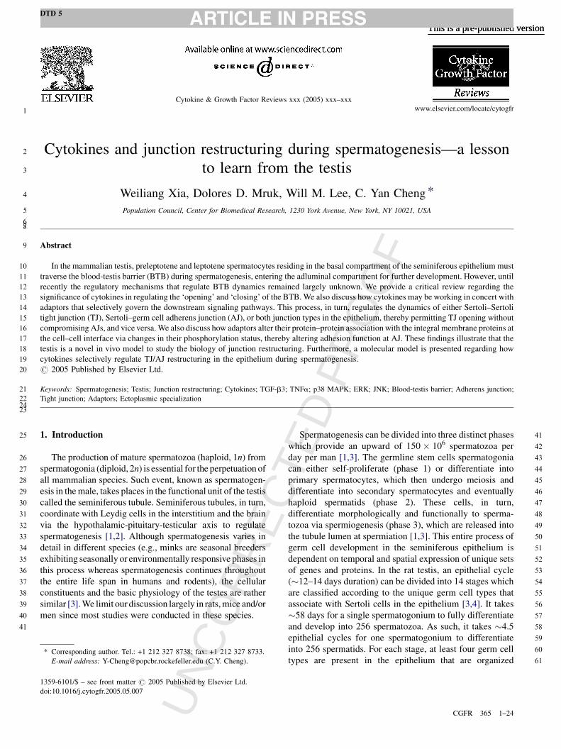
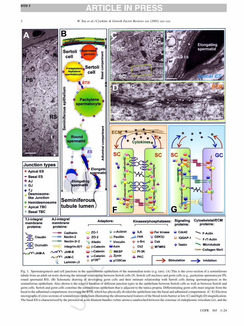
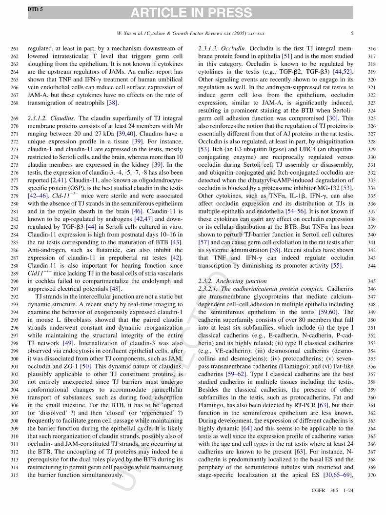
Fig. 1. Spermatogenesis and cell junctions in the seminiferous epithelium of the mammalian testis (e.g. rats). (A) This is the cross-section of a seminiferous
tubule from an adult rat testis showing the intimate relationship between Sertoli cells (N, Sertoli cell nucleus) and germ cells (e.g., pachytene spermatocyte PS,
round spermatid RS). (B) Schematic drawing of developing germ cells and their intimate relationship with Sertoli cells during spermatogenesis in the
seminiferous epithelium. Also shown is the relative location of different junction types in the epithelium between Sertoli cells as well as between Sertoli and
germ cells. Sertoli and germ cells constitue the seminiferous epithelium that is adjacent to the tunica propria. Differentiating germ cells must migrate from the
basal to the adluminal compartment, traversing the BTB, which has physically divided the epithelium into the basal and adluminal compartment. (C–E) Electron
micrographs of cross sections of seminiferous epithelium illustrating the ultrastructural features of the blood-testis barrier at low (C) and high (D) magnification.
The basal ES is characterized by the presence of actin filament bundles (white arrows) sandwiched between the cisternae of endoplasmic reticulum (er), and the
UN
CO
RR
EC
TED
PR
OO
F
spatially into layers from the base to the lumen of the
seminiferous tubule [3,4]. Furthermore, spermatogenesis
cannot completewithout the support of Sertoli cells, which are
the only other cell type in the seminiferous epithelium behind
the BTB besides germ cells (note: the BTB has physically
divided the epithelium into the basal and adluminal
compartment, see Fig. 1) [5–7]. Except for the spermatogonia,
developing germ cells move progressively toward the lumen
[8]. For instance, preleptotene and leptotene spermatocytes
that lie at the periphery of the tubule and outside the BTB must
traverse the BTB at late stages VIII and early IX of the
epithelial cycle [8].
It is conceivable that enormous Sertoli–germ cell
interactions take place in the seminiferous epithelium
throughout spermatogenesis [1–4,6,9,10]. If one views
spermatogenesis as a voyage of a germ cell that moves from
the basal to the adluminal compartment while developing to a
mature spermatozoon, this process involves numerous
decision makings and executions. It also requires signalings
in and out of germ cells to facilitate this event. Although it is
not entirely clear regarding the sequence of these signals,
there are at least two sources: external signals from outside the
tubule (e.g., via Leydig cells, peritubular myoid cells, and
both paracrine and hormonal factors including those from the
pituitary gland), and internal crosstalks between germ and
Sertoli cells (e.g., integrin-mediated signalings) [5,6,11,12].
The phenotypic consequence of these signalings is mani-
fested, at least in part, via the constant remodeling at the
Sertoli–Sertoli and Sertoli–germ cell interfacewhere different
cell junction types are present [1,2].
The identities of these signals and the details of the
remodeling events have become increasingly clear in recent
years [1,2]. For instance, there is accumulating evidence that
illustrates the crucial roles of cytokines pertinent to sperma-
togenesis and junction restructuring [2]. In this review, we first
give an update on the junction complexes that are found in the
testis, highlighting how cytokines (e.g., TGF-b3, TNFa) can
affect junction dynamics and how these signals are being fine-
tuned to allow their regulation of a particular junction type.
2. The seminiferous epithelium: Sertoli–germ cell
junctions and spermatogenesis
2.1. Seminiferous epithelium
The seminiferous epithelium is composed of Sertoli and
germ cells. The Sertoli cell is by and large a tall columnar
cell extending from the base to the apex of the seminiferous
tubule [3]. It is physically reshaped by germ cells to possess
many cytoplasic processes because each Sertoli cell is
‘nursing’ about 30–50 germ cells at different stages of their
development at any given time during the epithelial cycle
[13,14]. In the rat, Sertoli cells cease to proliferate at about
day 20 postnatal and the number of these nursing cells
determines how many germ cells can be supported and
produced via spermatogenesis in the testis [3], illustrating
the crucial function of Sertoli cells. For instance, Sertoli
cells provide structural support for germ cells and their
translocation, create the BTB and define the polarity of the
epithelium, secrete numerous biological factors and
nutrients for germ cells, and conduct other vital functions
pertinent to spermatogenesis (e.g., phagocytosis) [2,3,14].
2.2. Sertoli–Sertoli and Sertoli–germ cell junctions
The different junction types that are found in the
seminiferous epithelium have recently been reviewed [1,2].
Similar to other epithelia or endothelia, virtually all major
junction types are found in the testis. Besides the tight
junctions that are restricted to the BTB, several anchoring
junction types (four are found in most epithelia) are also
detected in the testis: (a) adherens junction (including basal
and apical ectoplasmic specialization [ES], basal and apical
tubulobulbar complex [TBC]); (b) desmosome-like junctions;
and (c) hemidesmosomes (for reviews, see [1,2,10,15]). ES is
a testis-specific, actin-based adherens junction localized at
two sites in the seminiferous epithelium: basal and apical
compartment (see Fig. 1) [2,9,10,16,17]. Basal ES is limited
to BTB and present side-by-side with TJ (Fig. 1). Apical ES is
found between elongating/elongate spermatids and Sertoli
cells. At least three protein complexes, namely, the cadherin/
catenin, the nectin/afadin, and the integrin/laminin, are known
to be ES components [2,17]. TBC is another modified AJ type
found in the testis [2,10,18]. Apical TBC only appears a few
days before spermiation in the epithelium at late stage VIII of
the epithelial cycle when apical ES begins to disappear
whereas basal TBC co-exists with TJ, basal ES, and
desmosomal-like junctions at the BTB site. Desmosome-like
junctions are present between Sertoli cells and spermatogo-
nia, spermatocytes and round spermatids, being most
prominent surrounding pachytene spermatocytes [10]. The
BTB is not fully formed until 16–19 days postnatal in the rat
testis [3]. Unlike barriers in other organs (e.g., the blood–brain
barrier, the blood–retinal barrier) where TJs are localized to
the apical region of the epithelium/endothelium, to be
W. Xia et al. / Cytokine & Growth Factor Reviews xxx (2005) xxx–xxx 3
DTD 5
CGFR 365 1–24
62
63
64
65
66
67
68
69
70
71
72
73
74
75
76
77
78
79
80
81
82
83
84
85
86
87
88
89
90
91
92
93
94
95
96
97
98
99
100
101
102
103
104
105
106
107
108
109
110
111
112
113
114
115
116
117
118
119
120
121
122
123
124
125
126
127
128
129
130
131
132
133
134
135
136
137
138
139
140
141
142
143
144
145
146
147
148
149
150
151
Sertoli cell membrane (apposing arrowheads represent the apposing Sertoli cell membranes), which can be found on both sides of the apposing Sertoli cells.
Tight junction (TJ) is found between the basal ES, the coexisting TJ and basal ES in turn constitute the BTB. Apical ES is shown in (E) which is typified by the
presence of actin filament bundles (white arrowheads) sandwiches between the cisternae of er and Sertoli cell membrane (apposing white arrowheads represent
the apposing Sertoli and germ cell membranes). However, this typical feature of ES, in contrast to the basal ES, is restictied only to the Sertoli cell side in apical
ES. (F–G) Schematic drawings that illustrate the molecular architecture of the constituent proteins at the BTB (F) and apical ES (G), which include cytokines
(e.g., TGF-b3 and TNFa) released from Sertoli and/or germ cells can mediate Sertoli–germ cell crosstalk during spermatogenesis. The protein complexes
known to exist at the apical ES site include cadherin/catenin, nectin/afadin, and a6b1 integrin/laminin g3; whereas occludin/ZO-1, JAM/ZO-1, claudin/ZO-1,
cadherin/catenin and nectin/afadin are found at the BTB site. Bar in E = 10 mm, C = 3 mm, D = 0.25 mm and E = 0.3 mm, respectively.
UN
CO
RR
EC
TED
PR
OO
F
followed by AJ, and TJs are furthest away from the ECM; TJs
at the BTB lie closest to the basement membrane (a modified
form of ECM). Furthermore, BTB is a dynamic structure
which must ‘open’ and ‘close’ to permit preleptotene/
leptotene spermatocyte transmigration. BTB is a rather
complex barrier when compared to other barriers (e.g.,
gastric–mucosal barrier which is formed by epithelial cells,
blood–retinal barrier and blood–brain barrier which are
formed by endothelial cells) [19–21] (see Fig. 1). Recent
studies have also shown that apical ES is constituted and
regulated by proteins that are usually restricted to the focal
contact in cell–matrix interface in other epithelia [22]. This
hybrid cell–matrix–cell junction type may indeed be essential
for rapid junction remodeling to facilitate spermatids
orientation and movement at spermiation.
2.3. Constituent proteins of different junction types in
the testis
2.3.1. Tight junction (TJ)
TJ is the only known example of occluding junction that
confers the barrier function of an epithelium or endothelium
by restricting the passages of molecules through the
intercellular spaces and creates a boundary that defines cell
polarity [23]. In the testis, TJ also creates an immunological
barrier that sequesters the post-meiotic germ cell antigens
from the immune system of the host animals. The currently
known TJ integral membrane proteins include JAMs
(junctional adhesion molecules), claudins and occludins,
which have recently been reviewed [1,2,23,24], as such, only
a brief update is provided in this section.
2.3.1.1. JAMs. JAMs are members of a distinct class of cell
adhesion molecules typified by the presence of two Ig-like
loops in the extracellular domain that are expressed in
leukocytes and are localized to tight junctions as integral
membrane proteins in epithelial and endothelial cells
[25,26]. Since the discovery of JAM-A in 1998 [27], other
members, including the more related JAM-B and JAM-C,
and the less related JAM4, coxsackie and adenovirus
receptor (CAR), and endothelial cell-selective adhesion
molecule (ESAM), have recently been added to the list
[25,26,28]. The presence of JAM-A, B and C in the testis
have now been confirmed [29,30]. JAM-A is present at the
BTB in the rat testis, co-localizing with ZO-1 [30].
Moreover, JAM-A expression is stage-specific, being
highest at IX–XIV, lowest at IV–VI [30]. This stage
specificity apparently is related to its possible involvement
in BTB dynamics, facilitating the passage of preleptotene/
leptotene spermatocytes across the BTB. Although Jam-A�/
� mice has been generated, it is not known if the BTB is
affected since a morphological examination of the testis has
yet to be reported [31]. A recent study on the Jam-C�/� mice
have shown that JAM-C is crucial to spermiogenesis since in
the viable mutants, mature spermatids are missing [29]. In
normal mice, JAM-C is localized to the developing round
and elongating spermatids [29]. Interestingly, JAM-B has
been localized both to the site of TJs at the basal
compartment and to the apical ES at the spermatid-Sertoli
cell interface in the seminiferous epithelium, outside the
BTB [29]. Besides their homophilic interactions amongst
JAM-A, B and C, JAM-C can interact with JAM-B
heterotypically [32]. Both JAM-B and JAM-C are localized
to the heads of spermatids at the apical ES, and this
heterophilic association may be important for the Sertoli
cell-spermatid adhesion function [29]. Based on currently
available data, two roles are suggested for JAMs: in the
immune system they are crucial to leukocyte transmigra-
tion; and in polarized epithelial and endothelial cells, they
seem to take part in organizing TJ and cell polarity [26].
This latter physiological role has been extended to the testis
since the cell polarity complex [partitioning-defective (Par)
3/atypical protein kinase C (aPKC)/Cdc42] apparently is
recruited by JAM-C to facilitate round spermatid polariza-
tion and thus differentiation [26]. How JAMs assist
preleptotene/leptotene spermatocytes to traverse the BTB
similar to neutrophil transmigration across the endothelial
TJ-barrier remains to be investigated since germ cells per se,
unlike neutrophils or macrophages, are not actively migrating
cells. It is possible that JAMs are associated with other
motor proteins (e.g., myosin VIIa) and cytoskeletons (e.g.,
actin, tubulin) that facilitate germ cell movement using
the locomotive apparatus in Sertoli cells that provides the
necessary protrusive force to guide germ cell movement
(for review, see [2]).
JAMs are expressed in multiple epithelia, endothelia,
leukocytes and platelets [25,26]. The regulation of JAMs in
the testis is largely unknown. In the rat testis, when the
intratesticular T was suppressed by placing testosterone and
estrogen implants subdermally, spermatids (step 8 and
beyond) were depleted because of a disruption of the cell
adhesion at the ES [30,33–36]. However, the tight junctions
at the BTB remained intact which were associated with a
significant surge in the levels of JAM-A, occludin and ZO-1
in the epithelium [30]. Indeed, the JAM-A distribution at the
BTB site in the basal compartment of the seminiferous
epithelium was significantly induced and intensified,
becoming a thickened and prominent ring surrounding the
entire tubule [30]. It is apparent that a depletion of androgen
in the testis triggers a novel mechanism that leads to two
distinctive events: germ cell loss and a reinforced BTB [30].
Another model using Adjudin to induce germ cell sloughing
from the epithelium in adult rat testes has yield similar
results in which JAM-A expression was induced at the time
of germ cell depletion (unpublished observations). Although
the compounds that were used to trigger the changes in the
epithelium are different in these two models, namely
androgen suppression and Adjudin, the signaling events
(e.g., both treatments activate the integrin/focal adhesion
kinase signaling pathway) and the phenotypic outcome (e.g.,
germ cell loss from the epithelium and a reinforced BTB) are
similar [36,37]. This seemingly suggests that JAM-A is
W. Xia et al. / Cytokine & Growth Factor Reviews xxx (2005) xxx–xxx4
DTD 5
CGFR 365 1–24
152
153
154
155
156
157
158
159
160
161
162
163
164
165
166
167
168
169
170
171
172
173
174
175
176
177
178
179
180
181
182
183
184
185
186
187
188
189
190
191
192
193
194
195
196
197
198
199
200
201
202
203
204
205
206
207
208
209
210
211
212
213
214
215
216
217
218
219
220
221
222
223
224
225
226
227
228
229
230
231
232
233
234
235
236
237
238
239
240
241
242
243
244
245
246
247
248
249
250
251
252
253
254
255
256
257
258
259
260
UN
CO
RR
EC
TED
PR
OO
F
regulated, at least in part, by a mechanism downstream of
lowered intratesticular T level that triggers germ cell
sloughing from the epithelium. It is not known if cytokines
are the upstream regulators of JAMs. An earlier report has
shown that TNF and IFN-g treatment of human umbilical
vein endothelial cells can reduce cell surface expression of
JAM-A, but these cytokines have no effects on the rate of
transmigration of neutrophils [38].
2.3.1.2. Claudins. The claudin superfamily of TJ integral
membrane proteins consists of at least 24 members with Mr
ranging between 20 and 27 kDa [39,40]. Claudins have a
unique expression profile in a tissue [39]. For instance,
claudin-1 and claudin-11 are expressed in the testis, mostly
restricted to Sertoli cells, and the brain, whereas more than 10
claudin members are expressed in the kidney [39]. In the
testis, the expression of claudin-3, -4, -5, -7, -8 has also been
reported [2,41]. Claudin-11, also known as oligodendrocyte-
specific protein (OSP), is the best studied claudin in the testis
[42–46]. Cld-11�/� mice were sterile and were associated
with the absence of TJ strands in the seminiferous epithelium
and in the myelin sheath in the brain [46]. Claudin-11 is
known to be up-regulated by androgens [42,47] and down-
regulated by TGF-b3 [44] in Sertoli cells cultured in vitro.
Claudin-11 expression is high from postnatal days 10–16 in
the rat testis corresponding to the maturation of BTB [43].
Anti-androgen, such as flutamide, can also inhibit the
expression of claudin-11 in prepubertal rat testes [42].
Claudin-11 is also important for hearing function since
Cld11�/� mice lacking TJ in the basal cells of stria vascularis
in cochlea failed to compartmentalize the endolymph and
suppressed electrical potentials [48].
TJ strands in the intercellular junction are not a static but
dynamic structure. A recent study by real-time imaging to
examine the behavior of exogenously expressed claudin-1
in mouse L fibroblasts showed that the paired claudin
strands underwent constant and dynamic reorganization
while maintaining the structural integrity of the entire
TJ network [49]. Internalization of claudin-3 was also
observed via endocytosis in confluent epithelial cells, after
it was dissociated from other TJ components, such as JAM,
occludin and ZO-1 [50]. This dynamic nature of claudins,
plausibly applicable to other TJ constituent proteins, is
not entirely unexpected since TJ barriers must undergo
conformational changes to accommodate paracellular
transport of substances, such as during food adsorption
in the small intestine. For the BTB, it has to be ‘opened
(or ‘dissolved’ ?) and then ‘closed’ (or ‘regenerated’ ?)
frequently to facilitate germ cell passage while maintaining
the barrier function during the epithelial cycle. It is likely
that such reorganization of claudin strands, possibly also of
occludin- and JAM-constituted TJ strands, are occurring at
the BTB. The uncoupling of TJ proteins may indeed be a
prerequisite for the dual roles played by the BTB during its
restructuring to permit germ cell passage while maintaining
the barrier function simultaneously.
2.3.1.3. Occludin. Occludin is the first TJ integral mem-
brane protein found in epithelia [51] and is the most studied
in this category. Occludin is known to be regulated by
cytokines in the testis (e.g., TGF-b2, TGF-b3) [44,52].
Other signaling events are recently shown to engage in its
regulation as well. In the androgen-suppressed rat testes to
induce germ cell loss from the epithelium, occludin
expression, similar to JAM-A, is significantly induced,
resulting in prominent staining at the BTB when Sertoli–
germ cell adhesion function was compromised [30]. This
also reinforces the notion that the regulation of TJ proteins is
essentially different from that of AJ proteins in the rat testis.
Occludin is also regulated, at least in part, by ubiquitination
[53]. Itch (an E3 ubiquitin ligase) and UBC4 (an ubiquitin-
conjugating enzyme) are reciprocally regulated versus
occludin during Sertoli cell TJ assembly or disassembly,
and ubiquitin-conjugated and Itch-conjugated occludin are
detected when the dibutyryl-cAMP-induced degradation of
occludin is blocked by a proteasome inhibitor MG-132 [53].
Other cytokines, such as TNFa, IL-1b, IFN-g, can also
affect occludin expression and its distribution at TJs in
multiple epithelia and endothelia [54–56]. It is not known if
these cytokines can exert any effect on occludin expression
or its cellular distribution at the BTB. But TNFa has been
shown to perturb TJ-barrier function in Sertoli cell cultures
[57] and can cause germ cell exfoliation in the rat testis after
its systemic administration [58]. Recent studies have shown
that TNF and IFN-g can indeed regulate occludin
transcription by diminishing its promoter activity [55].
2.3.2. Anchoring junction
2.3.2.1. The cadherin/catenin protein complex. Cadherins
are transmembrane glycoproteins that mediate calcium-
dependent cell–cell adhesion in multiple epithelia including
the seminiferous epithelium in the testis [59,60]. The
cadherin superfamily consists of over 80 members that fall
into at least six subfamilies, which include (i) the type I
classical cadherins (e.g., E-cadherin, N-cadherin, P-cad-
herin) and its highly related; (ii) type II classical cadherins
(e.g., VE-cadherin); (iii) desmosomal cadherins (desmo-
collins and desmogleins); (iv) protocadherins; (v) seven-
pass transmembrane cadherins (Flamingo); and (vi) Fat-like
cadherins [59–62]. Type I classical cadherins are the best
studied cadherins in multiple tissues including the testis.
Besides the classical cadherins, the presence of other
subfamilies in the testis, such as protocadherins, Fat and
Flamingo, has also been detected by RT-PCR [63], but their
function in the seminiferous epithelium are less known.
During development, the expression of different cadherins is
highly dynamic [64] and this seems to be applicable to the
testis as well since the expression profile of cadherins varies
with the age and cell types in the rat testis where at least 24
cadherins are known to be present [63]. For instance, N-
cadherin is predominantly localized to the basal ES and the
periphery of the seminiferous tubules with restricted and
stage-specific localization at the apical ES [30,65–69],
W. Xia et al. / Cytokine & Growth Factor Reviews xxx (2005) xxx–xxx 5
DTD 5
CGFR 365 1–24
261
262
263
264
265
266
267
268
269
270
271
272
273
274
275
276
277
278
279
280
281
282
283
284
285
286
287
288
289
290
291
292
293
294
295
296
297
298
299
300
301
302
303
304
305
306
307
308
309
310
311
312
313
314
315
316
317
318
319
320
321
322
323
324
325
326
327
328
329
330
331
332
333
334
335
336
337
338
339
340
341
342
343
344
345
346
347
348
349
350
351
352
353
354
355
356
357
358
359
360
361
362
363
364
365
366
367
368
369
370
UN
CO
RR
EC
TED
PR
OO
F
whereas E-cadherin is relatively more abundant in germ
cells [63,66]. A smaller amount of N-cadherin in the testis
appears also to be a component of desmosomal-like
junctions which is a hybrid junction type of desmosome
and gap junctions [65,67]. Indeed, N-cadherin has been
shown to link to both actin microfilament and microtubules
in the testis [66]. A recent report has also illustrated that
protocadherin a3 is associated with spermatids at the
acrosomal area, intercellular bridge as well as flagellum,
distinct from the distribution of classical cadherins [70].
Classical cadherin-based protein complex comprising of
the transmembrane protein cadherins and intracellular
adaptor catenins is a well defined focal point of cell
adhesion and signaling [59,71]. b-Catenin and g-catenin
connects cadherins to a-catenin and a-actinin, which are
two putative actin binding proteins [72]. Phosphorylation of
b-catenin can in turn regulate the integrity of the cadherin/
catenin complex [73]. In both Adjudin- and androgen
suppression-induced germ cell loss models, the event of
germ cell loss is facilitated by the dissociation of N-
cadherin from b-catenin [30,35,68]. Indeed, increased
tyrosine phosphorylation of b-catenin was detected at the
time of germ cell depletion in these models [30]. Kinases
and phosphatases are also known to regulate cadherin/
catenin association [35,74,75]. For instance, myotubularin-
related protein 2 (MTMR2), a lipid phosphoinositide
phosphatase, was shown to interact with the kinase c-Src
[35] and c-Src in turn associates with the N-cadherin/b-
catenin complex [74]. This illustrates a novel regulatory
mechanism may be in place in the testis regarding the
cadherin/catenin-mediated cell adhesion function in which
MTMR2 and c-Src regulate the phosphorylation status of
the cadherin/catenin, which in turn determines its cell
adhesive function. More recent studies have shown that the
N-cadherin/b-catenin adhesion unit can also be regulated
by the equilibrium between IQGAP-1 (IQ motif containing
GTPase activating protein, an effector of Cdc42 GTPase)
and Cdc42 in Sertoli–germ cell AJ [76]. For instance, using
a Ca2+ switch model, it has been demonstrated that at low
Ca2+ level, IQGAP-1 is released from Cdc42, and interacts
with b-catenin instead, causing the dissociation of b-
catenin from N-cadherin, and germ cell depletion from
Sertoli cells [76].
E-Cadherin is also a tumor suppressor which is down-
regulated while N-cadherin is up-regulated during epithelial
tumor progression [64,77,78]. This ‘cadherin switch’ further
illustrates the unique yet pivotal role of each cadherin in cell
adhesion and cell motility. It is not clear if such dynamic
switch-over between different cadherins occur during germ
cell movement in the seminiferous epithelium. However,
N-cadherin can become highly expressed in the testis of
Adjudin treated rats during germ cell loss from the
epithelium [66–68]. N-cadherin is also up-regulated in
androgen suppressed rat testes during germ cell loss [30,35].
Yet such a surge in N-cadherin cannot rescue germ cell loss
from the epithelium since a loss of association between N-
cadherin and b-catenin was detected at the time of germ cell
sloughing in both models [30,35]. It seems that such an
induction of cadherins reinforces the BTB integrity since N-
cadherin is also a component protein of the BTB in the rat
testis.
2.3.2.2. The nectin/afadin/ponsin/ADIP complex. The nec-
tin/afadin/ponsin complex is another actin-based cell
adhesion protein complex that plays a crucial role in the
testis during spermatogenesis. It confers Sertoli–germ cell
adhesion function particularly for elongating/elongate
spermatids [1,2,68,79]. Four nectins (nectin-1, -2,-3, and -
4) have been identified thus far, all of which are expressed in
the testis with nectin-2 and nectin-3 being the highly
expressed [2,80–82]. Nectin-3 is restricted exclusively to
elongating/elongate spermatids which can heterotypically
interacting with nectin-2 on the Sertoli cell side [68,83].
Spermatozoa from nectin-2�/� mice were morphologically
aberrant and functionally impotent [83–85]. Since nectins
are capable of activating Cdc42 via c-Src and a Cdc42 GEF
(GDP/GTP exchange factor) [86], or activating Rac, thus
recruiting the polarity complex Par3/aPKC/Par6 to the
apical ES site [87], the absence of nectin-3 may also lead to
malfunctioning of spermatid polarization, similar to Jam-
C�/� mice [29]. Nectins are known to initiate cell–cell
contacts by recruiting cadherin and JAM-A to establish
functional AJ and TJ in epithelial cells [79,87–89]. It is
likely that nectin-2/-3 and JAM-B/-C can also interact with
each other since they are all localized to the elongating/
elongate spermatids at the apical ES site, which should be
investigated in future studies. In the Adjudin-induced germ
cell loss model, it was found that the nectin-3/afadin
interaction became severely weakened before any obvious
reduction in their protein levels was detected [68],
illustrating this cell adhesion unit must be compromised
to facilitate spermatid loss (Table 1).
Besides afadin and ponsin, cytoplasmic adaptors that link
nectin to the actin-based cytoskeleton [79], a new adaptor
protein ADIP (afadin DIL domain-interacting protein) has
recently been localized to AJ sites that interacts with both a-
actinin and afadin, providing additional cytoplasmic link
between nectin- and cadherin-based cell adhesion units
[90,91]. ADIP is highly expressed in the mouse testis [90].
Another possible linker that binds to both afadin and a-
actinin is LMO7 (LIM domain only 7), however, its presence
in the rat testis failed to be confirmed by immunoblot
analysis [92].
Nectin-like (Necl) molecules are similar to nectins, but
do not bind to afadins [87,88]. This group of calcium-
independent cell adhesion molecules consists of five
members, capable of homo- or heterophilic interactions
with nectins, and are important cell–cell adhesion molecules
in various tissues [87,88]. At least Necl2 has been shown to
be highly expressed in the rat testis [93]. It will be important
to explore the significance of Necls in the testis, which is
likely to involve in Sertoli–germ cell adhesion function.
W. Xia et al. / Cytokine & Growth Factor Reviews xxx (2005) xxx–xxx6
DTD 5
CGFR 365 1–24
371
372
373
374
375
376
377
378
379
380
381
382
383
384
385
386
387
388
389
390
391
392
393
394
395
396
397
398
399
400
401
402
403
404
405
406
407
408
409
410
411
412
413
414
415
416
417
418
419
420
421
422
423
424
425
426
427
428
429
430
431
432
433
434
435
436
437
438
439
440
441
442
443
444
445
446
447
448
449
450
451
452
453
454
455
456
457
458
459
460
461
462
463
464
465
466
467
468
469
470
471
472
473
474
475
476
477
478
479
480
481

UN
CO
RR
EC
TED
PR
OO
F
2.3.2.3. The integrin/laminin complex. The integrin/lami-
nin protein complex has recently been identified at the apical
ES which confers Sertoli–germ cell adhesion and provides a
new platform regarding how these two cell types interact
with each other and coordinate spermatogenesis [37,94].
Integrin-based protein complexes are usually found at the
cell–matrix junctions, such as hemidesmosomes or focal
adhesion, which further connects to the intermediate
filament or actin bundles, with integrin also capable of
serving as a cell receptor for the ECM [95,96]. Interestingly,
the junctions between Sertoli and germ cells are not simple
cell–cell junction types; rather, they are a hybrid of both
cell–cell and cell–matrix junction types, probably to
facilitate rapid junction turnover and germ cell migration
during spermatogenesis [22]. Several recent reviews on the
role of integrins and ECM in the testis are available, thus this
information is not discussed herein [22,97].
3. Cytokines are key regulators of junction dynamicsin the testis
Cytokines are regulatory peptides (usually �30 kDa in
size) produced virtually by every nucleated cells in
mammals and have pleiotropic actions on cell physiology
as an autocrine or paracrine factor [98]. In the testis, Sertoli
and germ cells produce a number of cytokines, including
members of the TGF-b superfamily (e.g., TGF-bs, activins,
inhibins), platelet-derived growth factor (PDGF), interleu-
kins (e.g., IL-1, IL-6, IL-11), tumor necrosis factor (e.g.,
TNFa, Fas ligand), interferons (e.g., IFN-a, IFN-g),
fibroblast growth factor (FGF), nerve growth factor
(NGF), and stem cell factor (or steel factor) (for reviews,
see [1,2,11,99,100]) (see also Table 2). These cytokines likely
mediate crosstalk between Sertoli and germ cells to facilitate
germ cell movement across the seminiferous epithelium and
other cellular events in the epithelium during the epithelial
cycle such as germ cell differentiation. Herein, we critically
evaluate two best studied cytokines, namely TNFa and TGF-
b3, regarding their significance in spermatogenesis in the
testis and briefly summarize the action of other cytokines.
3.1. TNF
TNF, also known as TNFa or cachectin, is synthesized as
a 26 kDa type II transmembrane prepeptide (pro-TNF),
which is subsequently activated by proteolytic cleavage to
release the C-terminal 17 kDa mature protein by the TNF-
converting enzyme (TACE). The mature protein is formed
by aggregates creating a homotrimer that can bind to two
types of receptors: TNFR1 and TNFR2 [101,102]. The
major source of TNFa in mammalian body is immune cells
such as macrophage and monocytes, but TNFa is also
produced by other non-immune cells including astrocytes,
keratinotytes, Sertoli cells and germ cells [57,101]. TNF
signaling is mediated mainly through TNFR1, which has
distinct domains that facilitate the recruitment of other
intracellular adaptors to activate signaling pathways. The net
result of such activation can modulate apoptosis, inflamma-
tion and cell proliferation [101,103]. These adaptors include
TNFR1-associated death domain protein (TRADD) which
can recruit Fas-associated death domain protein (FADD),
TNF receptor associated factor-2 (TRAF-2), or receptor-
interacting protein (RIP), to induce the caspase-mediated
apoptosis, activate transcription factors (e.g., c-jun, c-fos,
ATF-2) via MAPK (ERK, JNK and p38), or activate nuclear
factor kappa B (NFkB) through inhibitor of NFkB kinase
(IKK), respectively [101–103]. A TNFR1 scaffolding
protein called TGFR-associated ubiquitous scaffolding
and signaling protein (TRUSS) has recently been cloned
and characterized [104]. The expression of TRUSS is
enriched in heart, liver and testes, it is also known to interact
with TRADD, TRAF-2 and IKK [104]. In addition to these
complex signaling networks that can be activated down-
W. Xia et al. / Cytokine & Growth Factor Reviews xxx (2005) xxx–xxx 7
DTD 5
CGFR 365 1–24
482
483
484
485
486
487
488
489
490
491
492
493
494
495
496
497
498
499
500
501
502
503
504
505
506
507
508
509
510
511
512
513
514
515
516
517
518
519
520
521
522
523
524
525
526
527
528
529
530
531
532
533
534
535
536
537
538
539
540
541
542
543
544
545
546
547
548
549
550
Table 1
Cytokine-mediated regulation of junction component proteins in epithelia including the testis
Junction component Protein Cytokine/hormone that modulates the steady-state
mRNA/protein level (+/�) or protein distribution pattern
(d) of the target junction protein
Selected references
TJ-integral membrane JAM-A TNF (d), IFN-g (d), T#(+) [30,38]
Occludin TGF-b3 (�), HGF (�/d), TNF (�/d), IFN-g (�/d), VEGF
(�/d, inhibited by ANP), IL-1b (�/d), IL-4 (�), IL-13 (�), MCP-1 (�), T#(+)
[44,52,54–56,223–225]
Claudin TGF-b3 (�), TNF (�), FSH/cAMP (�) [43,44]
AJ-integral membrane N-Cadherin HGF (+), EGF (+), TGF-b (+/d), T (+), T# (+), IL-6 (�) [30,66,78]
E-Cadherin TGF-b (�/d), T (+) [52,66,68]
Nectin-3 TGF-b3(�/d) [52,68]
Integrin-b1 TGF-b (+), T#(+), [30,226]
Adaptor ZO-1 TGF-b (d), IL-4 (�), IL-13 (�), T#(+) [30,225]
Afadin TGF-b3(�/d) [68]
b-Catenin TGF-b (d), T#(+) [30,139,227]
a-Catenin TGF-b (d), T#(+) [30,139,227]
T#, suppression of intratesticular testosterone level with the use of testosterone (T) and estradiol implants; +, stimulation; �, inhibition. Protein distribution pattern
was assessed by either immunofluorescent microscopy or immunohistochemistry using testicular cells cultured in vitro or seminiferous epithelium in vivo.
UNCORRECTED PROOF
W.
Xia
eta
l./Cyto
kine
&G
row
thF
acto
rR
eviews
xxx(2
00
5)
xxx–xxx
8 DT
D5
CG
FR
36
51
–2
4
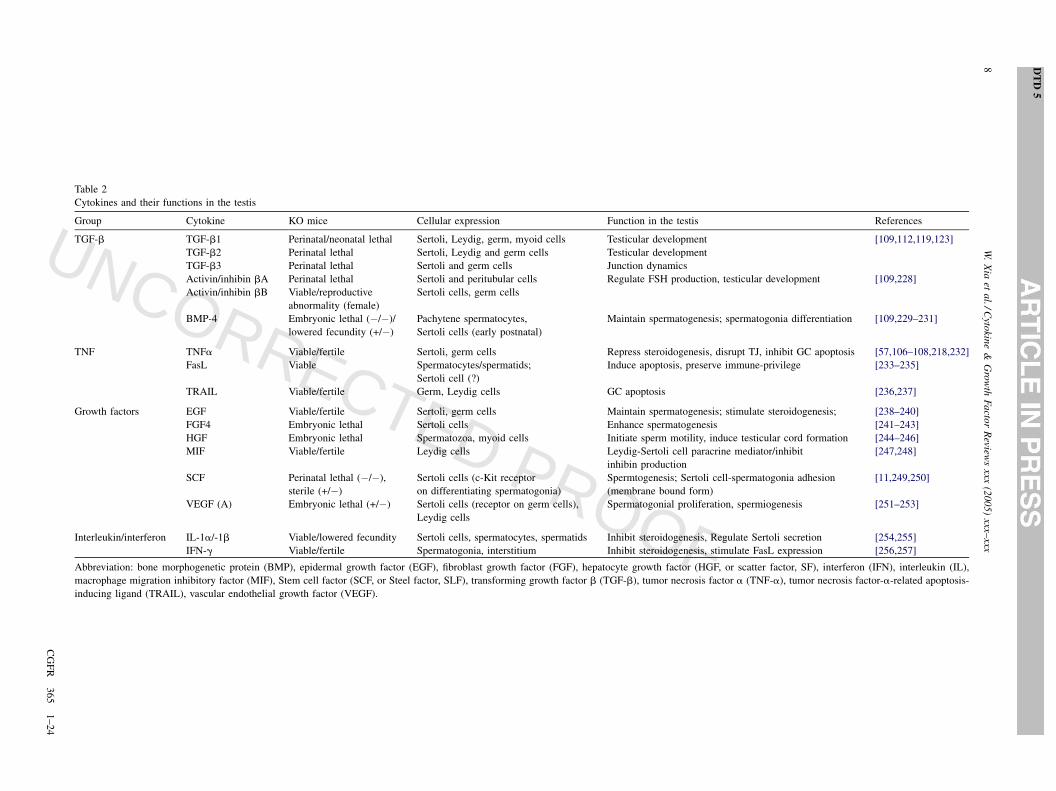
Table 2
Cytokines and their functions in the testis
Group Cytokine KO mice Cellular expression Function in the testis References
TGF-b TGF-b1 Perinatal/neonatal lethal Sertoli, Leydig, germ, myoid cells Testicular development [109,112,119,123]
TGF-b2 Perinatal lethal Sertoli, Leydig and germ cells Testicular development
TGF-b3 Perinatal lethal Sertoli and germ cells Junction dynamics
Activin/inhibin bA Perinatal lethal Sertoli and peritubular cells Regulate FSH production, testicular development [109,228]
Activin/inhibin bB Viable/reproductive
abnormality (female)
Sertoli cells, germ cells
BMP-4 Embryonic lethal (�/�)/
lowered fecundity (+/�)
Pachytene spermatocytes,
Sertoli cells (early postnatal)
Maintain spermatogenesis; spermatogonia differentiation [109,229–231]
TNF TNFa Viable/fertile Sertoli, germ cells Repress steroidogenesis, disrupt TJ, inhibit GC apoptosis [57,106–108,218,232]
FasL Viable Spermatocytes/spermatids;
Sertoli cell (?)
Induce apoptosis, preserve immune-privilege [233–235]
TRAIL Viable/fertile Germ, Leydig cells GC apoptosis [236,237]
Growth factors EGF Viable/fertile Sertoli, germ cells Maintain spermatogenesis; stimulate steroidogenesis; [238–240]
FGF4 Embryonic lethal Sertoli cells Enhance spermatogenesis [241–243]
HGF Embryonic lethal Spermatozoa, myoid cells Initiate sperm motility, induce testicular cord formation [244–246]
MIF Viable/fertile Leydig cells Leydig-Sertoli cell paracrine mediator/inhibit
inhibin production
[247,248]
SCF Perinatal lethal (�/�),
sterile (+/�)
Sertoli cells (c-Kit receptor
on differentiating spermatogonia)
Spermtogenesis; Sertoli cell-spermatogonia adhesion
(membrane bound form)
[11,249,250]
VEGF (A) Embryonic lethal (+/�) Sertoli cells (receptor on germ cells),
Leydig cells
Spermatogonial proliferation, spermiogenesis [251–253]
Interleukin/interferon IL-1a/-1b Viable/lowered fecundity Sertoli cells, spermatocytes, spermatids Inhibit steroidogenesis, Regulate Sertoli secretion [254,255]
IFN-g Viable/fertile Spermatogonia, interstitium Inhibit steroidogenesis, stimulate FasL expression [256,257]
Abbreviation: bone morphogenetic protein (BMP), epidermal growth factor (EGF), fibroblast growth factor (FGF), hepatocyte growth factor (HGF, or scatter factor, SF), interferon (IFN), interleukin (IL),
macrophage migration inhibitory factor (MIF), Stem cell factor (SCF, or Steel factor, SLF), transforming growth factor b (TGF-b), tumor necrosis factor a (TNF-a), tumor necrosis factor-a-related apoptosis-
inducing ligand (TRAIL), vascular endothelial growth factor (VEGF).
UN
CO
RR
EC
TED
PR
OO
F
stream of TNF, at least 19 ligands and more than 20
receptors have been identified in the TNF superfamily [103],
which can mediate an array of physiological processes and
diseases [103]. It has been known that TNF can disrupt TJ
integrity in multiple epithelial cells. As aforementioned,
TNF down-regulates occludin expression through its
promoter activity [55] or reduces JAM-A distribution on
the vascular endothelial cell surface [38]. TNF level is also
elevated in Crohn’s disease, a chronic granulomatous
inflammatory disease that affects the gastrointestinal tract,
which is manifested by impaired intestinal barrier function
with leaky TJs [105]. With the exception of TNF and Fas
ligand (FasL), the roles of other members of TNF superfamily
in the testis remain elusive.
In the testis, TNFa has been shown to play a role in
regulating germ cell apoptosis, junction remodeling and
Leydig cell steroidogenesis [57,106,107]. For instance, it is
known that TNFa represses the expression of steroidogenic-
enzyme genes in Leydig cells through an activation of
NFkB, which can in turn inhibit the transactivation of
orphan nuclear receptors [106]. Intratesticular injection of
TNF in normal and hypophysectomized rats has also
demonstrated its suppressive effect on testosterone produc-
tion in vivo [108]. Chronic infusion of TNF caused germ cell
(in particular spermatocytes and spermatids) depletion from
the epithelium, a loss of testis weight and a plunge in
testosterone level [58]. It remains unknown regarding the
mechanism(s) by which TNFa utilized to induce these
changes, but this could involve a suppression of Leydig cells
steroidogenesis, or an inhibition of Sertoli cell TJ protein
production at the BTB, or via its direct effect on germ cells.
Other recent studies have shown that TNFa can perturb the
TJ-permeability barrier in cultured Sertoli cells dose-
dependently and reversibly since the disrupted TJ-barrier
can be resealed upon the removal of the cytokine [57]. This
inhibitory effect of TNFa on Sertoli cell TJ function is likely
mediated via an induced production of collagen a3(IV),
matrix metalloprotease (MMP)-9 and tissue inhibitor of
metalloprotease (TIMP)-1 which collectively affect the
homeostasis of ECM, thereby altering the association of the
Sertoli cell epithelium with the basement membrane and
perturbing the TJ-barrier [57]. Also, TNFa can activate the
integrin/integrin linked kinase (ILK)/glycogen synthase
kinase (GSK) b-3/p130 Cas/JNK signaling pathway which
also contribute to changes in the TJ-protein expression and/
or distribution at the BTB [22,57,97].
3.2. TGF-b
The TGF-b superfamily comprises of TGF-bs, activins,
inhibins, bone morphogenetic proteins (BMPs), growth
differentiation factors (GDFs), Mullerian-inhibiting sub-
stance (MIS) and others, totaling more than 35 members
[109]. TGF-b superfamily proteins are crucial in the
regulation of a variety of biological processes, including
cell proliferation, differentiation, apoptosis, and tissue
remodeling [110]. Some members, like activins and
inhibins, were initially identified in the male gonad for
their ability to regulate the pituitary follicle stimulating
hormone (FSH) production [11]. MIS is known for its role in
sexual differentiation causing the regression of the
Mullerian ducts in the male [111]. The functions of TGF-
b superfamily proteins in reproduction have been recently
reviewed [109,112] hence we only focus on regulation of
junction restructuring by TGF-bs herein, which is elabo-
rated in Section 4. Table 2 summarizes other cytokines that
are known regulators of junction dynamics.
3.3. Cytokines working in concert with other ECM
proteins to regulate junction dynamics
Recent reviews have summarized how cytokines regulate
the homeostasis of proteases and their inhibitors, and ECM
proteins to coordinate spermatogenesis [2,22,97]. It is not at
all surprising that these molecules are working in concert
since their production, activation, and termination are all
interdependent and connected. Their homeostasis and
regulation are essential to almost all biological processes.
In the testis, for instance, when the BTB is disrupted by
cadmium, TGF-b3/p38 MAPK signaling is activated to
down-regulate the steady-state of TJ and AJ protein levels
that leads to the breakdown of both junctions and germ cell
exfoliation [44,52,113]. Proteases (e.g., cathepsin L) and
protease inhibitors (e.g., a2-macroglobulin) are induced to
coordinate the junction restructuring event [52]. Using a p38
MAPK inhibitor SB202190, the damage to the BTB and the
plunge of TJ and AJ proteins induced by CdCl2 can be
delayed but it cannot prevent the overexpression of protease
inhibitor a2-MG [52]. Further study revealed that a2-MG
production is regulated by JNK signaling pathway in the
testis, independent of the p38 MAPK pathway [114]. This
yin and yang relation of protease and protease inhibitor
regulation that utilizes distinct signaling pathways, and their
connection with cytokines (e.g., TGF-b3) have illustrated
that the testis is equipped with some delicate regulatory
mechanisms to orchestrate junction restructuring at sper-
matogenesis.
4. TGF-b3 as a junction regulator—versatility
realized through selectivity
4.1. Signaling conduits and versatile players in
biological processes
TGF-bs (b1, b2 and b3) are key regulators in a plethora
of biological processes (for reviews, see [110,115–120]).
These cytokines, when activated by releasing from the
latency-associated proteins (LAPs), can bind to their
receptors—first to the type II receptor, TbRII, which then
recruits the type I receptor, TbRI (or ALT5, activin-like
kinase)—although TGF-b2 requires binding of the two
W. Xia et al. / Cytokine & Growth Factor Reviews xxx (2005) xxx–xxx 9
DTD 5
CGFR 365 1–24
551
552
553
554
555
556
557
558
559
560
561
562
563
564
565
566
567
568
569
570
571
572
573
574
575
576
577
578
579
580
581
582
583
584
585
586
587
588
589
590
591
592
593
594
595
596
597
598
599
600
601
602
603
604
605
606
607
608
609
610
611
612
613
614
615
616
617
618
619
620
621
622
623
624
625
626
627
628
629
630
631
632
633
634
635
636
637
638
639
640
641
642
643
644
645
646
647
648
649
650
651
652
653
654
UN
CO
RR
EC
TED
PR
OO
F
receptors more or less at the same time and the assistance
from the type III receptor, betaglycan. The binding of the
cytokine to type I and type II receptors initiates a series of
phosphorylation mediated activation—autophosphorylation
of TbRII and TbRI phosphorylation by TbRII—and triggers
consequent intracellular signaling events (the canonical
Smad-mediated signalings and Smad-independent path-
ways). Despite their structural similarities and shared
signaling mechanisms, the three TGF-bs are spatiotempo-
rally expressed and play non-redundant roles, particularly
under in vivo conditions. This in part is attributed to their
unique promoter sequences [121]. For instance, in the mouse
testis, expression of TGF-b1 and TGF-b2 are much higher
in embryonic and early postnatal stages, and TGF-b3
becomes the highest expressed among the three isoforms in
adulthood [122]. Similarly, in postnatal day 5 to day 60 rats,
TGF-b1 and TGF-b2 expression are predominant in
immature testes, which decrease at the onset of puberty;
whereas TGF-b3 expression is most abundant at the pubertal
stage, coinciding with the initiation of spermatogenesis
[123]. These thus illustrate TGF-bs have unique roles in
distinct phases of testicular development: TGF-b1 and TGF-
b2 are important for the development while TGF-b3 takes
the center stage during spermatogenesis. Herein, we
summarize the TGF-b-mediated signaling conduits, focus-
ing on their regulation of junction remodeling.
4.1.1. Smad-mediated signaling
The smad-mediated TGF-b signaling pathways have
been extensively characterized and recently reviewed
[110,116,124,125]. Among the 8 Smad proteins (Smad1–
8), receptor-regulated R-Smad (Smad2 and Smad3),
common-partner Co-Smad (Smad4) and inhibitory I-Smad
(Smad 7) are involved in TGF-b/TbRII/TbRI signaling.
However, many of these Smad proteins have not been
subjected to rigorous investigation in the testis. In the testis,
the expression of Smad2 and Smad3 are developmentally
regulated and stage-specific: being more prominent in
prepubertal than in sexually mature rats, and at the lowest
levels at stages VII–VIII of the epithelial cycle in adult rats
[126]. Expression of Smad3, 4, 6 and 7 are also detected in
embryonic mouse testes [127]. It is not surprising that Smad
proteins are highly expressed in younger animals since TGF-
b superfamily members are essential for development. TGF-
b1 and TGF-b2 may be more important in the testis at the
early stages through Smad-mediated signaling pathways.
Yet the regulation and maintenance of spermatogenesis by
TGF-b3 in adult testes is likely mediated via Smad-
independent signalings, such as TJ and BTB dynamics [68].
For instance, TGF-b3 activates ERK without activation of
Smad2 and Smad3 in the Adjudin-induced germ cell loss
model [68].
4.1.2. MAPK-mediated signaling
There are accumulating evidence in the literature
regarding Smad-independent TGF-b signalings that regulate
diverse biological function, which has recently been reviewed
[115,116,118,128]. Amongst these, the best studied is the
MAPK signalings [129–131]. For instance, TGF-b is capable
of activating all three MAPK pathways [115,118,125]. In the
testis, all three pathways have been implicated in the
regulation of junction dynamics pertinent to spermatogenesis.
First, JNK pathway is involved in TNF-a-induced TJ
restructuring and a2-MG regulation [57,114]. Second, ERK
pathway can be activated via either integrin or TGF-b3, which
can in turn regulate AJ dynamics [36,37,68]. Third, p38
MAPK is responsible for TGF-b3-activated TJ and AJ
restructuring [52,113,132]. Nonetheless, little is known about
the expression and distribution of MAPKs and their upstream
kinases in the testis. ERK1/2 and p-ERK1/2 have been
localized to the elongate spermatids at the apical ES/TBC site
in the epithelium at stages VII–VIII [68,133], illustrating its
role in spermiation. ERK1/2 is also detected at the basal
compartment of the epithelium [133]. Indeed, when induced
by Adjudin, p-ERK1/2 is activated at the site of apical ES in
depleting elongate/elongating spermatids in tubules other
than stages VII–VIII, probably facilitating germ cell
exfoliation [68].
The complexity of TGF-b-mediated signaling pathways is
manifested by the presence of multiple intracellular inter-
acting points. Recent studies have identified different
interacting proteins with TGF-b receptors, illustrating these
proteins may play a role in selecting the downstream signaling
events. For example, occludin is known to associate with
TbRI and as such, TGF-b can efficiently regulate TJ
disruption during epithelial-mesenchymal transition (EMT)
[134]. Indeed, the proximity of TGF-b receptors with TJ
proteins has created an efficient regulatory mechanism where
TGF-b-induced TJ dissolution is mediated through the cell
polarity complex. Upon activation by TGF-b, TbRII is
recruited to the TbRI/occludin/Par6 complex, thereby
phosphorylating Par6, this in turn stimulates Par6 which
binds to Smurf1 (an E3 ubiquitin ligase), and causing
degradation of RhoA that leads to TJ disassembly [135,136].
Although it has not yet been confirmed for TbRII, proteins
that associate with type II receptor of BMP have recently been
identified, which include MAPK, PKC, and cytoskeleton
tubulin b5 [137]. These proteins associate not only with the
kinase domain of the receptor but also its C-terminus [137],
illustrating receptors of the TGF-b family proteins can affect
junction dynamics via protein-protein interactions with
junction protein complexes.
4.1.3. TGF-bs regulate junction restructuring
TGF-bs regulate junction dynamics in various cell types.
For instance, TGF-b1 can perturb the permeability of the
blood–retinal barrier via a stimulation of MMP-9 production
[138]. TGF-b1 also perturbs the TJ-permeability barrier in
pulmonary endothelial monolayers by inducing AJ proteins
to move away from the cell–cell contact site, possibly via a
myosin light chain kinase mediated mechanism [139]. TGF-
b1 and Ras can also work synergistically to promote cell
W. Xia et al. / Cytokine & Growth Factor Reviews xxx (2005) xxx–xxx10
DTD 5
CGFR 365 1–24
655
656
657
658
659
660
661
662
663
664
665
666
667
668
669
670
671
672
673
674
675
676
677
678
679
680
681
682
683
684
685
686
687
688
689
690
691
692
693
694
695
696
697
698
699
700
701
702
703
704
705
706
707
708
709
710
711
712
713
714
715
716
717
718
719
720
721
722
723
724
725
726
727
728
729
730
731
732
733
734
735
736
737
738
739
740
741
742
743
744
745
746
747
748
749
750
751
752
753
754
755
756
757
758
759
760
761
762
763
UN
CO
RR
EC
TED
PR
OO
F
invasiveness in intestinal epithelial cells by down-regulating
E-cadherin expression and subcellular redistribution of b-
catenin [140]. In addition, TGF-b1 can induce AJ disruption
in renal proximal tubular epithelial cells, which cannot be
reproduced by transient overexpression of Smad2/4 or
Smad3/4 [141], illustrating this is an Smad-independent
signaling event. On the other hand, a blockage of TGF-b
signaling by treatment of a TGF-b receptor kinase inhibitor
up-regulates TJ protein production (e.g., claudin-5) in
embryonic stem cell-derived endothelial cells [142].
Interestingly, in almost all of these epithelial/endothelial
cells, a disruption of either TJ or AJ can affect the integrity
of the other junction type following an induction by TGF-bs.
Yet the functional inter-relationship of AJ and TJ in the
seminiferous epithelium is significantly different from all
other epithelia and endothelia. For instance, TGF-b3 (and
also TGF-b2 in vitro) can disrupt the Sertoli–Sertoli TJ-
barrier by down-regulating TJ proteins (e.g., occludin) via
p38 MAPK signaling pathway and this effect is indeed
confirmed using an in vivo model to study the BTB
dynamics [44,113,132] (see Fig. 2). Analogous to other
epithelia and endothelia, a breakdown of TJ can indeed
affect the integrity of AJ, resulting in a loss of Sertoli–germ
cell adhesion [52]. However, a disruption of AJ between
Sertoli–germ and Sertoli–Sertoli cells seems to reinforce the
TJ at the BTB instead, let alone its disruption, in the
Adjudin- and intratesticular testosterone suppression-
induced germ cell loss models [2,30]. Recent studies have
shown that TGF-b3 can exert its effects on AJ integrity via a
signaling pathway different from the one that regulates TJ
dynamics in the testis [68], so that Sertoli–germ cell AJ can
undergo restructuring without perturbing the BTB integrity
(Fig. 2). This unique relation of AJ and TJ in the
seminiferous epithelium may be a physiological require-
ment for the testis to facilitate germ cell migration (i.e., AJ
restructuring) while maintaining TJ integrity. This concept
will be revisited and discussed in detail in Section 5.
4.2. Signaling regulation and selectivity
4.2.1. Multilayers of signal modulation
Regulation of TGF-b-mediated signalings occurs at
multiple levels: ligand production and activation, ligand–
receptor coupling, intracellular signal pathway selection,
nucleocytoplasmic shuttling of transcription factors, an
interaction of multiple transcription factors that finally
determines the activation or repression of gene expression,
and signal termination [110,143–146]. Less is known
regarding how the expression of TGF-bs is regulated. The
promoter sequences of human TGF-bs have been char-
acterized. For instance, TGF-b1 is mostly regulated by AP-1
site lacking TATA box, whereas TGF-b2 and TGF-b3 are
regulated by AP-2 site and cAMP-responsive elements,
containing TATA box [121], and the most potent activator of
TGF-b1 expression known thus far is the cytokine itself
[147]. It has been shown that JNK suppresses the autocrine
expression of TGF-b1 in fibroblasts [148]. A recent in vivo
study in the testis has shown that JNK signaling is required
for the production of a2-MG in the seminiferous epithelium,
which tethers TGF-b3 and antagonizes the cytokine [114].
These results thus illustrate the TGF-b action is regulated at
multiple levels and can induce diversified biological
responses. Upon secretion, TGF-bs are tightly but non-
covalently bound to LAPs, which are further tethered to
latent transforming growth factor-b binding proteins
(LTBPs) via covalent bonds [149]. LTBP can covalently
bind to ECM, enabling cytokines to be retained in the matrix
and creates a reservoir [149]. This biologically inactive
cytokine pool can be activated by low pH, protease (e.g.,
plasmin, MMP-2 and MMP-9), thrombospondin-1 (TSP-1),
integrin-avb6 or -avb8 [145,146,149,150]. At least one
LTBP called LTBP-1L (long form) is highly expressed in
testes [150]. MMP-2 and MMP-9 are also found in the testis
[97]. Other antagonists of TGF-bs include a2-MG and
decorin, which can ‘lock’ the ligand and prevent its binding
with receptors, and endoglin, which binds to TbRII-
associated TGF-b1 or -b3 and attenuates TbRI mediated
signaling [110,151,152]. After TGF-b binds to its receptors,
signaling is triggered but can be directed to a distinctive
pathway, and can sometimes activate multiple pathways.
Because of such diversified signaling capacity, a mechanism
must be in place to choose the needed downstream signaling
pathway. It is likely that adaptor proteins play the decision-
making role. For instance, activation of Smad2/3 is
facilitated by the adaptor SARA. Yet the detail of this
selection still remains elusive. To transmit the signaling to
the corresponding genes for their transcriptional induction,
activated transcription factors (e.g., Smad2 and Smad3)
must enter the nucleus. As such, there is constant
nucleocytoplasmic shuttling of the R-Smads between active
(phosphorylated) and inactive (dephosphorylated) status to
keep sensing the signals at real-time [110,144]. The cell-
specific and non-specific transcription factors/coactivator/
co-repressors can determine the final gene expression
outcome in a particular cell type at the end of TGF-b
activation [110,116]. Receptor internalization and degrada-
tion, Smad shuttling and ubiquitination, and expression
feedback can all contribute to the signal termination [118].
4.2.2. Adaptors as molecular switches for TGF-b
signaling in the testis
It is of interest to note that in the testis, the TGF-b3-
activated signaling can have distinctive effects on the
junction restructuring. When p38 MAPK is activated by
TGF-b3, the BTB in the seminiferous epithelium is
disrupted concomitant with Sertoli–germ cell AJ disas-
sembly [52] (Fig. 2). In contrast, when ERK1/2 is activated
by TGF-b3, only AJs are affected without affecting the BTB
integrity [68] (Fig. 2). Indeed, a blockade of the TGF-b3-
mediated signaling by using an antagonist (e.g., TbRII/Fc
conjugate) can prevent the activation of ERK1/2 and
significantly delay the Adjudin-induced germ cell loss from
W. Xia et al. / Cytokine & Growth Factor Reviews xxx (2005) xxx–xxx 11
DTD 5
CGFR 365 1–24
764
765
766
767
768
769
770
771
772
773
774
775
776
777
778
779
780
781
782
783
784
785
786
787
788
789
790
791
792
793
794
795
796
797
798
799
800
801
802
803
804
805
806
807
808
809
810
811
812
813
814
815
816
817
818
819
820
821
822
823
824
825
826
827
828
829
830
831
832
833
834
835
836
837
838
839
840
841
842
843
844
845
846
847
848
849
850
851
852
853
854
855
856
857
858
859
860
861
862
863
864
865
866
867
868
869
870
871
872
UN
CO
RR
EC
TED
PR
OO
F
W. Xia et al. / Cytokine & Growth Factor Reviews xxx (2005) xxx–xxx12
DTD 5
CGFR 365 1–24
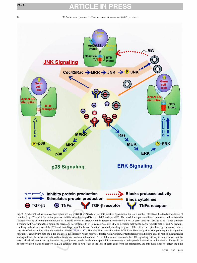
Fig. 2. A schematic illustration of how cytokines (e.g., TGF-b3, TNFa) can regulate junction dynamics in the testis via their effects on the steady-state levels of
proteins (e.g., TJ- and AJ-proteins, protease inhibitors such as a2-MG) at the BTB and apical ES. This model was prepared based on recent studies from this
laboratory using different animal models as reviewed herein. In brief, cytokines released from either Sertoli or germ cells can activate at least three different
signaling pathways upon their binding to receptors. For instance, TGF-b3 can activate p38 MAPK signaling pathway to down-regulate both TJ and AJ proteins,
resulting in the disruption of the BTB and Sertoli–germ cell adhesion function, eventually leading to germ cell loss from the epithelium (green sector), which
was identified in studies using the cadmium model [52,113,132]. This also illustrates that when TGF-b3 utilizes the p38 MAPK pathway for its signaling
function, it can perturb both the BTB and apical ES integrity. When rats were treated with Adjudin, or testosterone/estradiol implants to reduce intratesticular
androgen level, the testis responds to these treatments with an induction of TGF-b3 that can activate only the ERK signaling pathway to compromise Sertoli–
germ cell adhesion function by lowering the steady-state protein levels at the apical ES or weakening protein-protein interactions at this site via changes in the
phosphorylation status of adaptors (e.g., b-catenin); this in turn leads to the loss of germ cells from the epithelium, and this event does not affect the BTB
UN
CO
RR
EC
TED
PR
OO
F
the epithelium [68]. By blocking these two MAPK signaling
pathways using kinase inhibitors can also rescue the
epithelium from the disruptive effects of CdCl2 and Adjudin
on the BTB and Sertoli–germ cell AJ, respectively
[52,68,113]. As such, TGF-b3 serves as a key regulator
that decides whether BTB is affected or not. It is tempting to
speculate that this TGF-b-activated MAPK signaling
cascade requires the recruitment of adaptors to the site
which can in turn shuttle to the correct signaling pathway
downstream. Recent studies have shown that TGF-b-
induced p38 MAPK activation is mediated through a protein
scaffold complex XIAP (X-linked inhibitor of apoptosis)/
TAB1 (TAK binding protein)/TAK1 (TGF-b-activated
kinase), in which adaptor XIAP may link adaptor TAB1
and MAPKKK TAK1 to the TbRI. TbR1 can activate
TAK1, which further activates either MKK3/6 or MKK4 that
in turn activates p38 MAPK or JNK, respectively [118].
Besides, MAPKK-independent autophosphorylation of p38
is also possible, which is TAB1 dependent [153]. On the
other hand, TGF-b can activate Ras, which further activates
ERK signaling pathway and regulates various cellular
processes including junction dynamics [68]. TGF-b-induced
ERK activation also requires the adaptor CD2-associated
protein (CD2AP). When this adaptor is not involved, p38
MAPK is preferentially activated instead. In its presence,
TGF-b activates both the PI 3-kinase/Akt and the Ras/ERK
pathways [154]. Interestingly, CD2AP is not involved in PI
3-kinase/Akt activation by EGF and insulin, nor in the
activation of Smad2 by TGF-b, suggesting it plays a role in
TGF-b-activated, Smad-independent signaling [155]. The
association between TGF-b receptors and CD2AP is further
supported by the evidence that both are present in lipid rafts
[156,157]. Thus CD2AP can serve as a molecular switch to
determine the downstream signaling direction of TGF-b.
CD2AP belongs to a family of ubiquitously expressed
adaptors containing three Src-homology 3 (SH3) domains, a
proline-rich region and a coiled-coil domain [158] and is
expressed in human testes [159]. The SH3 domain mediates
interaction with the p85 subunit of PI 3-kinase and the
proline-rich region mediates association with p130 Cas and
Src family kinases (for a review, see [158]). p130 Cas and
Src are components of a signaling machinery connecting
FAK, paxillin, ERK and myosin light chain kinase (MLCK),
which, in turn regulate cell adhesion during cell migration
process [160]. Interestingly, virtually all of these proteins
have recently been found in the testis and they are likely
involved in junction dynamics during spermatogenesis (for
reviews, see [2,22,97]). It is likely that CD2AP is a crucial
adaptor of TGF-b mediated and integrin/FAK mediated
signaling events in the testis. Although much of the
information on CD2AP derives from studies in the kidney,
the testis may employ this molecular switch to select the
downstream signaling pathways to be activated by TGF-b3,
affecting either AJ alone or TJ and basal ES at the BTB. This
should be vigorously validated in future studies.
5. What lessons we learn from the testis as a model to
study junction restructuring?
As we have discussed above, the testis is an intriguing
organ where extensive junction restructuring occurs in the
seminiferous epithelium at each stage of the epithelial cycle.
Recent studies aiming to delineate the mechanisms that
regulate the junction restructuring events in the testis have
yielded some crucial information, which is likely applicable
to general cellular physiology as a whole. Herein, we
summarize several in vivo models that have been established
and used in recent studies (see Table 3). We only highlight
some of the latest development using these models and
readers are encouraged to refer to several recent reviews
[1,2,9,75,161,162].
5.1. Adjudin model
Formerly called AF-2364 [1-(2,4-dichlorobenzyl)-1H-
indazole-3-carbohydrazide], Adjudin is a molecule that
selectively induces adherens junction disruption. It is a well
studied potential male contraceptive derived from indazole-3-
carboxylic acid [163,164]. It is also one of the best studied
compounds that induce germ cell sloughing in the testis.
Adjudin apparently exerts its effects on the Sertoli–germ cell
adhesion unit to induce a loss of AJ function by triggering a
couple of signaling events, including a surge of the ES-
associated signaling molecule testin, an induction of integrin-
and cadherin-initiated pathways, as well as TGF-b3 activa-
tion [37,66–68,74,163,165–167]. Moreover, the ES-based
AJs are compromised due to a loss of protein-protein
association in the N-cadherin/b-catenin and nectin/afadin
protein complexes, which is likely the result of a coordinated
regulation by protein and lipid kinases and phosphatases,
proteases and protease inhibitors [35,67,68,74,94]. These
signalings are triggered within a few hours after adult male
rats are treated with a single or multiple doses of Adjudin at
W. Xia et al. / Cytokine & Growth Factor Reviews xxx (2005) xxx–xxx 13
DTD 5
CGFR 365 1–24
873
874
875
876
877
878
879
880
881
882
883
884
885
886
887
888
889
890
891
892
893
894
895
896
897
898
899
900
901
902
903
904
905
906
907
908
909
910
911
912
913
914
915
916
917
918
919
920
921
922
923
924
925
926
927
928
929
930
931
932
933
934
935
936
937
938
939
940
941
942
943
944
945
946
947
948
949
950
951
952
953
954
955
956
957
958
959
960
961
integrity (blue sector) [2,68]. This thus suggests that TGF-b3 can limit its action at the apical ES without compromising the BTB when the ERK signaling
pathway is being utilized. Using the cadmium model, it is presently known that JNK is activated during the cadmium-induced BTB damage. This induces the
production of a2-MG, which either bind to the free cytokines, limiting their biological action and/or blocking protease activity to limit the BTB damage. Since it
is known that by blocking the production of a2-MG, it can worsen the damaging effect of cadmium on testicular junctions (red sector), illustrating this JNK-a2-
MG pathway is crucial to maintain the normal physiology in the seminiferous epithelium [57,114]. This pathway is likely utilized by TNFa to regulate the
steady-state protein level of a2-MG. The coordinated action of these three interacting signaling pathways that are intriguingly regulated by cytokines (e.g., TGF-
b3 and TNFa) is crucial to maintain the integrity of the seminiferous epithelium during spermatogenesis, permitting selective disruption of either TJ, AJ, or TJ
and AJ. As reviewed herein, it is likely that adaptors play a crucial role upstream to select which signaling pathway should be activated by these cytokines, which
in turn determines if either BTB, apical ES, or both BTB and apical ES should be compromised during spermatogenesis.
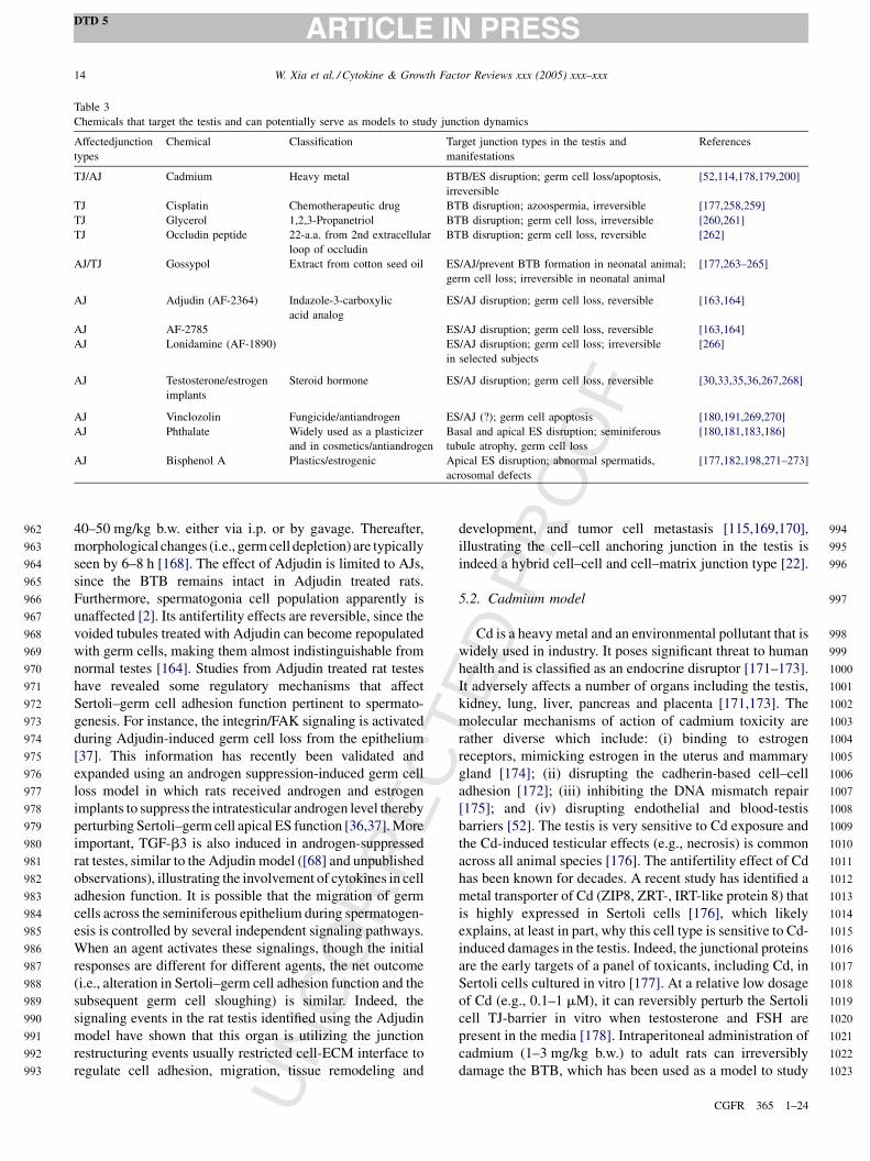
UN
CO
RR
EC
TED
PR
OO
F40–50 mg/kg b.w. either via i.p. or by gavage. Thereafter,
morphological changes (i.e., germ cell depletion) are typically
seen by 6–8 h [168]. The effect of Adjudin is limited to AJs,
since the BTB remains intact in Adjudin treated rats.
Furthermore, spermatogonia cell population apparently is
unaffected [2]. Its antifertility effects are reversible, since the
voided tubules treated with Adjudin can become repopulated
with germ cells, making them almost indistinguishable from
normal testes [164]. Studies from Adjudin treated rat testes
have revealed some regulatory mechanisms that affect
Sertoli–germ cell adhesion function pertinent to spermato-
genesis. For instance, the integrin/FAK signaling is activated
during Adjudin-induced germ cell loss from the epithelium
[37]. This information has recently been validated and
expanded using an androgen suppression-induced germ cell
loss model in which rats received androgen and estrogen
implants to suppress the intratesticular androgen level thereby
perturbing Sertoli–germ cell apical ES function [36,37]. More
important, TGF-b3 is also induced in androgen-suppressed
rat testes, similar to the Adjudin model ([68] and unpublished
observations), illustrating the involvement of cytokines in cell
adhesion function. It is possible that the migration of germ
cells across the seminiferous epithelium during spermatogen-
esis is controlled by several independent signaling pathways.
When an agent activates these signalings, though the initial
responses are different for different agents, the net outcome
(i.e., alteration in Sertoli–germ cell adhesion function and the
subsequent germ cell sloughing) is similar. Indeed, the
signaling events in the rat testis identified using the Adjudin
model have shown that this organ is utilizing the junction
restructuring events usually restricted cell-ECM interface to
regulate cell adhesion, migration, tissue remodeling and
development, and tumor cell metastasis [115,169,170],
illustrating the cell–cell anchoring junction in the testis is
indeed a hybrid cell–cell and cell–matrix junction type [22].
5.2. Cadmium model
Cd is a heavy metal and an environmental pollutant that is
widely used in industry. It poses significant threat to human
health and is classified as an endocrine disruptor [171–173].
It adversely affects a number of organs including the testis,
kidney, lung, liver, pancreas and placenta [171,173]. The
molecular mechanisms of action of cadmium toxicity are
rather diverse which include: (i) binding to estrogen
receptors, mimicking estrogen in the uterus and mammary
gland [174]; (ii) disrupting the cadherin-based cell–cell
adhesion [172]; (iii) inhibiting the DNA mismatch repair
[175]; and (iv) disrupting endothelial and blood-testis
barriers [52]. The testis is very sensitive to Cd exposure and
the Cd-induced testicular effects (e.g., necrosis) is common
across all animal species [176]. The antifertility effect of Cd
has been known for decades. A recent study has identified a
metal transporter of Cd (ZIP8, ZRT-, IRT-like protein 8) that
is highly expressed in Sertoli cells [176], which likely
explains, at least in part, why this cell type is sensitive to Cd-
induced damages in the testis. Indeed, the junctional proteins
are the early targets of a panel of toxicants, including Cd, in
Sertoli cells cultured in vitro [177]. At a relative low dosage
of Cd (e.g., 0.1–1 mM), it can reversibly perturb the Sertoli
cell TJ-barrier in vitro when testosterone and FSH are
present in the media [178]. Intraperitoneal administration of
cadmium (1–3 mg/kg b.w.) to adult rats can irreversibly
damage the BTB, which has been used as a model to study
W. Xia et al. / Cytokine & Growth Factor Reviews xxx (2005) xxx–xxx14
DTD 5
CGFR 365 1–24
962
963
964
965
966
967
968
969
970
971
972
973
974
975
976
977
978
979
980
981
982
983
984
985
986
987
988
989
990
991
992
993
994
995
996
997
998
999
1000
1001
1002
1003
1004
1005
1006
1007
1008
1009
1010
1011
1012
1013
1014
1015
1016
1017
1018
1019
1020
1021
1022
1023
Table 3
Chemicals that target the testis and can potentially serve as models to study junction dynamics
Affectedjunction
types
Chemical Classification Target junction types in the testis and
manifestations
References
TJ/AJ Cadmium Heavy metal BTB/ES disruption; germ cell loss/apoptosis,
irreversible
[52,114,178,179,200]
TJ Cisplatin Chemotherapeutic drug BTB disruption; azoospermia, irreversible [177,258,259]
TJ Glycerol 1,2,3-Propanetriol BTB disruption; germ cell loss, irreversible [260,261]
TJ Occludin peptide 22-a.a. from 2nd extracellular
loop of occludin
BTB disruption; germ cell loss, reversible [262]
AJ/TJ Gossypol Extract from cotton seed oil ES/AJ/prevent BTB formation in neonatal animal;
germ cell loss; irreversible in neonatal animal
[177,263–265]
AJ Adjudin (AF-2364) Indazole-3-carboxylic
acid analog
ES/AJ disruption; germ cell loss, reversible [163,164]
AJ AF-2785 ES/AJ disruption; germ cell loss, reversible [163,164]
AJ Lonidamine (AF-1890) ES/AJ disruption; germ cell loss; irreversible
in selected subjects
[266]
AJ Testosterone/estrogen
implants
Steroid hormone ES/AJ disruption; germ cell loss, reversible [30,33,35,36,267,268]
AJ Vinclozolin Fungicide/antiandrogen ES/AJ (?); germ cell apoptosis [180,191,269,270]
AJ Phthalate Widely used as a plasticizer
and in cosmetics/antiandrogen
Basal and apical ES disruption; seminiferous
tubule atrophy, germ cell loss
[180,181,183,186]
AJ Bisphenol A Plastics/estrogenic Apical ES disruption; abnormal spermatids,
acrosomal defects
[177,182,198,271–273]
UN
CO
RR
EC
TED
PR
OO
F
BTB dynamics in the testis [52,113,114,179]. Apparently,
when absorbed by Sertoli cells, Cd targets the microfila-
ment, causing a disorganization of actin bundles [179].
Furthermore, Cd induces the dissolution of TJ proteins (e.g.,
occludin) from the seminiferous epithelium, and down-
regulates AJ-proteins (e.g., cadherin, nectin) to induce a
secondary disruption of basal ES, leading to germ cell
sloughing [52]. Using this model, it has been shown that the
TGF-b3/MEKK/p38 MAPK mediated signaling pathway is
a putative mechanism that regulates TJ dynamics at the BTB
in vivo, and a disruption of this pathway using specific
inhibitors can indeed significantly delay the Cd damage to
the BTB as well as the subsequent germ cell loss from the
epithelium [44,113,132].
5.3. Possible in vivo models to study junction dynamics
in the testis
Recent studies have illustrated a number of chemicals
that can affect testicular junctions, which may be developed
into useful in vivo models. Many of these molecules are
endocrine disruptors, which include phthalate, bisphenol A,
vinclozolin and others [180–183]. Because of their wide-
spread distribution in the environment and potential health
hazards (e.g., reproductive organs), these compounds have
attracted great attention of research, particularly on their
effects to the reproductive organs (e.g., testes). They affect
the endocrine system either by acting as antiandrogens
(e.g., phthalate, vinclozolin) or estrogens (e.g., bisphenol A)
[180–183].
Phthalate and vinclozolin are compounds that can
antagonize androgens. However, they exert these effects
via different mechanisms: phthalate affects androgen
synthesis [183] whereas the metabolites of vinclozolin are
antagonists of androgen receptors [184,185]. Phthalate is
found in cosmetic products (e.g., nail polishes, perfumes,
hair sprays) and is widely used as a plasticizer, which can be
non-covalently bound to the matrix and thereby slowly
releases to the environment, and can be inhaled or adsorbed
dermally [181,183]. Its toxic effects in male neonatal
animals include hypospadias, reduced anogenital distance,
vaginal pouch, some of which (e.g., hypospadias) are
detected in humans [180,181,183,186]. When adults rats
were treated with a single dose of di-n-pentyl phthalate
(DPP) (2.2 g/kg b.w.), Sertoli cell junctions displayed
abnormalities with disrupted basal ES, and apical ES was
either absent or badly disorganized [187]. Interestingly, the
disrupted basal ES between apposing Sertoli cells were
reformed by 48 h after DPP treatment [187], illustrating this
is a potentially useful model to study basal ES dynamics if
adequately characterized. Furthermore, in DPP-treated
prepubertal rats, extensive vacuolation occurs in Sertoli
cells, to be followed by sloughing of germinal cells [188].
Apparently the observed effects of phthalate on germ cell
loss are mediated via disruption of Sertoli-Sertoli and
Sertoli–germ cell adhesion function.
Vinclozolin [3-(3,5-dichlorophenyl)-5-methyl-5-viny-
loxazolidine-2,4-dione] is a fungicide that is widely used
in farming industry. When adsorbed by humans or rodents,
vinclozolin is metabolized to M1 (2-[[(3,5-dichlorophenyl)-
carbamoyl]oxy]-2-methyl-3-butenoic acid) and M2 (30,50-dichloro-2-hydroxy-2-methylbut-3-enanilide), which can
bind to androgen receptors, antagonizing androgen function
in vivo [184,185]. Besides its disruptive effects on
reproductive organs (e.g., hypospadias, reduced anogenital
distance) in male rats when exposed to vinclozolin in utero, it
is neurotoxic and is an endocrine disruptor [189]. In the testis,
vinclozolin can induce Leydig cell hypertrophy, reduce testis
weight as a result of germ cell loss, and subsequently impair
sperm production [190–192]. The direct structural damage of
junctions at the Sertoli–Sertoli and Sertoli–germ cell interface
following vinclozolin treatment remains to be examined.
Bisphenol A is a xenoestrogen although its bioactivity is
1000–1500-fold lower than 17b-estradiol. An ultrastructural
examination of adult rat and mouse testes after treatment
with bisphenol A has revealed that the apical ES was absent
or badly damaged versus control animals; but, interestingly,
the basal ES and BTB were not affected [182]. This
information has also strengthened the notion that AJ
disruption in the seminiferous epithelium can be restricted
to the ES site without perturbing the TJ-barrier function at
the BTB [2,30] in contrast to other epithelia where a
disruption of AJ can lead to a secondary damage of the TJ-
barrier function and vice versa [193–197]. The disruptive
effects of bisphenol A and estrogens (17B-estradiol and b-
estradiol-3-benzoate) on apical ES (but not basal ES and
BTB, which remained intact) was also detected in
maturating rats and mice that had been exposed to bisphenol
A at neonatal [198]. However, these effects were not found
when rats were fully mature [198]. In short, one of the target
structures of this endocrine disruptor is the apical ES.
5.4. Why is the testis a vulnerable target of
environmental toxicants? A lesson to learn from the
testis
Studies on different environmental toxicants have
unequivocally demonstrated that the testis is extremely
vulnerable to these toxicants (for a review, see [199]). When
exposed to these chemicals, Sertoli-Sertoli and Sertoli–germ
cell junctions are the early targets and their subsequent
dissolution is likely the result of down-regulation of junction
proteins or changes in protein-protein association of the
junction protein complexes [177]. Indeed, recent studies
have shown that when adult rats were exposed to cadmium
chloride, the BTB damage had occurred at least 24 h before
the TJ-barrier of the microvessel in the interstitium
[114,200], indicating the BTB is more sensitive than the
endothelial TJ-barrier in microvessels to cadmium toxicity.
Subsequent analyses by immunohistochemistry and fluor-
escent microscopy using these rats have conclusively
demonstrated a significant loss of TJ- and AJ-integral
W. Xia et al. / Cytokine & Growth Factor Reviews xxx (2005) xxx–xxx 15
DTD 5
CGFR 365 1–24
1024
1025
1026
1027
1028
1029
1030
1031
1032
1033
1034
1035
1036
1037
1038
1039
1040
1041
1042
1043
1044
1045
1046
1047
1048
1049
1050
1051
1052
1053
1054
1055
1056
1057
1058
1059
1060
1061
1062
1063
1064
1065
1066
1067
1068
1069
1070
1071
1072
1073
1074
1075
1076
1077
1078
1079
1080
1081
1082
1083
1084
1085
1086
1087
1088
1089
1090
1091
1092
1093
1094
1095
1096
1097
1098
1099
1100
1101
1102
1103
1104
1105
1106
1107
1108
1109
1110
1111
1112
1113
1114
1115
1116
1117
1118
1119
1120
1121
1122
1123
1124
1125
1126
1127
1128
1129
1130
1131
UN
CO
RR
EC
TED
PR
OO
F
proteins from the BTB site, consistent with results of
immunoblot analyses [52,114]. Furthermore, a loss of
protein-protein interactions of the AJ integral membrane
proteins and their adaptors namely cadherin-catenin and
nectin-afadin was also detected when rats were exposed to
Adjudin [68], a chemical known to induce germ cell loss
from the seminiferous epithelium without disrupting the TJ-
barrier at the BTB (for reviews, see [1,2,9]). Taking
collectively, these data have clearly illustrated the vulner-
ability of the testis to environmental toxicants (e.g.,
cadmium) and that the proteins at the TJ and AJ sites are
some of the primary targets of these toxicants. While the
precise mechanism underlying such vulnerability is not fully
understood, recent studies have shed new lights on this issue,
which also highlights a unique opportunity to use these
toxicant-induced BTB or AJ damage to the testis as novel
models to study BTB dynamics, AJ restructuring pertinent to
spermatogenesis, and their regulation. Furthermore, these
studies can plausibly provide new insights in developing
preventive measures to antagonize these toxicants.
First, Sertoli cells are secretory cells that actively provide
virtually all the necessary nutrients for germ cell develop-
ment behind the BTB including metal transporters, such as
transferrin, ceruoplasmin, and metallothioneins (MTs). MTs
are small Mr proteins having high affinities for heavy metal
ions including cadmium, zinc, copper and mercury. MTs are
produced in virtually all mammalian tissues in response to
metal ions exposure, which can detoxify heavy metals, such
as cadmium (for reviews, see [201,202–205]). MTs are also
important to maintain the homeostasis of essential trace
elements, such as zinc and copper and are scavengers of free
radicals [202,204] and protect cells from the cytotoxic
effects of cadmium [206]. In the rat testis, MTs, such as MT1
and MT2, have been identified and isolated [207,208].
Recent studies have found a novel testis-specific MT-like
protein called tesmin which is specifically expressed by
spermatogenic cells [209]. MT1 and MT2 are products of
Sertoli and germ cells, which are significantly induced after
cadmium exposure [210]. Yet the production of MTs by
Sertoli and germ cells are significantly lower when
compared to hepatocytes in vitro in response to cadmium
exposure [210]. Indeed, the quiescence of MT expression in
the ventral prostate and the testis is the possible cause of
their susceptibility to cadmium cytotoxicity and carcino-
genicity [204,211–213].
Second, recent studies on the effects of cadmium on
different cell lines, including MDCK, LLC-PK1, and Caco-2
cells, have shown that its primary target is E-cadherin (for a
review, see [214]). For instance, cells that were exposed to
cadmium were found to have their E-cadherin moving away
from the cell–cell interface and became diffusely localized
in the cytoplasm. It was postulated that cadmium may be
competing to the binding of calcium to the E-cadherin,
thereby perturbing the AJ function [214–217]. If this is the
case, cadmium (and possibly other environmental toxicants)
must first gain access to AJ to disrupt E-cadherin. In all other
epithelia found in mammals, AJ is physically located behind
the TJ since the TJ-barrier is located to the apical portion of
the cell epithelium, and behind TJ lies desmosomes, which
collectively known as the junctional complex. Behind the
junctional complex are the gap junctions to be followed by
the cell–matrix adhesion complex. As such, the TJ would
seal most of the environmental toxicants off the epithelium
in virtually all organs. Yet in the testis, TJ coexists with AJ
and desmosome-like junctions at the BTB, which collec-
tively lies adjacent to the basement membrane (a modified
form of ECM, for a review, see [12]), closest to the
interstitium. Thus, toxicants (e.g., cadmium) diffuses from
the microvessels will have immediate access to the E-
cadherin in the AJ (which is the cellular target of cadmium)
at the BTB because there is no TJ-barrier that seals off
cadmium. This, in turn, disrupts AJ, inducing germ cell loss
from the epithelium as manifested by germ cell sloughing in
many of these animal models using environmental toxicants.
It is of interest to note that recent studies have begun to shed
light on the physiological significance of such coexisting TJ
and AJ at the BTB in relation to spermatogenesis. For
instance, it is well understood that spermatogenesis is
associated with extensive restructuring of Sertoli–Sertoli
and Sertoli–germ cell interface because of the constant
reshaping of germ cell shapes as a result of differentiation
and germ cell movement from the basal to the adluminal
compartment. If such AJ restructuring leads to TJ-barrier
disruption as it is the case in other epithelia [193,195,197],
the BTB integrity cannot be maintained, and haploid germ
cell antigens cannot be sequestered from the host immune
system; and such a disruption, even transiently, of the
immunological barrier is detrimental to spermatogenesis.
Thus, the fact that the BTB is constituted by co-existing AJ
and TJ is to ensure such transient disruption of TJ during AJ
restructuring in the seminiferous epithelium does not occur.
Recent studies have shown that a signal that induces AJ
disruption [e.g., via treatment of rats with Adjudin to induce
extensive AJ restructuring that leads to germ cell loss from
the epithelium, or a decline in endogenous intratesticular T
level using androgen/estradiol transdermal implants] can
lead to a surge in the production of both AJ (e.g., cadherins,
catenins) and TJ (e.g., occludin, ZO-1) proteins [30,35,68]
(Fig. 2). The increased TJ proteins are being used to
reinforce the TJ-barrier integrity at the BTB at the time of
extensive AJ restructuring. While the levels of AJ proteins
are also induced, germ cells can still be dissociated from
Sertoli cells because the AJ-integral membrane protein-AJ
adaptor (e.g., the N-cadherin-b-catenin protein complex)
association is found to be weakened via an increase in
tyrosine phosphorylation of b-catenin [30,35,68]. These
findings are significant because it depicts the presence of a
novel mechanism utilized by the testis to ensure TJ-barrier
integrity while permitting AJ restructuring within a
microenvironment such as the seminiferous epithelium.
Fig. 2 is a schematic drawing that illustrates this novel
mechanism of increasing AJ and TJ proteins, which is likely
W. Xia et al. / Cytokine & Growth Factor Reviews xxx (2005) xxx–xxx16
DTD 5
CGFR 365 1–24
1132
1133
1134
1135
1136
1137
1138
1139
1140
1141
1142
1143
1144
1145
1146
1147
1148
1149
1150
1151
1152
1153
1154
1155
1156
1157
1158
1159
1160
1161
1162
1163
1164
1165
1166
1167
1168
1169
1170
1171
1172
1173
1174
1175
1176
1177
1178
1179
1180
1181
1182
1183
1184
1185
1186
1187
1188
1189
1190
1191
1192
1193
1194
1195
1196
1197
1198
1199
1200
1201
1202
1203
1204
1205
1206
1207
1208
1209
1210
1211
1212
1213
1214
1215
1216
1217
1218
1219
1220
1221
1222
1223
1224
1225
1226
1227
1228
1229
1230
1231
1232
1233
1234
1235
1236
1237
1238
1239
1240
1241
1242
1243

UN
CO
RR
EC
TED
PR
OO
F
regulated by cytokines [e.g., TGF-b3, TNF-a released from
either Sertoli or germ cells [57,113,123,218]] via the ERK
signaling pathway that maintains the BTB integrity while
permitting AJ restructuring, facilitating germ cell movement
across the seminiferous epithelium. However, this same
mechanism that is physiologically necessary to facilitate
germ cell movement while maintaining BTB integrity
during spermatogenesis also makes the BTB extremely
vulnerable to environmental toxicants because of the
unusual exposure of AJ structural proteins (e.g., E-cadherin)
to the toxicants (e.g., cadmium).
6. Conclusion and future perspectives
In all animal species, cell migration and junction
remodeling are naturally occurring processes. For instance,
there are three types of signals that control different aspects
of Drosophila border cell migration: a global steroid-
hormone signal to determine the timing, a highly localized
cytokine signal to induce migration, and a growth factor to
guide cells to their destination (for a review, see [219]). In
the testis, FSH released from the pituitary and testosterone
from Leydig cells may serve as the global regulatory factors.
Cytokines that function as either paracrines or autocrines
can in turn regulate localized signaling and processes.
Several theories of germ cell movement during spermato-
genesis have been proposed, and recently, we have put forth
a junction restructuring theory in which cytokines, protease/
protease inhibitors, cytoskeleton regulators and junctional
complex proteins are all coordinated to facilitate germ cell
movement in the epithelium [2]. Stem cell research has also
offered new insights on Sertoli–germ cell interactions and
may facilitate the research regarding the local regulatory
function of cytokines in this event. When rat spermatogonia
are transplanted into recipient mouse testes, the rat stem
cells develop according to their only timing (�7 week
instead of �5 week), irrespective to the surrounding mouse
spermatogenesis milieu [220]. It seems that this internal
preprogrammed rhythm autonomously determines the fate
of rat spermatogonia differentiation, and creates a suitable
localized environment through dialogues with mouse Sertoli
cells, probably via cytokines for crosstalk. This intrinsic
preprogrammed timing may be controlled or executed by
homeobox genes. A homeobox gene cluster Rhox (repro-
ductive homeobox on the X chromosome) has recently been
identified in mice [221]. The 12 Rhox genes are expressed
mostly in reproductive organs (placenta, ovary, testis and
epididymis), arranged into three subclusters and manifested
temporal and quantitative colinearity in expression patterns
[221]. In the testis, the majority of Rhox genes are primarily
expressed in Sertoli cells and androgen responsive [221].
During the first wave of spermatogenesis, the timing of Rhox
genes expression corresponds to the specific phases of germ
cell differentiation. Hence these transcription factors may
direct the expression of an array of proteins required for
germ cell development, and may also define the correspond-
ing timing of epithelial cycle and length of spermatogenesis
in rodents [221].
Several approaches can be used in future studies to aid the
understanding of Sertoli–germ cell crosstalk and junction
restructuring. First, development of testis-specific knockout
mice against crucial proteins pertinent to junction restruc-
turing and spermatogenesis to identify the function of these
proteins in the testis. For many cytokines (e.g., TGF-bs),
their deletion can lead to lethality of the null mice. As such,
their roles in spermatogenesis at adulthood cannot be
examined. In the rat, the first wave of spermiation occurs
only by 30–40 days of age. Recently conditional knockout
technique has allowed investigators to elucidate protein
function in a tissue- and time-specific manner in testis using
specific Sertoli cell KOs, such as androgen receptor [222].
The generation of testis-specific KOs (e.g., TGF-b3) will
help define the roles of these cytokines in junction dynamics
at spermatogenesis. Second, germline stem cell transplanta-
tion with traceable markers to follow germ cell differentia-
tion as well as junction remodeling during spermatogenesis
can assist the study of cell–cell interactions pertinent to
germ cell movement. For instance, when spermatogonia are
transplanted into the recipient testis, they can migrate to the
basal niche and initiate spermatogenesis in the prepro-
grammed cycle independent of the host environment. This
migration must traverse the BTB, differing from gonocyte
migration in the tubule when BTB has not yet formed. Third,
using microarray technique to identify the expression
profiles of various cytokines, proteases and protease
inhibitors, junctional proteins, adaptors and transcription
factors in staged tubules and in testes obtained from selected
in vivo models. This approach can also pinpoint the leading
and supporting biological factors pertinent to spermatogen-
esis. It is hopeful that using these approaches, a better
understanding of spermatogenesis can emerge, which should
be helpful for various applications such as treating male
infertility or for contraception.
Acknowledgements
Supported in part by grants from the National Institutes of
Health (NICHD, 5U01 HD045908 to CYC; 5U54
HD029990, Project 3 to CYC), and the CONRAD Program
(CICCR, C1G 01-72 to CYC, CIG 01-74 to DDM).
References
[1] Cheng CY, Mruk DD. Cell junction dynamics in the testis: Sertoli–
germ cell interactions and male contraceptive development. Physiol
Rev 2002;82:825–74.
[2] Mruk DD, Cheng CY. Sertoli–Sertoli and Sertoli–germ cell interac-
tions and their significance in germ cell movement in the seminifer-
ous epithelium during spermatogenesis. Endocr Rev 2004;25:
747–806.
W. Xia et al. / Cytokine & Growth Factor Reviews xxx (2005) xxx–xxx 17
DTD 5
CGFR 365 1–24
1244
1245
1246
1247
1248
1249
1250
1251
1252
1253
1254
1255
1256
1257
1258
1259
1260
1261
1262
1263
1264
1265
1266
1267
1268
1269
1270
1271
1272
1273
1274
1275
1276
1277
1278
1279
1280
1281
1282
1283
1284
1285
1286
1287
1288
1289
1290
1291
1292
1293
1294
1295
1296
1297
1298
1299
1300
1301
1302
1303
1304
1305
1306
1307
1308
1309
1310
1311
1312
1313
1314
1315
1316
1317
1318
1319
1320
1321
1322
1323
1324
1325
1326
1327
1328
1329
1330
1331
1332
1333
1334
1335
1336
1337
1338
1339
1340
1341
1342
1343
1344
1345
1346
1347

UN
CO
RR
EC
TED
PR
OO
F
[3] de Kretser DM, Kerr JB. The cytology of the testis. In: Knobil E,
Neill JD, editors. The physiology of reproduction. New York: Raven
Press; 1994. p. 1177–300.
[4] Parvinen M. Regulation of the seminiferous epithelium. Endocr Rev
1982;3:404–17.
[5] Griswold M. Interactions between germ cells and Sertoli cells in the
testis. Biol Reprod 1995;52:211–6.
[6] Kierszenbaum AL. Mammalian spermatogenesis in vivo and in vitro:
a partnership of spermatogenic and somatic cell lineages. Endocr Rev
1994;15:116–34.
[7] Griswold MD. The central role of Sertoli cells in spermatogenesis.
Semin Cell Dev Biol 1998;9:411–6.
[8] Russell LD. Movement of spermatocytes from the basal to the adlum-
inal compartment of the rat testis. Am J Anat 1977;148:313–28.
[9] Mruk DD, Cheng CY. Cell–cell interactions at the ectoplasmic
specialization in the testis. Trends Endocr Metab 2004;15:439–47.
[10] Russell L. Sertoli–germ cell interactions: a review. Gamete Res
1980;3:179–202.
[11] Gnessi L, Fabbri A, Spera G. Gonadal peptides as mediators of
development and functional control of the testis: an integrated system
with hormones and local environment. Endocr Rev 1997;18:541–609.
[12] Dym M. Basement membrane regulation of Sertoli cells. Endocr Rev
1994;15:102–15.
[13] Weber JE, Russell LD, Wong V, Peterson RN. Three-dimensional
reconstruction of a rat stage V Sertoli cell. II. Morphometry of
Sertoli–Sertoli and Sertoli–germ-cell relationships. Am J Anat
1983;167:163–79.
[14] Hess RA, Franca LR. Structure of the Sertoli cell. In: Skinner MK,
Griswold MD, editors. Sertoli cell biology.. San Diego: Elsevier
Academic Press; 2005. p. 19–40.
[15] Alberts B, Johnson A, Lewis J, Raff M, Roberts K, Watter P.
Molecular Biology of the Cell. New York Garland Science, 2002.
[16] Toyama Y, Maekawa M, Yuasa S. Ectoplasmic specializations in the
Sertoli cell: news vistas based on genetic defects and testicular
toxicology. Anat Sci Int 2003;78:1–16.
[17] Lee NPY, Cheng CY. Ectoplasmic specialization, a testis-specific
cell–cell actin-based adherens junction type: is this a potential target
for male contraceptive development? Hum Reprod Update
2004;10:349–69.
[18] Guttman JA, Takai Y, Vogl AW. Evidence that tubulobulbar com-
plexes in the seminiferous epithelium are involved with internaliza-
tion of adhesion junctions. Biol Reprod 2004;71:548–59.
[19] Cunha-Vaz JG. The blood–retinal barriers system. Basic concepts
and clinical evaluation. Exp Eye Res 2004;78:715–21.
[20] Rubin LL, Staddon JM. The cell biology of the blood-brain barrier.
Annu Rev Neurosci 1999;22:11–28.
[21] Dejana E. Endothelial cell–cell junctions: happy together. Nat Rev
Mol Cell Biol 2004;5:261–70.
[22] Siu MKY, Cheng CY. Dynamic cross-talk between cells and the
extracellular matrix in the testis. BioEssays 2004;26:978–92.
[23] Tsukita S, Furuse M, Itoh M. Multifunctional strands in tight junc-
tions. Nat Rev Mol Cell Biol 2001;2:285–93.
[24] Schneeberger EE, Lynch RD. The tight junction: a multifunctional
complex. Am J Physiol Cell Physiol 2004;286:C1213–28.
[25] Bazzoni G. The JAM family of junctional adhesion molecules. Curr
Opin Cell Biol 2003;15:525–30.
[26] Ebnet K, Suzuki A, Ohno S, Vestweber D. Junctional adhesion
molecules (JAMs): more molecules with dual functions? J Cell
Sci 2004;117:19–29.
[27] Martin-Padura I, Lostaglio S, Schneemann M, Williams L, Romano
M, Fruscella P, et al. Junctional adhesion molecule, a novel member
of the immunoglobulin superfamily that distributes at intercellular
junctions and modulates monocyte transmigration. J Cell Biol
1998;142:117–27.
[28] Hirabayashi S, Tajima M, Yao I, Nishimura W, Mori H, Hata Y.
JAM4, a junctional cell adhesion molecule interacting with a tight
junction protein, MAGI-1. Mol Cell Biol 2003;23:4267–82.
[29] Gliki G, Ebnet K, Aurrand-Lions M, Lmhot BA, Adams RH.
Spermatid differentiation requires the assembly of a cell polarity
complex downstream of junctional adhesion molecule-C. Nature
2004;431:320–4.
[30] Xia W, Wong CH, Lee NPY, Lee WM, Cheng CY. Disruption of
Sertoli–germ cell adhesion function in the seminiferous epithelium of
the rat testis can be limited to adherens junctions without affecting
the blood-testis barrier integrity: an in vivo study using an androgen
suppression model. J Cell Physiol, in press.
[31] Cera MR, Del Prete A, Vecchi A, Corada M, Martin-Padura I,
Motoike T, et al. Increased DC trafficking to lymph nodes and
contact hypersensitivity in junctional adhesion molecule-A-deficient
mice. J Clin Invest 2004;114:729–38.
[32] Liang TW, Chiu HH, Gurney A, Sidle A, Tumas DB, Schow P, et al.
Vascular endothelial-junctional adhesion molecule (VE-JAM)/JAM
2 interacts with T, NK, and dendritic cells through JAM 3. J Immunol
2002;168:1618–26.
[33] McLachlan RI, Wreford NG, Meachem SJ, Kretser DMD, Robertson
DM. Effects of testosterone on spermatogenic cell populations in the
adult rat. Biol Reprod 1994;51:945–55.
[34] O’Donnell L, McLachlan RI, Wreford NG, de Kretser DM, Robert-
son DM. Testosterone withdrawal promotes stage-specific detach-
ment of round spermatids from the at seminiferous epithelium. Biol
Reprod 1996;55:895–901.
[35] Zhang J, Wong CH, Xia W, Mruk DD, Lee NPY, Lee WM, et al.
Regulation of Sertoli–germ cell adherens junction dynamics via
changes in protein-protein interactions of the N-cadherin-b-catenin
protein complex which are possibly mediated by c-Src and MTMR2:
an in vivo study using an androgen suppression model. Endocrinol-
ogy 2005;146:1268–84.
[36] Wong CH, Xia W, Lee NPY, Mruk DD, Lee WM, Cheng CY.
Regulation of ectoplasmic specialization dynamics in the semini-
ferous epithelium by focal adhesion-associated proteins in
testosterone-suppressed rat testes. Endocrinology 2005;146:
1192–204.
[37] Siu MKY, Mruk DD, Lee WM, Cheng CY. Adhering junction
dynamics in the testis are regulated by an interplay of b1-integrin
and the focal adhesion complex-associated proteins. Endocrinology
2003;144:2141–63.
[38] Shaw SK, Perkins BN, Lim YC, Liu Y, Nusrat A, Schnell FJ, et al.
Reduced expression of junctional adhesion molecule and platelet/
endothelial cell adhesion molecule-1 (CD31) at human vascular
endothelial junctions by cytokines tumor necrosis factor-a plus
interferon-gamma does not reduce leukocyte transmigration under
flow. Am J Pathol 2001;159:2281–91.
[39] Turksen K, Troy T-C. Barriers built on claudins. J Cell Sci 2004;
117:2435–47.
[40] Tsukita S, Furuse M. The structure and function of claudins, cell
adhesion molecules at tight junctions. Ann N Y Acad Sci
2000;915:129–35.
[41] Heiskala M, Peterson PA, Yang Y. The roles of claudin superfamily
proteins in paracellular transport. Traffic 2001;2:92–8.
[42] Florin A, Maire M, Bozec A, Hellani A, Chater S, Bars R,
et al. Androgens and postmeiotic germ cells regulate claudin-
11 expression in rat Sertoli cells. Endocrinology 2005;146:1532–
40.
[43] Hellani A, Ji J, Mauduit C, Deschildre C, Tabone E, Benahmed M.
Developmental and hormonal regulation of the expression of oligo-
dendrocyte-specific protein/claudin 11 in mouse testis. Endocrinol-
ogy 2000;141:3012–9.
[44] Lui WY, Lee WM, Cheng CY. Transforming growth factor-b3
perturbs the inter-Sertoli tight junction permeability barrier in vitro
possibly mediated via its effects on occludin, zonula occludens-1,
and claudin-11. Endocrinology 2001;142:1865–77.
[45] Morita K, Sasaki H, Fujimoto K, Furuse M, Tsukita S. Claudin-
11/OSP-based tight junctions of myelin sheaths in brain and Sertoli
cells in testis. J Cell Biol 1999;145:579–88.
W. Xia et al. / Cytokine & Growth Factor Reviews xxx (2005) xxx–xxx18
DTD 5
CGFR 365 1–24
1348
1349
1350
1351
1352
1353
1354
1355
1356
1357
1358
1359
1360
1361
1362
1363
1364
1365
1366
1367
1368
1369
1370
1371
1372
1373
1374
1375
1376
1377
1378
1379
1380
1381
1382
1383
1384
1385
1386
1387
1388
1389
1390
1391
1392
1393
1394
1395
1396
1397
1398
1399
1400
1401
1402
1403
1404
1405
1406
1407
1408
1409
1410
1411
1412
1413
1414
1415
1416
1417
1418
1419
1420
1421
1422
1423
1424
1425
1426
1427
1428
1429
1430
1431
1432
1433
1434
1435
1436
1437
1438
1439
1440
1441
1442
1443
1444
1445
1446
1447
1448
1449
1450
1451
1452
1453
1454
1455
1456
1457
1458
1459
1460
1461
1462
1463
1464
1465
1466
1467
1468
1469
1470
1471
1472
1473
1474
1475
1476
1477
1478
1479
1480
1481
UN
CO
RR
EC
TED
PR
OO
F
[46] Gow A, Southwood CM, Li JS, Pariali M, Riordan GP, Brodie SE, et
al. CNS myelin and Sertoli cell tight junction strands are absent in
Osp/claudin-11 null mice. Cell 1999;99:649–59.
[47] Gye MC. Changes in the expression of claudins and transepithelial
electrical resistance of mouse Sertoli cells by Leydig cell coculture.
Int J Androl 2003;26:271–8.
[48] Kitajiri S-I, Miyamoto T, Mineharu A, Sonoda N, Furuse K, Hata M,
et al. Compartmentalization established by claudin-11-based tight
junctions in stria vascularis is required for hearing through generation
of endocochlear potential. J Cell Sci 2004;117:5087–96.
[49] Sasaki H, Matsui C, Furuse K, Mimori-Kiyosue Y, Furuse M, Tsukita
S. Dynamic behavior of paired claudin strands within apposing
plasma membranes. Proc Natl Acad Sci USA 2003;100:3971–6.
[50] Matsuda M, Kubo A, Furuse M, Tsukita S. A peculiar internalization
of claudins, tight junction-specific adhesion molecules, during the
intercellular movement of epithelial cells. J Cell Sci 2004;117:1247–
57.
[51] Furuse M, Hirase T, Itoh M, Nagafuchi A, Yonemura S, Tsukita S.
Occludin: a novel integral membrane protein localizing at tight
junctions. J Cell Biol 1993;123:1777–88.
[52] Wong CH, Mruk DD, Lui WY, Cheng CY. Regulation of blood-testis
barrier dynamics: an in vivo study. J Cell Sci 2004;117:783–98.
[53] Lui WY, Lee WM. cAMP perturbs inter-Sertoli tight junction
permeability barrier in vitro via its effect on proteasome-sensitive
ubiquitination of occludin. J Cell Physiol 2005;203:564–72.
[54] Coyne CB, Vanhook MK, Gambling TM, Carson JL, Boucher RC,
Johnson LG. Regulation of airway tight junctions by proinflamma-
tory cytokines. Mol Biol Cell 2002;13:3218–34.
[55] Mankertz J, Tavalali S, Schmitz H, Mankertz A, Riecken EO, Fromm
M, et al. Expression from the human occludin promoter is affected by
tumor necrosis factor alpha and interferon gamma. J Cell Sci
2000;113:2085–90.
[56] Yamamoto T, Kojima T, Murata M, Takano K, Go M, Chiba H, et al.
IL-1beta regulates expression of Cx32, occludin, and claudin-2 of rat
hepatocytes via distinct signal transduction pathways. Exp Cell Res
2004;299:427–41.
[57] Siu MKY, Lee WM, Cheng CY. The interplay of collagen IV, tumor
necrosis factor-a, gelatinase B (matrix metalloprotease-9), and tissue
inhibitor of metalloproteases-1 in the basal lamina regulates Sertoli
cell-tight junction dynamics in the rat testis. Endocrinology 2003;
144:371–87.
[58] Mealy K, Robinson B, Millette CF, Majzoub J, Wilmore DW. The
testicular effects of tumor necrosis factor. Ann Surg 1990;211:470–5.
[59] Wheelock MJ, Johnson KR. Cadherins as modulators of cellular
phenotype. Annu Rev Cell Dev Biol 2003;19:207–35.
[60] Yagi T, Takeichi M. Cadherin superfamily genes: functions, genomic
organization, and neurologic diversity. Genes Dev 2000;14:1169–80.
[61] Gooding JM, Yap KL, Ikura M. The cadherin-catenin complex as a
focal point of cell adhesion and signalling: new insights from three-
dimensional structures. BioEssays 2004;26:497–511.
[62] Angst BD, Marcozzi C, Magee AI. The cadherin superfamily:
diversity in form and function. J Cell Sci 2001;114:629–41.
[63] Johnson KJ, Patel SR, Boekelheide K. Multiple cadherin superfamily
members with unique expression profiles are produced in rat testis.
Endocrinology 2000;141:675–83.
[64] Peinado H, Portillo F, Cano A. Transcriptional regulation of cadher-
ins during development and carcinogenesis. Int J Dev Biol
2004;48:365–75.
[65] Johnson KJ, Boekelheide K. Dynamic testicular adhesion junctions
are immunologically unique. II. Localization of classic cadherins in
rat testis. Biol Reprod 2002;66:992–1000.
[66] Lee NPY, Mruk D, Lee WM, Cheng CY. Is the cadherin/catenin
complex a functional unit of cell–cell actin-based adherens junctions
in the rat testis? Biol Reprod 2003;68:489–508.
[67] Lee NPY, Mruk DD, Conway AM, Cheng CY. Zyxin, axin, and
Wiskott-Aldrich syndrome protein are adaptors that link the cad-
herin/catenin protein complex to the cytoskeleton at adherens junc-
tions in the seminiferous epithelium of the rat testis. J Androl
2004;25:200–15.
[68] Xia W, Cheng CY. TGF-b3 regulates anchoring junction dynamics
in the seminiferous epithelium of the rat testis via the Ras/
ERK signaling pathway: an in vivo study. Dev Biol 2005;280:
321–43.
[69] Wine RN, Chapin RE. Adhesion and signaling proteins spatiotem-
porally associated with spermiation in the rat. J Androl 1999;20:198–
213.
[70] Johnson KJ, Zecevic A, Kwon EJ. Protocadherin a3 acts at sites
distinct from classic cadherins in rat testis and sperm. Biol Reprod
2004;70:303–12.
[71] Wheelock MJ, Johnson KR. Cadherin-mediated cellular signaling.
Curr Opin Cell Biol 2003;15:509–14.
[72] Perez-Moreno M, Jamora C, Fuchs E. Sticky business: orchestrating
cellular signals at adherens junctions. Cell 2003;112:535–48.
[73] Daniel JM, Reynolds AB. Tyrosine phosphorylation and cadherin/
catenin function. BioEssays 1997;19:883–91.
[74] Lee NPY, Cheng CY. Protein kinases and adherens junction dynamics
in the seminiferous epithelium of the rat testis. J Cell Physiol
2005;202:344–60.
[75] Zhang J, Mruk DD, Cheng CY. Myotubularin phosphoinositide
phosphatases, proteins phosphatases, and kinases: their roles in
junction dynamics and spermatogenesis. J Cell Physiol, in press.
[76] Lui WY, Mruk D, Cheng CY. Interactions among IQGAP1, Cdc42,
and the cadherin/catenin protein complex regulate Sertoli–germ cell
adherens junction dynamics in the testis. J Cell Physiol 2005;202:49–
66.
[77] Hazan RB, Qiao R, Keren R, Badano I, Suyama K. Cadherin switch
in tumor progression. Ann N Y Acad Sci 2004;1014:155–63.
[78] Derycke LDM, Bracke ME. N-cadherin in the spotlight of cell–cell
adhesion, differentiation, embryogenesis, invasion and signaling. Int
J Dev Biol 2004;48:463–76.
[79] Takai Y, Nakanishi H. Nectin and afadin: novel organizers of
intercellular junctions. J Cell Sci 2003;116:17–27.
[80] Satoh-Horikawa K, Nakanishi H, Takahashi K, Miyahara M, Nishi-
mura M, Tachibana K, et al. Nectin-3, a new member of immuno-
globulin-like cell adhesion molecules that shows homophilic and
heterophilic cell–cell adhesion activities. J Biol Chem 2000;275:
10291–9.
[81] Reymond N, Fabre S, Lecocq E, Adelaide J, Dubreuil P, Lopez M.
Nectin4/PRR4, a new afadin-associated member of the nectin family
that trans-interacts with nectin1/PRR1 through V domain Interaction.
J Biol Chem 2001;276:43205–15.
[82] Takahashi K, Nakanishi H, Miyahara M, Mandai K, Satoh K, Satoh
A, et al. Nectin/PRR: an immunoglobulin-like cell Adhesion mole-
cule recruited to cadherin-based adherens junctions through inter-
action with afadin, a PDZ domain–containing protein. J Cell Biol
1999;145:539–49.
[83] Ozaki-Kuroda K, Nakanishi H, Ohta H, Tanaka H, Kurihara H,
Mueller S, et al. Nectin couples cell–cell adhesion and the actin
scaffold at heterotypic testicular junctions. Curr Biol 2002;12:1145–
50.
[84] Bouchard MJ, Dong Jr Y, McDermott Jr BM, Lam D-H, Brown KR,
Shelanski M, et al. Defects in nuclear and cytoskeletal morphology
and mitochondrial localization in spermatozoa of mice lacking
nectin-2, a component of cell–cell adherens junctions. Mol Cell Biol
2000;20:2865–73.
[85] Mueller S, Rosenquist TA, Takai Y, Bronson RA, Wimmer E. Loss of
nectin-2 at Sertoli–spermatid junctions leads to male infertility and
correlates with severe spermatozoan head and midpiece malforma-
tion, impaired binding to the zona pellucida, and oocyte penetration.
Biol Reprod 2003;69:1330–40.
[86] Fukuhara T, Shimizu K, Kawakatsu T, Fukuyama T, Minami Y,
Honda T, et al. Activation of Cdc42 by trans interactions of the cell
adhesion molecules nectins through c-Src and Cdc42-GEF FRG. J
Cell Biol 2004;166:393–405.
W. Xia et al. / Cytokine & Growth Factor Reviews xxx (2005) xxx–xxx 19
DTD 5
CGFR 365 1–24
1482
1483
1484
1485
1486
1487
1488
1489
1490
1491
1492
1493
1494
1495
1496
1497
1498
1499
1500
1501
1502
1503
1504
1505
1506
1507
1508
1509
1510
1511
1512
1513
1514
1515
1516
1517
1518
1519
1520
1521
1522
1523
1524
1525
1526
1527
1528
1529
1530
1531
1532
1533
1534
1535
1536
1537
1538
1539
1540
1541
1542
1543
1544
1545
1546
1547
1548
1549
1550
1551
1552
1553
1554
1555
1556
1557
1558
1559
1560
1561
1562
1563
1564
1565
1566
1567
1568
1569
1570
1571
1572
1573
1574
1575
1576
1577
1578
1579
1580
1581
1582
1583
1584
1585
1586
1587
1588
1589
1590
1591
1592
1593
1594
1595
1596
1597
1598
1599
1600
1601
1602
1603
1604
1605
1606
1607
1608
1609
1610
1611
1612
1613
1614
1615
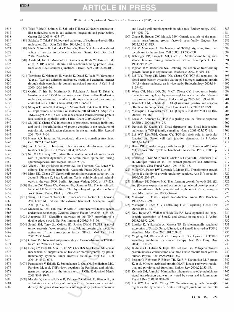
UN
CO
RR
EC
TED
PR
OO
F
[87] Takai Y, Irie K, Shimizu K, Sakisaka T, Ikeda W. Nectins and nectin-
like molecules: roles in cell adhesion, migration, and polarization.
Cancer Sci 2003;94:655–67.
[88] Sakisaka T, Takai Y. Biology and pathology of nectins and nectin-like
molecules. Curr Opin Cell Biol 2004;16:513–21.
[89] Irie K, Shimizu K, Sakisaka T, Ikeda W, Takai Y. Roles and modes of
action of nectins in cell–cell adhesion. Semin Cell Dev Biol
2004;15:643–56.
[90] Asada M, Irie K, Morimoto K, Yamada A, Ikeda W, Takeuchi M,
et al. ADIP, a novel afadin- and a-actinin-binding protein loca-
lized at cell–cell adherens junctions. J Biol Chem 2003;278:4103–
11.
[91] Tachibana K, Nakanishi H, Mandai K, Ozaki K, Ikeda W, Yamamoto
Y, et al. Two cell adhesion molecules, nectin and cadherin, interact
through their cytoplasmic domain-associated proteins. J Cell Biol
2000;150:1161–76.
[92] Ooshio T, Irie K, Morimoto K, Fukuhara A, Imai T, Takai Y.
Involvement of LMO7 in the association of two cell–cell adhesion
molecules, nectin and E-cadherin, through afadin and a-actinin in
epithelial cells. J Biol Chem 2004;279:31365–73.
[93] Shingai T, Ikeda W, Kakunaga S, Morimoto K, Takekuni K, Itoh S, et
al. Implications of nectin-like molecule-2/IGSF4/RA175/SgIGSF/
TSLC1/SynCAM1 in cell–cell adhesion and transmembrane protein
localization in epithelial cells. J Biol Chem 2003;278:35421–7.
[94] Siu MKY, Cheng CY. Interactions of proteases, protease inhibitors,
and the b1 integrin/laminin g3 protein complex in the regulation of
ectoplasmic specialization dynamics in the rat testis. Biol Reprod
2004;70:945–64.
[95] Hynes RO. Integrins: bidirectional, allosteric signaling machines.
Cell 2002;110:673–87.
[96] Jin H, Varner J. Integrins: roles in cancer development and as
treatment targets. Br J Cancer 2004;90:561–5.
[97] Siu MKY, Cheng CY. Extracellular matrix: recent advances on its
role in junction dynamics in the seminiferous epithelium during
spermatogenesis. Biol Reprod 2004;375–91.
[98] Vilcek J. The cytokines: an overview. In: Thomson AW, Lotze MT,
editors. The cytokine handbook. Academic Press; 2003. p. 3–18.
[99] Mruk DD, Cheng CY. Sertoli cell proteins in testicular paracriny. In:
Jegou B, Pineau C, Saez J, editors. Testis, epididymis and technol-
ogies in the year 2000. Berlin: Springer–Verlag; 2000. p. 197–228.
[100] Bardin CW, Cheng CY, Mustow NA, Gunsalus GL. The Sertoli cell.
In: Knobil E, Neill JD, editors. The physiology of reproduction. New
York: Raven Press; 1994. p. 1291–332.
[101] Wang H, Czura CJ, Tracey KJ. Tumor necrosis factor. In: Thomson
AW, Lotze MT, editors. The cytokine handbook. Academic Press;
2003. p. 837–60.
[102] Mocellin S, Rossi CR, Pilati P, Nitti D. Tumor necrosis factor, cancer
and anticancer therapy. Cytokine Growth Factor Rev 2005;16:35–53.
[103] Aggarwal BB. Signalling pathways of the TNF superfamily: a
double-edged sword. Nat Rev Immunol 2003;3:745–56.
[104] Soond SM, Terry JL, Colbert JD, Riches DWH. TRUSS, a novel
tumor necrosis factor receptor 1 scaffolding protein that mediates
activation of the transcription factor NF-kB. Mol Cell Biol
2003;23:8334–44.
[105] Gibson PR. Increased gut permeability in Crohn’s disease: is TNF the
link? Gut 2004;53:1724–5.
[106] Hong CY, Park JH, Ahn RS, Im SY, Choi H-S, Soh J, et al. Molecular
mechanism of suppression of testicular steroidogenesis by proin-
flammatory cytokine tumor necrosis factor a. Mol Cell Biol
2004;24:2593–604.
[107] Pentikainen V, Erkkila K, Suomalainen L, Otala M, Pentikainen MO,
Parvinen M, et al. TNFa down-regulates the Fas ligand and inhibits
germ cell apoptosis in the human testis. J Clin Endocrinol Metab
2001;86:4480–8.
[108] Morales V, Santana P, Diaz R, Tabraue C, Gallardo G, Blanco FL, et
al. Intratesticular delivery of tumor necrosis factor-a and ceramide
directly abrogates steroidogenic acute regulatory protein expression
and Leydig cell steroidogenesis in adult rats. Endocrinology 2003;
144:4763–72.
[109] Chang H, Brown CW, Matzuk MM. Genetic analysis of the mam-
malian transforming growth factor-b superfamily. Endocr Rev
2002;23:787–823.
[110] Shi Y, Massague J. Mechanisms of TGF-b signaling from cell
membrane to the nucleus. Cell 2003;113:685–700.
[111] Behringer RR, Finegold MJ, Cate RL. Mullerian-inhibiting sub-
stance function during mammalian sexual development. Cell
1994;79:415–25.
[112] Ingman WV, Robertson SA. Defining the action of transforming
growth factor b in reproduction. BioEssays 2002;24:904–14.
[113] Lui WY, Wong CH, Mruk DD, Cheng CY. TGF-b3 regulates the
blood-testis barrier dynamics via the p38 mitogen activated protein
(MAP) kinase pathway: an in vivo study. Endocrinology 2003;144:
1139–42.
[114] Wong CH, Mruk DD, Siu MKY, Cheng CY. Blood-testis barrier
dynamics are regulated by a2-macroglobulin via the c-Jun N-term-
inal protein kinase pathway. Endocrinology 2005;146:1893–908.
[115] Wakefield LM, Roberts AB. TGF-b signaling: positive and negative
effects on tumorigenesis. Curr Opin Genet Dev 2002;12:22–9.
[116] Massague J. How cells read TGF-b signals. Nat Rev Mol Cell Biol
2000;1:169–78.
[117] Leask A, Abraham DJ. TGF-b signaling and the fibrotic response.
FASEB J 2004;18:816–27.
[118] Derynck R, Zhang YE. Smad-dependent and Smad-independent
pathways in TGF-b family signaling. Nature 2003;425:577–84.
[119] Lui WY, Lee WM, Cheng CY. TGF-bs: their role in testicular
function and Sertoli cell tight junction dynamics. Int J Androl
2003;26:1–14.
[120] Howe PH. Transforming growth factor b. In: Thomson AW, Lotze
MT, editors. The cytokine handbook. Academic Press; 2003 . p.
1119–52.
[121] Roberts AB, Kim SJ, Noma T, Glick AB, Lafyatis R, Lechleider R, et
al. Multiple forms of TGF-b: distinct promoters and differential
expression. Ciba Found Symp 1991;157:7–15.
[122] Miller DA, Pelton RW, Derynck R, Moses HL. Transforming growth
factor-b-a family of growth regulatory peptides. Ann N Y Acad Sci
1990;593:209–17.
[123] Mullaney BP, Skinner MK. Transforming growth factor-b (b1, b2,
and b3) gene expression and action during pubertal development of
the seminiferous tubule: potential role at the onset of spermatogen-
esis. Mol Endocrinol 1993;7:67–76.
[124] Massague J. TGF-b signal transduction. Annu Rev Biochem
1998;67:753–91.
[125] Massague J, Chen Y-G. Controlling TGF-b signaling. Genes Dev
2000;14:627–44.
[126] Xu J, Beyer AR, Walker WH, McGee EA. Developmental and stage-
specific expression of Smad2 and Smad3 in rat testis. J Androl
2003;24:192–200.
[127] Luukko K, Ylikorkala A, Makela TP. Developmentally regulated
expression of Smad3, Smad4, Smad6, and Smad7 involved in TGF-b
signaling. Mech Dev 2001;101:209–12.
[128] Yingling JM, Blanchard KL, Sawyer JS. Development of TGF-b
signalling inhibitors for cancer therapy. Nat Rev Drug Disc
2004;3:1011–22.
[129] Widmann C, Gibson S, Jarpe MB, Johnson GL. Mitogen-activated
protein kinases: conservation of a three-kinase module from yeast to
human. Physiol Rev 1999;79:143–80.
[130] Peason G, Robinson F, Bibson TB, Xu B-E, Karandikar M, Berman
K, et al. Mitogen-activated protein (MAP) kinase pathways: regula-
tion and physiological functions. Endocr Rev 2001;22:153–83.
[131] Kyriakis JM, Avruch J. Mammalian mitogen-activated protein kinase
signal transduction pathways activated by stress and inflammation.
Physiol Rev 2001;81:807–69.
[132] Lui WY, Lee WM, Cheng CY. Transforming growth factor-b3
regulates the dynamics of Sertoli cell tight junctions via the p38
W. Xia et al. / Cytokine & Growth Factor Reviews xxx (2005) xxx–xxx20
DTD 5
CGFR 365 1–24
1616
1617
1618
1619
1620
1621
1622
1623
1624
1625
1626
1627
1628
1629
1630
1631
1632
1633
1634
1635
1636
1637
1638
1639
1640
1641
1642
1643
1644
1645
1646
1647
1648
1649
1650
1651
1652
1653
1654
1655
1656
1657
1658
1659
1660
1661
1662
1663
1664
1665
1666
1667
1668
1669
1670
1671
1672
1673
1674
1675
1676
1677
1678
1679
1680
1681
1682
1683
1684
1685
1686
1687
1688
1689
1690
1691
1692
1693
1694
1695
1696
1697
1698
1699
1700
1701
1702
1703
1704
1705
1706
1707
1708
1709
1710
1711
1712
1713
1714
1715
1716
1717
1718
1719
1720
1721
1722
1723
1724
1725
1726
1727
1728
1729
1730
1731
1732
1733
1734
1735
1736
1737
1738
1739
1740
1741
1742
1743
1744
1745
1746
1747
1748
1749

UN
CO
RR
EC
TED
PR
OO
F
mitogen-activated protein kinase pathway. Biol Reprod 2003;68:
1597–612.
[133] Chapin RE, Wine RN, Harris MW, Borchers CH, Haseman JK.
Structure and control of a cell–cell adhesion complex associated
with spermiation in rat seminiferous epithelium. J Androl 2001;
22:1030–52.
[134] Barrios-Rodiles M, Brown KR, Ozdamar B, Bose R, Liu Z, Donovan
RS, et al. High-throughput mapping of a dynamic signaling network
in mammalian cells. Science 2005;307:1621–5.
[135] Ozdamar B, Bose R, Barrios-Rodiles M, Wang H-R, Zhang Y, Wrana
JL. Regulation of the polarity protein Par6 by TGFb receptors
controls epithelial cell plasticity. Science 2005;307:1603–9.
[136] Wang H-R, Zhang Y, Ozdamar B, Ogunjimi AA, Alexandrova E,
Thomsen GH, et al. Regulation of cell polarity and protrusion
formation by targeting RhoA for degradation. Science 2003;302:
1775–9.
[137] Hassel S, Eichner A, Yakymovych M, Hellman U, Knaus P, Sou-
chelnytskyi S. Proteins associated with type II bone morphogenetic
protein receptor (BMPR-II) and identified by two-dimensional gel
electrophoresis and mass spectrometry. Proteomics 2004;4:1346–58.
[138] Behzadian MA, Wang X-L, Windsor LJ, Ghaly N, Caldwell RB.
TGF-b increases retinal endothelial cell permeability by increasing
MMP-9: possible role of glial cells in endothelial barrier function.
Invest Ophthalmol Vis Sci 2001;42:853–9.
[139] Hurst IV V, Goldberg PL, Minnear FL, Heimark RL, Vincent PA.
Rearrangement of adherens junctions by transforming growth factor-
b1: role of contraction. Am J Physiol Lung Cell Mol Physiol
1999;276:L582–95.
[140] Fujimoto K, Sheng H, Shao J, Beauchamp RD. Transforming growth
factor-b1 promotes invasiveness after cellular transformation with
activated Ras in intestinal epithelial cells. Exp Cell Res 2001;266:
239–49.
[141] Tian YC, Fraser D, Attisano L, Phillips AO. TGF-b1-mediated
alterations of renal proximal tubular epithelial cell phenotype. Am
J Physiol Renal Physiol 2003;285:F130–42.
[142] Watabe T, Nishihara A, Mishima K, Yamashita J, Shimizu K,
Miyazawa K, et al. TGF-b receptor kinase inhibitor enhances growth
and integrity of embryonic stem cell-derived endothelial cells. J Cell
Biol 2003;163:1303–11.
[143] Sheppard D. Roles of av integrins in vascular biology and pulmonary
pathology. Curr Opin Cell Biol 2004;16:552–7.
[144] Xu L, Massague J. Nucleocytoplasmic shuttling of signal transdu-
cers. Nat Rev Mol Cell Biol 2004;5:1–11.
[145] Keski-Oja J, Koli K, von Melchner H. TGF-b activation by traction?
Trends Cell Biol 2004;14:657–9.
[146] Gumienny TL, Padgett RW. The other side of TGF-b superfamily
signal regulation: thinking outside the cell. Trends Endocr Metab
2002;13:295–9.
[147] Pardoux C, Derynck R. JNK regulates expression and autocrine
signaling of TGF-b1. Mol Cell 2004;15:170–1.
[148] Ventura J-J, Kennedy NJ, Flavell RA, Davis RJ. JNK regulates
autocrine expression of TGF-b1. Mol Cell 2004;15:269–78.
[149] Annes JP, Munger JS, Rifkin DB. Making sense of latent TGFb
activation. J Cell Sci 2003;116:217–24.
[150] Rifkin DB. Latent transforming growth factor-b (TGF-b) binding
proteins: orchestrators of TGF-b availability. J Biol Chem 2005;280:
7409–12.
[151] Piek E, Heldin C-H, ten Dijke P. Specificity, diversity, and regulation
in TGF-b superfamily signaling. FASEB J 1999;13:2105–24.
[152] Lebrin F, Deckers M, Bertolino P, ten Dijke P. TGF-b receptor
function in the endothelium. Cardiovasc Res 2005;65:599–608.
[153] Ge B, Gram H, Di Padova F, Huang B, New L, Ulevitch RJ, et al.
MAPKK-independent activation of p38a mediated by TAB1-depen-
dent autophosphorylation of p38a. Science 2002;295:1291–4.
[154] Schiffer M, Mundel P, Shaw AS, Bottinger EP. A novel role for the
adaptor molecule CD2-associated protein in transforming growth
factor-b-induced apoptosis. J Biol Chem 2004;279:37004–12.
[155] Schiffer M, Mundel P, Shaw AS, Bottinger EP. A novel role
for the adaptor molecule CD2-associated protein in tranforming
growth factor-b-induced apoptosis. J Biol Chem 2004;279:37004–
12.
[156] Razani B, Zhang XL, Bitzer M, von Gersdorff G, Bottinger EP,
Lisanti MP. Caveolin-1 regulates transforming growth factor (TGF)-
b/Smad signaling through an interaction with the TGF-b type I
receptor. J Biol Chem 2001;276:6727–38.
[157] Schwarz K, Simons M, Reiser J, Saleem MA, Faul C, Kriz W,
et al. Podocin, a raft-associated component of the glomerular slit
diaphragm, interacts with CD2AP and nephrin. J Clin Invest 2001;
108:1621–9.
[158] Dikic I. CIN85/CMS family of adaptor molecules. FEBS Lett
2002;529:110–5.
[159] Kirsch KH, Georgescu M-M, Ishimaru S, Hanafusa H. CMS: An
adapter molecule involved in cytoskeletal rearrangements. Proc Natl
Acad Sci USA 1999;96:6211–6.
[160] Webb DJ, Donais K, Whitmore LA, Thomas SM, Turner CE, Parsons
JT, et al. FAK-Src signalling through paxillin, ERK and MLCK
regulates adhesion disassembly. Nat Cell Biol 2004;6:154–61.
[161] Lui WY, Mruk D, Lee WM, Cheng CY. Sertoli cell tight junction
danamics: their regulation during spermatogenesis. Biol Reprod
2003;68:1087–97.
[162] Lui WY, Mruk DD, Lee WM, Cheng CY. Adherens junction
dynamics in the testis and spermatogenesis. J Androl 2003;24:1–14.
[163] Grima J, Silvestrini B, Cheng CY. Reversible inhibition of sperma-
togenesis in rats using a new male contraceptive, 1-(2,4-dichloro-
benzyl)-indazole-3-carbohydrazide. Biol Reprod 2001;64:1500–8.
[164] Cheng CY, Silvestrini B, Grima J, Mo M-Y, Zhu L-J, Jahansson E, et
al. Two new male contraceptives exert their effects by depleting germ
cells prematurely from the testis. Biol Reprod 2001;65:449–61.
[165] Grima J, Wong CCS, Zhu L-J, Zong S-D, Cheng CY. Testin secreted
by Sertoli cells is associated with the cell surface, and its expression
correlates with the disruption of Sertoli–germ cell junctions but not
the inter-Sertoli tight junction. J Biol Chem 1998;273:21040–53.
[166] Grima J, Cheng CY. Testin induction: the role of cyclic 30,50-adenosine monophosphate/protein kinase A signaling in the regula-
tion of basal and lonidamine-induced testin expression by rat Sertoli
cells. Biol Reprod 2000;63:1648–60.
[167] Lui WY, Lee WM, Cheng CY. Sertoli–germ cell adherens junction
dynamics in the testis are regulated by RhoB GTPase via the ROCK/
LIMK signaling pathway. Biol Reprod 2003;68:2189–206.
[168] Chen Y-M, Lee NPY, Mruk DD, Lee WM, Cheng CY. Fer kinase/
FerT and adherens junction dynamics in the testis: an in vitro and in
vivo study. Biol Reprod 2003;69:656–72.
[169] Werner S, Grose R. Regulation of wound healing by growth factors
and cytokines. Physiol Rev 2003;83:835–70.
[170] Sahai E. Mechanisms of cancer cell invasion. Curr Opin Genet Dev
2005;15:87–96.
[171] Zalups RK, Ahmad S. Molecular handling of cadmium in transport-
ing epithelia. Toxicol Appl Pharmac 2003;186:163–88.
[172] Waisberg M, Joseph P, Hale B, Beyersmann D. Molecular and cellular
mechanisms of cadmium carcinogenesis. Toxicology 2003;192:95–
117.
[173] Henson MC, Chedrese PJ. Endocrine disruption by cadmium, a
common environmental toxicant with paradoxical effects on repro-
duction. Expt Biol Med 2004;229:383–92.
[174] Johnson MD, Kenney N, Stoica A, Hilakivi-Clarke L, Singh B,
Chepko G, et al. Cadmium mimics the in vivo effects of estrogen
in the uterus and mammary gland. Nat Med 2003;9:1081–4.
[175] Jin YH, Clark AB, Slebos RJC, Al-Refai H, Taylor JA, Kunkel TA, et
al. Cadmium is a mutagen that acts by inhibiting mismatch repair. Nat
Genet 2003;34:326–9.
[176] Dalton TP, He L, Wang B, Miller ML, Jin L, Stringer KF, et al.
Identification of mouse SLC39A8 as the transporter responsible for
cadmium-induced toxicity in the testis. Proc Natl Acad Sci USA
2005;102:3401–6.
W. Xia et al. / Cytokine & Growth Factor Reviews xxx (2005) xxx–xxx 21
DTD 5
CGFR 365 1–24
1750
1751
1752
1753
1754
1755
1756
1757
1758
1759
1760
1761
1762
1763
1764
1765
1766
1767
1768
1769
1770
1771
1772
1773
1774
1775
1776
1777
1778
1779
1780
1781
1782
1783
1784
1785
1786
1787
1788
1789
1790
1791
1792
1793
1794
1795
1796
1797
1798
1799
1800
1801
1802
1803
1804
1805
1806
1807
1808
1809
1810
1811
1812
1813
1814
1815
1816
1817
1818
1819
1820
1821
1822
1823
1824
1825
1826
1827
1828
1829
1830
1831
1832
1833
1834
1835
1836
1837
1838
1839
1840
1841
1842
1843
1844
1845
1846
1847
1848
1849
1850
1851
1852
1853
1854
1855
1856
1857
1858
1859
1860
1861
1862
1863
1864
1865
1866
1867
1868
1869
1870
1871
1872
1873
1874
1875
1876
1877
1878
1879
1880
1881
1882
1883
UN
CO
RR
EC
TED
PR
OO
F
[177] Fiorini C, Tilloy-Ellul A, Schevalier S, Charuel C, Pointis G. Sertoli
cell junctional proteins as early targets for different classes of
reproductive toxicants. Reprod Toxicol 2004;18:413–21.
[178] Chung NPY, Cheng CY. Is cadmium chloride-induced inter-Sertoli
tight junction permeability barrier disruption a suitable in vitro model
to study the events of junctional disassembly during spermatogenesis
in the rat testis? Endocrinology 2001;142:1878–88.
[179] Hew KW, Heath GL, Jiwa AH, Welsh MJ. Cadmium in vivo causes
disruption of tight junction-associated microfilaments in rat Sertoli
cells. Biol Reprod 1993;49:840–9.
[180] Gray LE, Ostby J, Furr J, Wolf CJ, Lambright C, Parks L, et al.
Effects of environmental antiandrogens on reproductive development
in experimental animals. Hum Reprod Update 2001;7:248–64.
[181] Boekelheide K, Johnson KJ, Richburg JH. Sertoli cell toxicants. In:
Skinner MK, Griswold MD, editors. Sertoli cell biology. San Diego:
Elsevier Academic Press; 2005. p. 345–82.
[182] Toyama Y, Suzuki-Toyota F, Maekawa M, Ito C, Toshimori K.
Adverse effects of bisphenol A to spermiogenesis in mice and rats.
Arch Histol Cytol 2004;67:373–81.
[183] Fisher JS. Environmental anti-androgens and male reproductive
health: focus on phthalates and testicular dysgenesis syndrome.
Reproduction 2004;127:305–15.
[184] Kelce WR, Monosson E, Gamcsik MP, Laws SC, Gray LEJ. Envir-
onmental hormone disruptors: evidence that vinclozolin develop-
mental toxicity is mediated by antiandrogenic metabolites. Toxicol
Appl Pharmacol 1994;126.
[185] Wong C-I, Kelce WR, Sar M, Wilson EM. Androgen receptor
antagonist versus agonist activities of the fungicide vinclozolin
relative to hydroxyflutamide. J Biol Chem 1995;270:19998–20003.
[186] Foster PMD, Mylchreest E, Gaido KW, Sar M. Effects of phthalate
esters on the developing reproductive tract of male rats. Hum Reprod
Update 2001;7:231–5.
[187] Creasy DM, Beech LM, Gray TJ, Butler WH. The ultrastructural
effects of di-n-pentyl phthalate on the testis of the mature rats. Exp
Mol Pathol 1987;46:357–71.
[188] Creasy DM, Foster JR, Foster PM. The morphological development
of di-N-pentyl phthalate induced testicular atrophy in the rat. J Pathol
1983;139:309–21.
[189] Hotchkiss AK, Ostby JS, Vandenbergh JG, Gray J, Earl L. An
environmental antiandrogen, vinclozolin, alters the organization of
play behavior. Physiol Behav 2003;79:151–6.
[190] Gray LEJ, Ostby J, Monosson E, Kelce WR. Environmental anti-
androgens: low doses of the fungicide vinclozolin alter sexual
differentiation of the male rat. Toxicol Ind Health 1999;15:48–64.
[191] Yu WJ, Lee BJ, Nam SY, Ahn B, Hong JT, Do JC, et al. Reproductive
disorders in pubertal and adult phase of the male rats exposed to
vinclozolin during puberty. J Vet Med Sci 2004;66:847–53.
[192] Kubota K, Ohsako S, Kurosawa S, Takeda K, Qing W, Sakaue M, et
al. Effects of vinclozolin administration on sperm production and
testosterone biosynthetic pathway in adult male rat. J Reprod Dev
2003;49:403–12.
[193] Venkiteswaran K, Xiao K, Summers S, Calkins CC, Vincent PA,
Pumiglia K, et al. Regulation of endothelial barrier function and
growth by VE-cadherin, plakoglobin, and b-catenin. Am J Physiol
Cell Physiol 2002;283:C811–21.
[194] Troxell ML, Chen Y-T, Cobb N, Nelson WJ, Marrs JA. Cadherin
function in junctional complex rearrangement and posttranslational
control of cadherin expression. Am J Physiol Cell Physiol 1999;
276:C404–18.
[195] Man Y, Hart VJ, Ring CJA, Sanjar S, West MR. Loss of epithelial
integrity resulting from E-cadherin dysfunction predisposes airway
epithelial cells to adenoviral infection. Am J Respir Cell Mol Biol
2000;23:610–7.
[196] Guo X, Rao JN, Liu L, Zou TT, Turner DJ, Bass BL, et al. Regulation
of adherens junctions and epithelial paracellular permeability: a
novel function for polyamines. Am J Physiol Cell Physiol 2003;
285:C1174–87.
[197] Gassler N, Rohr C, Schneider A, Kartenbeck J, Bach A, Obermuller
N, et al. Inflammatory bowel disease is associated with changes of
enterocytic junctions. Am J Physiol Gastrointest Liver Physiol
2001;281:G216–28.
[198] Toyama Y, Yuasa S. Effects of neonatal administration of 17b-
estradiol, b-estradiol 3-benzoate, or bisphenol A on mouse and rat
spermatogenesis. Reprod Toxicol 2004;19:181–8.
[199] Li LH, Heindel JJ. Sertoli cell toxicants. In: Korach KS, editor.
Reproductive and developmental toxicology. New York: Dekker
Marcel; 1998. p. 655–91.
[200] Setchell BP, Waites GMH. Changes in the permeability of testicular
capillaries and of the ‘‘blood-testis barrier’’ after injection of cad-
mium chloride in the rat. J Endocrinol 1970;47:81–6.
[201] Kagi J, Schaffer A. Biochemistry of metallothionein. Biochemistry
1988;27:8509–15.
[202] Nath R, Kambadur R, Gulati S, Paliwal V, Sharma M. Molecular
aspects, physiological function, and clinical significance of metal-
lothioneins. Crit Rev Food Nutr 1988;27:41–85.
[203] Nordberg G. Cadmium metabolism and toxicity. Environ Physiol
Biochem 1972;2:7–36.
[204] Vallee B. The function of metallothionein. Neurochem Int 1995;27:
23–33.
[205] Waalkes M, Goering P. Metallothionein and other cadmium-binding
proteins: Recent developments. Chem Res Toxicol 1990;3:281–8.
[206] Sciavolino P, Lee T, Vilcek J. Overexpression of metallothionein
confers resistance to the cytotoxic effect of TNF with cadmium in
MCF-7 breast carcinoma cells. Lymphokine Cytokine Res 1992;11:
265–70.
[207] Dufresne J, Cyr D. Effects of short-term methylmercury exposure on
metallothionein mRNA levels in the testis and epididymis of the rat. J
Androl 1999;20:769–78.
[208] Suzuki J, Kodama N, Molotkov A, Aoko E, Tohyama C. Isolation and
identification of metallothionein isoforms (MT-1 and MP-2) in the rat
testis. Biochem J 1998;334:695–701.
[209] Sugihara T, Wadhwa R, Kaul S, Mitsui Y. A novel testis-specific
metallothionein-like protein, tesmin, is an early marker of male germ
cell differentiation. Genomics 1999;57:130–6.
[210] Ren X, Zhou Y, Zhang J, Feng W, Jiao B. Metallothionein gene
expression under different time in testicular Sertoli and spermatogenic
cells of rats treated with cadmium. Reprod Toxicol 2003;17:219–27.
[211] Coogan T, Shiraishi N, Waalkes M. Minimal basal activity and lack
of metal-induced activation of the metallothionein gene correlates
with lobe-specific sensitivity to the carcinogenic effects of cadmium
in the rat prostate. Toxicol Appl Pharmacol 1995;132:164–73.
[212] Coogan T, Shiraishi N, Waalkes M. Metallothionein gene expression
in the reproductive tissues of the Wistar rat: Effects of treatment with
metals and glucocorticoids. Toxic Subst Mech 1997;16:357–70.
[213] Lee K, Lau K, Ho S. Effects of cadmium on metallothionein-I and
metallothionein-II mRNA expression in rat ventral, lateral, and
dorsal prostatic lobes: Quantification by competitive RT-PCR. Tox-
icol Appl Pharmacol 1999;154:20–7.
[214] Prozialeck WC. Evidence that E-cadherin may be a target for
cadmium toxicity in epithelial cells. Toxicol Appl Pharmacol
2000;164:231–49.
[215] Prozialeck WC, Lamar PC. Interaction of cadmium (Cd2+) with a 13-
residue polypeptide analog of a putative calcium-binding motif of E-
cadherin. Biochim Biophys Acta 1999;1451:93–100.
[216] Prozialeck WC, Niewenhuis RJ. Cadmium (Cd2+) disrupts intercel-
lular junctions and actin filaments in LLC-PK1 cells. Toxicol Appl
Pharmacol 1991;107:81–97.
[217] Plow EF, Haas TA, Zhang L, Loftus J, Smith JW. Ligand binding to
integrins. J Biol Chem 2000;275:21785–8.
[218] De SK, Chen HL, Pace JL, Hunt JS, Terranova PF, Enders GC.
Expression of tumor necrosis factor-a in mouse spermatogenic cells.
Endocrinology 1993;133:389–96.
[219] Montell DJ. Border-cell migration: the race is on. Nat Rev Mol Cell
Biol 2003;4:13–24.
W. Xia et al. / Cytokine & Growth Factor Reviews xxx (2005) xxx–xxx22
DTD 5
CGFR 365 1–24
1884
1885
1886
1887
1888
1889
1890
1891
1892
1893
1894
1895
1896
1897
1898
1899
1900
1901
1902
1903
1904
1905
1906
1907
1908
1909
1910
1911
1912
1913
1914
1915
1916
1917
1918
1919
1920
1921
1922
1923
1924
1925
1926
1927
1928
1929
1930
1931
1932
1933
1934
1935
1936
1937
1938
1939
1940
1941
1942
1943
1944
1945
1946
1947
1948
1949
1950
1951
1952
1953
1954
1955
1956
1957
1958
1959
1960
1961
1962
1963
1964
1965
1966
1967
1968
1969
1970
1971
1972
1973
1974
1975
1976
1977
1978
1979
1980
1981
1982
1983
1984
1985
1986
1987
1988
1989
1990
1991
1992
1993
1994
1995
1996
1997
1998
1999
2000
2001
2002
2003
2004
2005
2006
2007
2008
2009
2010
2011
2012
2013
2014
2015
2016
2017
UN
CO
RR
EC
TED
PR
OO
F
[220] Matzuk MM. Germ-line immortality. Proc Natl Acad Sci USA
2004;101:16395–6.
[221] MacLean IIJA, Chen MA, Wayne CM, Bruce SR, Rao M, Meistrich
ML, et al. Rhox: a new homeobox gene cluster. Cell 2005;120:369–82.
[222] De Gendt K, Swinnen JV, Saunders PTK, Schoonjans L, Dewerchin
M, Devos A, et al. A Sertoli cell-selective knockout of the androgen
receptor causes spermatogenic arrest in meiosis. Proc Natl Acad Sci
USA 2004;101:1327–32.
[223] Stamatovic SM, Keep RF, Kunkel SL, Andjelkovic AV. Potential role
of MCP-1 in endothelial cell tight junction ‘opening’: signaling via
Rho and Rho kinase. J Cell Sci 2003;116:4615–28.
[224] Pedram A, Razandi M, Levin ER. Deciphering vascular endothelial
cell growth factor/vascular permeability factor signaling to vascular
permeability. Inhibition by atrial natriuretic peptide. J Biol Chem
2002;277:44385–98.
[225] Ahdieh M, Vandenbos T, Youakim A. Lung epithelial barrier function
and wound healing are decreased by IL-4 and IL-13 and enhanced by
IFN-g. Am J Physiol Cell Physiol 2001;281:C2029–38.
[226] Kagami S, Kuhara T, Yasutomo K, Okada K, Loster K, Reutter W, et
al. Transforming growth factor-b (TGF-b) stimulates the expression
of b1 integrins and adhesion by rat mesangial cells. Exp Cell Res
1996;229:1–6.
[227] Tian YC, Fraser D, Attisano L, Phillips AO. TGF-b1-mediated
alterations of renal proximal tubular epithelial cell phenotype. Am
J Physiol Renal Physiol 2003;285:F130–42.
[228] de Kretser DM, Buzzard JJ, Okuma Y, O’Connor AE, Hayashi T, Lin
S-Y, et al. The role of activin, follistatin and inhibin in testicular
physiology. Mol Cell Endocr 2004;225:57–64.
[229] Shimasaki S, Moore RK, Otsuka F, Erickson GF. The bone morpho-
genetic protein system in mammalian reproduction. Endocr Rev
2004;25:72–101.
[230] Pellegrini M, Grimaldi P, Rossi P, Geremia R, Dolci S. Develop-
mental expression of BMP4/ALK3/SMAD5 signaling pathway in the
mouse testis: a potential role of BMP4 in spermatogonia differentia-
tion. J Cell Sci 2003;116:3363–72.
[231] Hua J, Chen Y-X, Wang D, Qi X, Li T-G, Hao J, et al. Developmental
expression and function of Bmp4 in spermatogenesis and in main-
taining epididymal integrity. Dev Biol 2004;276:158–71.
[232] Pasparakis M, Alexopoulou L, Episkopou V, Kollias G. Immune and
inflammatory responses in TNFa-deficient mice: a critical require-
ment for TNFa in the formation of primary B cell follicles, follicular
dendritic cell networks and germinal centers, and in the maturation of
the humoral immune response. J Exp Med 1996;184:1397–411.
[233] D’Alessio A, Riccioli A, Lauretti P, Padula F, Muciaccia B, De
Cesaris P, et al. Testicular FasL is expressed by sperm cells. Proc Natl
Acad Sci USA 2001;98:3316–21.
[234] Lee J, Richburg JH, Younkin SC, Boekelheide K. The Fas system is a
key regulator of germ cell apoptosis in the testis. Endocrinology
1997;138:2081–8.
[235] Adachi M, Suematsu S, Kondo T, Ogasawara J, Tanaka T, Yoshida N,
et al. Targeted mutation in the Fas gene causes hyperplasia in
peripheral lymphoid organs and liver. 1995;11:294–300.
[236] Grataroli R, Vindrieux D, Gougeon A, Benahmed M. Expression of
tumor necrosis factor-a-related apoptosis-inducing ligand and its
receptors in rat testis during development. Biol Reprod 2002;66:
1707–15.
[237] Cretney E, Takeda K, Yagita H, Glaccum M, Peschon JJ, Smyth MJ.
Increased susceptibility to tumor initiation and metastasis in TNF-
related apoptosis-inducing ligand-deficient mice. J Immunol 2002;
168:1356–61.
[238] Luetteke N, Qiu T, Fenton S, Troyer K, Riedel R, Chang A, et al.
Targeted inactivation of the EGF and amphiregulin genes reveals
distinct roles for EGF receptor ligands in mouse mammary gland
development. Development 1999;126:2739–50.
[239] Radhakrishnan B, Oke B, Papadopoulos V, DiAugustine R, Suarez-
Quian C. Characterization of epidermal growth factor in mouse testis.
Endocrinology 1992;131:3091–9.
[240] Wahab-Wahlgren A, Martinelle N, Holst M, Jahnukainen K, Parvinen
M, Soder O. EGF stimulates rat spermatogonial DNA synthesis in
seminiferous tubule segments in vitro. Mol Cell Endocr 2003;201:
39–46.
[241] Bottcher RT, Niehrs C. Fibroblast growth factor signaling during
early vertebrate development. Endocr Rev 2005;26:63–77.
[242] Yamamoto H, Ochiya T, Tamamushi S, Toriyama-Baba H, Takahama
Y, Hirai K, et al. HST-1/FGF-4 gene activation induces spermato-
genesis and prevents adriamycin-induced testicular toxicity. Onco-
gene 2002;21:899–908.
[243] Yamamoto H, Ochiya T, Takahama Y, Ishii Y, Osumi N, Sakamoto H,
et al. Detection of spatial localization of Hst-1/Fgf-4 gene expression
in brain and testis from adult mice. Oncogene 2000;19:3805–10.
[244] Ricci G, Catizone A, Galdieri M. Pleiotropic activity of hepatocyte
growth factor during embryonic mouse testis development. Mech
Dev 2002;118:19–28.
[245] Catizone A, Ricci G, Galdieri M. Expression and functional role of
hepatocyte growth factor receptor (C-MET) during postnatal rat testis
development. Endocrinology 2001;142:1828–34.
[246] Uehara Y, Minowa O, Mori C, Shiota K, Kuno J, Noda T, et al.
Placental defect and embryonic lethality in mice lacking hepatocyte
growth factor/scatter factor. Nature 1995;373:702–5.
[247] Meinhardt A, Bacher M, Wennemuth G, Eickhoff R, Hedger M,
Macrophage migration inhibitory factor (MIF) as a paracrine med-
iator in the interaction of testicular somatic cells. 2000;32:46–48.
[248] Honma N, Koseki H, Akasaka T, Nakayama T, Taniguchi M,
Serizawa I, et al. Deficiency of the macrophage migration inhibitory
factor gene has no significant effect on endotoxaemia. Immunology
2000;100:84–90.
[249] Marziali G, Lazzaro D, Sorrentino V. Binding of germ cells to mutant
SId Sertoli cells is defective and is rescued by expression of the
transmembrane form of the c-kit ligand. Dev Biol 1993;157:182–90.
[250] Bedell MA, Zama AM. Genetic analysis of Kit ligand functions
during mouse spermatogenesis. J Androl 2004;25:188–99.
[251] Nalbandian A, Dettin L, Dym M, Ravindranath N. Expression of
vascular endothelial growth factor receptors during male germ cell
differentiation in the mouse. Biol Reprod 2003;69:985–94.
[252] Rudolfsson SH, Wikstrom P, Jonsson A, Collin O, Bergh A. Hor-
monal regulation and functional role of vascular endothelial growth
factor A in the rat testis. Biol Reprod 2004;70:340–7.
[253] Ferrara N, Carver-Moore K, Chen H, Dowd M, Lu L, O’Shea KS, et
al. Heterozygous embryonic lethality induced by targeted inactiva-
tion of the VEGF gene. Nature 1996;380:439–42.
[254] Horai R, Asano M, Sudo K, Kanuka H, Suzuki M, Nishihara M, et al.
Production of mice deficient in genes for interleukin (IL)-1a, IL-1b,
IL-1a/b, and IL-1 receptor antagonist shows that IL-1b is crucial in
turpentine-induced fever development and glucocorticoid secretion. J
Exp Med 1998;187:1463–75.
[255] Hedger MP, Meinhardt A. Cytokines and the immune-testicular axis.
J Reprod Immunol 2003;58:1–26.
[256] Dalton D. IFN-g and IFN-g receptor knockout mice. In: Fantuzzi G,
editor. Cytokine knockouts. Totowa, NJ: Humana Press; 2003 . p.
347–59.
[257] Dejucq N, Lienard M-O, Guillaume E, Dorval I, Jegou B. Expression
of interferons-a and -g in testicular interstitial tissue and spermato-
gonia of the rat. Endocrinology 1998;139:3081–7.
[258] Pogach LM, Lee Y, Gould S, Giglio W, Meyenhofer M, Huang HF.
Characterization of cis-platinum-induced Sertoli cell dysfunction in
rodents. Toxicol Appl Pharmacol 1989;98:350–61.
[259] Peterson PM, Giwercman A, Skakkebaek NE, Rorth M. Gonadal
function in men with testicular cancer. Semin Oncol 1998;25:224–33.
[260] Wiebe JP, Kowalik A, Gallardi RL, Egeler O, Clubb BH. Glycerol
disrupts tight junction-associated actin microfilaments, occludin, and
microtubules in Sertoli cells. J Androl 2000;21:625–35.
[261] Wiebe JP, Barr KJ, Buckingham KD, Gedded PD, Kudo PA. Pro-
spects of a male contraceptive based on selective antispermatogenic
action of 1,2,3-trihydroxypropane (THP; glycerol). In: Zatuchni GI,
W. Xia et al. / Cytokine & Growth Factor Reviews xxx (2005) xxx–xxx 23
DTD 5
CGFR 365 1–24
2018
2019
2020
2021
2022
2023
2024
2025
2026
2027
2028
2029
2030
2031
2032
2033
2034
2035
2036
2037
2038
2039
2040
2041
2042
2043
2044
2045
2046
2047
2048
2049
2050
2051
2052
2053
2054
2055
2056
2057
2058
2059
2060
2061
2062
2063
2064
2065
2066
2067
2068
2069
2070
2071
2072
2073
2074
2075
2076
2077
2078
2079
2080
2081
2082
2083
2084
2085
2086
2087
2088
2089
2090
2091
2092
2093
2094
2095
2096
2097
2098
2099
2100
2101
2102
2103
2104
2105
2106
2107
2108
2109
2110
2111
2112
2113
2114
2115
2116
2117
2118
2119
2120
2121
2122
2123
2124
2125
2126
2127
2128
2129
2130
2131
2132
2133
2134
2135
2136
2137
2138
2139
2140
2141
2142
2143
2144
2145
2146
2147
2148
2149
2150
2151
UN
CO
RR
EC
TED
PR
OO
F
Goldsmith A, Spiler AJM, Sciarra JJ, editors. Male contraceptio-
n:advances and future prospects. Philadelphia: Harper and Row;
1986. p. 252–70.
[262] Chung NPY, Mruk D, Mo M-Y, Lee WM, Cheng CY. A 22-amino
acid synthetic peptide corresponding to the second extracellular loop
of rat occludin perturbs the blood-testis barrier and disrupts sperma-
togenesis reversibly in vivo. Biol Reprod 2001;65:1340–51.
[263] Waites GM, Wang C, Griffin PD. Gossypol: reasons for its failure to
be accepted as a safe, reversible male antifertility drug. Int J Androl
1998;21:8–12.
[264] Qian S, Wang Z. Gossypol: a potential antifertility agent for males.
Annu Rev Pharmacol Toxicol 1984;24:329–60.
[265] Pelletier RM, Friend DS. Sertoli cell junctional complexes in gos-
sypol-treated neonatal and adult guinea pigs. J Androl 1986;7:
127–39.
[266] Silvestrini B, Palazzo G, De Gregorio M. Lonidamine and related
compounds. Prog Med Chem 1984;21:111–35.
[267] O’Donnell L, Stanton PG, Bartles JR, Robertson DM. Sertoli cell
ectoplasmic specializations in the seminiferous epithelium of the
testosterone-suppressed adult rat. Biol Reprod 2000;63:99–108.
[268] McLachlan RI, O’Donnell L, Meachem SJ, Stanton PG, Kretser
DMD, Pratis K, et al. Hormonal regulation of spermatogenesis in
primates and man: insights for development of the male hormonal
contraceptive. J Androl 2002;23:149–62.
[269] Kelce WR, Lambright CR, Gray J, Earl L, Roberts KP. Vinclozolin
and p,p0-DDE alter androgen-dependent gene expression: in vivo
confirmation of an androgen receptor-mediated mechanism. Toxicol
Appl Pharmacol 1997;142:192–200.
[270] Uzumcu M, Suzuki H, Skinner MK. Effect of the anti-androgenic
endocrine disruptor vinclozolin on embryonic testis cord formation
and postnatal testis development and function. Reprod Toxicol
2004;18:765–74.
[271] Ashby J, Tinwell H, Lefevre PA, Joiner R, Haseman J. The effect on
sperm production in adult Sprague-Dawley rats exposed by gavage to
bisphenol A between postnatal days 91–97. Toxicol Sci 2003;74:
129–38.
[272] Williams K, McKinnell C, Saunders PTK, Walker M, Fisher JS, Turner
KJ, et al. Neonatal exposure to potent and environmental oestrogens
and abnormalities of the male reproductive system in the rat: evidence
for importance of the androgen-oestrogen balance and assessment of
the relevance to man. Hum Reprod Update 2001;7:236–47.
[273] Wang Y, Thuillier R, Culty M. Prenatal estrogen exposure differen-
tially affects estrogen receptor-associated proteins in rat testis gono-
cytes. Biol Reprod 2004;71:1652–64.
W. Xia et al. / Cytokine & Growth Factor Reviews xxx (2005) xxx–xxx24
DTD 5
CGFR 365 1–24
2152
2153
2154
2155
2156
2157
2158
2159
2160
2161
2162
2163
2164
2165
2166
2167
2168
2169
2170
2171
2172
2173
2174
2175
2176
2177
2178
2179
2180
2181
2182
2183
2184
2185
2186
2187
2188
2189
2190
2191
2192
2193
2194
2195
2196