UNIVERSITE CHEKH ANTA DIOP FACULTE DES SCIENCES ET TECHNOLOGIES DE L'EDUCATION ET DE LA FORMATION
Upload
khangminh22Category
view
3download
0
UNIVERSITE DE LA MEDITERRANEE
FACULTE DE PHARMACIE DE MARSEILLE
THESE DE DOCTORAT Spécialité : Immunologie
Présentée et publiquement soutenue le 14 Décembre 2010 Par M. LACROIX Romaric
Né le 02/11/80 à Toulon En vue d�obtenir le grade de Docteur de l�Université Aix-Marseille II
---oOo---
TITRE :
MICROPARTICULES : DE LA GENERATION DE PLASMINE A LA STANDARDISATION D�UN BIOMARQUEUR EMERGEANT.
---oOo---
JURY
Président : M. le Professeur Pierre CAU Rapporteur : M. le Professeur Jean-Baptiste MICHEL Rapporteur : M. le Professeur Pierre SIE Directeur de thèse: Mme le Professeur Françoise DIGNAT-GEORGE Examinateur : M. Christophe DUBOIS�
�

��
�
REMERCIEMENTS
�
�
� En tête de cette liste, je remercie celle qui a subi avec patience ce travail, ma
chère épouse Damaris avec une tendre pensée pour mes deux petits garçons Siméon et
Noé.
Je remercie mon directeur de thèse, Françoise, pour m�avoir guidé dans ce
travail et pour toute son attention quotidienne.
Je remercie les membres du jury de thèse d�avoir accepté de juger ce travail.
Je remercie le Professeur J. Sampol pour m�avoir acceuilli dans son laboratoire
Je remercie le Professeur L. Camoin-Jau pour m�avoir donné le goût de
l�hématologie.
Je remercie Florence pour son soutien constant.
Je remercie tous ceux avec qui je travaille au quotidien : la microparticule team
(Stephane, Philippe, Coralie et Tarik), Christophe et Laurence, mes partenaires de
bureau (Stephane et Lucas), Corine, mes co-assistantes (Stephanie, Magalie, Elise, Yaël
et Maud), toute l�équipe de séniors et juniors de l�u608 et toute l�équipe de biologistes et
de techniciens du service d�hématologie de la Conception, les secrétaires.
Je remercie ceux qui m�ont acceuilli pendant cette thèse : Eduardo Angles-Cano,
Bruce et Barbara Furie.
Je remercie mes parents et ma s�ur Karine
Je remercie mes anciens co-internes : Sofiane, Boris et Anne-Catherine
Je remercie mes amis de toujours : Adrien et Natacha, Titi et Vincent, TomTom
et Pauline, Kalou, Momo, Hélène et Fabien, Betty, TonTon, Marjorie et Franck,
Sandra.
Je remercie ceux qui m�ont guidé : Tonton et Tata, M.Tafani, Guillaume, Père
Host du Roure
Je remercie Dieu

��
�
�
« J�ai appliqué mon c�ur à rechercher et à explorer par la
sagesse tout ce qui se fait sous les cieux, c�est une occupation
ingrate que Dieu a donnée aux fils des hommes afin qu�ils s�y
fatiguent »
Ecclésiate 1v 13
�
�
�
�
�
�
�
�
�
�

��
�
SOMMAIRE
�
REMERCIEMENTS ............................................................ 1�
SOMMAIRE ......................................................................... 3�
TABLE DES FIGURES ....................................................... 8�
PRINCIPALES ABREVIATIONS .................................... 10�
PREMIERE PARTIE: REVUE BIBLIOGRAPHIQUE . 12�
CHAPITRE 1 : MICROPARTICULES, DEFINITION ET
ORIGINE ............................................................................ 13�
A. MICROPARTICULES : ASPECT HISTORIQUE .............................. 13�
1) Cellules hématopoïétiques et vasculaires .................................................. 13�
2) Cellules tumorales ....................................................................................... 15�
3) Autres types cellulaires ............................................................................... 15�
B. DEFINITION DES MICROPARTICULES .......................................... 17�
1) Définition ...................................................................................................... 17�
2) Terminologie ................................................................................................ 17�
C. FORMATION DES MICROPARTICULES ......................................... 19�
1) Mécanismes de formation des microparticules ........................................ 19�
a. Remaniement des phospholipides de la membrane plasmique ............ 20�
i. Asymétrie de la membrane plasmique .................................. 20�
ii. Activation cellulaire et externalisation de la
phosphatidylsérine .................................................................... 21�
iii. Lien entre l�exposition de la PS et la vésiculation cellulaire
.................................................................................................. 22�
iv. Limites du modèle actuel ..................................................... 22�
v. Autres mécanismes de vésiculation dépendant de la
composition en phospholipides ................................................. 23�

��
�
b. Réarrangement du cytosquelette ............................................................ 23�
i. Rôle des calpaïnes ................................................................. 25�
ii. Rôle de la Gelsoline .............................................................. 25�
iii. Rôle de l�interaction phospholipides/cytosquelette ............. 25�
iv. Mécanismes moléculaires aboutissant au remodelage du
cytosquelette ............................................................................. 26�
2) Les inducteurs de la vésiculation .............................................................. 27�
a. Activation cellulaire ................................................................................. 28�
i. Les plaquettes ........................................................................ 28�
ii. Les globules rouges .............................................................. 29�
iii. Les leucocytes ...................................................................... 29�
iv. Les cellules endothéliales .................................................... 30�
b. Apoptose .................................................................................................... 32�
c. Conclusion ................................................................................................. 33�

��
�
CHAPITRE 2 : MICROPARTICULES : STRUCTURE
ET FONCTION .................................................................. 34�
A. COMPOSITION DES MICROPARTICULES ..................................... 34�
B. FONCTIONS DES MICROPARTICULES ........................................... 36�
1) Rôle des microparticules dans l�hémostase ............................................. 36�
2) Rôle des microparticules dans la modulation du tonus vasculaire ........ 41�
3) Rôle des microparticules dans l�inflammation ......................................... 43�
4) Rôle des microparticules dans l�angiogenèse ........................................... 44�
5) Rôle des microparticules dans la protéolyse matricielle ......................... 46�
6) Rôle des microparticules dans la survie cellulaire .................................. 47�
7) Rôle des microparticules dans la modulation de la réponse immunitaire
........................................................................................................................... 48�
8) Rôle des microparticules dans la communication intercellulaire ........... 49�

��
�
CHAPITRE 3 : LA GENERATION DE PLASMINE ..... 51�
A. LE PLASMINOGENE ............................................................................. 51�
B. LES ACTIVATEURS DU PLASMINOGENE ...................................... 53�
1) L�activateur tissulaire du plasminogène (t-PA) ....................................... 53�
2) L�urokinase (uPA) ...................................................................................... 55�
3) Le récepteur de l�urokinase (uPAR) ........................................................ 56�
C. LA FORMATION DE PLASMINE ........................................................ 57�
1) Formation de plasmine par le t-PA .......................................................... 57�
2) Formation de plasmine par l�uPA ............................................................ 59�
D. LA REGULATION DE LA GENERATION DE PLASMINE. ........... 60�
1) Les serpines .................................................................................................. 61�
2) Les inhibiteurs de liaison du plasminogène ............................................. 65�
E. CONSEQUENCES DE L�ACTIVATION DU PLASMINOGENE ..... 66�
1) La fibrinolyse ............................................................................................... 66�
2) Le remodelage matriciel ............................................................................ 67�
3) Autres conséquences de l�activation du plasminogène ............................ 69�

��
�
CHAPITRE 4 : MICROPARTICULES :
BIOMARQUEUR EN PATHOLOGIE HUMAINE ........ 70�
A. INTERET DU DOSAGE DE MICROPARTICULES CIRCULANTES EN PATHOLOGIE HUMAINE ................................................................... 70�
1) Signification biologique des microparticules en pathologie. .................. 70�
2) Limites actuelles de l�utilisation des microparticules comme
biomarqueur en pathologie humaine. ............................................................ 77�
B. VARIABLES PRE-ANALYTIQUES POUVANT INFLUER SUR LES DOSAGES DE MICROPARTICULES....................................................... 77�
C. METHODOLOGIES DISPONIBLES POUR L�ANALYSE DES
MICROPARTICULES ................................................................................. 80�
1) Diversité des méthodologies disponibles pour l�analyse des
microparticules ................................................................................................ 80�
2) Avantages et limites des techniques de mesure des microparticules ..... 84�
DEUXIEME PARTIE: TRAVAUX PERSONNELS ........ 88�
INTRODUCTION ......................................................................................... 88�
ARTICLE 1 .................................................................................................... 90�
ARTICLE 2 .................................................................................................... 93�
ARTICLE 3 .................................................................................................... 96�
BREVET 1 ...................................................................................................... 98�
ARTICLE 4 .................................................................................................... 99�
ARTICLE 5 .................................................................................................. 102�
DISCUSSION-PERSPECTIVES ............................................................... 104�
ANNEXES .......................................................................... 111�
BIBLIOGRAPHIE ............................................................112�

�
�
Table des figures
Figure 1 : Images historiques de microparticules par microscopie. �������..16�
Figure 2 : Représentation schématique de la libération des différentes microvésicules
par la cellule dans l�espace extracellulaire. ......................................................................... 18�
Figure 3 : Implication du cytosquelette dans le mécanisme de vésiculation. ................... 24�
Figure 4 : Place de l�activation de ROCK dans le phénomène de vésiculation ............... 27�
Figure 5 : Mécanisme de formation des MPE après stimulation par la thrombine ........ 31�
Figure 6 : Composition moléculaire fonctionnelle des microparticules endothéliales. ... 35�
Figure 7 : Contribution de la génération des MPE à l�activité procoagulante. ............... 39�
Figure 8 : Contribution des MP tumorales à la formation du thrombus in vivo ............. 40�
Figure 9 : MPE et dysfonction endothéliale par diminution de la synthèse de NO. ....... 42�
Figure 10 : Lien entre propriétés pro-thrombotique et inflammatoire des MP. ............. 44�
Figure 11 : Augmentation de l�angiogenèse par des concentrations croissantes de MPP
dans un modèle d�anneaux aortiques de rat ........................................................................ 46�
Figure 12 : Différents modes d�interaction entre les MP et la cellule cible. ..................... 50�
Figure 13 : Structure du plasminogène. ............................................................................... 53�
Figure 14 : Structure du t-PA ............................................................................................... 54�
Figure 15 : Conversion de pro-urokinase en urokinase par la plasmine .......................... 56�
Figure 16 : Formation de plasmine par le t-PA ou l�u-PA à la surface des cellules
endothéliales. .......................................................................................................................... 59�
Figure 17 : Inhibiteurs du système fibrinolytique. ............................................................ 60�
Figure 18 : Inhibition du complexe u-PA/u-PAR par le PAI-1. ........................................ 62�
Figure 19 : Conséquences de la génération de plasmine par le système uPA/uPAR. ...... 68�
Figure 20 : Balance définissant la vasculocompétence. (Sabatier F, JCMM 2009) ......... 76�
Figure 21 : Etapes du processus pré-analytiques pouvant influer sur le dosage des MP.
.................................................................................................................................................. 80�

�
�
Figure 22 : Exemple de mesure des MPP par cytométrie de flux ..................................... 81�
Figure 23 : Principe d�une méthode hybride combinant capture et test fonctionnel. ..... 83�
Figure 24 : Shéma du principe d�un appareil utilisant le single particle tracking .......... 84�
Tableau 1 : Caractéristiques différentielles des différentes vésicules. .............................. 19�
Tableau 2 : Signification clinique de l�élévation des MPE en pathologie ......................... 73�
Tableau 3 : Résumé des principales caractéristiques des techniques utilisées pour la
mesure des MP ....................................................................................................................... 87�

���
�
Principales abréviations
�2-AP : �2-antiplasmine �-ACA : acide �-amino caproïque APCE : Enzyme de clivage de l�antiplasmine ATF: Fragment aminoterminal de l�urokinase CEC : Cellules endothéliales circulantes CMF : Cytométrie en flux CML : Cellules musculaires lisses CpB : Carboxypeptidase B CRP : Protéine C reactive DLS : Dynamic light scattering EGF : Epidermal growth factor EMLT : Enhanced laser microscopy tracking FGF : Facteur de croissance des fibroblastes fMPL : N-formylmethionyl-leucyl-phenylalanine FS : Forward Scatter FT : Facteur tissulaire GM-CSF : Facteur de croissance des monocytes et des granulocytes GPI : Glycero-phospho-inositol HTA : Hypertension artérielle HUVEC : Cellules endothéliales de veine ombilicale humaine IL : Interleukine INF� : Interferon gamma ISTH : International Society on thrombosis and Haemostasis LBS : Site de liaison des résidus lysine Lp(a) : Lipoprotéine (a) LPS : Lipopolysaccharide Lys C term : Lysine C terminals MDR : Multidrug resistance MLC : Chaines légères de la myosine MMP : Métalloprotéinase matricielle MP : Microparticules MPE : Microparticules endothéliales MPGR : Microparticules érythrocytaires MPP : Microparticules plaquettaires MT-MMP : Métalloprotéase de type membranaire NMDA : N-méthyl-D-aspartate NO : Monoxyde d�azote PAF/PMA : Platelet activation factor/ phorbol 12-myristate 13-acetate

���
�
PAI-1 : Inhibiteur du plasminogène type I PBS : Phosphate Buffered Saline PDGF : Platelet derived growth factor PE : Phosphatidyléthanolamine PEC : Progéniteurs endothéliaux PECAM : Platelet endothelium cellular adhesion molecule PFP : Platelet free plasma PIP2 :Phosphatidyl inositol de type II PS: Phosphatidylsérine PSGL-1 : P-selectin glycoprotein ligand PTAI : Purpura thrombopénique auto-immun PTT : Purpura thrombotique thrombocytopénique Scu-PA : Pro-urokinase SPT : Single particle tracking TAFI : Inhibiteur de la fibrinolyse activé par la thrombine Tcu-PA : Urokinase TFPI : Inhibiteur de la voie du facteur tissulaire TGF� : Tumor growth factor �TIMP : Inhibiteur tissulaire des métalloprotéases TLR : Toll-like receptor TM : Thrombomoduline TNF : Tumor necrosis factor t-PA : Activateur tissulaire du plasminogène uPA : Urokinase uPAR : Récepteur de l�urokinase VEGF : Facteur de croissance de l�endothélium vasculaire VIH : Virus de l�immunodéficience humaine

���
�
PREMIERE PARTIE: REVUE
BIBLIOGRAPHIQUE
� Dans cette première partie bibliographique nous nous attacherons après un bref
rappel historique à définir les microparticules (MP) d�origine cellulaire et à résumer la
connaissance actuelle sur leurs mécanismes de formation. Nous verrons ensuite en quoi
une meilleure connaissance de leur composition permet de découvrir un spectre de
fonctions biologiques de plus en plus large. Ceci inclura leurs propriétés protéolytiques
qui mettent en jeu les molécules du système fibrinolytique. Nous consacreront donc le
chapitre suivant aux acteurs et mécanismes de la génération de plasmine ainsi que ces
conséquences. Enfin, nous feront le point sur l�intérêt d�utiliser les MP comme
biomarqueur en pathologie en discutant les limites actuelles, essentiellement d�ordre
méthodologique, qu�il reste à franchir.

���
�
CHAPITRE 1 : MICROPARTICULES, DEFINITION ET
ORIGINE
�
A. MICROPARTICULES : ASPECT HISTORIQUE
Considérant les MP dans leur ensemble, il n�est pas vraiment possible de parler d�une
découverte unique, source de l�avancée de la connaissance dans ce domaine. En effet,
plusieurs voies indépendantes, focalisées chacune sur un sous-type cellulaire, ont caractérisé
l�avancée scientifique sur les microparticules. Tantôt constatation d�une perte de matériel
membranaire, tantôt découverte de la précipitabilité d�une activité supposée soluble ou simple
observation microscopique (Figure 1), l�observation scientifique princeps fut très variable. Le
lien entre toutes ces entités n�a finalement été fait qu�assez tardivement dans les années 1990.
Cette prise de conscience de l�universalité du phénomène de vésiculation cellulaire a entrainé
l�ouverture de cet axe de recherche à une multitude de milieux biologiques et de types
cellulaires.
1) Cellules hématopoïétiques et vasculaires
La notion selon laquelle le vieillissement et la destruction des globules rouges sont
associés à une perte de phospholipides et de cholestérol est très ancienne. La première
mention a été faite par M. Schultze dans un surprenant article de la revue allemande Archiv
für Mikroskopishe Anatomie datant de 1861 dans lequel il rapporte une observation de
fragments provenant de globules rouges à l�aide d�un nouvel objectif chauffant de
microscope16. Plus d�un siècle plus tard, Weed et co proposent la fragmentation de la
membrane du globule rouge comme mécanisme in vivo et in vitro de cette perte de
phospholipides17-21. En 1964, Michel rapporte la présence de matériel spécifique de groupes
sanguins dans le plasma de sang stocké22. En 1969, Cooper et Jandl23 décrivent la perte de
cholestérol et de phospholipides par des globules rouge incubés dans du sérum déprivé en
glucose, sans toutefois suspecter le phénomène de vésiculation. Les premiers à utiliser ce
terme et à décrire la production de vésicules incorporant des sialoglycoprotéines de

���
�
membrane de globules rouges sont les MacDonalds en collaboration avec Martin24-27. Dans le
même temps, les français Bessis et Mandon, publient des photos de microscopie électronique
montrant la formation de microsphérules (figure1A)28,29.
C�est en 1949 que Chargaff et co. constatent pour la première fois que le plasma
déplaquetté contient un facteur précipitable qui a la capacité d�accélérer la génération de
thrombine30. Plus tard en 1967, Wolf rapporte que du matériel dérivé de membranes
cellulaires riches en lipides sudanophiliques, obtenu par ultracentrifugation de plasma
déplaquetté permet de générer de la thrombine31. Il démontre une corrélation linéaire entre le
nombre de microparticules plaquettaire (MPP), alors appelées « platelet dusts », et la
numération plaquettaire d�origine dans les échantillons sanguins. Par microscopie
électronique il sera ensuite possible de confirmer que ces petites vésicules infra-
micrométriques proviennent des plaquettes activées (Figure 1B)32.
En 1985, des vésicules issues de granulocytes, nommées ectosomes, sont identifiées.
Elles sont associées alors à un mécanisme de réponses cellulaires aux attaques du
complément (Figure 1C et D)33-35. En 1994, Satta et al. rapportent l�existence de MP
circulantes dérivées de monocytes et de cellules THP-136 , générées après stimulation des
monocytes par le lipopolysaccharide (LPS). La même équipe décrira trois ans plus tard les
MP dérivées de lymphocytes dans le contexte de l�infection au VIH (Virus de
l�immunodéficience humaine)37.
La première évidence de l�existence des microparticules endothéliales (MPE) est la
démonstration par Hamilton et co en 1990 que le surnageant de culture des cellules
endothéliales de veine ombilicale humaine (HUVEC) en présence du complément contient
des éléments membranaires capables de générer de la thrombine38. Leur véritable
caractérisation morphologique, phénotypique et fonctionnelle est réalisée par Combes et co
en 1999 (Figure 1E)39.

���
�
2) Cellules tumorales
La description des MP d�origine tumorale est également assez ancienne. Les premiers
éléments proviennent d�images de microscopie électronique sans pour autant que les
observateurs n�aient particulièrement relevé la présence de ces éléments vésiculaires. Par
exemples, des vésicules tumorales sont clairement visibles sur des images de tératocarcinome
murin de 196240 ou de tumeur rénale induite chez le rat en 196741. Le premier à noter leur
importance est Tarin en 196742,43 qui les nomment « structures vésiculaires » ou « matériel
fragmenté ». Dans la décennie suivante, ces vésicules sont isolées de surnageant de culture de
lignées tumorales murines, le liquide d�ascite de patients atteint de cancer ovariens, des
sécrétions d�adénome villeux entre autres (Figure 1F)44,45. Très rapidement, l�intérêt se porte
sur la relation entre ces fragments et les métastases cancéreuses46,47.
3) Autres types cellulaires
Le phénomène de vésiculation est un processus commun à toutes les cellules
eucaryotes. Ainsi, depuis les premières descriptions précédemment citées, différentes sous-
populations de MP dérivées d�autres types cellulaires ont été décrites et étudiées comme par
exemple et par ordre chronologiques les MP provenant des chondrocytes48,
oligodendrocytes49, fibroblastes50, syncytiotrophoblastes51,52, podocytes53, adipocytes54,
cellules musculaires lisses55, mégacaryocytes56,57, cellules neuro-épithéliales58, cellules
souches embryonnaires59 et cellules souches mésenchymateuses60.
Différents liquides biologiques ont été utilisés pour caractériser les MP. A la suite des
surnageant de culture et du plasma, d�autres milieux comme les urines44, les liquides
d�ascite44, le liquide cérébro-spinal49, la plaque d�athérosclérose61, le liquide synovial62, le
liquide broncho-alvéolaire63, le muscle64, l�humeur vitrée65 ont permis leur mise en évidence.
Ces nombreuses études, initialement purement descriptives, aussi bien in vitro dans
des modèles cellulaires purifiés qu�in vivo dans différents contextes cliniques, ont permis de
dévoiler progressivement les caractéristiques structurelles et fonctionnelles de ces MP.

���
�
��
�
��
�
�
Fig
ure
1:
Images
his
tori
qu
es d
e m
icro
part
icu
les
par
mic
rosc
op
ie. A
. Mic
ropa
rtic
ules
éry
thro
cyta
ires
(B
essi
s, S
prin
ger-
Ver
lag,
197
3 et
Gre
enw
ald
R.L
iss
1980
) B
. Mic
ropa
rtic
ules
pla
quet
tair
es (
Geo
rge
JN e
t co.
Blo
od 1
982)
C. e
t D
. M
icro
part
icul
es le
ucoc
ytai
res
(Mor
gan
et c
o. J
Im
mun
ol 1
987)
E. M
icro
part
icul
es
endo
thél
iale
s (C
ombe
s et
co.
J C
lin
Inve
st 1
999)
. F
. Mic
ropa
rtic
ules
tum
oral
es (
Sco
tt e
t co.
J C
ell S
ci 1
979)
.

���
�
B. DEFINITION DES MICROPARTICULES
1) Définition
Même si quelques points de discussion persistent, le sous comité de standardisation
en biologie vasculaire de la société internationale de thrombose et d�hémostase (ISTH) définit
les MP comme des vésicules de taille hétérogène, comprise entre 0.1 et 1 �m, résultant du
bourgeonnement de la membrane cellulaire de la plupart si n�est toutes les cellules activées
ou apoptotiques. Elles sont retrouvées dans le culot d�une centrifugation rapide d�un plasma
déplaquetté ou d�un surnageant de culture. Elles expriment généralement de la
phosphatidylsérine (PS) et différentes molécules de la cellule d�origine permettant leur
identification et contribuant à leurs multiples fonctions. Les critères de définition concernant
leur taille précise et l�expression de PS restent débattus66-68.
2) Terminologie
Il faut rapporter ici une absence de consensus de la nomenclature des petites vésicules
parfois source de confusion. En effet, suivant les auteurs, le même terme est employé pour
désigner différentes entités spécifiques ou un ensemble de vésicules. Ainsi le terme
« microvésicules » est tantôt employé comme synonyme de microparticles ou pour désigner
l�ensemble des types de vésicules émises par une cellule quelque soit leur mécanisme de
formation ou leur composition (Figure 2). Les MP sont donc à distinguer d�autres petites
vésicules libérées par les cellules comme les exosomes ou les corps apoptotiques (Tableau
1). Les termes « argosomes »69 et « ectosomes »70,71 employés pour désigner des vésicules
d�origine spécifique dans la littérature sont à assimiler à des microparticules.
��
�
�
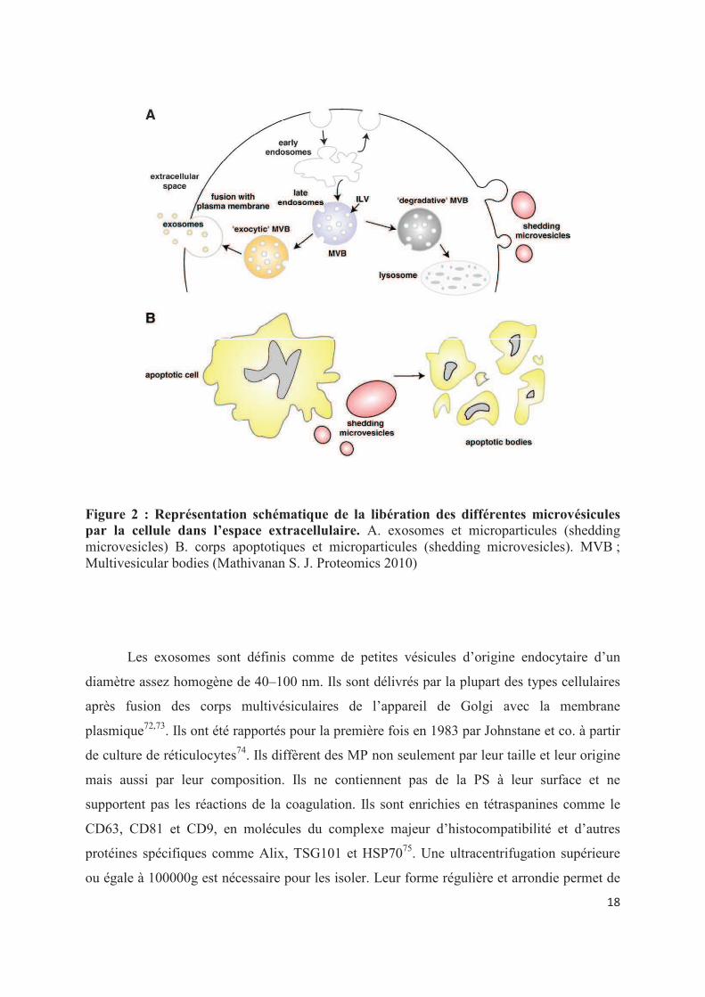
Figure 2 : Représentation schématique de la libération des différentes microvésicules par la cellule dans l�espace extracellulaire. A. exosomes et microparticules (shedding microvesicles) B. corps apoptotiques et microparticules (shedding microvesicles). MVB ; Multivesicular bodies (Mathivanan S. J. Proteomics 2010)
�
Les exosomes sont définis comme de petites vésicules d�origine endocytaire d�un
diamètre assez homogène de 40�100 nm. Ils sont délivrés par la plupart des types cellulaires
après fusion des corps multivésiculaires de l�appareil de Golgi avec la membrane
plasmique72,73. Ils ont été rapportés pour la première fois en 1983 par Johnstane et co. à partir
de culture de réticulocytes74. Ils diffèrent des MP non seulement par leur taille et leur origine
mais aussi par leur composition. Ils ne contiennent pas de la PS à leur surface et ne
supportent pas les réactions de la coagulation. Ils sont enrichies en tétraspanines comme le
CD63, CD81 et CD9, en molécules du complexe majeur d�histocompatibilité et d�autres
protéines spécifiques comme Alix, TSG101 et HSP7075. Une ultracentrifugation supérieure
ou égale à 100000g est nécessaire pour les isoler. Leur forme régulière et arrondie permet de

��
�
les isoler dans un gradient de sucrose dans un zone à faible densité de 1.10�1.21 g/mL, ce qui
les différencient des autres vésicules à forme plus irrégulière qui sont retrouvées dans des
zones de plus haute densité (>1.23 g/mL)76.
Les corps apoptotiques, de taille souvent plus importante que les microparticules (0.5-
4 �m) représentent le stade terminal des cellules en apoptose. Ils sont générés par
fragmentation de la cellule apoptotique et contiennent des résidus de matériel nucléaire
comme les histones77,78. Il n�est pas toujours aisé de les distinguer méthodologiquement des
microparticules sensus stricto, le phénomène d�apoptose étant également source de
microparticules.
�
Exosomes Microparticules Corps apoptotiques
Taille (diamètre) 30-100 nm 100-1000 nm 500-4000 nm Densité de flottaison 1.10-1.21 g/mL NA 1.16-1.28 g/mL
Morphologie Arrondies Hétérogène Hétérogène
Composition
lipidique
ALBP, PS faible, cholestérol, céramide,
shingomyéline PS fort, Cholestérol PS fort
Marqueurs
protéiques
Alix, TSG101, HSC70, CD63, CD81, CD9
Sélectines, intégrines, CD40, MMP
Histones
Origine Exocytose des MVB Vésiculation de la
membrane plasmique Contraction et mort
cellulaire
Composition Protéines, ARNm,
miARN Protéines, ARNm, miARN
Protéines, ARNm, miARN, ADN
�
Tableau 1 : Caractéristiques différentielles des différentes vésicules. (Modifié de Mathivanan S. J. Proteomics 2010)
C. FORMATION DES MICROPARTICULES
1) Mécanismes de formation des microparticules
Les mécanismes de formation des microparticules ne sont pas parfaitement élucidés.
La compréhension actuelle de ce phénomène fait intervenir au moins deux types
d�événement : un remaniement des phospholipides de la membrane plasmique et un
réarrangement du cytosquelette qui conduisent au bourgeonnement cellulaire.

���
�
a. Remaniement des phospholipides de la membrane plasmique
i. Asymétrie de la membrane plasmique
Les phospholipides ne sont pas repartis au hasard au sein de la membrane plasmique
mais de manière asymétrique79. Les phospholipides cationiques contenant de la choline
(phosphatidylcholine et sphingomyéline) sont localisés de façon prépondérante au niveau du
feuillet externe de la membrane plasmique. A l�inverse, les aminophospholipides
(phosphatidyléthanolamine (PE) et PS) sont concentrés au niveau du feuillet interne.
Cette asymétrie phospholipidique qui caractérise les cellules quiescentes est
maintenue par des activités enzymatiques transmembranaires. Ces enzymes sont regroupées
en trois groupes selon leur spécificité lipidique, la direction du transport et leur dépendance
ou non en énergie80. En 1984, les travaux de Seigneuret et Devaux puis de Daleke ont montré
l�importance de l�activité aminophospholipides translocase dans le maintien de l�asymétrie
phospholipidique81-83. Cette translocase ATP-dépendante permet le transport de la PE et de la
PS du feuillet externe vers le feuillet interne de la membrane plasmique contre le gradient de
concentration. Une molécule d�ATP est nécessaire pour chaque molécule de PS transporté.
Cette activité est inhibée lorsque les taux de calcium intra-cytoplasmique atteignent des
concentrations élevées de l�ordre du micromolaire.
Une autre enzyme ATP-dépendante a été décrite. Il s�agit de la floppase qui elle,
transporte les phospholipides du feuillet interne vers le feuillet externe de la membrane
plasmique de manière non spécifique84. Sa fonction n�est pas bien comprise. Elle fonctionne
probablement en conjonction avec l�aminophospholipide translocase. Leurs activités
combinées permettent de répondre aux altérations lipidiques de la membrane plasmique85.
Dans les globules rouges, l�activité floppase est portée par la protéine MDR1 (Multidrug
Resistance Protein 1) appartenant à la superfamille des transporteurs ABC (ATP Binding
Cassette)86. Par ailleurs, dans cette superfamille, la p-glycoprotéine (MDR2/3), a été
caractérisée au niveau des cellules des canaux biliaires comme transporteur de
phosphatidylcholine du feuillet interne vers le feuillet externe87. Enfin, la scramblase,
permet un transport aspécifique bidirectionnel des phospholipides88. Elle est fortement
présente dans la membrane des plaquettes et des globules rouges.

���
�
ii. Activation cellulaire et externalisation de la
phosphatidylsérine
L�activation cellulaire ou l�apoptose sont associées à une élévation du taux de calcium
intracellulaire. Celle-ci inhibe d�une part l�activité de l�aminophospholipide translocase et
d�autre part active les activités floppase et scramblase. Ainsi, un taux cytoplasmique de
calcium élevé induit l�externalisation de la PS et un équilibre des phospholipides négatifs
entre les deux feuillets de la membrane plasmique89.
Le gène ABCA1, impliqué dans le transport inverse du cholestérol, a été également
proposé comme élément clé de l�externalisation de PS dans la cellule activée ou apoptotique.
En effet, les cellules de souris ABCA1-KO externalisent la PS avec une efficacité très réduite
après activation cellulaire90,91.
D�autres mécanismes peuvent conduire à l�externalisation de la PS et à la libération de
MP comme, l�enrichissement de la membrane en cholestérol sur les monocytes qui entraîne
alors la libération de MP monocytaires porteuses de facteur tissulaire92. Il faut sans doute
rapprocher de ce mécanisme le rôle important joué par les radeaux lipidiques « rafts » dans
la vésiculation érythrocytaire93. Ces structures membranaires sont des domaines enrichis en
cholestérol, capables de s�assembler ou de se dissocier en réponse à différents stimuli, de
séquestrer certaines protéines et de recruter des molécules impliquées dans la signalisation.
Kunzelmann et co. ont démontré que l�intégrité de ces microdomaines est nécessaire à la
mobilisation du calcium intracellulaire et à l�externalisation de la phosphatidylsérine94. Ils
permettent le fonctionnement du canal calcique TRCP-1. De plus l�externalisation de la PS
dépend de l�activation de la voie des kinases ERK associée à ces microdomaines. Enfin, ces
zones de la membrane sont enrichies en protéines GPI (Glycéro-Phospho-Inositol) ancrées
qui sont retrouvées majoritairement sur les MP95.
Certaines études soulignent également l�importance du mécanisme de recapture du
calcium comme modulateur de l�exposition de PS après activaiton cellulaire : SOCE (store-
operated calcium entry)96. L�importance de SOCE a été illustrée récemment dans un modèle
animal. Les plaquettes des souris exprimant une forme mutée de Orai1 (une partie d�un canal
SOCE) montrent un défaut d�exposition de PS après stimulation97.

���
�
iii. Lien entre l�exposition de la PS et la vésiculation cellulaire
L�exposition de la PS et la vésiculation cellulaire ou émission de microparticules sont
des évènements fortement associés. Un modèle pathologique illustre bien cette association.
Le Scott syndrome est une maladie rare autosomale récessive avec tendance hémorragique
sévère, due à un défaut d�activité pro-coagulante des plaquettes98. L�anomalie sous jacente est
un défaut activité scramblase qui conduit à la réduction de l�expression de la PS et de la
production de MP99. L�agrégation plaquettaire à l�adrénaline, l�ADP (Adénosine
diphosphate), au collagène, à la ristocétine et l�acide arachidonique reste normale tandis que
la génération de thrombine est anormale100,101. Un défaut similaire avec activité pro-
thrombinasique normale a aussi été rapporté par Castaman102.
D�autre part l�inhibition de la randomisation des phospholipides supprime la
vésiculation des globules rouges induite par le calcium103.
iv. Limites du modèle actuel
A ce niveau, il faut reconnaitre que tous les acteurs impliqués dans le maintien ou la
rupture de l�asymétrie de la membrane n�ont pas été identifiés avec précision104. Ceci peut
être expliqué en partie par les difficultés techniques qui accompagnent les expériences de
modulation de la composition des membranes phospholipidiques. A l�heure actuelle, aucun
défaut d�internalisation de la PS n�a été rapporté, rendant incertaine l�identité de
l�aminophospholipide translocase. Comme nous l�avons vu, plusieurs candidats pour
l�activité floppase ont été proposés mais aucun n�a été confirmé par manque de tests
spécifiques. Il est également probable que plusieurs mécanismes existent en fonction du
mode d�induction. Le Scott syndrome est apparu comme bonne opportunité d�identifier la
floppase ou la scramblase. Si ABCA1 semble impliqué dans la vésiculation des globules
rouges murins et qu�une mutation non sens (ABCA1 R1925Q) a été trouvée dans un cas de
Scott syndrome anglais105, ABCA1 a été clairement exclue comme candidat au défaut de
floppase dans le Scott syndrome canin106. La protéine scramblase codée par le gène PLSCR1
a été initialement avancée comme potentiel candidat88. Cependant des récentes études
montrent que la famille de protéines initialement pressentie ne parait pas avoir la fonction

���
�
attendue. En effet, un modèle animal de souris PLSCR1 KO une hémostase normale et
aucune anomalie de l�externalisation de la PS107. Il a été montré recemment que cette protéine
appartient à une famille de facteurs de transcription attaché à la membrane108. Très
recemment une nouvelle protéine nommée TMEM16F été identifiée par une équipe japonaise
comme potentiellement la scramblase recherchée109.
v. Autres mécanismes de vésiculation dépendant de la
composition en phospholipides
D�autres mécanismes ont été proposés, liés à la composition en phospholipides de la
membrane. Il a été montré par exemple que le taux de vésiculation dépend de la concentration
en phosphatidyl inositol de type II (PIP2) dans la membrane des plaquettes110. Le PIP2 inhibe
la formation de PMP tandis qu�une diminution de sa concentration y contribue. De manière
intéressante, les PIP kinases de type I et II, de qui dépend la formation de PIP2, sont clivées
lors de l�activation cellulaire par la calpaïne. L�incubation des plaquettes avec un inhibiteur
de la calpaïne, la calpeptine, bloque la dégradation des PIP kinases, augmente la
concentration en PIP2 et diminue la génération de MP. Ceci souligne l�importance de la
composition de la membrane et des relations membrane-cytosquelette, qui seront détaillées
plus loin, dans le mécanisme de formation des MP.
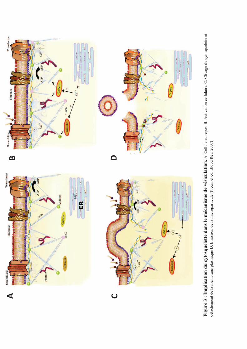
b. Réarrangement du cytosquelette
Si l�externalisation de la PS est donc un pré-requis pour la formation de MP, elle n�est
pas suffisante : différent travaux ont montré que la vésiculation cellulaire est également
dépendante de la protéolyse du cytosquelette (Figure 3). Cette notion a été initialement
suggérée par la présence de protéines contractiles dans les MPP, puis par les travaux de Fox
et al. démontrant que le cytosquelette membranaire stabilise les plaquettes au repos et
prévient la vésiculation111. Dans d�autres types cellulaires, le détachement de la membrane
plasmique du cytosquelette d�actine corticale est associé à la formation de protrusions au
niveau de la membrane et à la vésiculation112-115.
���
�
��
��
Fig
ure
3 :
Im
pli
cati
on
du
cyto
squ
elet
te d
an
s le
méc
an
ism
e d
e v
ésic
ula
tion
.A
. Cel
lule
au
repo
s. B
. Act
ivat
ion
cell
ulai
re. C
. Cli
vage
du
cyto
sque
lett
e et
déta
chem
ent d
e la
mem
bran
e pl
asm
ique
D. E
mis
sion
de
la m
icro
part
icul
e (P
icci
n et
co.
Blo
od R
ev. 2
007)

���
�
i. Rôle des calpaïnes
Le rôle de cystéine protéases calcium dépendantes de la famille de papaïnases, les
calpaïnes, a été démontré par l�utilisation d�inhibiteurs, comme la calpeptine, capable
d�empêcher au moins partiellement la formation des MP116. De plus, la présence de
calpaïnes actives a été mise en évidence par western blot dans les MP issues de plaquettes
activées117. L�activité calpaïne circulante est fortement associée aux MPP chez les patients
souffrant de purpura thrombotique thrombocytopénique (PTT)118,119. Leur activation se
produit à des concentrations de calcium cytosoliques élevées, supérieures à celles capables
d�induire l�externalisation de la PS et nécessitant un apport de calcium extracellulaire. Elles
dégradent les constituants du cytosquelette comme la filamine, la taline et la chaîne lourde
de la myosine. Elles activent aussi l�apoptose par la pro-caspase3 et BclxL66. Cependant le
fait que les MP puissent être formées par incubation avec le complément en présence
d�inhibiteurs de calpaïnes suggère qu�il existe aussi des mécanismes indépendants des
calpaïnes120.
ii. Rôle de la Gelsoline
La gelsoline est spécifique de la formation des MPP. Elle est induite par le calcium et
enlève les protéines chaperonnes comme l�adducine et la cap Z au bout des filaments
d�actine du cytosquelette plaquettaire lors de l�activation plaquettaire permettant ainsi le
remodelage du cytosquelette d�actine121.
iii. Rôle de l�interaction phospholipides/cytosquelette
Nous avons déjà évoqué la relation existant entre la calpaïne et les phospholipides
comme le PIP2. La relation entre le PIP2 et le cytosquelette est plus étroite encore. En effet,
sa concentration dans la membrane plasmique est directement liée aux forces de liaison
membrane-cytosquelette122 PIP2 se lie en fait à de nombreuses protéines liant l�actine
comme la � spectrine123, l��-actinine, l�ezrine, la radixine, et la moesine124,125.
L�interaction des aminophospholipides comme la PS avec les protéines du
cytosquelette comme la spectrine126 et la taline127 a aussi été impliquée dans d�adhésion
���
�
membrane/cytosquelette. Par exemple, la spectrine et l�actine sont plus difficile à extraire
lorsque les membranes sont enrichies en PS127. Ces résultats concordent avec le rôle des
calpaïnes dans la dégradation du cytosquelette aboutissant à la formation des
microparticules. L�élément essentiel de la vésiculation apparait ainsi la perte de l�adhésion
entre le cytosquelette et la membrane plasmique.
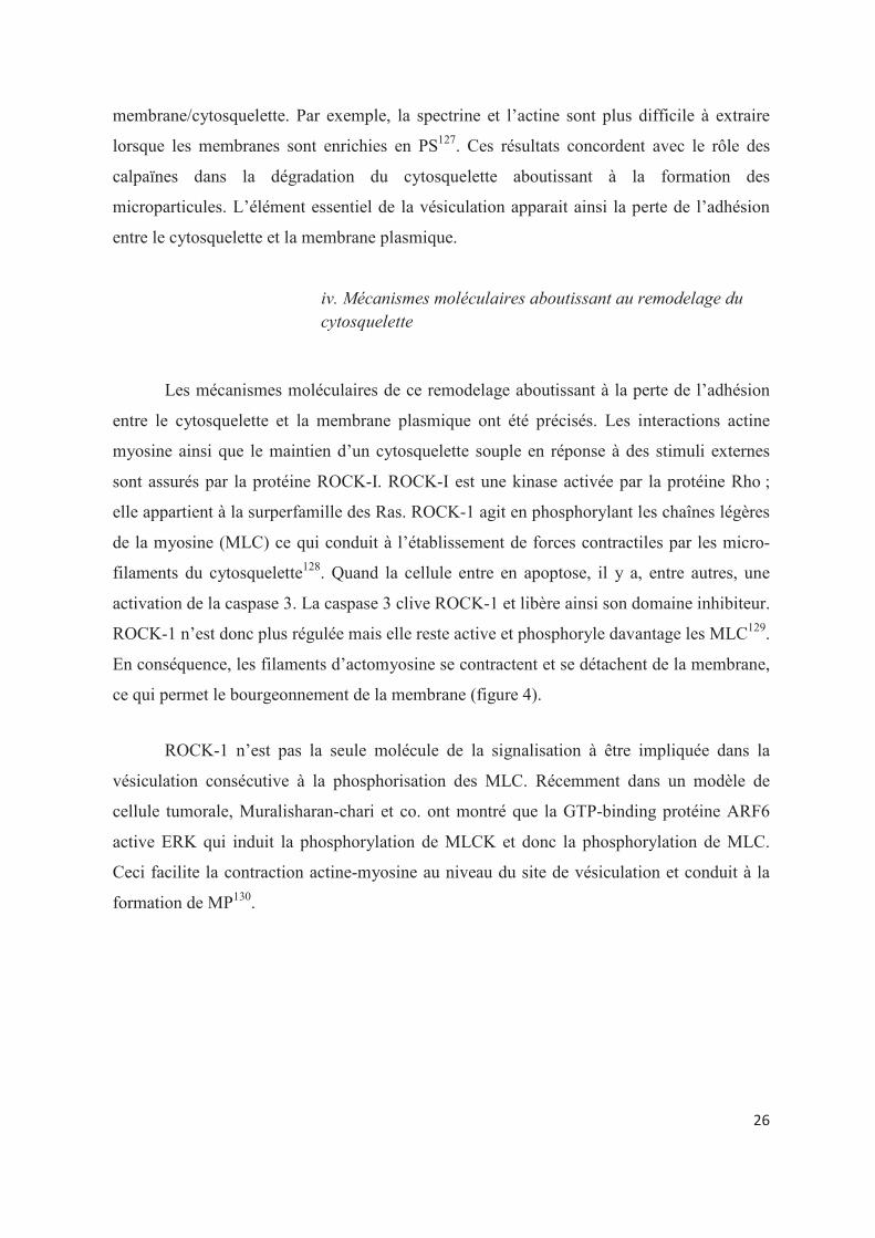
iv. Mécanismes moléculaires aboutissant au remodelage du
cytosquelette
Les mécanismes moléculaires de ce remodelage aboutissant à la perte de l�adhésion
entre le cytosquelette et la membrane plasmique ont été précisés. Les interactions actine
myosine ainsi que le maintien d�un cytosquelette souple en réponse à des stimuli externes
sont assurés par la protéine ROCK-I. ROCK-I est une kinase activée par la protéine Rho ;
elle appartient à la surperfamille des Ras. ROCK-1 agit en phosphorylant les chaînes légères
de la myosine (MLC) ce qui conduit à l�établissement de forces contractiles par les micro-
filaments du cytosquelette128. Quand la cellule entre en apoptose, il y a, entre autres, une
activation de la caspase 3. La caspase 3 clive ROCK-1 et libère ainsi son domaine inhibiteur.
ROCK-1 n�est donc plus régulée mais elle reste active et phosphoryle davantage les MLC129.
En conséquence, les filaments d�actomyosine se contractent et se détachent de la membrane,
ce qui permet le bourgeonnement de la membrane (figure 4).
ROCK-1 n�est pas la seule molécule de la signalisation à être impliquée dans la
vésiculation consécutive à la phosphorisation des MLC. Récemment dans un modèle de
cellule tumorale, Muralisharan-chari et co. ont montré que la GTP-binding protéine ARF6
active ERK qui induit la phosphorylation de MLCK et donc la phosphorylation de MLC.
Ceci facilite la contraction actine-myosine au niveau du site de vésiculation et conduit à la
formation de MP130.
���
�
�
Figure 4 : Place de l�activation de ROCK dans le phénomène de vésiculation
(Boulanger C., Hypertention 2006)�
2) Les inducteurs de la vésiculation
De très nombreux inducteurs capables in vitro d�engendrer la formation de MP sont
rapportés, et ces travaux, pour la plupart, concernent la vésiculation plaquettaire. Plus
récemment, la littérature décrit des circonstances associées à la vésiculation d�autres types
cellulaires, comme les globules rouges, les leucocytes, les cellules endothéliales et autres.
De façon schématique, la production de MP est observée dans deux grands types de
situations : l�activation cellulaire ou l�apoptose. Dans cette section, nous allons détailler les
différents inducteurs impliqués dans la vésiculation, les propriétés et les mécanismes
spécifiques qui en ressortent pour les MP en fonction de leur origine cellulaire.

��
�
a. Activation cellulaire
i. Les plaquettes
La plupart des agonistes activateurs des plaquettes sont capables, à des degrés divers,
d�induire la formation des MP131. L�effet est particulièrement marqué lorsque les plaquettes
sont activées par les calciums ionophores (A23187, ionomycine) et l�association
thrombine/collagène. Il est plus modéré en réponse au collagène, à la thrombine, à l�ADP ou
à l�épinéphrine. De plus, certains de ces agonistes peuvent avoir des effets additifs ou
synergiques. Le mécanisme de vésiculation induit par le collagène est sous la dépendance de
la phosphorylation de p38132.
Le complexe terminal d�attaque membranaire est un puissant inducteur de la
vésiculation plaquettaire in vitro. Les travaux de Sims et co.133 et Wiedner et co.120 ont
montré que la stimulation de plaquettes par des concentrations infra-lytiques de C5b9 induit
une activation plaquettaire avec sécrétion des granules denses et des granules alpha, associée
à la libération de vésicules membranaires. Ces MP semblent spécifiquement détachées des
sites membranaires dans lesquels s�insère le complexe d�attaque. In vivo, l�activation du
complément est vraisemblablement impliquée dans la vésiculation plaquettaire au cours de
pathologies auto-immunes comme le purpura thrombocytopénique auto-immun (PTAI). En
effet Horstman et co. ont montré que des plaquettes opsonisées par des anticorps
monoclonaux dirigés contre la GpIIbIIIa libèrent des MP en présence de sérum comme
source de complément. Cet effet est inhibé par le chauffage du sérum 30 min à 65°C et par
incubation avec alpha C1q confirmant le rôle du complément134.
Outre l�histamine et la thrombine, différents médiateurs de l�inflammation sont
capables d�induire la vésiculation cellulaire. Partant de l�observation que les cytokines
inflammatoires modulent l�activation cellulaire, Nomura et al ont étudié l�effet de ces
molécules sur la vésiculation de plaquettes normales en condition de flux. Ces auteurs ont
montré que l�Il-6, la thrombopoïétine et le GM-CSF sont des inducteurs de la vésiculation
plaquettaire in vitro135.
L�agrégation plaquettaire induite par des anticorps monoclonaux peut s�accompagner
de la formation de MP. Cette action est indépendante du complément, et a été observée avec
des anticorps dirigés contre le CD9, le récepteur pour le fragment Fc des immunoglobulines

��
�
(FcyRII) ou le CD41. Ces observations ont conduit à impliquer les anticorps dans les
mécanismes à l�origine de taux élevés de MP plaquettaires circulantes chez les patients
présentant une thrombopénie immune. La quantité de MP libérées par les plaquettes en
présence de sérum de patients présentant une thrombopénie induite par l�héparine est
directement corrélée à la présence d�anticorps anti-PF4/héparine d�isotype IgG136. Des
observations similaires relient la vésiculation plaquettaire induite par le sérum de patient
présentant un PTAI à la présence d�auto-anticorps dirigés contre les glycoprotéines
plaquettaires137. Le rôle des anticorps est conforté par les modèles animaux de thrombopénie
immune, puisque l�injection d�anticorps anti-plaquettes induit chez la souris une
thrombopénie associée à une augmentation des microparticules plaquettaires
circulantes138,139.
ii. Les globules rouges
En dehors du calcium ionophore, A23187140 et du calcium chloride utilisés in vitro,
les globules rouges vésiculent lors de leur maturation de réticulocyte à globule rouge mature
et lors du vieillissement que ce soit pendant le stockage ou in vivo141
. Il a été montré
qu�environ 20% de l�hémoglobine des érythrocytes était perdu par ce phénomène de
vésiculation durant leur durée de vie142. La vésiculation durant le vieillissement du globule
rouge in vivo est associée à des changements structuraux et le clivage protéolytique de la
protéine band 3. De plus, des études anciennes rapportent, la formation des vésicules
érythrocytaires in vitro par déplétion du milieu en glucose ou en ATP143,144.
iii. Les leucocytes
L�incubation en présence de cytokine inflammatoire comme l�interféron-gamma
(INF�), l�interleukine 2 (IL-2), l�IL-6 ou de facteur de croissance des granulocytes et des
monocytes (GM-CSF) accroît la libération de MP CD68+ par la lignée monocytaire
U937145. Par ailleurs, la stimulation de monocytes par du lipopolysaccharide (LPS), connue
pour sa capacité à induire dans ces cellules un double potentiel pro-coagulant et pro-adhésif,
s�accompagne d�une production de MP capables de disséminer ces deux activités36. Le LPS
est un ligand du récepteur toll-like 3 (TLR3). Une étude récente montre que non seulement

���
�
le LPS mais aussi le poly(I:C), un ligand du récepteur TLR3 induisent efficacement la
formation de MP dans un modèle macrophage. De plus ce phénomène dépend du monoxyde
d�azote (NO), lui-même pouvant induire la vésiculation146.
Le phénomène de vésiculation des granulocytes a été intialement observé suite à une
activation par les molécules du complément33. Le fMLP (N-formylmethionyl-leucyl-
phenylalanine) est souvent utilisé pour faire vésiculer les granulocytes147. De façon similaire
les associations PAF/PMA (Platelet activation factor/ phorbol 12-myristate 13-acetate) ou
PHA (phytohémagglutinine A)/PMA permettent d�induire une vésiculation efficace sur les
lymphocytes148. Dans certaines expériences, les lymphocytes T ont été activés par des billes
couplés à des anticorps anti-CD3 et anti-CD28149.
iv. Les cellules endothéliales
Les cellules endothéliales en culture peuvent générer des MPE, après plusieurs
heures de stimulation, en réponse à divers stimuli. L�assemblage du C5b-9 sur la membrane
de cellules endothéliales en culture induit, outre la sécrétion de facteur Von Willebrand, la
libération dans le surnageant de culture de microvésicules. Cette effet est plus marqué que
celui de la thrombine ou du A2318738. Les autres facteurs décrits pour induire de
phénomène de vésiculation endothéliale sont : l�absence de facteurs de croissance, les
cytokines telles que le TNF39,150,151, le PAI (inhibiteur de l�activation du plasminogène)152, la
thrombine153 , l�irradiation150, la camphothécine (un inhibiteur de topoiomérase I)151, l�acide
gras n-3 docosahexaénoique154, les toxines urémiques155 , la CRP156 et les radicaux libres
dérivés de l�oxygène150. Il apparaît dans certaines conditions, que le découplage de la NO
synthase endothéliale participe à la production de MP endothéliales156.
Les inducteurs in vivo de vésiculation des MPE sont beaucoup moins bien connus.
Amabile et co ont montré que les faibles forces de cisaillement étaient associées aux MPE
dans l�insuffisance rénale terminale9.
Récemment quelques études ont permis d�avancer dans la compréhension des
mécanismes moléculaires induit par ces activateurs dans les cellules endothéliales et
aboutissant à la formation de MP157. La vésiculation induite par le TNF-alpha dépend de la
voie p38MAP kinase158. L�activation par la thrombine comme montrée sur la figure 5 induit
l�activation de de la Rho kinase ROCKII par la caspase 2 à l�absence de mort cellulaire153 et

���
�
de la voie TRAIL/TRAIL-R2. TRAIL/Apo2L est une cytokine qui appartient à la famille du
TNF alpha qui est sécrétée par la cellule endothéliale stimulée. Cette forme soluble de
TRAIL va se lier au récepteur TRAIL-R2, recrutant des protéines adaptatrices à domaine de
mort (TRADD) puis TRAF2, RIP1 and NF-�B qui participent à une boucle d�amplification
pour la formation de MP. La stimulation des cellules endothéliales par la thrombine dans un
modèle HMEC-1, conduit également à une surexpression de Il-1R1. L�engagement de ce
récepteur induit un recrutement des protéines adaptatrices TRAF6 et IRAQ1 qui conduit à
une amplification du phénomène de vésiculation159 (Figure 5).
�
Figure 5 : Mécanisme de formation des MPE après stimulation par la thrombine. (Leroyer et al. Thromb Haemost. 2010) �
�
�
�
�

���
�
b. Apoptose
L�exposition de PS à la surface externe de la membrane cellulaire et le relargage de
MP font partie des altérations morphologiques précoces au cours du processus d�apoptose.
Des observations sur des lignées monocytaires THP-1 ou U937 ont montré qu�il existe une
relation entre la formation des MP et le degré d�apoptose, évalué par la proportion de
cellules hypodiploïdes, induit par les oxystérols, l�étoposide ou l�association
cycloheximide/actinomycine D37,160. De même, des cellules endothéliales en culture
incubées en présence de camptothécine, inhibiteur de la topoisomérase I, libèrent des MP.
La lignée leucémique Jurkat T génèrent plus efficacement des MP après induction par la
staurosporine ou son analogue 7-hydroxylé avec d�autres inhibiteurs de PKC (protein kinase
C) ou de CDK (cyclin dependent kinase)161. Cette vésiculation précède la fragmentation de
l�ADN151. Le lien entre apoptose et vésiculation a également été mis en évidence in vivo.
Dans la plaque d�athérosclérose, les cellules en apoptose, positives pour le marquage
TUNEL, colocalisent avec une expression de facteur tissulaire extracellulaire portée par des
MP PS+61. Par ailleurs, l�activation de la caspase-3, effecteur principal de l�apoptose, induit
la vésiculation plaquettaire in vitro, et les inhibiteurs des caspases réduisent la formation de
MP plaquettaires induite par le TNF chez la souris162. Comme nous l�avons déjà souligné, la
caspase 3 est induite par l�activation de Rho Kinase I et II128,129,153. Cette activation est
nécessaire à la relocalisation des fragments d�ADN de la région nucléaire. Ceci suggère que
les MP générées à partir de cellules en apoptose peuvent contenir du matériel nucléaire163,164.
La distinction avec les corps apoptotiques devient dans ce cas subtile. Un certain nombre
d�études vont dans le sens d�une composition spécifique des MP générées par un mécanisme
apoptotique comparé à un mécanisme inflammatoire. Les MPE générés par apoptose
semblent plus exprimer le marqueur constitutif PECAM-1 alors que le mécanisme
d�activation cellulaire aboutit à une expression de molécule inductible comme le CD62E165.
Ce dernier point est toutefois en contradiction avec des données qui montrent que à certaines
doses, le TNF induit une activation sans apoptose des cellules endothéliales conduisant à la
génération de MPE PECAM-1+ et CD62E+.

���
�
c. Conclusion
Les inducteurs de vésiculation agissant sur les multiples types cellulaires sont
couplés à des mécanismes de formation variés. Ceci aboutit à la génération de MP
spécifiques dans leur composition. Cette structure et cette composition sont étroitement liées
au panel de fonctions porté par les MP.

���
�
CHAPITRE 2 : MICROPARTICULES : STRUCTURE
ET FONCTION
A. COMPOSITION DES MICROPARTICULES
De part leur mécanisme de formation, les MP sont composées d�une membrane
phospholipidique qui porte des caractéristiques antigéniques spécifiques de la cellule
émettrice (cytoadhésines, glycoprotéines...). Néanmoins, la représentation relative des
antigènes entre la cellule mère et la MP sont susceptibles d�être différent166-168. La présence
de glycoprotéines membranaires représentatives de la cellule parentale169,170 permet
d�identifier l�origine des MP (plaquettaire, endothéliale, leucocytaire et érythrocytaire). Par
exemple, les MPP expriment GpIb (CD42b), platelet endothelium adhesion molecule
(PECAM-1; CD31)171, l�intégrine �IIbß3 (GpIIb-IIIa)172, la P-sélectine (CD62P)173, CD
63174, CD41a et CD 61175. Les MPE expriment les CD 31, 34, 51, 54, 62E (E-sélectine), 62
P (P-sélectine), CD 105 (endogline) et CD146 (S-endo1)66. Les MP transportent également
d�autres composants bioactifs comme de l�information génétique (ARNm) dérivé de la
cellule parentale176. Un exemple de composition de MPE est montré sur la figure 6.
Le spectre protéique porté par les MP a pu récemment être enrichi par les données
d�analyse protéomique. Ainsi des centaines de protéines vectorisées par les MP ont été
identifiées sur différentes sous populations: MP plasmatiques177, MPE178,179, MPP180, MP
lymphocytaire181, MP adipocytaires54 et MP de globules rouges182. Ces études ont permis de
montrer la présence dans les MP de protéines tels que des enzymes métaboliques, des
protéines impliquées dans les processus d�adhésion et de fusion, des protéines de la
signalisation, des protéines associées au cytosquelette ou appartenant au nucléosome. Des
profils protéiques comparatifs ont été réalisés entre différentes conditions de stimulation ou
d�origine183-185 et entre différentes tailles de MP186. Nous disposons ainsi de données
importantes permettant d�associer au profil protéique des différentes MP un profil
fonctionnel potentiel. Ainsi les MP dérivées de cellules tumorales comme le cancer du colon
possède un panel de molécules qui sont impliquées dans la tumorigénèse en induisant la
migration, l�invasion, la croissance des cellules tumorale, la modulation du système
immunitaire, le processus métastatique et l�angiogenèse187. Des molécules comme la
basigine et la métalloprotéinase matricielle (MMP-)14 ont récemment été identifiées sur des

���
�
MP issues de l�épithélium pigmentaire rétinien humain. Ces molécules peuvent être
impliquées dans le remodelage matriciel et donc peuvent participer au processus
pathologique de dégénération maculaire lié à l�âge188. Ces données peuvent également
faciliter l�identification de marqueurs d�une condition pathologique189. Cette stratégie a été
appliquée à la thrombose veineuse190,191. Une analyse récente par Miguet et co. sur des MP
dérivées de cellules B malignes a identifié le CD148 comme potentiel biomarqueur du
lymphome du manteau192.
Comme signalé précédemment la nature du stimulus conditionne la composition des
MP tant au niveau lipidique193 que protéique184,185. Ainsi, la composition phospholipidique
des MP du liquide synovial de patient attient de polyarthrite rhumatoïde est différente de
celle des MP plasmatique de sujets sains194,195. Le degré d�oxydation des phospholipides
dépend également du stimulus utilisé pour induire la vésiculation196.
Ces différences de composition, fortement dépendantes des conditions de stimulation
cellulaire initiale déterminent ainsi le profil fonctionnel des différentes sous-populations de
MP.
�
Figure 6 : Composition moléculaire fonctionnelle des microparticules endothéliales. TF : Facteur tissulaire, TM : Thrombomoduline ; EPC : Récepteur de la protéine C ; uPA : Urokinase ; uPAR : Récepteur de l�urokinase ; FcR : Récepteur du fragment Fc des immunoglobulines, MHC ; Molécules du complexe majeur d�histocompatibilité : CAM : Molécules d�adhésion cellulaire. MMP : Métalloprotéases matricielles (Leroyer et co Thromb Haemost 2010) �

���
�
B. FONCTIONS DES MICROPARTICULES
Loin de la perception initiale de débris cellulaires inactifs, les MP sont de véritables
effecteurs bioactifs jouant un rôle physiopathologique important dans de nombreuses
réponses de l�organisme comme l�hémostase, la thrombose, l�inflammation, le tonus
vasculaire, la réponse immunitaire, l�angiogenèse et la croissance tumorale. Grâce à leur
petite taille, les MP circulent tout le long de l�arbre vasculaire ce qui leur permet de
participer à la fois à des phénomènes locaux ou distants. Outre leurs fonctions propres, ces
éléments apparaissent véritablement comme un moyen de communication intercellulaire
important par interaction avec des récepteurs, transfert de molécules de surfaces ou de
contenu cytoplasmique protéique ou génétique197,198.
1) Rôle des microparticules dans l�hémostase
a. Fonction pro-coagulante et pro-thrombotique des
microparticules
Toutes les MP montrent à des degrés variables une activité de génération de
thrombine in vitro qui peut être significativement réduite en modifiant leur composition
phospholipidique. La PS est un phospholipide anionique que lient les protéines de la
coagulation par leur domaine �gla� permettant l�assemblage des complexes enzymatiques
de la coagulation « tenase » et « prothrombinase » et la génération de thrombine. Ainsi, les
MP circulantes, fournissent dans le compartiment vasculaire des surfaces phospholipidiques
additionnelles, riches en PS. Cette activité contribue au potentiel procoagulant du plasma.
Un certain nombre de donnée suggère que ce pouvoir pro-coagulant des MP joue un rôle
dans l�hémostase. En utilisant un test de génération de thrombine, Pereira et co., ont montré
que le PFP de patients avec un syndrome des antiphospholipides avait une capacité de
génération de thrombine augmentée comparé aux sujets sains. Cette différence était
dépendante de la présence des MPP199.
Cette question du rôle des MP dans l�équilibre hémostatique repose donc sur le profil
lipidique mais aussi protéique de la membrane particulaire. Une partie de ce pouvoir

���
�
procoagulant provient en effet de la vectorisation par les MP de facteur tissulaire (FT)200,201.
Ce récepteur transmembranaire est un puissant activateur de la cascade de la coagulation
exprimé par les monocytes, l�endothélium activé et les cellules tumorales. Cette activité FT
est proportionnelle à son taux d�exposition particulaire, le FT réellement soluble (non liée
aux MP) étant, quant à lui, inactif202. Les cellules monocytaires humaines stimulées par les
LPS émettent des MP enrichies en FT et en P-selectin glycoprotein ligand (PSGL)-1203. Les
MPE contribuent également au pool des MP FT+. Ceci a été prouvé dans différentes
situations pathologiques comme la drépanocytose4. Aucun ARNm de FT n�ayant pu être
détecté dans les mégacaryocytes201, l�acquisition de FT par les plaquettes et les MPP résulte
d�un transfert intercellulaire166,167. Plusieurs modalités de transferts intercellulaires ont été
décrites; les MP endothéliales générées après une stimulation proinflammatoire,
particulièrement riches en FT sont capables d�augmenter l�expression membranaire du FT à
la surface des monocytes204 (Figure 7). Les monocytes activés sont également une source
importante de FT capables de transférer le FT biologiquement actif aux plaquettes. Des
transports inverses de FT plaquettes-monocytes ont également été démontrés167. Ainsi, après
exposition au collagène, les plaquettes génèrent des MP riches en FT, qui vont se lier aux
macrophages205, aux granulocytes206 et à d�autres plaquettes par leurs ligands comme la P-
sélectine.
D�autres mécanismes contribuent à la propriété pro-coagulante de MP. Les
granulocytes génèrent des MP enrichies en Mac-1 capables d�induire une activation
plaquettaire207. Les MPE transportent des multimères de facteurs von Willebrand de haut
poids moléculaire qui induisent plus efficacement que la forme soluble l�agrégation
plaquettaire208.
En pathologie humaine, de nombreux exemples soulignent le rôle des MP dans
l�hémostase. Le défaut de génération de MP est associé à des syndromes hémorragiques
(syndrome de Scott, syndrome de dysvésiculation)101. À l�inverse, des taux élevés de MP
pourraient s�accompagner d�un rétablissement d�une hémostase efficace. De manière
intéressante, la stimulation de monocytes avec de la P-sélectine et des immunoglobulines
chimériques augmente le nombre de MP monocytaire circulante et restore l�hémostase dans
un modèle de souris hémophile A209. Ce gain de fonction est également retrouvé chez les
patients présentant des thrombopénies auto-immunes ou induites par des thérapeutiques
(anti-GPIIbIIIa, héparine)210,211.

��
�
Les conséquences thrombotiques de cette propriété des MP sont importantes sur les
versants veineux et artériels du compartiment vasculaire.
Au niveau artériel, les MP contenues dans la plaque d�athérome, sont
particulièrement riches en FT et constituent l�acteur majeur de la thrombogénicité de la
plaque61. Ces MP sont issues de cellules musculaires lisses ou de monocytes/macrophages
activés ou apoptotiques212,213. La rupture de plaque met en contact les plaquettes et les
facteurs de la coagulation circulants avec les MP hautement thrombogènes initialement
séquestrées dans la paroi, favorisant ainsi la formation d�un thrombus artériel214.
Au niveau veineux, il est maintenant relativement bien établi que les MP circulantes
porteuses de FT contribuent à la génération de fibrine lors de la formation du thrombus215.
L�adhésion de MP en plus de polynucléaires neutrophiles et de plaquettes au site de lésion
ou d�activation de l�endothélium est un mécanisme précocement associé à la formation d�un
thrombus veineux. Il a été montré que les MPL peuvent se lier au fibrinogène soluble ou
immobilisé au sein d�agrégats leuco-plaquettaires permettant de concentrer au site de
formation du thrombus l�activité FT. Celle-ci peut être transférée sur les plaquettes par les
MPL contribuant ainsi à la propagation du thrombus203. Cette propriété n�est pas exclusive
des MPL. Il a été montré que les MPP, les MPGR, les MP dérivées de cellules
hématopoïétiques contribuaient à la propagation du thrombus in vivo202,216. Les interactions
P-sélectine/MP sont particulièrement importantes dans l�initiation du processus
thrombotique. La P-sélectine permet en effet de recruter des MP porteuses de PSGL1,
particulièrement riches en FT. L�importance de ces mécanismes a été démontrée à l�aide de
modèles murins associant invalidation du gène de la P-sélectine et un suivi dynamique de la
croissance du thrombus par microscopie intravitale209,217-219. Ce phénomène possède de
nombreuses boucles d�amplifications. En effet, la thrombine générée in situ, le collagène
exposé par un sous-endothélium dénudé, le CD40 ligand soluble libéré par les plaquettes ou
les lymphocytes activés, la P-sélectine, les cytokines, le stress oxydatif favorisent au site de
la lésion vasculaire, la génération de nouvelles MP procoagulantes220.

��
�
�
Figure 7 : Contribution de la génération des MPE à l�activité procoagulante. EMPs : Microparticules endothéliales. TNF : Tumor necrosis factor (Sabatier et co. Blood 2002)
Les MP d�origine cancéreuse montrent également une activité procoagulante
dépendante du facteur tissulaire. Ainsi la problématique du lien entre les MP et les
thromboses associées aux cancers est un sujet important dans la littérature221. Bien qu�un
nombre important de données existe pour supporter l�implication physiopathologique des
MP dérivées de cellules cancéreuses dans les thromboses liées aux cancers222,223(Annexe 1),
le lien de causalité n�est pas encore totalement établi. Davila et co. ont montrés que
l�injection de MP dérivées de cellules cancéreuses portant une activité FT très élevée chez la
souris, provoque un syndrome proche d�une coagulation intravasculaire disséminée in vivo.
De manière intéressante, cette étude démontre également qu�un taux minimal de MP
injectées est nécessaire pour dépasser les mécanismes protecteurs anti-thrombotiques224. Ces
conclusions concordent avec celles de Thomas et co. qui montrent que l�injection de MP de
lignées tumorales pancréatiques humaines et murines induit dans un modèle de thrombus
chez la souris un raccourcissement du temps d�occlusion veineux et artériel. Ce même
travail démontre par microscopie intravitale que les MP spécifiquement dérivées de la
tumeur se lie au thrombus de manière dépendante de la PSGL-1225(Figure 8 et Annexe 2).

���
�
La contribution relative des MP dérivées de la tumeur ou des MP dérivées des cellules de
l�hôte dans la formation du thrombus in vivo ainsi que la démonstration formelle du rôle du
FT reste cependant des questions ouvertes.
�
b. Fonction anticoagulante des microparticules
Si la résultante des activités des MP sur l�équilibre hémostatique semble clairement
pro-coagulante, des protéines anticoagulantes ont été également mises en évidence à leur
surface. Ainsi les MP dérivées de monocytes sont porteuses de thrombomoduline226 et
d�inhibiteur de la voie du facteur tissulaire (TFPI)227. Le récepteur à la protéine C activée a
été montré à la surface des MPE. Ce récepteur endothélial de la protéine C est présent sous
une forme fonctionnelle dans la MP différente de sa forme clivée qui ne peut plus lier la
Figure 8 : Contribution des MP tumorales à la formation du thrombus in vivo (Thomas G. J. Exp. Med 2009). Détection de MP provenant d�une tumeur pancréatique marquée au GFP (vert) dans la circulation (images du haut) et dans le thrombus (images du bas) par microscopie intravitale.�

���
�
protéine C activée228. En physiopathologie il paraît donc nécessaire de vérifier la résultante
de l�expression concomitante des récepteurs anti- et pro-coagulants à la surface des MP sur
l�équilibre hémostatique, particulièrement dans les lignées cellulaires capables d�exhiber les
deux types d�activité (cellules endothéliales et monocytes-macrophages). L�activité
fibrinolytique des MP, récemment mise en évidence peut aussi impacter sur cette balance
hémostatique (cf infra).
2) Rôle des microparticules dans la modulation du tonus vasculaire
Les MP participent à la régulation du tonus vasculaire, notamment en diminuant la
production du NO par les cellules endothéliales229,230. Une diminution du NO endothélial à
pour conséquence de limiter les propriétés vasculoprotectrices et antiathérogènes de
l�endothélium vasculaire (Figure 9). Cet effet a été observé sur des artères exposées in vitro
à des concentrations circulantes de MP isolées à partir de sang de patients coronariens,
insuffisants rénaux, prééclamptiques, ou insuffisants pulmonaires9,11,231-233. Il est à noter que
dans ce même test, les MP issues de volontaires sains n�ont aucun effet. La dysfonction
endothéliale induite par les MP plasmatiques humaines dont essentiellement la sous-
population d�origine endothéliale, est associée à une diminution de la production de NO
mais pas à une altération de l�expression de la NO synthase endothéliale. Des résultats
similaires ont été observés avec des MP générées in vitro à partir de cellules musculaires
lisses ou d�une lignée lymphocytaire234-236.
D�autres mécanismes impliqués dans la réactivité vasculaire ont été décrit. Les MP
plaquettaires, source de thromboxane A2, induisent une contraction vasculaire237. Les MP
issues d�une lignée lymphocytaire T ou isolées à partir de plasma de patients diabétiques
induisent une hyporéactivité vasculaire, sur des artères murines isolées, associée à une
production de NO et de prostacycline238,239. Dans cette étude, les auteurs ont montré que les
MP induisent la NO synthase inductible et la cyclooxygénase de type 2 en activant le facteur
de transcription NFkB. L�ensemble de ces mécanismes peut contribuer à la dysfonction
endothéliale induite par les MP.
Par ailleurs, de manière surprenante, certaines MPE générées in vitro et portant sonic
hedgehog semblent stimuler la production de NO endothélial et diminuent celle de radicaux
libres oxygénés dans les vaisseaux240,241. Aucune information n�est encore disponible quant

���
�
à la présence de ce morphogène sur les MP plasmatiques ni sur l�évolution de son
expression sur les MP au cours des pathologies cardiovasculaires. D�autres MP pourraient
avoir également un effet favorable sur la fonction endothéliale. Le transfert d�acide
arachidonique par les MPP pourrait ainsi induire l�expression de la cyclooxygénase-2 et la
production de prostacycline impliquée dans l�inhibition de l�activation plaquettaire et la
vasodilatation242. L�importance de ce dernier mécanisme n�est pas clairement établie en
clinique humaine.
.
Figure 9 : MPE et dysfonction endothéliale par diminution de la synthèse de NO. (Amabile et co. Ann Cardiol Angeol 2008)
���
�
3) Rôle des microparticules dans l�inflammation
De nombreux travaux ont démontré, le rôle des MP dans la modulation de
l�inflammation dans le compartiment vasculaire. L�effet pro-inflammatoire des MP implique
par exemple les phospholipides oxydés capables d�activer les récepteurs du PAF (facteur
d�activation plaquettaire) présents sur les cellules endothéliales et les leucocytes243. Les MP
constituent également une source importante de substrat aminophospholipidique pour la
phospholipase A2 sécrétoire, massivement libérée au cours du sepsis, conduisant à la
génération d�acide lysophosphatidique, un puissant médiateur pro-inflammatoire et agoniste
plaquettaire236. L�acide arachidonique transporté par les MP induit une augmentation de
l�expression membranaire d�ICAM-1 sur l�endothélium198,244,245. Ces MP induisent
également la néosynthèse de contre-récepteurs (CD11a/CD18, CD11b/CD18) monocytaires
favorisant les interactions leuco-endothéliales. Les MP d�origine plaquettaire ou leucocytaire
induisent la libération de plusieurs cytokines endothéliales (Il-1b, Il-6, Il-8, MCP-1) ou
monocytaires (Il-1b, TNFa, Il-8)246-248 renforçant ainsi la réponse inflammatoire249. Les MP
plaquettaires peuvent par ailleurs faciliter le recrutement de nombreuses cellules immunes
(monocytes, lymphocytes T et B, cellules NK) 250-252. Les plaquettes interagissent
directement avec l�endothélium vasculaire activé en augmentant arrêt des
leucocytes/monocytes après tranfert par les MP de la chémiokine RANTES (Regulated on
Activation, Normal T Expressed and Secreted)253.
A l�inverse, les MP libérées par des neutrophiles activés semble limiter la réponse
inflammatoire aux stades précoces et participer aux défenses antimicrobiennes en se liant
aux bactéries opsonisées, aux monocytes et aux cellules endothéliales71 focalisant ainsi
l�activité anti-microbienne. Ces MPL moduleraient l�équilibre entre cytokines pro et anti-
inflammatoires sécrétées par les macrophages (Il-8 et TNFa vs TGF� et Il-10)254,255. Les MP
dérivées de lymphocytes T peuvent aussi induire l�apoptose des macrophages provoquant en
retour leur vésiculation256. Bien que ces dernières observations nécessitent une confirmation
dans d�autres modèles cellulaires et in vivo, elles posent la question du rôle des MP comme
acteurs potentiels de « limitation » et/ou de « résolution » de la réponse inflammatoire.
Le lien entre les activités inflammatoires, pro-thrombotiques et angiogénique des MP
est très fort (figure 10). Les mécanismes permettant de montrer clairement ce lien dans le
contexte endothélial ont été récemment résumés dans une revue157(annexe 3).

���
�
4) Rôle des microparticules dans l�angiogenèse
L�angiogenèse se définit comme la formation de nouveaux vaisseaux sanguins à
partir de vaisseaux préexistants. C�est un phénomène biologique très important et généralisé
au cours de l�organogenèse embryonnaire alors qu�au stade adulte, ce processus ne sera
activé que dans des conditions limitées ou pathologiques. Des travaux ont montré que les
MP interviennent à trois étapes cruciales de l�angiogenèse : la dégradation matricielle, le
recrutement et la différenciation des progéniteurs endothéliaux et la prolifération et la
migration des cellules endothéliales.
Figure 10 : Lien entre propriétés pro-thrombotique et inflammatoire des MP. PMP : Microparticules plaquettaires, MMP : Microparticules monocytaires, EMP : Microparticules endothéliales : EPC : Progéniteurs endothéliaux, EPCMP : Microparticules de progéniteurs endothéliaux, COX2 : Cycloxygénase 2, ICAM-1 : Molécule adhésion intercellulaire 1, IL : Interleukine, TF : Facteur tissulaire (N.Mackman, Circ Res 2009) �

���
�
Le rôle des MP dans la protéolyse et la dégradation matricielle est développé dans le
paragraphe suivant.
Concernant les deux autres étapes, plusieurs mécanismes ont été décrits. La
sphingomyéline portée par les MP tumorales pourrait être un composant actif de ces
processus d�angiogenèse en favorisant la migration de cellules endothéliales, l�invasion et la
formation de tubes257. De même, le FT porté par les MP est un puissant régulateur de
l�angiogenèse258. En particulier, il augmente la synthèse du VEGF (vascular endothelial
growth factor). De nombreux agonistes vasculaires comme le VEGF, le PAI-1 et le
fibroblast growth factor (FGF) sont par ailleurs impliqués dans la formation des MPE
porteuses de métalloprotéinases (MMP-2 et MMP-9). Plus récemment, il a été montré que
les MP issues de cellules progénitrices endothéliales stimulaient l�angiogenèse par transfert
horizontal de microARN176. De plus les MPE stimulent les mécanismes de réparation
tissulaire. Ceci a été montré dans le tissu musculaire. Cet effet est dépendant d�une
augmentation de la différentiation des progéniteurs endothéliaux par un mécanisme
impliquant la génération de radicaux libres oxygénés64.
Les MPP également sont capables d�induire l�angiogenèse in vitro (figure 11) et de
stimuler la revascularisation post-ischémique in vivo259-261. Un mécanisme avancé est la
vectorisation de récepteurs membranaires tels que Flt-1 (VEGF-R1) 262. Les MP qui portent
le Sonic Hedgehog favorisent l�angiogenèse en régulant positivement les molécules
d�adhésion et les facteurs proangiogéniques263. In vivo, dans un modèle de souris, elles
favorisent la néovascularisation par l�activation de la voie du NO264.
La promotion de l�angiogenèse par les MP peut avoir des effets délétères comme
dans l�invasion tumorale, la rétinopathie diabétique65 ou la déstabilisation de la plaque
d�athérosclérose265.
Inversement, dans certaines conditions, les MP libérées par les cellules endothéliales
en culture altèrent l�angiogenèse in vitro266. Il a aussi été montré que les MP plasmatiques
isolées de sujets hypercholestérolémiques diminuent la formation de progéniteurs
endothéliaux267.

���
�
�
Figure 11 :�Augmentation de l�angiogenèse par des concentrations croissantes de MPP dans un modèle d�anneaux aortiques de rat (Brill et co. Cardiovasc. Res. 2005)
5) Rôle des microparticules dans la protéolyse matricielle
Les cellules tumorales malignes possèdent un potentiel protéolytique important,
caractérisé par la production de MMP et d�uPA, nécessaire à leur dissémination et à
l�invasion tissulaire. Dans une série d�études publiées depuis 1994, la présence de ces
enzymes protéolytiques a été identifiée, in vitro et in vivo268-275, sur la membrane de MP
tumorales isolées de surnageants de culture de lignées tumorales humaines ou de liquides
d�ascite obtenus chez des patientes atteintes de cancer ovarien268,273. La détection de

���
�
l�activité enzymatique par zymographie et de la protéine support par immuno-empreinte a
permis d�identifier sur ces MP, l�uPA et son récepteur uPAR, la proMMP-2, la proMMP-9
et leurs formes actives, ainsi que des complexes entre les MMP et leur inhibiteur TIMP-1.
Une bande protéolytique ayant la masse moléculaire du tPA a été également détectée par
zymographie dans des MP provenant d�une lignée de carcinome de la prostate276. Un modèle
de matrice extracellulaire en trois dimensions (Matrigel) a permis de mettre en évidence une
action concertée entre l�uPA et les pro-MMP des MP dans la dégradation de protéines
matricielles en présence de plasminogène. Le mécanisme proposé suggère que la membrane
des MP fonctionnerait en offrant une large surface d�activation de MMP via la formation de
plasmine par l�uPA268,274,275. Ces résultats ont été confirmés plus récemment par l�étude des
MP isolées d�ascites prélevées chez des sujets atteints de cancer de l�ovaire277.
Les MMP sont également portées par des MP non tumorales, provenant de cellules
endothéliales humaines. En effet, la présence de proMMP-2, proMMP-9, de leurs formes
actives ainsi que celle de la MT1-MMP et des inhibiteurs TIMP-1 et TIMP-2 sur des MP
obtenues de surnageants de culture de ces cellules a été démontrée par Taraboletti et al.278.
Ces MP favorisent la migration de cellules endothéliales et modulent leur capacité
angiogénique dans le Matrigel : l�effet stimulateur sur la formation de tubes, observé à de
faibles concentrations de MP, se transforme en effet inhibiteur à des concentrations plus
fortes, suggérant un défaut d�implantation cellulaire sur la matrice dégradée par l�activité
protéolytique des MP. Brodsky et al. ont également rapporté la présence de l�uPAR sur des
MP obtenues par stimulation de cellules endothéliales avec des concentrations élevées de
PAI-1152.
Enfin, certaines MP peuvent stimuler la production de MMP par des fibroblastes279,
des cellules endothéliales280, ou des cellules malignes281 et contribuer ainsi de manière
indirecte à la protéolyse péricellulaire.
6) Rôle des microparticules dans la survie cellulaire
Les MP peuvent être considérées comme un moyen pour la cellule parente d�éliminer
de sa surface des molécules potentiellement délétères. Ceci a été montré il y a déjà plus de
20 ans comme un mécanisme de défense des granulocytes contre le complexe d�attaque
membranaire du complément33-35. Plus récemment un mécanisme similaire a été observé sur

��
�
un modèle de cellules érythroleucémique K562282. D�autres molécules sont ainsi expulsées
de la surface cellulaire comme Fas ligand par des cellules tumorales ovariennes283 ou la
caspase-3 par les cellules endothéliales induisant ainsi une protection contre l�apoptose284.
Les MP pourraient également être impliquées dans la résistance aux chimiothérapies. La
doxorubicine par exemple est accumulée dans des MP émises par des cellules tumorales. La
génération de MP, enrichies en drogues cytotoxiques pourrait ainsi fournir à la cellule
tumorale un mécanisme supplémentaire de résistance285. De manière intéressante, la survie
des cellules hématopoïétiques est favorisée par les MPP252. Une étude récente montre
également que les statines améliorent la survie des cellules endothéliales vasculaires en
facilitant leur vésiculation in vitro286. Il faut toutefois souligner que l�effet des statines reste
débattu287,288.��
�
7) Rôle des microparticules dans la modulation de la réponse
immunitaire
Plusieurs travaux ont suggéré un rôle pour les MP dans l�induction d�une tolérance
immune. Ainsi, au cours du premier trimestre de la grossesse, les cellules trophoblastiques
génèrent des MP porteuses de Fas-ligand capables d�induire l�apoptose de lymphocytes T
par l�intermédiaire d�interactions Fas/Fas-ligand289. L�apoptose induite par ce type de MP
pourrait aussi constituer un processus d�échappement des cellules cancéreuses à la réponse
immune en ciblant les cellules du système immunitaire porteuses de Fas et en diminuant le
taux d�expression de Fas-ligand disponible à la surface tumorale. Dans ce sens, il a été
démontré que des cellules épithéliales ovariennes cancéreuses étaient capables d�exporter du
Fas ligand fonctionnel à la surface des MP alors que leurs homologues non néoplasiques
exprimaient Fas-ligand sans le sécréter283.
À l�inverse, la réponse immune exacerbée pourrait résulter d�une production
soutenue de MP ou d�une diminution de leur clairance. La pérennisation de complexes
antigéniques phospholipides�protéines colocalisés par la membrane particulaire pourrait
faciliter la reconnaissance par le système immun et contribuer à la pathogénie du syndrome
des anticorps antiphospholipides. Des taux élevés de MP endothéliales riches en FT sont
ainsi rapportés chez les patients présentant un anticoagulant lupique39. Il n�est actuellement
pas établi dans ce type de situation, si les MP participent à la formation de néoantigènes ou
si elles ne reflètent qu�une stimulation endothéliale particulière.

��
�
Enfin, il faut noter, comme pour les exosomes, la présence sur des microparticules de
molécules du CMH. Ces molécules peuvent être transférées à des cellules dendritiques
receveuses permettant l�activation allogénique des lymphocytes T290. Ces vecteurs sont
probablement responsables du transfert de molécules de CMH II déjà observé en culture il y
a 10 ans291. Les MPP contribuent également la réponse humorale par activation des
lymphocytes B dépendante du CD40 ligand292. D�autre part les MPE favorisent la
maturation des cellules dendritiques plasmocytoïdes avec expression de molécules de
costimulation pouvant induire une réponse immune T293.
8) Rôle des microparticules dans la communication intercellulaire
Initialement décrite sur les exosomes, cette propriété de transfert horizontal de
protéine, de lipides et d�information génétique d�une cellule à une autre est aussi démontrée
pour les MP294. Les mécanismes impliqués dans ces interactions sont mal connus. Certaines
études montrent un mode de transfert direct de protéines à la surface des cellules
endothéliales253,295. D�autres suggèrent des mécanismes proches de la phagocytose
impliquant des récepteurs scavangers ou des récepteurs à la PS296. Une fusion des
membranes entre les MP et les cellules cibles a aussi été avancée297-300 (figure 12). Une
meilleure compréhension de ces mécanismes d�interaction est importante car cette étape est
potentiellement pharmacologiquement modulable.
Les MP d�origine monocytaires sont ainsi capables de transférer le récepteur de la
chémiokine CCR5, un corécepteur du virus VIH, à des cellules ne l�exprimant pas comme
les cellules endothéliales301. L�acquisition de ce corécepteur par les cellules cibles pourrait
les rendre sensibles à l�infection par le VIH. Plus récemment, la fonctionnalité d�un tel
transfert particulaire a été démontré à partir de MP dérivés des plaquettes ou des
mégacaryocytes porteuses du récepteur CXCR457. De même, les lymphocytes T recevant la
molécule Fas ligand par des MP de cellules tumorales peuvent subir l�apoptose302. Plus
récemment, ce mécanisme à permis d�expliquer l�acquisition par des cellules de gliome
humain de la forme tronquée du récepteur au facteur de croissance épidermique EGFRvIII
alors qu�elles ne l�exprimaient pas initialement303. De plus, les PMP vectorisent des facteurs
de transcriptions fonctionnels capables de maintenir un état d�activation des monocytes

���
�
élevé après endocytose304. Ce type de transfert explique également le risque de
contamination de progéniteurs humains destinés à la thérapie cellulaire par des protéines
animales apportées par des MP 168 et les discordances de phénotypes parfois observées après
culture cellulaire comme la contamination par des protéines plaquettaires de progéniteurs
endothéliaux précoces305. Ces transferts moléculaires complexifient l�identification des
cellules sources d�une protéine particulière comme par exemple le FT.
Lors des interactions avec leurs cellules cibles, les MP peuvent libérer dans le
cytoplasme de la cellule réceptrice tout leur contenu cytosolique. Celui-ci peut inclure non
seulement des protéines mais également des acides nucléiques comme des ARN messagers
ou des microARN298. Ces derniers ont pour fonction de contrôler l�expression des gènes en
régulant la production d�ARN messagers. Ainsi, il a été montré que les MP de progéniteurs
de cellules différentiées et de cellules tumorales transfèrent des ARNm59,176,252. Un transfert
similaire a également été observé entre les cellules de Schwann et les axones des nerfs
périphériques au niveau des n�uds de Ranvier306,307. Les conséquences de ce mécanisme
sont diverses en fonction du matériel transmis. Par exemple, il peut aboutir à la maturation et
à la différentiation des cellules cibles ou un effet inverse308.
Figure 12 : Différents modes d�interaction entre les MP et la cellule cible. (Camussi et co. Kidney International 2010)

���
�
CHAPITRE 3 : LA GENERATION DE PLASMINE
Le système d�activation du plasminogène joue un rôle fondamental dans l�homéostasie
vasculaire en contrôlant la dissolution du caillot et le maintien d�un système vasculaire
fonctionnel. L�activation du plasminogène permet de générer l�enzyme clé de ce système : la
plasmine. Ce système est hautement régulé par de nombreux inhibiteurs qui permettent de
limiter la diffusion de ses activités protéolytiques. Un jeu subtil d�interactions moléculaires
où les surfaces supports (fibrine, membranes cellulaires ou matrice extracellulaire) ont un
rôle essentiel, permet son équilibre.
A. LE PLASMINOGENE
Le plasminogène humain, précurseur de la plasmine, est une glycoprotéine
plasmatique monocaténaire de 92 kDa, synthétisée par le foie et circulant à une
concentration comprise entre 1,5 et 2 �M. Sa demi-vie plasmatique est de 2,2 jours.
La forme native du plasminogène a été baptisée Glu-plasminogène en raison de la
présence d�un acide glutamique à son extrémité N-terminale. Deux formes plasmatiques
existent en fonction de leur degré de glycosylation, qui peut se situer au niveau de Asn289 et
de la Thr346 (type I),ou au niveau de la Thr346 seulement (type II). Sept cent quatre-vingt onze
acides aminés et 24 ponts disulfures s�organisent en sept domaines structurels (figure 13):
une chaîne peptidique N terminale appelée peptide de préactivation, correspondant aux
acides aminés 1 à 77, puis cinq séquences homologues d�environ 80 acides aminés chacune,
appelées « kringles » K1 à K5, nommées ainsi par analogie avec un biscuit danois et enfin,
au niveau de l�extrémité C terminale, le site responsable de l�activité protéolytique sérine
protéase, formé des acides aminés 562 à 791309 contenant la triade catalytique caractéristique
des sérines protéases : His603, Asp646 et Ser741310.
Chacun de ces différents domaines confère au plasminogène des propriétés
spécifiques. Ainsi, le peptide N-terminal et les domaines �kringles� définissent la
conformation spatiale du plasminogène. Ces derniers possèdent trois ponts disulfures intra-

���
�
domaines qui leur confèrent une très grande stabilité de conformation. Des sites de liaison
dépendant de la lysine (LBS : Lysin Binding Site) sont présents sur les K1 et K4. Ils
permettent la fixation du plasminogène sur la fibrine ou sur les surfaces cellulaires et
l�interaction de la plasmine avec l��2-antiplasmine, jouant ainsi un rôle majeur dans la
régulation de la fibrinolyse311.
Un LBS de faible affinité est présent sur le K5. Il présente une affinité particulière
pour les analogues de la lysine et il est impliqué dans une interaction inter-domaine avec le
peptide N terminal du glu-plasminogène qui favorise une conformation compacte312,313. Des
études physico-chimiques indiquent que cette forme fermée du glu-plasminogène est
prédominante dans la circulation et qu�elle est en équilibre avec une forme ouverte de très
courte demi-vie314,315. La rupture chimique de cette interaction par clivage protéolytique du
peptide N-terminal par la plasmine (clivage soit de Lys 62, Arg 68 ou Lys 77) conduit à une
forme tronquée ouverte, connue sous le nom de lys-plasminogène, incapable d�adopter une
conformation compacte, qui constitue le substrat préférentiel pour les activateurs du
plasminogène316. De la même manière, la saturation avec l��-ACA (acide �-amino
caproïque) du LBS de faible affinité du glu-plasminogène K5 gène son interaction avec le
peptide N-terminal et entraîne une transition du glu-plasminogène de sa forme compacte
fermée à une conformation ouverte caractéristique du lys-plasminogène312,317.
Le clivage, dans le domaine sérine protéase, du pont peptidique Arg561-Val 562 par les
activateurs du plasminogène génère la plasmine, une sérine protéase à large spectre qui clive
ses substrats au niveau des résidus lysyl et arginyl. La plasmine est donc une molécule
bicaténaire composée d�une chaine lourde N-terminale (la chaine A, 58kDa) et d�une chaine
légère C-terminale (la chaine B, 25 kDa) reliées par deux ponts disulfures.
Il existe également des formes variantes de bas poids moléculaire comme le
miniplasmin(ogène) et le microplasmin(ogène), avec respectivement seulement un kringle
ou pas de kringle du tout attaché à la chaine légère.

���
�
B. LES ACTIVATEURS DU PLASMINOGENE
1) L�activateur tissulaire du plasminogène (t-PA)
Le t-PA est une glycoprotéine monocaténaire de 527 acides aminés et de masse
moléculaire 70kDa synthétisée par les cellules endothéliales et les cellules musculaires lisses
(CML) 318,319. La forme native contient au niveau de l�extrémité N-terminale une extension
de 3 acides aminés (Gly-Ala-Arg). Le t-PA est composé de 5 domaines structuraux (figure
14) : un domaine « finger » (4-50), un domaine « epidermal growth factor-like » (EGF-like)
(51-87), deux domaines « kringle » (88-176 et 177-262) et un domaine sérine-protéase (276-
527) à l�extrémité C-terminale, comprenant la triade catalytique His322, Asp371 et Ser478. Le
clivage du pont peptidique Arg275-Ile276 par la plasmine convertit le t-PA simple chaine (sct-
PA) en une forme double chaine (tct-PA). Mais au contraire de la plupart des enzymes de
cette famille, il faut noter que la forme précurseur monocaténaire est déjà active320. Chacun
Figure 13 : Structure du plasminogène. (D�après Rejante et Llinas, Eur J Biochem 1994)

���
�
des domaines du t-PA assure une fonction spécifique telle que l�activité enzymatique, la
liaison à la fibrine et à des récepteurs cellulaires ou encore l�interaction avec les inhibiteurs
des activateurs du plasminogène (PAI). La liaison à la fibrine se fait par l�intermédiaire du
domaine finger et du kringle 2321,322. En solution, le t-PA présente une faible affinité pour le
plasminogène. Mais celle-ci est fortement augmentée en présence de fibrine (x1000 à 1500)
sur laquelle les deux molécules sont concentrées et prennent une conformation favorable316.
En plus de son rôle fibrinolytique dans les vaisseaux, le t-PA semble être un acteur
important dans le système nerveux central323. Il est exprimé par les neurones et les cellules
de la microglie. Il induit localement une génération de plasmine responsable de la
dégradation de substrats différents de la fibrine comme par exemple la protéine �-
amyloide324. Il peut agir également directement sur certain substrat comme le PDGF-CC
Figure 14 : Structure du t-PA (D�après Gething et co. Embo J 1988)
���
�
(Platelet derived growth factor)325 ou remplir des fonctions non protéolytiques comme la
modulation des récepteurs N-méthyl-D-aspartate (NMDA)326.
2) L�urokinase (uPA)
L�uPA est également une sérine-protéase produite sous forme monocaténaire (scuPA
ou pro-urokinase), de 54kDa et composée de 411 acides aminés327. Elle est sécrétée par de
nombreux types cellulaires dont les cellules épithéliales, endothéliales, les polynucléaires
neutrophiles, les monocytes et les cellules tumorales. L�urokinase possède 3 domaines : un
domaine présentant des homologies avec l�EGF dans la partie N-terminale (5-49), un
domaine kringle (50-131) et un domaine sérine protéase comprenant la triade catalytique
His204, Asp255 et Ser356 à l�extrémité C-terminale. Le domaine kringle ne contient pas de site
de liaison aux résidus lysines, ce qui explique qu�à la différence du tPA, l�uPA ne se lie pas
à la fibrine. Sous sa forme monocaténaire, l�uPA est insensible aux inhibiteurs PAI-1 et PAI-
2. La pro-urokinase a la capacité d�activer le plasminogène mais ce potentiel activateur
demeure très faible (inférieur à 1% de celui de l�urokinase)328.
Le clivage de la liaison peptidique Lys158-Ile159 par la plasmine, la kallikréine
plasmatique ou la cathepsine B, convertit la pro-urokinase en une forme bicaténaire
considérée comme l�enzyme active, l�urokinase ou tcuPA (Figure 15). Les deux chaines sont
liées par un pont disulfure (Cys194-Cys222). La chaine légère (20kDa), composée de 158
acides aminés, contient le fragment amino-terminal (ATF) incluant les domaines EGF et
kringle et donc le site de liaison au récepteur de l�urokinase, uPAR. La chaine lourde
(34kDa) est composée de 253 acides aminés comprenant le domaine sérine-protéase.
En présence d�une forte concentration de plasmine, celle-ci clive la liaison peptidique
Lys135-Lys136, de la tcuPA qui génère une forme courte d�urokinase (33kDa) ne possédant
pas ATF. Cette forme d�urokinase appelée urokinase de bas poids moléculaire, conserve
l�intégrité de son activité mais perd sa capacité à interagir avec l�uPAR. Une autre forme de
bas poids moléculaire (32Kd) est générée par clivage de la liaison Glu143�Leu144 par des
métalloprotéinases spécifiques. La thrombine peut également cliver la pro-urokinase au
niveau de la liaison peptidique Arg156-Phe157 ce qui aboutit cette fois à une protéine
bicaténaire inactive. Cette réaction est fortement accélérée en présence de
thrombomoduline329.

���
�
Il est important de noter que la conversion de la pro-urokinase en urokinase n�est pas
requise pour l�initiation de l�activation du plasminogène au niveau de la surface de fibrine et
de la membrane cellulaire. En effet, la liaison du plasminogène au caillot de fibrine induit
des changements de conformation qui stimulent l�activité intrinsèque de la pro-urokinase et
lui permettent ainsi d�initier l�activation du plasminogène lié aux résidus lysines
carboxyterminaux330. La conversion par la plasmine de la pro-urokinase en urokinase, plus
active, conduit à une accélération de ce processus, d�autant plus que l�urokinase active le
plasminogène tant en solution qu�au niveau du caillot de fibrine et des membranes
cellulaires.
3) Le récepteur de l�urokinase (uPAR)
Le récepteur spécifique de la pro-uPA /uPA, uPAR (CD87), est exprimé à la surface
des monocytes, les lymphocytes T, dans les granules des neutrophiles, sur l�endothélium, les
épithéliums, les CML et les fibroblastes. Son expression à la membrane est régulée
positivement en cas d�infection bactérienne et en réponse aux cytokines inflammatoires
telles que le TNF�, l�IFN �, et IL-1327.
Figure 15 : Conversion de pro-urokinase en urokinase par la plasmine

���
�
L�uPAR est une protéine dont la masse moléculaire varie de 55 à 60 kDa en raison de
glycosylations hétérogènes. La forme exprimée à la membrane contient une chaine
peptidique de 283 résidus et une ancre glycérophosphoinositol (GPI) à sa partie C-terminale.
Trois modules homologues, les domaines D1 (domaine aminoterminal), D2 (domaine
central) et D3 (domaine carboxyterminal) sont impliqués dans l�interaction avec la séquence
Asp12-Asn32 du domaine EGF-like de la pro-urokinase331. La résolution des structures
cristallines de l�uPAR en complexe avec un peptide antagoniste332 ou avec le fragment ATF
de l�uPA a permis de montrer que les 3 domaines de l�uPAR étaient impliqués dans la
constitution du site de liaison de haute affinité pour l�u-PA333. Cette liaison induit un
changement conformationnel de l�uPAR qui va ainsi interagir avec d�autres composants
cellulaires comme la vitronectine et certaines intégrines par la partie externe du récepteur334.
Enfin, l�uPAR peut être clivé par l�urokinase, la plasmine ou les MMPs libérant
diverses formes solubles qui possèdent des effets propres indépendants de l�urokinase
comme la stimulation de la migration des monocytes335.
C. LA FORMATION DE PLASMINE
1) Formation de plasmine par le t-PA
L�efficacité de la génération de plasmine par le t-PA est dépendante de la formation
d�un complexe ternaire entre le t-PA, le plasminogène et la fibrine dans lequel les deux
premiers partenaires sont fixés à la surface commune que représente le troisième336. La
fibrine est donc cofacteur de la génération de plasmine avant d�en devenir le substrat
terminal.
Deux phases peuvent être distinguées dans la lyse du caillot induite par le t-PA337.
Dans la première phase, le t-PA et le plasminogène se lient à des sites de faible
affinité (Kd d�environ 1 �M) situés dans chaque région D de la fibrine (résidus �312�324 et
A�148�160, respectivement)338. La structure cristallographique de cette région indique que
ces sites sont très proches l�un de l�autre (environ 45 Å) ce qui facilite l�activation du
plasminogène par le t-PA. D�autres études suggèrent l�implication dans ce mécanisme d�une
autre région dans le domaine �C (A�221-610) qui a une affinité importante pour le
plasminogène et le t-PA338. Tous ces sites sont au départ masqués sur la molécule de

��
�
fibrinogène et ne sont révélés que lors de la formation du réseau de fibrine par des
modifications conformationelles339. Le sct-PA active ainsi lentement le plasminogène sur
une surface de fibrine intacte.
Lors de la deuxième phase, la fibrine est partiellement dégradée par la plasmine
formée exposant ainsi de nouveaux sites de liaison (lysines C terminale) pour le
plasminogène et potentiellement pour le t-PA. La concentration en plasminogène à la
surface de la fibrine et donc le taux de lyse peuvent ainsi être multipliés par trente340. Cette
lyse est dépendante des caractéristiques du réseau de fibrine. Il a été montré qu�un caillot
constitué de fibres plus fines avec de nombreux n�uds de branchement était lysé plus
lentement qu�un caillot à fibres épaisses341.
Au niveau de la surface cellulaire, le mécanisme est très proche. De nombreux types
cellulaires lient le plasminogène et ses activateurs pour générer de la plasmine et la protéger
de l��2-antiplasmine (�2-AP). Les sites de liaison sont nombreux mais de faible affinité. Les
gangliosides ainsi que les protéines de membrane qui possèdent des lysines C-terminale
comme l��-énolase joue un rôle important dans cette interaction342. Les autres récepteurs du
plasminogène comprennent le TIP49a343, des intégrines comme l��Mß2344 et l�histone
H2B345, ce dernier jouant un rôle important sur les macrophages346. Le clivage par le t-PA
du plasminogène lié à une surface cellulaire est environ 10 fois plus important qu�en
solution, du au changement de conformation du plasminogène vers une forme plus
facilement activable proche du lys-plasminogène347,348.
L�annexine A2, une protéine membranaire de 36-kd a été proposée comme le
récepteur fonctionnel du t-PA sur les HUVEC349 (figure 16). Ce récepteur accélère 6 fois
l�activation du plasminogène par le t-PA dans un système purifié. Dans un modèle de
thrombus carotidien chez le rat, l�injection d�annexine A2 recombinante diminue la
formation du thrombus350. De plus, les souris KO pour cette protéine, montrent des dépôts
de fibrine dans les petits vaisseaux et une clairance incomplète des thrombi artériels générés
par blessure351. Chez l�homme, des anticorps anti-annexine A2 sont associés aux thromboses
dans le syndrome des anti-phospholipides352. Les travaux d�autres équipes suggèrent qu�une
autre protéine proche de l�annexine A2, la S100A10 serait en fait le récepteur attendu353,354.

��
�
2) Formation de plasmine par l�uPA
Dans le plasma, en l�absence de fibrine, la scu-PA est stable et n�active pas le
plasminogène. C�est en présence d�un caillot de fibrine, que la scu-PA (mais pas la tcu-PA)
induit une lyse spécifique du caillot355. Cette enzyme est inefficace pour activer le
plasminogène lié aux résidus lysine interne sur la fibrine intacte mais présente une plus
grande activité envers le plasminogène lié aux lysine C-terminales de la fibrine partiellement
dégradée330.
Au niveau des membranes cellulaires, la liaison de la scu-PA à son récepteur u-PAR
augmente son activité de génération de plasmine356 (Figure 16). La plasmine générée peut en
retour activer la scu-PA en tcu-PA, augmentant ainsi l�efficacité de la réaction. Ces
interactions sont strictement dépendantes de la liaison du plasminogène à la surface
Figure 16 : Formation de plasmine par le t-PA ou l�u-PA à la surface des cellules
endothéliales. Plg : Plasminogène, Pli : Plasmine, Plg-R : Récepteur du plasminogène, t-PA : Activateur tissulaire du plasminogène, PAI-1 : Inhibiteur des activateurs du plasminogène type 1, u-PA : urokinase, Prou-PA : Pro-urokinase (N. Semeraro and M. Colucci, M. Curie Bioscience Database)

���
�
cellulaire. La plasmine formée qui reste liée à la surface cellulaire est efficacement protégée
d�une rapide inhibition par l��2-AP.
L�initiation de l�activation du plasminogène peut impliquer d�autres enzymes qui
permettent d�activer la scu-PA en tcu-PA : des métalloprotéases matricielles, comme la
MMP-2357, des sérines protéases transmembranaires de type II (TTSPs) comme la
matriptase358, la sérase-1B359 et l�hepsine360 ainsi que la kallikréine ou la cathepsine G.
Bien que l�u-PAR soit le récepteur cellulaire principal de l�uPA, celle-ci peut être
activée à la surface cellulaire de manière indépendante de l�uPAR. Par exemple, l�intégrine
�Mß2 présente sur les neutrophiles peut lier l�u-PA et induire de façon efficace une
génération de plasmine344.
D. LA REGULATION DE LA GENERATION DE PLASMINE.
Les différents inhibiteurs du système fibrinolytique agissent soit en inhibant
directement les acteurs de la fibrinolyse (tPA, uPA, plasminogène/plasmine), ce qui est le
cas des PAI et de l��2-anti-plasmine (�2-AP), ou alors de façon indirecte en interagissant sur
les sites de liaison du plasminogène, ce qui est le cas de la lipoprotéine(a) et de l�inhibiteur
de la fibrinolyse activé par la thrombine (TAFI)361 (Figure 17). Tous ces inhibiteurs
n�agissent pas chacun de leur côté mais il existe une synergie d�action du TAFIa, �2-AP et
du PAI-1 pour mener de façon optimale la lyse du thrombus362.
Figure 17 : Inhibiteurs du système fibrinolytique. T-PA : Activateur tissulaire du plasminogène, u-PA : Urokinase, a2AP : �2-antiplasmine, TAFI : Inhibiteur de la fibrinolyse activé par la thrombine, PAI-1 : Inhibiteur des activateurs du plasminogène type I, FDP : Fragments de dégradation de la fibrine. (Rijken et co. J.Thromb Haemost 2009) �

���
�
1) Les serpines
Le premier groupe d�inhibiteurs appartient à la superfamille des serpines (serine
protease inhibitors). Elles ont en commun une boucle réactive dans la région C terminale qui
contient une liaison peptidique clivée par leurs enzymes cibles. Ceci entraîne la libération
d�un peptide et la formation d�un complexe inactif enzyme/inhibiteur. Le site actif de
l�enzyme cible est bloqué par le complexe363.
a. Inhibiteur des activateurs du plasminogène de type 1 (PAI-1)
Le PAI-1 est l�inhibiteur majeur des activateurs du plasminogène, uPA et tPA. Plus
précisément, il réagit avec la sct-PA, la tct-PA, la tcu-PA mais pas avec la scu-PA364 Il est
constitué d�une simple chaine glycoprotéique de 52-kd, contenant 379 acides aminés365. Sa
structure cristallographique est très semblable aux autres serpines avec 3 feuillets � (A, B et
C), 9 hélices � (A à I) et une boucle réactive contenant la liaison peptidique piège270,366,367.
La forme active se transforme spontanément en une forme latente inactive368. Cette
forme peut être réactivée en forme active qui se trouve stabilisée après liaison avec la
protéine S ou la vitronectine par son domaine somatomedine B369. Il existe également une
forme clivée du PAI-1 au niveau de la liaison peptidique370. Comme pour la forme latente, la
boucle réactive n�est pas accessible dans cette conformation. Il ne peut donc pas y avoir de
formation de complexe stable avec le tPA ou l�uPA371. La stabilisation de ces formes de
transition par la liaison avec les ligands module le comportement du PAI-1372.
La concentration plasmatique du PAI-1 est faible, de l�ordre 0.4 nM mais augmente
dans de nombreuses conditions pathologiques373 et dans le thrombus plaquettaire en raison
de sa sécrétion et d�une synthèse de novo par les plaquettes374. Le PAI-1 peut également
s�accumuler au site du thrombus par son interaction étroite avec la vitronectine375.
L�accumulation de PAI-1 dans le thrombus prévient la lyse prématurée du thrombus en
cours de formation. Cependant, une fois que le thrombus s�est formé et que la fibrinolyse
s�amorce, le tPA et le plasminogène se lient à la fibrine et échappent au contrôle du PAI-1. Il
s�en suit une importante génération de plasmine et la dissolution de la fibrine.

���
�
Au niveau cellulaire, les complexes formés entre uPAR, uPA et PAI-1 sont
rapidement internalisés via le récepteur membranaire LRP (récepteur des LDL). Ce
processus d�internalisation est important, car il permet la dégradation de l�uPA et du PAI-1
et le recyclage d�uPAR à la surface cellulaire, où il peut reprendre ses fonctions (Figure 18).
Figure 18 : Inhibition du complexe u-PA/u-PAR par le PAI-1. LRP : Récepteur des LDL, u-PA : urokinase, uPAR : Récepteur de l�urokinase, t-PA : Activateur tissulaire du plasminogène, PAI-1 : Inhibiteur des activateurs du plasminogène (Binder et co. News Physiol Sci 2002)
�

���
�
b. Inhibiteur des activateurs de plasminogène de type 2 (PAI-2)
Le PAI-2 inhibe l�uPA et le tPA, mais avec une efficacité 10 à 50 fois moindre que le
PAI-1364. Le PAI-2 est produit par les monocytes et les cellules trophoblastiques
placentaires. Dans le plasma, sa concentration est très faible voire indétectable. La majorité
du PAI-2 est stockée dans la cellule sous forme d�une protéine de 43 kDa non glycosylée et
n�ayant pas de séquence signal à la membrane376. Néanmoins, le PAI-2 peut être sécrété
après stimulation par la thrombine. Il contient une séquence unique riche en glutamine
faisant de lui un substrat des transglutaminases dont le facteur XIIIa. Le facteur XIIIa
permet donc la liaison du PAI-2 aux résidus lysines de la fibrine et du fibrinogène. Des
effets du PAI-2 en dehors de la fibrinolyse ont été rapportés dont notamment une activité
protéolytique intracellulaire377.
c. L��2-antiplasmine
L��2-AP est aussi un membre de la superfamille des serpines. Il est le principal
inhibiteur de la plasmine. Il est synthétisé par le foie et circule à une concentration de 70
µg/ml (7µM) avec une demi-vie de 3 jours. Il est formé par une simple chaine de 70 kDa
avec 464 acides aminés dont 4 cystéines378. Seulement deux d�entre elles forment un pont
disulfure379.
L��2-AP est synthétisée sous une forme active qui est partiellement convertie dans la
circulation en une forme moins active par perte de sa partie C- terminale. De multiples
résidus lysines dans cette partie de la molécule contribuent à sa liaison avec les kringles du
plasmin(ogène)380,381. La structure cristallographique de l��2-AP révèle que cette partie C-
termine est très proche de la boucle réactive et qu�elle doit accélérer l�interaction avec la
plasmine382. L��2-AP exerce son activité anti-fibrinolytique selon 2 mécanismes : elle
inhibe de façon stable la plasmine en formant un complexe �2-AP-plasmine avec une
constance d�association de 107 M-1s-1 et elle se lie à la fibrine de façon covalente rendant
ainsi la fibrinolyse par la plasmine plus difficile383. Cette dernière réaction a lieu en présence
de calcium et de facteur XIIIa. Dans la circulation, l��2-AP subit un clivage protéolytique au
niveau de la liaison peptidique Pro12�Asn13 par un enzyme découverte récemment appelée

���
�
enzyme de clivage de l�antiplasmine (APCE) qui est une forme soluble d�une sérine protéase
agissant au niveau des prolines, la protéine d�activation des fibroblastes384. Cette forme
clivée se lie 13 fois plus rapidement à la fibrine pendant la formation du thrombus que la
forme native et protège le caillot contre la fibrinolyse plus efficacement. Toujours en rapport
avec cette forme clivée, il existe un polymorphisme au niveau du 6ième acide aminé qui est
soit Arg soit Trp. La forme Arg6 est clivée 8 fois plus vite par l�APCE que la forme Trp6 et
donc mieux incorporée à la fibrine385.
L�inhibition de la plasmine par l��2-AP se fait par 2 réactions successives : une
réaction rapide aboutissant à un complexe inactif réversible suivie par une réaction plus
lente qui aboutit à un complexe inactif de façon irréversible. La vitesse d�inhibition dépend
de disponibilité des LBS et du site actif sur la molécule de plasmine. Ainsi la demi-vie des
molécules de plasmine générée à la surface de fibrine ou de cellules et qui ont donc leur
LBS et site actif occupés est d�un ordre de grandeur 2 à 3 fois plus long que la plasmine
libre386. De même, l�inhibition de la miniplasmine et de la microplasmine par l��2-AP est
plus lente de la plasmine intacte.
d. Autres serpines
D�autres serpines sont impliquées dans l�inhibition des activateurs du plasminogène à
un degré moindre ou moins bien connu. Une neuro-serpine régule l�activité du t-PA dans le
système nerveux central387. Dans le plasma, le t-PA et la tcu-PA sont également lentement
inhibés par l��2-antiplasmine, l��1-antitrypsine, le C1-inhibitor et le plasminogen activator
inhibitor-3 (PAI-3).
La protéase nexine 1 pourrait aussi jouer un rôle dans certaines conditions. C�est une
glycoprotéine de 43-50 kDa, présente en faible quantité dans le plasma qui inhibe de
nombreuses protéases à sérine : trypsine, thrombine, uPA, plasmine, tPA, facteurs XIa et Xa
et protéine C activée. Cette protéase est à peine détectable dans le plasma mais exprimée par
les plaquettes. Une étude récente montre que la formation du thrombus est significativement
augmentée chez la souris déficiente en PN-1 dans un modèle de blessure au chlorure de
fer388. Ceci démontre que les propriétés anti-thrombotiques de cette molécule ont été sans
doute sous-estimées jusque là.

���
�
2) Les inhibiteurs de liaison du plasminogène
a. L�inhibiteur de la fibrinolyse activé par la thrombine (TAFI)
Le TAFI appartient à la famille des carboxypeptidases plasmatiques. Il est synthétisé
par le foie sous la forme d�un propeptide monocaténaire de 60 kDa comprenant 401 acides
aminés389. Il contient 4 sites de N-glycosylation (Asn22, Asn51, Asn63 et Asn86) contre un
seulement pour le domaine catalytique (Asn219)390. La structure cristallographique du TAFI
est disponible depuis peu pour les formes humaine391, bovine392 et bovine activée393. Elles
fournissent une base structurale de l�activité intrinsèque de la pro-enzyme392 bien que son
rôle anti-fibrinolytique soit mis en doute394,395. Elles mettent également en évidence
l�existence de 1 ou 2 sites de liaison à l�héparine sur le TAFI et le TAFIa392,393. Son activité
protéolytique est exercée par sa forme activée TAFIa396. Le zymogène est activé par clivage
protéolytique en TAFIa dont l�activité enzymatique est très instable avec une demi-vie de
l�ordre de la minute à 37°C. Après clivage, le peptide d�activation reste lié de façon non
covalente au domaine catalytique au zinc397.
Il existe 2 variants naturels du TAFI (Thr325 et Ile325) dont l�activité diffère
sensiblement après activation de la forme principale en termes de stabilité thermique et
d�activité antifibrinolytique398.
Le TAFI est l�inhibiteur de la fibrinolyse découvert le plus récemment montrant un
lien entre la coagulation et la fibrinolyse399. En effet son activation en TAFIa se fait par la
thrombine ou la plasmine par clivage au niveau de l�Arg92400. La thrombine est un
activateur faible du TAFI mais en présence de la thrombomoduline, l�activation du TAFI
augmente plus de mille fois401,402. Ce mécanisme d�activation du TAFI est un phénomène
calcium-dépendant403 au contraire de celui par la plasmine404.
L�activité antifibrinolytique du TAFIa passe par plusieurs mécanismes. Le TAFIa
élimine les résidus lysines C-terminaux à la surface de la fibrine et à la surface des cellules
empêchant ainsi le plasminogène de s�y lier405. Il participe à la clairance de la plasmine en
éliminant les résidus lysines et complète ainsi l�action de l��2-AP. Il freine la conversion du
Glu-plasminogène en Lys-plasminogène qui est un meilleur substrat pour ses activateurs. Et
���
�
enfin, de forte concentration de TAFIa inhibent directement la plasmine. Secondairement le
TAFIa inhibe les processus cellulaires de migration in vivo406 et d�angiogenèse in vitro
407.
b. La lipoprotéine (a)
L�apolipoprotéine (a) est la glycoprotéine spécifique de la lipoprotéine(a) (Lp(a)), un
facteur majeur de risque d�athéro-thrombose. La Lp(a) est constituée par l�association, via
un pont disulfure, d�une lipoprotéine de faible densité (LDL) donc un c�ur lipidique et une
molécule d�apoliprotéine B-100 avec une molécule d�apo(a). La Lp(a) présente une forte
homologie structurale avec le plasminogène avec un enchainement de nombreux motifs
kringle. Par conséquent, la Lp(a) rentre en compétition avec le plasminogène pour la liaison
aux résidus lysine de la fibrine. Elle est par ailleurs incapable d�exercer une activité
protéolytique. Elle présente donc un effet anti-fibrinolytique expliquant en partie son effet
athérothrombogène. Cet effet se manifeste également à la surface des cellules
endothéliales408 et des monocytes/macrophages409.
E. CONSEQUENCES DE L�ACTIVATION DU PLASMINOGENE
1) La fibrinolyse
La fibrine est un polymère insoluble issu de la conversion du fibrinogène sous
l�action de la thrombine et stabilisé par le facteur XIIIa. Elle constitue le support physique
du caillot sanguin. La fibrine est également le déclencheur de la fibrinolyse. La plasmine se
forme selon un processus très précisément contrôlé, sa génération devant rester strictement
localisée au niveau du thrombus. Comme nous l�avons vu, un certain nombre d�interactions
moléculaires complexes sont responsables cette régulation. L�activation du plasminogène
incorporé au thrombus est facilitée par la liaison du tPA à la fibrine. Les traces de plasmine
formée vont amplifier l�activité fibrinolytique locale ; la fibrine partiellement digérée libère
des sites lysine qui vont permettre de fixer un nombre plus important de molécules de

���
�
plasminogène. La pro-urokinase va être localement transformée en urokinase. Une lyse
prématurée du caillot est prévenue par l�incorporation dans le caillot d�une certaine quantité
d�inhibiteur. Les inhibiteurs de la phase soluble (�2-AP et PAI) vont éviter la dissémination
systémique du processus en inhibant rapidement la plasmine ou les activateurs du
plasminogène libérés du thrombus361.
2) Le remodelage matriciel
L�activation du plasminogène est conditionnée par une interaction entre ses sites de
liaison et les résidus lysine en position carboxy-terminale présents sur les glycoprotéines de
la membrane cellulaire, mais aussi de la matrice extracellulaire410. La plasmine générée in
situ est capable de cliver des glycoprotéines membranaires, de dégrader directement
certaines protéines de la matrice extracellulaire et la fibrine, mais aussi d�activer des
précurseurs inactifs dont la pro-urokinase, des facteurs de croissance, ainsi que des MMP.
Le système d�activation du plasminogène, en étroite interaction avec le système des MMP,
constitue ainsi la plus importante source d�activité protéolytique péricellulaire (dégradation
de protéines matricielles, libération de facteurs de croissance de la cellule ou de la matrice
extracellulaire, génération de molécules bioactives) au cours de l�inflammation, de
l�angiogenèse411 et de processus pathologiques comme le cancer412,413 et l�athérosclérose414.
Une défaillance de ce système peut conduire à des accidents ischémiques vasculaires.
Le système d�activation du plasminogène exerce la plupart de ces effets matriciels
par l�activation des MMPs415 (figure 19). Ce sont des endopeptidases sécrétées sous la forme
de pro-enzymes (pro-MMP), dont l�activité dépend de leur activation et de l�incorporation
du zinc dans le site catalytique416. Les pro-MMPs peuvent être activées en MMPs par la
plasmine, la thrombine417, d�autres MMPs comme les MT-MMPs (membrane-type MMPs)
et par les espèces réactives à l�oxygène. Les 23 MMP actuellement décrites chez l�homme
ont été initialement classifiées en fonction de leur spécificité pour un substrat (collagénase,
gélatinase, stromelysine, matrilysine, métallo-élastase) et de leur localisation sur la
membrane cellulaire (les MT-MMP)418. Cependant, la multiplicité des substrats pour un
même type de MMP a conduit à leur classification numérique (MMP-1, MMP-2 etc.). La
fonction des MMP activées est régulée par 4 types d�inhibiteurs de type tissulaire (TIMP-1 à
TIMP-4).

��
�
Bien que la protéolyse de la matrice extracellulaire apparaisse comme la fonction
principale des MMPs, elles régulent également l�activité biologique d�autres substrats. A
titre d�exemple, elles clivent l�uPA dont elles abolissent l�interaction avec l�uPAR419, le
plasminogène en générant ainsi de l�angiostatine420, le tumor growth factor � (TGF�)
qu�elles activent421. A travers toutes ces actions et en association avec d�autres protéases
dont celles impliquées dans le système d�activation du plasminogène, les MMPs participent
à de nombreux phénomènes physiologiques tels que le remodelage tissulaire,
l�inflammation, la différentiation, la prolifération et la migration cellulaires422, la plasticité
neuronale, l�angiogenèse423, la croissance et la dissémination tumorale424 et la mort
cellulaire425.
Figure 19 : Conséquences de la génération de plasmine par le système uPA/uPAR. ECM : Matrice extracellulaire, MMPs : Métalloprotéases, TGF-�1 : Tumor growth factor �1, uPA : urokinase, uPAR : Récepteur de l�urokinase. (Blasi et co. Nature review 2002)�
��
�
3) Autres conséquences de l�activation du plasminogène
La plasmine participe directement à l�activation de facteurs de croissance comme
l�HGF426 ou le TGF�427. L�activation du TFG� tissulaire par protéolyse de son domaine
protéique latent associé (LAP) ou de sa protéine de liaison, la LTBP, a particulièrement été
étudiée428,429. Cette activation peut se faire par l�intermédiare de la thrombospondine 1 qui
est connu pour lier le fibrinogène et faciliter sa conversion en plasmine par le t-PA sur les
macrophages430. L�affinité particulière de la LTBP pour des molécules matricielles comme
la fibrilline 1, adresse rapidement le TGF� libéré par les cellules sur la matrice431. L�action
protéolytique de la plasmine permet de séparer le TGF� de son complexe protéique et de la
libérer de la matrice extracellulaire. La mise a disposition de TGF� actif est essentielle dans
la mise en route des phénomènes de cicatrisation physiologique, voir de fibrose
pathologique. Dans un modèle animal de fibrose rénale chez la souris KO pour le PAI-1, des
niveaux élevés de TGF� ont été détectés suggérant une implication de cette voie d�activation
in vivo432
.
La plasmine joue également un rôle dans la régulation de l�adhésion cellulaire.
Différentes études montrent les conséquences dramatiques d�une protéolyse péricellulaire
exacerbée sur l�adhérence, l�organisation et la survie cellulaires, pouvant aboutir au
détachement de cellules de la matrice extracellulaire et à la mort cellulaire par apoptose433.
En effet, en dégradant les protéines d�adhésion extracellulaire, la plasmine induit une perte
des intéractions cellule/matrice extracellulaire avec pour conséquence une rétraction
cellulaire et un détachement conduisant à l�apoptose des cellules mésenchymateuses
adhérentes dans un processus appelé anoïkis434-436. L�apoptose des cellules
mésenchymateuses est un processus qui peut être impliqué dans l�initiation des phénomènes
de calcification437. Récemment, il a été montré que la plasmine pouvait induire l�apoptose
des myofibroblastes de valves aortiques438.

���
�
CHAPITRE 4 : MICROPARTICULES :
BIOMARQUEUR EN PATHOLOGIE HUMAINE
Selon le NIH Biomarkers Definitions Working Group, un biomarqueur se définit
comme une caractéristique biologique qui est objectivement mesurée et évaluée comme un
indicateur des processus biologiques normaux, pathogènes, ou pharmacologiques en réponse
à une intervention thérapeutique. Le biomarqueur doit subir un processus de développement
précis permettant de valider sa capacité d�indicateur de facteur pronostique. Si une telle
démarche est largement engagée pour les MP par une accumulation d�argument en faveur de
l�intérêt de leur dosage en pathologie humaine, elle reste pénalisée par un manque de
standardisation méthodologique. Ce manque de standardisation représente à ce jour une
limite importante affectant le critère d�objectivité de la mesure définissant un biomarqueur.
A. INTERET DU DOSAGE DE MICROPARTICULES CIRCULANTES EN PATHOLOGIE HUMAINE
L�objectif de ce premier sous-chapitre est de montrer l�avancement des données dans
le processus de développement du dosage des MP comme biomarqueur en pathologie
humaine. Les études citées le sont donc à titre d�exemples sans l�objectif d�être exhaustif ni
de développer l�intérêt des MP dans une pathologie particulière.
1) Signification biologique des microparticules en pathologie.
a. Elévation des microparticules en situations pathologiques
Les MP sont présentes dans le plasma de sujets sains et sont élevées dans de
nombreuses circonstances pathologiques : maladies cardiovasculaires, thrombotiques, auto-
immunes, inflammatoires ou cancéreuses220,439. A titre d�exemples, la vésiculation des
érythrocytes est importante dans les désordres hémolytiques. Des augmentations de MPE
���
�
sont rapportées dans les syndromes coronariens aigues214,440,441, les accidents vasculaires
cérébraux2,442, le syndrome métabolique443, le purpura thrombotique thrombocytopénique3,
le syndrome des anti-phospholipides5,39,444, les vascularites445, l�insuffisance veineuse
chronique446, les phlébites447, l�hémoglobinurie paroxystique nocturne448. Toutes ces
circonstances ont comme point commun une dérégulation des réponses inflammatoires et
hémostatiques449. De plus, les facteurs de risque associées aux maladies cardiovasculaires
tels que le diabète12,450,451, l�obésité, l�hypertension artérielle (HTA)13,452,453, les
dyslipidémies ou l�insuffisance rénale s�accompagnent d�une élévation des taux
plasmatiques de MP et plus spécifiquement de leur contingent endothélial.
Ces observations ont conduit à rechercher un lien entre l�élévation de ce paramètre
avec l�activation cellulaire et l�activité de la maladie.
b. Microparticules : Marqueur d�activation cellulaire et de
dysfonction endothéliale
Une élévation d�un sous-type de MP dans la circulation sanguine reflète une
activation de la population cellulaire d�origine. Ainsi l�élévation des MPP est corrélée au
facteur 4 plaquettaire et à la P-sélectine. De nombreuses études se sont concentrées sur les
MPE comme biomarqueur de l�activation endothéliale et secondairement de la dysfonction
endothéliale dans les pathologies cardiovasculaires. Par exemple, chez les patients souffrant
d�insuffisance rénale terminale, les niveaux circulants de MPE corrèlent avec la rigidité
artérielle et la détérioration de la dilatation vasculaire induite par le flux. Une telle relation
n�est pas observée avec les MPP ou les MP annexine V+9. Des résultats similaires ont été
retrouvés dans les syndromes coronariens aigues214,454, chez des patients diabétiques455, chez
des femmes obèses15, dans la préeclamsie456 et dans la sclérose en plaques457 où il existe une
corrélation entre les MPE et la dysfonction endothéliale mesurée par l�angiographie ou la
vasodilatation induite par le flux. Les MPE augmentent de manière concomitante avec la
dysfonction endothéliale chez les patients exposés au tabac. Ceci indique que la vésiculation
endothéliale est un phénomène rapide in vivo, dynamique et réversible qui est indicateur
d�une lésion endothéliale au moment du prélèvement458.
�

���
�
c. Microparticules : Marqueur d�activité d�un état pathologique
�
Le tableau 2 résume les données de la littérature montrant une relation entre
l�élévation des MPE et l�activité d�un état pathologique. Dans les syndromes coronariens, les
MPE corrèlent avec l�étendue et la sévérité de la sténose après angiographie1. Dans
l�hypertension pulmonaire, les niveaux de MPE sont positivement corrélés aux pressions
pulmonaires et donc à la sévérité de la maladie et inversement corrélés à l�index cardiaque8.
D�autres sous types de MP ont été reliés à la sévérité de la maladie. Par exemple,
chez les patients avec une hypertension sévère non traitée, les MPE et les MPP corrèlent
avec les pressions systoliques et diastoliques13. Les niveaux plasmatiques de MPL sont reliés
à la sévérité de l�athérosclérose chez les sujets asymptomatiques459. Plusieurs études ont
démontré également que le nombre et l�activité pro-coagulante dépendante du FT des MP
sont augmentés dans différents types de cancer où ils sont corrélés à la survenue de
thromboses222,460-462.
Bien qu�une association entre MP et l�activité de la maladie ou la survenue d�un
évènement collatéral comme la thrombose soit établit, le lien de causalité ou la signification
pronostique de ce paramètre reste souvent à démontrer.

���
�
��
��
��
� �
��
���
���
��
��
��
�
�
��� ���
���
���
�
Tab
leau
2:
Sig
nif
icati
on
cli
niq
ue
de
l�él
évati
on
des
MP
E e
n p
ath
olo
gie
(S
abat
ier
et c
o, R
ev M
ed I
nt 2
010)

���
�
d. Microparticules : Marqueur pronostique et indicateur du risque
vasculaire.
i. Utilisation des MP comme marqueur isolé
Elément clé pour l�intérêt clinique d�un biomarqueur, le rôle pronostique des taux
initiaux de MP a été montré dans plusieurs études cliniques. En effet, les niveaux élevés de
MPE sont associés à un pronostique défavorable chez les patients souffrant d�hypertension
pulmonaire452. La mesure des MPE prédit également la survenue d�évènements
cardiovasculaires chez les patients avec une pathologie cardiaque chronique sévère441. Les
MPE sont aussi pronostiques d�une récidive d�accidents vasculaires cérébraux,
d�évènements cardiaques aigus et de la survenue de complications vasculaires chez les
patients diabétiques2,11. Des données très récentes montrent que chez les patients en
insuffisance rénale terminale, des niveaux élevés de MPE sont un facteur prédictif robuste et
indépendant de mortalité, plus puissant que les autres facteurs de risque classique comme le
score de Framingham463. Un tel potentiel n�a pas été retrouvé pour les autres sous types de
MP jusqu�à présent. De nombreuses études prospectives sont en cours pour essayer de
démontrer que l�activité ou le nombre des MP procoagulantes est prédictif du risque
thrombotique dans des cohortes de patients bien définies, en cancérologie principalement464.
ii. Utilisation des MP dans des approches combinées
Le dosage des MPE comme outil d�identification des patients à risques de
complications vasculaires peut également s�envisager dans le cadre de stratégies combinées
associant des marqueurs de lésions (MP et cellules endothéliales circulantes) et de
régénération de l�endothélium (progéniteurs endothéliaux circulants) (Annexe 4). La balance
entre ces marqueurs montre un profil de « vasculocompétence » du patient définit comme le
résultat net de l�équilibre entre la lésion et la réparation de l�endothélium (figure 20). En
faveur de cette hypothèse, une mobilisation des progéniteurs endothéliaux (PEC), au cours
de l�ischémie myocardique ou cérébrale, a été associée à un pronostic fonctionnel favorable.

���
�
À l�inverse, une réponse régénérative insuffisante, associée ou non à une élévation
persistante des marqueurs de lésions, suggère une réponse inadaptée de l�endothélium ou «
vasculo-incompétence » conduisant à un état pathologique à risque vasculaire. Une étude,
menée chez des patients subissant une angioplastie coronaire avec implantation de stent, a
montré une élévation simultanée des cellules endothéliales circulantes (CEC) et des PEC au
cours de la procédure465. L�amplitude de mobilisation était inversement corrélée à l�étendue
de la lésion reflétée par les CEC. Cela suggère que des dommages endothéliaux importants
associés à une réparation endothéliale insuffisante pourraient caractériser les patients à
risque de complications post-procédure. À l�inverse des situations aiguës, les lésions
endothéliales résultant de l�exposition chronique à des stimuli pathogènes sont associées à
une réduction en précurseurs efficaces pour la réparation. Ce déséquilibre, objectivé par une
augmentation des MPE et/ou CEC associée à une réduction du nombre et/ou de la
fonctionnalité des PEC, met en évidence un déficit de réparation compensatoire qui
contribue au développement et à la progression de l�athérosclérose. Par exemple, chez des
patients présentant une insuffisance ventriculaire gauche post-ischémique466, ou chez des
patients dyslipidémiques267, un index vasculaire combinant la mesure des PEC et des
marqueurs de lésion tel que les MPE, offre une meilleure appréciation du risque vasculaire.
d. Microparticules : cible thérapeutique et indicateur de réponse
pharmacologique.
La reconnaissance des MP comme un vecteur d�information pathogénique en fait une
nouvelle cible pharmacologique et un indicateur de réponse thérapeutique. En effet, il a été
montré que plusieurs médicaments bénéfiques dans les pathologies cardiovasculaires
réduisent les niveaux circulants de MP. Par exemple, dans le diabète de type 2, le traitement
par nifédipine (inhibiteur calcique) réduit les concentrations circulantes de MPP, MPE et
MP monocytaires467. L�administration de bénidipine, un autre inhibiteur des canaux
calciques, diminue les concentrations de MPE chez les patients hypertendus avec diabète de
type 2468. Dans la même population de patients, le traitement par l�acide eicosapentanoïque
réduit également les concentrations de MPE469. Les MPE sont aussi diminuées par
d�administration de pioglitazone dans le syndrome métabolique470. L�injection intravitréenne
d�anti-VEGF dans la rétinopathie diabétique proliférative diminue les taux de MPE dans

���
�
l�humeur vitrée65. De même, dans certaines pathologies inflammatoires (maladie de Crohn,
rectocolite ulcérohémorragique), l�utilisation d�anticorps monoclonaux anti-TNF
(infliximab) s�accompagne d�une réduction marquée des taux de MP procoagulantes471. De
nombreux autres traitements sont accompagnés d�une réduction des taux circulants de MP :
thienopyridine, abciximab, losartan, simvastatine, bézafibrate472-476. Il est tentant de
supposer qu�une partie au moins de l�effet bénéfique de ces traitements puisse résulter d�une
diminution du pouvoir pathogène des MP. L�intérêt de l�utilisation des MP dans le suivi
thérapeutique est conditionné par la démonstration claire de son rôle pronostique dans une
situation clinique et population précise.�
�Figure 20 : Balance définissant la vasculocompétence. (Sabatier F, JCMM 2009) L�équilibre entre les lésions de l�endothélium, reflétées par les cellules endothéliales circulantes (CEC), les microparticules endothéliales (MPE) et la capacité de réparation endogène, attestée par les progéniteurs endothéliaux circulants (PEC), conditionne l�intégrité de l�endothélium. IVC : Index de vasculocompétence.�

���
�
2) Limites actuelles de l�utilisation des microparticules comme
biomarqueur en pathologie humaine.
A l�heure actuelle, l�interprétation et la comparaison des résultats des études
cliniques menées sur les MP sont limitées par au moins deux points critiques qui
conditionnent l�avenir de ce marqueur : 1/ un défaut de connaissance sur les mécanismes et
les facteurs agissant sur la vésiculation cellulaire entrainant une méconnaissance des
paramètres pré-analytiques influant sur la mesure des microparticules et la persistance de
zones d�ombre pour l�interprétation des variations inter-individuelles, 2/ des limites
techniques dues aux caractéristiques propres des microparticules comme leur petite taille qui
constitue un challenge méthodologique encore non parfaitement résolu. En conséquence, les
protocoles pré-analytiques et les méthodologies sont en constantes évolutions limitant
fortement les démarches de standardisation pourtant indispensables à la réalisation d�études
multicentriques. Ces limites rendent l�interprétation des résultats des études cliniques sur les
MP difficiles. Elle doit être effectuée à la lumière d�une expertise technique importante. La
réalisation d�études cliniques de qualité permettant de convaincre de l�intérêt des MP en
pathologie humaine passe donc par une meilleure connaissance du phénomène de
vésiculation in vivo et in vitro et des progrès technologiques permettant de mettre en place
d�une démarche de standardisation. C�est une des missions que s�est fixé le sous-comité de
standardisation pour la biologie vasculaire de l�ISTH.
B. VARIABLES PRE-ANALYTIQUES POUVANT INFLUER SUR
LES DOSAGES DE MICROPARTICULES
Les données de la littérature sur les variables pré-analytiques qui peuvent influer sur
les dosages de MP sont parcellaires et insuffisantes pour émettre des recommandations
consensuelles. Elles proviennent généralement de revues qui effectuent une synthèse des
protocoles appliqués dans les études, les données justifiant le choix d�un protocole restant
généralement non publiées66,477-480. Un forum paru en 2004 dans Journal of Thrombosis and
Haemostasis, présente la méthodologie employée par six laboratoires experts dans le

��
�
domaine481. Quelques grands principes peuvent en être retirés. L�objectif principal des
étapes pré-analytiques est d�éviter toute génération artéfactuelle de MP in vitro tout en
préservant au maximum les MP d�intérêt. Les méthodes présentent donc comme point
commun un prélèvement fait avec précautions et une étape de séparation du plasma
contenant les MP, des cellules circulantes par centrifugation. Le sang total a été utilisé dans
des méthodologies plus anciennes482-486. Les résultats ainsi obtenus ne sont pas comparables
avec ceux obtenus sur plasma. Cette alternative ne devrait plus être retenue pour le dosage
des MP circulantes.
Prenant en compte le schéma de traitement du prélèvement défini précédemment, de
nombreux facteurs sont potentiellement sources de variabilité (figure 21): le choix de
l�anticoagulant, la nature du tube de prélèvement, le mode de prélèvement, la taille de la
seringue utilisée, le transport, le délai entre le prélèvement et la première centrifugation, le
nombre, les vitesses et durées de centrifugation, la congélation avec sa température et sa
durée et le mode de décongélation, etc�. L�importance relative de chacun de ces paramètres
sur la mesure des MP est inconnue à l�heure actuelle et de ce fait aucune standardisation ne
peut être proposée.
Des données permettent cependant d�avancer certaines règles générales. Le
prélèvement doit être effectué avec une aiguille assez large de gauge 21 au minimum en
rejetant les premiers millilitres. Les tubes en plastique semblent moins activer les plaquettes
que les tubes en verre66. Il y a consensus général sur le fait d�utiliser le citrate comme
anticoagulant avec des arguments tirés de l�hémostase plasmatique et des études sur les
plaquettes. Il chélate le calcium et prévient ainsi la dégranulation leucocytaire et plaquettaire
et dans une certaine mesure la vésiculation487. Il faut noter que dans une étude sur la
réactivité plaquettaire, des niveaux différents de marqueurs d�activation enzymatique, de �-
thromboglobuline et de facteur 4 plaquettaire ont été notés en fonction de la nature du tube
et du fournisseur488. L�utilisation des tubes CTAD (Citrate-Théophylline-Adénosine-
Dipyridamole) contenant un antiagrégant plaquettaire est sujet à discussion, les résultats de
la littérature étant contradictoires489. L�EDTA semble moins adapté à la mesure des MP. En
effet il provoque une activation plaquettaire dépendante de la P-sélectine478 et induit une
dissociation de certaines molécules dépendantes du calcium comme l�intégrine �2b�3,
antigène potentiellement cible pour le dosage des MPP490.
Une étude sur l�impact de l�agitation sur des concentrés de globules rouges montre
un fort retentissement sur la vésiculation182.

��
�
Les étapes de centrifugation ont pour objectif de séparer le plasma des cellules en
éliminant toutes les plaquettes pour obtenir un « platelet free plasma » PFP. En effet les
plaquettes résiduelles sont responsables d�une génération artéfactuelle de MP principalement
après un cycle de congélation/décongélation. Pour éliminer les plaquettes une seule
centrifugation est insuffisante. Un protocole d�une centrifugation de 1500g 15 min suivie par
une deuxième centrifugation de 13000g 2 min, a été particulièrement recommandé491,492.
D�autres alternatives ont été présentées comme une double centrifugation de 1500g-2500g
15min493.
La réalisation d�études cliniques nécessite dans la plupart des cas une étape de
congélation. Les expériences évaluant l�effet d�un cycle de congélation à -80ºC ont aboutit à
des résultats variables. Dans les conditions où les plaquettes ont été adéquatement éliminées,
l�effet semble cependant modéré. Toutefois les cycles répétés de congélation/décongélation
sont à éviter. Une étude, réalisée sur des surnageants de concentrées de globules rouges, n�a
pas montré de variation significative entre aliquotes frais et congelés182. Shet et al. ne
trouvent pas de différence avant et après congélation pour des MPP plasmatiques4. Les
données sur les MPE sont contradictoires. Simak et al. n�observent pas de variation des
comptes de MPE après congélation14, alors que des fluctuations importantes sont observées
par Van Ierssel et al.494. Pour limiter ces variations, certaines équipes pratiquent une
congélation rapide dans l�azote liquide495. D�autres recommandent une décongélation lente
des PFP dans la glace pour assurer une meilleure préservation de la structure et la fonction
des MP202. Ceci est en contradiction avec une étude récente montrant l�impact majeur d�une
telle pratique comparée à une décongélation rapide à 37ºC au bain marie496
Après décongélation, les MP sont soit directement mesurées dans le plasma soit
isolées et lavées par centrifugation rapide avant d�être reprises dans un tampon PBS
(Phosphate Buffered Saline)495. Cette deuxième méthode présente l�avantage de limiter
l�interférence avec les protéines plasmatiques lors du dosage et de concentrer les MP
permettant une identification plus aisée des sous-populations faiblement représentées.
Cependant, la reproductibilité du rendement de cette étape de lavage n�a jamais été
démontrée.
En conclusion, les données de la littérature sont insuffisantes pour établir des
recommandations suffisamment complètes pour permettre d�initier une démarche de
standardisation des étapes pré-analytiques du dosage des MP.

��
�
�
C. METHODOLOGIES DISPONIBLES POUR L�ANALYSE DES MICROPARTICULES
1) Diversité des méthodologies disponibles pour l�analyse des
microparticules
La cytométrie en flux (CMF) est la technique la plus largement utilisée pour la
numération et la caractérisation des MP4,39,482,497-499. Cette technique permet une analyse des
cellules et des particules en suspension dans un flux de liquide les entrainant vers une cellule
(flow cell) où elles interagissent une à une avec un faisceau laser. La détection de la
dispersion de la lumière (scattering) et de la fluorescence émise permet une analyse
multiparamétrique simultanée des caractéristiques physiques et de la composition
antigénique de plus de 1000 évènements par seconde (Figure 22).
Figure 21 : Etapes du processus pré-analytiques pouvant influer sur le dosage des MP.
�
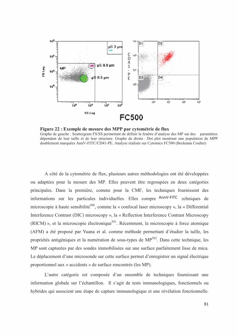
��
�
A côté de la cytométrie de flux, plusieurs autres méthodologies ont été développées
ou adaptées pour la mesure des MP. Elles peuvent être regroupées en deux catégories
principales. Dans la première, comme pour la CMF, les techniques fournissent des
informations sur les particules individuelles. Elles comprennent des techniques de
microscopie à haute sensibilité500, comme la « confocal laser microscopy », la « Differential
Interference Contrast (DIC) microscopy », la « Reflection Interference Contrast Microscopy
(RICM) », et la microscopie électronique501. Récemment, la microscopie à force atomique
(AFM) a été proposé par Yuana et al. comme méthode permettant d�étudier la taille, les
propriétés antigéniques et la numération de sous-types de MP502. Dans cette technique, les
MP sont capturées par des sondes immobilisées sur une surface parfaitement lisse de mica.
Le déplacement d�une microsonde sur cette surface permet d�enregistrer un signal électrique
proportionnel aux « accidents » de surface rencontrés (les MP).
L�autre catégorie est composée d�un ensemble de techniques fournissant une
information globale sur l�échantillon. Il s�agit de tests immunologiques, fonctionnels ou
hybrides qui associent une étape de capture immunologique et une révélation fonctionnelle.
������� ��
��������������
Figure 22 :�Exemple de mesure des MPP par cytométrie de flux Graphe de gauche : Scattergram FS/SS permettant de définir la fenêtre d�analyse des MP sur des paramètres dépendant de leur taille et de leur structure. Graphe de droite : Dot plot montrant une population de MPP doublement marquées AnnV-FITC/CD41-PE. Analyse réalisée sur Cytomics FC500 (Beckman Coulter)

��
�
Les premiers utilisent une capture des MP par anticorps ou par l�annexine V dirigée
contre des antigènes spécifiques ou la PS respectivement. Les MP sont ainsi mesurées en
fonction de leur origine cellulaire et/ou de leur contenu en PS503-507. A titre d�exemple, un kit
ELISA pour MPP a été commercialisé par la société japonaise JIMRO Co. Ltd (Gunma,
Japan). Michelsen et al. ont décrit une méthode qui quantifie les MPP ciblées par l�antigène
GPIIb par time�resolved immunofluorometric assay (TR-IFMA)508. La bonne sensibilité de
cette méthode repose sur les propriétés du rapporteur fluorescent utilisé, un lanthanide
(europium chelate).
Les tests fonctionnels sont exclusivement basés sur l�activité pro-coagulante des MP
soit dépendante de la PS, soit du FT509,510. La génération de thrombine par les MP peut être
réalisée en microplaque avec un substrat fluorescent sur le Calibrated Automated
Thrombogram® system (Thrombinoscope BV, Maastricht, NL and Diagnostica Stago,
Asnieres, F)511. Très recemment, un nouveau test a été commercialisé par Diagnostica Stago,
le STA®-procoag PPL®. Il mesure un temps de coagulation dépendant des phospholipides
dans un PFP512.
Enfin, il existe des tests hybrides comme le test Zymuphen-MP®, commercialisé par
la société Hyphen Biomed (Andresy, F). Il combine une capture en phase solide des MP par
de l�annexine V ou un anticorps et mesure l�activité prothrombinasique des MP214,513.

��
�
�
Figure 23 : Principe d�une méthode hybride combinant capture et test fonctionnel. TF : Facteur tissulaire, AV : Annexine V, BI : Biotine, SA : Streptavidine, PS : Phosphatidylsérine. (Zymuphen*, Hyphen Biomed). D�après J. M. freyssinet.
A la frontière entre ces deux grandes catégories, le Dynamic Light Scattering (DLS)
permet de déterminer la taille et la concentration des MP par la mesure des fluctuations de la
lumière dispersée produites par les mouvements browniens à différents angles514.
Récemment deux instruments, basés sur le principe du DLS, ont été commercialisés : le
Zetasizer Nano S de Malvern Instruments Ltd., et le N5 submicron particle Size Analyser de
Beckman Coulter. Cette technique a été adaptée au suivi des particules individuelles par
Harrison et al. : le « single particle tracking » (SPT)515 (Figure 24). L�origine cellulaire peut
être déterminée par l�utilisation de sondes fluorescentes adaptées comme dans le « enhanced
laser microscopy tracking » (ELMT)516.
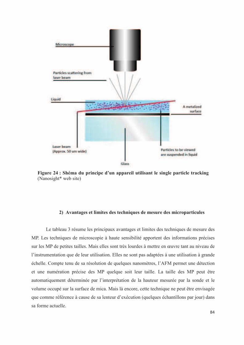
��
�
2) Avantages et limites des techniques de mesure des microparticules
Le tableau 3 résume les principaux avantages et limites des techniques de mesure des
MP. Les techniques de microscopie à haute sensibilité apportent des informations précises
sur les MP de petites tailles. Mais elles sont très lourdes à mettre en �uvre tant au niveau de
l�instrumentation que de leur utilisation. Elles ne sont pas adaptées à une utilisation à grande
échelle. Compte tenu de sa résolution de quelques nanomètres, l�AFM permet une détection
et une numération précise des MP quelque soit leur taille. La taille des MP peut être
automatiquement déterminée par l�interprétation de la hauteur mesurée par la sonde et le
volume occupé sur la surface de mica. Mais là encore, cette technique ne peut être envisagée
que comme référence à cause de sa lenteur d�exécution (quelques échantillons par jour) dans
sa forme actuelle.
Figure 24 : Shéma du principe d�un appareil utilisant le single particle tracking(Nanosight* web site) �
��
�
Les tests ELISA ou apparentés offrent au contraire une possibilité d�analyse à plus
haut débit de nombreux échantillons. Ils sont assez sensibles pour détecter des antigènes
faiblement présents et sont plus faciles d�utilisation que l�immunophénotypage par CMF.
Cependant, ces tests ne fournissent aucune information sur la taille des MP mesurée ni
aucun compte absolu de particules. Une quantification indirecte est cependant possible504.
La quantification globale est dépendante d�un seul biomarqueur mesuré. Des interférances
avec les antigènes solubles sont possibles et peuvent conduire à une surestimation des
niveaux de MP491. Les tests fonctionnels mesurent la contribution générale des MP à une
activité spécifique, non nécessairement reliée à leur nombre. Ceci est particulièrement vrai
quand ce sont les sous-populations minoritaires qui portent le plus d�activité.
La technique DLS est bien adaptée à la fourchette théorique de taille des MP.
Cependant dans sa configuration standard, cette méthodologie parait plus adaptée à la
détermination de la taille de population homogène. La distribution hétérogène de la taille des
MP plasmatiques entraîne souvent des résultats ininterprétables. Ainsi, les instruments
commercialisés sur le principe du SPT, déterminent avec précision la taille de billes de
calibration. Les résultats obtenus avec du plasma restent plus difficile à interpréter. De plus,
cette technique mesure tous les types de particules dans la fenêtre de taille d�analyse
comprenant par exemple les lipoprotéines et les débris. L�introduction de sondes
fluorescentes dans les techniques dérivées de type SPT ou ELMT ont pour but de palier à
cette limite.
Comparée à ces diverses techniques, la CMF présente l�avantage d�une analyse
multiparamétrique de très haut débit de chaque MP individuelle. Cependant, les limites cette
technique sont de plusieurs niveaux : une difficulté à détecter les évènements à faible
antigénicité, un comptage absolu des MP couteux et délicat et une difficulté à détecter des
évènements de faible taille (inférieure à 0.5µm). Ainsi, les cytomètres utilisés jusqu�à
présent ne mesurent qu�une partie des MP (celle de grosse taille). Cependant de récentes
améliorations technologiques permettre de dépasser dans une certaine mesure ces limites et
de maintenir la compétitivité de cette technique pour la numération des MP. La diversité des
performances des cytomètres ainsi que la multiplicité des protocoles appliqués et des
marqueurs choisis empêche une comparaison directe des résultats d�une équipe à l�autre.

��
�
Actuellement, aucune de ces techniques ne remplit tous les critères désirés pour la
mesure des MP plasmatique. En absence de méthode idéale, cette diversité doit être
comprise et utilisée dans un sens de complémentarité. Une comparaison directe entre ces
techniques est nécessaire afin de déterminer objectivement la place relative de chacune
d�elles.
La réalisation d�études cliniques multicentriques afin de confirmer l�intérêt de MP en
pathologie humaine rend indispensable une démarche de standardisation de ces
méthodologies.

��
�
Tab
leau
3:
Rés
um
é d
es p
rin
cip
ale
s ca
ract
éris
tiq
ues
des
tec
hn
iqu
es u
tili
sées
pou
r la
mes
ure
des
MP
(F
reys
sine
t et c
o. J
Thr
omb
Hae
mos
t 201
0)

�
�
DEUXIEME PARTIE: TRAVAUX PERSONNELS
INTRODUCTION
Les microparticules sont des vésicules qui résultent du bourgeonnement des
membranes des cellules activées ou apoptotiques. Leur intérêt est double. Elles sont tout à la
fois des vecteurs actifs d�effecteurs biologiques ayant de multiples conséquences sur
l�homéostasie de différents systèmes et également des biomarqueurs dosables dans le sang
périphérique d�un état cellulaire activé ou d�une lésion d�un tissu particulier.
Ce travail sur les microparticules comprend ces deux aspects. Les trois premiers
articles constituent un ensemble plus fondamental qui met en évidence une activité de
génération de plasmine à leur surface. Les deux articles suivants traitent un aspect plus
appliqué sur les microparticules visant à franchir les limites méthodologiques pour une
meilleure évaluation de ce biomarqueur émergeant en pratique clinique. Une stratégie
de standardisation de leur mesure par cytométrie de flux est proposée. Cette association
entre travail fondamental et appliqué se justifie par une mission mixte universitaire et
hospitalière menée en parallèle pendant ces années de thèse.
Leur capacité à vectoriser des systèmes fonctionnels exprimés par leurs cellules
d�origine nous a conduit à explorer, la génération de plasmine à leur surface. Dans un premier
travail, nous montrons que les MP dérivées de cellules endothéliales (MPE) sont des surfaces
catalytiques capables d�activer le plasminogène par le système de l�urokinase et de son
récepteur (uPA/uPAR). Les MPE agissent comme des vecteurs de plasmine constituant une
nouvelle voie dans la régulation des activités protéolytiques de l�endothélium (Article 1).
Dans un second travail, nous avons montré que l�uPA portée par les cellules ou par les MPE
est spécifiquement impliquée dans un mécanisme reconnaissance du plasminogène lié à une

�
�
autre surface biologique générant de la plasmine in situ avec une grande efficacité (Article
2).
Dans un troisième travail, nous montrons que cette activité fibrinolytique est détectable sur
les MP extraites de la circulation sanguine, où elle est plus spécifiquement portée par les sous
populations endothéliales et leucocytaires. Cette activité qui est dépendante de l�uPA mais
aussi de l�activateur tissulaire du plasminogène, est modulée dans des pathologies
cardiovasculaires et auto-immunes (Article 3). La détection de cette activité dans le sang
périphérique de patient a conduit à un dépôt de brevet sur une méthode de mesure de
l�activité plasmine des microparticules présentes dans un échantillon de fluide biologique et
utilisation de dosage approprié (Brevet 1).
Dans une seconde partie, la pertinence de la mesure des MP comme biomarqueur en
pratique clinique nous a amené à nous focaliser sur leurs méthodes d�analyse. En effet, à
l�heure actuelle, l�évaluation du bénéfice apporté par les MP est limitée par un manque de
standardisation des méthodologies. Dans ce travail, nous présentons une nouvelle stratégie de
standardisation de la cytométrie en flux (CMF) (Annexe 5) et son évaluation dans le cadre
d�une étude multicentrique utilisant des micro-billes fluorescentes calibrées en taille (Article
4). Enfin, nous discutons dans une revue les limites actuelles de la CMF et les stratégies ou
améliorations technologiques permettant de les dépasser (Article 5).
Pendant cette période de travail de thèse, nous avons également apporté notre
contribution à des travaux majeurs soit clinique (Annexe 1), soit fondamental (Annexe 2) sur
l�implication des MP tumorales dans la thrombose.
Notre implication dans le domaine des MPE s�est également traduite par la co-
rédaction d�articles de revue sur le sujet (Annexes 3 et 4).

��
�
ARTICLE 1
Activation of plasminogen into plasmin at the surface of endothelial
microparticles: a mechanism that modulates angiogenic properties of
endothelial progenitor cells in vitro.
Romaric Lacroix1, Florence Sabatier1, Agnès Mialhe1, Agnès Basire1, Ralph Pannell2,
Hélène Borghi3, Stéphane Robert1, Edouard Lamy1, Laurent Plawinski4, Laurence Camoin-Jau1, Victor Gurewich2, Eduardo Angles-Cano4, and Françoise Dignat-George1.
1Unité Mixte de Recherche S 608 (UMR-S 608), Institut National de la Santé et de la Recherche Médicale (INSERM), Université de la Méditerranée, Unité de Formation et de Recherche (UFR) de Pharmacie, Marseille, France; 2Vascular Research Laboratory, Beth Israel Deaconess Medical Center, Harvard Medical School, Boston, MA; 3Service de Microscopie Electronique, UFR de Médecine, Université de la Méditerranée, Marseille, France; 4INSERM, Cyceron, Université de Caen, Caen, France.
Blood. 2007;110:2432-2439
Les microparticules (MP) sont des vésicules qui résultent du bourgeonnement des
membranes des cellules activées ou apoptotiques. Elles ont été décrites dans de nombreux
modèles cellulaires et dans différentes conditions pathologiques où elles sont le signe d�une
activation ou d�une apoptose cellulaire. Parce qu�elles vectorisent diverses molécules
provenant de la cellule d�origine, les MP peuvent exercer un spectre d�activité biologique :
elles propagent des réponses biologiques impliquées dans la balance hémostatique,
participent aux réponses inflammatoires et à l�angiogenèse. La capacité de l�endothélium à
libérer des MP après une stimulation inflammatoire a été démontrée. Des niveaux élevés de
MPE ont été rapportés dans des conditions pathologiques variées comme les syndromes
coronariens, l�insuffisance rénale, le diabète, le syndrome des antiphospholipides, le purpura
thrombotique thrombocytopénique, et la drépanocytose, situations dans lesquelles ces valeurs
élevées de MP reflètent la dysfonction endothéliale et sont associées à un mauvais pronostic.
Les MPE fournissent une surface phospholipidique procoagulante pour l�assemblage
et l�activation des facteurs de la coagulation. Ceci est dû à l�expression de la
phosphatidylsérine sur la couche externe de la membrane cellulaire. Leur implication dans la
génération de thrombine résulte aussi de leur capacité à porter, délivrer ou induire une
activité facteur tissulaire. Toutefois, une contribution plus complexe des MPE à la balance

��
�
hémostatique est suggérée par l�expression à leur surface de molécules anticoagulante comme
la thrombomoduline, le TFPI et le récepteur endothélial à la protéine C.
Le système d�activation du plasminogène constitue un autre régulateur clé de
l�homéostasie vasculaire. Il repose sur 2 sérines protéases ; le t-PA qui est principalement
impliqué dans la fibrinolyse et l�uPA qui joue un rôle majeur dans la protéolyse péricellulaire
du à son affinité élevée pour son récepteur cellulaire, uPAR. La génération de plasmine
induite par l�uPA et l�activation secondaire de MMP favorise la migration cellulaire à travers
la matrice interstitielle et participe à des processus tels que le remodelage tissulaire,
l�invasion métastatique et l�angiogenèse. Il faut également noter qu�une activation
incontrôlée du plasminogène peut avoir des conséquences délétères en induisant un
détachement cellulaire et une apoptose. La régulation de la génération de plasmine à la
surface endothéliale est donc d�une haute importance dans le contrôle de l�homéostasie
vasculaire.
La capacité des MP à vectoriser des protéines et systèmes fonctionnels exprimés par les
cellules d�origine, nous a amené à poser l�hypothèse que les MPE pouvaient représenter
des surfaces catalytiques susceptibles d�interagir avec le plasminogène et de former de
la plasmine.
L�objectif de ce premier travail a donc été d�étudier la capacité des MPE à lier le
plasminogène et de la convertir en plasmine active et de définir l�impact de cette génération
de plasmine sur la capacité des MPE à réguler les réponses angiogéniques des PEC in vitro.
Ce travail a permis de montrer que des MPE dérivées d�HMEC stimulées au TNF-�
représentent des surfaces catalytiques capables de générer de la de plasmine. Cette génération
implique l�expression d�uPA et de son récepteur uPAR à la surface des MPE. Celle-ci est
amplifiée par leur capacité à lier de l�uPA exogène sur des molécules d�uPAR restées libres.
Le plasminogène est activé à la surface des MPE de manière dose dépendante, saturable et
spécifique comme l�indique l�inhibition par l�acide �-amino-caproïque et la carboxypeptidase
B.
La génération de plasmine dépendante des MPE modifie la formation des tubes
vasculaires par les PEC. Alors que les faibles concentrations de MPE augmentent la
formation des tubes, les fortes concentrations la diminuent. Le blocage de ces effets par des

��
�
inhibiteurs d�uPA ou de plasmine souligne le rôle clé de la génération de plasmine induite par
les MPE.
En conclusion, nous avons démontré que les MPE agissent comme des vecteurs
supportant la génération de plasmine et sa dissémination. Ceci constitue une nouvelle voie
dans la régulation des activités protéolytiques de l�endothélium avec des implications
potentielles dans l�inflammation, l�angiogenèse et l�athérosclérose.

HEMOSTASIS, THROMBOSIS, AND VASCULAR BIOLOGY
Activation of plasminogen into plasmin at the surface of endothelialmicroparticles: a mechanism that modulates angiogenic properties of endothelialprogenitor cells in vitro
Romaric Lacroix,1 Florence Sabatier,1 Agnes Mialhe,1 Agnes Basire,1 Ralph Pannell,2 Helene Borghi,3 Stephane Robert,1
Edouard Lamy,1 Laurent Plawinski,4 Laurence Camoin-Jau,1 Victor Gurewich,2 Eduardo Angles-Cano,4
and Francoise Dignat-George1
1Unite Mixte de Recherche S 608 (UMR-S 608), Institut National de la Sante et de la Recherche Medicale (INSERM), Universite de la Mediterranee, Unite de
Formation et de Recherche (UFR) de Pharmacie, Marseille, France; 2Vascular Research Laboratory, Beth Israel Deaconess Medical Center, Harvard Medical
School, Boston, MA; 3Service de Microscopie Electronique, UFR de Medecine, Universite de la Mediterranee, Marseille, France; 4INSERM, Cyceron, Universite
de Caen, Caen, France
The regulation of plasmin generation on
cell surfaces is of critical importance in
the control of vascular homeostasis. Cell-
derived microparticles participate in the
dissemination of biological activities.
However, their capacity to promote plas-
min generation has not been documented.
In this study, we show that endothelial
microparticles (EMPs) from tumor necro-
sis factor a (TNFa)–stimulated endothe-
lial cells served as a surface for the
generation of plasmin. The generation of
plasmin involved expression of urokinase-
type plasminogen activator (uPA) and its
receptor (uPAR) at the surface of EMPs
and was further increased by their ability
to bind exogenous uPA on uPAR. Plasmin-
ogen was activated at the surface of EMPs
in a dose-dependent, saturable, and spe-
cific manner as indicated by the inhibition
of plasmin formation by e-amino-caproic
acid (e-ACA) and carboxypeptidase B.
EMP-induced plasmin generation affects
tube formation mediated by endothelial
progenitor cells. However, low amounts
of EMPs increased tube formation,
whereas higher concentrations inhibited
it. Prevention of these effects by inhibi-
tors of either uPA or plasmin underscore
the key role of EMP-induced plasmin gen-
eration. In conclusion, we demonstrated
that EMPs act as vectors supporting effi-
cient plasmin generation and dissemina-
tion, a new pathway in the regulation of
endothelial proteolytic activities with po-
tential involvement in inflammation, an-
giogenesis, and atherosclerosis. (Blood.
2007;110:2432-2439)
© 2007 by The American Society of Hematology
Introduction
Microparticles (MPs) are vesicles resulting from the blebbing of the
cellular membrane of most activated or apoptotic cells.1 These mi-
crovesicles have been described in various cellular models and in
different pathological conditions as reliable hallmarks of cell damage.2
Because they convey various bioactive effectors originating from the
parent cells, MPs may exhibit a spectrum of biological activities: they
regulate endothelial or blood cell functions, participate in inflammatory
responses or angiogenesis, and propagate biological responses involved
in hemostatic balance.3We previously reported the capacity of endothe-
lial cells to releasemicroparticles after inflammatory stimulation and the
presence of increased levels of circulating endothelial microparticles
(EMPs) in patients with thrombotic disorders.4 Since this initial report,
elevated levels of EMPs have been documented in various pathological
conditions including coronary syndromes,5 renal failure,6 diabetes,7
antiphospholipid syndrome,8 thrombotic thrombocytopenic purpura,9
and sickle cell disease,10 in which they reflect endothelial dysfunction
and are associated with a poor clinical outcome.
EMPs provide procoagulant phospholipid surfaces for the
assembly and activation of coagulation factors, mainly through
phosphatidylserine translocation to the exoplasmic leaflet as a
result of membrane remodeling. Their involvement in thrombin
generation also results from their capacity to harbor, deliver, or
induce tissue factor activity.11-13 However, a more complex contri-
bution to the hemostatic balance is suggested by their expression of
thrombomodulin, tissue factor pathway inhibitor, and endothelial
protein C receptor, thus providing a possible antithrombotic
counterbalance.14,15
Another key regulator of the vascular homeostasis is the
plasminogen activation system. Plasminogen activation is medi-
ated by 2 serine proteases: tissue-type plasminogen activator (tPA),
which is mainly implicated in fibrinolysis, and urokinase-type
plasminogen activator (uPA), which is critically involved in
pericellular proteolysis due to its high affinity cell-surface receptor
uPAR.16 Plasmin generation induced by uPA and subsequent
activation of matrix metalloproteinases (MMPs) promote cell
migration through interstitial matrix and participate in processes
such as tissue remodeling, cancer invasion, and angiogenesis.17-19
Importantly, we have shown that uncontrolled plasminogen activa-
tion can have deleterious consequences by inducing cell detach-
ment and apoptosis.20,21 The regulation of plasmin generation at the
endothelial surface is therefore of critical importance in the control
of vascular homeostasis.
Submitted February 1, 2007; accepted June 22, 2007. Prepublished online as
Blood First Edition paper, July 2, 2007; DOI 10.1182/blood-2007-02-069997.
An Inside Blood analysis of this article appears at the front of this issue.
The online version of this article contains a data supplement.
The publication costs of this article were defrayed in part by page charge
payment. Therefore, and solely to indicate this fact, this article is hereby
marked ‘‘advertisement’’ in accordance with 18 USC section 1734.
© 2007 by The American Society of Hematology
2432 BLOOD, 1 OCTOBER 2007 z VOLUME 110, NUMBER 7

Because MPs convey protein and functional systems expressed
by the parent cell, we hypothesized that EMPs may serve as active
surfaces for interaction with plasminogen and plasmin formation,
a hitherto undescribed function. The objective of this study was
therefore to analyze the capacity of EMPs to bind and convert
plasminogen into active plasmin and to define the impact of
plasmin generation on EMP capacity to regulate angiogenic
responses mediated by endothelial progenitor cells (EPCs) in vitro.
Materials and methods
Preparation of plasminogen and plasminogen-free fetal
calf serum
Plasminogen was purified by lysine-affinity chromatography and molecular
sieving as reported previously.22 Plasminogen-free fetal calf serum (FCS)
was obtained by 3 cycles of affinity adsorption on lysine-Sepharose (4 FCS
vol/1 gel vol) and was subsequently checked and found to contain less than
0.1 nM plasmin(ogen) activity.
Cell culture
The human microvascular endothelial cell line (HMEC-1), obtained from
Dr Ades23 (Centers for Disease Control, Atlanta, GA), was cultured in
MCDB 131 medium (Invitrogen Life Technologies, Cergy Pontoise,
France) supplemented with 10% MP-free FCS, 10 ng/mL human recombi-
nant epithermal growth factor (Upstate Cell Signaling Solutions, Lake
Placid, NY), and 1 mg/mL hydrocortisone (Sigma, St Quentin Fallavier,
France). Human vein endothelial cells (HUVECs) were obtained by
collagenase digestion as previously described,24 cultured into 0.2% gelatin-
coated flasks in EGM2 medium and used at passage 2. Human saphenous
vein endothelial cells were purchased from Clonetics (Grand Island, NY)
and cultured into 0.2% gelatin-coated flasks in EGM2-MV medium and
used at passage 6.
Generation, harvesting, and flow cytometry enumeration
of EMPs
For most experiments, EMPs were from HMEC-1 origin, as this cell line is
well characterized and considered representative of the microcirculation,
the core of the vascular tree.23,25 EMPs were purified from culture medium
conditioned by subconfluent HMEC-1 stimulated for 48 hours with
100 ng/mL TNF-a (PeproTech, Rocky Hill, NJ) as previously described,
with minor modifications.4 Culture supernatants from flasks were collected
and cleared from detached cells or large cell fragments by centrifugation at
4300g for 5 minutes. The supernatants were then centrifuged at 20 000g for
120 minutes at 4°C. Pelleted EMPs were washed 2 times and resuspended
in phosphate-buffered saline (PBS). The absence of residual TNF-a in this
EMP sample was verified using an enzyme-linked immunosorbent assay
(ELISA) assay (R&D Systems, Minneapolis, MN). Aliquots of 10 mL EMP
suspension, 1/100 diluted, were labeled using fluorescein isothiocyanate
(FITC)–conjugated annexin V (Abcys, Paris, France), and EMPs were
enumerated by flow cytometry as previously described.26 The same
protocol was used to obtain EMPs from quiescent HMEC-1, saphenous
endothelial cells, and endothelial progenitor-derived cells (EPDCs).
Isolation and culture of endothelial progenitor cells from
cord blood
Human umbilical cord blood samples (30-50 mL) were collected from
donors, in compliance with French legislation, in a sterile tube containing
heparin (200 UI/mL). Mononuclear cells (MNCs) were isolated by density
gradient centrifugation. Briefly, blood was diluted 1:1 in PBS containing
2 mM ethylenediaminetetraacetic (PBS/EDTA) and layered over lympho-
cyte separation medium (Eurobio, Les Ulis, France). After a 30-minute
centrifugation at 400g, MNCs were washed 3 times in PBS/EDTA. Cord
blood MNCs were preplated in RPMI/10% FCS for 24 hours in plastic
flasks. Nonadherent cells were plated onto 0.2% gelatin-coated 24-well
plates (5.106 cells per well) and maintained in endothelial basal medium-2
(EBM-2) supplemented with EGM-2 SingleQuots (EGM-2medium; Clonet-
ics, Walkersville, MD). The medium was changed every 4 days. The
appearance of well-circumscribed colonies with a cobblestone morphology
was monitored daily. For expansion of EPDCs, colonies were trypsinized
and cells were replated on a 6-well plate (passage 1). Subsequently,
confluent cells were trypsinized and replated in T75 flasks for further
passages. Cells were maintained under standard conditions (humidified
atmosphere, 5% CO2, 37°C).
Progenitor endothelial-cell tube formation in matrigel
Flat-bottom 96-well plates were precoated with 1:1 mixture of cold matrigel
basement membrane (10 mg/mL, BD Biosciences, Bedford, MA) and
RMPI-10% FCS medium. After 45 minutes of polymerization at 37°C,
EPDCs were plated at 2.105 cells/well in RPMI/10% FCS with increasing
concentrations of EMP from TNF-a–stimulated HMEC-1 or an identical
volume of the supernatant from the last EMP washing. To test the role of
FCS plasminogen in this system, native FCS was replaced by plasminogen-
free FCS supplemented or not with 1 mM human plasminogen. For
inhibition experiments, EMPs were pre-incubated with either an anti-uPA
antibody (394O, American Diagnostica, Greenwich, CT, 25 mg/mL) or an
irrelevant control IgG1 (25 mg/mL) or with serine-protease inhibitors
(aprotinine, 100 kui/mL or a2-antiplasmin, 1 mM).After 24 hours, capillary
tube formation was evaluated by measuring the number of polygons formed
in a well under an inverted light microscope (Olympus) at 400 3
magnification. This time point was chosen following previous kinetics
experiments indicating that it is optimum to analyze tube formation in the
presence of EMPs. All experiments were performed in triplicate.
EMP immobilization
Aphysico-chemical adsorption principle was used to immobilize negatively
charged EMPs on a polycation surface. For that purpose, round-bottom
PVC plates were activated with 25 mg/mL poly-L-lysine, and various
concentrations of EMP in PBS were then incubated overnight at 4°C with
the activated surface. The plate was then washed, and the immobilized
EMPs were tested.
Plasmin generation test
In round-bottom 96-well PVC plates, various concentrations of EMPs,
either in suspension in PBS supplemented with 0.8% bovine serum albumin
(PBSA) or immobilized on poly-L-lysine–coated plates, were incubated
with 50 mL of 1 mM plasminogen and 0.75 mM of a chromogenic substrate
selective for plasmin (methyl-malonyl)-hydroxypropylarginine-
p-nitroanilide (CBS0065; Stago, Asniere, France). An identical volume
of supernatant from the last EMP washing was used as control. In order
to determine the Michaelis constant of plasmin generation by EMPs, a
fixed number (2.105) of EMPs and varying concentrations of plasmino-
gen (0 to 5 mM) were used. Kinetics of plasmin generation were
followed during 9 hours in a multiwell plate counter (MX5000, Dynex)
at 37°C by measuring the change in absorbance at 405 nm produced by
the release of p-nitroaniline. In the case of activation experiments
performed on immobilized EMPs, the plate was washed with PBSA to
eliminate unbound reactants, and the plasmin bound to the immobilized
EMPs was detected by adding 50 mL/well of 0.75 mM CBS0065 and
measuring the change in absorbance at 405 nm. When indicated, the
following inhibitors were pre-incubated with the EMPs: 1 mg/mL goat
antihuman tPA (Biopool, Uppsala, Sweden), 50 mg/mL mouse antihu-
man uPA (394O), 50 mg/mL rabbit anti-avb3 (gift of J.C. Lissitzky),
100 mg/mL anti–a-enolase (gift of R. Lopez Alemany), and the
respective irrelevant control IgGs (Biocytex, Marseille, France); the
uPA inhibitor, amiloride, was used at 100 mM and carboxypeptidase B
(CpB) at 50 mg/mL, final concentrations. In separate experiments, the
inhibitors a2-antiplasmin, aprotinin, and e-amino-caproic acid (e-ACA)
were added to the plasminogen activation solutions at 100 nM,
100 kiu/mL and 0.1 M final concentrations, respectively.
PLASMIN GENERATION ON ENDOTHELIALMICROPARTICLES 2433BLOOD, 1 OCTOBER 2007 z VOLUME 110, NUMBER 7

Characterization of uPA and uPAR in cell and EMP extracts
HMEC-1 and pelleted MPs were lysed in 100 mM tris(hydroxymethyl)ami-
nomethane (Tris)–HCl buffer pH 8.1, containing 0.5% Triton X-100 and
supplemented with complete protease inhibitor mixture (Roche Diagnostic,
Mannheim, Germany) except for zymography samples. Lysates were
clarified and protein concentrations were determined using the BCA kit
(Pierce, Rockford, IL). uPA and uPAR total antigen levels were assayed by
ELISA (894 and 893 IMUBIND ELISA kits, American Diagnostica)
according to the manufacturer’s instruction. The results were expressed as
ng of uPA or uPAR per mg of total proteins. For zymography, protein
extracts (5 mg), molecular weight markers, and purified scuPA were
electrophoresed under nonreducing conditions in 10% sodium dodecyl
sulfate (SDS)–polyacrylamide gel copolymerised with 1 mg/mL a-casein
from bovine milk (Sigma) and 20 mg/mL human plasminogen. After
electrophoresis, SDS was eluted from the gel by washing for 1 hour in 2.5%
Triton X-100 buffer. The gels were then incubated for 40 hours at 37°C in
50 mM Tris-HCl buffer, pH 8, containing 5 mM CaCl2, 138 mM NaCl, and
0.03% Brij 35. Zymograms were developed by staining with Coomassie
Brillant Blue Dye and destained to reveal clear bands of casein lysis,
indicative of enzymatic activity. Samples run on casein gels without
plasminogen or gels incubated in lysis buffer supplemented with 1 mM
amiloride (Sigma) served as controls of uPA activity. For Western blot
analysis, protein extracts and purified human uPAR (American Diagnos-
tica) used as positive control (20 mg/lane) were separated under nonreduc-
ing conditions by 8% SDS-polyacrylamide gel and electrotransferred onto a
nitrocellulose membrane. After blocking, the membranes were incubated
overnight at 4°C with rabbit antihuman uPAR (399R, American Diagnos-
tica, 0.5 mg/mL) antibodies, followed by the horseradish peroxidase–
conjugated secondary antibodies. Immunocomplexes were visualized with
the Supersignal West Pico chemiluminescence kit (Pierce).
Immunoelectron microscopy of EMPs
For uPA/uPAR immunogold labeling of EMPs, droplets of MP isolated
from TNFa-stimulated HMEC-1 were applied to 300 mesh nickel Formvar-
carbon-coated grids (Agar Scientific, Essex, United Kingdom) for 10 min-
utes. Samples were immunolabeled with either anti-uPA (3689, American
Diagnostica, 10 mg/mL) or anti-uPAR (3932, American Diagnostica,
10 mg/mL) antibodies for 1 hour, washed with PBS, and then reacted with
the 15-nm beads gold-labeled secondary antibody (BBInternational, Cardiff,
United Kingdom) for 1 hour. Grids were rinsed and negatively stained with
0.3% phosphotungstic acid (pH 7) before observation with a JEOL 1220
electron microscope. Specificity of immunolabeling was determined in
comparison to results obtained with an irrelevant control antibody or with
gold-labeled secondary antibody alone.
Flow cytometry for uPA and uPAR on EMPs
EMPs (10 mL) were labeled with either anti-uPA (394O, 25 mg/mL) or
anti-uPAR (3932, 25 mg/mL) antibodies for 1 hour, then FITC-labeled
secondary antibody was added and incubated for 30 minutes before samples
were analyzed on a Cytomics FC500* flow cytometer (Beckman Coulter,
Fullerton, CA). MPs were analyzed according to the FSC/SSC characteris-
tic in a gate with the upper limit defined by beads of 0.9 mm and the lower
limit defined by 0.3 mm. Specificity of labeling was determined in
comparison to results obtained with an irrelevant control antibody or with
the secondary antibody alone.
Binding of uPA to EMPs
A fixed concentration of EMPs was immobilized on poly-L-lysine–coated
96-well plates. Increasing concentrations (0 to 18.5 nM) of sc-uPA(a kind gift of
DrH.R. Lijnen,University of Leuven)were incubated during 1 hour at 37°Cwith
the immobilized microparticles in the presence of 4 mg/mL of poly-L-lysine
in order to eliminate nonspecific binding of sc-uPA to the poly-L-lysine surface.
To verify the specificity of the binding, a fixed concentration of native sc-uPA
(0.1 nM) was mixed with a 10 molar excess (1 nM) of a recombinant inactive
form “rsc-uPA Ile 1593Gly” (H.R. Lijnen, University of Leuven, Leuven,
Belgium). The native sc-uPA, specifically bound to EMPs, was detected by
measuring plasmin generation as described above.
Statistical analysis
Data are expressed as mean 6 standard deviation. Statistical analysis was
performed with Prizm software (GraphPad Software, San Diego, CA) and
with KaleidaGraph software (Synergy Software, Reading, PA). Significant
differences were determined using nonparametric Mann-Whitney test. A
P value less than 0.05 was considered significant.
Results
EMPs are able to activate plasminogen into plasmin
To investigate the ability of EMPs to generate plasmin, the
microparticles were incubated with plasminogen and a plasmin-
selective chromogenic substrate. As shown in Figure 1A,
plasmin generation occurred as a function of time and was
proportional to the number of EMPs. At 106 EMP/well the
plasmin generation rate was 47.6 6 1.1 A405nmx1023/min,
53-fold higher than at 103 EMP/well (0.9 6 0.3 A405nmx1023/
min) (Figure 1B). At identical EMP concentrations (2 105/50
mL) comparable results were obtained using EMP derived from
quiescent or TNFa-stimulated HMEC-1. In contrast, the level of
plasmin generated by EMPs was shown to vary according to
their endothelial cell origin (Table 1). Thus, a more pronounced
activity was produced by HMEC-1–derived MPs as compared
with MPs of macrovascular origin (saphenous vein, HUVEC),
whereas intermediate values were obtained for EPDC-derived
MPs. Plasminogen incubated with EMPs was activated in a
dose-dependent, saturable, and specific manner (Figure 1C,
Km 5 0.122 mM, Vmax 5 25.2 A405 nm/min 103). As ex-
pected, plasmin activity was completely blocked in the presence
of a2-antiplasmin or aprotinin (Figure 1D).
Plasminogen is activated by uPA at the surface of EMPs
Supernatants from EMP washing failed to generate plasmin,
ruling out the contribution of soluble factors and suggesting that
the activation of plasminogen was dependent on factors associ-
ated with the EMP surface. To test this hypothesis, plasminogen
activation experiments were performed on immobilized micro-
particles. In this system, plasmin generation was a function of
the number of immobilized EMPs (Figure 2A). Plasmin was
already detected on immobilized EMPs at a concentration of
25 000 EMP/well and increased progressively in a dose-
dependent manner. At the end of the activation experiments the
immobilized EMPs were washed, and bound plasmin was
detected using a plasmin-selective chromogenic substrate. The
amount of EMP-bound plasmin increased as a function of the
number of immobilized EMPs (Figure 2B). Since some plasmin
was released into the medium during its generation, the amount
bound after washing was lower than the total amount of plasmin
formed.
In parallel experiments using EMP in suspension, plasmin
generation was inhibited by the lysine analog e-ACA, suggesting a
lysine-dependent mechanism for plasminogen binding and activa-
tion that was confirmed by abrogation of plasmin formation upon
cleavage of C-terminal lysine residues on EMPs by CpB (Figure
1D).Among proteins bearing C-terminal lysine residues, a-enolase
was identified as a major binding protein as indicated by 65%
inhibition of plasmin formation with the specific antibody 11G1
(Figure 1D). The integrin avb3 that may influence plasminogen
activation, mainly through interactions with the uPA/uPAR system,
was shown not to be involved in plasmin generation by EMPs as
2434 LACROIX et al BLOOD, 1 OCTOBER 2007 z VOLUME 110, NUMBER 7

indicated by the absence of effect of a specific neutralizing
antibody (Figure 1D).
The uPA/uPAR complex is present on EMPs
The value determined for the apparent Km of plasminogen
activation on EMPs was within the range determined for the
uPA/uPAR system on endothelial cells and a variety of other cell
types.27,28 The plasminogen activator conveyed by EMPs was
identified as uPA as indicated by the inhibition of plasmin
generation by (1) amiloride, a specific inhibitor of uPA, and by
(2) an antibody specific for uPA ( . 95% inhibition) (Figure
1D). By contrast, an antibody directed against tPA had no
significant effect on plasmin generation. The presence of uPA
activity on EMPs was further confirmed by a lytic band on
casein zymography corresponding to the molecular mass of uPA
(Figure 2C), which was also absent in gels into which amiloride
(1 mM) was incorporated (data not shown). Comparison of the
lytic bands from the EMPs and the parent cells, normalized for
protein content, showed a 3- to 4-fold increase of uPA in the
EMPs (representative experiment in Figure 2C). Similarly, when
uPA antigen in EMPs and cells were measured by ELISA, a
3-fold increase in uPA was found in the EMPs (23.9 6 12.6
versus 8.3 6 3.17 ng uPA/mg protein, respectively, P , .05).
UPAR was identified in EMPs and cell lysates by Western blot
and was found to comigrate with mammalian uPAR. As for uPA,
the EMPs had more uPAR than the cells when normalized for
equivalent total protein concentrations (Figure 2D). The uPAR
antigen level measured by ELISAwas 4-fold higher in the EMPs
than in the parent HMEC-1 (57.7 6 14.54 versus 4.4 6 1.17
ng/mg protein, respectively, P , .05). The presence of uPA or
uPAR on the EMP surface also was analyzed by flow cytometry
(Figure 3A,B). The shift of fluorescence histogram after specific
labeling of EMPs with uPA and uPAR antibody compared with
the irrelevant staining confirmed the presence of uPA and uPAR
at the EMP surface.
EMPs also were analyzed by electron microscopy. Negative
staining of the pellet revealed intact irregular-shaped EMPs
ranging from 100 to 500 nm in size. Molecules of uPA and uPAR
were detected on the outer surface after immunogold labeling with
specific antibodies (Figure 3C). No labeling was observed in
experiments using control antibodies.
Binding of sc-uPA to EMP-uPAR
Since the amount of uPAR measured by ELISA was higher on
EMPs than on cells, we tested the capacity of immobilized
EMPs to bind exogenous scuPA specifically. A range of concen-
trations of scuPA was incubated with the immobilized EMPs,
washed, and then tested in a plasminogen activation assay.
Figure 3D shows that scuPA binding was dose-dependent,
saturable, and specific, as indicated by the inhibition of the
binding by a modified form of scuPA that resists activation
(Figure 3D, insert). Analysis of raw data with the Langmuir
equation for single-site binding allowed calculation of a very
low dissociation constant (Kd 5 0.1 nM), indicating a very high
affinity of scuPA for its EMP anchored receptor, in agreement
with previous published data for the uPA/uPAR system.28 Since
elution of intrinsic uPA could not be performed before the
binding experiments, these results indicate that the amount of
uPA originally bound is relatively small compared with the high
uPAR binding capacity of EMPs.
Table 1. Plasmin generation by endothelial microparticles
Source from which EMPs were derived
Plasmin generation,
mean % plus or minus SD
TNFa-stimulated HMEC-1 100
Quiescent HMEC-1 98.0 6 6.3
EPDC 27.7 6 5.5
Saphenous vein endothelial cells 13.0 6 2.6
HUVEC 7.6 6 3.3
*23 105/well.
Figure 1. EMPs are able to activate plasminogen into
plasmin. (A) Plot of plasmin generated versus time at
varying EMP amounts per 50 mL/well (E 5 106;M 5 105;
e 5 104; x 5 103; 1i5 control without EMPs) and fixed
final concentrations of plasminogen (1mM) and a plasmin-
selective chromogenic substrate (0.75 mM). Representa-
tive graph of 4 independent experiments. (B) Similar
experiment as in panel A expressed as change in absor-
bance at 405 nm per minute versus EMP amount per well
(S: EMP last washing supernatant used as control).
(C) Plasmin generated at varying plasminogen concentra-
tions (0-5 mM) and a fixed amount (2.105/50 uL) of EMPs
was detected with a chromogenic substrate as in panelA.
Raw data (E) were fitted to the Michaelis-Menten equa-
tion allowing calculation of nonspecific activity (e) and a
Km 5 0.122 mM for specific plamin generation ( M ).
(D) Effect of various inhibitors on the generation of
plasmin by EMPs (2.105/50 mL) at 0.5 mM plasminogen
(a2AP 5 a2-antiplasmin; e-ACA 5 e-amino-caproıc acid;
CPB 5 carboxypeptidase B; antibodies to uPA and tPA
avb3 and a-enolase as compared with an isotype control
IgG1). Results are the mean 6 SD of 3 independent
experiments.
PLASMIN GENERATION ON ENDOTHELIALMICROPARTICLES 2435BLOOD, 1 OCTOBER 2007 z VOLUME 110, NUMBER 7

EMPs affect tube formation by endothelial progenitor cells
in vitro
EPCs were supplemented with increasing amounts of EMPs (2.103
to 8.105), and tube formation in matrigel was evaluated using the
supernatant from the last EMP washing as control (100% tube
formation). As shown on Figure 4A, a biphasic effect was
observed: at low EMP concentrations (# 2.103/well), polygon
number and tube formation increased by 20% over control
(P 5 .012), whereas higher amounts of EMP ($ 2.105 EMPs/well)
had the reverse effect. Thus, tube formation decreased progres-
sively with the increase in the number of EMPs reaching 55%
decrease below control (P , .001) at 8.105 EMPs/well. Concomi-
tant with the decrease in tube formation, morphologic cell changes
were observed (rounded cells and retracted cell clusters) (Figure
4B). The moderate pro-angiogenic effect of MP seems to be less
potent as compared with vascular endothelial growth factor (VEGF)
(see the Supplemental Materials link at the top of the online article
available on the Blood website).
Plasmin is involved in the EMP effects on tube formation
An anti-uPA antibody and a2-antiplasmin were used to deter-
mine whether the observed effects of EMPs on EPC tube
formation were related to plasmin generation. As illustrated in
Figure 4C, in the presence of these inhibitors, EMPs had no
more effect on tube formation.
We also considered the presence of plasmin in the matrigel
medium, because the gel itself and the culture medium (RPMI/
10% FCS) contain plasminogen,29 and the EMPs are a source of
uPA. The plasmin chromogenic substrate was added with the
EMPs in matrigel, allowing detection of increased plasmin
generation from 105 EMP (P , .001) at 2 hours of incubation
(Figure 5A). The contribution of FCS plasminogen to plasmin
Figure 2. Plasminogen is activated at the surface of EMPs by uPA. (A,B) A variable number of EMPs immobilized on poly-L-lysine surfaces were incubated with fixed
concentrations of plasminogen (0.5 mM) and the plasmin-selective chromogenic substrate (0.75 mM). The graph in panel A shows an increase in the formation of plasmin as a
function of the number of immobilized microparticles. Unbound reagents were then washed off and the chromogenic substrate added to detect plasmin that remained bound to
the immobilized EMPs; the graph (B) shows an increase in bound plasmin as a function of the number of immobilized EMPs. Representative graphs (mean 6 SD) of 3
independent experiments. (C,D) Protein extracts from HMEC-1 and its derived EMPs were processed for zymography (panel C) to detect plasminogen activator activity (5 mg
protein/lane; S: EMP last washing supernatant) and for immunoblot (panel D) using rabbit antibodies against uPAR and purified uPAR as reference (20 mg protein/lane). The
;50 kDa and upper bands in panel C were inhibited by antibodies to urokinase (not shown). A space has been inserted to indicate where a gel lane was cut. These gels came
from different experiments as indicated by the space between the gels.
Figure 3. Identification of uPA, uPAR, and uPA/uPAR
complex on EMP. (A,B) Measured fluorescence intensity
by flow cytometry analysis of EMPs with anti-uPA (A) and
anti-uPAR (B) antibodies (black) and the corresponding
isotype controls (gray). (C) Representative images of
immunogold labeling of uPA (top) and uPAR (bottom)
analysis on EMPs by transmission electron microscopy.
Slides were viewed with a JEOL 1220 transmission
electron microscope (JEOL, Tokyo, Japan). Images were
acquired using a MegaView III camera (Soft Imaging
System, Munster, Germany) and were processed with
Analysis software (Soft Imaging System) and Adobe
Photoshop version 7.0 software (Adobe Systems, San
Jose, CA). The dimension of the bars indicate the relative
small size of these EMPs, and the clusters of 15-nm gold
particles (black dots) indicate the presence of uPA and
uPAR at the surface of EMPs. (D) Main graph: isotherm of
the binding of varying amounts of scuPA incubated with
EMPs immobilized on a poly-L-lysine surface. The amount
of bound scuPA was detected by its ability to activate
plasminogen using a chromogenic substrate selective for
plasmin. Data fitted to the Langmuir equation as indicated
in “Materials and methods” allowed calculation of a
dissociation constant, Kd 5 0.1 nM, for the interaction of
scuPA with its receptor. Representative graph
(mean 6 SD) of 3 independent experiments. Inset: speci-
ficity of the binding of native scuPA is demonstrated by its
inhibition with a modified recombinant form of scuPA
(r-scuPA, ILe1593Gly) that bind to its receptor but
cannot be activated. The bars represent the mean
(6 SD) of 3 independent experiments.
2436 LACROIX et al BLOOD, 1 OCTOBER 2007 z VOLUME 110, NUMBER 7

formation was estimated using plasminogen-free FCS (Figure
5B). Under these conditions plasmin generation was moderately
reduced (211.8%, P 5 .001) compared with native FCS and
was not influenced by the amount of EMPs added, suggesting
the presence of a basal plasmin generation in the matrigel. The
addition of plasminogen to plasminogen-free FCS restored
plasmin formation to the level observed with native FCS in the
absence of EMPs and was again importantly increased as a
function of the number of added EMPs (more than 75% increase
at 2.105 EMP/well, Figure 5B). These data confirmed the role of
matrigel and plasminogen from FCS in plasmin generation in
the absence of EMPs, and the strong potential capacity of EMPs
to enhance plasmin generation.
Altogether, these data indicate that the uPA/uPAR system of
EMPs is responsible for plasmin generation and that plasmin
proteolytic activity is involved in the EMP effects on tube
formation by EPCs.
Discussion
The plasminogen activation system plays a pivotal role in maintain-
ing vascular patency and facilitating cell migration and angiogen-
esis. Binding of plasminogen to fibrin or the cell surface is of
critical importance to regulate and target the proteolytic activity.
Although the presence of uPAR on endothelial microparticles
previously has been described,30 its functional consequences, to our
knowledge, have not been investigated as yet. The present study is
the first demonstration that EMPs provide a catalytic surface for the
conversion of plasminogen into plasmin by expressing uPA and
uPAR. Plasmin generation was EMP surface-dependent and could
be augmented by binding of exogenous uPA to EMP-uPAR. As a
result, EMPs were shown to modulate the angiogenic responses of
endothelial progenitors in vitro.
Figure 4. EMPs affect the angiogenic properties of endothelial progenitors in vitro: effects on tube formation in matrigel. EPDC in RPMI/10% FCS and varying
amounts of EMPs were plated on matrigel, and tube formation was evaluated after 24 hours of incubation. (A) Biphasic effect of EMPs on tube formation: at low EMP
concentrations ( # 2.103/well), polygon number and tube formation increased by 20% (P 5 .012, n 5 12) over control (S: EMP last washing supernatant). However, higher
amounts of EMPs ( $ 2.105 EMP/well) had the reverse effect and progressively decreased the number of tubes to 50% at 8.105 EMP/well. Stars indicate significant changes
(*P , .05, **P , .01, ***P , .001). Results are the mean (6 SD) of 12 independent experiments. (B) Inverted light microscope representative images (400 3 magnification)
showing EPDC tube formation in matrigel in the absence and presence of 2.103 or 2.105 EMPs per well. Asterisks indicate retracted cell clusters. Slides were viewed with an
inverted Nikon light microscope (Nikon, Melville, NY) using an Olympus lens (403/0.60; Olympus, Tokyo, Japan). Images were acquired using a Nikon model DXM 1200F
camera and were processed with Lucia G software (both from Nikon) and Adobe Photoshop version 7.0 software (Adobe Systems, San Jose, CA). (C) Experiments were
performed in the presence of inhibitors of either plasmin formation (anti-uPAantibody) or activity (a2-antiplasmin, a2-AP). The biphasic effect of EMPs on tube formation (left bar
panel) was abolished by the anti-uPA antibody (middle) and by a2-AP (right). Asterisks indicate significant changes (P , .05). Results are the mean (6 SD) of 3 independent
experiments.
Figure 5. EMPs are involved in plasmin generation in
matrigel. (A)Plasmingeneration inmatrigel is dependent on
the amount of EMPs. The chromogenic substrate CBS0065
was added to the matrigel with the EMPs, and under these
conditions an increase in plasmin formation was detected
from 105 EMPs (P , .001) after 2 hours of incubation.
Asterisks indicate significant changes (P , .05). Results are
themean (6SD) of 3 independent experiments. (B) Plasmin
generation in matrigel at different EMP amounts is depen-
dent on the supply of plasminogen by FCS. The graph
shows plasmin activity detected after 3 hours of incubation
, medium with 10% FCS; , medium with plasminogen-
free FCS; , medium with plasminogen-free FCS supple-
mented with 1 mM plasminogen). Activity in the absence of
EMPs may represent low levels of plasmin and/or limited
amounts of activators in matrigel. Asterisks indicate signifi-
cant changes (P , .05). Results are the mean (6 SD) of 3
independent experiments.
PLASMIN GENERATION ON ENDOTHELIALMICROPARTICLES 2437BLOOD, 1 OCTOBER 2007 z VOLUME 110, NUMBER 7
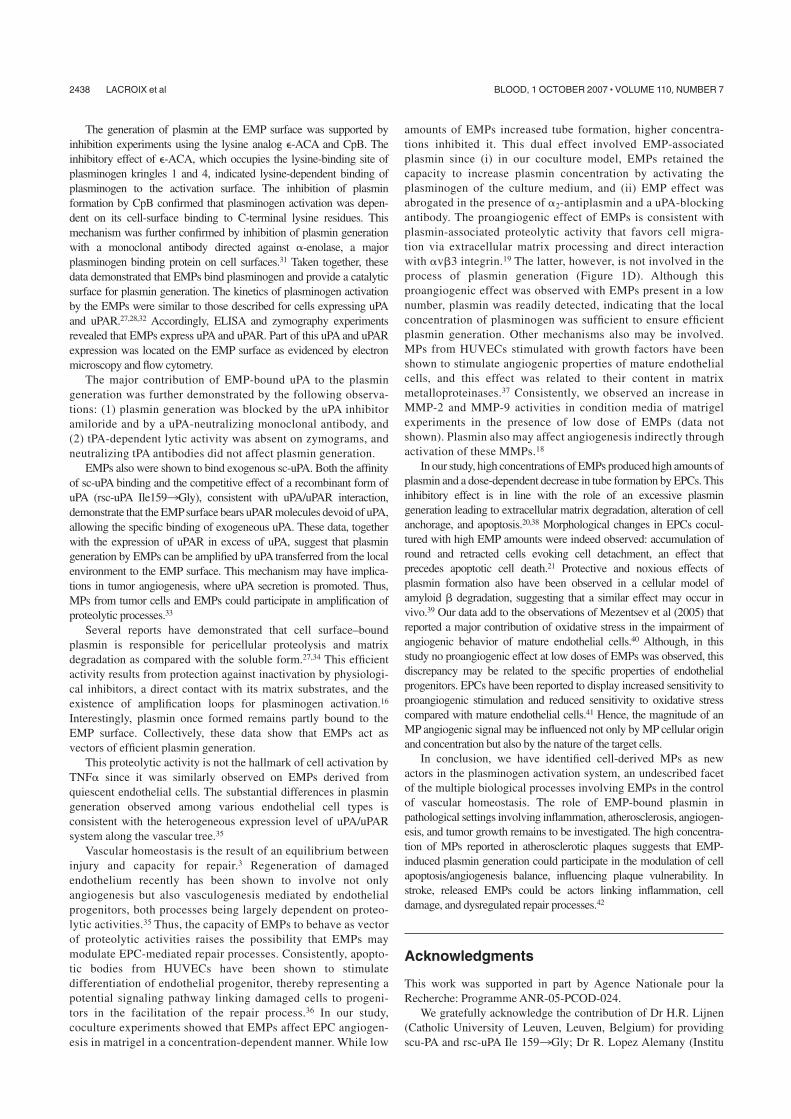
The generation of plasmin at the EMP surface was supported by
inhibition experiments using the lysine analog e-ACA and CpB. The
inhibitory effect of e-ACA, which occupies the lysine-binding site of
plasminogen kringles 1 and 4, indicated lysine-dependent binding of
plasminogen to the activation surface. The inhibition of plasmin
formation by CpB confirmed that plasminogen activation was depen-
dent on its cell-surface binding to C-terminal lysine residues. This
mechanism was further confirmed by inhibition of plasmin generation
with a monoclonal antibody directed against a-enolase, a major
plasminogen binding protein on cell surfaces.31 Taken together, these
data demonstrated that EMPs bind plasminogen and provide a catalytic
surface for plasmin generation. The kinetics of plasminogen activation
by the EMPs were similar to those described for cells expressing uPA
and uPAR.27,28,32 Accordingly, ELISA and zymography experiments
revealed that EMPs express uPAand uPAR. Part of this uPA and uPAR
expression was located on the EMP surface as evidenced by electron
microscopy and flow cytometry.
The major contribution of EMP-bound uPA to the plasmin
generation was further demonstrated by the following observa-
tions: (1) plasmin generation was blocked by the uPA inhibitor
amiloride and by a uPA-neutralizing monoclonal antibody, and
(2) tPA-dependent lytic activity was absent on zymograms, and
neutralizing tPA antibodies did not affect plasmin generation.
EMPs also were shown to bind exogenous sc-uPA. Both the affinity
of sc-uPA binding and the competitive effect of a recombinant form of
uPA (rsc-uPA Ile1593Gly), consistent with uPA/uPAR interaction,
demonstrate that theEMPsurface bears uPARmolecules devoid of uPA,
allowing the specific binding of exogeneous uPA. These data, together
with the expression of uPAR in excess of uPA, suggest that plasmin
generation by EMPs can be amplified by uPA transferred from the local
environment to the EMP surface. This mechanism may have implica-
tions in tumor angiogenesis, where uPA secretion is promoted. Thus,
MPs from tumor cells and EMPs could participate in amplification of
proteolytic processes.33
Several reports have demonstrated that cell surface–bound
plasmin is responsible for pericellular proteolysis and matrix
degradation as compared with the soluble form.27,34 This efficient
activity results from protection against inactivation by physiologi-
cal inhibitors, a direct contact with its matrix substrates, and the
existence of amplification loops for plasminogen activation.16
Interestingly, plasmin once formed remains partly bound to the
EMP surface. Collectively, these data show that EMPs act as
vectors of efficient plasmin generation.
This proteolytic activity is not the hallmark of cell activation by
TNFa since it was similarly observed on EMPs derived from
quiescent endothelial cells. The substantial differences in plasmin
generation observed among various endothelial cell types is
consistent with the heterogeneous expression level of uPA/uPAR
system along the vascular tree.35
Vascular homeostasis is the result of an equilibrium between
injury and capacity for repair.3 Regeneration of damaged
endothelium recently has been shown to involve not only
angiogenesis but also vasculogenesis mediated by endothelial
progenitors, both processes being largely dependent on proteo-
lytic activities.35 Thus, the capacity of EMPs to behave as vector
of proteolytic activities raises the possibility that EMPs may
modulate EPC-mediated repair processes. Consistently, apopto-
tic bodies from HUVECs have been shown to stimulate
differentiation of endothelial progenitor, thereby representing a
potential signaling pathway linking damaged cells to progeni-
tors in the facilitation of the repair process.36 In our study,
coculture experiments showed that EMPs affect EPC angiogen-
esis in matrigel in a concentration-dependent manner. While low
amounts of EMPs increased tube formation, higher concentra-
tions inhibited it. This dual effect involved EMP-associated
plasmin since (i) in our coculture model, EMPs retained the
capacity to increase plasmin concentration by activating the
plasminogen of the culture medium, and (ii) EMP effect was
abrogated in the presence of a2-antiplasmin and a uPA-blocking
antibody. The proangiogenic effect of EMPs is consistent with
plasmin-associated proteolytic activity that favors cell migra-
tion via extracellular matrix processing and direct interaction
with avb3 integrin.19 The latter, however, is not involved in the
process of plasmin generation (Figure 1D). Although this
proangiogenic effect was observed with EMPs present in a low
number, plasmin was readily detected, indicating that the local
concentration of plasminogen was sufficient to ensure efficient
plasmin generation. Other mechanisms also may be involved.
MPs from HUVECs stimulated with growth factors have been
shown to stimulate angiogenic properties of mature endothelial
cells, and this effect was related to their content in matrix
metalloproteinases.37 Consistently, we observed an increase in
MMP-2 and MMP-9 activities in condition media of matrigel
experiments in the presence of low dose of EMPs (data not
shown). Plasmin also may affect angiogenesis indirectly through
activation of these MMPs.18
In our study, high concentrations of EMPs produced high amounts of
plasmin and a dose-dependent decrease in tube formation byEPCs.This
inhibitory effect is in line with the role of an excessive plasmin
generation leading to extracellular matrix degradation, alteration of cell
anchorage, and apoptosis.20,38 Morphological changes in EPCs cocul-
tured with high EMP amounts were indeed observed: accumulation of
round and retracted cells evoking cell detachment, an effect that
precedes apoptotic cell death.21 Protective and noxious effects of
plasmin formation also have been observed in a cellular model of
amyloid b degradation, suggesting that a similar effect may occur in
vivo.39 Our data add to the observations of Mezentsev et al (2005) that
reported a major contribution of oxidative stress in the impairment of
angiogenic behavior of mature endothelial cells.40 Although, in this
study no proangiogenic effect at low doses of EMPs was observed, this
discrepancy may be related to the specific properties of endothelial
progenitors. EPCs have been reported to display increased sensitivity to
proangiogenic stimulation and reduced sensitivity to oxidative stress
compared with mature endothelial cells.41 Hence, the magnitude of an
MPangiogenic signal may be influenced not only byMPcellular origin
and concentration but also by the nature of the target cells.
In conclusion, we have identified cell-derived MPs as new
actors in the plasminogen activation system, an undescribed facet
of the multiple biological processes involving EMPs in the control
of vascular homeostasis. The role of EMP-bound plasmin in
pathological settings involving inflammation, atherosclerosis, angiogen-
esis, and tumor growth remains to be investigated. The high concentra-
tion of MPs reported in atherosclerotic plaques suggests that EMP-
induced plasmin generation could participate in the modulation of cell
apoptosis/angiogenesis balance, influencing plaque vulnerability. In
stroke, released EMPs could be actors linking inflammation, cell
damage, and dysregulated repair processes.42
Acknowledgments
This work was supported in part by Agence Nationale pour la
Recherche: ProgrammeANR-05-PCOD-024.
We gratefully acknowledge the contribution of Dr H.R. Lijnen
(Catholic University of Leuven, Leuven, Belgium) for providing
scu-PA and rsc-uPA Ile 1593Gly; Dr R. Lopez Alemany (Institu
2438 LACROIX et al BLOOD, 1 OCTOBER 2007 z VOLUME 110, NUMBER 7

Recerca Oncologica, Barcelona, Spain) for providing the monoclo-
nal antibody 11G1 directed against a-enolase; and Dr J.C. Lis-
sitzky (UFR Pharmacie, Marseille, France) for providing the
anti-avb3 antibody.We also thank Prof Marc Gamerre and his staff
(Department of Gynecology and Obstetrics, CHU Conception,
Marseille, France) for collection of cord blood samples.
E.A.-C. and his team are funded by an Inserm-Avenir/Lower-
Normandy Council grant.
Authorship
Contribution: R.L. performed research (plasmin generation by
microparticles, angiogenesis assays), collected data, analysed and
interpreted data, and participated in manuscript drafting; F.S.
contributed to isolation and culture of endothelial progenitors and
participated in data interpretation and in manuscript drafting; A.M.
performed research (characterisation of uPA-uPAR expression by
microparticles); A.B. performed research (flow cytometry experi-
ments, statistical analysis); R.P. performed research (zymography);
H.B. contributed to electronical microscopy experiments; S.R.
performed research (flow cytometry experiments); E.L. performed
research (zymography); L.C.-J. contributed to research design and
microparticle production; L.P. performed research (plasminogen
preparations and fibrin-agar zymography); V.G. contributed to
study design and editing of the manuscript; E.A.-C. designed
research, contributed analytical tools, analyzed data, and partici-
pated in manuscript drafting; and F.D.-G. designed research,
analyzed data, and participated in manuscript drafting.
F.D.-G. and E.A.-C. share senior authorship for this study.
Conflict-of-interest disclosure: The authors declare no compet-
ing financial interests.
Correspondence: Francoise Dignat-George, Inserm UMR 608,
UFR de Pharmacie, 27 Bd Jean Moulin, 13385-cdx Marseille 5,
France; e-mail: [email protected]; or EduardoAngles-
Cano, Inserm/Cyceron/Universite de Caen, Bd Henri Becquerel,
14074, Caen, France; e-mail: [email protected].
References
1. Freyssinet JM. Cellular microparticles: what are
they bad or good for? J Thromb Haemost. 2003;
1:1655-1662.
2. Piccin A, Murphy WG, Smith OP. Circulating mi-
croparticles: pathophysiology and clinical implica-
tions. Blood Rev. 2007;21:157-171.
3. Morel O, Toti F, Hugel B, et al. Procoagulant mi-
croparticles: disrupting the vascular homeostasis
equation? Arterioscler Thromb Vasc Biol. 2006;
26:2594-2604.
4. Combes V, SimonAC, GrauGE, et al. In vitro gen-
eration of endothelial microparticles and possible
prothrombotic activity in patients with lupus antico-
agulant. J Clin Invest. 1999;104:93-102.
5. Bernal-Mizrachi L, Jy W, Jimenez JJ, et al. High
levels of circulating endothelial microparticles in
patients with acute coronary syndromes. Am
Heart J. 2003;145:962-970.
6. Faure V, Dou L, Sabatier F, et al. Elevation of cir-
culating endothelial microparticles in patients with
chronic renal failure. J Thromb Haemost. 2006;4:
566-573.
7. Sabatier F, Darmon P, Hugel B, et al. Type 1 and 2
diabetic patients display different patterns of cellular
microparticles. Diabetes. 2002;51:2840-2845.
8. Dignat-George F, Camoin-Jau L, Sabatier F, et al.
Endothelial microparticles: a potential contribution to
the thrombotic complications of antiphospholipid syn-
drome. ThrombHaemost. 2004;91:667-673.
9. Jimenez JJ, JyW,Mauro LM, Horstmann LL, Soder-
land C,AhnYS. Endothelial microparticles released
in Thrombotic thromcytopenic purpura express von
Willebrand factor andmarkers of endothelial activa-
tion. Br J Haematol. 2003;123:896-902.
10. Shet AS, Aras O, Gupta K, et al. Sickle blood con-
tains tissue factor–positive microparticles derived
from endothelial cells and monocytes. Blood.
2003;102:2678-2683.
11. Giesen PL, Rauch U, Bohrmann B, et al. Blood-
borne tissue factor: another view of thrombosis. Proc
NatlAcad Sci U SA. 1999;96:2311-2315.
12. Muller I, KlockeA,AlexM, et al. Intravascular tissue
factor initiates coagulation via circulatingmicrovesicles
and platelets. FASEB J. 2003;17:476-478.
13. Biro E, Sturk-Maquelin KN, Vogel GMT, et al. Hu-
man-cell derivedmicroparticles promote thrombus
formation in vivo in a tissue factor-dependent man-
ner. J ThrombHaemost. 2003;197:1585-1598.
14. Steppich B, Mattisek C, Sobczyk D, KastratiA,
SchomigA, Ott I. Tissue factor pathway inhibitor on
circulatingmicroparticles in acute myocardial infarc-
tion. ThrombHaemost. 2005;93:35-39.
15. Perez-Casal M, Downey C, Fukudome K, Marx
G, Toh CH. Activated protein C induces the re-
lease of microparticle-associated endothelial pro-
tein C receptor. Blood. 2005;105:1515-1522.
16. Lijnen HR. Elements of the fibrinolytic system.
Ann N YAcad Sci. 2001;936:226-236.
17. Tkachuk V, Stepanova V, Little PJ, Bobik A.
Regulation and role of urokinase plasminogene
activator in vascular remodelling. Clin Exp Phar-
macol Physiol. 1996;23:759-765.
18. Lijnen HR. Plasmin and matrix metalloprotein-
ases in vascular remodeling. Thromb Haemost.
2001;86:324-33.
19. Tarui T, Majumdar M, Miles LA, Ruf W, Takada Y.
Plasmin-induced migration of endothelial cells: a
potential target for the anti-angiogenic action of
angiostatin. J Biol Chem. 2002;277:33564-33570.
20. Rossignol P, Ho-Tin-Noe B, Vranckx R, et al. Pro-
tease nexin-1 inhibits plasminogen activation-
induced apoptosis of adherent cells. J Biol Chem.
2004;279:10346-10356.
21. Rossignol P, Luttun A, Martin-Ventura JL, et al.
Plasminogen activation: a mediator of vascular
smooth muscle cell apoptosis in atherosclerotic
plaques. J Thromb Haemost. 2006;4:664-670.
22. Fleury V, Angles-Cano E. Characterization of the
binding of plasminogen to fibrin surfaces: the role
of carboxy-terminal lysines. Biochemistry. 1991;
30:7630-7638.
23. Ades EW, Candal FJ, Swerlick RA, et al.
HMEC-1: establishment of an immortalized hu-
man microvascular endothelial cell line. J Invest
Dermatol. 1992;99:683-690.
24. Jaffe EA, Nachman RL, Becker CG, Minick CR. Cul-
ture of human endothelial cells derived from umbilical
veins: identification bymorphologic and immunologic
criteria. J Clin Invest. 1973;52:2745-2756.
25. Bouis D, Hospers GA, Meijer C, MolemaG,Mulder
NH. Endothelium in vitro: a review of human vascular
endothelial cell lines for blood vessel–related re-
search.Angiogenesis. 2001;4:91-102.
26. Sabatier F, Roux V, Anfosso F, Camoin L, Sampol
J, Dignat-George F. Interaction of endothelial mi-
croparticles with monocytic cells in vitro induces
tissue factor–dependent procoagulant activity.
Blood. 2002;991:3962-3970.
27. Plow EF, Herren T, Redlitz A, Miles LA, Hoover-
Plow JL. The cell biology of the plasminogen sys-
tem. FASEB J. 1995;9:939-945.
28. Ellis V. Plasminogen activation at the cell surface.
Curr Top Dev Biol. 2003;54:263-312.
29. Farina AR, Tiberio A, Tacconelli A, Cappabianca
L, Gulino A, Mackay AR. Identification of plasmin-
ogen in Matrigel and its activation by reconstitu-
tion of this basement membrane extract. Biotech-
niques. 1996;21:904-909.
30. Brodsky SV, Malinowski K, Golightly M, et al. Plas-
minogen activator inhibitor-1 promotes formation of
endothelial microparticles with procoagulant poten-
tial. Circulation. 2002;106:2372-2378.
31. Lopez-Alemany R, Longstaff C, Hawley S, et al. Inhi-
bition of cell surfacemediated plasminogen activa-
tion by amonoclonal antibody against alpha-Eno-
lase.Am J Hematol. 2003;72:234-242.
32. Lamanuzzi LB, Mtairag el M, Pepe G, Angles-
Cano E. Neutrophils stimulated by apolipopro-
tein(a) generate fragments that are stronger in-
hibitors of plasmin formation than apo(a). Thromb
Haemost. 2004;92:1066-1075.
33. Angelucci A, D’Ascenzo S, Festuccia C, et al.
Vesicle-associated urokinase plasminogen acti-
vator promotes invasion in prostate cancer cell
lines. Clin Exp Metastasis. 2000;18:163-170.
34. Ellis V, Murphy G. Cellular strategies for proteo-
lytic targeting during migration and invasion.
FEBS Lett. 2001;506:1-5.
35. BasireA, Sabatier F, Ravet S, et al. High urokinase
expression contributes to the angiogenic properties
of endothelial cells derived from circulating progeni-
tors. ThrombHaemost. 2006;95:678-688.
36. Hristov M,Weber C. Endothelial progenitor cells:
characterization, pathophysiology, and possible clini-
cal relevance. J Cell Mol Med. 2004;8:498-508.
37. Taraboletti G, D’Ascenzo S, Borsotti P, et al.
Shedding of the matrix metalloproteinases
MMP-2, MMP-9, and MT1-MMP as membrane
vesicle–associated components by endothelial
cells. Am J Pathol. 2002;160:673-680.
38. Meilhac O, Ho-Tin-Noe B, Houard X, Philippe M,
Michel JB, Angles-Cano E. Pericellular plasmin
induces smooth muscle cell anoıkis. FASEB J.
2003;17:1301-1303.
39. Davis J, Wagner MR, Zhang W, Xu F, Van Nos-
trand WE. Amyloid beta-protein stimulates the
expression of urokinase-type plasminogen activa-
tor (uPA) and its receptor (uPAR) in human cere-
brovascular smooth muscle cells. J Biol Chem.
2003;278:19054-19061.
40. Mezentsev A, Merks RM, O’Riordan E, et al. En-
dothelial microparticles affect angiogenesis in
vitro: role of oxidative stress. Am J Physiol Heart
Circ Physiol. 2005;289:H1106-H1114.
41. Bompais H, Chagraoui J, Canron X, et al. Human
endothelial cells derived from circulating progeni-
tors display specific functional properties com-
pared with mature vessel wall endothelial cells.
Blood. 2004;103:2577-2584.
42. Simak J, GeldermanMP, Yu H,Wright V, BairdAE.
Circulating endothelial microparticles in acute isch-
emic stroke: a link to severity, lesion volume and out-
come. J ThrombHaemost. 2006;4:1296-1302.
PLASMIN GENERATION ON ENDOTHELIALMICROPARTICLES 2439BLOOD, 1 OCTOBER 2007 z VOLUME 110, NUMBER 7

0
20
40
60
80
100
120
140
160
S 1 5 10 20 50
Tu
be
form
atio
n (
%)
VEGF (ng/ml)
VEGF (1ng/ml) VEGF (50ng/ml)
A
B

��
�
ARTICLE 2
Fibrinolytic cross-talk: a new mechanism for plasmin formation
*Tiphaine Dejouvencel,1,2
*Loïc Doeuvre,1,3
Romaric Lacroix,4 Laurent Plawinski,
1,5
Françoise Dignat-George,4
H. Roger Lijnen,6 and Eduardo Anglès-Cano
1,5
* Ces 2 auteurs ont contribués de manière égale à ce travail.
1Inserm, U919, Caen, France;
2Universite´ Paris Diderot-Paris 7, Paris, France;
3Université de
Caen, Caen, France; 4Inserm, UMR608, Aix-Marseille Université, Marseille, France;
5Centre
National de la Recherche Scientifique (CNRS) UMR6232, Caen, France; and 6Center for
Molecular and Vascular Biology, KU Leuven, Leuven, Belgium.
Blood. 2010 ; 115 : 2048-2056
La fibrinolyse et la protéolyse péricellulaire dépendent de l�activation du
plasminogène sur la fibrine ou à la surface des cellules. Le plasminogène est une protéine de
92 kDa composé de 791 acides aminés répartis en une région sérine protéase, 5 structures à 3
boucles appelées domaines Kringle (K) et une séquence amino-terminale. Les domaines K1
et K4 contiennent des sites de liaison à la lysine (LBS) qui permettent la liaison du
plasminogène aux cellules, aux MP, à la fibrine et aux composant de la matrice
extracellulaire avec une affinité modérée (Kd = 0.5-1 �M). Cette interaction est inhibée par la
carboxypeptidase B (CpB), une exopeptidase qui cible l�activation de surface en clivant les
résidus lysines carboxy-terminaux exposés (Lys C-term) et par l��-ACA, un analogue de la
lysine qui cible les LBS du plasminogène.
D�un point de vue structurel, l�existence sur le domaine K5, d�un LBS de faible
affinité, qui interagit avec le peptide amino-terminal du Glu-plasminogène, favorise une
conformation « compacte » ou « fermée » du plasminogène majoritaire par rapport à une
��
�
forme « ouverte » de courte durée de vie. Le clivage protéolytique par la plasmine de la
région amino-terminale du plasminogène, aboutit à une forme tronquée stable de
conformation « ouverte », le Lys-plasminogène, incapable d�adopter la conformation
« fermée ». D�une manière semblable, la saturation du LBS de faible affinité du Glu-
plasminogène avec l� �-ACA provoque une transition de la forme compacte « fermée » à une
forme « ouverte » caractéristique de Lys-plasminogène. Ainsi, parce que l��-ACA est un
analogue des Lys C-term, il est reconnu que le Glu-plasminogène adopte une conformation
semblable au Lys-plasminogène lorsqu�il se lie aux Lys C term des surfaces biologiques.
Ainsi, la liaison du plasminogène à ces récepteurs de surface est un pré-requis indispensable à
une activation efficace en plasmine par ses activateurs (t-PA ou u-PA).
L�endothélium quiescent et les neurones expriment du t-PA, alors que l�u-PA est
exprimée par les cellules endothéliales des capillaires ou les cellules inflammatoires. Nous
avons montré dans le premier travail, qu�un mécanisme similaire de formation de plasmine
était fonctionnel à la surface des MPE produites à partir d�une lignée endothéliale
microvasculaire humaine. La plasmine formée sur ces membranes cellulaires est impliquée
soit directement, soit par l�intermédiaire de MMP dans les processus protéolytiques
aboutissant au remodelage tissulaire, la migration cellulaire, l�angiogenèse et le détachement
entrainant l�apoptose.
Des études précédentes ont montré que l�uPA active le Lys-plasminogène et le Glu-
plasminogène lié à de l� �-ACA plus rapidement que le Glu-plasminogène natif. De plus,
l�uPA simple chaine active spécifiquement le plasminogène lié aux résidus Lys-C term de la
fibrine. Ceci suggère une relation entre la conformation moléculaire du plasminogène et la
génération de plasmine.
Nous avons donc formulé l�hypothèse de l�existence d�un mécanisme de
formation de plasmine permettant d�outrepasser la nécessité prédéfinie d�un co-
assemblage moléculaire sur la même surface entre l�u-PA et le plasminogène, par un
« cross-talk » protéolytique entre surface des microparticules et surfaces cellulaires,
fibrine ou matrice extracellulaire.
Dans ce second travail, nous montrons que malgré une inhibition de la liaison du Glu-
plasminogène à la surface cellulaire par l� �-ACA ou par des anticorps monoclonaux
spécifiques des LBS, la plasmine est contre toute attente générée par l�u-PA cellulaire.

��
�
Ensuite nous montrons que l�uPA exprimée par les monocytes ou vectorisée par les MPE est
capable d�activer le Glu-plasminogène lié aux résidus lys C term de la fibrine, des
composants de la matrice extracellulaire (fibronectine, laminine) mais aussi des plaquettes.
Une telle activation n�est pas observée en présence de neurones exprimant le t-PA. Ce
nouveau mécanisme d�activation du plasminogène semble plus efficace que lors d�une
activation sur la même surface du plasminogène par l�u-PA. La vitesse de formation de
plasmine est doublée. En résumé, ces résultats montrent que, à l�inverse du t-PA, l�uPA
portée par les cellules ou par les MPE est spécifiquement impliquée dans la reconnaissance
des changements conformationnels du Glu-plasminogène lié à une autre surface biologique,
par un mécanisme dépendant des lys-C term. Ce mécanisme de « cross-talk » lié à l�uPA
permet de générer de la plasmine in situ avec une grande efficacité, soulignant ainsi sa
relevance physiopathologique dans la fibrinolyse et la protéolyse matricielle induites par les
cellules inflammatoires ou les MP.

THROMBOSIS AND HEMOSTASIS
Fibrinolytic cross-talk: a newmechanism for plasmin formation
*Tiphaine Dejouvencel,1,2 *Loïc Doeuvre,1,3 Romaric Lacroix,4 Laurent Plawinski,1,5 Francoise Dignat-George,4
H. Roger Lijnen,6 and Eduardo Angles-Cano1,5
1Inserm, U919, Caen, France; 2Universite Paris Diderot-Paris 7, Paris, France; 3Universite de Caen, Caen, France; 4Inserm, UMR608, Aix-Marseille Universite,
Marseille, France; 5Centre National de la Recherche Scientifique (CNRS) UMR6232, Caen, France; and 6Center for Molecular and Vascular Biology, KULeuven,
Leuven, Belgium
Fibrinolysis and pericellular proteolysis
depend on molecular coassembly of plas-
minogen and its activator on cell, fibrin,
or matrix surfaces. We report here the
existence of a fibrinolytic cross-talk
mechanism bypassing the requirement
for their molecular coassembly on the same
surface. First, we demonstrate that, despite
impaired binding of Glu-plasminogen to the
cell membrane by e-aminocaproic acid
(e-ACA) or by a lysine-binding site–specific
mAb, plasmin is unexpectedly formed by
cell-associated urokinase (uPA). Second,
we show that Glu-plasminogen bound to
carboxy-terminal lysine residues in plate-
lets, fibrin, or extracellular matrix compo-
nents (fibronectin, laminin) is transformed
into plasmin by uPA expressed on mono-
cytes or endothelial cell–derived micropar-
ticles but not by tissue-type plasminogen
activator (tPA) expressed on neurons. A 2-
fold increase in plasmin formation was ob-
served over activation on the same surface.
Altogether, these data indicate that cellular
uPA but not tPA expressed by distinct cells
is specifically involved in the recognition of
conformational changes and activation of
Glu-plasminogen bound to other biologic
surfaces via a lysine-dependent mecha-
nism. This uPA-driven cross-talk mecha-
nism generates plasmin in situ with a high
efficiency, thus highlighting its potential
physiologic relevance in fibrinolysis and
matrix proteolysis induced by inflammatory
cells or cell-derived microparticles. (Blood.
2010;115:2048-2056)
Introduction
Fibrinolysis and pericellular proteolysis depend on plasminogen
activation at fibrin and cell surfaces, respectively. Plasminogen is a
92-kDa protein composed of 791 amino acids (Glu1-Asn791;
Glu-plasminogen) arranged in a serine protease region, 5 triple-
loop structures (approximately 80 amino acid residues constrained
by 3 disulfide bridges) called kringle (K) domains and an amino-
terminal sequence (Glu1-Lys77/Val79).1 Domains K1 and K4 contain
lysine-binding sites (LBSs)2 that allow binding of plasminogen to
cells,3 fibrin,4 and extracellular matrix (ECM) components5 with a
moderate affinity (overall dissociation constant, Kd 5 0.5-1mM).
This interaction is inhibited by carboxypeptidase B (CpB), an
exopeptidase that targets the activation surface by cleaving ex-
posed carboxy-terminal lysines (C-ter Lys), and by e-aminocaproic
acid (e-ACA), a lysine analog that targets the LBS of plasminogen.
Domain K5 of plasminogen contains a LBS of weak affinity that
interacts with the amino-terminal peptide of Glu-plasminogen and
favors a predominant compact “closed” conformation6-8 over a
very short-lived extended “open” form.8,9 Proteolytic removal of
the amino-terminal peptide by plasmin (cleavage at either Lys62,
Arg68, or Lys77) yields a stable truncated open form (Lys-
plasminogen), unable to adopt the compact conformation.10-12 In a
similar manner, saturation of the weak LBS in Glu-plasminogen
with e-ACA causes a transition from the compact closed form to the
open extended conformation8,9 characteristic of Lys-plasminogen.12-14
Because e-ACA is an analog of C-ter Lys, it has been accepted that
Glu-plasminogen adopts an open Lys-like conformation on binding
to C-ter Lys on surface biologic receptors.8,9 Plasminogen binding
to these receptors is a prerequisite for its efficient transformation
into plasmin by a plasminogen activator localized on the same cell
surface (either the tissue-type, tPA, or the urokinase-type, uPA,
plasminogen activator). Quiescent endothelial cells and neurons
characteristically express tPA,15,16 whereas uPA is expressed by
migrating capillary endothelial cells17 and inflammatory cells.18 It
has recently been shown that a similar mechanism of plasmin
formation is also functional at the surface of microparticles (EMPs)
derived from a human microvascular endothelial cell line.19
Plasmin formed on these cellular membranes is implicated either
directly or by activation of matrix metalloproteinases, in proteo-
lytic processing of ECM proteins,20 cell migration, angiogenesis,
and cell detachment-induced apoptosis.21,22
Previous studies have shown that uPA activates Lys-plasminogen
and e-ACA–liganded Glu-plasminogen faster than native Glu-
plasminogen.10,11,23-25 Furthermore, single-chain uPA (scuPA) spe-
cifically activates plasminogen bound to C-ter Lys in fibrin,26
suggesting a relation between its molecular conformation and
plasmin generation. We therefore hypothesized the existence of a
new mechanism of plasmin formation bypassing the requirement
for molecular coassembly on the same surface, by a proteolytic
cross-talk. In this study, we show that conformational changes
induced in Glu-plasminogen by either e-ACA or its binding to
fibrin, platelets, or matrix proteins are readily recognized by the
uPA/uPA receptor (uPAR) system of either EMPs, monocytes or
THP-1 cells but not by tPA-bearing cells. This cellular uPA-driven
cross-talk mechanism results in a high efficiency in plasmin
formation, thus highlighting its potential physiologic relevance in
fibrinolysis and ECM degradation. This novel mechanism may be
Submitted June 19, 2009; accepted November 9, 2009. Prepublished online as
Blood First Edition paper, December 7, 2009; DOI 10.1182/blood-2009-06-228817.
*T.D. and L.D. contributed equally to this study (alphabetical order).
The online version of this article contains a data supplement.
The publication costs of this article were defrayed in part by page charge
payment. Therefore, and solely to indicate this fact, this article is hereby
marked ‘‘advertisement’’ in accordance with 18 USC section 1734.
© 2010 by The American Society of Hematology
2048 BLOOD, 11 MARCH 2010 z VOLUME 115, NUMBER 10

an intermediary pathway in pathophysiologically relevant inflam-
matory processes such as atherothrombosis, angiogenesis, and cell
migration.
Methods
Reagents
Human Glu- and Lys-plasminogens were purified as described27 and were
greater than 99% pure as assessed by sodium dodecyl sulfate–polyacrylam-
ide gel electrophoresis (SDS-PAGE) and by amino-terminal sequence
analysis. Recombinant inactive human plasminogen with Ser741 mu-
tagenized to Ala (r-Pg-Ala741) and recombinant scuPA with Ile159 mu-
tagenized to Gly (r-scuPA-Gly159) were obtained and characterized as
described.28,29 uPA and tPA (both greater than 99% single-chain form), a
sheep antibody directed against uPA, and an immunoglobulin G1 mAb
34D3 were obtained as described.20,30 The mAb 34D3 reacts with plasmin-
ogen fragment K1 1 2 13, blocks the LBS function of K1, and shows no
cross-reaction with plasminogen K4.31 The chromogenic substrate selective
for plasmin (methylmalonyl)-hydroxyprolylarginine-para-nitroaniline
(CBS0065) was from Stago. The lysine analog e-ACA, CpB (porcine
pancreas), ZnCl2, amiloride (A7410), and poly-L-lysine (P-9155) were
from Sigma. D-Valyl-L-phenylalanyl-L-lysine chloro-methyl ketone (VFK)
was from Calbiochem. Surfactant-free white aliphatic amine latex beads
were from Interfacial Dynamics Corp.
Preparation of fibrin surfaces and immobilization of matrix
proteins
Fibrin surfaces were generated on polyglutaraldehyde-activated aliphatic
amine latex beads or microtiter plates as described previously.27 The matrix
proteins fibronectin and laminin (10 mg/mL) were immobilized onto
polyglutaraldehyde-activated microtiter plates with a similar procedure.
To generate C-ter Lys residues on fibrin for the binding of
plasminogen, the surfaces were treated with 10nM plasmin for 15
minutes at 37°C. Plasmin was discarded, and the surfaces were treated
with a 0.1M phosphate buffer, pH 7.4, containing 1mM VFK and 0.1M
e-ACA to inhibit and elute the remaining bound plasmin. The surfaces
were then stored at 4°C in 50mM phosphate buffer, pH 7.4, containing
80mM NaCl, 0.2% bovine serum albumin, 0.05% Tween-20, 0.01%
Azide, and 1mM VFK.
Cell culture
The human monocytic cell line THP-1 (ATCC) was grown in RPMI-
1640 medium supplemented with 10% fetal calf serum, 4mM glutamine,
0.5mM sodium pyruvate, 0.5% nonessential amino acids, and 1%
antibiotics (penicillin, streptomycin). Cortical neurons were obtained
from Swiss mice embryos at 16 days of gestation, seeded in 96-well
plates coated with 25 mg/mL poly-L-lysine, cultured with the use of
neurobasal medium (Invitrogen) supplemented with 2% B27 (Invitro-
gen) and 0.5mM L-glutamine as described.32 Neurons used for experi-
ments 5 days after seeding contained less than 2.5% astrocytes.33 The
human microvascular endothelial cell line (HMEC-1)34 (obtained from
Dr Ades, Centers for Disease Control and Prevention) was cultured in
MCDB 131 medium (Invitrogen) supplemented with 10% MP-free fetal
calf serum, 10 ng/mL human recombinant epidermal growth factor
(Upstate Cell Signaling Solutions), and 1 mg/mL hydrocortisone (Sigma).
Cells were maintained at 37°C in a humidified atmosphere of 5% CO2.
Isolation of monocytes and platelets
Peripheral blood mononuclear cells were isolated from citrated blood of
healthy donors by Lymphoprep (Axis-Shield PoC AS) separation at 600g
for 20 minutes at room temperature. Monocytes were then purified by cell
sorting with CD14-coupled magnetic beads (Miltenyi Biotec), following
the supplier’s instructions. The purified cell population contained more than
98% monocytes, as assessed by flow cytometry, with the use of CD14
monoclonal antibodies (Immunotech). Purified monocytes were allowed to
adhere 2 hours onto 96-well plates (Techno Plastic Products AC).
Platelets were isolated from blood collected into acid-citrate-dextrose
from healthy volunteers. Platelet-rich plasma was obtained by centrifuga-
tion at 200g for 15 minutes at room temperature, followed by a second
centrifugation at 1200g for 12 minutes to pellet platelets. Platelets were then
washed twice in 36mM citric acid buffer, pH 6.5, containing 100mM NaCl,
5mM KCl, 1mM MgCl2, and 5mM glucose.
Generation, harvesting, and flow cytometry of EMPs
EMPs were purified from culture medium conditioned by subconfluent
HMEC-1 cells stimulated for 48 hours with 100 ng/mL tumor necrosis
factor a (PeproTech Inc) as previously described with minor modifica-
tions.35 Culture supernatants from flasks were collected and cleared from
detached cells or large cell fragments by centrifugation at 4300g for
5 minutes. The supernatants were then centrifuged at 20 000g for
90 minutes at 4°C. Pelleted EMPs were washed twice and resuspended in
phosphate-buffered saline (PBS). Aliquots of 10 mL of EMP suspension,
1/100 diluted, were labeled with the use of fluorescein isothiocyanate–
conjugated annexin V (Abcys), and EMPs were quantitated by flow
cytometry as previously described.36
Characterization of plasminogen activators in cell and MP
extracts
THP-1 cells, cortical neurons, HMEC-1 cells, pelleted EMPs, and
platelets were lysed in 100mM Tris-HCl buffer, pH 8.1, containing 1%
Triton X-100. Lysates were clarified by centrifugation, and their protein
concentration was determined with the use of the BCA kit (Pierce
Chemical). Fibrin autography-electrophoresis was performed as de-
scribed previously.37 Briefly, 10 mg of proteins in cell lysates and 10 mL
of reference proteins (tPA 1 IU/mL, uPA 3 IU/mL, and plasmin 200nM)
were electrophoresed in an 8% polyacrylamide gel under nonreducing
conditions. SDS was then exchanged with 2.5% Triton X-100. After
washing off excess Triton X-100 with distilled water, the gel was
carefully overlaid on a 1% agarose support containing 1 mg/mL bovine
fibrinogen, 100nM bovine plasminogen, and 0.2 NIH unit/mL bovine
thrombin. Zymograms were allowed to develop at 37°C during 24 hours
and were photographed at regular intervals using dark-ground illumina-
tion. Active proteins in cell lysates were identified by reference to the
migration of known markers (uPA, tPA, plasmin), and inhibition of their
activity with specific antibodies incorporated into the fibrin-agarose gel.
The presence of uPA and uPAR on EMPs was further identified both by
electron microscopy and by flow cytometry as described.19
Effects of LBS ligands on plasmin formation by cells
Glu- or Lys-plasminogen at various concentrations (0-3mM) was incubated
at 37°C with uPA-bearing THP-1 cells or tPA-bearing cortical neurons, in
the presence of 0.75mM CBS0065, a plasmin-selective chromogenic
substrate. The effect of various inhibitors (mAb anti–K1-LBS; amiloride,
an uPA inhibitor; e-ACA; PMSF-treated CpB) on plasminogen binding and
activation was determined with the use of a fixed final plasminogen
concentration. Kinetics of plasmin formation was followed by measuring
the release of p-nitroaniline from the chromogenic substrate, detected as a
change in absorbance (DA405nm/minute), using a multiwell plate reader
(MX5000; Dynex) thermostated at 37°C. Rates of plasmin formation were
calculated from the slopes of A405nm versus time. After the activation
reaction, the cells were washed in PBS, and residual bound plasmin was
detected by adding 0.75mM CBS0065.
Activation of platelet-bound plasminogen by monocytes or
neurons
Isolated platelets were treated with 5nM plasmin for 30 minutes at room
temperature to generate plasminogen-binding sites (C-ter Lys residues) at
their surface as reported previously.38 Platelets were then washed, treated
FIBRINOLYTIC CROSS-TALK 2049BLOOD, 11 MARCH 2010 z VOLUME 115, NUMBER 10

with 1mM (final concentration) aprotinin (Trasylol; Bayer) to inhibit
residual plasmin, and were finally incubated with 2mM plasminogen for
30 minutes at room temperature. Excess unbound plasminogen was
removed by washing.
Isolated monocytes and neurons used as a source of distinct plasmino-
gen activator, respectively uPA and tPA, were plated at 105 cells/well in
96-well plates and treated with 50 mg/mL CpB for 30 minutes at 37°C. The
cells were then washed and incubated with various amounts of plasminogen-
bearing platelets (0-15 3 106/well) in cell medium supplemented with
0.75mM of the plasmin substrate CBS0065. The kinetics of plasmin
formation on platelets was followed during 12 hours by measuring the
release of p-nitroaniline from the chromogenic substrate as a change in
A405nm as a function of time. The cells were then carefully rinsed with PBS
to discard platelets. A volume of 100 mL of culture medium containing
0.75mM CBS0065 was then added to determine whether plasmin was
present at the surface of monocytes or neurons.
Activation on plasminogen bound to fibrin-coated beads by
monocytes, neurons, or EMPs
Fibrin-coated beads were incubated with 1mM plasminogen for 30 minutes
at 37°C. Excess unbound plasminogen was removed by washing. Isolated
monocytes, neurons, and EMPs were treated with 50 mg/mL CpB for
30 minutes at 37°C. Cells were then washed and incubated with
2.5 3 105 beads/well in cell medium supplemented with 0.75mM of the
plasmin substrate CBS0065. The kinetics of plasmin formation on fibrin-
coated beads was followed during 12 hours at 37°C by measuring the
release of p-nitroaniline.
In parallel experiments, fibrin-coated beads were incubated with 1mM
native or r-Pg-Ala741 for 30 minutes at 37°C. Fibrin-coated beads with
bound plasminogen were then incubated with 106 EMPs in a final volume
of 200 mL.After overnight incubation at 22°C, the fibrin-coated beads were
sedimented by centrifugation and resuspended in 10mM Tris-HCL, pH 6.8,
containing 1% SDS to elute fibrin-bound plasminogen derivatives. The
supernatant was electrophoresed under nonreducing conditions, proteins
were transferred to PVDF membranes and revealed with a horseradish
peroxidase–conjugated mAb (150 ng/mL) directed against plasminogen
K1. Purified plasmin was used as reference.
Activation of plasminogen bound to fibrin surfaces and matrix
proteins by monocytes or EMPs
Glu-plasminogen at 1mM was incubated with the fibrin surfaces or with
immobilized fibronectin or laminin. In parallel experiments, fibronectin
(50 mg/mL) and plasminogen (1mM) were simultaneously incubated and
allowed to bind to the surface of fibrin. Unbound proteins were discarded by
washing, and plasminogen bound to fibrin surfaces or to immobilized
fibronectin and laminin was incubated with various concentrations of either
THP-1 cells or EMPs in the presence of CBS0065. The transformation of
bound plasminogen into plasmin was detected by measuring the release of
p-nitroaniline as indicated in “Activation of platelet-bound plasminogen by
monocytes or neurons.” After the activation reaction, the supernatant was
collected, the plates were washed with PBS, and matrix-bound plasmin was
detected by adding 0.75mM CBS0065. To quantitate degradation of the
fibrin surface, the plates were again washed and then incubated with a mAb
(FDP-14) that specifically recognizes fibrin fragmentation, as described.30
Degradation of fibronectin in the supernatant was detected by Western blot
as described.22
Statistical analysis
Data are expressed as mean plus or minus SEM. The statistics were
performed with the use of nonparametric tests of Kruskall-Wallis and of
Mann-Whitney (StatView 5 software; SAS Institute). Statistical signifi-
cance was set at P less than .05.
Results
Plasminogen activation on cells
To illustrate the accepted mechanism of plasmin formation by
coassembly of plasminogen and its activators at the same cell
surface, plasminogen activation experiments were performed on
cultured cells. THP-1 and endothelial cells were shown to express
active uPA at their membrane, identified by fibrin zymography of
cell lysates (Figure 1A) on the basis of its molecular mass (Mr
54 000) and its inhibition with polyclonal anti-uPA immunoglobu-
lin G added to the indicator fibrin gel (not shown). Similar results
were obtained with human monocytes (not shown). A fibrinolytic
band corresponding to tPA (Mr 70 000) was detected in lysates of
mouse cortical neurons. In contrast, neither tPA nor uPA activity
could be detected in the lysates of human platelets. The presence of
uPA and uPAR on EMPs was shown by immunoelectron micros-
copy and flow cytometry as described previously.19 THP-1 and
cortical neurons in culture were able to activate plasminogen at
their surface in a specific and dose-dependent manner until
saturation was reached (Figure 1B; THP-1, Km 5 492nM; neu-
rons, Km 5 49 6 9nM, not shown), in agreement with previous
studies.33,39,40
Figure 1. Identification of activators and cellular activation of plasminogen.
(A) Fibrin autography of platelets, cortical neurons, HMEC-1 cells, and THP-1 cells.
The samples were electrophoresed on 8% (wt/vol) polyacrylamide, SDS was then
exchanged with 2.5% (wt/vol) Triton X-100, and the gel was overlaid on a fibrin-
agarose indicator gel. The picture was taken after 4 hours at 37°C. The position of
purified controls (Pn indicates plasmin; tPA, and uPA) is indicated on top. The thin
vertical line indicates assembly from the same gel. The thick vertical line separates
2 different gels. (B) THP-1 cells (105 cells/well) were incubated with various concen-
trations of plasminogen (0-3mM) and 0.75mM CBS0065. Kinetics of plasmin
formation (mOD/minute) was followed by measuring the release of p-nitroaniline.
Data were fitted according to the Michaelis-Menten equation (Km 5 492nM).
2050 DEJOUVENCEL et al BLOOD, 11 MARCH 2010 z VOLUME 115, NUMBER 10

Contrasting effects of e-ACA on plasmin formation by cell
plasminogen activators
The activation of plasminogen by neuronal tPA was completely
inhibited by the lysine-analog e-ACA (Figure 2A), in agreement
with a previous report.33 These data indicate that inhibition of
plasminogen binding to the cell surface by e-ACA prevents the
formation of plasmin in situ by cells that express tPA.21,22
Surprisingly, cells that express uPA activate Glu-plasminogen
despite the presence of e-ACA (THP-1; Figure 2B). Because
plasmin could not be shown at the cell surface (Figure 2B dotted
line), our data suggest that plasminogen was activated on contact of
e-ACA–liganded plasminogen with uPA-bearing THP-1 cells.
Because e-ACA blocks both K1- and K4-LBS of plasminogen, we
further explored the role of K1-LBS, known to be directly
implicated in plasminogen binding, using the specific mAb anti–K1-
LBS, 34D3. Neutralization of K1-LBS by mAb 34D3 impaired
binding of plasminogen to the cell surface, and, as a consequence,
cell-bound plasmin could not be detected on THP-1 cells (Figure
2C dotted line). However, and similarly to the effect of e-ACA
(Figure 2B), the formation of plasmin was apparent in the
supernatant despite the inhibition of plasminogen binding (Figure
2C). These results indicate that Glu-plasminogen in complex with
molecular probes that selectively block the LBS of K1 (e-ACA or
the anti–LBS-K1 mAb) adopts a conformation that could be
recognized and activated by uPA-bearing cells.
Distinct effects of e-ACA on Glu- and Lys-plasminogen
activation by cellular uPA
To understand the role of conformational transitions of plasmino-
gen (closedº open) on plasmin formation by cellular uPA, we
compared the activation of equimolar amounts of either Glu- or
Lys-plasminogen in the absence and presence of e-ACA (Figure 3).
In the absence of e-ACA, Lys-plasminogen is activated 4.5-fold
faster than native Glu-plasminogen (Figure 3A), and the amount of
plasmin that remained bound to THP-1 cells shows a similar
relationship (Figure 3B). The addition of e-ACAhas no stimulating
effect on Lys-plasminogen. In contrast, e-ACA produced a 2.3-fold
increase in plasmin formation from Glu-plasminogen, in agreement
with data shown in Figure 2B. The rate of Glu-plasminogen
Figure 2. Cellular activation of plasminogen: effect of LBS ligands. (A) Corti-
cal neurons (105 cells/well) and (B) THP-1 cells (105/well) were incubated with
125nM Glu-plasminogen supplemented with various concentrations of e-ACA
(0-25mM) and 0.75mM CBS0065. Plasmin formation (F) was detected by
measuring the release of p-nitroaniline. (C) THP-1 cells (105/well) were incubated
with 125nM Glu-plasminogen supplemented with of 0 to 1mM anti–K1-LBS mAb
34D3 and 0.75mM CBS0065. (A-C) After detection of plasmin formation, the cells
were washed twice with PBS and incubated with 0.75mM CBS0065 to detect
cell-associated plasmin (M). Results are expressed as a percentage (mean
6 SD; n 5 3) of plasmin formation or of cell-associated plasmin activity in the
absence of e-ACA or mAb.
Figure 3. Effect of e-ACA and carboxipeptidase B on Lys- and Glu-plasminogen
activation by cellular uPA. THP-1 cells (105/well) were incubated with 500nM Lys-
( ) or Glu-plasminogen (M) in medium alone or supplemented with e-ACA (5 or
25mM) and 0.75mM CBS0065. Carboxypeptidase B, CpB (50 mg/mL), pretreated
THP-1 cells were incubated with plasminogen and CBS0065. Rate of plasmin
formation (A) and amount of cell-associated plasmin (B) were detected as indicated
in Figure 2. Bars represent the amount of plasmin formed or associated to the cells
(mOD/minute) versus the concentration of e-ACA or after CpB treatment (mean
6 SEM; n 5 3). Significant changes compared with Lys-Pg (*P , .05)/Glu-Pg
(§P , .05) alone.
FIBRINOLYTIC CROSS-TALK 2051BLOOD, 11 MARCH 2010 z VOLUME 115, NUMBER 10

activation approaches the value obtained for Lys-plasminogen,
whereas cell-bound plasmin was undetectable for both Glu- and
Lys-plasminogens (Figure 3B).
The activation experiments were then performed in the presence
of CpB to eliminate plasminogen binding and activation onto C-ter
Lys residues. Our data (Figure 3) indicate that, at the concentration
of CpB used (50 mg/mL), the activation of Glu-plasminogen
(Figure 3A) at the cell surface and the amount of cell-bound
plasmin (Figure 3B) were markedly decreased. In contrast, the
activation of Lys-plasminogen (Figure 3A) was only moderately
modified by CpB despite an important reduction in cell-bound
plasmin (Figure 3B), suggesting that activation at the cell mem-
brane was practically absent. These results indicate that cell-bound
uPA is able to recognize and activate Lys-plasminogen and the
Lys-like Glu-plasminogen conformation induced by e-ACA with-
out requirement for molecular coassembly on the same cell surface.
Plasmin generation via a proteolytic cell-to-matrix cross-talk
It has been generally accepted that the extended open conformation
adopted by e-ACA–liganded Glu-plasminogen mimics plasmino-
gen bound to lysine residues in fibrin, ECM, or cells. We therefore
hypothesized on the basis of the above data (Figures 2–3) that
Glu-plasminogen bound to fibrin, matrix proteins, or cell mem-
branes may be recognized and activated by other cells or cell-
derived MPs present in the microenvironment and expressing
uPA/uPAR complexes at their membrane. To verify this hypothesis,
we analyzed the activation of r-Pg-Ala741 a recombinant active
site–inactivated plasminogen, bound to fibrin-coated beads, by
uPA-bearing EMPs. The r-Pg-Ala741 can bind to surface C-ter Lys
and be cleaved into inactive Glu-plasmin by activators.26 There-
fore, if its binding to fibrin-coated beads induces a conformational
change, it should be recognized and activated by uPA present on
EMPs. Functionality of the cross-talk was verified using native
Glu-plasminogen and measuring plasmin activity. Figure 4A shows
that the recognition and activation of fibrin-bound r-Pg-Ala741 by
uPA-bearing EMPs results in the formation of inactive Glu-plasmin
at the surface of fibrin. Formation of Lys-plasminogen was not an
intermediary step in the cross-talk mechanism, supporting the view
that Glu-plasminogen bound to fibrin adopts a conformation that is
recognized by EMPs bearing uPA. To further verify this hypothesis,
we determined the generation of plasmin activity during a matrix-
to-cell cross-talk between matrix-bound native Glu-plasminogen
and EMPs or monocytes. Fibrin-bound plasminogen was efficiently
activated, proportionally to the number of EMPs incubated with the
fibrin surface (Figure 4B). In a similar manner, plasminogen-bound to
the ECM protein fibronectin (Figure 4C) and laminin (supplemen-
tal Figure 1, available on the Blood website; see the Supplemental
Materials link at the top of the online article) was efficiently
activated by EMPs. This activation was inhibited in the presence of
amiloride or an anti-uPA specific antibody. Plasminogen bound to
fibrin could also be specifically activated by uPA-bearing THP-1
cells in a cell concentration–dependent manner (Figure 4D main
graph) and by adherent monocyte-borne uPA (Figure 4D inset). In
contrast tPA-bearing adherent neurons failed to generate plasmin
(Figure 4D inset). Plasmin formed on fibrin surfaces resulted in
fibrinolysis as indicated by the specific binding of the mAb FDP-14
directed against fibrin degradation products, in agreement with
previous published data.27 In a similar fashion, the fibronectin in
complex with fibrin was degraded by plasmin as detected by
Western blot (data not shown).
Plasmin generation via a proteolytic cell-to-cell cross-talk
Cell-to-cell cross-talks were studied between platelets bearing
plasminogen and cells bearing either uPA or tPA (monocytes and
neurons, respectively) or EMPs. Platelets were pretreated with
plasmin to enhance the number of plasminogen binding sites.38
After binding of plasminogen to platelets no conversion to plasmin
was detected in agreement with a previous report.41 To avoid
Glu-plasminogen transfer from platelets to cells bearing the
plasminogen activators, monocytes and neurons were pretreated
Figure 4. Fibrinolytic cross-talk: activation of fibrin- and
fibronectin-bound plasminogen by cellular MPs. (A) Native or
recombinant active site–inactivated Glu-plasminogen (Glu-Pg,
r-Pg-Ala741) at 1mM was bound to fibrin-coated beads for 1 hour
at 37°C. Fibrin-coated beads with bound plasminogen were then
incubated with 106 EMPs in a final volume of 200 mL. After
overnight incubation at 22°C, the fibrin-coated beads were
sedimented by centrifugation and resuspended in 10mM Tris-
HCl, pH 6.8, containing 10% SDS to elute fibrin-bound plasmino-
gen derivatives. The supernatant was electrophoresed under
nonreducing conditions, proteins were transferred to PVDF mem-
branes and revealed with a horseradish peroxidase–conjugated
mAb (150 ng/mL) directed against plasminogen K1. The Western
blot shows Glu-plasmin formation by EMPs. Purified plasmin is
shown as reference. (B-C) Glu-plasminogen (1mM) was bound
to fibrin (B) or fibronectin (C) surfaces. After 3 washes with
PBS, EMPs were added at various concentrations. (D) Glu-
plasminogen (1mM) was bound to fibrin surfaces (main graph)
or to fibrin-coated beads (inset). THP-1 cells were then added
to fibrin surfaces at various concentrations (main graph) and
2.5 3 105 fibrin-coated beads were incubated with 105 adher-
ent monocytes or neurons (inset). The formation of plasmin
was detected by measuring the release of p-nitroaniline from
the chromogenic substrate CBS0065 added at 0.75mM. Bars
represent the amount of plasmin formed (mOD/minute; mean
6 SEM; n 5 3) by THP-1 cells on fibrin (B,D) and fibro-
nectin (C), and by adherent monocytes or neurons on fibrin-
coated beads (D inset). Amil indicates amiloride; IgG, antibody
against uPA and its nonimmune IgG control. *Significant
changes compared with activation without THP-1 (A) or
EMPs (B-C) or activation on neurons (P , .05); §changes with
inhibitors compared with activation at 5 3 105 EMPs (P , .05).
2052 DEJOUVENCEL et al BLOOD, 11 MARCH 2010 z VOLUME 115, NUMBER 10

with CpB. Under these conditions, the extent of platelet-bound
Glu-plasminogen transformation into plasmin by uPA-bearing
monocytes is dependent on platelet number added until saturation
(Figure 5A). In contrast to full plasminogen activation on 1.8 3 106
platelets by monocyte-bearing uPA, plasmin formation on platelets
incubated with tPA-bearing neurons was approximately 10%
(Figure 5A) and did not reach saturation at higher platelet
concentrations. Moreover, Lys-plasminogen bound to platelets
could not be activated by neuronal tPA as well (supplemental
Figure 2). No plasmin was detected on monocytes or neurons
surfaces, indicating that plasmin was indeed generated on the
platelet surface and remained associated with the platelet (not
shown). Plasmin formation on platelets in the absence of cells was
undetectable at 1.8 3 106 plasminogen-bearing platelets (Figure
5A inset).
Platelet-bound plasminogen could also be activated by EMPs as
indicated in Figure 5B. Because EMPs were pretreated with CpB,
we exclude the possibility of plasminogen exchange during the
cross-talk. Plasmin was therefore formed at the surface of platelets
in contact with EMPs. The rate of plasmin formation during the
platelet/EMPcross-talk (Vi 5 0.54 6 0.14 mOD/minute) was higher
than the rate of plasminogen activation (Vi 5 0.32 6 0.06 mOD/
minute) on EMPs supplemented with an amount of plasminogen
equivalent to that present on platelets (Figure 5B). To further
investigate the relevance of the intercellular cross-talk mechanism,
the formation of plasmin by a platelet/monocyte cross-talk was
compared with activation of plasminogen by uPA on the same cell
surface (monocytes); for the purpose of demonstration, we also
studied the activation of platelet-bound plasminogen by free uPA.
Equivalent amounts of either plasminogen or uPA were used in all
cases. The results are shown in Figure 6A. The cross-talk mecha-
nism resulted in a higher rate (Vi 5 1.1 6 0.26 mOD/minute) of
plasmin formed compared with activation on the same cell surface
(Vi 5 0.6 6 0.2 mOD/minute; P 5 .005), thus qualifying the effi-
ciency of the cross-talk mechanism. Addition of free uPA to
platelets resulted in a nonsignificant (P 5 .078) increase in plasmin
formation compared with the cross-talk. However, free uPA
cannot be a relevant plasminogen activator, because most
unbound uPA in physiologic fluids is complexed to plasminogen
activator inhibitor-1. Specificity of the cross-talk between
platelet-bound plasminogen and uPA-bearing monocytes was
established by the ability of a recombinant inactive form of
scuPA (r-scuPA-Gly159) to inhibit the formation of plasmin (64%
inhibition at 5nM; Figure 6B).
Figure 5. Proteolytic cross-talk: activation of platelet-bound plasminogen by
cells bearing uPA (monocytes) or tPA (neurons). Glu-plasminogen (2mM) was
bound to platelets as indicated in “Activation of platelet-bound plasminogen by
monocytes or neurons.” (A) After treatment with 50 mg/mL CpB, monocytes (F) or
neurons (f) were incubated with plasminogen-bearing platelets at various concentra-
tions (0-1.5 106/well) in the presence of 0.75mM CBS0065. The formation of plasmin
(mOD/minute) was detected bymeasuring the release of p-nitroaniline. (Inset) Detec-
tion of plasmin formation on platelets (1.8 3 106) in the absence of cells or incubated
with monocytes or neurons (105 cells). Results are expressed as the rate of plasmin
formation (mean 6 SEM; n 5 3). (B) After treatment with 50 mg/mL CpB, EMPs were
incubated with 5 3 106 platelets bearing plasminogen or with 3nM plasminogen
(concentration equivalent to that bound to platelets), or with buffer in the presence of
0.75mM CBS0065. The formation of plasmin (mOD/minute) was detected by
measuring the release of p-nitroaniline. Results are expressed as rate of plasmin
formation (n 5 3). A representative experiment is shown.
Figure 6. Efficiency and specificity of the plasminogen cross-talk. Glu-
plasminogen (2mM) was bound to platelets as indicated in “Activation of platelet-
bound plasminogen by monocytes or neurons.” (A) After treatment with 50 mg/mL
CpB, adherent monocytes were incubated with 5 3 106 platelets bearing plasmino-
gen (cross-talk) or with plasminogen (concentration equivalent to plasminogen bound
to platelets; same surface) in the presence of 0.75mM CBS0065. In a parallel
experiment, uPA (concentration equivalent to uPA bound to monocytes) was
incubated with 5 3 106 platelets bearing plasminogen (free uPA) in the presence of
0.75mM CBS0065. Results are expressed as the rate of plasmin formation
(mean 6 SEM; n 5 3; triplicates). NS indicates nonsignificant (P 5 .078), *P , .005,
#P , .014. (B) Monocytes were incubated with 1nM of native uPA and various
concentrations of a nonactive mutant uPA (r-scuPA-Gly159). Monocytes were then
incubated with 5 3 106 platelets bearing plasminogen, in the presence of 0.75mM
CBS0065. The formation of plasmin (mOD/minute) was detected by measuring the
release of p-nitroaniline (mean 6 SEM; n 5 2; triplicates). A representative experi-
ment is shown (P 5 .023; 0 vs 5nM r-scuPA-Gly159).
FIBRINOLYTIC CROSS-TALK 2053BLOOD, 11 MARCH 2010 z VOLUME 115, NUMBER 10
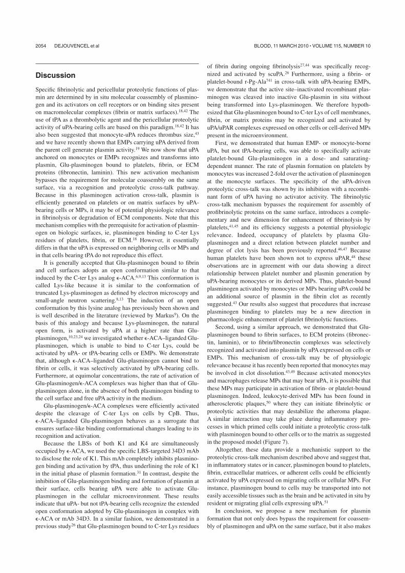
Discussion
Specific fibrinolytic and pericellular proteolytic functions of plas-
min are determined by in situ molecular coassembly of plasmino-
gen and its activators on cell receptors or on binding sites present
on macromolecular complexes (fibrin or matrix surfaces).18,42 The
use of tPA as a thrombolytic agent and the pericellular proteolytic
activity of uPA-bearing cells are based on this paradigm.18,42 It has
also been suggested that monocyte-uPA reduces thrombus size,43
and we have recently shown that EMPs carrying uPA derived from
the parent cell generate plasmin activity.19 We now show that uPA
anchored on monocytes or EMPs recognizes and transforms into
plasmin, Glu-plasminogen bound to platelets, fibrin, or ECM
proteins (fibronectin, laminin). This new activation mechanism
bypasses the requirement for molecular coassembly on the same
surface, via a recognition and proteolytic cross-talk pathway.
Because in this plasminogen activation cross-talk, plasmin is
efficiently generated on platelets or on matrix surfaces by uPA-
bearing cells or MPs, it may be of potential physiologic relevance
in fibrinolysis or degradation of ECM components. Note that this
mechanism complies with the prerequisite for activation of plasmin-
ogen on biologic surfaces, ie, plasminogen binding to C-ter Lys
residues of platelets, fibrin, or ECM.18 However, it essentially
differs in that the uPA is expressed on neighboring cells or MPs and
in that cells bearing tPA do not reproduce this effect.
It is generally accepted that Glu-plasminogen bound to fibrin
and cell surfaces adopts an open conformation similar to that
induced by the C-ter Lys analog e-ACA.6,9,13 This conformation is
called Lys-like because it is similar to the conformation of
truncated Lys-plasminogen as defined by electron microscopy and
small-angle neutron scattering.8,13 The induction of an open
conformation by this lysine analog has previously been shown and
is well described in the literature (reviewed by Markus9). On the
basis of this analogy and because Lys-plasminogen, the natural
open form, is activated by uPA at a higher rate than Glu-
plasminogen,10,23,24 we investigated whether e-ACA–liganded Glu-
plasminogen, which is unable to bind to C-ter Lys, could be
activated by uPA- or tPA-bearing cells or EMPs. We demonstrate
that, although e-ACA–liganded Glu-plasminogen cannot bind to
fibrin or cells, it was selectively activated by uPA-bearing cells.
Furthermore, at equimolar concentrations, the rate of activation of
Glu-plasminogen/e-ACA complexes was higher than that of Glu-
plasminogen alone, in the absence of both plasminogen binding to
the cell surface and free uPA activity in the medium.
Glu-plasminogen/e-ACA complexes were efficiently activated
despite the cleavage of C-ter Lys on cells by CpB. Thus,
e-ACA–liganded Glu-plasminogen behaves as a surrogate that
ensures surface-like binding conformational changes leading to its
recognition and activation.
Because the LBSs of both K1 and K4 are simultaneously
occupied by e-ACA, we used the specific LBS-targeted 34D3 mAb
to disclose the role of K1. This mAb completely inhibits plasmino-
gen binding and activation by tPA, thus underlining the role of K1
in the initial phase of plasmin formation.31 In contrast, despite the
inhibition of Glu-plasminogen binding and formation of plasmin at
their surface, cells bearing uPA were able to activate Glu-
plasminogen in the cellular microenvironment. These results
indicate that uPA- but not tPA-bearing cells recognize the extended
open conformation adopted by Glu-plasminogen in complex with
e-ACA or mAb 34D3. In a similar fashion, we demonstrated in a
previous study26 that Glu-plasminogen bound to C-ter Lys residues
of fibrin during ongoing fibrinolysis27,44 was specifically recog-
nized and activated by scuPA.26 Furthermore, using a fibrin- or
platelet-bound r-Pg-Ala741 in cross-talk with uPA-bearing EMPs,
we demonstrate that the active site–inactivated recombinant plas-
minogen was cleaved into inactive Glu-plasmin in situ without
being transformed into Lys-plasminogen. We therefore hypoth-
esized that Glu-plasminogen bound to C-ter Lys of cell membranes,
fibrin, or matrix proteins may be recognized and activated by
uPA/uPAR complexes expressed on other cells or cell-derived MPs
present in the microenvironment.
First, we demonstrated that human EMP- or monocyte-borne
uPA, but not tPA-bearing cells, was able to specifically activate
platelet-bound Glu-plasminogen in a dose- and saturating-
dependent manner. The rate of plasmin formation on platelets by
monocytes was increased 2-fold over the activation of plasminogen
at the monocyte surfaces. The specificity of the uPA-driven
proteolytic cross-talk was shown by its inhibition with a recombi-
nant form of uPA having no activator activity. The fibrinolytic
cross-talk mechanism bypasses the requirement for assembly of
profibrinolytic proteins on the same surface, introduces a comple-
mentary and new dimension for enhancement of fibrinolysis by
platelets,41,45 and its efficiency suggests a potential physiologic
relevance. Indeed, occupancy of platelets by plasma Glu-
plasminogen and a direct relation between platelet number and
degree of clot lysis has been previously reported.46,47 Because
human platelets have been shown not to express uPAR,48 these
observations are in agreement with our data showing a direct
relationship between platelet number and plasmin generation by
uPA-bearing monocytes or its derived MPs. Thus, platelet-bound
plasminogen activated by monocytes or MPs bearing uPA could be
an additional source of plasmin in the fibrin clot as recently
suggested.43 Our results also suggest that procedures that increase
plasminogen binding to platelets may be a new direction in
pharmacologic enhancement of platelet fibrinolytic functions.
Second, using a similar approach, we demonstrated that Glu-
plasminogen bound to fibrin surfaces, to ECM proteins (fibronec-
tin, laminin), or to fibrin/fibronectin complexes was selectively
recognized and activated into plasmin by uPA expressed on cells or
EMPs. This mechanism of cross-talk may be of physiologic
relevance because it has recently been reported that monocytes may
be involved in clot dissolution.43,49 Because activated monocytes
and macrophages release MPs that may bear uPA, it is possible that
these MPs may participate in activation of fibrin- or platelet-bound
plasminogen. Indeed, leukocyte-derived MPs has been found in
atherosclerotic plaques,50 where they can initiate fibrinolytic or
proteolytic activities that may destabilize the atheroma plaque.
A similar interaction may take place during inflammatory pro-
cesses in which primed cells could initiate a proteolytic cross-talk
with plasminogen bound to other cells or to the matrix as suggested
in the proposed model (Figure 7).
Altogether, these data provide a mechanistic support to the
proteolytic cross-talk mechanism described above and suggest that,
in inflammatory states or in cancer, plasminogen bound to platelets,
fibrin, extracellular matrices, or adherent cells could be efficiently
activated by uPA expressed on migrating cells or cellular MPs. For
instance, plasminogen bound to cells may be transported into not
easily accessible tissues such as the brain and be activated in situ by
resident or migrating glial cells expressing uPA.51
In conclusion, we propose a new mechanism for plasmin
formation that not only does bypass the requirement for coassem-
bly of plasminogen and uPA on the same surface, but it also makes
2054 DEJOUVENCEL et al BLOOD, 11 MARCH 2010 z VOLUME 115, NUMBER 10

Lys-plasminogen dispensable. These heterotypic cell-to-cell (plate-
lets/monocytes; platelets/MPs), cell-to-matrix (THP-1/fibrin), or
MPs-to-matrix (MPs/fibrin) proteolytic cross-talk represents an
alternative pathway for localized plasmin formation that may be
relevant to processes implicating cell migration and MP dissemina-
tion, ie, inflammation or angiogenesis. Finally, our data provide
additional evidence for a novel role of MPs and platelets, as vectors
that generate and propagate plasmin fibrinolytic and/or proteolytic
activity, and could thereby constitute a pharmacologic tool.
Acknowledgments
This work was supported by funds from Inserm and the Lower-
Normandy Regional Council. E.A.-C., L.D., and L.P. are members
of the European Community’s Seventh Framework Program (FP7/
2007-2013) under grant agreement no. 201024. The Center for
Molecular and Vascular Biology is supported by the Excellentie
financiering, KULeuven. T.D. is a recipient of a PhD fellowship
awarded by the Nouvelle Societe Francaise d’Atherosclerose. L.D.
is a recipient of a PhD fellowship from the Education and Research
French Ministry.
Authorship
Contribution: T.D. and L.D. performed research, analyzed data, and
wrote the manuscript; R.L. performed research and participated in
manuscript drafting; L.P. provided experimental support and partici-
pated in manuscript drafting; F.D.-G. analyzed data and partici-
pated in manuscript drafting; H.R.L. contributed analytical tools
and participated in manuscript drafting; and E.A-C. designed the
research, analyzed data, and wrote the manuscript.
Conflict-of-interest disclosure: The authors declare no compet-
ing financial interests.
The current affiliation for T.D. is Inserm, U698, Paris, France.
Correspondence: Eduardo Angles-Cano, Inserm U919 Serine
Proteases and Pathophysiology of the Neurovascular Unit, GIP
Cyceron/Bd Henri Becquerel, 14074-cdx, Caen, France; e-mail:
[email protected] or [email protected].
References
1. Petersen TE, Martzen MR, Ichinose A, Davie EW.
Characterization of the gene for human plasmino-
gen, a key proenzyme in the fibrinolytic system.
J Biol Chem. 1990;265(11):6104-6111.
2. Tulinsky A, Park CH, Mao B, Llinas M. Lysine/
fibrin binding sites of kringles modeled after the
structure of kringle 1 of prothrombin. Proteins.
1988;3(2):85-96.
3. Herren T, Swaisgood C, Plow EF. Regulation of
plasminogen receptors. Front Biosci. 2003;8:d1-
d8.
4. Lucas MA, Fretto LJ, McKee PA. The binding of
human plasminogen to fibrin and fibrinogen.
J Biol Chem. 1983;258(7):4249-4256.
5. Knudsen BS, Silverstein RL, Leung LL, Harpel
PC, Nachman RL. Binding of plasminogen to ex-
tracellular matrix. J Biol Chem. 1986;261(23):
10765-10771.
6. Cockell CS, Marshall JM, Dawson KM,
Cederholm-Williams SA, Ponting CP. Evidence
that the conformation of unliganded human plas-
minogen is maintained via an intramolecular in-
teraction between the lysine-binding site of krin-
gle 5 and the N-terminal peptide. Biochem J.
1998;333(Pt 1):99-105.
7. Tranqui L, Prandini MH, Chapel A. The structure
of plasminogen studied by electron microscopy.
Biol Cell. 1979;34:39-42.
8. Weisel JW, Nagaswami C, Korsholm B,
Petersen LC, Suenson E. Interactions of plas-
minogen with polymerizing fibrin and its deriva-
tives, monitored with a photoaffinity cross-linker
and electron microscopy. J Mol Biol. 1994;
235(3):1117-1135.
9. Markus G. Conformational changes in plasmino-
gen, their effect on activation, and the agents that
modulatie activation rates—a review. Fibrinolysis.
1996;10(2):75-85.
10. Claeys H, Vermylen J. Physico-chemical and
proenzyme properties of NH2-terminal glutamic
acid and NH2-terminal lysine human plasmino-
gen. Influence of 6-aminohexanoic acid. Biochim
Biophys Acta. 1974;342(2):351-359.
11. Thorsen S, Mullertz S. Rate of activation and
electrophoretic mobility of unmodified and
partially degraded plasminogen. Effects of
6-aminohexanoic acid and related compounds.
Scand J Clin Lab Invest. 1974;34(2):167-
176.
12. Markus G, Evers JL, Hobika GH. Comparison of
some properties of native (Glu) and modified
(Lys) human plasminogen. J Biol Chem. 1978;
253(3):733-739.
13. Mangel WF, Lin BH, Ramakrishnan V. Character-
ization of an extremely large, ligand-induced con-
formational change in plasminogen. Science.
1990;248(4951):69-73.
14. Sjoholm I. Studies on the conformational changes
of plasminogen induced during activation to plas-
min and by 6-aminohexanoic acid. Eur J Bio-
chem. 1973;39(2):471-479.
15. Sappino AP, Madani R, Huarte J, et al. Extracellu-
lar proteolysis in the adult murine brain. J Clin
Invest. 1993;92(2):679-685.
16. Angles-Cano E, Balaton A, Le Bonniec B,
Figure 7. Model of fibrinolytic and proteolytic cross-talk. In
inflammatory processes of the vascular wall, activation of coagu-
lation leads to fibrin deposits. Within this setting and according to
data presented here, the fibrinolytic cross-talk mechanism could
be an intermediary pathway for fibrinolysis and pericellular
proteolysis. Fibrinolytic effects (top): plasminogen (Pg) bound to
fibrin or to platelets could be activated by either monocyte- or
MP-borne uPA(EMPor monocyte-derivedmicroparticle [MoMP]).
In ECM remodeling in the vascular wall (bottom), plasminogen
bound to matrix components could be activated by MP-borne
uPA (EMP or MoMP) or by macrophage-borne uPA.
FIBRINOLYTIC CROSS-TALK 2055BLOOD, 11 MARCH 2010 z VOLUME 115, NUMBER 10
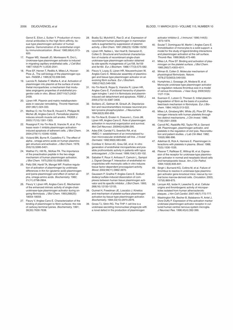
Genot E, Elion J, Sultan Y. Production of mono-
clonal antibodies to the high fibrin-affinity, tis-
sue-type plasminogen activator of human
plasma. Demonstration of its endothelial origin
by immunolocalization. Blood. 1985;66(4):913-
920.
17. Pepper MS, Vassalli JD, Montesano R, Orci L.
Urokinase-type plasminogen activator is induced
in migrating capillary endothelial cells. J Cell Biol.
1987;105(6 Pt 1):2535-2541.
18. Plow EF, Herren T, Redlitz A, Miles LA, Hoover-
Plow JL. The cell biology of the plasminogen sys-
tem. FASEB J. 1995;9(10):939-945.
19. Lacroix R, Sabatier F, Mialhe A, et al. Activation of
plasminogen into plasmin at the surface of endo-
thelial microparticles: a mechanism that modu-
lates angiogenic properties of endothelial pro-
genitor cells in vitro. Blood. 2007110(7):2432-
2439.
20. Lijnen HR. Plasmin and matrix metalloprotein-
ases in vascular remodeling. Thromb Haemost.
2001;86(1):324-333.
21. Meilhac O, Ho-Tin-Noe B, Houard X, Philippe M,
Michel JB, Angles-Cano E. Pericellular plasmin
induces smooth muscle cell anoikis. FASEB J.
2003;17(10):1301-1303.
22. Rossignol P, Ho-Tin-Noe B, Vranckx R, et al. Pro-
tease nexin-1 inhibits plasminogen activation-
induced apoptosis of adherent cells. J Biol Chem.
2004;279(11):10346-10356.
23. Violand BN, Byrne R, Castellino FJ. The effect of
alpha-, omega-amino acids on human plasmino-
gen structure and activation. J Biol Chem. 1978;
253(15):5395-5401.
24. Walther PJ, Hill RL, McKee PA. The importance
of the preactivation peptide in the two-stage
mechanism of human plasminogen activation.
J Biol Chem. 1975;250(15):5926-5933.
25. Peltz SW, Hardt TA, Mangel WF. Positive regula-
tion of activation of plasminogen by urokinase:
differences in Km for (glutamic acid)-plasminogen
and lysine-plasminogen and effect of certain al-
pha, omega-amino acids. Biochemistry. 1982;
21(11):2798-2804.
26. Fleury V, Lijnen HR, Angles-Cano E. Mechanism
of the enhanced intrinsic activity of single-chain
urokinase-type plasminogen activator during on-
going fibrinolysis. J Biol Chem. 1993;268(25):
18554-18559.
27. Fleury V, Angles-Cano E. Characterization of the
binding of plasminogen to fibrin surfaces: the role
of carboxy-terminal lysines. Biochemistry. 1991;
30(30):7630-7638.
28. Busby SJ, Mulvihill E, Rao D, et al. Expression of
recombinant human plasminogen in mammalian
cells is augmented by suppression of plasmin
activity. J Biol Chem. 1991;266(23):15286-15292.
29. Lijnen HR, Nelles L, Van Hoef B, Demarsin E,
Collen D. Structural and functional characteriza-
tion of mutants of recombinant single-chain
urokinase-type plasminogen activator obtained
by site-specific mutagenesis of Lys158, Ile159
and Ile160. Eur J Biochem. 1988;177(3):575-582.
30. Fleury V, Loyau S, Lijnen HR, Nieuwenhuizen W,
Angles-Cano E. Molecular assembly of plasmino-
gen and tissue-type plasminogen activator on an
evolving fibrin surface. Eur J Biochem.
1993;216(2):549-556.
31. Ho-Tin-Noe B, Rojas G, Vranckx R, Lijnen HR,
Angles-Cano E. Functional hierarchy of plasmin-
ogen kringles 1 and 4 in fibrinolysis and plasmin-
induced cell detachment and apoptosis. FEBS J.
2005;272(13):3387-3400.
32. Siciliano JC, Gelman M, Girault JA. Depolariza-
tion and neurotransmitters increase neuronal pro-
tein tyrosine phosphorylation. J Neurochem.
1994;62(3):950-959.
33. Ho-Tin-Noe B, Enslen H, Doeuvre L, Corsi JM,
Lijnen HR, Angles-Cano E. Role of plasminogen
activation in neuronal organization and survival.
Mol Cell Neurosci. 200942(4)288-295.
34. Ades EW, Candal FJ, Swerlick RA, et al.
HMEC-1: establishment of an immortalized hu-
man microvascular endothelial cell line. J Invest
Dermatol. 1992;99(6):683-690.
35. Combes V, Simon AC, Grau GE, et al. In vitro
generation of endothelial microparticles and pos-
sible prothrombotic activity in patients with lupus
anticoagulant. J Clin Invest. 1999;104(1):93-102.
36. Sabatier F, Roux V, Anfosso F, Camoin L, Sampol
J, Dignat-George F. Interaction of endothelial mi-
croparticles with monocytic cells in vitro induces
tissue factor-dependent procoagulant activity.
Blood. 2002;99(11):3962-3970.
37. Gaussem P, Grailhe P, Angles-Cano E. Sodium
dodecyl sulfate-induced dissociation of com-
plexes between human tissue plasminogen acti-
vator and its specific inhibitor. J Biol Chem. 1993;
268(16):12150-12155.
38. Ouimet H, Freedman JE, Loscalzo J. Kinetics
and mechanism of platelet-surface plasminogen
activation by tissue-type plasminogen activator.
Biochemistry. 1994;33(10):2970-2976.
39. Gross TJ, Sitrin RG. The THP-1 cell line is a
urokinase-secreting mononuclear phagocyte with
a novel defect in the production of plasminogen
activator inhibitor-2. J Immunol. 1990;144(5):
1873-1879.
40. Soulat T, Dominguez M, Martin I, Angles-Cano E.
Immobilisation of monocytes to a solid support: a
model for the study of ligand-binding interactions
and plasminogen activation at the cell surface.
Thromb Res. 1999;96(6):473-480.
41. Miles LA, Plow EF. Binding and activation of plas-
minogen on the platelet surface. J Biol Chem.
1985;260(7):4303-4311.
42. Wiman B, Collen D. Molecular mechanism of
physiological fibrinolysis. Nature.
1978;272(5653):549-550.
43. Humphries J, Gossage JA, Modarai B, et al.
Monocyte urokinase-type plasminogen activator
up-regulation reduces thrombus size in a model
of venous thrombosis. J Vasc Surg. 2009;50(5):
1127-1134.
44. Suenson E, Lutzen O, Thorsen S. Initial plasmin-
degradation of fibrin as the basis of a positive
feed-back mechanism in fibrinolysis. Eur J Bio-
chem. 1984;140(3):513-522.
45. Miles LA, Ginsberg MH, White JG, Plow EF. Plas-
minogen interacts with human platelets through
two distinct mechanisms. J Clin Invest. 1986;
77(6):2001-2009.
46. Carroll RC, Radcliffe RD, Taylor FB Jr, Gerrard
JM. Plasminogen, plasminogen activator, and
platelets in the regulation of clot lysis. Reconstitu-
tion and patient studies. J Lab Clin Med. 1982;
100(6):986-996.
47. Adelman B, Rizk A, Hanners E. Plasminogen in-
teractions with platelets in plasma. Blood. 1988;
72(5):1530-1535.
48. Plesner T, Ralfkiaer E, Wittrup M, et al. Expres-
sion of the receptor for urokinase-type plasmino-
gen activator in normal and neoplastic blood cells
and hematopoietic tissue. Am J Clin Pathol.
1994;102(6):835-841.
49. Singh I, Burnand KG, Collins M, et al. Failure of
thrombus to resolve in urokinase-type plasmino-
gen activator gene-knockout mice: rescue by nor-
mal bone marrow-derived cells. Circulation. 2003;
107(6):869-875.
50. Leroyer AS, Isobe H, Leseche G, et al. Cellular
origins and thrombogenic activity of micropar-
ticles isolated from human atherosclerotic
plaques. J Am Coll Cardiol. 2007;49(7):772-777.
51. Washington RA, Becher B, Balabanov R, Antel J,
Dore-Duffy P. Expression of the activation marker
urokinase plasminogen-activator receptor in cul-
tured human central nervous system microglia.
J Neurosci Res. 1996;45(4):392-399.
2056 DEJOUVENCEL et al BLOOD, 11 MARCH 2010 z VOLUME 115, NUMBER 10
��
�
ARTICLE 3
Leukocyte- and endothelial-derived microparticles: a circulating source for
Fibrinolysis.
Romaric Lacroix1, Laurent Plawinski
2,3, Stephane Robert
1, Loïc Doeuvre
2,4, Florence
Sabatier1, Francine Anfosso
1, Aurelie S. Leroyer
5, Chantal M. Boulanger
5,6, Eduardo Angles-
Cano2,3
and Françoise Dignat-George1
1Inserm, UMR-S608, Université Aix-Marseille, Marseille, France.
2Inserm, U919,
3CNRS,
UMR6232, 4Université de Caen, Caen, France.
5Inserm, U970, Paris Cardiovascular Research
Center � PARCC, Paris, France. 6Université Paris Descartes, UMR-S970, Paris, France
Blood. Article en révision.
Nos travaux précédents ont permis d�assigner une nouvelle fonction fibrinolytique
aux MP dérivées d�HMEC-1. Nous avons montré l�existence d�un mécanisme original par
lequel ces MPE constituent une surface catalytique efficace pour l�activation du
plasminogène par l�u-PA fixée sur son récepteur u-PAR. Nous avons montré que ces MPE
participent à un mécanisme de cross-talk entre surface des MP et surfaces cellulaires, fibrine
ou matrice extracellulaire. Cependant la possibilité que des MP dérivées de cellules
endothéliales primaires humaines ou des cellules hématopoïétiques du sang circulant puissent
également servir de support à une activité protéolytique conduisant à la formation de
plasmine in vivo n�a pas encore été explorée.
Nous avons donc formulé l�hypothèse que chez l�homme, les microparticules
présentent dans la circulation sanguine ou dérivées de cellules endothéliales primaires
pouvaient constituer un support efficace pour la formation de plasmine in vivo.
Dans ce travail, nous avons donc étudié la capacité de génération de plasmine de MP
isolées de plasma de patients en situations pathologiques ou purifiées à partir des différentes
sous-populations de cellules circulantes et de cellules endothéliales primaires.
Chez des patients souffrant de pathologies caractérisées par une vésiculation
endothéliale accrue, nous avons mis en évidence une génération de plasmine détectable à

��
�
partir des MP circulantes. Nous avons montré que la formation de plasmine par les MP
circulantes de patients présentant une athérosclérose ou une pathologie auto-immune comme
le lupus érythémateux disséminé ou le PTT, est plus importante que pour les contrôles sains.
L�identification des activateurs responsables de cette activité plasminogénolytique a révélé
que non seulement l�u-PA mais aussi le t-PA sont impliqués, à des niveaux variables en
fonction des patients et de l�existence de complexes entre le t-PA et son inhibiteur (PAI-1).
Nous avons démontré que l�activité de génération de plasmine était portée par les MP
dérivées de cellules endothéliales (MPE) et de leucocytes (MPL) mais était absente des MP
d�origine plaquettaire ou érythrocytaire. Les MPL expriment de l�uPA et son récepteur
l�uPAR tandis que les MPE expriment du t-PA et des complexes t-PA/PAI-1.
En conclusion, cette étude montre que dans la circulation sanguine, ce sont les sous
populations minoritaires de MP, les MP d�origine endothéliale et leucocytaire, qui supportent
l�activité fibrinolytique. Cette activité est modulée dans des pathologies cardiovasculaires et
auto-immunes.
1
Leukocyte- and endothelial-derived microparticles: a circulating source for
fibrinolysis
Short title: Fibrinolytic microparticles
Romaric Lacroix1*
, Laurent Plawinski2,3*
, Stephane Robert1, Loïc Doeuvre
2,4, Florence
Sabatier1, Francine Anfosso
1, Aurelie S. Leroyer
5, Chantal M. Boulanger
5,6, Eduardo Angles-
Cano2,3**
and Françoise Dignat-George1**
1Inserm, UMR-S608, Université Aix-Marseille, Marseille, France.
2Inserm, U919,
3CNRS,
UMR6232, 4Université de Caen, Caen, France.
5Inserm, U970, Paris Cardiovascular Research
Center – PARCC, Paris, France. 6Université Paris Descartes, UMR-S970, Paris, France
* These authors (alphabetical order) contributed equally to this work.
** Françoise Dignat-George ([email protected]) and **Eduardo Angles-Cano
([email protected]) are corresponding authors and share senior authorship.
Françoise Dignat-George, Inserm UMR 608, UFR de Pharmacie, 27 Bd Jean Moulin, 13385-
cdx Marseille 5, France, Tel: 33 4 91 83 56 00 Fax: 33 4 91 83 56 02
Eduardo Anglés-Cano, Inserm U919, GIP Cyceron, Bd Henri Becquerel, 14074, Caen,
France. Tel 33 2 31 47 01 64 Fax 33 2 31 47 02 22
Word counts: Total text: 1287. Abstract: 146
Number of figures: 2
Number of references: 18
Scientific category: Hemostasis, Thrombosis, and Vascular Biology
2
Abstract
We recently assigned a fibrinolytic function to endothelial-derived microparticles (MPs). We
report now that plasmin generation is also detectable on circulating MPs from patients with
diseases featuring increased vesiculation. For instance, plasmin formation on circulating MPs
from patients with atherosclerotic or autoimmune disease was significantly higher than in
healthy controls. Moreover, circulating MPs from thrombotic thrombocytopenic purpura
patients generate a range of plasmin activity and express a variable content in either
urokinase- (uPA) or tissue-type plasminogen activator (tPA) and complexes between tPA and
its inhibitor (PAI-1). We further demonstrate that plasmin was generated on endothelial- and
leukocyte-derived MPs, but was absent on MPs of platelet or erythrocyte origin. Leukocyte-
derived MPs express uPA and its receptor uPAR whereas endothelial-derived MPs expressed
tPA and tPA-PAI-1 complexes. In conclusion, our study indicates that endothelial- and
leukocyte-derived MPs support fibrinolytic activity in the circulation that is modulated in
pathological situations.
Key words: fibrinolysis; microparticle, plasmin(ogen), uPA, tPA, TTP

3
Introduction
Microparticles (MPs) are small vesicles resulting from membrane blebbing of most
activated or apoptotic cells.1 MPs detectable in the circulation are derived from blood and
vascular cells. Over the past decade, they have been accepted as diagnostic and prognostic
biomarkers in cardiovascular disorders. Accumulating evidence indicates that beyond their
biomarker quality, MPs also convey various bioactive effectors implicating them in activation
of coagulation, inflammation, vascular remodelling and angiogenesis.2-6
We recently assigned
a new fibrinolytic function to MPs derived from the human microvascular endothelial cell line
HMEC-1. We provided evidence for a novel mechanism by which these endothelial MPs
(EMPs) constitute an efficient catalytic surface for the activation of plasminogen by the
urokinase-type plasminogen activator (uPA) anchored to its receptor uPAR.7 However, the
possibility that MPs derived from native endothelial and blood cells also serve as a template
for plasmin formation and proteolytic activity in vivo, remain unsolved questions. We
therefore sought to investigate whether MPs from pathological situations and MPs purified
from different blood cell subpopulations and primary endothelial cells could be the source of
fibrinolysis in the circulation.
Materials and Methods
Detailed materials and methods are described in a supplemental document (available online).
Briefly, MPs from patients with thrombotic thrombocytopenic purpura (TTP, n=11) were
isolated and characterized by zymography, ELISA, flow cytometry and photometric assays
for plasminogen activators and plasmin generation activity as described previously.7 We also
performed MP analyses in patients with systemic lupus erythematosus (n=12) or with
atherosclerosis (n=16), and in healthy subjects (n=36). Furthermore, MPs derived from human

4
circulating and primary endothelial cells were generated and evaluated using the previously
quoted techniques.
Results and discussion
1. Circulating microparticles support in vivo a fibrinolytic activity that is modulated in
pathological settings.
To investigate whether human MPs are involved in plasmin generation in vivo, we
studied MPs isolated from patients with TTP a disease featuring increased vesiculation.8,9
A
majority of the isolated MP samples (8 out of 11) generate plasmin activity upon incubation
with 1 µM plasminogen. Figure 1A shows plasmin generation (1.6-5.9 mOD/minute) in
representative samples (2.105 MPs/well) that display a variable content in plasminogen
activator activity. Platelet- (PMPs) or erythrocyte-derived MPs may not contribute to this
activity as samples depleted in these MPs displayed a slight increase rather than a decrease in
plasmin formation (figure1B). The observed increase in plasmin formation is most probably
related to depletion in active PAI-1/vitronectin complexes present in PMPs.10
The inhibition
of plasmin generation by ε-ACA was consistent with a lysine-dependent mechanism for
plasminogen binding and activation (figure 1C).
The plasminogen activator activity in distinct samples was identified either as uPA or as
tPA in fibrin-agarose gels as indicated by the position of their fibrinolytic band relative to
known markers (figure 1D, upper panel). For instance, the single fibrinolytic band apparent in
sample TTP C and the corresponding uPA standard were absent in a fibrin-agarose gel
containing anti-uPA antibodies (figure 1D, lower panel). This antibody also inhibited plasmin
formation by MPs from TTP 1 in a chromogenic assay (figure 1C). PTT sample D in figure
1D (representative of 3 samples) contained tPA and a main fibrinolytic band of high
molecular mass corresponding to the position of tPA in complex with its inhibitor. This

5
sample also contained a minor uPA fibrinolytic band that was blocked in the presence of anti-
uPA antibodies whereas tPA and its complexes retained their activity (figure 1D, lower
panel). No direct plasmin fibrinolytic band could be observed in the MP samples.
We then investigated whether this plasminogen activator activity differs between
healthy subjects and patients. Previously documented pathological situations associated with
increased levels of MPs namely atherosclerotic complications (n=16) or systemic lupus
erythematosus (n=22) were selected.11,12
As shown in figure 1E, the plasminogen activator
activity of MPs was significantly higher in the two groups of patients compared to healthy
controls (1.51 ± 1.43 mOD/minute and 1.62 ± 1.92 mOD/minute versus 0.57 ± 0.24
mOD/minute, p<0.001).
These results provide the first evidence that human circulating MPs isolated from the
plasma compartment, convey plasminogen activators, activate membrane-bound plasminogen
and may generate plasmin activity in vivo. This MP activity suggests a compensatory
fibrinolytic response to coagulation that maintains vessel patency. Its relevance in local or
systemic fibrinolysis remains to be elucidated. As a first step in this endeavor, we determined
the cell origin of the plasminogen activator activity.
2. The fibrinolytic activity is borne by microparticles originating from endothelial cells
and leukocytes.
To determine which subpopulations of MPs activate plasminogen into plasmin, we
generated MPs from both primary human endothelial cells and circulating blood cell
subpopulations. Plasmin generation (figure 2A) was detected on endothelial MPs (EMPs)
with variations according to their anatomical origin. The highest plasminogen activator
activity was expressed by EMPs of renal artery origin (1.2±0.2 mOD/minute) and the lowest
by EMPs of dermal microvascular origin (0.03±0.01 mOD/minute). By zymography, two
fibrinolytic bands resistant to anti-uPA antibodies were observed in lysates of EMPs (figure

6
2B). One band corresponded to the migration of purified tPA whereas the other, of high
molecular mass and higher intensity than the fibrinolytic activity of free tPA, corresponded to
the migration of tPA in complex with its inhibitor (Cs in figure 2B), in agreement with a
previous report.13
PAI-1 and tPA antigens were indeed detected in lysates of EMPs with a
specific ELISA (figure 2C). The presence of tPA was further confirmed by flow cytometry
(figure 2D). In contrast, uPA activity and antigen (figure 2E) were undetectable.
Plasminogen activator activity was also detected on leukocyte MPs (LMPs) of monocyte,
lymphocyte and neutrophil origin but was absent in platelet- or erythrocyte-derived MPs
(figure 2A). The presence of plasminogen activator activity on LMPs was further confirmed
by a lytic band on zymography corresponding to the position of uPA (figure 2B) that was
absent in the presence of antibodies to uPA (not shown). The expression of uPA and its
receptor uPAR on the surface of LMPs was evidenced by flow cytometry (figure 2E). In
parallel experiment, tPA antigen was undetectable on LMPs, PMPs or erythrocyte-derived
MPs (figure 2D). Platelet and erythrocyte MPs (up to 5x10⁶ MPs per well) failed to generated
plasmin (figure 2A) or a fibrinolytic band (2C). Similar negative results on plasminogen
activator activity were obtained when PMPs were generated using different platelet agonist
combinations (collagen, thrombin and A23187) (not shown).
Collectively, given that both platelet and erythrocyte MP populations account for the
bulk of circulating MPs,14
this is the first demonstration that plasminogen activator activity is
borne by a minor fraction of MPs circulating in human blood i.e. endothelial and leukocyte
MPs expressing either tPA or uPA/uPAR. This selectivity contrast with the well-described
procoagulant potential common to all MPs.15
In summary, our study indicates that the plasminogen activator activity of circulating
human MPs is supported by subpopulations of leukocyte or endothelial origin. This
fibrinolytic activity was found increased in various pathological conditions, most probably as
a compensatory response to coagulation. In contrast, we did not detect plasmin generation on

7
erythrocyte and platelet MPs. Accordingly, plasminogen activators have not been described
on red blood cells and their expression by human platelets is not clearly demonstrated except
for the ectopic production of uPA in the platelet Quebec syndrome.16
It is therefore possible
that platelet-derived MPs may, in some cases, develop plasminogen activator activity. Indeed,
since platelets bind plasminogen,17-19
PMPs may be a source of substrate for enhanced
fibrinolysis by scuPA20
via a fibrinolytic cross-talk mechanism demonstrated recently.21
3. Conclusion
We provide the first evidence that surface plasminogen activation is a functional
feature of plasma MPs that significantly differ between healthy subjects and pathological
samples. This activity is specifically supported by endothelial and leukocyte subpopulations
of MPs, the quantitatively minor contributors to the pool of circulating MPs. Although the
clinical signification of this activity remains to be defined, its measurement may be indicative
of pathological processes involving endothelial- and leukocyte-derived MP proteolytic
responses.
References
1. Piccin A, Murphy WG, Smith OP. Circulating microparticles: pathophysiology and
clinical implications. Blood Rev. 2007;21:157-171. 2. Sabatier F, Darmon P, Hugel B, et al. Type 1 and type 2 diabetic patients display
different patterns of cellular microparticles. Diabetes. 2002;51:2840-2845.
3. Taraboletti G, D'Ascenzo S, Borsotti P, Giavazzi R, Pavan A, Dolo V. Shedding of the matrix metalloproteinases MMP-2, MMP-9, and MT1-MMP as membrane vesicle-associated
components by endothelial cells. Am J Pathol. 2002;160:673-680.
4. Boulanger CM, Scoazec A, Ebrahimian T, et al. Circulating microparticles from
patients with myocardial infarction cause endothelial dysfunction. Circulation.
2001;104:2649-2652.
5. MacKenzie A, Wilson HL, Kiss-Toth E, Dower SK, North RA, Surprenant A. Rapid
secretion of interleukin-1beta by microvesicle shedding. Immunity. 2001;15:825-835.
6. Mause SF, von Hundelshausen P, Zernecke A, Koenen RR, Weber C. Platelet
microparticles: a transcellular delivery system for RANTES promoting monocyte recruitment on endothelium. Arterioscler Thromb Vasc Biol. 2005;25:1512-1518.
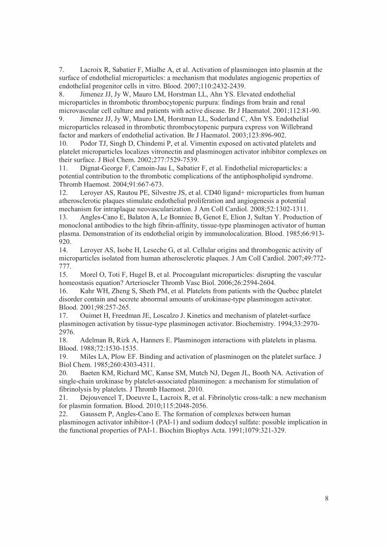
8
7. Lacroix R, Sabatier F, Mialhe A, et al. Activation of plasminogen into plasmin at the surface of endothelial microparticles: a mechanism that modulates angiogenic properties of
endothelial progenitor cells in vitro. Blood. 2007;110:2432-2439.
8. Jimenez JJ, Jy W, Mauro LM, Horstman LL, Ahn YS. Elevated endothelial microparticles in thrombotic thrombocytopenic purpura: findings from brain and renal
microvascular cell culture and patients with active disease. Br J Haematol. 2001;112:81-90.
9. Jimenez JJ, Jy W, Mauro LM, Horstman LL, Soderland C, Ahn YS. Endothelial
microparticles released in thrombotic thrombocytopenic purpura express von Willebrand
factor and markers of endothelial activation. Br J Haematol. 2003;123:896-902.
10. Podor TJ, Singh D, Chindemi P, et al. Vimentin exposed on activated platelets and
platelet microparticles localizes vitronectin and plasminogen activator inhibitor complexes on
their surface. J Biol Chem. 2002;277:7529-7539.
11. Dignat-George F, Camoin-Jau L, Sabatier F, et al. Endothelial microparticles: a potential contribution to the thrombotic complications of the antiphospholipid syndrome.
Thromb Haemost. 2004;91:667-673.
12. Leroyer AS, Rautou PE, Silvestre JS, et al. CD40 ligand+ microparticles from human atherosclerotic plaques stimulate endothelial proliferation and angiogenesis a potential
mechanism for intraplaque neovascularization. J Am Coll Cardiol. 2008;52:1302-1311. 13. Angles-Cano E, Balaton A, Le Bonniec B, Genot E, Elion J, Sultan Y. Production of
monoclonal antibodies to the high fibrin-affinity, tissue-type plasminogen activator of human
plasma. Demonstration of its endothelial origin by immunolocalization. Blood. 1985;66:913-920.
14. Leroyer AS, Isobe H, Leseche G, et al. Cellular origins and thrombogenic activity of
microparticles isolated from human atherosclerotic plaques. J Am Coll Cardiol. 2007;49:772-777.
15. Morel O, Toti F, Hugel B, et al. Procoagulant microparticles: disrupting the vascular
homeostasis equation? Arterioscler Thromb Vasc Biol. 2006;26:2594-2604.
16. Kahr WH, Zheng S, Sheth PM, et al. Platelets from patients with the Quebec platelet
disorder contain and secrete abnormal amounts of urokinase-type plasminogen activator.
Blood. 2001;98:257-265.
17. Ouimet H, Freedman JE, Loscalzo J. Kinetics and mechanism of platelet-surface
plasminogen activation by tissue-type plasminogen activator. Biochemistry. 1994;33:2970-
2976. 18. Adelman B, Rizk A, Hanners E. Plasminogen interactions with platelets in plasma.
Blood. 1988;72:1530-1535.
19. Miles LA, Plow EF. Binding and activation of plasminogen on the platelet surface. J Biol Chem. 1985;260:4303-4311.
20. Baeten KM, Richard MC, Kanse SM, Mutch NJ, Degen JL, Booth NA. Activation of
single-chain urokinase by platelet-associated plasminogen: a mechanism for stimulation of fibrinolysis by platelets. J Thromb Haemost. 2010.
21. Dejouvencel T, Doeuvre L, Lacroix R, et al. Fibrinolytic cross-talk: a new mechanism
for plasmin formation. Blood. 2010;115:2048-2056.
22. Gaussem P, Angles-Cano E. The formation of complexes between human
plasminogen activator inhibitor-1 (PAI-1) and sodium dodecyl sulfate: possible implication in
the functional properties of PAI-1. Biochim Biophys Acta. 1991;1079:321-329.

9
Figure 1. Fibrinolytic activity of microparticles isolated from patients with thrombotic
thrombocytopenic purpura (TTP), systemic lupus erythematosus (SLE) and
cardiovascular disease.
Microparticles (2.105
per well) were isolated as indicated in Methods (supplemental file on
line) and then incubated with plasminogen (1µM) in the presence of a plasmin-selective
chromogenic substrate (CBS0065, 0.75mM). Bars represent the amount of plasmin formed
(mOD/minute) of representative samples. Results are the mean ± SD of a duplicate
experiment. A. Plasmin generated by microparticles from TTP patients. B. Plasmin generation
by TTP microparticles depleted (>90%) in erythrocyte- and platelet-derived microparticles. A
volume of 25µL of total (gray bars) or depleted (dark gray bars) microparticle samples tested.
C. Effect of inhibitors (ε -amino-caproïc acid, ε-ACA and anti-uPA antibody, α-uPA) and
IgG, a control immunoglobulin, on the generation of plasmin by TTP 1 microparticles. D.
Fibrin-zymography analysis of TTP microparticles. Proteins in microparticle lysates were
separated by SDS-PAGE and their fibrinolytic activity revealed on fibrin-agarose gels.
Reference standards are purified tPA, plasmin and uPA. The fibrinolytic activity was tested in
the absence (upper panel) or presence (lower panel) of 10 µg/mL of a polyclonal antibody
against uPA. The high molecular mass fibrinolytic band observed in the tPA lane correspond
to tPA dimers.22
E. Plasmin generated by microparticle samples of patients with
cardiovascular disease and SLE. Experiments were performed as indicated in A. The mean of
samples for each category is indicated by the dotted lines ( 1.51 ± 1.43 mOD/minute and
□1.62 ± 1.92 mOD/minute versus control 0.57 ± 0.24 mOD/minute, *p<0.001)

10
Figure 2. Fibrinolytic activity of microparticles derived from human circulating blood
cells and primary endothelial cells.
Circulating blood microparticles and endothelial cell-derived microparticles were obtained
and isolated as indicated in Methods (supplemental file on line). Endothelial cell-derived
microparticles were obtained from primary cultures of coronary artery (CA), pulmonary
artery (PA), renal artery (RA) and dermal microvascular (DMV) endothelial cells. Circulating
blood cells-derived microparticles were obtained from isolated polymorphonuclear cells
(PMN), monocytes (MC), lymphocytes (LC), red cells (RC) and platelets (PLA). Bars
represent the amount of plasmin formed (mOD/minute). Results are the mean ± SD of
duplicate experiments. MFI: mean fluorescence intensity.
A. Plasmin generation by microparticles (2.105
per well) was tested as indicated in figure 1.
B. Fibrin-zymography analysis of endothelial and circulating microparticles. Zymography
was performed as indicated in figure 1D. Reference standards are purified tPA, plasmin, uPA
and tPA in complex with PAI-1 (Cs). C. PAI-1 and tPA concentrations by ELISA for 106
endothelial cell-derived microparticle lysates per well. Results are the mean ± SD of duplicate
experiments. D. tPA expression at the surface of endothelial and circulating microparticles by
flow cytometry. Graphs represent overlays of representative fluorescence histograms obtained
with isotype controls (grey) and tPA antibody (black) on annexin V+ gated events. E. uPAR
and uPA expression at the surface of endothelial and circulating microparticles by flow
cytometry. Graphs represent overlays of representative fluorescence histograms obtained with
isotype controls (grey) and uPAR or uPA antibodies (black) on annexin V+ gated events.

FIGURE 1
A
D E
B
C
0
1
2
3
4
5
6
1 2 3 4 5
Pla
sm
in g
en
era
tio
n (
mO
D/m
in)
0
0.5
1
1.5
2
2.5
TTP A TTP B
Pla
sm
in (
mO
D/m
in)
0
1
2
3
4
5
TTP 1 α-uPA ε-ACA IgG
Pla
sm
in (
mO
D/m
in)
0
2
4
6
8
Cardiovascular SLE Healthy control
Pla
sm
in g
en
era
tio
n (
mO
D/m
in)
**
anti-uPA tPA Pln uPA TTPC TTPD
-
+

RAMVD PACA
t-PA (MFI)
Even
ts
PLA Leukocyte RC
Even
ts
A
B
C
D
0
0.5
1
1.5
2
2.5
CA PA RA DMV PMN MC LC RC PLA
Pla
sm
in g
en
era
tio
n (
mO
D/m
in)
0
5
10
15
20
25
0
5
10
15
20
25
DMV CA RA PAP
AI-
1 (
ng
/10
6 M
P) tP
A (n
g/1
06 M
P)

PL
A
LCRC
Even
ts
MC PMN
RAMVD PACA
Even
ts
uPAR (MFI)
PL
A
LCRC
Even
ts
M PMN
RAMVD PACA
Even
ts
uPA (MFI)
E
FIGURE 2

Supplemental Materials and Methods
Isolation of microparticles from human plasma
Plasma was separated (1500g, 15 minutes) from blood collected in 0.119 M sodium
citrate from healthy volunteers (n=36) and from patients with autoimmune diseases (systemic
lupus erythematosus, SLE, with, n=10, or without, n=12, antiphospholipid syndrome, APLS,
according to the Sapporo criteria) or with atherosclerotic complications (n=16). MPs were
also purified from 36 mL of citrated plasma obtained by plasmapheresis from patients (n=11)
suffering from acute thrombotic thrombocytopenic purpura (TTP). Informed consent was
obtained from all patients.
MPs were isolated by sequential centrifugation of platelet-poor plasma, first at
13000g for 2 minutes to remove residual platelets. The platelet-free plasma was then
centrifuged at 20000g for 90 minutes at 4°C. Pelleted microparticles were washed twice
(20000g for 90 minutes at 4°C) and re-suspended in phosphate-buffered saline (PBS).
In some experiment circulating MPs were depleted in erythrocyte- and platelet-derived
microparticles by magnetic immuno-separation with beads coated with CD41 and CD235a
antibodies. Depletion in MPs was checked by FCM superior at 90%. Control experiments
were performed in parallel with beads coated with irrelevant antibodies.
Generation of microparticles from blood cells
To investigate which subtype of circulating MPs may support plasminogen activation
activity, MPs were generated in vitro from different blood cell types. Whole blood from
healthy volunteers or patients who had not taken anti-platelet medication for at least 2 weeks,
was collected into 0.119 sodium citrated tubes.

Human platelets prepared as already described1 were incubated without stirring for 15
minutes at 37°C with 1 NIH U/mL of thrombin and/or 1 µM of A23187 (Sigma, Saint Louis,
MO), and/or 10 µg/mL of collagen (Stago, Asnière, France,). Platelets were subsequently
separated by centrifugation at 20000g for 2 minutes, and MPs isolated and washed as
described above.
Erythrocyte-derived MPs were generated from human red blood cells as previously
described by Salzer et al. with minor modifications2. Erythrocytes pelleted at 200 g for 10
minutes and subsequently washed in sodium chloride 0.9%, were resuspended in 9 volumes of
PBS containing 1 mM CaCl2 and 5 µM ionophore A23187 and incubated at 37°C for 30
minutes. Erythrocytes were subsequently separated by centrifugation at 20000g for 2 minutes,
and MPs isolated and washed as described above.
Leukocyte-derived microparticles were prepared as previously described with
some modifications 3. Monocytes were isolated for PBMC (Lymphocytes separation medium
1077) by CD14+ immuno-magnetic separation on MS columns (Miltenyi Biotec, Bergish
Gladbach, Germany). Purity superior at 95 % was checked by flow cytometry. Monocytes
were stimulated by LPS (1�g/ml) overnight. Vesiculation was evaluated by FCM by AnnV-
FITC/CD11b staining. Neutrophils were purified from citrated buffy coat and were stimulated
by N-formylmethionyl-leucyl-phenylalanine (fMPL), 1 �M during 2h at 37ºC. Lymphocytes
were purified from PBMC (Lymphocytes separation medium 1077) and were stimulated by
PAF/PMA (Platelet activation factor/ phorbol 12-myristate 13-acetate) 500nM/50nM during
2h at 37ºC. MPs were isolated and washed as described above.

Generation and harvesting of microparticles from endothelial cells
Several subtypes of human endothelial cells originating form distinct vascular beds
were purchased from Clonetics (Grand Island, NY) and cultured into 0.2 % gelatin coated
flasks in EGM2-MV medium: renal artery endothelial cells (RAEC) used at passage 8,
coronary artery endothelial cells (HCAEC) used at passage 7, dermal human microvascular
vein endothelial cells (HMVEC-d) and human pulmonary artery endothelial cells (HPAEC)
used at passage 6. Endothelial microparticles were purified from culture medium conditioned
by sub-confluent endothelial cells stimulated or not for 24 hours with 10 ng/mL TNF-α
(PeproTech Inc, Rocky Hill, NJ, USA) as previously described with some modifications 4.
Culture supernatants were centrifugated at 300g 5 min and 2500g 10 min to remove detached
cells and debris. MPs were isolated as described above, were washed twice and re-suspended
in HEPES buffer.
Characterization of microparticles by flow cytometry and ELISA
Aliquots of 10 µL microparticles suspension, 1/10 diluted, were labeled using
fluorescein isothiocyanate (FITC)-conjugated annexin V (Abcys, Paris, France) or specific
monoclonal antibodies. Phycoerythrin (PE)-labeled anti-CD41 (BioCytex, Marseille, France),
FITC anti-CD31, PE anti-CD11b and PE anti-glycophorin A were from Beckman Coulter
(Miami, FL). Flow cytometry analysis was performed on Cytomics FC500* instrument
(Beckman Coulter). MP were analyzed as previously described5.
u-PA, u-PAR and t-PA expression were evaluated by FCM using directly FITC
labeled antibodies on MPs gated by CD146-PE (endothelial-MP), CD59-PE (endothelial,
erythrocytes and leukocytes MP or CD41-PE (Platelet-MP) positivity. Matched antibodies for
protein concentration and fluorescence/protein ratio were used as control.
Pelleted MP were lysed in 100 mmol/L Tris-HCl buffer pH 8.1, containing 0.5% Triton
X-100 and supplemented with complete protease inhibitor mixture (Roche Diagnostic GmbH,
Mannheim, Germany). uPA and uPAR total antigen levels were assayed by ELISA (894 and
893 IMUBIND®
ELISA kits, American Diagnostica) according to the manufacturer’s
instruction. The results were expressed as ng of uPA or uPAR per 107 MP. T-PA and PAI-1
total antigen level were measured by ELISA (Asserachrom* t-PA and Asserachrom* PAI-1,
Diagnostica Stago, Asniere, France). Results were expressed as ng/106 MPs.
Determination of plasminogen activators and plasminogen activation activity on
microparticles
The presence and identity of plasminogen activators of MPs were analysed by fibrin
autography of MPs lysates ran on SDS-PAGE under non-reducing conditions as previously
described.6 When indicated the fibrin-agarose indicator gel was supplemented with antibodies
(10µg/mL) directed against specific plasminogen activators.
The capacity of MP to activate plasminogen was determined by incubating a fixed
concentration of plasminogen (1 µM) with the MPs in the presence of a chromogenic
substrate selective for plasmin, as described previously7. Plasmin formed from plasminogen
bound at the surface of the MPs cleaves the chromogenic substrate and the release of pNA is
detected by measuring A405nm as a function of time. Thus, results are expressed in A405nmx10-
3/minute. When indicated, the following inhibitors were pre-incubated with the MPs: 10
�g/mL goat anti-human tPA (Biopool, Uppsala, Sweden), 50 �g/mL mouse anti-human uPA
(American Diagnostica 394O), and respective irrelevant control IgGs (Biocytex, Marseille,
France); amiloride and �-aminocaproïc acid (�-ACA) were used at 100 µM and 0.1 M, final
concentrations respectively.

Statistical analysis
Data are expressed as mean +/- standard deviation. Statistical analysis was performed
with KaleidaGraph software (Synergy Software, Reading, PA, USA). Significant differences
were determined using non parametric Mann-Whitney test. A p value less than 0.05 was
considered significant.
REFERENCES
1. Dubois C, Steiner B, Kieffer N, Reigner SC. Thrombin binding to GPIbalpha induces
platelet aggregation and fibrin clot retraction supported by resting alphaIIbbeta3 interaction
with polymerized fibrin. Thromb Haemost. 2003;89:853-865.
2. Salzer U, Hinterdorfer P, Hunger U, Borken C, Prohaska R. Ca(++)-dependent vesicle
release from erythrocytes involves stomatin-specific lipid rafts, synexin (annexin VII), and
sorcin. Blood. 2002;99:2569-2577.
3. Cerri C, Chimenti D, Conti I, Neri T, Paggiaro P, Celi A. Monocyte/macrophage-
derived microparticles up-regulate inflammatory mediator synthesis by human airway
epithelial cells. J Immunol. 2006;177:1975-1980.
4. Combes V, Simon AC, Grau GE, et al. In vitro generation of endothelial
microparticles and possible prothrombotic activity in patients with lupus anticoagulant. J Clin
Invest. 1999;104:93-102.
5. Sabatier F, Darmon P, Hugel B, et al. Type 1 and type 2 diabetic patients display
different patterns of cellular microparticles. Diabetes. 2002;51:2840-2845.
6. Gaussem P, Angles-Cano E. The formation of complexes between human
plasminogen activator inhibitor-1 (PAI-1) and sodium dodecyl sulfate: possible implication in
the functional properties of PAI-1. Biochim Biophys Acta. 1991;1079:321-329.
7. Lacroix R, Sabatier F, Mialhe A, et al. Activation of plasminogen into plasmin at the
surface of endothelial microparticles: a mechanism that modulates angiogenic properties of
endothelial progenitor cells in vitro. Blood. 2007;110:2432-2439.

�
�
BREVET 1
Méthode de mesure de l�activité plasmine des microparticules présentes
dans un échantillon de fluide biologique et utilisation.
Romaric Lacroix1,2
, Florence Sabatier1,2
, Eduardo Angles-Cano3
et Françoise Dignat-
George1,2
1Inserm, UMR-S608, Université Aix-Marseille, Marseille, France.
2APHM, Marseille,
France, 3Université de Caen, Caen, France.
Brevet 07/04060
Les travaux précédents sur la génération de plasmine par les MP circulantes ont
conduit à un dépôt de brevet ayant pour objet une méthode de mesure de l�activité plasmine
des MP présentes dans un échantillon de fluide biologique ou extrait tissulaire pouvant servir
comme méthode de diagnostic ou de suivi d�un traitement.


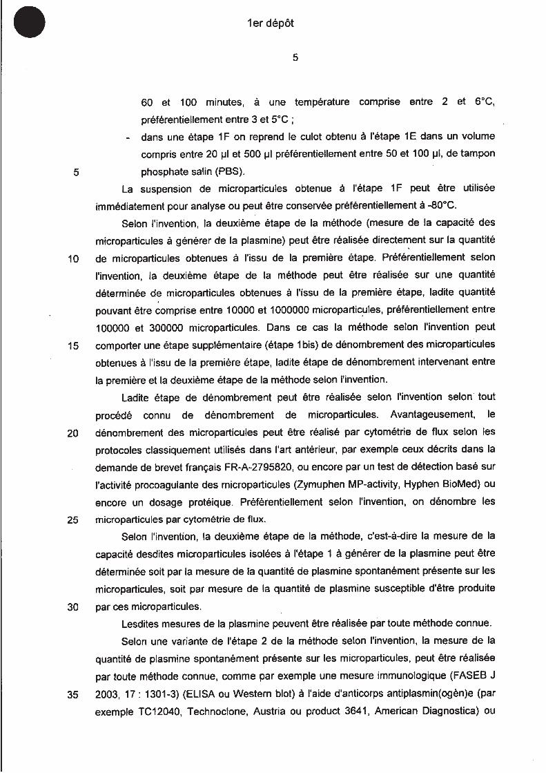


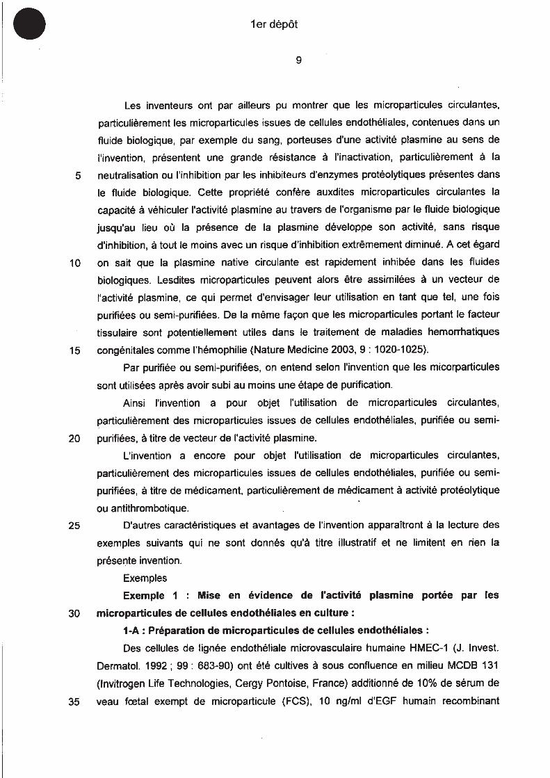


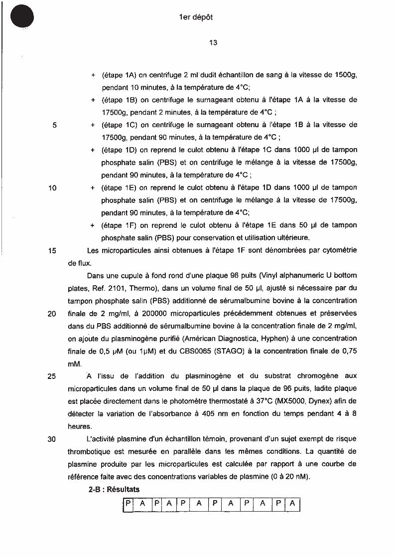


�
�
ARTICLE 4
Standardization of platelet-derived microparticle enumeration by flow
cytometry using calibrated beads: results of ISTH SSC Collaborative
workshop.
R. Lacroix1, S. Robert1, P. Poncelet2, R.S. Kasthuri3, N. S. Key3, F. Dignat-George1�, on behalf
of the ISTH SSC Workshop �.
1UMR-S 608 INSERM-Aix-Marseille Université, UFR de Pharmacie, Marseille, France 2Biocytex, 140, Chemin de l'Armée d'Afrique, Marseille, France, 3�Department of Medicine,
University of North Carolina, Chapel Hill, NC, United States
J. Thromb. Haemost. 2010, doi: 10.1111/j.1538-7836.2010.04047.x. (Disponible en ligne)
Les MP sont de petites vésicules, de taille hétérogène de 0.1 à 1µm, libérées par les
cellules activées ou en apoptose. Leur rôle de biomarqueur et de biovecteur dans la
coagulation, l�inflammation et le cancer est de mieux en mieux établi. Ce qui accroît l�intérêt
de leur mesure en pratique clinique. Cependant, à l�heure actuelle, l�évaluation du bénéfice
apporté par les MP dans l�identification des sujets à risque vasculaire ou dans le suivi
thérapeutique est limitée par un manque de standardisation des méthodologies.
Parmi les différentes technologies disponibles, la CMF est la plus largement utilisée.
Cependant les données de la littérature sont difficiles à comparer en raison de l�hétérogénéité
des protocoles en termes de réglage des instruments et de leur sensibilité respective. Par
exemple, les valeurs de MPP pour les individus sains varient de 100-4000 MPP/µl.
Le paramètre le plus approprié pour l�analyse des MP par CMF est le forward scatter
(FS). Cependant, l�identification des MP sur la base de leur taille relative en utilisant ce
paramètre est restreinte par la limite de sensibilité des appareils. Même si les cytomètres de
dernière génération ont une limite de détection inférieure à 500 nm, la mesure à ce niveau de
sensibilité demeure incertaine en pratique courante. Dans ces conditions extrêmes
d�utilisation de la CMF pour la numération des MP, la limite de résolution du FS entre les
MP et le bruit de fond dépend de l�ajustement fin de l�optique et de la propreté du système
fluidique et optique du cytomètre. Elle peut donc varier avec le temps en fonction des

����
�
maintenances effectuées sur chaque appareil, et donc, être différente entre plusieurs
plateformes de CMF. Des outils et des stratégies sont donc nécessaires pour définir et
contrôler de manière reproductible la limite inférieure d�analyse des MP sur le paramètre FS
afin de numérer les MP de façon standardisée.
Dans ce but, un mélange de plusieurs billes fluorescentes appelé Mégamix, a été
développé par la compagnie BioCytex (Marseille, France). Il est constitué de billes
fluorescentes calibrées en taille de 0,5 et 0,9 µm dont la formulation respecte un ratio précis
de 2 : 1 respectivement. La bille de 0,9 µm aide à placer la limite supérieure de la région
d�analyse des MP ; La bille de 0,5 µm aide à régler de manière standardisée le seuil FS et
donc la limite inférieure de la fenêtre MP. La valeur médiane de la bille 0,5 µm est choisie
comme cible. Ceci est facilement obtenu en utilisant le ratio intrinsèque des billes 0,5/0,9 µm
de 2 : 1 et en incluant toujours le même pourcentage de bille 0,5 µm (50%) dans l�analyse.
Cette stratégie de calibration par un ratio de bille permet un contrôle à long terme du
protocole de mesure des MP par CMF et d�obtenir des comptes reproductibles de MP dans le
temps. Nous avons démontrée son efficacité sur des comptes de MPP sur un cytomètre
Cytomics FC500 (Beckman Coulter). En utilisant cette stratégie, les réglages optimums ont
facilement été transférés entre 3 instruments de même type et des comptes similaires ont été
obtenus entre les cytomètres (Annexe5)517
.
A la suite de ce travail, trois sous-comités de standardisation de l�ISTH (Biologie
vasculaire, Hémostase et Cancer et DIC) ont initié un projet ayant pour objectif la
standardisation des MP par CMF. Dans ce cadre, nous avons organisé un premier atelier
collaboratif international ayant pour objectif, de définir la reproductibilité inter-instrument
des numérations de MPP dans le plasma humain en appliquant la stratégie Mégamix
précédemment décrite.
Nous avons donc posé l�hypothèse qu�une stratégie de standardisation utilisant
des billes fluorescentes calibrées en tailles (Mégamix) permettait d�obtenir des
numérations des MPP reproductibles entre les différents cytomètres et opérateurs.
Cette étude a fédéré 40 laboratoires internationaux, soit 59 cytomètres, composés de marques
et de générations différentes. Elle s�est déroulée en 2 phases : L�objectif de la phase initiale
était de vérifier si les instruments utilisés pour la numération des MPP montraient les
performances requises pour l�utilisation des billes fluorescentes. Dans cette phase A, les

����
�
laboratoires participant ont reçu les billes Mégamix avec lesquelles ils ont réglé sur leurs
cytomètres le protocole pré-défini et l�ont validé. Basé sur les performances de résolution sur
le paramètre FS et le niveau de bruit de fond, les instruments testés ont été acceptés ou rejetés
pour la phase B. Dans la phase B, les laboratoires sélectionnés (n=29) ont reçu des aliquotes
de plasma humain dépourvus de plaquettes correspondant à trois niveaux de MPP. Ces
échantillons ont été analysés avec les instruments précédemment validés, sur le protocole
établi avec les billes Mégamix et en utilisant des réactifs communs. Toutes les données brutes
ont ensuite été ré-analysées par notre laboratoire.
Les résultats de ce travail ont montré qu�il était possible de définir les performances
de la CMF en termes de résolution du FS et de bruit de fond quel que soit le type
d�instrument en utilisant les billes Mégamix. Ces billes de calibration représentent un outil
efficace pour standardiser la mesure de MPP, dont le coefficient de variation se situe entre 10
et 20% sur les cytomètres qui mesurent le paramètre FS avec un angle relativement large (1-
19º) comme les instruments de type Beckman Coulter. Cette stratégie s�est cependant révélée
inapplicable sans modifications substantielles sur des instruments mesurant le paramètre FS
avec un angle plus réduit (1-8º Becton-Dickinson).
�

OFFICIAL COMMUNICATION OF THE SSC
Standardization of platelet-derived microparticle enumerationby flow cytometry with calibrated beads: results of theInternational Society on Thrombosis and Haemostasis SSCCollaborative workshop
R . LACROIX ,* S . ROBERT ,* P . PONCELET , R . S . KASTHURI ,à N. S . KEYà and F . D IGNAT-GEORGE*
ON BEHALF OF THE I STH SSC WORKSHOP*UMR-S 608 INSERM-Aix-Marseille Universite, UFR de Pharmacie and Laboratoire d’hematologie, CHU Conception, Marseille; Biocytex,
Marseille, France; and àDepartment of Medicine, University of North Carolina, Chapel Hill, NC, USA
To cite this article: Lacroix R, Robert S, Poncelet P, Kasthuri RS, Key NS, Dignat-George F, on behalf of the ISTH SSC Workshop. Standardization of
platelet-derived microparticle enumeration by flow cytometry with calibrated beads: results of the International Society on Thrombosis and
Haemostasis SSC Collaborative workshop. J Thromb Haemost 2010; 8: 2571–4.
Microparticles (MPs) are sub-micrometer sized vesicles re-
leased from cell membranes in response to activation or
apoptosis [1]. MPs originating from several cell sources have
been described in human plasma. Among them, platelet-
derivedMPs (PMPs) are believed to account for themajority of
circulating MPs in healthy subjects [2]. Their levels are
increased in several prothrombotic and inflammatory disorders
[1]. In these clinical settings, PMP counts may be useful for
identifying patients at risk for vascular disorders and for
monitoring response to treatment [3]. However, their clinical
use is not fully established, because standardized methodolo-
gies for PMP counting are lacking.
A previous International Society on Thrombosis and
Haemostasis (ISTH) Vascular Biology Subcommittee survey
indicated that approximately 75% of laboratories use flow
cytometry (FCM) to enumerate MPs in clinical samples.
However, a wide variety of preanalytic variables and analytic
variables have been reported in the literature, resulting in a
wide range of PMP values in platelet-free plasma (PFP) of
healthy subjects (100–4000 PMPs lL–1). This lack of consen-
sus stresses the need for standardization [4].
Three ISTH Scientific and Standardization Subcommittees
(SSC Vascular Biology, DIC, and Haemostasis &Malignancy)
have initiated a project aimed at standardizing the enumeration
of cellularMPs by FCM.Afirstcollaborative workshopwas set
up, to: first, establish the resolution and the level of background
noise of the flow cytometers currently used in laboratories with
respect to the strategy requirements; and second, define the
interinstrument reproducibility of PMP enumeration in human
plasma. This strategy was based on the use of fluorescent
calibrated sub-micrometer beads (Megamix beads; BioCytex,
Marseille, France), which allow the window of MP analysis to
be reproducibly set [5].
The study included 40 laboratories accounting for 59 flow
cytometers, and was performed in two stages (Fig. S1): in
stage A, participating laboratories received Megamix beads
and were asked to set up the FCM protocol and to validate an
instrument protocol adapted from a previously described
method [5]. On the basis of forward scatter (FS)/FS channeling
(FSC) resolution and background characteristics, stage A
results led to acceptance or rejection of the tested instruments,
with some time being allowed for technical intervention in
order to improve any deficient performance. In stage B,
selected laboratories received PFP samples prepared as frozen
aliquots by the core laboratory, and were asked to analyze
them with the previously validated instrument(s), common
reagents, and the FCM protocol established in stage A. A
detailed description of the methodology is available in the
Supporting Information (Data S1).
The purpose of this initial phase was to check whether the
instrument to be used to enumerate PMPs demonstrated the
required performance with a blend of fluorescent beads with
well-known sizes and relative amounts. The instruments were
validated on the basis of their capacity to discriminate between
0.5-lm and 0.9-lm Megamix beads using the FS/FSC param-
eter, as well as their background noise. Instruments detecting
< 0.1% of fluorescent bead events among total events were
rejected, because such a level of background may impede the
electronics functions and induce amajor loss of events owing to
coincidences and electronic aborts (Fig. S2A,B). Analysis of
the results demonstrated that instruments were heterogeneous
with respect to FS/FSC resolution and background noise.
Furthermore, the level of performance could vary over time
(Fig. S2C). Some of the parameters affecting FS/FSC resolu-
tion were identified with Megamix beads. Among them, FS/
FSC gain, FS/FSCmode, neutral density (intensity scavenging)
Correspondence: Francoise Dignat-George, UMR-S608, 27 Bd Jean
Moulin, 13005 Marseille, France.
Tel.: +33 491 385600; fax: +33 491 385602.
E-mail: [email protected]
Journal of Thrombosis and Haemostasis, 8: 2571–2574 DOI: 10.1111/j.1538-7836.2010.04047.x
Ó 2010 International Society on Thrombosis and Haemostasis

40
49
59
3633
2930
40
50
60
70 Registered
Tested
0
10
20
Validated
12
10
14
6
4
8
Gro
up s
ize
Registered
Tested
0
Gallio
s
LSRII
FC50
0
FACSC
anto
ll
FACSAria
Cya
n
EPICS X
L
FACSca
libur
EPICS E
lite
FACSVan
tage
FACSsc
an
2
Validated
Newest to oldest flow cytometer generation
Gro
up s
ize
Cytometers Laboratories
4
EP
ICS
XL
FC500
BC instruments
Gallio
s
4
FA
CS
Calib
ur
FACS
Aria
BD instruments
LSRIIFACSCanto
2
3
2
3
1
PM
P levels
× 1
03 µ
L–1
1
PM
P levels
× 1
03 µ
L–1
0 0
BC instruments BD instruments
3
4
With MP gate
upper limit
Without MP gate
upper limit
3
4
CV = 17%
1
2
1
2CV = 15%
PM
P levels
× 1
03 µ
L–1
PM
P levels
×1 0
3 µ
L–1
0
1
12
1 2 3 4 5 6 7 8 9 1011 12 13 14 15 16 17 18 19 20 21 22
18 22 18 22170
Low Medium High
CV = 30%
A
B
C E
D F
2572 R. Lacroix et al
Ó 2010 International Society on Thrombosis and Haemostasis

filters, flow rate and osmolarity of the Megamix suspension
proved to be important (Fig. S3). Megamix bead analysis
allowed easy optimization of these parameters.
Finally, among 59 registered instruments from 40 candidate
laboratories, 49 were tested, and 33 (67%) from 29 laboratories
were ultimately validated (Fig. 1A). As expected, a higher
validation rate was obtained with instruments of later gener-
ations [Gallios fromBeckman Coulter (BC),Miami, FL, USA;
LSRII fromBectonDickinson (BD), Franklin Lake, NJ, USA;
FC500 from BC; FACSCantoII from BD] than with instru-
ments of older generations (FACSCalibur from BD;
EPICS XL from BC): 84% vs. 35%, respectively. However,
as shown in Fig. 1B, some of the most sophisticated instru-
ments failed to qualify [FACSCantoII (1/10), LSRII (1/5), and
FC500 (2/10)]. Conversely, some instruments of older gener-
ations reached the required criteria, as seen with EPICS XL (4/
6) and FACSCalibur (2/8). This outcome shows that instru-
ment response related to the FS/FSC parameter is highly
variable, and depends on the status of an individual instrument,
which needs to be recalibrated on a regular basis.
To avoid any preanalytic-linked variability, the core labo-
ratory prepared and validated PFP samples (Fig. S4). All
participants received six PFP aliquots from three different
samples accounting for three different PMP levels (low,
medium and high). PMPs were enumerated on validated
instruments, with common reagents and the same FCM
protocol that was previously calibrated with Megamix beads.
The analyses obtained from 22 instruments (11 BC and 11
BD) are shown in Fig. 1C,E. As illustrated in Fig. 1D,
interlaboratory reproducibilities of 30% (low), 15% (medium)
and 17% (high) were found among BC instruments with
Megamix beads, whereas highly dispersed values were reported
with BD instruments (Fig. 1E): ) 24%, coefficient of variation
(CV) = 78% (low); ) 60%, CV = 87% (medium); and
) 66%, CV = 91% (high).
A potential explanation for such a discrepancy between the
two types of instrument was suggested by the observation of
the location of the PMP population on the standardized size-
related window of analysis. As shown in representative
examples (Fig. S5A), the PMP population was located inside
the MP gate defined by the Megamix beads for BC instru-
ments, whereas it varied from being partly inside to entirely
outside the MP gate on all BD instruments (Fig. S5B,C).
Interestingly, as illustrated in Fig. 1F, when listmode files were
reanalyzed without setting an upper limit on the MP gate with
three BD instruments (no. 12, FACSCalibur; no. 18, FAC-
SCantoII; and no. 22, LSRII), a similar range of PMP values
as determined by BC instruments was apparent. Taken
together, these findings showed that PMP concentrations
appeared to be consistent among instruments that measure the
FS parameter with a relatively wide solid angle (1–19°,
Beckman-Coulter; hereafter referred to as Ôwide FS platformsÕ).
However, at present, this strategy could not be applied without
substantial modifications for instruments with FSC signals
collected with a lower solid angle (1–8°, Becton-Dickinson; so-
called Ôlow FS platformsÕ). These discrepancies between BD
and BC intruments are also consistent with the notion that size-
related information derived from plastic beads and that derived
from biological particles are not comparable.
To conclude, these data indicate that standardization of
PMP enumeration by FCM is feasible but is dependent on
intrinsic characteristics of both the flow cytometer and the
calibration strategy. Calibrated beads such as Megamix are
useful standards that allow instrument qualification and
follow-up. However, they do not constitute a universal
biological standard for PMP enumeration. At present, this
strategy has proved to be applicable for instruments that
measure the FS parameter with Ôwide FS platformsÕ.
Moreover, additional questions are raised by this study. Is it
adequate to focus only on MPs larger than about half a
micrometer in size? If so, how representative is this Ôvisible part
of theMP icebergÕ for the clinically relevant biomarkers that we
are seeking? Can we look forward to a newer generation or
types of flow cytometer (or alternative technologies with similar
immunologic capabilities) that would allow enumeration and
characterization of particles of smaller sizes? Finally, a critical,
but so far unaddressed, area is the impact of preanalytic
conditions onMP enumeration. These questions constitute the
basis for future SSC workshops.
Disclosure of Conflict of Interests
The authors state that they have no conflict of interest.
Supporting Information
Additional Supporting Informationmay be found in the online
version of this article:
Data S1. Supplemental materials and methods.
Fig. S1. Design of the study.
Fig. S2. Resolution and background noise heterogeneity.
Fig. S3. Parameters affecting forward scatter (FS)/FS
channeling (FSC) resolution.
Fig. 1. Instrument validation (stage A) (A) Histogram showing the number of flow cytometers and laboratories that were registered in the workshop
(black bars), were tested (light gray bars), and were validated (dark gray bars). (B) Histogram showing the number of flow cytometers that were registered
in the workshop (black bars), were tested (light gray bars), and were validated (dark gray bars), according to their type, classified from newest to oldest
generation. (C–F) Interlaboratory reproducibility (stage B). (C) Platelet-derived microparticle (PMP) levels obtained for the three tested platelet-free
plasmas (PFP) (diamonds, low; squares, medium; triangles, high) for each validated Beckman Coulter (BC) instrument. (D) Histogram showing the
interlaboratory reproducibility obtained for the three PFPs with BC instruments. (E) PMP levels obtained for the three PFPs (diamonds, low; squares,
medium; triangles, high) for each validated BectonDickinson (BD) instrument. (F) PMP levels for the three tested PFPs (diamonds, low; squares, medium;
triangles, high) before and after removing the upper limit on the microparticle (MP) gate on three selected BD instruments. CV, coefficient of variation.
Standardization of platelet-derived microparticle enumeration by flow cytometry 2573
Ó 2010 International Society on Thrombosis and Haemostasis
Fig. S4. Interaliquot variability.
Fig. S5. Platelet-derived microparticle (PMP) location on the
standardized size-related window of analysis between Becton
Dickinson (BD) and Beckman Coulter (BC) instruments.
Please note:Wiley-Blackwell are not responsible for the content
or functionality of any supporting materials supplied by the
authors. Any queries (other than missing material) should be
directed to the corresponding author for the article.
Appendix
ISTH SSC Workshop: A. Leyte, M. van Schilfgaarde, A.
Falanga, A.Vignoli, A. Enjeti, L. Lincz, A. Maraveyas, L.
Madden, A. Bonnefoy, J. Rivard, K. Ben-Zion, J. Lopez, P.
Davizon-Castillo, E. Maurer-Spurej, F. Mullier, J. Marvin, H.
Kwaan, I. Weiss, I. Bosch, M. Woda, J. Pereira, P. Hidalgo, J.
Antovic, F. Mobarrez, K. Ghosh, L. Macchi, G. Franck, M.
Perez-Casal, M. G. Huisse, M. Macey, M. Harrison, A.
Amirkhosravi, M. Davila, N. Li, N. Matijevic, W. Wang, P.
Harrison, A. N. Boing, H. Buller, R. Nieuwland,W. Chandler,
W.Yeung, D. Stirling, S. Baird, S. Zahra, R.Krueger, R. Ross,
J. Thom, S. Osanto, Y. Yuana, V.Martinez-Sales, V. Virtudes,
V. Proulle, V. Latger-Cannard, M. De Carvalho, S. Susen, M.
S. Carraway, and A. Charpentier.
References
1 Piccin A, Murphy WG, Smith OP. Circulating microparticles: patho-
physiology and clinical implications. Blood Rev 2007; 21: 157–71.
2 Lynch SF, Ludlam CA. Plasma microparticles and vascular disorders.
Br J Haematol 2007; 137: 36–48.
3 Morel O, Toti F, Hugel B, Bakouboula B, Camoin-Jau L, Dignat-
George F, Freyssinet JM. Procoagulant microparticles: disrupting the
vascular homeostasis equation? Arterioscler Thromb Vasc Biol 2006;
26: 2594–604.
4 Simak J, Gelderman MP. Cell membrane microparticles in blood and
blood products: potentially pathogenic agents and diagnostic markers.
Transfus Med Rev 2006; 20: 1–26.
5 Robert S, Poncelet P, Lacroix R, Arnaud L, Giraudo L, Hauchard A,
Sampol J, Dignat-George F. Standardization of platelet-derived
microparticle counting using calibrated beads and a Cytomics FC500
routine flow cytometer: a first step towards multicenter studies? J
Thromb Haemost 2009; 7: 190–7.
2574 R. Lacroix et al
Ó 2010 International Society on Thrombosis and Haemostasis
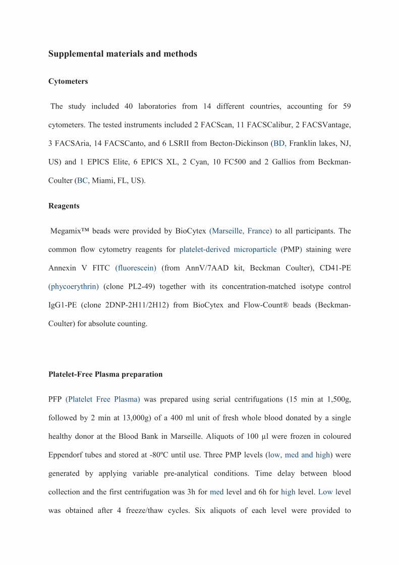
Supplemental materials and methods
Cytometers
The study included 40 laboratories from 14 different countries, accounting for 59
cytometers. The tested instruments included 2 FACScan, 11 FACSCalibur, 2 FACSVantage,
3 FACSAria, 14 FACSCanto, and 6 LSRII from Becton-Dickinson (BD, Franklin lakes, NJ,
US) and 1 EPICS Elite, 6 EPICS XL, 2 Cyan, 10 FC500 and 2 Gallios from Beckman-
Coulter (BC, Miami, FL, US).
Reagents
Megamix™ beads were provided by BioCytex (Marseille, France) to all participants. The
common flow cytometry reagents for platelet-derived microparticle (PMP) staining were
Annexin V FITC (fluorescein) (from AnnV/7AAD kit, Beckman Coulter), CD41-PE
(phycoerythrin) (clone PL2-49) together with its concentration-matched isotype control
IgG1-PE (clone 2DNP-2H11/2H12) from BioCytex and Flow-Count® beads (Beckman-
Coulter) for absolute counting.
Platelet-Free Plasma preparation
PFP (Platelet Free Plasma) was prepared using serial centrifugations (15 min at 1,500g,
followed by 2 min at 13,000g) of a 400 ml unit of fresh whole blood donated by a single
healthy donor at the Blood Bank in Marseille. Aliquots of 100 µl were frozen in coloured
Eppendorf tubes and stored at -80ºC until use. Three PMP levels (low, med and high) were
generated by applying variable pre-analytical conditions. Time delay between blood
collection and the first centrifugation was 3h for med level and 6h for high level. Low level
was obtained after 4 freeze/thaw cycles. Six aliquots of each level were provided to

participating labs for each validated instrument. Inter-aliquot reproducibility was evaluated
after one freezing/thawing cycle, and stability over time of the three levels was regularly
checked on the same instrument during the study.
Flow cytometry protocol for stage A
In order to evaluate the quality of FS(C) (Forward Scatter (Channel)) resolution of the
cytometers using Megamix™ beads, the following protocol was designed: (a) Flow
cytometry (FCM) analysis was set with a discriminator (also called threshold) on FL1
fluorescence detector (FITCdetection channel) fluorescence in order to focus FCM analysis
only on the Megamix fluorescent beads, and thus avoid any background originating from all
other non fluorescent particles; (b) bead singlets (3/0.9/0.5 µm) were selected on FL1 x
SS(C) (Side scatter (Channel)) cytograms; and (c) instrument resolution (ability to
discriminate between 0.5 and 0.9 µm beads) was checked on the FS(C) histogram as well as
dual scatter plots (FS(C) x SS(C)) gated on submicron beads.
To check whether background level was acceptable in these conditions, threshold was
switched to the FS/FSC-H parameter and the FS/FSC settings (threshold, gains) were
adjusted until the 0.5 µm bead peak represented ~50% (range 48% - 52%) of the total sub-
micron beads. Due to the initial intrinsic 2:1 ratio of the 0.5 �m and 0.9 �m beads in
Megamix, this setting corresponds to a cut centered at the median of the 0.5 �m beads
histogram and thus positioned at the level of their nominal size.

FCM protocol for stage B
To enumerate PMP in PFP aliquots prepared by the core center, participating laboratories
were instructed to use the protocol established in stage A with the following exception: BD
instrument users were asked to modify the protocol in order to reduce the high background
noise generally observed in standard conditions. Thus, a combined size-related threshold was
triggered on both the FSC-H and SSC-H parameters rather than the FS threshold alone as
recommended for BC instruments. SSC PMTv (photomultiplier voltage) was increased so
that 0.9 µm beads reached 4x10e3 a.u. (arbitrary units) in a 5.5 decade log scale, while SSC
threshold was set at the minimum possible value (200 a.u.). Based on previous tests on BD
FACSCanto II and feedback from participating labs, this setting had seemed compatible with
detection of all PMP seen in FC500 using the FS threshold alone.
On all instruments, FS/FSC threshold was set in order to reach 50% for the 0.5 µm beads just
before PFP analysis. A rectangular dual scatter gate to measure MP was created with an
upper limit defined by the tangents to an elliptical autogate set on 0.9 µm beads and the lower
limit by the FS/FSC threshold. PMP were analysed in a dual fluorescence cytogram
(FL1xFL2) gated on the MP scatter gate.
For each PFP level, 3 tubes were prepared. 30 µl of PFP was incubated with (a) 10 µL CD41-
PE + 10 µL AnnV-FITC (diluted with PBS (phosphate buffered saline) without Ca²+), (b) 10
µL IgG1-PE + 10 µL AnnV-FITC (diluted with Annexin kit Binding Buffer – BB), (c) 10 µL
CD41-PE + 10 µL AnnV-FITC (diluted with BB). Incubation time was 30 min at room
temperature with samples protected from light. Each stained tube was run in the standardized
protocol (time stop set to 1 mn; low flow-rate), with a maximum delay of 30 min after the
end of staining. Compensation was adjusted using tubes a and b. PMP were defined as
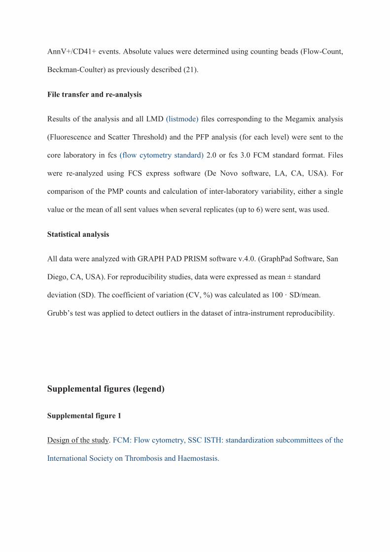
AnnV+/CD41+ events. Absolute values were determined using counting beads (Flow-Count,
Beckman-Coulter) as previously described (21).
File transfer and re-analysis
Results of the analysis and all LMD (listmode) files corresponding to the Megamix analysis
(Fluorescence and Scatter Threshold) and the PFP analysis (for each level) were sent to the
core laboratory in fcs (flow cytometry standard) 2.0 or fcs 3.0 FCM standard format. Files
were re-analyzed using FCS express software (De Novo software, LA, CA, USA). For
comparison of the PMP counts and calculation of inter-laboratory variability, either a single
value or the mean of all sent values when several replicates (up to 6) were sent, was used.
Statistical analysis
All data were analyzed with GRAPH PAD PRISM software v.4.0. (GraphPad Software, San
Diego, CA, USA).�For reproducibility studies, data were expressed as mean ± standard
deviation (SD). The coefficient of variation (CV, %) was calculated as 100 · SD/mean.
Grubb’s test was applied to detect outliers in the dataset of intra-instrument reproducibility.
�
�
�
Supplemental figures (legend)
Supplemental figure 1
Design of the study. FCM: Flow cytometry, SSC ISTH: standardization subcommittees of the
International Society on Thrombosis and Haemostasis.

Supplemental figure 2
Resolution and background noise heterogeneity; representative graphs of FC500 and
FACSCanto II cytometers. A; FS/FSC resolution heterogeneity in the capacity to discriminate
between 0.5 and 0.9 µm beads. Left panel: FS(C)/SS(C) scattergrams of the 0.5 and 0.9 �m
beads. Right panel: FS(C) histograms of the 0.5 and 0.9 �m beads. Threshold is set on FL1 B;
FS/FSC background noise heterogeneity. FS(C)/SS(C) scattergrams of the 0.5 and 0.9 �m
beads. Threshold is set on FS(C) C; Variation over a 2 year period of a FC500 cytometer
performance. Upper panel: FS(C) histograms of the 0.5 and 0.9 �m beads with threshold on
FL1 showing the resolution between the two beads over time. Lower panel: FS(C)/SS(C)
scattergrams of the 0.5 and 0.9 �m beads with threshold on FS(C) showing the background
noise over-time.
Supplemental figure 3
Parameters affecting FS/FSC resolution; A. FS/FSC gain. Increasing the FS gain provided
improved resolution due to a higher number of measurement channels per decade in the upper
decades. B. FS/FSC mode. During FSC signal treatment on FACS instruments that provide a
choice, use of signal height (FSC-H) parameter provided better resolution than signal area
(FSC-A). C. Neutral density (light reducing) filters. On a FACSAria, FSC signal absorption
due to the involuntary use of a neutral density filter impeded measurement of small particles.
D. Flow rate. With all cytometers, a low flow rate gave the best results compared to a higher
speed option E. Osmolarity of the Megamix suspension. On BD instruments, Megamix low
osmolarity seemed to influence resolution. In some cases, dilution 1:2 in salted buffer/sheath
fluid produced improved instrument performance in some cases.

Supplemental figure 4
Inter-aliquot variability; A. Inter-aliquot reproducibility after one freeze/thaw cycle using the
same cytometer, reagents, methodology and operator for the three levels of PMP including
inter-run variability of the cytometer on a core lab instrument (FC500). B. Stability of frozen
aliquots over time on a FC500. C. Stability of frozen aliquots over-time on a FACSCanto II.
D. Overall variability (mean and range values) inter-aliquots including data obtained on
13/14 cytometers with 2 to 6 PMP enumerations each.
Supplemental figure 5
PMP location on the standardized size-related window of analysis between BD and BC
instruments. A. EPICS XL. B. FACSCanto a. C. FACSCanto b.�
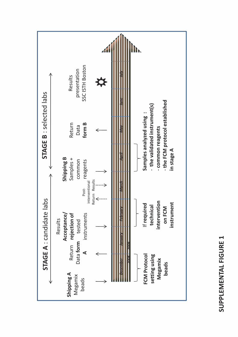
�����������
��������
���
���������
��
���
��
����
����
�������
������
����������
��� �����
������
��������
����
���
�����������
���������������
������
�������
�
�
������
�����
��
�
�������
�������������
������
������������
����
���
������������
�����������
������
�����������
�����
����� ��������
�������������
����
���������
��
���
��
����
����
�������
������
�����
�����
�������������
�������������
���� ��
��� �
�!���!���� �
����������
�����"�������
�������
������ ���
�� ��������#$� �����%
�����"��� ��� ������ ���&�'
��� ����������
���������������������������
����������
�(��)���*��)��+�(,��-

FC
50
0FA
CS
Can
to I
I
AdequateInadequate
A BF
C5
00
FA
CS
Can
to I
I
AdequateInadequate
B 0
.9+
0.5
= 0
.01
%
B 0
.9+
0.5
= 7
2%
B 0
.9+
0.5
= 0
.1%
B 0
.9+
0.5
= 1
%
�(��)���*��)��+�(,��.

Mar0
7N
ov07
Feb08
Mar0
9O
ct0
8
Para
mete
rC
yto
mete
r
Tim
eF
C500
Resolu
tion
C
Backgro
und n
ois
e
Mai0
7D
ec07
Mai0
8Jan09
�(��)���*��)��+�(,��.

Para
mete
r(N
on o
ptim
ized/
Optim
ized)
Cyto
mete
r
FS
/FS
C g
ain
(Hig
h/L
ow
)FA
CS
Ca
lib
ur
FS
/FS
C m
od
e(F
SC
-A/F
SC
-H)
FA
CS
Ca
nto
II
No
n o
ptim
ize
dO
ptim
ize
d
M1
= 0
.5 µ
m b
ea
ds
/M2
= 0
.9 µ
m b
ea
ds
M1
M2
A B(F
SC
-A/F
SC
-H)
FA
CS
Ca
nto
II
FS
/FS
C f
ilte
r(N
eutr
al density filt
er
+/-
)FA
CS
Ari
a
B C �(��)���*��)��+�(,��/

Para
mete
r(N
on o
ptim
ized/
Optim
ized)
Cyto
mete
r
Flo
w r
ate
(Mediu
m/L
ow
)FA
CS
Ca
nto
II
No
n o
ptim
ize
dO
ptim
ize
d
M1
= 0
.5 µ
m b
ea
ds
/M2
= 0
.9 µ
m b
ea
ds
M1/
M2
D
Os
mo
lari
ty(W
ithout/w
ith M
egam
ix
dilu
tion in
salted b
uffer)
FA
CS
Ca
nto
II
M1
M2
M2
E �(��)���*��)��+�(,��/
M1
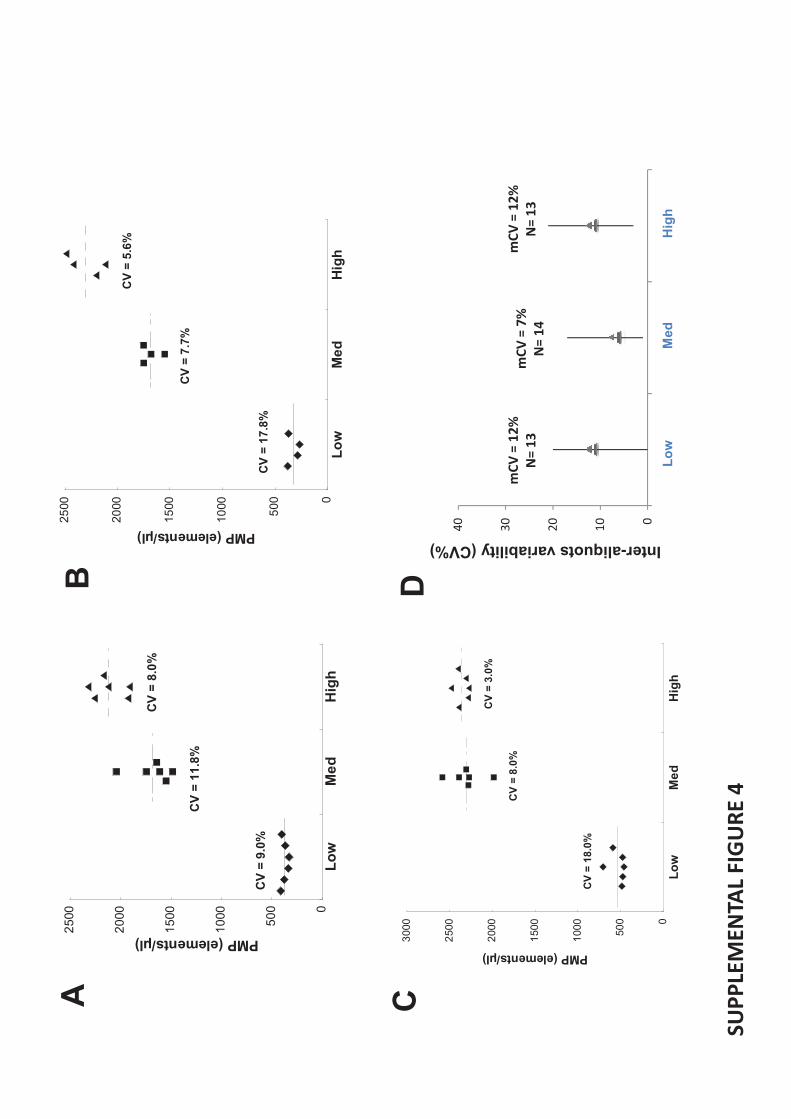
0
500
1000
1500
2000
2500
Lo
wM
ed
Hig
h
PMP (elements/µl)
CV
= 1
1.8
%
CV
= 8
.0%
CV
= 9
.0%
0
500
10
00
15
00
20
00
25
00
Lo
wM
ed
Hig
h
CV
= 5
.6%
CV
= 7
.7%
CV
= 1
7.8
%
PMP (elements/µl)
AB
CD
0
50
0
1000
1500
2000
2500
3000
Lo
wM
ed
Hig
h
CV
= 1
8.0
%
CV
= 3
.0%
CV
= 8
.0%
PMP (elements/µl)
"
#"
$"
%"
&"
'� ���#
'� ���$
'� ���%
�01�-.2
*1�-/
�0�1�32
*1�-4
�0�1�-.2
*1�-/
Inter-aliquots variability (CV%)
CD
�(��)���*��)��+�(,��4
Lo
wM
ed
Hig
h
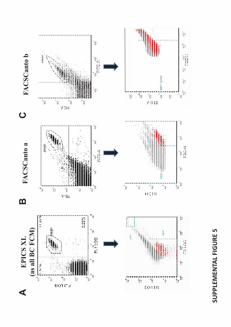
EP
ICS
XL
(as
all
BC
FC
M)
FA
CS
Ca
nto
aF
AC
SC
an
to b
AB
C
�(��)���*��)��+�(,��5

����
�
ARTICLE 5
Overcoming limitations of microparticle measurement by flow cytometry.
Dr Romaric Lacroix1,2
(PharmD), Stephane Robert 1, Dr. Philippe Poncelet
3 (PhD), Pr.
Françoise Dignat-George 1,2*
(PharmD,PhD)
1 UMR-S608 INSERM, F-Marseille, France, Université de la Méditerranée, Faculté de
Pharmacie, F-Marseille, France 2 Service d�hématologie, CHU La Conception, AP-HM, F-
Marseille, France 3 Biocytex, Marseille, France
Seminar in Thromb. Haemost. 2010; 36 : 807-18
Le CMF est à l�heure actuelle la technique la plus utilisée pour la mesure des MP
circulantes grâce à sa capacité d�analyse multiparamétrique de très haut débit de chaque MP
individuelle. Cependant cette méthodologie présente également des limites qui expliquent
que les cytomètres utilisés jusqu�à présent ne mesurent qu�une partie des MP (celles de
grosse taille). Ces limites peuvent être de trois ordres : une difficulté à détecter les
évènements de faible antigénicité, un comptage absolu des MP couteux et sujet à variations et
une difficulté à détecter des évènements de faible taille (inférieure à 0.5µm).
L�objectif de cette revue a été de discuter des limites actuelles de la cytométrie en
flux et de faire un état des lieux des stratégies ou améliorations technologiques
permettant de dépasser ces limites.
Cette revue est focalisée sur les étapes analytiques de la mesure de MP par CMF et
exclue les étapes pré-analytiques pourtant source majeure de variabilité et de potentiels
artéfacts. Après une courte revue des méthodologies disponibles, elle se concentre sur les
limites de la CMF qui relèvent de différentes problématiques : 1/la détermination de la taille,
2/ le marquage, 3/la numération absolue. Cette revue couvre également les diverses stratégies
de standardisation et les améliorations technologiques proposées pour dépasser ces limites.

����
�
Des stratégies originales de calibration et les récents progrès de l�instrumentation
ouvrent de nouvelles voies pour permettre la détection des MP de plus petites tailles,
jusqu�alors irréalisable, avec les cytomètres de dernière génération. Une optimisation
significative de la détection des MP est aussi attendue de l�utilisation de nouveaux marqueurs
fluorescents et de l�établissement de recommandations pratiques. Enfin le comptage absolu
des MP va bénéficier également de stratégies adaptées basées sur des billes de comptage et de
la généralisation des systèmes volumétriques. De façon globale, les récentes améliorations
technologiques permettent à la CMF de rester hautement compétitive pour la mesure des MP.
Le réel impact de ces améliorations pour la pratique clinique reste à démontrer dans des
situations pathologiques.
Le contenu de ce manuscrit dépasse largement le cadre d�une simple revue de la
littérature. Il contient de nombreux éléments et synthèses, fruits de plusieurs années
d�expérience propre à notre laboratoire, de dialogues étroits avec les acteurs industriels et
académiques de ce domaine et d�une prospection active amenant à une expertise directe des
innovations proposées dans cette application. Ce travail est également le fruit d�une aventure
humaine au sens figuré comme au sens propre quand il s�agit d�aller faire dans la même
journée 1200 km de voiture dont 50 km avec des chaines dans les neiges de l�hiver pyrénéen
pour expertiser un nouveau cytomètre. Ceci justifie donc pleinement la place de cette revue
dans la partie travail personnel de cette thèse.


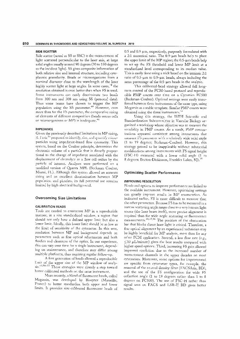

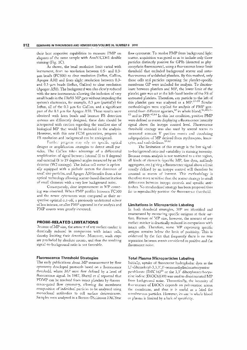


Gallio
sF
C500
Influ
xA
pogee A
50
LS
1
Beads resolutionBackground Noise
AB
Background NoisePMP
An
nV
(FIT
C)
CD41 (PE)
C

����
�
DISCUSSION-PERSPECTIVES
Dans la première partie de ce travail, nous avons identifié les MPE comme nouveaux
acteurs dans le système d'activation du plasminogène, une facette jusqu�alors inconnue parmi
les différentes réponses biologiques impliquant les MP dans le contrôle de l'homéostasie
vasculaire. Notre travail de thèse a été le premier à démontrer les mécanismes moléculaires
responsables de la génération de plasmine dépendante du système uPA-uPAR à la surface des
MP générées à partir d�une lignée endothéliale. Nous avons ensuite établi la preuve de
concept in vivo de cette nouvelle propriété des MP en montrant qu�une génération de
plasmine était détectable à la surface des MP circulantes de patients atteints d�athérosclérose,
de lupus ou de PTT. Nous avons identifié les activateurs responsables de cette propriété in
vivo en démontrant la présence d�urokinase (uPA/uPAR) et/ou d�activateur tissulaire du
plasminogène (t-PA) à la surface des MP issues des cellules sanguines circulantes et de
cellules endothéliales primaires. A l�inverse de la génération de thrombine, cette activité est
vectorisée par les MP endothéliales (MPE) et leucocytaires (MPL) mais absente sur les MP
plaquettaires et érythrocytaires. Par ailleurs, nos travaux ont contribué à mettre en lumière
un nouveau mode d�activation du plasminogène par communication croisée entre la surface
des MP et les surfaces cellulaires, la fibrine ou la matrice extracellulaire.
Microparticules : Pourvoyeurs de surface catalytiques bioactives
Les surfaces cellulaires ou matricielles sont des éléments clés dans la régulation de
l�homéostasie vasculaire. Elles constituent le support des réactions moléculaires
enzymatiques de la plupart des systèmes biologiques parmi lesquels la coagulation et la
fibrinolyse. Par la colocalisation de récepteurs membranaires, elles assurent une probabilité
de rencontre entre les divers éléments d�un même système et rendent accessibles les seuils
énergétiques des réactions mises en jeu. Elles assurent ainsi une catalyse efficace, localisée et
contrôlée permettant de générer les acteurs clés de systèmes protéolytiques comme ceux de la
thrombine et de la plasmine.
La vésiculation cellulaire par activation ou par apoptose, multiplie la disponibilité des
surfaces d�origine et donc la possibilité d�assemblages bioactifs dans la circulation ou les
tissus. Si une telle implication des MP avait parfaitement été démontrée pour la constitution

����
�
des complexes tenase et prothrombinasique responsables de la génération de thrombine,
l�assemblage des complexes aboutissant à la génération de plasmine n�avait pas été démontré
dans le système vasculaire. L�existence au préalable d�un certain nombre de travaux montrant
la présence de certains acteurs protéolytiques fonctionnels (uPAR, PAI-1, MMP) sur des MP
d�origine tumorale doit toutefois être rappelée. Les MP possèdent à la fois des récepteurs
pour les activateurs (u-PAR) et le plasminogène lui-même (Lys C term de l��-énolase)
permettant la formation du complexe moléculaire nécessaire à la génération de plasmine.
L�importance de la surface des MP est montrée par l�inhibition de la génération de plasmine
lorsque le plasminogène ne peut plus s�y fixer en cas d�inhibition de ces sites de fixation par
la CpB ou des LBS des K1 et K4 du plasminogène par l��-ACA. La surface des MP constitue
également un système de protection pour la fraction de plasmine qui reste fixée contre les
inhibiteurs de la phase soluble comme l��2-AP. La génération de plasmine à la surface des
MP est donc efficace. Nous avons également montré que la majorité des récepteurs uPAR à la
surface des MPE était libre. L�uPA exogène peut être captée par ces récepteurs ce qui
amplifie considérablement la génération de plasmine. Ces données suggèrent que de l�uPA
sécrétée dans l�environnement local peut être transférée à la surface des MP. Ce mécanisme
peut avoir des implications dans l�angiogenèse tumorale où la sécrétion d�uPA est
importante. Ainsi les MP de cellules tumorales et les MPE pourraient participer à
l�amplification des processus protéolytiques et à la dissémination métastatique. ��
Ainsi, par rapport aux surfaces cellulaires activées et aux matrices (fibrine ou matrice
extracellulaire), les MP constituent un support supplémentaire efficace et abondant pour la
génération de plasmine.
Microparticules : Biovecteur protéolytique
Les activateurs du plasminogène et la plasmine ainsi formée peuvent donc être
vectorisés par les MP localement ou à distance. L�activité protéolytique des MP contribue
ainsi au remodelage matriciel, de façon directe ou indirecte, par l�activation de MMP. Nous
avons montré que cette génération de plasmine dépendante des MP module in vitro les
propriétés angiogéniques des progéniteurs endothéliaux dans un test qui consiste à mesurer la
formation de tube dans une matrice très dépendante des activités protéolytiques. Ainsi en
présence de faible quantité de plasmine générée, la protéolyse limitée est suffisante pour
faciliter la migration des progéniteurs endothéliaux et la formation des tubes vasculaires. En

����
�
présence d�une grande quantité d�activité protéolytique, la matrice est désorganisée, la
migration cellulaire est perturbée et la formation de tube diminue. Dans ce modèle in vitro,
ces observations sont consistantes avec les effets de la plasmine sur l�angiogenèse. Ainsi les
MP par la vectorisation d�une activité protéolytique dépendante de la plasmine peuvent
réguler la croissance vasculaire ;
Ce rôle protéolytique des MP est également relevant dans le remodelage matriciel au
sein de la plaque d�athérosclérose. En effet, les MPE et MPL sont présentes en fortes
concentrations dans la plaque d�athérosclérose518
. Dans un travail en cours, nous avons
formulé l�hypothèse que la génération de plasmine induite par ces concentrations élevées de
MP pourrait induire un remodelage tissulaire de la plaque et ainsi concourir à sa
déstabilisation. Nous montrons la présence des activateurs du plasminogène et d�une
génération de plasmine à la surface de MP dans les plaques d�athérosclérose humaine
prélevées par endartectomie par des techniques ELISA, de CMF, de tests de génération de
plasmine colorimétrique et par zymographie. De manière intéressante ces MP ont la capacité
de dégrader les composants de la plaque sur des tests de dégradation matricielle in vitro
(fibrine, fibronectine, laminine) et sur artère de rat désendothélialisé. L�activité
plasminogénolytique des MP semble donc jouer un rôle dans les processus de remodelage
vasculaire et détermine le caractère instable de la plaque d�athérome.
Comme nous l�avons montré, le système uPA-uPAR ne nécessite pas la présence de
plasminogène sur la même surface que son activateur. La reconnaissance de ce système est
strictement conformationnelle. Ainsi, l�uPA vectorisé par les MP pourra reconnaitre une
molécule de plasminogène fixé sur une surface cellulaire (leucocytes, plaquettes) ou
matricielle (fibrine, matrice extracellulaire) lors d�une réaction de croisée entre MP/cellules
ou MP/matrice. Ainsi, les MP présentent dans le caillot activent potentiellement le
plasminogène déposé sur la fibrine ou sur les plaquettes, contribuant ainsi à la fibrinolyse.
La contribution réelle de la génération de plasmine dépendante des MP dans la
fibrinolyse in vivo est inconnue à l�heure actuelle. Cette question constitue la suite de ce
travail fondamental. Notre hypothèse est que par l�activité protéolytique vectorisée à la
surface des MPE et MPL contribue efficacement à la lyse du caillot de fibrine. Ainsi, les
prochaines étapes de notre travail sont de réaliser des études in vitro de lyse d�un caillot
fluorescent par pesée, de mesurer de la libération de fluorescence et d�étudier la cinétique de
rétraction du front de lyse par laser scanning confocal microscopy. L�impact des MPE et
MPL sur la phase de décroissance d�un caillot par thrombo-élastogramme sera également

����
�
mesurée. Ces expériences seront complétées in vivo par l�évaluation de la contribution des
MPE et MPL sur la fibrinolyse par microscopie intra-vitale dans un modèle de caillot induit
par une blessure LASER. Cette technique, apprise lors d�un stage doctoral à Boston (nov
2007- nov 2008), dans le laboratoire d�hémostase du Pr. B. Furie (Université d�Harvard) nous
permettra de montrer le niveau d�implication des MP dans la fibrinolyse.
Microparticules : Biomarqueur fonctionnel
Nous avons montré que les MP d�origine endothéliale et leucocytaire supportent
l�activité fibrinolytique dans la circulation humaine. Cette activité est non détectable en
situation physiologique mais détectable et modulée en situations pathologiques
cardiovasculaires et auto-immunes. Si ceci constitue la preuve de concept de cette activité in
vivo, la signification d�une augmentation de la génération de plasmine dépendante des MP est
actuellement inconnue. On peut noter que cette activité est extrêmement faible voire
indétectable chez les sujets sains. En situation pathologique les niveaux détectés sont très
variables dans une même pathologie. Ils pourraient refléter une activation leuco-endothéliale
et donc indirectement être en relation avec une situation inflammatoire ou à risque vasculaire.
Ils pourraient également refléter une activation des systèmes coagulation/fibrinolyse en phase
aiguë. En phase chronique, ils pourraient contribuer à un équilibre dans la balance
hémostatique en réduisant le risque thrombotique. De manière intéressante cette activité est
portée par des sous-populations de MP circulantes plus difficilement accessibles par les
méthodologies de mesure actuelle comme la CMF. La mesure de cette activité apporte donc
une information différente d�une simple numération de ces populations.
La mesure de cette activité plasminogénolytique des MP pourrait donc avoir un intérêt
clinique de biomarqueur dans différents contextes pathologiques. Comprendre la relevance de
cette nouvelle activité dans ces contextes pathologiques et transposer ces connaissance vers le
développement de nouveaux outils diagnostics ou pronostics nécessite la maitrise d�une
méthodologie fiable. Ce qui n�est pas totalement le cas à l�heure actuelle. En effet, les tests
réalisés jusqu�à présent dépendent d�une étape de purification des MP par centrifugation et
lavages dont le rendement ne saurait en aucun cas être parfaitement reproductible d�un
patient à l�autre.

���
�
L�objectif principal est donc de développer un test pour la détection des MP associées
à une activité protéolytique de type plasmine adaptée à la pratique de routine des laboratoires
afin d�offrir un outil sensible et reproductible. Ce projet technologique se fait actuellement en
collaboration étroite avec un industriel (Stago/BioCytex) dans le cadre d�une prise d�option
sur le contrat de licence du brevet correspondant. Ceci constitue une première étape pour
pouvoir déterminer l�intérêt de cette activité des MP en pathologie humaine.
Dans la deuxième partie plus appliquée de notre travail, nous avons développé
puis évalué une méthode de standardisation par billes fluorescentes calibrées en taille, pour la
mesure des MP par CMF. Nous avons également évalué les différentes possibilités
disponibles actuellement pour dépasser les limites actuelles de cette technologie dans cette
application.
En effet, la volonté de porter les MP au stade d�un biomarqueur impose comme pré
requis la maîtrise des paramètres pré-analytiques et analytiques. Ceci est associé à une mise
au point de techniques de pointes performantes prenant en compte les spécificités physico-
biologiques de ces biomarqueurs cellulaires comme en particulier leur faible taille ou leur
faible nombre dans l�échantillon biologique. La validation de leur intérêt en clinique
nécessite une étape importante de standardisation à l�échelle internationale.
Maîtrise de la phase analytique de la mesure des microparticules
Dans ce cadre, notre stratégie a été de développer un outil adapté aux limites actuelles
de la cytométrie en flux qui permette de maîtriser les variations intra et inter-instruments.
L�évaluation internationale de cet outil a montré que ces billes de calibration représentent un
outil efficace pour standardiser la mesure de MPP sur les cytomètres qui mesurent le
paramètre FS avec un angle relativement large (1-19º) comme les instruments de type
Beckman Coulter. Cependant cette stratégie s�est cependant révélée inapplicable sans
modifications substantielles sur des instruments mesurant le paramètre FS avec un angle plus
réduit (1-8º Becton-Dickinson). Ceci est le reflet d�une grande variabilité entre les
informations récoltées. En effet, la mesure du FS ne correspond pas exactement à la même
���
�
information entre des cytomètres de conception différente. Ainsi, le processus de
standardisation se trouve limité. Cette différence de mesure entraine une différence de rapport
entre la taille apparente des billes de latex et des MP entre les différents appareils. Une
solution peut venir du développement de standard biologique de même nature que les MP qui
permettraient de s�affranchir de ces différences.
Dans cette stratégie de standardisation, nous nous sommes limités aux performances
de la plupart des cytomètres utilisés en routine. Ainsi la mesure est limitée aux MP de
grandes tailles (supérieure à 0.5�m). Or la grande majorité des MP est de taille inférieure
comme le confirme les techniques innovantes de microscopie et de DLS. Comme développé
dans la revue, les récents progrès des performances de la CMF en terme de résolution ouvrent
de nouvelles perspectives pour la mesure des MP de petites tailles jusqu�alors inaccessibles.
Le bénéfice apporté par la mesure des MP de petites tailles par rapport à la méthodologie
actuelle dans l�utilisation de ce biomarqueur en clinique doit être évalué. Une standardisation
de cette nouvelle mesure devra être amorcée.
Les progrès réalisés en termes de résolution de taille déplacent les n�uds de la
technologie vers la limite de résolution de fluorescence. En effet, du fait de leur petite taille,
les MP ne génèrent souvent qu�un signal de faible intensité en CMF. Nous avons donc pour
objectif de développer en collaboration avec l�industrie (Beckman Coulter, BioCytex) de
nouveaux outils plus sensibles pour la détection des MP soit génériques soit spécifiques de
sous populations. A partir des connaissances actuelles sur le phénomène de remodelage de la
membrane plasmique des cellules mères, l�approche principale consiste à définir la
probabilité de concentration d�une molécule à la surface des MP. Puis de valider cette
hypothèse par des marquages optimisés sur des MP purifiées avant de l�appliquer au plasma
humain.
Maîtrise de la phase pré-analytique de la mesure des microparticules
L�étape pré-analytique dans le dosage des microparticules circulantes est une source
importante d�artéfacts dans l�évaluation de l�intérêt clinique de ces biomarqueurs. Il n�existe
pas de consensus dans ce domaine. Ce point n�a pas été développé dans ce travail. Toutefois,
dans un travail en cours, nous évaluons l�impact des différents paramètres pré-analytiques sur
la numération des MP circulantes par CMF et par des tests fonctionnels chez le sujet sain et
en pathologie afin de proposer un protocole pré-analytique complet. Ce protocole de

����
�
standardisation sera évalué par une étude internationale afin de servir de base à des
recommandations.
La maîtrise des paramètres analytiques et pré-analytiques, étape indispensable du
développement d�un biomarqueur, devrait permettre la mise en place d�études
multicentriques pour évaluer et convaincre de l�intérêt des MP en clinique.

����
�
ANNEXES
Annexe 1 :
Tumor-derived tissue factor-bearing microparticles are associated with venous
thromboembolic events in malignancy.
Zwicker JI, Liebman HA, Neuberg D, Lacroix R, Bauer KA, Furie BC, Furie B.
Clin Cancer Res. 2009;15(22):6830-40.
Annexe 2 :
Cancer cell-derived microparticles bearing P-selectin glycoprotein ligand 1 accelerate
thrombus formation in vivo.
Thomas GM, Panicot-Dubois L, Lacroix R, Dignat-George F, Lombardo D, Dubois C.
J Exp Med. 2009;206(9):1913-27.
Annexe 3 :
Endothelial-derived microparticles: Biological conveyors at the crossroad of
inflammation, thrombosis and angiogenesis.
Leroyer AS, Anfosso F, Lacroix R, Sabatier F, Simoncini S, Njock SM, Jourde N, Brunet P,
Camoin-Jau L, Sampol J, Dignat-George F.
Thromb Haemost. 2010;104(3):456-63.
Annexe 4 :
Circulating endothelial cells, microparticles and progenitors: Towards the definition of
vascular competence.
Sabatier F, Lacroix R, Camoin-Jau L, Anfosso F, Sampol J, Dignat-George F.
Rev Med Interne. 2010 Jun 10.
Annexe 5 :
Standardization of platelet-derived microparticle counting using calibrated beads and a
Cytomics FC500 routine flow cytometer: a first step towards multicenter studies?
Robert S, Poncelet P, Lacroix R, Arnaud L, Giraudo L, Hauchard A, Sampol J, Dignat-
George F.
J Thromb Haemost. 2009 ;7(1):190-7.

Human Cancer Biology
Tumor-Derived Tissue Factor–Bearing Microparticles Are Associated
With Venous Thromboembolic Events in Malignancy
Jeffrey I. Zwicker,1 Howard A. Liebman,3 Donna Neuberg,2 Romaric Lacroix,1
Kenneth A. Bauer,1 Barbara C. Furie,1 and Bruce Furie1
Abstract Purpose: Despite the strong association between malignant disease and thromboem-
bolic disorders, the molecular and cellular basis of this relationship remains uncertain.
We evaluated the hypothesis that tumor-derived tissue factor–bearing microparticles in
plasma contribute to cancer-associated thrombosis.
Experimental Design: We developed impedance-based flow cytometry to detect, quan-
titate, and size microparticles in platelet-poor plasma. We evaluated the number of tis-
sue factor–bearing microparticles in a cohort of cancer patients of different histologies
(N = 96) and conducted a case-control study of 30 cancer patients diagnosed with an
acute venous thromboembolic event (VTE) compared with 60 cancer patients of similar
age, stage, sex, and diagnosis without known VTE, as well as 22 patients with an idio-
pathic VTE.
Results: Tissue factor–bearing microparticles were detected in patients with advanced
malignancy, including two thirds of patients with pancreatic carcinoma. Elevated levels
of tissue factor–bearing microparticles were associated VTE in cancer patients (adjust-
ed odds ratio, 3.72; 95% confidence interval, 1.18-11.76; P = 0.01). In cancer patients
without VTE, a retrospective analysis revealed a 1-year cumulative incidence of VTE
of 34.8% in patients with tissue factor–bearing microparticles versus 0% in those with-
out detectable tissue factor–bearing microparticles (Gray test P = 0.002).The median
number of tissue factor–bearing microparticles in the cancer VTE cohort (7.1 × 10⁴ mi-
croparticles/μL) was significantly greater than both the idiopathic VTE and cancer–no
VTE groups (P = 0.002 and P = 0.03, respectively). Pancreatectomy in three patients
eliminated or nearly eliminated these microparticles which coexpressed the epithelial
tumor antigen, MUC-1.
Conclusion: We conclude that tumor-derived tissue factor–bearing microparticles are
associated with VTE in cancer patients and may be central to the pathogenesis of
cancer-associated thrombosis. (Clin Cancer Res 2009;15(22):6830–40)
Thromboembolic disease is a recognized complication of can-cer (1–8). Patients with cancer experience venous thromboem-bolic events (VTE) at a rate four to seven times higher than thatof the general population (9). Although all tumor types are as-
sociated with thrombosis, glioblastoma and carcinomas of the
ovary and pancreas are consistently associated with the highest
incidence of VTE (10, 11). Despite the strong association be-
tween malignant disease and thromboembolic disorders, the
molecular and cellular basis of this relationship remains uncer-
tain. Hypotheses regarding the mechanism have included the
activation of blood coagulation by tissue factor in tumors
(12), a factor X–activating cysteine protease (13), mucinous gly-
coproteins (8), and MET oncogene activation (14). Laboratory
markers of coagulation activation are often elevated in patients
with cancer (15–21) but are of little clinical utility in assessing
individual risk for thrombosis.Tissue factor, a receptor protein constitutively expressed on
the plasma membrane of most nonvascular cells and inducibly
expressed on monocytes and endothelial cells, initiates blood
coagulation in vivo (22). In addition to its expression in the ves-
sel wall, tissue factor is associated with cell-derived vesicles
in blood that bind to the developing thrombus and play a role
in fibrin propagation (23, 24). These vesicles, known as micro-
particles based on their submicron diameter, express protein
surface components from the parent cell from which they are
Authors' Affiliations: 1Department of Medicine, Beth Israel DeaconessMedical
Center and Harvard Medical School, and 2Department of Biostatistics and
Computational Biology, Dana Farber Cancer Institute, Boston, Massachusetts;
and 3Divisionof Hematology, Norris Comprehensive Cancer Center, University
of Southern California-Keck School of Medicine, Los Angeles, California
Received 2/12/09; revised 8/5/09; accepted 8/10/09; published OnlineFirst
10/27/09.
Grant support: NIH grants K23 HL084052 (J. Zwicker) and HL095084 (B.C.
Furie, B. Furie).
The costs of publication of this article were defrayed in part by the payment of
page charges. This article must therefore be hereby marked advertisement
in accordance with 18 U.S.C. Section 1734 solely to indicate this fact.
Note: Supplementary data for this article are available at Clinical Cancer
Research Online (http://clincancerres.aacrjournals.org/).
Requests for reprints: Jeffrey I. Zwicker, Beth Israel Deaconess Medical
Center, 330 Brookline Avenue, Boston, MA 02215. Phone: 617-667-9299;
Fax: 617-667-9922; E-mail: [email protected].
F 2009 American Association for Cancer Research.
doi:10.1158/1078-0432.CCR-09-0371
6830Clin Cancer Res 2009;15(22) November 15, 2009 www.aacrjournals.org

derived. Monocyte-derived microparticles that express tissue
factor accumulate in the developing thrombus through the in-
teraction of P-selectin and PSGL-1, thus delivering tissue factor
to the site of vascular injury and facilitating fibrin propagation
(24). Microparticles derived from blood and vascular cells cir-
culate in the blood of normal subjects, and these populations
are altered qualitatively and quantitatively in specific disease
states (25–28). Tissue factor–bearing microparticles have been
observed in the plasma of cancer patients, and a possible role
for these microparticles in cancer-associated thrombosis has
beenproposed (29, 30). To evaluatewhether tissue factor–bearing
microparticles are a risk factor for and a possible cause of cancer-
associated thrombosis, we quantified tissue factor–bearing
microparticles in cancer patients using impedance-based flow
cytometry and analyzed their association with acute venous
thromboembolic disease. We show that impedance-based flow
cytometry allows identification and quantitation of tissue
factor–bearing microparticles, that most of these tissue factor–
bearing microparticles are tumor-derived, and that the presence
of detectable tissue factor–bearing microparticles is associated
with an increased risk of thromboembolic disease accompanying
malignant disease.
Materials and Methods
Materials. Humanized monoclonal antibody cH36 against human
tissue factor was the generous gift of Dr. Hing Wong (Altor Biosciences,
Miramar FL). Antibodies against MUC1 were generously provided by
Dr. Donald Kufe (Dana Farber Cancer Institute, Boston MA); these anti-
bodies were labeled with Alexa 488 using standard methods. ASPC-1,
a human pancreatic carcinoma cell line, was obtained from the
American Type Culture Collection. Goat anti-human Ig coupled to
Alexa 488 was obtained from Invitrogen, anti-CD41a-FITC antibody
and phycoerythrin-conjugated mouse anti-human monoclonal TF an-
tibody from BD Biosciences, and all other immunochemical reagents
from Sigma-Aldrich. Fluorescently labeled polystyrene microspheres
were purchased from Bangs Laboratories. These microspheres are
homogeneous with regard to size by 10%.
Generation of tissue factor–bearingmicroparticles via cell culture. ASPC-1
cells were incubated at 37°C in RPMI media for 72 h. The cells were re-
moved from conditioned media by centrifugation twice at 2,100× g. Ali-
quots (10 μL) of supernatant were labeled with (0.25 μg) humanized
anti–tissue factor monoclonal antibody cH36 and labeled with a goat
anti-human IgG-Alexa 488 antibody.
Flow cytometry analyses of platelets and platelet microparticles. Platelet-
rich plasma was centrifuged for 2 min at 2,100× g. A 40 μL aliquot of
plasma, containing both platelets and platelet microparticles, was incu-
bated with 0.125 μg of anti-CD41a-FITC antibody for 30 min. The
plasma samples were diluted 1:50 in NPE Iso-Diluent with 0.78-μm
polystyrene microspheres at a final concentration of 6,000/μL. Samples
were analyzed on a Beckman Coulter Quanta flow cytometer equipped
with a 40-μm flow cell, a Becton Dickinson LSRII flow cytometer, and a
Becton Dickinson FACSCalibur flow cytometer.
Impedance-based flow cytometry for the detection of microparticles. A
Quanta (NPE Systems) or Beckman Coulter SC Quanta flow cytometer
(Beckman Coulter) were used to measure particle size by impedance.
Both flow cytometry instruments required postmarket installation of
smaller flow cells (25 μm or 40 μm, respectively) to analyze particles
between 0.3 and 1.0 μm in diameter. A 0.5 μg aliquot of humanized
monoclonal antibody cH36 against human tissue factor or a purified
human IgG antibody control was added to platelet-poor plasma, and
the sample was incubated for 30 min at room temperature. Subse-
quently, a secondary goat anti-human IgG coupled to Alexa 488 was
added, and the sample was incubated for 30 min at room temperature.
An aliquot of the antibody-treated platelet-poor plasma (10 μL) was
diluted 1:100 into filtered NPE Iso-Diluent (Beckman-Coulter) and as-
sayed in triplicate in the Quanta flow cytometer. Microparticle concen-
tration, particle size, and particle fluorescence were recorded.
Fluorescent 0.78-μm microspheres were used for instrument calibra-
tion. The cell-free supernatant of ASPC-1 pancreatic cells containing tis-
sue factor–bearing microparticles served as positive controls. The lower
limit of detection of tissue factor–bearing microparticles at these dilu-
tions was estimated to be 1 × 10⁴ microparticles per μL of plasma. Dual
label experiments were done using 5 μL of platelet-poor plasma and
0.05 μg phycoerythrin-conjugated mouse anti-human monoclonal tis-
sue factor antibody (BD Biosciences) and 0.5 μg mouse anti-human
MUC-1 monoclonal antibody labeled with Alexa 488 (Invitrogen).
Isotype-matched mouse IgG controls were similarly labeled.
Survey of patients with advanced malignancy for plasma tissue factor–
bearing microparticles. The plasma of cancer-free subjects and subjects
with surgically unresectable or metastatic cancer were assayed for tissue
factor–bearing microparticles. Malignancy diagnosis required histologic
documentation as well as radiographic evidence of disease. Cancer di-
agnoses included pancreatic carcinoma, non–small cell lung cancer,
ovarian carcinoma, colorectal cancer, and breast cancer. Cancer-free
controls were cancer-free by history, without a history of VTE, and could
not be taking warfarin for any indication.
Clinical trial to determine whether tissue factor–bearing microparticles
in cancer patients are more likely associated with those with thrombosis
than those free of thrombosis. Patients with an active malignancy
who presented with symptomatic proximal deep venous thrombosis
or pulmonary embolism were enrolled. A qualifying VTE required ra-
diographic evidence of proximal deep vein thrombosis by compression
ultrasound and/or a pulmonary embolism by high probability ventila-
tion/perfusion lung scan or computed tomography angiogram of the
pulmonary arteries. Patients who experienced a thrombotic event with-
in 30 d of a surgical procedure or following trauma were excluded.
The control population was a cohort of cancer patients without evi-
dence of venous thromboembolic disease in the previous 5 y, matched
for cancer diagnosis, cancer stage at time of blood draw (equal stage or
higher), age (within 10 y), and sex, with each cancer patient in the acute
VTE cohort. Two control patients were enrolled for each cancer patient
with acute venous thromboembolic disease. This provided a control
group with a similar distribution of diagnoses, age, and sex. Radiother-
apy or chemotherapy administered within 30 d prior to enrollment was
Translational Relevance
There is a clear association between cancer and
venous thromboembolic events, but the prognostic
risk factors and underlying mechanisms of this asso-
ciation are poorly understood. Procoagulant vesicles
expressing tissue factor, known as microparticles
based on their submicron diameter, are known to
propagate a fibrin clot in vivo and may be qualita-
tively or quantitatively altered in various disease
states. We describe the initial application of an
impedance-based flow cytometer to accurately char-
acterize these microparticles and show that tumor-
derived tissue factor–bearing microparticles are
associated with venous thromboembolic events in
cancer patients. Based on our observations, a ran-
domized clinical trial has been initiated to investigate
the benefit of primary thromboprophylaxis in cancer
patients with high levels of circulating tissue factor–
bearing microparticles.
6831 Clin Cancer Res 2009;15(22) November 15, 2009www.aacrjournals.org
Tissue Factor Microparticles and Cancer-Associated VTE

documented. For patients in the cancer–no VTE group, a retrospective
analysis was done to evaluate the incidence of VTE; all radiographic re-
ports for the cancer–no VTE group were analyzed by a reviewer blinded
to microparticle status. Only documented evidence of a new proximal
extremity deep vein thrombosis or pulmonary embolism was included
in the analysis. A third comparator group included patients who pre-
sented with an acute idiopathic symptomatic VTE. An idiopathic event
was defined as not occurring within 30 d of surgery or trauma and no
history of malignancy within the last 5 y. Patients with hereditary
thrombophilias were not specifically excluded.
All venipunctures were done within 72 h of initial diagnosis of acute
VTE. Blood anticoagulated in citrate buffer was centrifuged at 2,100× g
for 20 min twice to obtain platelet-poor plasma and stored at -80°C. All
study protocols were approved by the Human Investigation Review
Board at Beth Israel Deaconess Medical Center and the University of
Southern California prior to the enrollment of study subjects. All pa-
tients signed informed consent prior to laboratory testing and personal
health information was protected in accordance with Health Insurance
Portability and Accountability Act guidelines.
Statistical analysis. Based on our preliminary data that tissue factor–
bearing microparticles were measurable in patients with prothrombo-
tic malignancies (31), the study was powered to detect a difference of
40 percentage points between the percentage of individuals in the
cancer-VTE group versus the cancer–no VTE group who had measur-
able tissue factor–bearing microparticles. We anticipated enrollment of
a minimum of 21 patients with cancer–acute VTE with a 2:1 matched
enrollment of no VTE–cancer controls (n = 42) to achieve a statistical
power of 80% with a two-sided α level of 0.05%. Matching was im-
plemented to assure similar age, sex, and cancer diagnosis distribution
in the VTE and no-VTE groups; there was no plan to do a matched
analysis. We also anticipated comparing the cancer-VTE group with
an idiopathic VTE group, with planned enrollment of 21 patients with
acute idiopathic VTE. To account for this second comparison involv-
ing the cancer-VTE group, significance by Fisher's exact testing was
defined a priori as P ≤ 0.025.
The determination of significance between groups based on the pres-
ence or absence of detectable tissue factor–bearing microparticles was
done using Fisher's exact analysis. The odds ratio with 95% confidence
intervals for thrombotic risk attributable to tissue factor–bearing micro-
particles was calculated by logistic regression; stepwise logistic regres-
sion was used to identify factors that significantly contributed to
thromboembolic risk in addition to tissue factor–bearing microparti-
cles. Absolute differences in tissue factor–bearing microparticles were
analyzed by the Wilcoxon rank-sum test as the median values for the
cancer–no VTE control and idiopathic VTE groups fell within the unde-
tectable range. The incidence of VTE among cancer patients without VTE
at the time of blood sampling was estimated by the method of cumu-
lative incidence, identifying death without VTE as a competing risk;
only patients alive without VTE were censored. Differences in the time
to VTE using the cumulative incidence method were assessed by the
Gray test.
Results
To investigate whether tissue factor–bearing microparticlesare associated with VTE in cancer patients, we first evaluatedimpedance-based flow cytometry as an accurate method for mi-croparticle detection and subsequently carried out a series ofclinical investigations to establish a link between elevated tissuefactor–bearing microparticles and cancer-associated thrombosisas outlined in Supplementary Fig. S1 (supplementary data).We previously showed in a mouse model of thrombosis that
tissue factor–bearing microparticles accumulate in the develop-ing thrombus in an interaction that is mediated by plateletP-selectin and microparticle PSGL-1 (24). However, our efforts
to detect, quantitate, and size these tissue factor–bearing mi-croparticles by light scattering using flow cytometry were lim-ited by conventional methods. When particles are of the sameorder of magnitude as the incident wavelength, the angular
Fig. 1. Analysis of fluorescent microspheres using impedance-based flowcytometry. A, histogram of 0.52-μm polystyrene fluorescent microspheresshowing particle distribution as a function of particle size. B, dot plot ofparticle diameter versus relative fluorescence for 0.52-μmmicrospheres.Some aggregateswere observed.C, histograms generated bymeasurementof electronic volumes of a mixed population of microspheres withdiameters of 0.78 μm (blue) and 1.01 μm (green) showing full resolutionof the two populations. Fluorescence was monitored at 488 nm.
6832Clin Cancer Res 2009;15(22) November 15, 2009 www.aacrjournals.org
Human Cancer Biology

distribution of forward light scatter is not dependent on par-ticle size (32, 33). Moreover, the sizing of particles by lightscatter is influenced by the refractive index, internal structure,and presence of absorptive material on the particle surface. Theabsolute sizing of cells or particles by flow cytometry is typical-ly done by referencing the light scatter characteristics of a uni-form population of sized beads, ignoring the fact that therefractive index of polystyrene microspheres is much greaterthan cellular membranes (33). These issues have been recog-nized by others, but for lack of better alternatives, light scatter-ing–based flow cytometry has remained a standard method formicroparticle detection and measurement.Impedance-based flow cytometry: application to fluorescent
beads of defined size. For these reasons, we have explored anovel method for identifying, sizing, and quantitating micro-particles utilizing impedance-based flow cytometry. This sys-tem, based on the Coulter principle, determines the electronicvolume of a particle based on the fact that the electronic vol-ume is proportional to the change in the impedance associatedwith the displacement of electrolyte in a flow cell by the particleof interest. The system is calibrated using fluorescent polysty-rene microspheres of uniform size. In the example shown, poly-
styrene microspheres 520 nm in diameter were monitored byimpedance-based flow cytometry. A dominant population witha diameter of 520 nm is observed in the histogram (Fig. 1A) asare smaller populations of microsphere aggregates. A plot offluorescence against particle size illustrates the size distributionof fluorescently labeled microspheres (Fig. 1B). Using polysty-rene microspheres of known concentration, analyses of solu-tions containing these microspheres determined concentrationwithin 5% of the theoretical concentration value. The mean co-efficient of variation for microspheres with a diameter of0.78 μm included an electronic volume and fluorescence of25.2% and 9.2%, respectively (n = 15). Populations of polysty-rene microspheres of two sizes can be resolved from each otherbased on their diameter, and independently quantitated (Fig. 1C).To compare microsphere and microparticle sizing, we mea-
sured platelet microparticles, calibrated microspheres, andplatelets in human plasma both by impedance- and lightscatter–based flow cytometry. When characterized by forwardlight scatter, there is poor size discrimination between plateletsand platelet microparticles (Fig. 2A and B). Furthermore, theplatelet population and the platelet microparticle populationoverlap with the 0.78-μmmicrospheres. Using impedance-based
Fig. 2. Particle size comparisons. Platelet microparticles, platelets, and 0.78-μm fluorescent microspheres were analyzed using impedance-based and lightscatter–based flow cytometry (top). Platelets were removed by centrifugation; platelet microparticles and 0.78-μm microspheres alone were detected(bottom). A, light scatter flow cytometry with Becton Dickinson FACSCalibur. Platelets (blue) and platelet microparticles (black) were partially resolved, andboth overlap with 0.78-μm microspheres (red). The platelet microparticles appear within the tail of the platelet population (x-axis, forward scatter, y-axis,fluorescence). B, light scatter flow cytometry with the Becton Dickinson LSRII. Platelets (blue) and platelet microparticles (black) were partially resolved,and both overlap with the population of 0.78-μm microspheres (red; X-axis, forward scatter, Y-axis, fluorescence). C, impedance-based flow cytometry.Platelets (blue) and platelet microparticles (black) were resolved (X-axis, diameter; Y-axis, fluorescence), and can be compared with the 0.78-μmmicrospheres (red). Platelets and platelet microparticles were labeled with CD41a-FITC antibody. Platelets (blue), platelet microparticles (blue), and plateletmicroparticles (black). Dot plots depict events accumulated over 30 s.
6833 Clin Cancer Res 2009;15(22) November 15, 2009www.aacrjournals.org
Tissue Factor Microparticles and Cancer-Associated VTE

flow cytometry, the dot plot of the diameter versus fluorescenceshowed a platelet microparticle population measuring less thanthe 0.78-μm microspheres that is completely resolved from thepopulation of platelets (Fig. 2C).Impedance-based flow cytometry: application for detection of
tissue factor–bearing microparticles. Dvorak et al. has previ-
ously shown that procoagulant microvesicles are releasedfrom malignant cells in culture (34). Based on this observa-
tion, we examined conditioned media from ASPC-1 pancreat-ic cells in culture for the presence of tissue factor–bearing
microparticles. Using a monoclonal antibody specific for hu-man tissue factor and a FITC-labeled secondary antibody di-
rected against the anti–tissue factor antibody, tissue factor–bearing microparticles were detected, sized, and quantitated
by impedance-based flow cytometry (Fig. 3A). Tissue factor–bearing microparticles derived from the ASPC-1 pancreatic cells
varied in size from about 800 nm to 300 nm. As a control, puri-fied human IgG was used in place of the anti–tissue factor anti-
body (Fig. 3B).Tissue factor–bearing microparticles in cancer patients. To de-
termine whether tissue factor–bearing microparticles could bedetected and quantitated in platelet-poor plasma from cancerpatients, blood samples were obtained from patients with ad-vanced disease for an initial survey. Four histograms from theanalysis of samples from patients with metastatic breast carci-noma, non–small cell lung carcinoma, and pancreatic carcino-ma are shown. Tissue factor–bearing microparticles weredetected in these four samples, and the microparticle size variedfrom 0.30 μm to 0.75 μm, with a median value of about 0.40 to0.45 μm (Fig. 4A).Blood samples were randomly collected from a variety of
cancer patients as well as a cohort of cancer-free controls toprovide preliminary information concerning the prevalence oftissue factor–bearing microparticles in these disorders. Thesedata were subsequently used to adequately power a case-control study. All cancer patients had metastatic or locallyadvanced disease whereas the control population was cancerfree by history. Tissue factor–bearing microparticles were de-tected in the platelet-poor plasma of subjects in all categories(Fig. 4B). Tissue factor–bearing microparticles were detected in68% of patients with pancreatic cancer, seven of whom had pre-viously incurred a VTE. About half of the patients withmetastaticovarian carcinoma, breast carcinoma, and colorectal carcinomahad detectable tissue factor–bearing microparticles. The concen-tration of tissue factor–bearing microparticles varied from20,000 to 10 million per μL, with the lower limits of detectionat 10,000 microparticles/μL. Tissue factor–bearing microparti-cles were detectable in significantly fewer cancer-free controlsand patients with non–small cell lung carcinoma compared withpatients with advanced pancreatic cancer (P < 0.001) or colorec-tal cancer (P = 0.03). These data show that tissue factor–bearingmicroparticles can be measured in a significant percentage ofcancer populations using impedance-based flow cytometry.Tissue factor–bearing microparticles in cancer patients with and
without venous thromboembolic disease. Given that tissuefactor–bearing microparticles can be detected in a high percent-age of patients with malignant diseases using impedance-basedflow cytometry, we explored the statistical association betweentissue factor–bearing microparticles and thromboembolic dis-ease in patients with cancer. A case-control study was conductedto establish whether there is a relationship between the presence
of tissue factor–bearing microparticles and cancer-associatedthrombosis.Tissue factor–bearing microparticles were measured in 30
cancer patients with acute VTE and 60 cancer patients withoutVTE selected to reflect a similar distribution of age, stage, sex,and diagnosis (Table 1). The median age in this cohort was60 years. In the cancer-VTE group, one half of qualifying eventswere pulmonary emboli and five patients presented with both aproximal deep vein thrombosis (DVT) and pulmonary embolusat time of enrollment. Nine patients were enrolled with an iso-lated deep vein thrombosis; three were of the lower extremityand one patient was simultaneously diagnosed with upper andlower extremity DVT. A single patient was enrolled with a largeinferior vena cava thrombus. The cancer stages of the cancer-VTE group were as follows: two myeloma patients at stage Iby International Staging System, one case of chronic lympho-cytic leukemia (Rai stage II), three patients with non-Hodgkinslymphoma at stage III and one at stage II, and the patient withovarian cancer with active recurrent disease. The patient withglioblastoma had active disease, and all of the other solid tu-mor malignancies were metastatic except one patient with stage
Fig. 3. Tissue factor–bearing microparticles were measured in theconditioned media from the ASPC-1 pancreatic cancer cell line. The X-axispresents particle diameter and the Y-axis presents fluorescence using ahumanized anti-TF monoclonal antibody. The scatter plots for anti–tissuefactor-Alexa 488 (A) and IgG-Alexa 488 control (B) are shown.
6834Clin Cancer Res 2009;15(22) November 15, 2009 www.aacrjournals.org
Human Cancer Biology

I breast cancer. A greater percentage of controls were actively re-ceiving either chemotherapy or radiation therapy. With regard toantiangiogenesis agents associated with thrombosis, one indi-vidual in the acute VTE group was receiving lenalidomide where-as two control patients were taking thalidomide. Bevacizumabwas administered to one individual in the acute VTE group andfour in the control group. The detection of tissue factor–bearingmicroparticles was not associated with gender, active therapy,smoking, or diabetes by Fisher's exact test (P > 0.05), or withage, complete blood count parameters (white cell count, hemat-ocrit, platelet count) by Wilcoxon rank sum analysis (P > 0.05).In this case-control study, tissue factor–bearing microparticles
were identified in 60% (18 of 30) of patients with cancer-
associated VTE compared with 27% (16 of 60) of patients withcancer without VTE (P = 0.01). This corresponded to a >4-foldincrease risk of thrombosis associated with detectable tissuefactor–bearing microparticles (odds ratio, 4.13; 95% confidenceinterval, 1.63-10.43). Multivariable models suggest that in addi-tion to the presence of tissue factor–bearing microparticles,lower platelet count, active therapy for underlying disease,and lower hemoglobin were also significantly associated withVTE. The adjusted odds ratio for the effect of detectable tissuefactor–bearing microparticles on occurrence of VTE in thismodel is 3.72 (95% confidence interval, 1.18-11.76).Microparticles predict the development of thrombosis in cancer
patients initially free of venous thromboembolic disease. To
Fig. 4. Survey of tissue factor–bearingmicroparticles in cancer patients. A,histograms of tissue factor–bearingmicroparticle detected in four cancerpatients. Tissue factor microparticlecount versus particle diameter; theX-axis is linear for particle volume butis presented in particle diameter inmicrometers (0-1.0 μm). The patientshad advanced disease, and sampleswere drawn prior to a cycle ofchemotherapy. The cancer diagnoseswere (a) breast, (b) pancreatic, (c)pancreatic, and (d) non–small cell lungcancer. Microparticle size varied from0.3 μm (the lower limits of detection) to500 to 800 μm. B, tissue factor–bearingmicroparticle concentrations inadvanced malignant disease. Thepercentage of patients with elevatedlevels of tissue factor–bearingmicroparticles was significantly greaterfor pancreatic carcinoma (25 of 39)and colorectal carcinoma (7 of 12)compared with cancer-free subjects(6 of 31; P < 0.001 and P = 0.03respectively). Significant differenceswere not observed between cancer-freesubjects and non–small cell lungcarcinoma (5 of 28), breast carcinoma(4 of 9), and ovarian cancer (5 of 8).Tissue factor–bearing microparticleconcentrations <1 × 10⁴/μL wereconsidered undetectable (gray). TheP values were calculated using Fisher'sexact test and cancer-free controls werecompared with each cancer category(pancreatic, non–small cell lung cancer,colorectal, breast, and ovary).
6835 Clin Cancer Res 2009;15(22) November 15, 2009www.aacrjournals.org
Tissue Factor Microparticles and Cancer-Associated VTE

further assess the relationship between tissue factor–bearing mi-croparticles and thrombosis, we performed a retrospective anal-ysis of deep venous thrombosis or pulmonary emboli diagnosedin the cancer–no VTE group in the two years following enroll-ment. Only documented evidence of a new proximal extremitydeep vein thrombosis or pulmonary embolism was included inthe analysis. Of the 60 patients in this group, 16 had measurabletissue factor–bearing microparticles, 4 of whom subsequentlydeveloped radiographic evidence of VTEwithin 12months of en-rollment (Fig. 5). One thrombotic event was recorded among the44 patients without detectable tissue factor–bearing microparti-cles, but after 15 months following enrollment into the study.The tissue factor–bearing microparticle–positive group and thetissue factor–bearingmicroparticle–negative group did not differsignificantly for age, sex, active cancer treatment, smoking status,diabetes, or the presence of metastatic disease at time of enroll-ment. Identifying death without VTE as a competing risk, the 1-year estimate of the rate of VTE in cancer patients with detectabletissue factor–bearingmicroparticles was 34.8%; among the samegroup without detectable tissue factor–bearing microparticles,the 1-year rate was 0% (Gray test P = 0.002). The presence oftissue factor–bearing microparticles in cancer patients initiallythrombosis-free predicted a 7-fold increased risk of thrombosisover cancer patients who were negative for tissue factor–bearingmicroparticles (odds ratio, 7.00; 95% confidence interval, 0.85-82.74; P = 0.02). Elevated levels of tissue factor–bearing micro-particles were associated with a sensitivity of 0.80 and specificityof 0.78 for the development of VTE with a positive predictivevalue of 0.25 and negative predictive value of 0.97 for a popu-
lation with disease site distribution that matches that of ourstudy.Tissue factor–bearing microparticles in idiopathic VTE. A to-
tal of 22 patients with idiopathic venous thromboembolic dis-ease without cancer were compared with the 30 cancer patientscomplicated by acute venous thromboembolic disease to deter-mine whether the presence of tissue factor–bearing microparti-cles was a feature of venous thromboembolic disease. Themedian age in the idiopathic VTE group was 55.5 years, whichwas younger than 59.5 years in the cancer VTE group (P = 0.01),and 54.5% subjects were female (12 of 22; P = 0.47). Nine pa-tients were diagnosed with a pulmonary embolism; this num-ber did not differ significantly from that of the cancer VTEgroup (P = 0.71). Tissue factor–bearing microparticles were de-tectable in 23% patients with acute idiopathic VTE (5 of 22),significantly fewer than the 60% (18 of 30) in the cancer pa-tients with acute thromboembolic disease (P = 0.01). The me-dian number of tissue factor–bearing microparticles in thecancer VTE cohort (7.1 × 10⁴microparticles/μL) was significant-ly greater than both the idiopathic VTE and cancer–no VTEgroups whose levels fell within the undetectable range (P =0.002 and P = 0.03, respectively). These results confirm thatthe high prevalence of tissue factor–bearing microparticles inpatients who present with an acute VTE is associated with can-cer patients and not VTE alone.Decrease in tissue factor–bearing microparticles following can-
cer resection. To examine whether the measured tissuefactor–bearing microparticles are derived from the underlyingmalignancy, we measured tissue factor–bearing microparticleconcentrations before and after definitive surgery in three pa-tients with pancreatic carcinoma. In one patient, the preoperative
Table 1. Characteristics of cancer patients with
acute VTE compared with matched cancer controls
Cancer
VTE
(n = 30)
Cancer
controls
(n = 60)
P*
Median age (y) 59.5 59 0.93
Female (%) 60 58 0.94
Cancer diagnosis:
Breast 4 8
Non-Hodgkin's lymphoma 4 8
Ovarian 4 8
Renal cell 4 8
Non–small cell lung cancer 2 4
Myeloma 2 4
Pancreatic 2 4
Colon 1 2
Rectal 1 2
Thyroid 1 2
Glioblastoma 1 2
Hepatocellular 1 2
Gastric 1 2
Chronic lymphocytic leukemia 1 2
Uterine 1 2
WBC count (×109/L) 6.4 6.1 0.90
Hemoglobin (g/dL) 10.8 12.8 <.01
Platelet count (×109/L) 220 295 0.01
Active cancer therapy 16 (53%) 47 (78%) 0.02
Diabetes 6 (20%) 9 (15%) 0.56
Current smoker 2 (6.7%) 6 (10%) 0.71
*Age and complete blood counts were compared by Wilcoxon rank
sum analysis.
Fig. 5. Cumulative incidence of VTE for cancer patients initially withoutVTE according to the presence of tissue factor–bearing microparticles.Tissue factor–bearing microparticle–positive (dashed line; n = 16) andtissue factor–bearing microparticle–negative (solid line; n = 44) cancerpatients were assessed for radiographic evidence of thromboembolicdisease. In the year following enrollment, thromboembolic disease onlydeveloped in a subset of patients who had detectable tissue factor–bearingmicroparticles. Median follow up was 8.9 mo (range, 1.4-27.9 mo), and75% of the patients were followed for ≥5 mo. There were 11 deaths onrecord, at a median of 4 mo after study entry (range, 4 d to 23 mo).Dashed line, cancer patients with high levels of tissue factor bearingmicroparticles (TFMP+); solid line, those with undetectable levels (TFMP-).
6836Clin Cancer Res 2009;15(22) November 15, 2009 www.aacrjournals.org
Human Cancer Biology

concentration of tissue factor–bearing microparticles was5.40 × 105/μL. Forty-four days following distal pancreatecto-my, there was a >10-fold decrease of these microparticles to3.92 × 104/μL (Fig. 6A). In a second patient, the preoperativeconcentration of tissue factor–bearing microparticles was 1.02 ×10⁶/μL. These microparticles were undetectable 26 days post-
operatively. In a third patient, the preoperative concentrationof tissue factor–bearing microparticles was 7.06 × 10⁵/μL, andsimilarly these microparticles were undetectable 27 days post-operatively. These results are consistent with the concept thatthe tissue factor–bearing microparticles in these patients aretumor-derived.
Fig. 6. Concentration of tissue factor–bearing microparticles and MUC-1 expression in patients with pancreatic carcinoma undergoing surgery with curativeintent. A, patient A was evaluated on postoperative day 44, patient B on day 26, patient C on postoperative day 27. Pre, preoperative measurements(black); Post, postoperative measurements (white). B, the preoperative plasma samples from three pancreatic cancer patients were analyzed by flowcytometry for the expression of tissue factor and MUC-1 on microparticles. Samples were labeled with either anti–tissue factor and anti–MUC-1 antibodiesor IgG isotype-matched controls. FL1 represents the fluorescence associated with anti-MUC1 antibody conjugated with Alexa 488 (X-axis) and FL2represents the fluorescence associated with anti–tissue factor conjugated with phycoerythrin (Y-axis).
6837 Clin Cancer Res 2009;15(22) November 15, 2009www.aacrjournals.org
Tissue Factor Microparticles and Cancer-Associated VTE
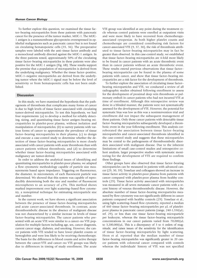
To further explore this question, we examined the tissue fac-tor–bearing microparticles from these patients with pancreaticcancer for the presence of the tumor marker, MUC-1. The MUC-1 antigen is a transmembrane glycoprotein overexpressed in ep-ithelial malignancies such as pancreatic cancer but is not foundon circulating hematopoietic cells (35, 36). The preoperativesamples were labeled with the anti–tissue factor antibody anda monoclonal antibody directed against the MUC-1 antigen. Inthe three patients tested, approximately 50% of the circulatingtissue factor–bearing microparticles in these patients were alsopositive for the MUC-1 antigen (Fig. 6B). These results supportthe premise that a population of microparticles is derived fromthe underlying malignancy. Whether the tissue factor–positive,MUC-1–negative microparticles are derived from the underly-ing tumor where the MUC-1 signal may be below the level ofsensitivity or from hematopoietic cells has not been estab-lished.
Discussion
In this study, we have examined the hypothesis that the path-ogenesis of thrombosis that complicates many forms of canceris due to high levels of tissue factor associated with circulatingtumor-derived microparticles. To test this hypothesis, we hadfour requirements: (a) to develop a method for reliably detect-ing, sizing, and quantitating tissue factor antigen–bearing mi-croparticles in platelet-poor plasma, without isolation andmanipulation of microparticles; (b) to survey patients with var-ious forms of cancer to approximate the prevalence of tissuefactor–bearing microparticles in their plasma; (c) to designand execute a case-control study to determine whether the de-tection of tissue factor–bearing microparticles is more closelyassociated with cancer patients with acute thrombosis than withcancer patients without thrombosis; and (d) to determinewhether tissue factor–bearing microparticles associated withmalignancy are tumor-derived.In order to address the analytical issues of identifying and
quantitating microparticles in platelet-poor plasma, we adapteda flow cytometric methodology capable of particle size mea-surements based upon impedance. Triggering on fluorescence,the diameter, in micrometers, of each fluorescent particle wasdetermined. We showed that this system was capable of repro-ducibly determining both the size and number of fluorescentmicrospheres to an accuracy of ±5%. This method showsmarked improvement over light scattering–based flow cytome-try, a nonoptimal technique for estimating microparticle sizeand number.In the current work, we have shown a significant association
between the presence of tissue factor–bearing microparticlesand acute cancer-associated thrombosis. In contrast, acutethromboembolic disease in the absence of malignant diseasewas not characterized by a similar increase in levels of tissuefactor–bearing microparticles. The cancer patients who pre-sented with an acute VTE were similar to a cancer–no VTE pop-ulation for multiple factors including age, sex, cancer diagnosis,current cancer stage, diabetes, and smoking. However, the can-cer patients with VTE tended to have lower platelet counts orhemoglobin and were less likely to be receiving chemotherapy.The basis for the differences in blood counts and active therapybetween the cancer-VTE and cancer–no VTE groups was likelydue to differences in timing of study enrollment. The acute
VTE group was identified at any point during the treatment cy-cle whereas control patients were enrolled at outpatient visitsand were more likely to have recovered from chemotherapy-associated cytopenias. As both higher platelet counts andchemotherapy are considered contributing risk factors forcancer-associated VTE (9, 37, 38), the risk of thrombosis attrib-uted to tissue factor–bearing microparticles may in fact begreater than observed. In this case-control study, we establishedthat tissue factor–bearing microparticles are 4-fold more likelyto be found in cancer patients with an acute thrombotic eventthan in cancer patients without an acute thrombotic event.These results extend previous observations that tissue factor–bearing microparticles can be found in the plasma of somepatients with cancer, and show that tissue factor–bearing mi-croparticles are a risk factor for the development of thrombosis.To further explore the association of circulating tissue factor–
bearing microparticles and VTE, we conducted a review of allradiographic studies obtained following enrollment to assessfor the development of proximal deep vein thrombosis or pul-monary emboli in cancer patients who had not incurred VTE attime of enrollment. Although this retrospective review wasdone in a blinded manner, the patients were not systematicallyassessed for the development of VTE. However, the potential forsystematic bias was low as this was a nonintervention trial andenrollment did not impact the subsequent management ofthese patients. Only those cancer patients with detectable tissuefactor–bearing microparticles subsequently developed a throm-botic event in the year following enrollment. This analysis cor-roborated the association between tissue factor–bearingmicroparticles and cancer-associated thrombosis identified inthe case-control study and suggests that these microparticlesmay be central to the pathogenesis of thromboembolic disor-ders associated with malignant disease. Due to the inherentlimitations of small case-control studies and retrospective co-hort analysis, larger prospective studies with systematic moni-toring for the development of VTE are required to confirmthese findings.Other groups have also observed that tissue factor–bearing
microparticles can be measured in patients with advanced can-cer (29, 30, 39). Tesselaar and colleagues showed an increase intissue factor activity in platelet-poor plasma from patients withcancer compared with platelet-poor plasma from healthy con-trols (29). Tissue factor activity associated with microparticleswas measured in all seven metastatic cancer patients with a re-cent history of venous thromboembolic disease. However, theabsolute number of tissue factor–bearing microparticles mea-sured by flow cytometry was not significantly different in cancerpatients compared with healthy controls (29). Tesselaar et al.,using light scattering–based flow cytometry, reported a medianof 460 tissue factor–bearing microparticles per μL of platelet-poor plasma in pancreatic cancer patients (range, 240-1,550/μL;ref. 29), or less than one tissue factor–bearing microparticleper leukocyte, whereas the tissue factor–bearing microparticleconcentrations in our cancer patients varied from 70,000/μLto 3,200,000/μL. This is a discrepancy of 3 to 4 orders of mag-nitude, and raises issues of the sensitivity for the identificationof tissue factor–bearing microparticles by light scattering.Hron et al. reported that the median number of tissuefactor–bearing microparticles was significantly greater in 20 can-cer patients with colorectal cancer compared with controlswhereas the individuals' history of VTE was not specified
6838Clin Cancer Res 2009;15(22) November 15, 2009 www.aacrjournals.org
Human Cancer Biology

(30). More recently, Khorana and colleagues observed that twopatients with pancreatic cancer with increased levels of plasmatissue factor subsequently developed a venous thromboembolicevent.(39)We present two independent arguments that the tissue fac-
tor–bearing microparticles are largely derived from the tumoritself rather than from inflammatory cells. First, in three pa-tients with pancreatic carcinoma undergoing pancreatectomywith curative intent, the microparticle concentration before sur-gery was reduced to very low or unmeasurable levels about amonth after surgery. Second, in three patients with pancreaticcarcinoma, we showed that about 50% of the tissue factor–bearing microparticles coexpress MUC-1, a tumor marker forpancreatic carcinoma. Tumor cells express tissue factor (12),the expression of tissue factor activity on tumor cells correlateswith thrombotic risk in pancreatic cancer patients (40), cancercells in culture shed procoagulant vesicular structures (34), andcirculating tumor-derived tissue factor–bearing microparticlescan be detected in mice xenografted with human pancreatic tu-mors (41). These results provide compelling evidence that tis-sue factor–bearing microparticles are derived from certaintumors. Indeed, we suspect that the variability of microparticlegeneration from different tumors, as well as the total tumorburden, is the basis for the variability of thrombotic events incancer-associated thrombosis. However, the presence of circu-lating tissue factor–bearing microparticles is not unique to can-cer patients as high levels were identified in a subset of
noncancer controls as well as individuals with an idiopathicVTE. The clinical significance of these elevations in noncancerpopulations is not known.Venous thromboembolic disease is a leading cause of death
in patients with malignant disease (42), but several recent ran-domized studies have failed to show the benefit of prophylacticanticoagulation in cancer patients even in the presence of in-dwelling central catheters (43–45). The methodologies andconclusions of this work provide a rational basis to determinewhether the detection of tissue factor–bearing microparticles incancer patients and their use as a biomarker predict an in-creased risk of a thromboembolic event. If so, it is critical todetermine whether patients identified with this biomarker willbenefit from primary thromboprophylaxis.
Disclosure of Potential Conflicts of Interest
No potential conflicts of interest were disclosed.
Acknowledgments
We thank Cynthia Kos, Koryn Johnson, and Thomas LaRocca for excellent
technical assistance and Marie Mahony RN for clinical support; Michael
Brochu, Sr., Rick Thomas, Michael Brochu, Jr., and Raquel Cabana at NPE Sys-
temsandGusAlbanatBeckman-Coulter for their collaboration in flowcytometerdevelopment for this application, and Vasilis Toxavidis at the BIDMC Flow Core
Facility; Dr. Hing Wong at Altor Biosciences for the gift of humanized tissue
factor antibody; and Dr. Donald Kufe for the monoclonal antibody to MUC1.
References1. Rickles FR, Edwards RL. Activation of blood co-agulation in cancer: Trousseau's syndrome re-visited. Blood 1983;62:14–31.
2. Donati MB. Cancer and thrombosis: fromPhlegmasia alba dolens to transgenic mice.Thromb Haemost 1995;74:278–81.
3. Francis JL, Biggerstaff J, Amirkhosravi A. He-mostasis and malignancy. Semin Thromb He-most 1998;24:93–109.
4. Arkel YS. Thrombosis and cancer. Semin Oncol2000;27:362–74.
5. Rickles FR, Falanga A. Molecular basis for therelationship between thrombosis and cancer.Thromb Res 2001;102:V215–24.
6. Rickles FR, Patierno S, Fernandez PM. Tissuefactor, thrombin, and cancer. Chest 2003;124:58–68S.
7. Khorana AA, Fine RL. Pancreatic cancer andthromboembolic disease. Lancet Oncol 2004;5:655–63.
8. Varki A. Trousseau's syndrome: multiple defini-tions and multiple mechanisms. Blood 2007;110:1723–9.
9. Heit JA, Silverstein MD, Mohr DN, PettersonTM, O'Fallon WM, Melton LJ III. Risk factors fordeep vein thrombosis and pulmonary embolism:a population-based case-control study. Arch In-tern Med 2000;160:809–15.
10. Blom JW, Vanderschoot JP, Oostindier MJ,Osanto S, van der Meer FJ, Rosendaal FR. Inci-dence of venous thrombosis in a large cohort of66,329 cancer patients: results of a record link-age study. J Thromb Haemost 2006;4:529–35.
11. Levitan N, Dowlati A, Remick SC, et al. Rates ofinitial and recurrent thromboembolic diseaseamong patients with malignancy versus thosewithout malignancy. Risk analysis using Medi-care claims data. Medicine (Baltimore) 1999;78:285–91.
12. Callander NS, Varki N, Rao LV. Immunohisto-chemical identification of tissue factor in solidtumors. Cancer 1992;70:1194–201.
13. Gordon SG, Cross BA. A factor X-activatingcysteine protease from malignant tissue. J ClinInvest 1981;67:1665–71.
14. Boccaccio C, Sabatino G, Medico E, et al. TheMET oncogene drives a genetic programmelinking cancer to haemostasis. Nature 2005;434:396–400.
15. Sun NC, McAfee WM, Hum GJ, Weiner JM.Hemostatic abnormalities in malignancy, a pro-spective study of one hundred eight patients.Part I. Coagulation studies. Am J Clin Pathol1979;71:10–6.
16. Nand S, Fisher SG, Salgia R, Fisher RI. Hemo-static abnormalities inuntreated cancer: incidenceand correlation with thrombotic and hemo-rrhagic complications. J Clin Oncol 1987;5:1998–2003.
17. Buccheri G, Ferrigno D, Ginardi C, Zuliani C.Haemostatic abnormalities in lung cancer:prognostic implications. Eur J Cancer 1997;33:50–5.
18. Johnson MJ, Walker ID, Sproule MW, ConkieJ. Abnormal coagulation and deep venous throm-bosis in patients with advanced cancer. Clin LabHaematol 1999;21:51–4.
19. Sallah S, Wan JY, Nguyen NP, Hanrahan LR,Sigounas G. Disseminated intravascular coagu-lation in solid tumors: clinical and pathologicstudy. Thromb Haemost 2001;86:828–33.
20. Sallah S, Husain A, Sigounas V, et al. Plasmacoagulation markers in patients with solid tu-mors and venous thromboembolic disease re-ceiving oral anticoagulation therapy. ClinCancer Res 2004;10:7238–43.
21. Miller GJ, Bauer KA, Howarth DJ, Cooper JA,Humphries SE, Rosenberg RD. Increased inci-dence of neoplasia of the digestive tract in menwith persistent activation of the coagulant path-way. J Thromb Haemost 2004;2:2107–14.
22. Furie B, Furie BC. Mechanisms of thrombusformation. N Engl J Med 2008;359:938–49.
23. Giesen PL, Rauch U, Bohrmann B, et al. Blood-
borne tissue factor: another view of thrombosis.Proc Natl Acad Sci U S A 1999;96:2311–5.
24. Falati S, Liu Q, Gross P, et al. Accumulation oftissue factor into developing thrombi in vivo isdependent upon microparticle P-selectin glyco-protein ligand 1 and platelet P-selectin. J ExpMed 2003;197:1585–98.
25. Jimenez JJ, Jy W, Mauro LM, Horstman LL,Ahn YS. Elevated endothelial microparticles inthrombotic thrombocytopenic purpura: findingsfrom brain and renal microvascular cell cultureand patients with active disease. Br J Haematol2001;112:81–90.
26. Shet AS, Aras O, Gupta K, et al. Sickle bloodcontains tissue factor-positive microparticles de-rived from endothelial cells and monocytes.Blood 2003;102:2678–83.
27. Combes V, Simon AC, Grau GE, et al.In vitro generation of endothelial microparticlesand possible prothrombotic activity in patientswith lupus anticoagulant. J Clin Invest 1999;104:93–102.
28. Chironi G, Simon A, Hugel B, et al. Circulatingleukocyte-derived microparticles predict subclin-ical atherosclerosis burden in asymptomaticsubjects. Arterioscler Thromb Vasc Biol 2006;26:2775–80.
29. Tesselaar ME, Romijn FP, Van Der Linden IK,Prins FA, Bertina RM, Osanto S. Microparticle-associated tissue factor activity: a link betweencancer and thrombosis? J Thromb Haemost2007;5:520–7.
30. Hron G, Kollars M, Weber H, et al. Tissue fac-tor-positive microparticles: cellular origin andassociation with coagulation activation inpatients with colorectal cancer. Thromb Hae-most 2007;97:119–23.
31. Zwicker JI, Furie BC, Kos C, LaRocca T, BauerKA, Furie B. Trousseau's syndrome revisited: tis-sue factor-bearing microparticles in pancreaticcancer [abstract 259]. Blood 2005;:106.
32. Hercher M, Mueller W, Shapiro HM. Detection
6839 Clin Cancer Res 2009;15(22) November 15, 2009www.aacrjournals.org
Tissue Factor Microparticles and Cancer-Associated VTE

and discriminationof individual viruses by flowcy-tometry. J Histochem Cytochem 1979;27:350–2.
33. Shapiro HM. Practical flow cytometry. 4th ed.New York: Wiley-Liss; 2003.
34. Dvorak HF, Quay SC, Orenstein NS, et al. Tu-mor shedding and coagulation. Science 1981;212:923–4.
35. Ho SB, Niehans GA, Lyftogt C, et al. Heteroge-neity of mucin gene expression in normal andneoplastic tissues. Cancer Res 1993;53:641–51.
36. Levi E, Klimstra DS, Andea A, Basturk O,Adsay NV. MUC1 and MUC2 in pancreatic neo-plasia. J Clin Pathol 2004;57:456–62.
37. Khorana AA, Francis CW, Culakova E, KudererNM, Lyman GH. Frequency, risk factors, andtrends for venous thromboembolism amonghospitalized cancer patients. Cancer 2007;110:2339–46.
38. Khorana AA, Francis CW, Culakova E, LymanGH. Risk factors for chemotherapy-associatedvenous thromboembolism in a prospective ob-servational study. Cancer 2005;104:2822–9.
39. Khorana AA, Francis CW, Menzies KE, et al.Plasma tissue factor may be predictive of ve-nous thromboembolism in pancreatic cancer.J Thromb Haemost 2008;6:1983–5.
40. Khorana AA, Ahrendt SA, Ryan CK, et al. Tis-sue factor expression, angiogenesis, and throm-bosis in pancreatic cancer. Clin Cancer Res 2007;13:2870–5.
41. Davila M, Amirkhosravi A, Coll E, et al. Tissuefactor-bearing microparticles derived fromtumor cells: impact on coagulation activation.J Thromb Haemost 2008;6:1517–24.
42. Donati MB. Cancer and thrombosis. Haemos-tasis 1994;24:128–31.
43. Couban S, Goodyear M, Burnell M, et al.Randomized placebo-controlled study of low-dose warfarin for the prevention of cen‐
tral venous catheter-associated thrombosis inpatients with cancer. J Clin Oncol 2005;23:4063–9.
44. Karthaus M, Kretzschmar A, Kroning H, et al.Dalteparin for prevention of catheter-relatedcomplications in cancer patients with cen-tral venous catheters: final results of a double-blind, placebo-controlled phase III trial. AnnOncol 2006;17:289–96.
45. Verso M, Agnelli G, Bertoglio S, et al. Enoxa-parin for the prevention of venous thromboem-bolism associated with central vein catheter: adouble-blind, placebo-controlled, randomizedstudy in cancer patients. J Clin Oncol 2005;23:4057–62.
6840Clin Cancer Res 2009;15(22) November 15, 2009 www.aacrjournals.org
Human Cancer Biology
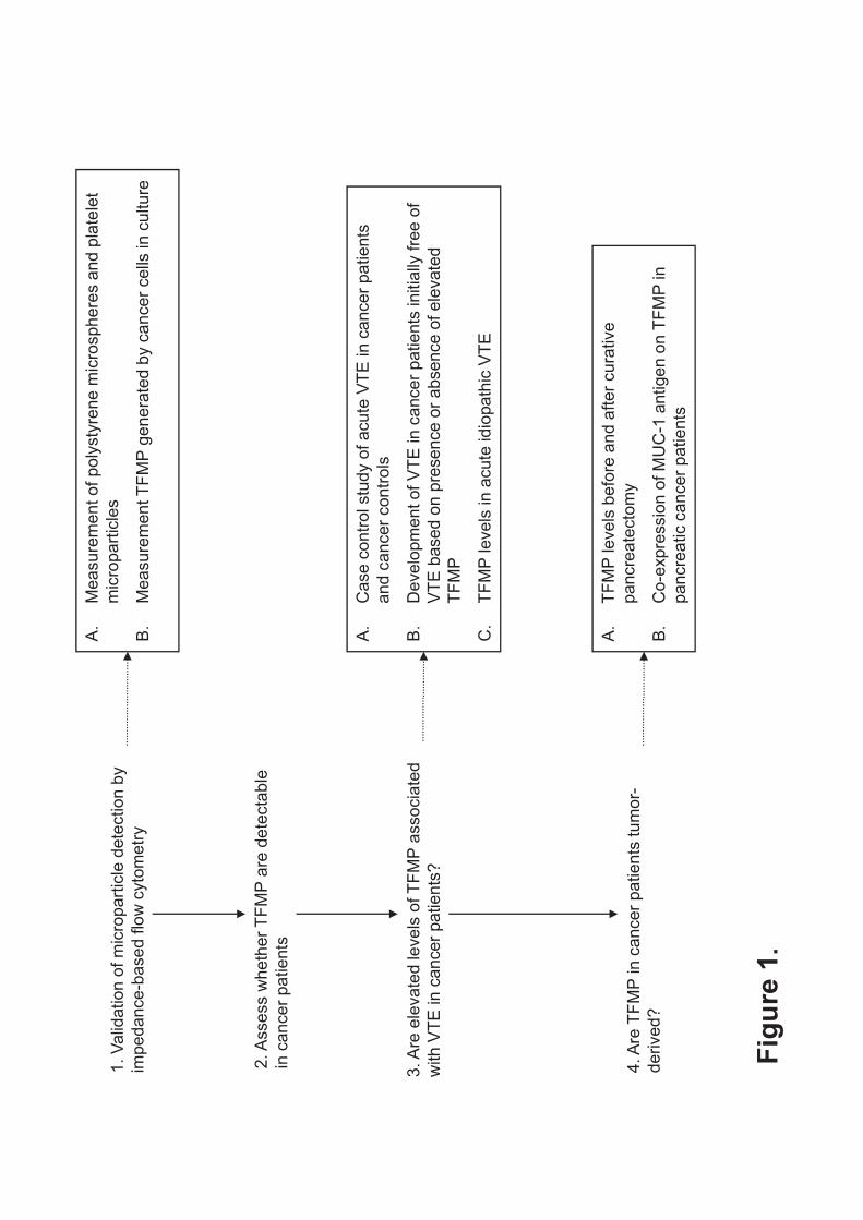
1. V
alid
ation o
f m
icro
part
icle
dete
ction b
y
impedance-b
ased f
low
cyto
metr
y
A.
Measure
ment of
poly
sty
rene m
icro
sphere
s a
nd p
late
let
mic
ropart
icle
s
B.
Measure
ment T
FM
P g
enera
ted b
y c
ancer
cells
in c
ulture
2. A
ssess w
heth
er
TF
MP
are
dete
cta
ble
in c
ancer
patients
A.
Case c
ontr
ol stu
dy o
f acute
VT
E in c
ancer
patients
dt
l
3. A
re e
levate
d levels
of T
FM
P a
ssocia
ted
with V
TE
in c
ancer
patients
?
and c
ancer
con
trols
B.
Develo
pm
ent of
VT
E in c
ancer
patients
initia
lly fre
e o
f
VT
E b
ased o
n p
resence o
r absence o
f ele
vate
d
TF
MP
CT
FM
Ple
vels
inacute
idio
path
icV
TE
C.
TF
MP
levels
inacute
idio
path
icV
TE
4. A
re T
FM
P in c
ancer
patients
tum
or-
derived?
A.
TF
MP
levels
befo
re a
nd a
fter
cura
tive
pancre
ate
cto
my
B.
Co-e
xpre
ssio
n o
f M
UC
-1 a
ntigen o
n T
FM
P in
pancre
atic c
ancer
patients
Fig
ure
1.

ARTICLE
The Rockefeller University Press $30.00
J. Exp. Med. Vol. 206 No. 9 1913-1927
www.jem.org/cgi/doi/10.1084/jem.20082297
1913
The association between the development of
metastasis and the risk of thrombotic com-
plications has been documented since 1865.
Armand Trousseau was the !rst to establish a
direct correlation between thrombophlebitis
and the development of cancer (Trousseau,
1865). A rather common complication and one
of the leading causes of death in patients with
cancer is the risk of developing thromboem-
bolic diseases (Kakkar and Williamson, 1999;
Stein et al., 2006; Schiavetti et al., 2008). The
incidence of thrombosis is high in adenocarci-
nomas such as ovarian, prostate, lung, and gas-
trointestinal carcinomas (Blom et al., 2006b),
and it is particularly high (up to 57%) in pa-
tients su"ering from pancreatic cancer (Sack
et al., 1977; Blom et al., 2006a). In the latter
case, thromboembolic diseases are the second
most common cause of mortality, accounting
for 44% of total deaths after cancer progression
(Neoptolemos et al., 2001). The recurrence of
thrombotic complications may also be the !rst
manifestation of underlying malignant disease
(Prandoni et al., 1992).
The pathogenesis of the thrombotic state in
cancer is associated with the generation of a
local and systemic hypercoagulable/thrombotic
state that confers a growth advantage to tumor
cells. It is now known that activation of the
coagulation cascade and aggregation of blood
platelets around cancer cells protects the cells
from the di"erent degradative pathways pres-
ent in the blood, and also facilitates dissemina-
tion of cancer cells to various sites of metastasis
(Gasic et al., 1976; Sierko and Wojtukiewicz,
2007). This supports a model in which the pres-
ence of tissue factor (TF), generation of throm-
bin, and activation of platelets favor the aggressive
CORRESPONDENCE Christophe Dubois: [email protected]
Abbreviations used: MP, mic-
roparticle; PPP, platelet-poor
plasma; PSGL-1, P-selectin
glycoprotein ligand 1; Qdot,
quantum dot ; TF, tissue factor.
Cancer cell–derived microparticles bearing
P-selectin glycoprotein ligand 1 accelerate
thrombus formation in vivo
Grace M. Thomas,1,2 Laurence Panicot-Dubois,1,2 Romaric Lacroix,3 Françoise Dignat-George,3 Dominique Lombardo,1,2 and Christophe Dubois1,2
1Institut National de la Santé et de la Recherche Médicale (INSERM) UMR911, Centre de Recherche en Oncologie Biologique et Oncopharmacologie, 13385 Marseille, France
2Aix-Marseille Université, Faculté de Médecine La Timone, 13385 Marseille, France3INSERM UMR608, Laboratoire d’Hématologie et d’Immunologie, 13385 Marseille, France
Recent publications have demonstrated the presence of tissue factor (TF)–bearing mic-
roparticles (MPs) in the blood of patients suffering from cancer. However, whether these
MPs are involved in thrombosis remains unknown. We show that pancreatic and lung
cancer cells produce MPs that express active TF and P-selectin glycoprotein ligand 1
(PSGL-1). Cancer cell–derived MPs aggregate platelets via a TF-dependent pathway. In vivo,
cancer cell–derived MPs, but not their parent cells, infused into a living mouse accumulate
at the site of injury and reduce tail bleeding time and the time to occlusion of venules and
arterioles. This thrombotic state is also observed in mice developing tumors. In such mice,
the amount of circulating platelet-, endothelial cell–, and cancer cell–derived MPs is
increased. Endogenous cancer cell–derived MPs shed from the growing tumor are able to
accumulate at the site of injury. Infusion of a blocking P-selectin antibody abolishes the
thrombotic state observed after injection of MPs or in mice developing a tumor. Collec-
tively, our results indicate that cancer cell–derived MPs bearing PSGL-1 and TF play a key
role in thrombus formation in vivo. Targeting these MPs could be of clinical interest in the
prevention of thrombosis and to limit formation of metastasis in cancer patients.
© 2009 Thomas et al. This article is distributed under the terms of an Attribu-tion–Noncommercial–Share Alike–No Mirror Sites license for the #rst six months after the publication date (see http://www.jem.org/misc/terms.shtml). After six months it is available under a Creative Commons License (Attribution–Noncom-mercial–Share Alike 3.0 Unported license, as described at http://creativecommons .org/licenses/by-nc-sa/3.0/).
The J
ourn
al of Experi
menta
l M
edic
ine
on A
ugust 2
0, 2
010
jem
.rupre
ss.o
rgD
ow
nlo
aded fro
m
Published August 10, 2009
http://jem.rupress.org/content/suppl/2009/08/10/jem.20082297.DC1.htmlSupplemental Material can be found at:

1914 A THROMBOTIC ROLE FOR CANCER MICROPARTICLES | Thomas et al.
biology of cancer. Nevertheless, the cause of this association
remains unclear.
Di!erent reports suggest a potential role for circulating
microparticles (MPs) in the establishment of a thrombotic state
in cancer (Kim et al., 2003; Del Conde et al., 2007; Tilley
et al., 2008). MPs are de"ned as cell-derived membrane frag-
ments and range in size from 0.1 to 1 µm in diameter. They
are characterized by their presence at the surface of negative
phospholipid moieties that are essential for initiation of blood
coagulation (Ghosh et al., 2008); they also bear at least one
of the antigenic markers distinctive of the parent cell (Abid
Hussein et al., 2003). Aggregated platelets, leukocytes, eryth-
rocytes, and endothelial lineages constitute the most impor-
tant sources of circulating MPs under many pathophysiological
situations, including thrombosis, in$ammation, and angiogen-
esis (Müller et al., 2003). Recent clinical studies have shown
that the concentration of circulating TF-bearing MPs is sig-
ni"cantly greater in patients with cancer (Tilley et al., 2008),
including patients with pancreatic cancer (Del Conde et al.,
2007; Hron et al., 2007; Tesselaar et al., 2007), suggesting that
these MPs may be responsible for the thrombotic state associ-
ated with cancers. Meanwhile, the cellular origins of such MPs
have not been determined (Hron et al., 2007; Zwicker et al.,
2007; Langer et al., 2008) and may include platelets (Hron
et al., 2007; Tesselaar et al., 2007), cancer cells (Dvorak et al.,
1981), or monocytes (Falati et al., 2003; Myers et al., 2003;
Vandendries et al., 2007). At their surface, platelet-derived
MPs express activated integrins (e.g., IIb 3 or v 3) and
receptors (i.e., GPVI and GPIb-IX-V) known to be involved
in thrombus formation. Monocyte-derived MPs that have
been isolated, labeled, and infused into a recipient mouse ac-
cumulate at the site of a laser-induced injury by binding to
P-selectin expressed on activated platelets through P-selectin
glycoprotein ligand 1 (PSGL-1; Falati et al., 2003; Vandendries
et al., 2007). To date, no study has determined the role of
cancer cell–derived MPs in thrombus formation in vivo.
In the present study, we hypothesize that cancer cell–
derived MPs bearing PSGL-1 are involved in the development
of a thrombotic state. We demonstrate that pancreatic cancer
cells are able to produce MPs in vitro that express active TF
and PSGL-1. We show that cancer cell–derived MPs circu-
late in the bloodstream and accumulate at the site of injury in
a P-selectin–dependent manner in vivo. Finally, we observe
that cancer cell–derived MPs, but not their parental cells, ac-
celerate thrombus formation in a mouse model of cancer.
RESULTSHuman and mouse pancreatic and lung cancer cell lines are able to shed MPs that express TF and PSGL-1 in vitroTo determine whether pancreatic or lung cancer cells could
shed MPs, ultracentrifuged supernatants of human (SOJ-6)
and mouse (Panc02) adenocarcinoma pancreatic cell lines or
mouse Lewis lung carcinoma cancer cell lines (LLC1) were
evaluated by $ow cytometry. All cancer cell lines produced
vesicles ranging in size from 0.4 to 1 µm in diameter, and these
particles were able to bind annexin V (Fig. 1 A). Previous
Figure 1. Pancreatic and lung cancer cell lines shed MPs that express TF in vitro. (A) Flow cytometry analysis showing annexin V–FITC
labeling of SOJ-6, Panc02, and LLC1 cell–derived MPs. Negative controls
were realized without addition of calcium. (B) Immuno!uorescence micros-
copy of cancer cells using an anti-TF antibody. (C) Western blot analysis of
protein extracts from SOJ-6–derived MPs with a speci"c anti-TF antibody.
All of the experiments were independently performed three times.
on A
ugust 2
0, 2
010
jem
.rupre
ss.o
rgD
ow
nlo
aded fro
m
Published August 10, 2009
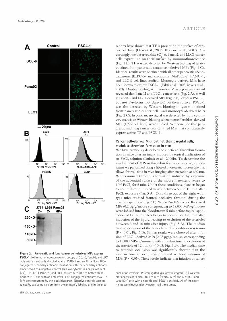
JEM VOL. 206, August 31, 2009
ARTICLE
1915
reports have shown that TF is present on the surface of can-
cer cell lines (Haas et al., 2006; Khorana et al., 2007). Ac-
cordingly, we observed that SOJ-6, Panc02, and LLC1 cancer
cells express TF on their surface by immuno!uorescence
(Fig. 1 B). TF was also detected by Western blotting of lysates
obtained from pancreatic cancer cell–derived MPs (Fig. 1 C).
Identical results were obtained with all other pancreatic adeno-
carcinoma (BxPC-3) and carcinoma (MiaPaCa-2, PANC-1,
and LLC1) cell lines studied. Monocyte-derived MPs have
been shown to express PSGL-1 (Falati et al., 2003; Myers et al.,
2003). Double labeling with annexin V as a positive control
revealed that Panc02 and LLC1 cancer cells (Fig. 2 A), as well
as Panc02- and LLC1-derived MPs (Fig. 2 B), express PSGL-1
but not P-selectin (not depicted) on their surface. PSGL-1
was also detected by Western blotting in lysates obtained
from pancreatic cancer cell– and monocyte-derived MPs
(Fig. 2 C). In contrast, no signal was detected by !ow cytom-
etry analysis or Western blotting when mouse "broblast–derived
MPs (L929 cell lines) were studied. We conclude that pan-
creatic and lung cancer cells can shed MPs that constitutively
express active TF and PSGL-1.
Cancer cell–derived MPs, but not their parental cells, modulate thrombus formation in vivoWe have previously described the kinetics of thrombus forma-
tion in mice after an injury induced by topical application of
an FeCl3 solution (Dubois et al., 2006b). To determine the
involvement of MPs in thrombus formation in vivo, experi-
ments we performed using a "bered !uorescent microscope that
allows for real-time in vivo imaging after excitation at 660 nm.
We examined thrombus formation induced by exposure
of the adventitial surface of the mouse mesenteric vessels to
10% FeCl3 for 5 min. Under these conditions, platelets began
to accumulate in injured vessels between 5 and 15 min after
FeCl3 exposure (Fig. 3 A). Only three out of the eight wild-
type mice studied formed occlusive thrombi during the
35-min experiment (Fig. 3 B). When Panc02 cancer cell–derived
MPs (0.2 µg/g/mouse corresponding to 18,000 MPs/g/mouse)
were infused into the bloodstream 5 min before topical appli-
cation of FeCl3, platelets began to accumulate 1–5 min after
induction of the injury, leading to occlusion of the arterioles
between 3 and 10 min after injury (Fig. 3 A). The median
time to occlusion of the arteriole in this condition was 6 min
(P < 0.01; Fig. 3 B). Similar results were observed after infu-
sion of LLC1-derived MPs (0.08 µg/g/mouse, corresponding
to 18,000 MPs/g/mouse), with a median time to occlusion of
the arteriole of 12 min (P < 0.05; Fig. 3 B). The median time
to arteriole occlusion was signi"cantly shorter than the
median time to occlusion observed without infusion of
MPs (P < 0.05). These results indicate that infusion of cancer
Figure 2. Pancreatic and lung cancer cell–derived MPs express PSGL-1. (A) Immuno!uorescence microscopy of SOJ-6, Panc02, and LLC1
cells with an antibody directed against PSGL-1 and an Alexa Fluor 488–
conjugated secondary antibody. Incubation with the secondary antibody
alone served as a negative control. (B) Flow cytometric analysis of J774
(C+), L929 (C ), Panc02-, and LLC1-derived MPs labeled both with an-
nexin V–FITC and with an anti–PSGL-1 PE-conjugated antibody. PSGL-1+
MPs are represented by the black histogram. Negative controls were ob-
tained by excluding calcium from the annexin V labeling and in the pres-
ence of an irrelevant PE-conjugated IgG (gray histogram). (C) Western
blot analysis of Panc02-derived MPs (Panc02 MPs) and J774 (C+) and
L929 (C ) cells with a speci"c anti–PSGL-1 antibody. All of the experi-
ments were independently performed three times.
on A
ugust 2
0, 2
010
jem
.rupre
ss.o
rgD
ow
nlo
aded fro
m
Published August 10, 2009

1916 A THROMBOTIC ROLE FOR CANCER MICROPARTICLES | Thomas et al.
Figure 3. Cancer cell–derived MPs accelerate thrombus formation in vivo. Thrombus formation was assessed after infusion of Alexa Fluor 660–
conjugated anti-CD41 antibody into wild-type mice in the presence of Panc02-derived MPs (MPs Panc02, 0.2 µg of MP-associated proteins/g/mouse),
LLC1-derived MPs (MPs LLC1, 0.08 µg of MP-associated proteins/g/mouse), or Panc02 cells (Cells Panc02, 5,000 cells/g/mouse) or LLC1 cells (Cells LLC1,
5,000 cells/g/mouse). Injury of the mesenteric vessels was induced with 10% FeCl3 for 5 min. (A) Representative composite images of "uorescence depicting
on A
ugust 2
0, 2
010
jem
.rupre
ss.o
rgD
ow
nlo
aded fro
m
Published August 10, 2009

JEM VOL. 206, August 31, 2009
ARTICLE
1917
cell–derived MPs a�ected the kinetics of thrombus formation.
To determine whether cancer cells may also be involved in
thrombus formation, we determined the time to occlusion in
arterioles after infusion of cancer cells (5,000 cells/g/mouse,
equivalent to the maximum concentration of cells that may be
infused without altering blood !ow; not depicted; Fig. 3 A).
In this condition, no statistical di�erence was found in com-
parison with the kinetics observed when wild-type mice were
injured. Indeed, platelet accumulation was detected between
5 and 10 min after injury. Only three out of seven mice stud-
ied after infusion of Panc02 cells and only one out of eight
mice studied after infusion of LLC1 cells formed occlusive
thrombi (Fig. 3 B). To con"rm the involvement of cancer
cell–derived MPs in thrombus formation, tail bleeding time
experiments were performed as previously described (Panicot-
Dubois et al., 2007). After injection with Panc02- and LLC1-
derived MPs, the median tail bleeding time observed in
wild-type mice was 63 s (n = 8) and 79 s (n = 7), respectively.
These values were signi"cantly lower (P < 0.01) than the
bleeding times (close to 150 s) obtained for wild-type mice
that were not infused with cells or MPs (n = 13), wild-type
mice infused with Panc02 cells (n = 8), or wild-type mice in-
fused with LLC1 cells (n = 8; Fig. 3 C). We next determined
the number of MPs su#cient enough to a�ect kinetics of
thrombosis. We observed that an amount as low as 0.2 ng/g/
mouse of cancer cell–derived MPs signi"cantly accelerated ki-
netics of thrombosis in vivo in our model (Fig. 3 D). Collec-
tively, these results indicate that infusion of cancer cell–derived
MPs, but not infusion of their parental cells, accelerates throm-
bus growth in vivo, suggesting that cancer cell–derived MPs
may participate in thrombus formation.
To determine whether cancer cell–derived MPs accu-
mulate at the site of FeCl3-induced injury in vivo, MPs were
isolated, labeled with the DiD !uorochrome, and infused
into a recipient mouse. First, we determined the half-life of
exogenous MPs in the blood circulation of healthy mice
(Fig. 4 A). The results show that 50% of exogenous MPs
were eliminated from the blood circulation in <15 min, con-
"rming the results previously described for erythrocyte-
derived MPs infused into the rat (Willekens et al., 2005).
After infusion of exogenous MPs and induction of a chemi-
cal injury to mesenteric arterioles, a !uorescent signal corre-
sponding to DiD-labeled MPs was detected (Fig. 4 B). This
accumulation of cancer cell–derived MPs at the site of in-
jury was further con"rmed by intravital microscopy (Dubois
et al., 2007; Panicot-Dubois et al., 2007). Using such a system,
we simultaneously imaged DiO-labeled cancer-derived MPs
(0.2 µg/g/mouse, corresponding to 18,000 MPs/g/mouse)
and DiD-labeled cancer cells (5,000 Panc02 cells/g/mouse)
infused into the bloodstream of a recipient mouse. Both
cancer cell–derived MPs and cancer cells were detected cir-
culating in the blood (Fig. 4 C). However, only cancer cell–
derived MPs were found to accumulate at the site of the
FeCl3-induced injury. 8 min after injury, the number of exog-
enous MPs accumulating at the site of injury increased, indi-
cating that thrombus-associated MPs remained in the animal
for a longer time period than did circulating MPs (Fig. 4 C).
We conclude that cancer cell–derived MPs, but not their
parental cells, accumulate at the site of injury in vivo and
accelerate thrombus growth.
Characterization of a thrombotic state in mice developing ectopic tumorsNext, we created a mouse model of cancer induced by subcu-
taneous injection of pancreatic (Panc02) or lung cancer (LLC1)
cells. 5 wk after induction of the ectopic tumor, the number
of endogenous circulating MPs in both healthy mice and mice
developing tumors were compared by !ow cytometry analy-
sis. To characterize the cellular origin of MPs, we "rst labeled
MPs using antibodies speci"c for platelets (CD41+), erythro-
cytes (TER 119+), leukocytes (CD45+), cells likely of endo-
thelial origin (CD31+CD41 ), and cancer cells (MUC-1+). The
data are presented in Table I (see also Fig. S1). As expected,
the number of endogenous circulating annexin V+ MPs was
signi"cantly increased in mice developing tumors in compari-
son with control mice (median = 9,383 MPs/µl and 10,018
MPs/µl of plasma for Panc02- and LLC1-induced-tumor
mice, respectively, and 8,951 MPs/µl of plasma in control
mice; P < 0.05). The amount of platelet- and endothelial cell–
derived MPs was signi"cantly increased in mice developing
tumors in comparison with control mice. However, neither
the number of leukocyte-derived circulating MPs nor the
quantity of endogenous erythrocyte-derived MPs increased in
comparison with the control. We also addressed the presence
of endogenous circulating cancer cell–derived MPs using
Muc-1, a protein that is highly expressed in various epithelial
cancers. As expected, the amount of Muc-1–bearing MPs was
signi"cantly increased in tumor-induced mice in comparison
with healthy mice. We also detected MPs bearing the carbo-
hydrate CA19-9, which is highly expressed in lung and pan-
creatic cancers. Of note, this antigen was only present on MPs
in blood obtained from Panc02- and LLC1-tumor-induced
thrombus formation of labeled platelet accumulation (white) on mesenteric vessels (Pre, before injury). (B) Time to vessel occlusion reported in minutes
for mesenteric arterioles in wild-type mice (n = 10 thrombi in 10 mice), wild-type mice infused with Panc02-derived MPs (n = 10 thrombi in 7 mice), or
with LLC1-derived MPs (n = 6 thrombi in 6 mice), Panc02 cells (n = 7 thrombi in 7 mice), or LLC1 cells (n = 8 thrombi in 8 mice). (C). Bleeding time in sec-
onds determined in wild-type mice (n = 13 mice), wild-type mice infused with Panc02-derived MPs (n = 8 mice), or with LLC1-derived MPs (n = 7 mice),
Panc02 cells (n = 8 mice), or LLC1 cells (n = 8 mice). (D) Times to arteriole occlusion were observed in wild-type mice (n = 10 thrombi in 10 mice) and in
wild-type mice infused with different concentrations of Panc02-derived MPs (MPs 0.02 ng/g/mouse, n = 7 thrombi in 6 mice; MPs 0.2 ng/g mouse, n = 7
thrombi in 7 mice; and MPs 200 ng/g mouse, n = 10 thrombi in 9 mice). Horizontal bars indicate median values. Experiments were independently per-
formed 10 times (A and B) and at least six times (C and D). **, P < 0.01; *, P < 0.05.
on A
ugust 2
0, 2
010
jem
.rupre
ss.o
rgD
ow
nlo
aded fro
m
Published August 10, 2009

1918 A THROMBOTIC ROLE FOR CANCER MICROPARTICLES | Thomas et al.
and venules of control and tumor-bearing mice (Fig. 5 B).
A signi!cant decrease in the time to occlusion of the arterioles
(P < 0.05; Fig. 5 C) and venules (P < 0.05; Fig. 5 D) was ob-
served in mice developing tumors (a median time to occlu-
sion of 8 and 15 min in arterioles and 13 and 8 min in venules
in mice developing Panc02- and LLC1-induced tumors, re-
spectively) in comparison with control mice (median time > 35
and 18 min in arterioles and venules, respectively). Interest-
ingly, the tail bleeding time and time to occlusion of arteri-
oles and venules were similar in mice developing tumors
(median volume = 239 mm3) and mice for which Panc02-
derived MPs were infused into the circulation (compare
Fig. 3 B with 5 C and Fig. 3 C with 5 A). Furthermore, we
did not observe any linear correlation between the volume of
the tumor and the bleeding time (Fig. 5 E). Our results indi-
cate that the presence of an ectopic tumor in mice increases
the quantity of MPs present in the bloodstream and acceler-
ates thrombus formation, as compared with healthy mice.
mice (Table I). Because we have previously observed that
Panc02- and LLC1-derived MPs express PSGL-1 on their sur-
face (Fig. 2) and because the quantity of leukocyte-derived
MPs, which also bear PSGL-1, was not increased in tumor-in-
duced mice in comparison with control mice, we determined
the quantity of circulating PSGL-1–bearing MPs in the di"er-
ent populations of mice. The number of MPs bearing PSGL-1
was signi!cantly increased in mice developing tumors in com-
parison with healthy mice. Collectively, our results indicate
that the elevation of endogenous cell–derived MPs positive for
annexin V results from a selective increase in MPs originating
from platelets, endothelial cells, and tumor cells.
Tail bleeding times were signi!cantly reduced in mice
developing a tumor (median times for mice that received
Panc02 or LLC1 cancer cells were 70 and 78 s, respectively)
in comparison with mice that did not (median time = 150 s;
P < 0.01; Fig. 5 A). We determined the kinetics of thrombus
formation after FeCl3 injury to both the mesenteric arterioles
Figure 4. Cancer cell–derived MPs accumulate at the site of injury in mice. (A) Half-life of DiO-labeled Panc02-derived MPs in the blood circula-
tion of a healthy mouse (n = 4 mice, performed independently) reported as the mean ± SD. (B) Imaging of DiD-labeled Panc02-derived MPs using "bered
#uorescence microscopy. Panc02-derived MPs were labeled with DiD (left) before infusion into the blood circulation of a living mouse. Although no #uo-
rescent signal was detected in the microcirculation before injury (middle), MPs were detected accumulating at the site of FeCl3-induced injury (right).
Images are representative of six injuries performed in three mice, independently. (C) Intravital videomicroscopy visualization of mesenteric vessels. DiO-
labeled MPs (depicted in green; 0.2 µg of MP-associated proteins/g/mouse) and their DiD-labeled parental cells (depicted in red; 5,000 cells/g/mouse)
were simultaneously infused into a recipient mouse. Circulating MPs and cells were detected in the microcirculation before injury (second from left). Note
that no #uorescent signal was detected before infusion of MPs and cells (left). The experiment was independently performed three times in three mice.
on A
ugust 2
0, 2
010
jem
.rupre
ss.o
rgD
ow
nlo
aded fro
m
Published August 10, 2009
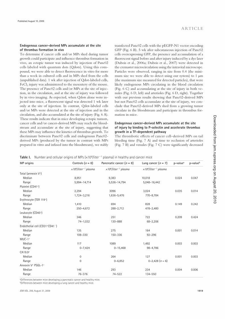
JEM VOL. 206, August 31, 2009
ARTICLE
1919
transfected Panc02 cells with the pEGFP-N1 vector encoding
GFP (Fig. 6 B). 5 wk after subcutaneous injection of Panc02
cells overexpressing GFP, the presence and accumulation of a
!uorescent signal before and after injury induced by a dye laser
(Dubois et al., 2006a; Dubois et al., 2007) were detected in
the cremaster microcirculation using the intravital microscope.
Particles were observed, ranging in size from 0.4 (the mini-
mum size we were able to detect using our system) to 1 µm
(the maximum size measured for detected particles), that were
likely endogenous MPs circulating in the blood circulation
(Fig. 6 C) and accumulating at the site of injury in both ve-
nules (Fig. 6 D, left) and arterioles (Fig. 6 D, right). Together
with our previous results showing that Panc02-derived MPs
but not Panc02 cells accumulate at the site of injury, we con-
clude that Panc02-derived MPs shed from a growing tumor
circulate in the bloodstream and participate in thrombus for-
mation in mice.
Endogenous cancer-derived MPs accumulate at the site of injury by binding to P-selectin and accelerate thrombus growth in a TF-dependent pathwayThe thrombotic e#ects of cancer cell–derived MPs on tail
bleeding time (Fig. 7 A) and time to occlusion of arterioles
(Fig. 7 B) and venules (Fig. 7 C) were signi$cantly decreased
Endogenous cancer-derived MPs accumulate at the site of thrombus formation in vivoTo determine if cancer cells and/or MPs shed during tumor
growth could participate and in!uence thrombus formation in
vivo, an ectopic tumor was induced by injection of Panc02
cells labeled with quantum dots (Qdots). Using this com-
pound, we were able to detect !uorescence in vitro for more
than a week in cultured cells and in MPs shed from the cells
(unpublished data). 1 wk after injection of Qdot-labeled cells,
FeCl3 injury was administered to the mesentery of the mouse.
The presence of Panc02 cells and/or MPs at the site of injec-
tion, in the circulation, and at the site of injury was followed
by in vivo imaging. As expected, when Qdots alone were in-
jected into mice, a !uorescent signal was detected 1 wk later
only at the site of injection. In contrast, Qdot-labeled cells
and/or MPs were detected at the site of injection and in the
circulation, and also accumulated at the site of injury (Fig. 6 A).
These results indicate that in mice developing ectopic tumors,
cancer cells and/or cancer-derived MPs may reach the blood-
stream and accumulate at the site of injury, suggesting that
these MPs may in!uence the kinetics of thrombus growth. To
discriminate between Panc02 cells and endogenous Panc02-
derived MPs (produced by the tumor in contrast with MPs
prepared in vitro and infused into the bloodstream), we stably
Table I. Number and cellular origins of MPs (×106/liter 1 plasma) in healthy and cancer mice
MP origins Controls (n = 8) Pancreatic cancer (n = 8) Lung cancer (n = 7) p-valuea p-valueb
×106/liter 1 plasma ×106/liter 1 plasma ×106/liter 1 plasma
Total (annexin V+)
Median 8,951 9,383 10,018 0.024 0.047
Range 5,994–14,714 5,536–14,794 5,048–16,442
Platelet (CD41+)
Median 2,294 3096 3,024 0.035 0.014
Range 1,724–3,216 1,636–5,476 770–8,784
Erythrocyte (TER 119+)
Median 1,410 694 828 0.149 0.242
Range 250–4,672 288–2,712 478–2,480
Leukocyte (CD45+)
Median 346 251 722 0.209 0.424
Range 74–1,032 130–888 68–2,206
Endothelial cell (CD31+CD41 )
Median 135 275 164 0.001 0.014
Range 108–330 150–336 92–296
MUC-1+
Median 117 1089 1,492 0.003 0.003
Range 0–7,424 0–15,468 98–4,786
CA19.9+
Median 0 264 127 0.001 0.003
Range 0 0–6,852 0–2,428 (n = 6)
Annexin V+ PSGL-1+
Median 146 293 234 0.004 0.006
Range 76–376 74–522 134–550
aDifferences between mice developing a pancreatic cancer and healthy mice.bDifferences between mice developing a lung cancer and healthy mice.
on A
ugust 2
0, 2
010
jem
.rupre
ss.o
rgD
ow
nlo
aded fro
m
Published August 10, 2009

1920 A THROMBOTIC ROLE FOR CANCER MICROPARTICLES | Thomas et al.
TF was present in a decrypted (active) form on the surface of
cancer cells and MPs, the activity of TF present on SOJ-6,
Panc02, and LLC1 cells, as well as MPs, was determined.
when proteins at the surface of MPs were stripped by trypsin
before infusion into the recipient mouse. Cancer cells and MPs
express TF on their surfaces (Fig. 1). To determine whether
Figure 5. Characterization of a thrombotic state in mice developing tumors. Kinetics of thrombosis in wild-type mice or in mice developing
Panc02-derived (Cancer Panc02) or LLC1-derived (Cancer LLC1) tumors. (A) Bleeding times were determined in wild-type mice (n = 13 mice), mice devel-
oping a Panc02 tumor (n = 11 mice), or mice developing an LLC1 tumor (n = 6 mice). (B) Thrombus formation was studied after infusion of Alexa Fluor
660–conjugated anti-CD41 Fab fragment and Syto 62 in wild-type mice and in mice developing tumors. Injury was induced in mesenteric vessels by topi-
cal application of 10% FeCl3 for 5 min. Representative !uorescence images depicting the kinetics of thrombus formation of labeled platelet accumulation
(white) on mesenteric arterioles (Pre, before injury). (C and D) Time to vessel occlusion reported in minutes for mesenteric arterioles (n = 13, 7, and 6
thrombi for WT, Cancer Panc02, and Cancer LLC1, respectively; C) and venules (n = 13, 11, and 6 thrombi for WT, Cancer Panc02, and Cancer LLC1, respec-
tively; D). One injury was performed per mouse. (E) Bleeding times (in seconds) in function of the tumor volume (in mm3) in Panc02 tumor–bearing mice
(n = 11). The linear coef"cient of determination value is represented by R2. Horizontal bars in A, C, and D indicate median values. Experiments were inde-
pendently performed at least six times (A–D) and 11 times (E). **, P < 0.01; *, P < 0.05.
on A
ugust 2
0, 2
010
jem
.rupre
ss.o
rgD
ow
nlo
aded fro
m
Published August 10, 2009

JEM VOL. 206, August 31, 2009
ARTICLE
1921
cancer cells and MPs. Our results indicate that pancreatic
and lung cancer cells are able to shed MPs expressing TF and
PSGL-1 on their surface. In vivo, in a mouse model of cancer,
these MPs circulate in the blood, accumulate at the site of injury
in a P-selectin–dependent manner, and may participate in
As previously described by others (Milsom et al., 2007; Pawlinski
and Mackman, 2008), we observed that cancer cells express
active TF, demonstrating activity ranging from 7 × 10 4 pmol/
cell for the two mouse cancer cell lines tested to 17 × 10 4
pmol/cell for human SOJ-6 cancer cells (Fig. 7 D). All MPs
studied expressed similar concentrations of active TF (between
2.3 × 10 4 and 4.3 × 10 4 pmol/MP). We then determined
the activity of TF as a ratio of TF concentration to surface area
to control for the di"erence in size between a cancer cell (di-
ameter = 20 µm) and an MP (diameter = 1 µm). Results ob-
tained show that the density of active TF is increased >100-fold
in MPs as compared with cancer cells. (Fig. 7 D). These cancer
cell–derived MPs were able to activate platelets and to induce
platelet aggregation and formation of a $brin mesh after addi-
tion of platelet-poor plasma (PPP) to a washed platelet prepa-
ration (Fig. 7 E). This e"ect was slightly inhibited when a
$brinogen-de$cient plasma was added to the platelet prepara-
tion and was abolished by using a factor VII–depleted PPP
(Fig. 7 E). These data demonstrate that MPs activate and ag-
gregate platelets via a TF-dependent pathway in vitro.
To determine whether endogenous MPs arising from
cancer cells accumulate in the thrombus growth via an inter-
action between PSGL-1 and P-selectin, the accumulation of
endogenous Panc02-derived MPs was compared at the site
of injury before and after infusion of a blocking P-selectin
monoclonal antibody, as previously described (Falati et al.,
2003; Vandendries et al., 2007). Although accumulation of
endogenous MPs was observed after laser- or FeCl3-induced
injury (Fig. 8 A, left) to the mesentery, no signal corresponding
to endogenous cancer cell–derived MPs bound to a throm-
bus was detected upon infusion of the P-selectin antibody
(Fig. 8 A, right). The same results were observed when exog-
enous DiD-labeled cancer cell–derived MPs were infused
into the circulation in the presence of blocking antibody (un-
published data). We conclude that cancer cell–derived MPs
accumulate at the site of thrombus formation via interactions
between the PSGL-1 expressed at their surface and P-selectin
expressed on activated platelets and endothelial cells. Further-
more, the median times to occlusion in arterioles (Fig. 8 B)
and venules (Fig. 8 C) were signi$cantly higher when cancer
cell–derived MPs were infused in the presence of blocking
P-selectin antibody than when they were infused alone (P <
0.05). These results were con$rmed by determining the tail
bleeding time, which was signi$cantly higher in the presence of
P-selectin (P < 0.01; Fig. 8 D). Collectively, our results indicate
that tumor-shed MPs reach the bloodstream, accumulate at the
site of injury in a P-selectin-dependent manner, and accelerate
thrombus growth mainly via a TF-dependent pathway.
DISCUSSIONIn the present paper, we studied the role of cancer cell–derived
MPs in thrombus formation. To characterize the throm-
botic/coagulation state in vivo, we measured the kinetics of
thrombus formation after a chemical- or laser-induced in-
jury. We also compared the tail bleeding time of healthy
mice, mice developing tumors, and mice after infusion of
Figure 6. Endogenous cancer cell–derived MPs accumulate at the site of thrombus formation in vivo. (A) Qdot-labeled Panc02 cells (2 ×
106 cells) or Qdots alone (Control) were injected subcutaneously into the
right "ank of a mouse. 1 wk later, "uorescent signals were detected at the
site of injection (left), and in the mesentery before (middle panel) and after
(right) FeCl3-induced injury. (B) Fluorescence microscopy of Panc02 cells
overexpressing GFP before subcutaneous injection into wild-type mice.
(C and D) 2 × 10⁵ Panc02 cells overexpressing GFP were injected subcuta-
neously into the right "ank of a mouse. 5 wk later, the cremaster was iso-
lated and "uorescent circulating GFP-labeled MPs were detected in the
blood microcirculation (red arrow, direction of the blood "ow; red circle,
GFP microparticle; C) and accumulating at the site of injury in venules
(D, left) and arterioles (D, right). All images are representative of three
independent experiments observed for nine thrombi formed in three mice.
on A
ugust 2
0, 2
010
jem
.rupre
ss.o
rgD
ow
nlo
aded fro
m
Published August 10, 2009

1922 A THROMBOTIC ROLE FOR CANCER MICROPARTICLES | Thomas et al.
cells to characterize their respective roles in the kinetics of
thrombus formation in vivo.
Among the di!erent proteins able to activate and aggre-
gate platelets while simultaneously favoring the process of
metastasis, TF expressed on cancer cells has been suggested to
play a key role in the development of a cancer-associated
thrombotic state (Riewald and Ruf, 2001; Haas et al., 2006;
the generation of thrombin, likely via the expression of ac-
tive TF at their surface. The presence of MPs is necessary for
the formation of a thrombotic state in vivo. Finally, our re-
sults show that cancer cells do not accumulate at the site of
injury and do not accelerate the kinetics of thrombosis in
our model. To our knowledge, this is the "rst study to use
imaging of endogenous cancer cell–derived MPs and cancer
Figure 7. Endogenous cancer-derived MPs accelerate thrombus growth through a TF-dependent pathway. (A–C) Kinetics of thrombosis in mice
perfused with control Panc02 MPs or trypsinized Panc02 MPs (18,000 MPs/g/mouse) compared with wild-type mice. (A) Bleeding time of wild-type mice
and of mice perfused with control MPs (WT+MPs) or trypsinized MPs (WT+trypsinized MPs; n = 6 mice for each group). Time to vessel occlusion reported
in minutes for mesenteric arterioles (B) and venules (C; n = 6 thrombi in six mice for each group). Injury of the mesenteric vessels was induced by topical
application of 10% FeCl3 for 5 min. Horizontal bars indicate median values. **, P < 0.01; *, P < 0.05. (D) Measurement of TF activity on the surface of can-
cer cells and MPs in pmol/cell 1 and pmol/MP 1 (left) or in pmol/mm2 (right), reported as the mean ± SD. (E) Aggregation of 2.5 × 10⁸ human washed
platelets/ml induced by addition of thrombin (Curve 1, red), SOJ-6 MPs (1 µg of MP-associated proteins/ml) in the absence (Curve 2, black) or presence of
5% vol/vol ($nal) of PPP (Curve 3, blue), factor VII–depleted PPP (Curve 4, green), or $brinogen-de$cient PPP (Curve 5, gray). Experiments were indepen-
dently performed six times (A–C) and three times (D and E).
on A
ugust 2
0, 2
010
jem
.rupre
ss.o
rgD
ow
nlo
aded fro
m
Published August 10, 2009

JEM VOL. 206, August 31, 2009
ARTICLE
1923
Versteeg et al., 2008). However, no direct correlation could
be established between the presence of TF on cancer cells and
the development of thrombosis complications associated with
cancer (Furie and Furie, 2006), suggesting that other impor-
tant mechanisms may link cancer and thrombosis. Recently,
various reports have shown that the number of circulating
MPs is increased in patients developing di!erent types of can-
cers known to be associated with a thrombotic state (Kim et al.,
2003; Del Conde et al., 2007; Zwicker et al., 2007). These
observations suggest that MPs may play a key role in the de-
velopment of the thrombotic complications associated with
cancer. Our results indicate that MPs derived from cancer
cells enhance thrombus formation. The presence of PSGL-1
targets MPs to sites of thrombosis, whereas active TF actively
participates in the formation of the thrombus by generating
thrombin and "brin. Two recent reports have shown, in mice
models, a direct correlation between the size of a tumor and
the level of TF present in cell-free plasma (Yu et al., 2005;
Davila et al., 2008). We did not observe such correlation
when comparing the bleeding time associated with the vol-
ume of the tumors (Fig. 5 E). Indeed, even relatively small
tumors are su#cient enough to strongly diminish the tail
bleeding time and the occlusion time in our model. These
results con"rm that the presence in the bloodstream of few
tumor-derived MPs is su#cient enough to induce a throm-
botic state in mice. Last, in the two papers, authors have not
discriminated the origin of circulating TF that may come
from platelets or monocyte-derived MPs. Other studies have
previously shown that such MPs may originate from platelets
(Hron et al., 2007; Tesselaar et al., 2007). In this study, we
observed that the quantities of circulating platelet-, endothe-
lial cell–, and cancer cell–derived MPs are increased in mice
developing tumors compared with healthy mice. With the
knowledge that platelets release MPs only when they aggre-
gate (Piccin et al., 2007), it not surprising that the amount of
circulating MPs derived from platelets increases when patients/
mice develop a thrombotic state. Indeed, this pathological
Figure 8. Endogenous cancer-derived MPs accelerate thrombus formation in a P-selectin–dependent manner. (A) 2 × 10⁵ Panc02 cells
overexpressing GFP were injected subcutaneously into the right "ank of a
mouse. 5 wk later, chemical injury of the mesentery was induced, and
"uorescence was detected in the absence (left, Control) or presence of
circulating P-selectin blocking antibody (right, P-selectin antibody; n = 6
thrombi in three mice). (B–D). Kinetics of thrombosis in wild-type mice,
wild-type mice infused with Panc02 MPs (WT+MPs, 0.2 µg of MP-associ-
ated proteins/g/mouse), or with Panc02 MPs plus 2 µg/g/mouse of anti
P-selectin blocking antibody (WT+MPs+anti P-sel). Controls were performed
by infusion of anti–P-selectin antibody into wild-type mice (WT+anti
P-sel) or by infusing an irrelevant antibody into wild-type mice perfused
with MPs (WT+MPs+Irr Ab). Injury of the mesenteric vessels was chemi-
cally induced, and the time to vessel occlusion for arterioles (n = 10 for
WT, n = 7 for WT+anti Psel, n = 10 for WT+MPs, n = 7 for WT+MPs+anti
Psel, and n = 8 for WT+MPs+Irr Ab; B) and veinules (n = 13 for WT, n = 7
for WT+anti Psel, n = 7 for WT+MPs, n = 7 for WT+MPs+anti Psel, and
n = 8 for WT+MPs+Irr Ab; C) was measured. (D) Bleeding times were deter-
mined for all conditions studied (n = 13 for WT, n = 7 for WT+anti Psel,
n = 8 for WT+MPs, n = 6 for WT+MPs+Anti Psel, and n = 8 for
WT+MPs+Irr Ab). One thrombus was performed per mouse. Horizontal
bars designate median values. Experiments were independently performed
three times (A) and at least seven times (B–D). **, P < 0.01; *, P < 0.05.
on A
ugust 2
0, 2
010
jem
.rupre
ss.o
rgD
ow
nlo
aded fro
m
Published August 10, 2009

1924 A THROMBOTIC ROLE FOR CANCER MICROPARTICLES | Thomas et al.
at least partially, by the levels of active TF expressed by
Panc02- and LLC1-derived MPs. Indeed, activation of the
coagulation cascade resulting in thrombin generation is known
to play a more important role in thrombus formation induced
in arteries than in veins.
We demonstrated that PSGL-1 is present on the surface of
MPs and showed that injection of a P-selectin blocking anti-
body prevents the accumulation of MPs at the site of injury
and reduces thrombus formation. The P-selectin–PSGL-1
axis has previously been described as being involved in the
binding of MPs to platelets. Indeed, Falati et al. (2003) have
previously shown that exogenous monocyte-derived MPs ex-
press PSGL-1 and bind to a growing thrombus in a P-selectin–
dependent manner in a laser injury model. We determined
that inhibition of P-selectin did not a!ect time to occlusion
in the FeCl3 model in healthy mice, showing that accumula-
tion of monocyte-derived MPs by the P-selectin–PSGL-1
axis may have a minor implication when cancer-derived MPs
are not involved in thrombus formation after a FeCl3-induced
injury. Our results identify P-selectin as a potentially good
pharmaceutical target for the prevention of thrombosis asso-
ciated with cancer, and may also explain why heparin, which
binds to and blocks selectin, is much more e!ective than war-
farin, a vitamin K antagonist, in preventing thrombosis in pa-
tients su!ering from Trousseau’s syndrome (Del Conde et al.,
2007). MPs also express negatively charged phospholipids on
their surface that may participate with TF in activation of the
coagulation cascade. However, based on our results, the ac-
tivity of TF seems to be critical for the generation of throm-
bin. Indeed, we found that (a) in vitro TF activity per surface
unit is 100-fold more important in MPs than in their parental
cells (Fig. 7 D), (b) the use of factor VII–de"cient plasma
abolishes the e!ect of MPs on platelet aggregation and the
formation of a "brin mesh (Fig. 7 E), and (c) in vivo hydroly-
sis of the surface proteins but not the negatively charged phos-
pholipids present on MPs completely abrogated their e!ect
on the kinetics of thrombosis (Fig. 7, A–C). Based on our re-
sults, we propose a two-step mechanism to describe the in-
volvement of cancer cell–derived MPs in thrombosis. First,
MPs bind to the site of injury by interactions between PSGL-1
and P-selectin present on activated platelets and endothelial
cells. Second, MPs a!ect thrombus formation, likely via the
expression of active TF on their surface. Other proteins and
lipids may also be of importance in this process. Among the
di!erent proteins expressed by cancer cell–derived MPs, feto-
acinar pancreatic protein and "bronectin were identi"ed (un-
published data; Ni et al., 2003). Both of these may play an
important role in thrombus formation. Fetoacinar pancreatic
protein, an onco-isoform of bile salt–dependent lipase, increases
the kinetics of thrombus formation by binding to CXCR4
on the surface of activated platelets (Panicot-Dubois et al.,
2007). Overall, our data provide a new molecular basis for es-
tablishing relationships between cancer and thrombosis, and
may facilitate the development of new pharmaceutical drugs
designed to prevent thrombosis and metastasis in patients suf-
fering from cancer.
state leads to the activation and aggregation of platelets, either
directly via cancer cells or indirectly via production of throm-
bin by TF. Therefore, it is likely that platelet-derived MPs are
a consequence of thrombotic complications and do not cause
the development of this phenotype. However, platelet-
derived MPs, when present in the blood, may also participate
in the stability of a thrombotic state associated with cancer.
Endothelial cells activated by tumoral MPs may provide a sup-
plementary procoagulant surface by TF exposure. Although
several reports have shown that TF is expressed on cancer cells,
there is con#icting evidence as to the presence of this factor
at the surface of platelets and their MPs (Furie and Furie,
2006). To our knowledge, this is the "rst paper to demon-
strate an increase in circulating endothelial cell–derived MPs
in association with cancer. However, previous publications
have shown that the level of endothelial cell–derived MPs is
increased in in#ammatory-related diseases (Combes et al., 1999;
Chamouard et al., 2005). These MPs, by expressing proteins
such as metalloproteases, urokinase plasminogen activator,
and urokinase plasminogen activator receptor (Lacroix et al.,
2007), may potentially take part in and amplify the processes
of in#ammation, angiogenesis, and "brinolysis. Further ex-
periments are needed to determine the actual role of platelet-
derived MPs in concert with cancer cell– and endothelial
cell–derived MPs.
When cancer cells and cancer cell–derived MPs were
infused into the circulation of a living mouse, only MPs
were observed to accumulate at the site of a laser- or chemi-
cal-induced injury, suggesting that cancer cells may not
participate in thrombus formation. These cells were not
eliminated from the blood, because we were able to detect
them circulating in the vessels (Fig. 4 C, before injury). This
result was con"rmed by our data showing that injection of
cancer cells did not in#uence either the kinetics of throm-
bus formation or bleeding times. Our results are consistent
with previous observations showing that monocyte-derived
MPs, but not their parent cells, may indeed interact with a
growing thrombus (Falati et al., 2003). Although circulating
cancer cells did not bind to the site of injury, they may in-
teract with the endothelium at sites of premetastatic niches
to form sites of metastasis (Kaplan et al., 2006). Di!erent
hypotheses may explain why MPs and not their parental
cells bind to a growing thrombus. It is possible that this
interaction occurs via the negative phospholipid moieties
commonly expressed on the surface of MPs. Furthermore,
the quantity and density of binding partners present, such as
PSGL-1, may be more important for MPs than for their par-
ent cells. This point is illustrated by our results showing that
even if the activity of cell TF was greater than that of the
MP, when the results were reported per surface unit, the
activity of TF was >100-fold higher in MPs than in cells.
This result may also explain why the cells were not throm-
bogenic compared with MPs. In our conditions, Panc02-
derived MPs have a more dramatic e!ect on thrombosis in
arterioles compared with venules, whereas LLC1-derived
MPs have the opposite e!ect. This tendency may be caused,
on A
ugust 2
0, 2
010
jem
.rupre
ss.o
rgD
ow
nlo
aded fro
m
Published August 10, 2009
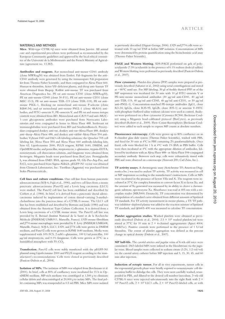
JEM VOL. 206, August 31, 2009
ARTICLE
1925
as previously described (Dignat-George, 2006). L929 and J774 cells were ac-
tivated with 10 ng/ml TNF- before MP isolation. Concentrations of MPs
were determined by protein quanti!cation using the bicinchoninic acid assay
(Thermo Fisher Scienti!c).
PAGE and Western blotting. SDS-PAGE performed on gels of poly-
acrylamide (7.5% acrylamide in the presence of 0.1% sodium dodecyl sulfate)
and Western blotting were performed as previously described (Panicot-Dubois
et al., 2007).
Flow cytometry. Platelet-free plasma (PFP) samples were prepared as pre-
viously described (Sabatier et al., 2002) using serial centrifugations and stored
at 80°C until use. For MP labeling, 30 µl of freshly thawed PFP or of the
MP suspension was incubated for 30 min with 10 µl FITC–annexin V or
PE–anti–mouse monoclonal antibodies (30 µg/ml anti-CD41, 40 µg/ml
anti–TER-119, 40 µg/ml anti-CD45, 40 µg/ml anti-CD31, or 50 µg/ml
anti–PSGL-1). Concentration-matched PE-isotype antibodies (IgG1, clone
R3-34; IgG2a, clone R35-95; IgG2b, clone A95-1) or annexin V–FITC
with phosphate-bu$ered saline without calcium were used as control. Analy-
ses were performed on a &ow cytometer (Cytomics FC500; Beckman Coul-
ter) using a Megamix bead–calibrated protocol (BioCytex), as previously
described (Robert et al., 2009). Flow-Count &uorospheres (Beckman Coul-
ter) were added to each sample to express MP counts as absolute numbers.
Fluorescence microscopy. Cells were grown up to 80% con&uence on an
8-chamber glass slide system (Thermo Fisher Scienti!c), washed with PBS,
and !xed for 30 min at 4°C in PBS containing 2% paraformaldehyde. Once
!xed, cells were blocked for 1 h at 4°C with 1% BSA in PBS bu$er. Cells
were then incubated at 4°C with the appropriate dilution of antibodies, fol-
lowed by incubation with an Alexa Fluor 488– or Alexa Fluor 594–conjugated
secondary antibody. Between each step, cells were exhaustively rinsed with
PBS and were observed on a microscope (Axiovert 200; Carl Zeiss, Inc.).
TF activity assays. A chromogenic assay (Actichrome TF; American Diag-
nostica Inc.) was used to analyze TF activity. TF activity was measured in cell
or MP suspension according to the manufacturer’s instructions. Cells or MPs
were incubated in the presence of factors VIIa and X. The reagents were in-
cubated at 37°C for complex formation to convert factor X to factor Xa, and
the amount of Xa generated was measured by its ability to cleave a chromo-
genic substrate, spectrozyme Xa. Absorbance was read at 405 nm with a mi-
croplate reader (MR5000; Dynatech). TF concentration was determined by
interpolation of a standard curve obtained from di$erent amounts of lipidated
TF standards. For TF activity measurement in mouse plasma, a TF/TF path-
way inhibitor–depleted plasma was added to the reaction mixture of lipidated
TF standards, and A405–490 was measured to calculate TF concentration.
Platelet aggregation studies. Washed platelets were obtained as previ-
ously described (Dubois et al., 2004). 2.5 × 10⁸ washed platelets/ml were
stirred at 37°C for 10 min in 2 × 4-channel aggregometers (Apact 4004;
LABiTec). Positive controls were performed in the presence of 1 U/ml
thrombin. The extent of platelet aggregation was de!ned as the percent
change in optical density (Dubois et al., 2007).
MP half-life. The carotid arteries and jugular veins of 8-wk-old mice were
cannulated. DiO-labeled MPs were infused in the bloodstream via the jugu-
lar veins. Blood samples were collected in sodium citrate (3.2%, 1:2 vol/vol)
via the carotid artery catheter before MP injection and 5, 15, 30, 45, and 60
min after injection.
Induction of ectopic tumor. For all in vivo experiments, tumor cells in
the exponential growth phase were brie&y exposed to nonenzymatic cell dis-
sociation bu$er to dislodge the cells. They were next carefully washed, resus-
pended in PBS, and diluted to the desired cell number/inoculum. 3-wk-old
C57BL/6 mice were injected subcutaneously into the right &ank with 2 ×
105 Panc02 cells, 2 × 10⁵ LLC1 cells, 2 × 10⁶ Panc02-labeled cells, or with
MATERIALS AND METHODSMice. Wild-type C57BL/6J mice were obtained from Janvier. All animal
care and experimental procedures were performed as recommended by the
European Community guidelines and approved by the local ethical commit-
tee of the Université de la Méditerranée and the French Ministry of Agricul-
ture (agreement no. 13.382).
Antibodies and reagents. Rat monoclonal anti–mouse CD41 antibody
(clone MWReg30) was obtained from Emfret. Fab fragments for the anti-
CD41 antibody were generated by using the immunopure Fab preparation
kit from Thermo Fisher Scienti!c, and then conjugated to Alexa Fluor 660.
Human -thrombin, factor VII–de!cient plasma, and sheep anti–human TF
were obtained from Biopep. Rabbit anti–mouse TF was purchased from
American Diagnostica Inc. PE rat anti–mouse CD41 (clone MWReg30),
PE rat anti–mouse CD45 (clone 30-F11), PE rat anti–mouse CD31 (clone
MEC 13.3), PE rat anti–mouse TER-119 (clone TER-119), PE rat anti–
mouse PSGL-1, blocking rat monoclonal anti–mouse P-selectin (clone
RB40.34), and rat monoclonal anti–mouse PSGL-1 (clone 4RA10) anti-
bodies, and FITC–annexin V, PE–annexin V, and PE rat and mouse isotype
controls were obtained from BD. Monoclonal anti-CA19.9 and anti–MUC-
1–core glycoprotein antibodies were purchased from Novocastra Labo-
ratories and were conjugated in house to Alexa Fluor 488. Irrelevant rat
immunoglobulins were purchased from BD and SouthernBiotech. Peroxy-
dase-conjugated donkey anti–rat, donkey anti–rat–Alexa Fluor 488, donkey
anti–sheep–Alexa Fluor 488, and donkey anti–rabbit–Alexa Fluor 594 anti-
bodies; Vybrant DiD and DiO cell-labeling solutions; the Qtracker 705 cell
labeling kit; Alexa Fluor 594 and Alexa Fluor 660 antibody-labeling kits;
Syto 62, Lipofectamine 2000, PLUS reagent, RPMI 1640, DMEM, and
OptiMEM media; and penicillin, streptomycin, l-glutamine, trypsin-EDTA,
nonenzymatic cell dissociation solution, and fungizone were obtained from
Invitrogen. Megamix beads were purchased from BioCytex. Prostaglandin
I2 was obtained from EMD. BSA, apyrase grade VI, Gly-Pro-Arg-Pro, and
FeCl3 were purchased from Sigma-Aldrich. pEGFP-N1 vector was obtained
from Clontech Laboratories, Inc. Tiro!ban (Aggrastat) was purchased from
Iroko Pharmaceuticals.
Cell lines and culture conditions. One cell line from human pancreatic
adenocarcinomas (SOJ-6; Fujii et al., 1990), and two cell lines from a mouse
pancreatic adenocarcinoma (Panc02) and a Lewis lung carcinoma (LLC1)
were studied. The Panc02 cell line has been established and described by
Corbett et al. (1984). In brief, it is derived from a pancreatic ductal adeno-
carcinoma induced by implantation of cotton thread–carrying 3-methyl-
cholanthrene into the pancreas tissue of a C57BL/6 mouse. The LLC1 cell
line has been established and described by Bertram and Janik (1980) and was
obtained from the American Type Culture Collection. It is derived from a
Lewis lung carcinoma of a C57BL mouse strain. The Panc02 cell line was
provided by E. Beraud (Institut National de la Santé et de la Recherche
Médicale [INSERM] UMR911, Marseille, France). L929 mouse !broblasts
and J774 mouse macrophages were provided by F. Levy (INSERM UMR608,
Marseille, France). SOJ-6, LLC1, L929, and J774 cells were grown in DMEM
medium, and Panc02 cells were grown in RPMI 1640 medium. Media were
supplemented with 10% FCS, 2 mM l-glutamine, 100 U/ml penicillin, 100
µg/ml streptomycin, and 0.1% fungizone. Cells were grown at 37°C in a
humidi!ed atmosphere with 5% CO2.
Transfection. Panc02 cells were stably transfected with the pEGFP-N1
plasmid using Lipofectamine 2000 and PLUS reagent according to the man-
ufacturer’s recommendations. Cells were cloned as previously described
(Panicot-Dubois et al., 2004).
Isolation of MPs. The isolation of MPs was adapted from Berckmans et al.
(2001). In brief, cells at 80% of con&uency were incubated for 15 h in Op-
tiMEM medium. MP-rich medium was centrifuged at 1,500 g to eliminate
cellular debris and ultracentrifuged at 20,000 g to isolate MPs. The !nal pel-
let containing MPs was resuspended in 0.5 ml PBS. Mice MPs were isolated
on A
ugust 2
0, 2
010
jem
.rupre
ss.o
rgD
ow
nlo
aded fro
m
Published August 10, 2009

1926 A THROMBOTIC ROLE FOR CANCER MICROPARTICLES | Thomas et al.
humans and support low grade thrombin generation. Thromb. Haemost.
85:639–646.
Bertram, J.S., and P. Janik. 1980. Establishment of a cloned line of Lewis
Lung Carcinoma cells adapted to cell culture. Cancer Lett. 11:63–73.
Blom, J.W., S. Osanto, and F.R. Rosendaal. 2006a. High risk of venous
thrombosis in patients with pancreatic cancer: a cohort study of 202
patients. Eur. J. Cancer. 42:410–414.
Blom, J.W., J.P. Vanderschoot, M.J. Oostindiër, S. Osanto, F.J. van der
Meer, and F.R. Rosendaal. 2006b. Incidence of venous thrombosis in a
large cohort of 66,329 cancer patients: results of a record linkage study.
J. Thromb. Haemost. 4:529–535.
Chamouard, P., D. Desprez, B. Hugel, C. Kunzelmann, C. Gidon-Jeangirard,
M. Lessard, R. Baumann, J.M. Freyssinet, and L. Grunebaum. 2005.
Circulating cell-derived microparticles in Crohn’s disease. Dig. Dis. Sci.
50:574–580.
Combes, V., A.C. Simon, G.E. Grau, D. Arnoux, L. Camoin, F. Sabatier, M.
Mutin, M. Sanmarco, J. Sampol, and F. Dignat-George. 1999. In vitro
generation of endothelial microparticles and possible prothrombotic ac-
tivity in patients with lupus anticoagulant. J. Clin. Invest. 104:93–102.
Corbett, T.H., B.J. Roberts, W.R. Leopold, J.C. Peckham, L.J. Wilko",
D.P. Griswold Jr., and F.M. Schabel Jr. 1984. Induction and chemo-
therapeutic response of two transplantable ductal adenocarcinomas of
the pancreas in C57BL/6 mice. Cancer Res. 44:717–726.
Davila, M., A. Amirkhosravi, E. Coll, H. Desai, L. Robles, J. Colon, C.H.
Baker, and J.L. Francis. 2008. Tissue factor-bearing microparticles de-
rived from tumor cells: impact on coagulation activation. J. Thromb.
Haemost. 6:1517–1524.
Del Conde, I., L.D. Bharwani, D.J. Dietzen, U. Pendurthi, P. Thiagarajan,
and J.A. López. 2007. Microvesicle-associated tissue factor and Trousseau’s
syndrome. J. Thromb. Haemost. 5:70–74.
Dignat-George, F. 2006. Detection of circulating endothelial cells and en-
dothelial progenitor cells by $ow cytometry. Cytometry B Clin. Cytom.
70:104–105.
Dubois, C., B. Steiner, and S.C. Meyer Reigner. 2004. Contribution of
PAR-1, PAR-4 and GPIbalpha in intracellular signaling leading to the
cleavage of the beta3 cytoplasmic domain during thrombin-induced
platelet aggregation. Thromb. Haemost. 91:733–742.
Dubois, C., B. Atkinson, B.C. Furie, and B. Furie. 2006a. Real time in
vivo imaging of platelets during thrombus formation. In Platelets. A.D.
Michelson, editor. Elsevier/Academic Press, Worcester, MA. 611–628.
Dubois, C., L. Panicot-Dubois, G. Merrill-Skolo", B. Furie, and B.C.
Furie. 2006b. Glycoprotein VI-dependent and -independent pathways
of thrombus formation in vivo. Blood. 107:3902–3906.
Dubois, C., L. Panicot-Dubois, J.F. Gainor, B.C. Furie, and B. Furie. 2007.
Thrombin-initiated platelet activation in vivo is vWF independent during
thrombus formation in a laser injury model. J. Clin. Invest. 117:953–960.
Dvorak, H.F., S.C. Quay, N.S. Orenstein, A.M. Dvorak, P. Hahn, A.M.
Bitzer, and A.C. Carvalho. 1981. Tumor shedding and coagulation.
Science. 212:923–924.
Falati, S., Q. Liu, P. Gross, G. Merrill-Skolo", J. Chou, E. Vandendries, A.
Celi, K. Croce, B.C. Furie, and B. Furie. 2003. Accumulation of tissue
factor into developing thrombi in vivo is dependent upon microparticle
P-selectin glycoprotein ligand 1 and platelet P-selectin. J. Exp. Med.
197:1585–1598.
Fujii, Y., M. Sekiguchi, Y. Shiroko, H. Shimizu, I. Sugawara, K. Hasumi, M.
Eriguchi, T. Ikeuchi, and H. Uchida. 1990. Establishment and characteriza-
tion of human pancreatic adenocarcinoma cell line SOJ producing carcino-
embryonic antigen and carbohydrate antigen 19-9. Hum. Cell. 3:31–36.
Furie, B., and B.C. Furie. 2006. Cancer-associated thrombosis. Blood Cells
Mol. Dis. 36:177–181.
Gasic, G.J., P.A. Koch, B. Hsu, T.B. Gasic, and S. Niewiarowski. 1976.
Thrombogenic activity of mouse and human tumors: e"ects on plate-
lets, coagulation, and %brinolysis, and possible signi%cance for metasta-
ses. Z. Krebsforsch. Klin. Onkol. Cancer Res. Clin. Oncol. 86:263–277.
Ghosh, A., W. Li, M. Febbraio, R.G. Espinola, K.R. McCrae, E. Cockrell,
and R.L. Silverstein. 2008. Platelet CD36 mediates interactions with
endothelial cell-derived microparticles and contributes to thrombosis in
mice. J. Clin. Invest. 118:1934–1943.
PBS alone for control mice. Cells were labeled with Qdots (Qtraker 705),
DiD, or DiO according to the manufacturer’s instructions (Invitrogen).
Intravital microscopy. Mice were anesthetized with an intraperitoneal in-
jection of 100 mg/kg ketamine and 12.5 mg/kg Xylazine (Bayer). The jugu-
lar vein was cannulated to maintain anesthesia by injection of 50 mg/kg
pentobarbital injection and to infuse antibodies, cells, and MPs as needed. The
trachea was intubated to facilitate spontaneous respiration by the mouse, and
a thermocontrolled blanket maintained the mouse at 37°C. Cremaster and
mesentery preparations were performed as previously described by Dubois
et al. (2007). Intravital videomicroscopy was performed as previously de-
scribed (Dubois et al., 2007; Panicot-Dubois et al., 2007). Data were obtained
using a microscope (BX61WI; Olympus) with a 60 × 0.9 or a 20 × 0.5 NA water
immersion objective. Alternatively, a %bered $uorescence microscopy imag-
ing system (Cellvizio; Mauna Kea Technologies) was used to image platelets.
The optical probe used in this study has a tip diameter of 4.2 mm (MiniO/100;
Mauna Kea Technologies), providing images below the surface of biological
tissue and at a depth 100 ± 5 µm, with a lateral resolution of 1.8 µm.
FeCl3-induced injury. The FeCl3-induced model of thrombosis has been
shown to cause substantial damage to the endothelium and exposure of un-
derlying collagen (Dubois et al., 2006b). Vessel injuries were generated by us-
ing a 1 × 2–mm %lter paper soaked with a 10% FeCl3 solution and placed over
the vessel for 5 min, as previously described (Dubois et al., 2006b). Platelet
accumulation at the site of injury was detected by infusion in blood circula-
tion of 250 ng/g/mouse of anti-CD41 Fab fragment coupled with Alexa
Fluor 660 for 35 min or until blood $ow cessation lasted for >10 s (occlusion).
Typically, one venous and one arterial injury were performed per mouse.
Laser-induced injury. Vessel-wall injuries were induced with a nitrogen
dye laser (Micropoint; Photonics Instruments) focused through the micro-
scope objective, parfocal with the focal plane and aimed at the vessel wall, as
previously described (Dubois et al., 2006b; Dubois et al., 2007). Typically,
one or two pulses were required to induce vessel wall injury.
Tail bleeding time. Tail bleeding times were performed as previously de-
scribed on anesthetized 8-wk-old mice (Dubois et al., 2007).
Statistics. Signi%cance was determined by a paired two-tailed Student’s
t test for the in vitro experiments and by Wilcoxon’s rank-sum test for the in
vivo experiments. The di"erence was considered signi%cant at P < 0.05.
Online supplemental material. Fig. S1 shows the box plot distribution of
CD31+CD41 MPs and TER119+ MPs in healthy and tumor-bearing mice
(data are from Table I). Online supplemental material is available at http://
www.jem.org/cgi/content/full/jem.20082297/DC1.
The authors are indebted to L. Arnaud for the quanti!cation of MPs. The authors are
grateful to J. Varin, J. Courageot, and C. Prévot for expert technical assistance, and
to M.-C. Alessi for the use of the aggregometers. The authors thank P.-A. Dancer,
B. Brisson, J. Rebouillat, A. Hembury, and Mauna Kea Technologies for the use of the
Cellvizio !bered #uorescence microscopy imaging system.
This work was supported by institutional funding from INSERM and Aix-
Marseille Université. This work was supported by the Association pour la Recherche
sur le Cancer (grant ARC-1038 to C. Dubois).
The authors have no con#icting !nancial interests.
Submitted: 14 October 2008
Accepted: 17 July 2009
REFERENCESAbid Hussein, M.N., E.W. Meesters, N. Osmanovic, F.P. Romijn, R.
Nieuwland, and A. Sturk. 2003. Antigenic characterization of endothelial
cell-derived microparticles and their detection ex vivo. J. Thromb. Haemost.
1:2434–2443.
Berckmans, R.J., R. Neiuwland, A.N. Böing, F.P. Romijn, C.E. Hack,
and A. Sturk. 2001. Cell-derived microparticles circulate in healthy
on A
ugust 2
0, 2
010
jem
.rupre
ss.o
rgD
ow
nlo
aded fro
m
Published August 10, 2009

JEM VOL. 206, August 31, 2009
ARTICLE
1927
CXCR4 and modulates thrombus formation in mice and humans.
J. Clin. Invest. 117:3708–3719.
Pawlinski, R., and N. Mackman. 2008. Use of mouse models to study the role
of tissue factor in tumor biology. Semin. Thromb. Hemost. 34:182–186.
Piccin, A., W.G. Murphy, and O.P. Smith. 2007. Circulating microparti-
cles: pathophysiology and clinical implications. Blood Rev. 21:157–171.
Prandoni, P., A.W. Lensing, H.R. Büller, A. Cogo, M.H. Prins, A.M.
Cattelan, S. Cuppini, F. Noventa, and J.W. ten Cate. 1992. Deep-vein
thrombosis and the incidence of subsequent symptomatic cancer.
N. Engl. J. Med. 327:1128–1133.
Riewald, M., and W. Ruf. 2001. Mechanistic coupling of protease signaling
and initiation of coagulation by tissue factor. Proc. Natl. Acad. Sci. USA.
98:7742–7747.
Robert, S., P. Poncelet, R. Lacroix, L. Arnaud, L. Giraudo, A. Hauchard,
J. Sampol, and F. Dignat-George. 2009. Standardization of platelet-
derived microparticle counting using calibrated beads and a Cytomics
FC500 routine "ow cytometer: a #rst step towards multicenter studies?
J. Thromb. Haemost. 7:190–197.
Sabatier, F., P. Darmon, B. Hugel, V. Combes, M. Sanmarco, J.G. Velut,
D. Arnoux, P. Charpiot, J.M. Freyssinet, C. Oliver, et al. 2002. Type
1 and type 2 diabetic patients display di$erent patterns of cellular mic-
roparticles. Diabetes. 51:2840–2845.
Sack, G.H., Jr., J. Levin, and W.R. Bell. 1977. Trousseau’s syndrome and
other manifestations of chronic disseminated coagulopathy in patients
with neoplasms: clinical, pathophysiologic, and therapeutic features.
Medicine (Baltimore). 56:1–37.
Schiavetti, A., M. Foco, A. Ingrosso, E. Bonci, L. Conti, and M. Matrunola.
2008. Venous thrombosis in children with solid tumors. J. Pediatr. Hematol.
Oncol. 30:148–152.
Sierko, E., and M.Z. Wojtukiewicz. 2007. Inhibition of platelet function: does
it o$er a chance of better cancer progression control? Semin. Thromb.
Hemost. 33:712–721.
Stein, P.D., A. Beemath, F.A. Meyers, E. Skaf, J. Sanchez, and R.E. Olson.
2006. Incidence of venous thromboembolism in patients hospitalized with
cancer. Am. J. Med. 119:60–68.
Tesselaar, M.E., F.P. Romijn, I.K. Van Der Linden, F.A. Prins, R.M. Bertina,
and S. Osanto. 2007. Microparticle-associated tissue factor activity: a link
between cancer and thrombosis? J. Thromb. Haemost. 5:520–527.
Tilley, R.E., T. Holscher, R. Belani, J. Nieva, and N. Mackman. 2008.
Tissue factor activity is increased in a combined platelet and micropar-
ticle sample from cancer patients. Thromb. Res. 122:604–609.
Trousseau, A. 1865. Phlegmasia alba dolens. In Clinique Médicale de
l’Hôtel-Dieu. J.-B. Baillière, Paris. 654–712.
Vandendries, E.R., J.R. Hamilton, S.R. Coughlin, B. Furie, and B.C. Furie.
2007. Par4 is required for platelet thrombus propagation but not #brin
generation in a mouse model of thrombosis. Proc. Natl. Acad. Sci. USA.
104:288–292.
Versteeg, H.H., F. Scha$ner, M. Kerver, H.H. Petersen, J. Ahamed, B. Felding-
Habermann, Y. Takada, B.M. Mueller, and W. Ruf. 2008. Inhibition of
tissue factor signaling suppresses tumor growth. Blood. 111:190–199.
Willekens, F.L., J.M. Werre, J.K. Kruijt, B. Roerdinkholder-Stoelwinder, Y.A.
Groenen-Döpp, A.G. van den Bos, G.J. Bosman, and T.J. van Berkel.
2005. Liver Kup$er cells rapidly remove red blood cell-derived vesicles
from the circulation by scavenger receptors. Blood. 105:2141–2145.
Yu, J.L., L. May, V. Lhotak, S. Shahrzad, S. Shirasawa, J.I. Weitz, B.L.
Coomber, N. Mackman, and J.W. Rak. 2005. Oncogenic events regu-
late tissue factor expression in colorectal cancer cells: implications for
tumor progression and angiogenesis. Blood. 105:1734–1741.
Zwicker, J.I., B.C. Furie, and B. Furie. 2007. Cancer-associated thrombosis.
Crit. Rev. Oncol. Hematol. 62:126–136.
Haas, S.L., R. Jesnowski, M. Steiner, F. Hummel, J. Ringel, C. Burstein,
H. Nizze, S. Liebe, and J.M. Löhr. 2006. Expression of tissue factor in
pancreatic adenocarcinoma is associated with activation of coagulation.
World J. Gastroenterol. 12:4843–4849.
Hron, G., M. Kollars, H. Weber, V. Sagaster, P. Quehenberger, S. Eichinger,
P.A. Kyrle, and A. Weltermann. 2007. Tissue factor-positive micropar-
ticles: cellular origin and association with coagulation activation in pa-
tients with colorectal cancer. Thromb. Haemost. 97:119–123.
Kakkar, A.K., and R.C. Williamson. 1999. Prevention of venous thrombo-
embolism in cancer patients. Semin. Thromb. Hemost. 25:239–243.
Kaplan, R.N., S. Ra#i, and D. Lyden. 2006. Preparing the “soil”: the pre-
metastatic niche. Cancer Res. 66:11089–11093.
Khorana, A.A., S.A. Ahrendt, C.K. Ryan, C.W. Francis, R.H. Hruban, Y.C.
Hu, G. Hostetter, J. Harvey, and M.B. Taubman. 2007. Tissue factor ex-
pression, angiogenesis, and thrombosis in pancreatic cancer. Clin. Cancer
Res. 13:2870–2875.
Kim, H.K., K.S. Song, Y.S. Park, Y.H. Kang, Y.J. Lee, K.R. Lee, H.K.
Kim, K.W. Ryu, J.M. Bae, and S. Kim. 2003. Elevated levels of cir-
culating platelet microparticles, VEGF, IL-6 and RANTES in patients
with gastric cancer: possible role of a metastasis predictor. Eur. J. Cancer.
39:184–191.
Lacroix, R., F. Sabatier, A. Mialhe, A. Basire, R. Pannell, H. Borghi, S.
Robert, E. Lamy, L. Plawinski, L. Camoin-Jau, et al. 2007. Activation
of plasminogen into plasmin at the surface of endothelial microparticles:
a mechanism that modulates angiogenic properties of endothelial pro-
genitor cells in vitro. Blood. 110:2432–2439.
Langer, F., B. Spath, K. Haubold, K. Holstein, G. Marx, J. Wierecky, T.H.
Brümmendorf, J. Dierlamm, C. Bokemeyer, and B. Eifrig. 2008. Tissue
factor procoagulant activity of plasma microparticles in patients with
cancer-associated disseminated intravascular coagulation. Ann. Hematol.
87:451–457.
Milsom, C., J. Yu, L. May, B. Meehan, N. Magnus, K. Al-Nedawi, J.
Luyendyk, J. Weitz, P. Klement, G. Broze, et al. 2007. The role of
tumor-and host-related tissue factor pools in oncogene-driven tumor
progression. Thromb. Res. 120(Suppl. 2):S82–S91.
Müller, I., A. Klocke, M. Alex, M. Kotzsch, T. Luther, E. Morgenstern, S.
Zieseniss, S. Zahler, K. Preissner, and B. Engelmann. 2003. Intravascular
tissue factor initiates coagulation via circulating microvesicles and plate-
lets. FASEB J. 17:476–478.
Myers, D.D., A.E. Hawley, D.M. Farris, S.K. Wrobleski, P. Thanaporn,
R.G. Schaub, D.D. Wagner, A. Kumar, and T.W. Wake#eld. 2003.
P-selectin and leukocyte microparticles are associated with venous
thrombogenesis. J. Vasc. Surg. 38:1075–1089.
Neoptolemos, J.P., D.D. Stocken, J.A. Dunn, J. Almond, H.G. Beger, P.
Pederzoli, C. Bassi, C. Dervenis, L. Fernandez-Cruz, F. Lacaine, et al;
European Study Group for Pancreatic Cancer. 2001. In"uence of resec-
tion margins on survival for patients with pancreatic cancer treated by
adjuvant chemoradiation and/or chemotherapy in the ESPAC-1 ran-
domized controlled trial. Ann. Surg. 234:758–768.
Ni, H., P.S. Yuen, J.M. Papalia, J.E. Trevithick, T. Sakai, R. Fässler, R.O.
Hynes, and D.D. Wagner. 2003. Plasma #bronectin promotes thrombus
growth and stability in injured arterioles. Proc. Natl. Acad. Sci. USA.
100:2415–2419.
Panicot-Dubois, L., M. Aubert, C. Franceschi, E. Mas, F. Silvy, C. Crotte,
J.P. Bernard, D. Lombardo, and M.O. Sadoulet. 2004. Monoclonal an-
tibody 16D10 to the C-terminal domain of the feto-acinar pancreatic
protein binds to membrane of human pancreatic tumoral SOJ-6 cells
and inhibits the growth of tumor xenografts. Neoplasia. 6:713–724.
Panicot-Dubois, L., G.M. Thomas, B.C. Furie, B. Furie, D. Lombardo,
and C. Dubois. 2007. Bile salt-dependent lipase interacts with platelet
on A
ugust 2
0, 2
010
jem
.rupre
ss.o
rgD
ow
nlo
aded fro
m
Published August 10, 2009

JEM
Th
e Jo
urn
al o
f E
xp
erim
en
ta
l M
ed
ic
in
e
S1
SUPPLEMENTAL MATERIAL
Thomas et al., http://www.jem.org/cgi/content/full/jem.20082297/DC1
[ID]FIGS1[/ID]
Figure S1. Statistical distribution of endothelial cell– and erythrocyte-derived MPs in healthy and cancer-bearing mice. (A and B) Ranges
of observed numbers of MPs (×10 6 /liter 2 1 plasma) represented by box plot. The red rhombs inside boxes represent the median number of events. The
boxes represent the interquartile range (50% of values). Tails extend to values within 1.5× the interquartile range. CD31 + CD41 2 MPs (A) and TER119 +
MPs (B) are shown. The red dots represent an outlier >1.5× the interquartile range. Experiments were independently performed at least seven times. †,
difference between mice developing a pancreatic cancer and healthy mice; ‡, difference between mice developing a lung cancer and healthy mice.
on A
ugust 2
0, 2
010
jem
.rupre
ss.o
rgD
ow
nlo
aded fro
m
Published August 10, 2009

1 © Schattauer 2010
Thrombosis and Haemostasis 104.3/2010
Theme Issue Article
Endothelial-derived microparticles: biological conveyors at the crossroad of inflammation, thrombosis and angiogenesis Aurélie S. Leroyer1; Francine Anfosso1; Romaric Lacroix1,2; Florence Sabatier1,3; Stéphanie Simoncini1; Sébastien M. Njock1;
Noémie Jourde1,4; Philippe Brunet1,4; Laurence Camoin-Jau1,2; José Sampol1; Françoise Dignat-George1,2 1Laboratoire de Physiopathologie de l'Endothélium - UMR_S 608 INSERM, Université de la Mediterranée, Marseille, France; 2Laboratoire d’Hématologie, Hôpital de La Conception,
Assistance publique – hôpitaux de Marseille, Marseille, France; 3Laboratoire de Culture et Thérapie Cellulaire, INSERM CIC-BT 510, Hôpital de la Conception, Assistance publique
–hôpitaux de Marseille, Marseille, France; 4Centre de néphrologie et de transplantation rénale, hôpital de la Conception, Assistance publique – Hôpitaux de Marseille, Marseille,
France
Summary Endothelial microparticles (EMP) are complex vesicular structures that
can be shed by activated or apoptotic endothelial cells. EMP are
composed of a phospholipid bilayer that exposes transmembrane pro-
teins and receptors and encloses cytosolic components such as
enzymes, transcription factors and mRNA derived from their parent
cells. Thus, EMP behave as biological conveyors playing a key role in the
tuning of vascular homeostasis. This review focuses on the multifaceted
roles of EMP, notably in coagulation, inflammation and angiogenesis
and also on the mechanisms that trigger their formation. In this con-
Correspondence to:
Prof. Françoise Dignat-George
Laboratoire d’Hématologie, Hôpital de La Conception
Assistance publique –hôpitaux de Marseille
Marseille, F-13385 France
E-mail: [email protected]
or
Laboratoire de Physiopathologie de l'Endothélium
UMR_S 608 INSERM
UFR de Pharmacie
27, bd Jean Moulin
Marseille, F-13385 France
text, EMP could compromise vascular homeostasis and then represent
key players in the pathogenesis of several inflammatory and thrombotic
diseases. Consequently, elucidating their role and their mechanisms of
formation will bring new insights into the understanding of endothe-
lial-associated diseases. Moreover, in the future, it can open novel
therapeutic perspectives based on the inhibition of EMP release.
Keywords Microparticles, endothelium, coagulation, inflammation, thrombosis,
vascular homeostasis, vesiculation
Financial support:
This work was supported by grants from Inserm and Université de la Méditerranée,
L’Agence nationale de la recherche grant AO5064AS MIPRAMET and Assistance Pub-
lique-Hopitaux de Marseille Programme Hospitalier de Recherche Clinique grant
02/07. MS Njock was a recipient of Region PACA-Inserm doctoral funds.
Received: February 11, 2010
Accepted after minor revision: April 15, 2010
Prepublished online: July 20, 2010
doi:10.1160/TH10-02-0111
Thromb Haemost 2010; 104: ■■■
Introduction
It is now well established that disruption of endothelial integrity represents a crucial event in the initiation and development of car-diovascular diseases. The endothelium is able to respond to physi-cal and chemical signals by production of a wide range of factors that regulate vascular tone, thrombo-resistance, cell proliferation and vessel wall inflammation. In addition to these soluble mol-ecules, the endothelium can shed microparticles (MP) as a result of cell activation or apoptosis. Since the first description of MP as circulating ‘dust’ in 1967 (1), numerous studies have reported the in vitro release of MP from different subtypes and their presence in human plasma. MP were defined as small vesicular structures with an heterogeneous diameter (from 0.1 to 1 micron), resulting from the remodelling of membrane phospholipids and expressing phos-phatidylserine (PS) and antigens representative of their parent cells. MP harbouring endothelial-associated molecules were ident-ified both in vitro and in human plasma and therefore, were called endothelial-derived MP (EMP). Moreover, the exposure of pro-coagulant phospholipids and specific receptors at their surface also indicate that EMP behave as bio-messengers linking inflam-
mation, thrombosis and angiogenesis. This brief review summa-rises the mechanisms leading to EMP formation, their structure and function relationship as well as their physiopathological in-volvement in vascular homeostasis.
Mechanisms of EMP formation
The current knowledge on EMP formation derives mainly from experiments on isolated or cultured endothelial cells. Indeed, in
vivo mechanisms involved in EMP generation still remain mostly unclear.
Remodelling of membrane phospholipids
Under physiological conditions, cell membrane is defined by an asymmetric distribution of negative phospholipids being se-questred into the inner leaflet of the membrane. Disruption of

Thrombosis and Haemostasis 104.3/2010 © Schattauer 2010
2 Leroyer et al. Endothelial-derived microparticles
phospholipids asymmetry is a universal feature of cells undergoing activation or apoptosis, leading to exposure of phosphatidylserine (PS) on the outer leaflet as a consequence of the calcium-depend-ent activation of scramblase and floppase/ABC1 and the inhibition of translocase/flippase activities (2–4). Moreover, MP formation and shedding necessitate modifications in cell structural architec-ture involving disruption of cytoskeleton protein organisation. Al-together, these events lead to plasma membrane budding, formation of membrane blebs and MP release into the extracellu-lar fluid.
Inducers of endothelial vesiculation
Cultured endothelial cells can release EMP after activation by a variety of prolonged stimuli, essentially inflammatory stimuli. Combes et al. first described the generation of EMP from human umbilical endothelial cells (HUVECs) stimulated by tumour ne-crosis factor-α (TNF-α) (5). Inflammatory cytokines (6), bacterial lipopolysaccharides, reactive oxygen species (6), plasminogen acti-vator inhibitor (7), thrombin (8), camptothecin (9), C-reactive protein (10) and ureamic toxins (11) are also able to induce EMP generation in vitro.
The in vivo mechanisms of EMP release were documented through a recent study showing that low shear stress is associated with in vivo EMP release in end-stage renal disease (12).
Molecular mechanisms involved in EMP release
Very few studies have analysed the intrinsic mechanisms leading to the formation and release of MP from endothelial cells. A study based on gene profiling analysis has identified an original pathway of endothelial vesiculation induced by thrombin, involving an ac-tivation of the Rho-kinase ROCK-II by caspase 2 in the absence of cell death (8). This mechanism of EMP generation depends on the nuclear factor (NF)-κB activation and proceeds with several steps: a first phase that occurs early after thrombin binding to its receptor PAR-1, and a second phase that depends on transcriptional events mediated by thrombin and involving TRAIL/Apo2L, a cytokine that belongs to the TNF-α superfamily (13). This pathway implies the soluble form of TRAIL, secreted by endothelial cells under thrombin- or inflammatory- stimulation. The interaction between sTRAIL and its endothelial receptor TRAIL-R2 initiates the re-cruitment of death-domain-containing adaptor proteins TRADD and then TRAF2, RIP1 and NF-κB, which participate to EMP re-lease. Moreover, the engagement of this signalling pathway con-trols the thrombin-mediated up-regulation of the inflammatory mediators ICAM-1 and IL-8 (13). Moreover, gene profiling also identified IL-1 and IL-1Ra as additional players of thrombin-in-duced EMP. Indeed, thrombin stimulation of HMEC-1 resulted in an increase in IL-1R1 expression, a concomitant decrease in IL-1Ra and a low secretion of IL-1 (PFig. 1A). Thrombin-induced EMP release was inhibited by specifically silencing of IL-1R1 or IL-1 (Fig. 1B). The engagement of IL-1R1 resulted in the recruitment of
Figure 1: Involvement
of IL-1 and IL-1R1 in
EMP generation me-
diated by thrombin.
A) Time-dependent
mRNA expression of
IL-1R1 and IL-Ra in
HMEC-1 in response to
thrombin stimulation.
B) EMP generation by
thrombin in HMEC-1
transiently transfected
with IL-1 or IL-1R1
siRNA. C) EMP gener-
ation by thrombin after
engagement of IL-1R1 in
HMEC-1 specifically
knocked-down for TRAF6
and IRAQ-1 (n=3).

© Schattauer 2010 Thrombosis and Haemostasis 104.3/2010
3 Leroyer et al. Endothelial-derived microparticles
adaptors proteins TRAF6 and IRAQ1 that transduced a signalling pathway leading to amplification of EMP release by thrombin (Fig. 1C). Consequently, the inflammatory mediators regulated by thrombin are key partners linking coagulation and inflammation, and represent an autocrine pathway that amplifies endothelial ve-siculation (PFig. 2).
Whether intracellular pathways regulating EMP release and the general inflammatory response can be related remains an open question. A recent study identified p38 mitogen-activated protein kinase (MAPK) as a critical molecule in the production of pro-in-flammatory EMP (14). Indeed, the authors showed that EMP trig-gered by TNF-α activation induced an increase of soluble ICAM-1 from endothelial cells, thus providing a paracrine loop enhancing the endothelial response to inflammation.
Another open question is whether EMP released from apop-totic cells differ in lipid and protein composition from those shed following cell activation, and could possibly have different physio-logical effects. Blebbing of the cellular membrane occurs rapidly after cells enter the apoptotic process. Bleb formation depends upon actin cytoskeleton and actin-myosin contraction, which is regulated by caspase3-induced Rho Kinase I and II activation (8, 15, 16). Rho kinase activation is required for re-localisation of DNA fragments from the nuclear region to membrane blebs, sug-gesting that MP from apoptotic cells may contain nuclear materi-al (15, 17, 18). Among the classical apoptosis mechanisms, the sig-nalling pathways controlling EMP formation still remain mostly unknown. It has been suggested that EMP generated in response to apoptosis are more likely to express the constitutive endothelial cell marker PECAM-1, whereas EMP released by endothelial cell acti-vation are characterised by increased expression of inducible en-dothelial markers such as CD62E (19, 20). Due to this difference, the analysis of EMP phenotypic signatures could provide clinically useful information on the status of the endothelium.
EMP structure and functions
EMP composition
Endothelial-derived microparticules are complex vesicular struc-tures that express a large repertoire of molecules representative of their parent cells. Their composition may vary depending upon the cell they originate from and the type of stimulus that caused their formation (21, 22). EMP harbour at their surface a panel of phos-pholipids and proteins including oxidised bioactive lipids, mem-brane receptors controlling the coagulation equilibrium (23–27) (endothelial protein C receptor [EPCR], thrombomodulin [TM] and tissue factor [TF]), adhesion molecules (5) (intercellular cell adhesion molecule-1 [ICAM-1], platelet endothelial cell adhesion molecule-1 [PECAM-1], vascular cell adhesion molecule [VCAM-1], endothelial-selectin [E-selectin]). In addition, enzymes such as matrix metalloproteases (MMP) (28), nicotina-mide adenine dinucleotide phosphate (NADPH) oxydase (29) or proteolytic systems such as urokinase plasminogen activator/uPA receptor (uPA/uPAR) plasminogenic activity (30) and also growth factor receptors (19, 31) (endoglin, Hedgehog morphogen [32]) are also expressed on EMP surface. Presence of immunoglobulins, complement molecules or major histocompatibility complex (MHC) molecules were also reported at the MP surface (33–35). Moreover, recent studies have demonstrated that MP can enclose transcription factors, mRNA and microRNA (36–41), suggesting a possible regulatory role for MP. All together, the phospholipids, proteins, receptors and genetic information are selectively sorted during the vesiculation process. This selective enrichment of sur-face molecules may be due to the accumulation of membrane li-pids and proteins in cholesterol-rich raft domains prone to bleb-bing during MP formation. Thus, EMP released after endothelial cell activation or apoptosis could not only be considered as
Figure 2: Mechanisms
of EMP formation.
Thrombin stimulation of
endothelial cells induces
a complex release of
EMP that requires kinase
and NF-kB activation,
and the expression of
pro-inflammatory cyto-
kines, which amplifies
the endothelial vesicu-
lation.

Thrombosis and Haemostasis 104.3/2010 © Schattauer 2010
4 Leroyer et al. Endothelial-derived microparticles
«miniature versions of endothelial cells» but also as specific bio-logical entities by their capacity to enrich specific molecules from parent cells and then to display different properties.
Expression of this large repertoire of molecules supports the multifaceted functions of EMP. In this section, we will summarise the proposed functions of EMP, in vascular homeostasis, that de-rived from in vitro and in vivo observations (PFig. 3).
Role of EMP in thrombosis
A direct consequence of PS expression on EMP is that PS can bind to coagulation factors and promote their activation, consistent with a pro-coagulant potential for EMP. In addition to PS expo-sure, EMP harbour tissue factor (TF), the initiator of the extrinsic coagulation pathway, suggesting that EMP could promote the as-sembly of clotting enzymes leading to thrombin generation. This capacity of EMP to mediate thrombin generation was first demon-strated by the reduction of the clotting time of normal plasma in-cubated with increasing amounts of EMP released in vitro (5). The pro-coagulant activity of EMP was then confirmed by the demon-stration that EMP from activated cells triggered TF-dependent thrombin formation in vitro and thrombus formation in vivo (42). Moreover, the contribution of endothelial cells to the circulating pool of TF-bearing MP was demonstrated in patients with endot-oxaemia (43) and intravascular coagulopathies (27). Pro-coagu-lant EMP have also been found in atherosclerotic plaques (44, 45) and in patients with acute coronary syndrome (46). Thus, by ex-posing PS and TF, EMP behave as biological vectors in the dissemi-nation of pro-coagulant potential. However, recent studies dem-onstrated that EMP can also expose endothelial protein C receptor and exhibit anticoagulant properties (25, 26), suggesting that EMP participate in the tuning of the pro-coagulant / anti-coagulant equilibrium.
Moreover, because of the expression of endothelial adhesive molecules, EMP can bind to other cell types and transfer bioactive molecules such as TF (31), so participating to the dissemination
and amplification of pro-inflammatory and pro-coagulant re-sponses. Indeed, it was reported that TF-positive EMP bind to the monocytic cell line THP-1 and then induce monocyte TF ex-pression and pro-coagulant activity (31). A possible TF transfer from TF-bearing EMP to activated platelets could be also involved in this pro-coagulant response, since such a mechanism was re-ported for TF-exposing leukocyte MP (47). Consequently, EMP can induce pro-coagulant pathways leading to thrombin gener-ation, directly due to their intrinsic TF and PS-dependent pro-co-agulant activity, but also indirectly, through dynamic interactions with monocytes able to trigger TF-dependent pathways. As a whole, EMP can amplify the bi-directional relationship between inflammation and thrombosis by conveying cell information.
Involvement of EMP in the tuning of angiogenesis
Besides their pro-coagulant activity, EMP also behave as a surface supporting plasmin generation by expressing the urokinase-type plasminogen activator (uPA) and its receptor (uPAR) (30), which could counteract the thrombin generated by EMP. In this context, EMP support a link between haemostasis and angiogenesis. In-deed, the plasminogen activation system plays a pivotal role in maintaining vascular patency and facilitating cell migration and angiogenesis. This proteolytic potential affects the angiogenic po-tential of endothelial progenitor cells (30). By conveying plasmin, EMP activate matrix metalloproteases (MMP) which are involved in the extracellular matrix degradation and the release of growth factors that play a crucial role in tissue remodelling, angiogenesis and cancer spreading. Other in vitro studies provided evidence that EMP convey angiogenic messages supported by proteases belong-ing to the MMP family (28) but also by a horizontal transfer of mRNA, able to activate a pro-angiogenic program in endothelial cells (37). These findings were confirmed in vivo by a recent study demonstrating that EMP derived from ischaemic muscle promote in vitro endothelial proliferation and in vivo post natal vasculogen-esis (29). However, the involvement of EMP in angiogenic re-
Figure 3: EMP harbour a panel of recep-
tors, enzymes and molecules which confer
them a role in coagulation, inflammation,
angiogenesis and immune regulation . TF,
tissue factor; TM, thrombomodulin; EPCR,
endothelial protein C receptor; uPAR,
urokinase plasminogen activator recep-
tor; uPA, urokinase plasminogen acti-
vator; FcR, Fc receptor; MHC, major histo-
compatibility complex; E-selectin, en-
dothelial selectin; ICAM-1, intercellular
cell adhesion molecule-1; VCAM-1, vascu-
lar cell adhesion molecule-1; PECAM-1,
platelet endothelial cell adhesion mol-
ecule-1; MMP, matrix metalloproteases.

© Schattauer 2010 Thrombosis and Haemostasis 104.3/2010
5 Leroyer et al. Endothelial-derived microparticles
sponse and vascular repair is still controversy. For example, Me-zentsev et al. demonstrated that EMP reduced endothelial prolifer-ation as well as formation of new vessels in vitro (48). Taraboletti et al. reported that MP isolated from HUVECs promoted formation of capillary-like structures by endothelial cells in low concen-trations whereas high levels of EMP abolished angiogenesis (28). Consequently, the pro-angiogenic role of EMP and its associated mechanisms need to be further elucidated, and will open new per-spectives for MP pathological implications. Indeed, EMP could be endogenous survival signals responsible of vascular repair in is-chaemic tissues. However, promotion of angiogenic response may also have deleterious effects in cancer spreading, proliferative dia-betic retinopathy (49) or atherosclerotic plaque destabilisation by promoting intraplaque neovascularisation (50).
Role of EMP in vascular tone
EMP have also been reported to be critical inducers of endothelial dysfunction, both in vitro and in vivo. Indeed, total plasma MP im-pair the release of nitric oxide (NO) from vascular endothelial cells, which was observed on isolated arteries exposed in vitro to circulating concentrations of MP from patients with acute coron-ary syndromes, end-stage renal failure or preeclampsia, but not with MP from healthy subjects (51–53). It was shown that EMP subpopulation levels were correlated with endothelial dysfunction in patients with chronic renal failure (51). These data identify EMP not only as bystanders but also as mediators of endothelial dys-function, by direct alteration of NO production and impairment of endothelium dependent vasorelaxation.
Role of EMP in cell survival
Abid-Hussein et al. demonstrated that EMP could play a role in cell survival. Indeed, they showed that EMP release can protect en-dothelial cell apoptosis, diminishing levels of caspase-3 in cultured endothelial cells by enclosing caspase-3 during vesiculation (54). In a more recent study, they demonstrated that statins improve the overall condition of the remaining vascular endothelium by facili-tating this EMP release in vitro (55). Nevertheless, the effect of sta-tins on EMP release is still a matter of debate (56, 57). Finally, EMP-associated EPCR could also promote cell survival by induction of cytoprotective and anti-inflammatory effects (25).
Role of EMP in the transfer of cell information
The mechanisms by which MP exert an effect on their target cells remain mostly unknown. It is not known yet whether they act on surrounding cells by a paracrine pathway or cell-to-MP contact. Some studies indicated that MP operate by transferring proteins to the surface of endothelial cells (58, 59). Other studies suggested mechanisms similar to phagocytosis involving either scavenger re-ceptors or phosphatidylserine receptor (60). Also membrane fusion between MP and target cells might trigger cell activation. Finally, more recent studies bring new insights into these mechan-isms showing that MP are able to transfer their RNA content and specially their microRNA to endothelial cells, so modulating the response of the target cells (38, 41, 61, 62).
On the one hand, EMP can promote beneficial effects on vascu-lar repair and cell survival, but on the other hand, they could initi-
Figure 4: EMP
released after
endothelial activation
are at the crossroad
of thrombosis and
angiogenesis and
participate in a cross-
talk between
inflammation and co-
agulation.
CAM, cell adhesion
molecule; TF, tissue
factor; TNF, tumour
necrosis factor; LPS,
lipopolysaccharide; EPCR,
endothelium protein C
receptor; uPA, urokinase
plasminogen activator;
uPAR, urokinase
plasminogen activator
receptor.

6 Leroyer et al. Endothelial-derived microparticles
Thrombosis and Haemostasis 104.3/2010 © Schattauer 2010
ate and perpetuate endothelial dysfunction as well as coagulation dissemination. Consequently, pathophysiological functions at-tributed to EMP may contribute to the tuning of vascular homeos-tasis (PFig. 4).
EMP pathological implications
Increased EMP levels in diseases
Because EMP can compromise vascular homeostasis, they may represent new players in the initiation / amplification of several in-flammatory and thrombotic diseases. Circulating EMP are detect-able in the plasma of healthy subjects and their amount increase under pathological conditions associated with increased throm-botic risk and endothelial dysfunction. Thus, elevated levels of EMP were reported in acute coronary syndromes (46, 63, 64), acute ischaemic stroke (65, 66), diabetes (67–70), metabolic syn-drome (71), severe hypertension with endorgan damage (72–74), thrombotic thrombocytopenic purpura (75), antiphospholipid syndrome (76, 77), lupus anticoagulant (5), vasculitis (78), chronic venous insufficiency (79), venous thromboembolism (80), par-oxysmal nocturnal haemoglobinuria (81), all conditions defined by deregulated inflammatory and homeostatic responses.
Relationship between EMP levels and endothelial dysfunction
The release of EMP has also been associated with endothelial dys-function assessed by abnormal flow mediated dilatation in patho-logical settings such as end stage renal failure or preeclampsia (51, 82) but also in obese women (83) and multiple sclerosis (84). Ac-cumulating evidence showing pro-coagulant, pro-inflammatory and pro-angiogenic role for EMP indicate that EMP not only con-stitute an emerging marker of endothelial injury useful to identify patients with vascular risk, but can also be considered as patho-genic vectors. Accordingly, sustained elevated amount of EMP after myocardial infarction or diabetes are indicative of a poor clinical outcome (85). In a recent study, EMP levels were reported to be an independent predictor of the presence of coronary artery disease (69).
EMP as biomarkers of vascular risk
Accumulating data showed that elevated levels of EMP have been detected in several diseases, particularly cardiovascular diseases in-cluding acute coronary syndrome (46, 63, 64), stroke (65, 66) and hypertension (73), but also in other vascular settings associated with a thrombotic propensity (86). Elevated EMP are associated with most of the cardiovascular risk factors such as obesity, hyper-
tension, diabetes (87), and appear indicative of a poor clinical out-come. In the future, provided that standarisation of available methodologies could be achieved, EMP measurement will offer new perspectives to assess vascular risk. Indeed, EMP measure-ment remains a technical challenge due to the lack of standard-isation. First, several pre-analytical variables such as blood collec-tion, sample processing, transportation and centrifugation may have a major impact on MP measurement. Second, flow cytometry measurement presents some limitations such as threshold for par-ticle size detection or standardised instrument settings. However, this limit will be overcome with the new generation of flow cyto-meters. Moreover, the choice of specific antibodies against en-dothelial antigens is critical for EMP quantification in pathological settings (88, 89). Therefore, developing standardised protocols for EMP measurement is a prerequisite for the full definition of their clinical interest as prognosis markers.
Moreover, another critical point, still debated, is whether EMP can be considered as a cause or a consequence in these diseases. In patients with end-stage renal disease, it was recently suggested (90) that EMP could both reflect and induce endothelial dysfunction. Indeed, EMP could promote vascular dysfunction by decreasing NO production, and thus trigger the disease. On the other hand, patients with end-stage renal failure present low shear stress and increased concentrations of uremic toxins, both participating in endothelial vesiculation.
EMP as new therapeutic targets?
The recognition of EMP as vectors in the transcellular exchange of pathogenic information offers new pharmacological perspectives. Indeed, several therapies beneficial in cardiovascular disorders were reported to reduce circulating MP levels. For example, type 2 diabetic patients treated with the calcium antagonist nifepidine showed a reduced level of platelet-, monocyte- and endothelial-cell derived MP (91). Administration of benidipine, another channel calcium blocker, decreased concentrations of EMP in hypertensive patients with type 2 diabetes (92). Moreover, diabetic patients treated with eicosapentaenoic acid showed a significant decrease of their EMP plasma concentrations (93). In patients with metabolic syndrome, pioglitazone administration diminished EMP levels (94). Intra-vitreal anti-VEGF injection decreased vitreous EMP shed following proliferative diabetic retinopathy (49). Thus, these studies bring new insights into the understanding of the mechan-isms of EMP generation and the development of associated dis-eases.
In conclusion, EMP can be considered as complex structures expressing a large repertoire of endothelial molecules and biologi-cal functions that are related to their potential involvement in the tuning of vascular homeostasis. The notion that EMP are con-veyors of biological activities with major role in inflammation, thrombosis and angiogenesis is an exciting prospect. However, proof of this concept remains to be fully established in vivo using animal models, a critical step to better understand the pathophysi-

7 Leroyer et al. Endothelial-derived microparticles
© Schattauer 2010 Thrombosis and Haemostasis 104.3/2010
24. Kushak RI, Nestoridi E, Lambert J, et al. Detached endothelial cells and micropar-ticles as sources of tissue factor activity. Thromb Res 2005; 116: 409–419.
25. Perez-Casal M, Downey C, Cutillas-Moreno B, et al. Microparticle-associated en-dothelial protein C receptor and the induction of cytoprotective and anti-inflam-matory effects. Haematologica 2009; 94: 387–394.
26. Perez-Casal M, Downey C, Fukudome K, et al. Activated protein C induces the re-lease of microparticle-associated endothelial protein C receptor. Blood 2005; 105: 1515–1522.
27. Shet AS, Aras O, Gupta K, et al. Sickle blood contains tissue factor-positive micro-particles derived from endothelial cells and monocytes. Blood 2003; 102: 2678–2683.
28. Taraboletti G, D'Ascenzo S, Borsotti P, et al. Shedding of the matrix metallopro-teinases MMP-2, MMP-9, and MT1-MMP as membrane vesicle-associated com-ponents by endothelial cells. Am J Pathol 2002; 160: 673–680.
29. Leroyer AS, Ebrahimian TG, Cochain C, et al. Microparticles from ischemic muscle promotes postnatal vasculogenesis. Circulation 2009; 119: 2808–2817.
30. Lacroix R, Sabatier F, Mialhe A, et al. Activation of plasminogen into plasmin at the surface of endothelial microparticles: a mechanism that modulates angio-genic properties of endothelial progenitor cells in vitro. Blood 2007; 110: 2432–2439.
31. Sabatier F, Roux V, Anfosso F, et al. Interaction of endothelial microparticles with monocytic cells in vitro induces tissue factor-dependent procoagulant activity. Blood 2002; 99: 3962–3970.
32. Agouni A, Mostefai HA, Porro C, et al. Sonic hedgehog carried by microparticles corrects endothelial injury through nitric oxide release. Faseb J 2007; 21: 2735–2741.
33. Biro E, Nieuwland R, Tak PP, et al. Activated complement components and com-plement activator molecules on the surface of cell-derived microparticles in pa-tients with rheumatoid arthritis and healthy individuals. Ann Rheum Dis 2007; 66: 1085–1092.
34. Mayr M, Grainger D, Mayr U, et al. Proteomics, metabolomics, and immunomics on microparticles derived from human atherosclerotic plaques. Circ Cardiovasc Genet 2009; 2: 379–388.
35. Smalley DM, Root KE, Cho H, et al. Proteomic discovery of 21 proteins express-ed in human plasma-derived but not platelet-derived microparticles. Thromb Haemost 2007; 97: 67–80.
36. Chen TS, Lai RC, Lee MM, et al. Mesenchymal stem cell secretes microparticles enriched in pre-microRNAs. Nucleic Acids Res 2010; 38: 215–224.
37. Deregibus MC, Cantaluppi V, Calogero R, et al. Endothelial progenitor cell de-rived microvesicles activate an angiogenic program in endothelial cells by a hori-zontal transfer of mRNA. Blood 2007; 110: 2440–2448.
38. Hunter MP, Ismail N, Zhang X, et al. Detection of microRNA expression in human peripheral blood microvesicles. PLoS One 2008; 3: e3694.
39. Koh W, Sheng CT, Tan B, et al. Analysis of deep sequencing microRNA expression profile from human embryonic stem cells derived mesenchymal stem cells reveals possible role of let-7 microRNA family in downstream targeting of hepatic nu-clear factor 4 alpha. BMC Genomics 2010; 11 (Suppl 1): S6.
40. Ratajczak J, Miekus K, Kucia M, et al. Embryonic stem cell-derived microvesicles reprogram hematopoietic progenitors: evidence for horizontal transfer of mRNA and protein delivery. Leukemia 2006; 20: 847–856.
41. Yuan A, Farber EL, Rapoport AL, et al. Transfer of microRNAs by embryonic stem cell microvesicles. PLoS One 2009; 4: e4722.
42. Abid Hussein MN, Boing AN, Biro E, et al. Phospholipid composition of in vitro endothelial microparticles and their in vivo thrombogenic properties. Thromb Res 2008; 121: 865–871.
43. Aras O, Shet A, Bach RR, et al. Induction of microparticle- and cell-associated in-travascular tissue factor in human endotoxemia. Blood 2004; 103: 4545–4553.
44. Leroyer AS, Isobe H, Leseche G, et al. Cellular origins and thrombogenic activity of microparticles isolated from human atherosclerotic plaques. J Am Coll Cardiol 2007; 49: 772–777.
45. Mallat Z, Hugel B, Ohan J, et al. Shed membrane microparticles with procoagu-lant potential in human atherosclerotic plaques: a role for apoptosis in plaque thrombogenicity. Circulation 1999; 99: 348–353.
46. Mallat Z, Benamer H, Hugel B, et al. Elevated levels of shed membrane micropar-ticles with procoagulant potential in the peripheral circulating blood of patients with acute coronary syndromes. Circulation 2000; 101: 841–843.
47. Del Conde I, Shrimpton CN, Thiagarajan P, et al. Tissue-factor-bearing microve-sicles arise from lipid rafts and fuse with activated platelets to initiate coagulation. Blood 2005; 106: 1604–1611.
References 1. Wolf P. The nature and significance of platelet products in human plasma. Br J
Haematol 1967; 13: 269–288. 2. Daleke DL. Regulation of transbilayer plasma membrane phospholipid asym-
metry. J Lipid Res 2003; 44: 233–242. 3. Hamon Y, Broccardo C, Chambenoit O, et al. ABC1 promotes engulfment of
apoptotic cells and transbilayer redistribution of phosphatidylserine. Nat Cell Biol 2000; 2: 399–406.
4. Zwaal RF, Schroit AJ. Pathophysiologic implications of membrane phospholipid asymmetry in blood cells. Blood 1997; 89: 1121–1132.
5. Combes V, Simon AC, Grau GE, et al. In vitro generation of endothelial micropar-ticles and possible prothrombotic activity in patients with lupus anticoagulant. J Clin Invest 1999; 104: 93–102.
6. Szotowski B, Antoniak S, Goldin-Lang P, et al. Antioxidative treatment inhibits the release of thrombogenic tissue factor from irradiation- and cytokine-induced en-dothelial cells. Cardiovasc Res 2007; 73: 806–812.
7. Brodsky SV, Malinowski K, Golightly M, et al. Plasminogen activator inhibitor-1 promotes formation of endothelial microparticles with procoagulant potential. Circulation 2002; 106: 2372–2378.
8. Sapet C, Simoncini S, Loriod B, et al. Thrombin-induced endothelial micropar-ticle generation: identification of a novel pathway involving ROCK-II activation by caspase-2. Blood 2006; 108: 1868–1876.
9. Simak J, Holada K, Vostal JG. Release of annexin V-binding membrane micropar-ticles from cultured human umbilical vein endothelial cells after treatment with camptothecin. BMC Cell Biol 2002; 3: 11.
10. Wang JM, Wang Y, Huang JY, et al. C-Reactive protein-induced endothelial micro-particle generation in HUVECs is related to BH4-dependent NO formation. J Vasc Res 2007; 44: 241–248.
11. Faure V, Dou L, Sabatier F, et al. Elevation of circulating endothelial micropar-ticles in patients with chronic renal failure. J Thromb Haemost 2006; 4: 566–573.
12. Boulanger CM, Amabile N, Guerin AP, et al. In vivo shear stress determines circu-lating levels of endothelial microparticles in end-stage renal disease. Hyperten-sion 2007; 49: 902–908.
13. Simoncini S, Njock MS, Robert S, et al. TRAIL/Apo2L mediates the release of pro-coagulant endothelial microparticles induced by thrombin in vitro: a potential mechanism linking inflammation and coagulation. Circ Res 2009; 104: 943–951.
14. Curtis AM, Wilkinson PF, Gui M, et al. p38 mitogen-activated protein kinase tar-gets the production of proinflammatory endothelial microparticles. J Thromb Haemost 2009; 7: 701–709.
15. Coleman ML, Sahai EA, Yeo M, et al. Membrane blebbing during apoptosis results from caspase-mediated activation of ROCK I. Nat Cell Biol 2001; 3: 339–345.
16. Sebbagh M, Renvoize C, Hamelin J, et al. Caspase-3-mediated cleavage of ROCK I induces MLC phosphorylation and apoptotic membrane blebbing. Nat Cell Biol 2001; 3: 346–352.
17. Abid Hussein MN, Nieuwland R, Hau CM, et al. Cell-derived microparticles con-tain caspase 3 in vitro and in vivo. J Thromb Haemost 2005; 3: 888–896.
18. Holdenrieder S, Stieber P, Bodenmuller H, et al. Circulating nucleosomes in serum. Ann NY Acad Sci 2001; 945: 93–102.
19. Abid Hussein MN, Meesters EW, Osmanovic N, et al. Antigenic characterization of endothelial cell-derived microparticles and their detection ex vivo. J Thromb Haemost 2003; 1: 2434–2443.
20. Garcia S, Chirinos J, Jimenez J, et al. Phenotypic assessment of endothelial micro-particles in patients with heart failure and after heart transplantation: switch from cell activation to apoptosis. J Heart Lung Transplant 2005; 24: 2184–2189.
21. Jimenez JJ, Jy W, Mauro LM, et al. Endothelial cells release phenotypically and quantitatively distinct microparticles in activation and apoptosis. Thromb Res 2003; 109: 175–180.
22. Miguet L, Pacaud K, Felden C, et al. Proteomic analysis of malignant lymphocyte membrane microparticles using double ionization coverage optimization. Pro-teomics 2006; 6: 153–171.
23. Banfi C, Brioschi M, Wait R, et al. Proteome of endothelial cell-derived procoagu-lant microparticles. Proteomics 2005; 5: 4443–4455.
ological implications of EMP. Moreover, extending our knowledge on the mechanisms controlling endothelial vesiculation will cer-tainly open novel therapeutic perspectives based on the inhibition of EMP release.

Thrombosis and Haemostasis 104.3/2010 © Schattauer 2010
8 Leroyer et al. Endothelial-derived microparticles
48. Mezentsev A, Merks RM, O'Riordan E, et al. Endothelial microparticles affect an-giogenesis in vitro: role of oxidative stress. Am J Physiol Heart Circ Physiol 2005; 289: H1106–1114.
49. Chahed S, Leroyer AS, Benzerroug M, et al. Increased vitreous shedding of micro-particles in proliferative diabetic retinopathy stimulates endothelial proliferation. Diabetes 2010; 59: 694-701.
50. Leroyer AS, Rautou PE, Silvestre JS, et al. CD40 ligand+ microparticles from human atherosclerotic plaques stimulate endothelial proliferation and angiogen-esis a potential mechanism for intraplaque neovascularization. J Am Coll Cardiol 2008; 52: 1302–1311.
51. Amabile N, Guerin AP, Leroyer A, et al. Circulating endothelial microparticles are associated with vascular dysfunction in patients with end-stage renal failure. J Am Soc Nephrol 2005; 16: 3381–3388.
52. Boulanger CM, Scoazec A, Ebrahimian T, et al. Circulating microparticles from patients with myocardial infarction cause endothelial dysfunction. Circulation 2001; 104: 2649–2652.
53. VanWijk MJ, Nieuwland R, Boer K, et al. Microparticle subpopulations are in-creased in preeclampsia: possible involvement in vascular dysfunction? Am J Ob-stet Gynecol. 2002; 187: 450–456.
54. Abid Hussein MN, Boing AN, Sturk A, et al. Inhibition of microparticle release triggers endothelial cell apoptosis and detachment. Thromb Haemost 2007; 98: 1096–1107.
55. Diamant M, Tushuizen ME, Abid-Hussein MN, et al. Simvastatin-induced en-dothelial cell detachment and microparticle release are prenylation dependent. Thromb Haemost 2008; 100: 489–497.
56. Nomura S. Statin and endothelial cell-derived microparticles. Thromb Haemost 2008; 100: 377–378.
57. Tramontano AF, O'Leary J, Black AD, et al. Statin decreases endothelial micropar-ticle release from human coronary artery endothelial cells: implication for the Rho-kinase pathway. Biochem Biophys Res Commun 2004; 320: 34–38.
58. Majka M, Kijowski J, Lesko E, et al. Evidence that platelet-derived microvesicles may transfer platelet-specific immunoreactive antigens to the surface of endothe-lial cells and CD34+ hematopoietic stem/ progenitor cells--implication for the pathogenesis of immune thrombocytopenias. Folia Histochem Cytobiol 2007; 45: 27–32.
59. Mause SF, von Hundelshausen P, Zernecke A, et al. Platelet microparticles: a trans-cellular delivery system for RANTES promoting monocyte recruitment on en-dothelium. Arterioscler Thromb Vasc Biol 2005; 25: 1512–1518.
60. Kirsch T, Woywodt A, Beese M, et al. Engulfment of apoptotic cells by microvas-cular endothelial cells induces proinflammatory responses. Blood 2007; 109: 2854–2862.
61. Aliotta JM, Pereira M, Johnson KW, et al. Microvesicle entry into marrow cells mediates tissue-specific changes in mRNA by direct delivery of mRNA and induc-tion of transcription. Exp Hematol 2010; 38: 233–245.
62. Zernecke A, Bidzhekov K, Noels H, et al. Delivery of microRNA-126 by apoptotic bodies induces CXCL12-dependent vascular protection. Sci Signal 2009; 2: ra81.
63. Bulut D, Tuns H, Mugge A. CD31+/Annexin V+ microparticles in healthy off-springs of patients with coronary artery disease. Eur J Clin Invest 2009; 39: 17–22.
64. Nozaki T, Sugiyama S, Koga H, et al. Significance of a multiple biomarkers strat-egy including endothelial dysfunction to improve risk stratification for cardiovas-cular events in patients at high risk for coronary heart disease. J Am Coll Cardiol 2009; 54: 601–608.
65. Jung KH, Chu K, Lee ST, et al. Circulating endothelial microparticles as a marker of cerebrovascular disease. Ann Neurol 2009; 66: 191–199.
66. Simak J, Gelderman MP, Yu H, et al. Circulating endothelial microparticles in acute ischemic stroke: a link to severity, lesion volume and outcome. J Thromb Haemost 2006; 4: 1296–1302.
67. Bernard S, Loffroy R, Serusclat A, et al. Increased levels of endothelial micropar-ticles CD144 (VE-Cadherin) positives in type 2 diabetic patients with coronary noncalcified plaques evaluated by multidetector computed tomography (MDCT). Atherosclerosis 2009; 203: 429–435.
68. Feng B, Chen Y, Luo Y, et al. Circulating level of microparticles and their cor-relation with arterial elasticity and endothelium-dependent dilation in patients with type 2 diabetes mellitus. Atherosclerosis 2010; 208: 264–269.
69. Koga H, Sugiyama S, Kugiyama K, et al. Elevated levels of VE-cadherin-positive endothelial microparticles in patients with type 2 diabetes mellitus and coronary artery disease. J Am Coll Cardiol 2005; 45: 1622–1630.
70. Nomura S. Dynamic role of microparticles in type 2 diabetes mellitus. Curr Dia-betes Rev 2009; 5: 245–251.
71. Agouni A, Lagrue-Lak-Hal AH, Ducluzeau PH, et al. Endothelial dysfunction caused by circulating microparticles from patients with metabolic syndrome. Am J Pathol 2008; 173: 1210–1219.
72. Amabile N, Heiss C, Chang V, et al. Increased CD62e(+) endothelial microparticle levels predict poor outcome in pulmonary hypertension patients. J Heart Lung Transplant 2009; 28: 1081–1086.
73. Preston RA, Jy W, Jimenez JJ, et al. Effects of severe hypertension on endothelial and platelet microparticles. Hypertension 2003; 41: 211–217.
74. Wang JM, Su C, Wang Y, et al. Elevated circulating endothelial microparticles and brachial-ankle pulse wave velocity in well-controlled hypertensive patients. J Hum Hypertens 2009; 23: 307–315.
75. Jimenez JJ, Jy W, Mauro LM, et al. Endothelial microparticles released in throm-botic thrombocytopenic purpura express von Willebrand factor and markers of endothelial activation. Br J Haematol 2003; 123: 896–902.
76. Dignat-George F, Camoin-Jau L, Sabatier F, et al. Endothelial microparticles: a po-tential contribution to the thrombotic complications of the antiphospholipid syndrome. Thromb Haemost 2004; 91: 667–673.
77. Pericleous C, Giles I, Rahman A. Are endothelial microparticles potential markers of vascular dysfunction in the antiphospholipid syndrome? Lupus 2009; 18: 671–675.
78. Erdbruegger U, Grossheim M, Hertel B, et al. Diagnostic role of endothelial microparticles in vasculitis. Rheumatology (Oxford) 2008; 47: 1820–1825.
79. Georgescu A, Alexandru N, Popov D, et al. Chronic venous insufficiency is associ-ated with elevated level of circulating microparticles. J Thromb Haemost 2009; 7: 1566–1575.
80. Chirinos JA, Heresi GA, Velasquez H, et al. Elevation of endothelial micropar-ticles, platelets, and leukocyte activation in patients with venous thromboembol-ism. J Am Coll Cardiol 2005; 45: 1467–1471.
81. Helley D, Peffault de Latour R, Porcher R, et al. Evaluation of hemostasis and en-dothelial function in patients with paroxysmal nocturnal hemoglobinuria receiv-ing eculizumab. Haematologica 2010; 95: 574-581.
82. Gonzalez-Quintero VH, Smarkusky LP, Jimenez JJ, et al. Elevated plasma en-dothelial microparticles: preeclampsia versus gestational hypertension. Am J Ob-stet Gynecol 2004; 191: 1418–1424.
83. Esposito K, Ciotola M, Schisano B, et al. Endothelial microparticles correlate with endothelial dysfunction in obese women. J Clin Endocrinol Metab 2006; 91: 3676–3679.
84. Minagar A, Jy W, Jimenez JJ, et al. Elevated plasma endothelial microparticles in multiple sclerosis. Neurology. 2001; 56: 1319–1324.
85. Morel O, Hugel B, Jesel L, et al. Sustained elevated amounts of circulating pro-coagulant membrane microparticles and soluble GPV after acute myocardial in-farction in diabetes mellitus. Thromb Haemost 2004; 91: 345–353.
86. Sabatier F, Camoin-Jau L, Anfosso F, et al. Circulating endothelial cells, micropar-ticles and progenitors: key players towards the definition of vascular competence. J Cell Mol Med 2009; 13: 454–471.
87. Morel O, Toti F, Hugel B, et al. Procoagulant microparticles: disrupting the vascu-lar homeostasis equation? Arterioscler Thromb Vasc Biol 2006; 26: 2594–2604.
88. Duval A, Helley D, Capron L, et al. Endothelial dysfunction in systemic lupus pa-tients with low disease activity: evaluation by quantification and characterization of circulating endothelial microparticles, role of anti-endothelial cell antibodies. Rheumatology (Oxford) 2010; 49: 1049-1055.
89. van Ierssel SH, Van Craenenbroeck EM, Conraads VM, et al. Flow cytometric de-tection of endothelial microparticles (EMP): Effects of centrifugation and storage alter with the phenotype studied. Thromb Res 2010; 125: 332-339.
90. Boulanger CM, Leroyer AS, Amabile N, et al. Circulating endothelial micropar-ticles: a new marker of vascular injury. Ann Cardiol Angeiol (Paris) 2008; 57: 149–154.
91. Nomura S, Inami N, Kimura Y, et al. Effect of nifedipine on adiponectin in hyper-tensive patients with type 2 diabetes mellitus. J Hum Hypertens 2007; 21: 38–44.
92. Nomura S, Shouzu A, Omoto S, et al. Benidipine improves oxidized LDL-depend-ent monocyte and endothelial dysfunction in hypertensive patients with type 2 diabetes mellitus. J Hum Hypertens 2005; 19: 551–557.
93. Nomura S, Shouzu A, Omoto S, et al. Effects of eicosapentaenoic acid on endothe-lial cell-derived microparticles, angiopoietins and adiponectin in patients with type 2 diabetes. J Atheroscler Thromb 2009; 16: 83–90.
94. Esposito K, Ciotola M, Giugliano D. Pioglitazone reduces endothelial micropar-ticles in the metabolic syndrome. Arterioscler Thromb Vasc Biol 2006; 26: 1926.
Pour citer cet article : Sabatier F, et al. Cellules endothéliales circulantes, microparticules et progéniteurs : vers la définition de la« vasculocompétence ». Rev Med Interne (2010), doi:10.1016/j.revmed.2010.03.341
ARTICLE IN PRESSG Model
REVMED3865; No. of Pages 10
La Revue de médecine interne xxx (2010) xxx–xxx
Idées et débats
Cellules endothéliales circulantes, microparticules et progéniteurs : vers ladéfinition de la « vasculocompétence »
Circulating endothelial cells, microparticles and progenitors: Towards the definition of
vascular competence
F. Sabatier a,b, R. Lacroix a,b, L. CamoinJau a,b, F. Anfosso a, J. Sampol a, F. DignatGeorge a,∗,b
a UMRS 608 Inserm, laboratoire d’hématologie et d’immunologie, UFR de pharmacie, université de la Méditerranée, 27, boulevard JeanMoulin, 13385 Marseille cedex 5, Franceb Laboratoire d’hématologie, CHU La Conception, 13005 Marseille, France
i n f o a r t i c l e
Historique de l’article :
Disponible sur Internet le xxx
Mots clés :
Cellules endothéliales circulantesMicroparticules endothélialesProgéniteurs endothéliauxBiomarqueurs endothéliauxLésion/réparation de l’endothéliumVasculocompétence
r é s u m é
L’exposition à des processus délétères d’origine métabolique, infectieuse, autoimmune ou mécaniquealtère l’endothélium qui progresse vers une activation proinflammatoire et procoagulante, une sénescence accélérée et une mort par apoptose. Cette « réponse à la lésion » de l’endothélium joue un rôlemajeur dans l’initiation et le développement des maladies cardiovasculaires. Au cours des dix dernièresannées, l’identification dans le sang périphérique de cellules endothéliales matures (CEC) et de microparticules (MPE) reflétant les processus de lésion de l’endothélium a ouvert la voie à de nouvelles méthodesd’exploration non invasives. En effet, ces biomarqueurs sont associés à la plupart des facteurs de risquecardiovasculaire, sont corrélés à des marqueurs établis de dysfonction endothéliale et constituent desindicateurs de mauvais pronostic. De plus, ils représentent des vecteurs biologiquement actifs capablesde transmettre des signaux délétères dans le compartiment vasculaire. Plus récemment, ce concept a étérevisité par la découverte d’un puissant mécanisme de réparation, basé sur la mobilisation à partir dela moelle osseuse, de progéniteurs endothéliaux (PEC) capables de régénérer les cellules endothélialeslésées. Les facteurs de risque cardiovasculaires altèrent le nombre et les propriétés fonctionnelles de cesPEC. Le rôle critique de l’équilibre lésion/régénération dans l’homéostasie vasculaire permet aujourd’huid’intégrer CEC, MPE et PEC dans un phénotype endothélial définissant la « vasculocompétence » à l’échelleindividuelle. La standardisation des méthodes disponibles pour l’étude de ces biomarqueurs endothéliauxémergeants est un prérequis majeur pour confirmer leur intérêt dans la définition du risque vasculaireet l’évaluation de thérapeutiques ciblées sur le vaisseau.
© 2010 Société nationale française de médecine interne (SNFMI). Publié par Elsevier Masson SAS.Tous droits réservés.
Keywords:
Circulating endothelial cellsEndothelialderived microparticlesCirculating endothelial progenitor cellsBiomarkers
a b s t r a c t
Exposure to deleterious processes of metabolic, infectious, autoimmune or mechanical origin, alters theendothelium which progresses towards a proinflammatory and procoagulant activation, senescence andapoptosis. This “response to injury” of the endothelium plays a key role in the initiation and progression ofcardiovascular disorders. In the last 10 years, identification in peripheral blood of circulating endothelialcells (CEC) and endothelialderived microparticles (EMP) reflecting endothelium damage has led to thedevelopment of new noninvasive methods for endothelium exploration. Indeed, these biomarkers wereassociated with most of the cardiovascular risk factors, were correlated with established parametersof endothelial dysfunction, and were indicative of a poor clinical outcome. Moreover, they behave asbiological vectors able to disseminate deleterious signals in the vascular compartment. More recently,this concept has been enlarged by the discovery of a potent repair mechanism based on the recruitmentof the circulating endothelial progenitors cells (EPC) from the bone marrow, able to regenerate injured
∗ Auteur correspondant.Adresse email : [email protected]mrs.fr (F. DignatGeorge).
02488663/$ – see front matter © 2010 Société nationale française de médecine interne (SNFMI). Publié par Elsevier Masson SAS. Tous droits réservés.doi:10.1016/j.revmed.2010.03.341

Pour citer cet article : Sabatier F, et al. Cellules endothéliales circulantes, microparticules et progéniteurs : vers la définition de la« vasculocompétence ». Rev Med Interne (2010), doi:10.1016/j.revmed.2010.03.341
ARTICLE IN PRESSG Model
REVMED3865; No. of Pages 10
2 F. Sabatier et al. / La Revue de médecine interne xxx (2010) xxx–xxx
endothelial cells. Cardiovascular risk factors alter EPC number and function. Because the damage/repairbalance plays a critical role in the endothelium homeostasis, CEC, EMP and EPC could be combined in anendothelium phenotype that defines the “vascular competence” of each individual. In the future, progressin standardization of available methodologies to measure these emerging biomarkers is a crucial stepto establish their clinical interest for assessment of vascular risk and monitoring of vasculardirectedtherapeutics.
© 2010 Société nationale française de médecine interne (SNFMI). Published by Elsevier Masson SAS. Allrights reserved.
1. Introduction
De part sa position anatomique, l’endothélium vasculaire sertd’interface entre le sang et les tissus. Il constitue de ce fait la cibleprivilégiée de stimulations physiopathologiques et joue un rôleessentiel dans la régulation du tonus vasomoteur, de l’hémostase,de l’inflammation et de la croissance vasculaire. Les données accumulées ces 20 dernières années ont clairement démontré qu’enl’absence de régulation adaptive, les altérations de l’endothéliumjouent un rôle majeur dans l’initiation et le développement desmaladies cardiovasculaires (MCV) [1]. Ce rôle clé de l’endothélium,comme cible et acteur des pathologies vasculaires, a motivé le développement de méthodes d’exploration du vaisseau, une des limitesétant le caractère difficilement accessible de l’endothélium. Cesméthodes consistent soit à évaluer directement la fonctionnalité del’endothélium, soit à mesurer des biomarqueurs circulants reflétantles altérations de son intégrité. Le premier groupe inclut la mesurede la vasodilatation dépendante de l’endothélium, de la rigiditéartérielle et de la propagation de l’onde de pouls [2]. La secondecatégorie comprend tout un panel de marqueurs solubles en relation avec les principales fonctions régulatrices de l’endothéliumcomme l’hémostase (facteur von Willebrand [vWF], facteur tissulaire [FT], thrombomoduline, activateur tissulaire du plasminogène[tPA], inhibiteur de l’activation du plasminogène de type 1 [PAI1]), l’inflammation (molécule d’adhérence intercellulaire de type 1[ICAM1], molécule d’adhérence des cellules vasculaires de type1 [VCAM1], Esélectine, Psélectine. . .) ou le tonus vasomoteur(oxyde nitrique, endothéline, prostacycline. . .). Toutefois, malgréla simplicité de leur dosage, ces marqueurs se sont montrés décevants par manque de spécificité pour les réponses endothéliales, cequi limite leur intérêt dans la pratique clinique. Plus récemment, lamise en évidence, dans le sang périphérique, de souspopulationscellulaires dérivées de l’endothélium, a conduit à une nouvellevision de l’endothélium : celle d’un tissu dynamique, en équilibreavec un compartiment circulant, reflétant les processus de lésionet de régénération de l’arbre vasculaire. Ce compartiment circulantdonne accès à une troisième catégorie de biomarqueurs cellulairescomportant les cellules endothéliales circulantes (CEC), les microparticules (MPE) et les progéniteurs endothéliaux circulants (PEC)[3]. Cette revue a pour but de définir chacun de ces marqueurs, defaire le point sur les méthodes actuelles permettant de les mesureret sur leur intérêt respectif pour évaluer l’intégrité endothéliale demanière spécifique et non invasive. Cette revue discute égalementl’intérêt potentiel de ces biomarqueurs émergeants dans la définition ou prédiction du risque vasculaire et dans la prise en chargethérapeutique.
2. Lésion et régénération de l’endothélium : la théorie de laréponse à la lésion revisitée
Longtemps considéré comme une monocouche de cellules tapissant l’arbre vasculaire, l’endothélium est reconnu aujourd’huicomme une interface dynamique, capable de répondre à descontraintes physiques mais aussi à des stimulations autocrinesou paracrines résultant d’interactions avec des cellules et des
molécules circulantes. Cette capacité de l’endothélium à s’adapteraux conditions physiologiques le rend aussi particulièrement sensible aux modifications pathologiques. Ainsi, selon la théorie dela réponse à la lésion de Russel Ross, les atteintes mécaniques oul’exposition chronique de l’endothélium à des facteurs de risquealtèrent les propriétés de régulation de l’endothélium, qui vaprogressivement évoluer vers une activation proinflammatoire,une sénescence et une apoptose [4]. Ainsi, l’endothélium stimulés’engage dans un spectre complexe de réponses délétères regroupées sous le terme de dysfonction endothéliale qui participent àla perte de son intégrité. Ces réponses comprennent la formation de microparticules, qui résultent de processus d’activationou d’apoptose [5], et l’apparition de cellules endothéliales qui sedétachent de la paroi vasculaire [6]. Plus récemment, cette théorie de la réponse à la lésion a été revisitée par la découverted’un puissant mécanisme de réparation basé sur la mobilisationde progéniteurs endothéliaux. Les travaux pionniers d’Asahara etal. [7] ont clairement établi que les PEC représentent un mécanisme complémentaire de l’angiogenèse pour assurer la réparationendothéliale. Recrutées à partir de la moelle osseuse, ces cellulessont capables de se différencier en cellules matures et de restaurer l’intégrité endothéliale. Au total, ce spectre complexe deréponses endothéliales peut être intégré dans une triade dynamique « activation/lésion/régénération » qui a fait évoluer notrevision de la biologie endothéliale.
3. Biomarqueurs endothéliaux de la lésion et de larégénération endothéliale
L’évolution des connaissances sur la physiopathologie de laréponse endothéliale s’est accompagnée du développement denouvelles méthodes d’explorations non invasives basées sur lamesure des CEC, des MPE et des PEC. Leurs caractéristiques comparées sont résumées dans le Tableau 1.
4. Cellules endothéliales circulantes
En 1970, c’est sur des critères morphologiques que les CEC ontété décrites dans le sang périphérique par Bouvier et Lhadovec.Grâce au développement d’anticorps monoclonaux spécifiques,les CEC ont été caractérisées sur des critères immunologiques en1992, chez des patients subissant une angioplastie coronaire, etainsi identifiées comme un marqueur du traumatisme secondaireà l’agression mécanique de l’endothélium [6]. Les CEC sont définiescomme des cellules matures détachées de la paroi vasculaire [8].Elles présentent une taille hétérogène (de 10 à 50 mm), exprimentdes marqueurs endothéliaux (vWF, CD31, CD144, CD146) mais sontnégatives pour les marqueurs leucocytaires [9]. Par opposition auxPEC, elles n’expriment pas de marqueur d’immaturité comme leCD133 et sont incapables de donner naissance à des colonies decellules à haut potentiel prolifératif.
Les CEC appartiennent à la catégorie des évènements rares présents dans le sang périphérique. La technique de référence pour leurnumération est l’immunoséparation magnétique (IMS) [10]. Cetteméthode combine une première étape d’enrichissement grâce à

Pour citer cet article : Sabatier F, et al. Cellules endothéliales circulantes, microparticules et progéniteurs : vers la définition de la« vasculocompétence ». Rev Med Interne (2010), doi:10.1016/j.revmed.2010.03.341
ARTICLE IN PRESSG Model
REVMED3865; No. of Pages 10
F. Sabatier et al. / La Revue de médecine interne xxx (2010) xxx–xxx 3
Tableau 1
Caractéristiques comparées des CEC, MPE et PEC.
CEC MPE PEC
Origine Paroi du vaisseau sanguin Paroi du vaisseau sanguinCEC ? PEC ?
Moelle, autres niches PEC précoces
Morphologie Cellules matures15–50 mm de diamètre
Vésicules dérivées de l’endothélium dediamètre 0,1 à 1 mm
Cellules immatures dediamètre < 15 mm
Phénotype CD31+, CD105+, CD146+Ulex Europaeus lectin+, vWF+eNOS+CD45−, CD133−, CD34+
CD144+, CD146+, CD62E+, CD51+CD31+/CD42−CD31+/CD51+
CD133+, CD34+, KDR+, CD117+CD146±CD45±
Méthodes de détection Gradient de densité Cytométrie en flux Cytométrie en fluxCD146 IMS et fluorescence Elisa Tests clonogéniquesCD146 IMS et cytométrie en flux Capture en phase solide CFUEC ou culture (« PEC
myéloides »)CD146 IMS et analyse d’image HPPECFC (« PEC non myéloides »)Cytométrie en flux
Physiopathologie Lésion endothéliale Activation/apoptose de l’endothélium Néovascularisation, réparation
CEC : cellules endothéliales circulantes ; MPE : microparticules endothéliales ; PEC : progéniteurs endothéliaux circulants ; IMS : séparation immunomagnétique ; CFUEC :colony forming unitendothelial cells ; HPPECFC : high proliferation potentialendothelial colony forming cells.
des microbilles magnétiques couvertes d’anticorps antiCD146 etune seconde étape de comptage des cellules utilisant la microscopie à fluorescence, après marquage par un marqueur spécifiquede l’endothélium. Chez le sujet normal, des valeurs de CEC inférieures à dix cellules par millilitre ont été rapportées de manièreconsensuelle grâce à cette technique [11]. Plus récemment ontété proposées des méthodes hybrides combinant l’enrichissementimmunomagnétique à une identification multiparamétrique desCEC en cytométrie de flux ou par analyse d’image [12,13], cesapproches fournissant des valeurs du même ordre de grandeur quel’IMS. Des méthodologies alternatives basées sur la cytométrie deflux ont été largement utilisées chez les patients atteints de cancer[14–16].
Le détachement des cellules endothéliales de la paroi vasculairerésulte de mécanismes multiples et non exclusifs parmi lesquelson peut citer une agression mécanique de la paroi vasculaire, unealtération des propriétés d’adhérence aux cellules adjacentes, uneprotéolyse accrue de la matrice, une levée de l’inhibition de contactdes cellules endothéliales pouvant conduire soit à une apoptose(phénomène d’Anoikis), soit à l’activation de réponses prolifératives comme observé dans l’angiogenèse tumorale [11].
Sur le plan clinique, une élévation du nombre des CEC a été rapportée dans différentes pathologies associées à des altérations del’endothélium (Tableau 2) telles que les pathologies cardiovasculaires, mais aussi en relation avec la plupart des facteurs de risquecardiovasculaire comme le diabète, l’hypertension et l’insuffisancerénale chronique. À côté de ces pathologies, des valeurs élevées deCEC ont été mentionnées au cours des maladies autoimmunes àcomposante vasculaire, au cours des pathologies hématologiqueset infectieuses mais aussi en transplantation et en cancérologie.De facon générale, les principales études cliniques, rapportéesdans le Tableau 2, indiquent que le niveau des CEC est un signed’activité de la maladie. Les nombres de CEC sont corrélés auxmarqueurs évaluant la fonctionnalité de l’endothélium comme lamesure de la dilatation dépendante du flux. Ils sont également corrélés à des marqueurs plasmatiques traduisant une lésion (vWF)ou une activation proinflammatoire (Esélectine) et procoagulante (FT) [17–19]. Ainsi les CEC sont le reflet d’une altération del’endothélium en réponse à une agression qui selon le contextepathologique peut résulter de la présence chronique des facteurs derisque ou d’un agent microbien, d’une réponse aiguë inflammatoireet/ou ischémique ou d’un préconditionnement de l’endothéliumpar des agents cytotoxiques. En supposant que les CEC soientreprésentatives des vaisseaux dont elles dérivent, l’analyse de leurphénotype pourrait fournir des renseignements intéressants nonseulement sur leur territoire vasculaire d’origine et la pathogénie
sousjacente mais aussi sur leur capacité à vectoriser des activitésdélétères dans le courant circulatoire. Si certaines études ont rapporté l’expression de molécules bioactives par les CEC telles que lefacteur tissulaire ou la Esélectine [20,21], leur rôle pathogène resteencore à démontrer.
Audelà de leur rôle de marqueur d’une lésion endothéliale, l’intérêt pronostique des CEC a été rapporté dans dessituations pathologiques comme les syndromes coronaires aigus,l’hémodialyse ou le choc septique où des valeurs élevées de CECsont prédictives de mortalité ou de complications vasculairesmajeures (Tableau 2). De plus, au cours des syndromes coronairesaigus (SCA), l’élévation des CEC est plus précoce et indépendantepar rapport à la troponine cardiaque suggérant qu’une stratégiecombinant ces deux marqueurs pourrait permettre d’optimiserle diagnostic précoce chez des patients présentant un angorinstable. L’intérêt diagnostique des CEC a également été rapportérécemment, pour identifier des forme irréversibles d’hypertensionpulmonaires chez des enfants présentant des cardiopathies congénitales, en évitant le recours à une biopsie pulmonaire [22].
5. Les microparticules endothéliales
Les microparticules endothéliales (MPE) ont été décrites pour lapremière fois chez des patients présentant un lupus anticoagulant[5]. Elles sont définies comme des vésicules de petite taille avecun diamètre hétérogène compris entre 0,1 et un micron. Les MPErésultent d’importantes modifications des flux calciques entraînant un remaniement des phospholipides membranaires au coursde l’activation ou de l’apoptose cellulaire. Elles expriment à leursurface des phospholipides anioniques, notamment la phosphatidylsérine (PS) qui lie l’annexine V, et des antigènes caractéristiquesdes cellules endothéliales [3]. Les MPE ne sont pas seulementdes marqueurs d’activation ou de mort endothéliale mais représentent également de véritables effecteurs cellulaires capablesd’amplifier ou de moduler l’hémostase, l’inflammation, le tonusvasculaire et l’angiogenèse [23]. La caractérisation phénotypiquedes MPE, réalisée à partir de cellules endothéliales en cultureindique que les MPE expriment des molécules identiques auxcellules d’origine. Ainsi, des récepteurs comme VCAM1 ou la Esélectine sont détectables sur les MPE générées à partir de cellulesendothéliales en culture activées au tumor necrosis factor (TNF)indiquant qu’elles représentent une signature des réponses proinflammatoires de l’endothélium. Par ailleurs, l’expression de PSqui permet l’assemblage des complexes enzymatiques de la coagulation et du FT, confère aux MPE un potentiel procoagulant [5].Chez l’homme, l’expression de FT par les MPE a été démontrée

Pour citer cet article : Sabatier F, et al. Cellules endothéliales circulantes, microparticules et progéniteurs : vers la définition de la« vasculocompétence ». Rev Med Interne (2010), doi:10.1016/j.revmed.2010.03.341
ARTICLE IN PRESSG Model
REVMED3865; No. of Pages 10
4 F. Sabatier et al. / La Revue de médecine interne xxx (2010) xxx–xxx
Tableau 2
Variation du niveau sanguin des cellules endothéliales circulantes en pathologie.
Pathologie/Circonstance Variation Signification clinique [référence]
Pathologies cardiovasculaires
Angioplastie coronaire ↑ Traumatisme de la paroi vasculaire [6]Syndromes coronariens aigus ↑ Marqueur de lésion [56]
Marqueur précoce et indépendant de la troponineCorrélation aux taux plasmatiques de vWF et d’IL6 [57]Prédiction de la survenue d’évènements cardiovasculaires majeurs [58]
Insuffisance cardiaque ↑ Corrélation inverse à la dilatation induite par le flux et positive au vWF et au FT [59]Accident vasculaire cérébral ↑ Corrélation à la Esélectine soluble et au vWF [18]Hypertension pulmonaire ↑ Corrélation aux pressions systoliques et diastoliques [20]
Facteurs de risques cardiovasculaires
Diabètes ↑ Marqueur indépendant de la glycémie et du taux d’HbA1c [60]Hémodialyse ↑ Prédiction de la survenue d’évènements cardiovasculaires [61]
Pathologies hématologiques
Microangiopathie thrombotique ↑ Valeur pronostique [62]Drépanocytose ↑ Corrélation aux crises drépanocytaires [63]
Transplantation
Transplantation rénale ↑ Marqueur du rejet vasculaire [64]↓ Marqueur corrélé à l’histoire cardiovasculaire du patient qui diminue à un an après
transplantation et est dépendant du traitement immunosuppresseur [65]Greffe hématopoïétique ↑ Corrélation avec l’intensité du conditionnement [66]
Maladies autoimmunes
Sclérodermie ↑ Corrélation avec le score de sévérité de la maladie et l’hypertension pulmonaire [67]Vascularites ↑ Corrélation à la sévérité de la maladie et la réponse au traitement immunosuppresseur [21]
Pathologies infectieuses
Infection par le CMV ↑ Marqueur de l’activité de la maladie [68]Rickettsiose ↑ Marqueur d’activité ; diminution après traitement [69]
Cancérologie
Cancer rectal ↑ Marqueur corrélé à la survie après traitement [70]Tumeur solide ↑ [15]Cancer métastatique ↑ [16]
IMS : immunoséparation magnétique ; CMF : cytométrie de flux ; vWF : facteur von Willebrand ; FTs : facteur tissulaire soluble ; HbA1c : hémoglobine A1c ; CMV : cytomégalovirus.
dans un modèle d’endotoxémie [24] et chez des patients atteintsde coagulopathies intravasculaires, et cette expression est corrélée aux marqueurs circulants d’activation de la coagulation [25]. Laprésence de MPE procoagulantes a également été décrite dans lesplaques d’athérosclérose [26] et dans la circulation de patients souffrant de SCA [27]. À côté de cette activité procoagulante, des étudesin vitro ont démontré que des MPE générées à partir de cellulesendothéliales stimulées par de la protéine C activée expriment lerécepteur endothélial à la protéine C fonctionnel, ce qui leur confèreune propriété anticoagulante [28]. Bien que ce mécanisme ne soitpas encore documenté in vivo, il pourrait indiquer l’existenced’un potentiel anticoagulant capable de contrebalancer le caractère prothrombotique des MPE. La capacité des MPE à générer dela plasmine a été démontrée plus récemment [29]. Ce caractèreprotéolytique confère potentiellement aux MPE un rôle dans lafibrinolyse intravasculaire et la dégradation du caillot mais aussidans les processus de remodelage vasculaire associées à la protéolyse extravasculaire.
Les MPE sont présentes chez le sujet sain et leur concentrationest accrue dans différentes situations pathologiques. La cytométriede flux est la méthode la plus largement utilisée pour caractériser les MPE à l’aide d’anticorps monoclonaux parmi lesquels lesplus utilisés sont le CD144, le CD146, le CD105 ou la combinaisonCD31+/CD41−. À côté de la cytométrie de flux, qui permet de compter les MPE, il existe des tests fonctionnels basés sur la mesure deleur activité procoagulante [30,31]. Quelle que soit la technique utilisée, l’étude des MPE exige le respect de règles strictes aussi biensur les étapes préanalytiques qu’analytiques.
En clinique humaine, des niveaux élevés de MPE ont été rapportés dans les maladies cardiovasculaires et dans d’autres pathologies
autoimmunes ou hématologiques présentant une atteinte endothéliale (Tableau 3). De plus, les facteurs de risque des maladiescardiovasculaires tels que le diabète, l’HTA, les dyslipidémiesou l’insuffisance rénale s’accompagnent d’une élévation des tauxplasmatiques de MPE. Chez le sujet sain, l’hypertriglycéridémiepostprandiale s’accompagne d’une élévation rapide (une à troisheures après l’ingestion) et transitoire des taux plasmatiques deMPE. Le rôle des MPE comme marqueur de la dysfonction endothéliale a été rapporté dans différentes situations cliniques commel’insuffisance rénale terminale, la maladie coronarienne ou chez desfemmes atteintes d’obésité où il existe une corrélation entre lesMPE et la dysfonction endothéliale mesurée par l’angiographie oula vasodilatation induite par le flux. Par ailleurs, plusieurs étudescliniques suggèrent un rôle pronostique des taux initiaux de MPEdans la récidive d’accidents vasculaires cérébraux, dans les SCA oudans la survenue de complications vasculaires chez les patientsdiabétiques [32,33]. Audelà de leur rôle de marqueurs, les MPEsont des effecteurs biologiques pathogènes capables d’induire oud’aggraver une dysfonction vasculaire préexistante. Ainsi, les MPEgénérées sous l’action de nombreux agonistes entraînent une altération de la relaxation vasculaire à l’acétylcholine, une majorationdu stress oxydatif et une diminution de la production de monoxyded’azote, puissant vasodilatateur, antiagrégant plaquettaire et facteur majeur de survie des cellules endothéliales [34].
Ce potentiel délétère des MPE ouvre de nouvelles perspectivespharmacologiques. Plusieurs thérapeutiques largement utiliséespour leur effet bénéfique dans les maladies cardiovasculairescomme les antioxydants, les bêtabloquants ou les statines [23]réduisent les concentrations de MPE associées à différentes pathologies.
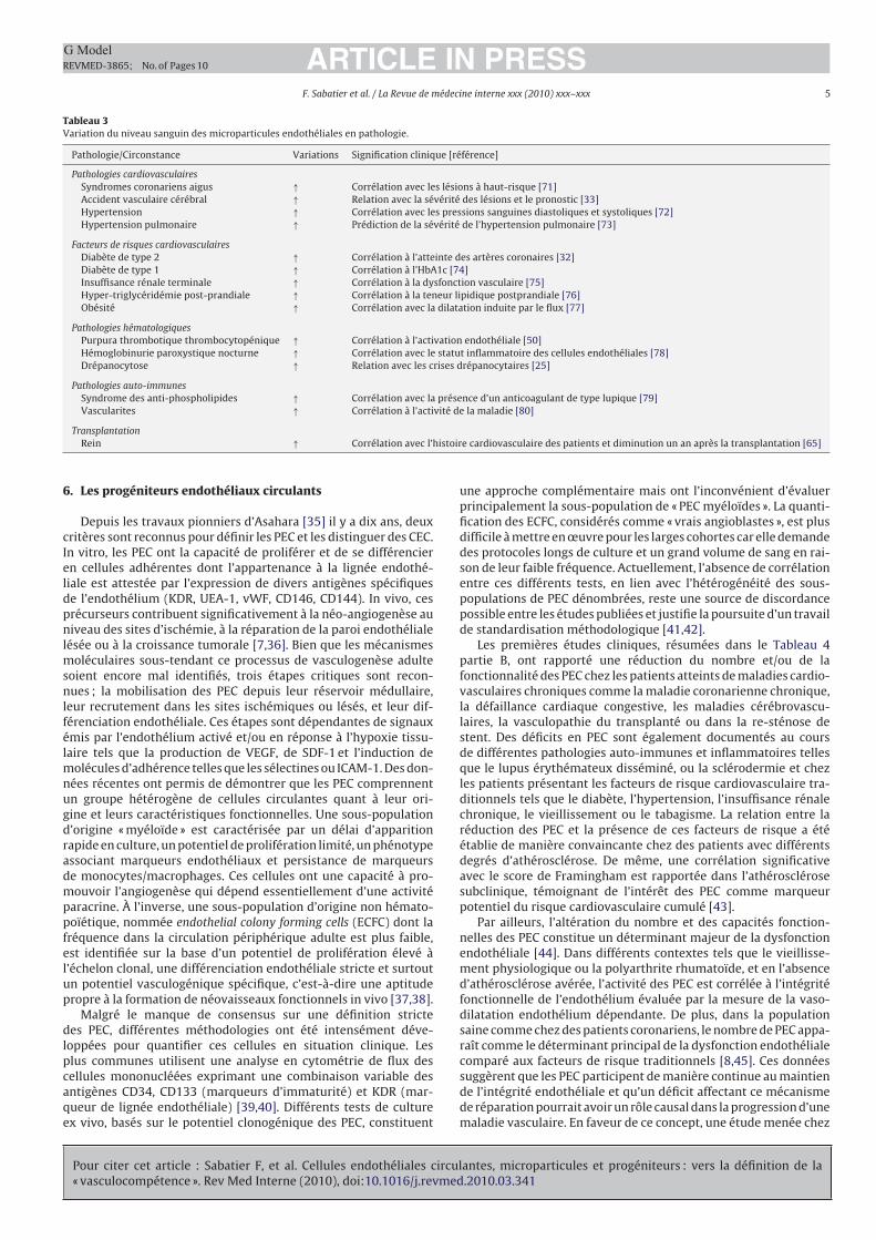
Pour citer cet article : Sabatier F, et al. Cellules endothéliales circulantes, microparticules et progéniteurs : vers la définition de la« vasculocompétence ». Rev Med Interne (2010), doi:10.1016/j.revmed.2010.03.341
ARTICLE IN PRESSG Model
REVMED3865; No. of Pages 10
F. Sabatier et al. / La Revue de médecine interne xxx (2010) xxx–xxx 5
Tableau 3
Variation du niveau sanguin des microparticules endothéliales en pathologie.
Pathologie/Circonstance Variations Signification clinique [référence]
Pathologies cardiovasculaires
Syndromes coronariens aigus ↑ Corrélation avec les lésions à hautrisque [71]Accident vasculaire cérébral ↑ Relation avec la sévérité des lésions et le pronostic [33]Hypertension ↑ Corrélation avec les pressions sanguines diastoliques et systoliques [72]Hypertension pulmonaire ↑ Prédiction de la sévérité de l’hypertension pulmonaire [73]
Facteurs de risques cardiovasculaires
Diabète de type 2 ↑ Corrélation à l’atteinte des artères coronaires [32]Diabète de type 1 ↑ Corrélation à l’HbA1c [74]Insuffisance rénale terminale ↑ Corrélation à la dysfonction vasculaire [75]Hypertriglycéridémie postprandiale ↑ Corrélation à la teneur lipidique postprandiale [76]Obésité ↑ Corrélation avec la dilatation induite par le flux [77]
Pathologies hématologiques
Purpura thrombotique thrombocytopénique ↑ Corrélation à l’activation endothéliale [50]Hémoglobinurie paroxystique nocturne ↑ Corrélation avec le statut inflammatoire des cellules endothéliales [78]Drépanocytose ↑ Relation avec les crises drépanocytaires [25]
Pathologies autoimmunes
Syndrome des antiphospholipides ↑ Corrélation avec la présence d’un anticoagulant de type lupique [79]Vascularites ↑ Corrélation à l’activité de la maladie [80]
Transplantation
Rein ↑ Corrélation avec l’histoire cardiovasculaire des patients et diminution un an après la transplantation [65]
6. Les progéniteurs endothéliaux circulants
Depuis les travaux pionniers d’Asahara [35] il y a dix ans, deuxcritères sont reconnus pour définir les PEC et les distinguer des CEC.In vitro, les PEC ont la capacité de proliférer et de se différencieren cellules adhérentes dont l’appartenance à la lignée endothéliale est attestée par l’expression de divers antigènes spécifiquesde l’endothélium (KDR, UEA1, vWF, CD146, CD144). In vivo, cesprécurseurs contribuent significativement à la néoangiogenèse auniveau des sites d’ischémie, à la réparation de la paroi endothélialelésée ou à la croissance tumorale [7,36]. Bien que les mécanismesmoléculaires soustendant ce processus de vasculogenèse adultesoient encore mal identifiés, trois étapes critiques sont reconnues ; la mobilisation des PEC depuis leur réservoir médullaire,leur recrutement dans les sites ischémiques ou lésés, et leur différenciation endothéliale. Ces étapes sont dépendantes de signauxémis par l’endothélium activé et/ou en réponse à l’hypoxie tissulaire tels que la production de VEGF, de SDF1 et l’induction demolécules d’adhérence telles que les sélectines ou ICAM1. Des données récentes ont permis de démontrer que les PEC comprennentun groupe hétérogène de cellules circulantes quant à leur origine et leurs caractéristiques fonctionnelles. Une souspopulationd’origine « myéloïde » est caractérisée par un délai d’apparitionrapide en culture, un potentiel de prolifération limité, un phénotypeassociant marqueurs endothéliaux et persistance de marqueursde monocytes/macrophages. Ces cellules ont une capacité à promouvoir l’angiogenèse qui dépend essentiellement d’une activitéparacrine. À l’inverse, une souspopulation d’origine non hématopoïétique, nommée endothelial colony forming cells (ECFC) dont lafréquence dans la circulation périphérique adulte est plus faible,est identifiée sur la base d’un potentiel de prolifération élevé àl’échelon clonal, une différenciation endothéliale stricte et surtoutun potentiel vasculogénique spécifique, c’estàdire une aptitudepropre à la formation de néovaisseaux fonctionnels in vivo [37,38].
Malgré le manque de consensus sur une définition strictedes PEC, différentes méthodologies ont été intensément développées pour quantifier ces cellules en situation clinique. Lesplus communes utilisent une analyse en cytométrie de flux descellules mononucléées exprimant une combinaison variable desantigènes CD34, CD133 (marqueurs d’immaturité) et KDR (marqueur de lignée endothéliale) [39,40]. Différents tests de cultureex vivo, basés sur le potentiel clonogénique des PEC, constituent
une approche complémentaire mais ont l’inconvénient d’évaluerprincipalement la souspopulation de « PEC myéloïdes ». La quantification des ECFC, considérés comme « vrais angioblastes », est plusdifficile à mettre en œuvre pour les larges cohortes car elle demandedes protocoles longs de culture et un grand volume de sang en raison de leur faible fréquence. Actuellement, l’absence de corrélationentre ces différents tests, en lien avec l’hétérogénéité des souspopulations de PEC dénombrées, reste une source de discordancepossible entre les études publiées et justifie la poursuite d’un travailde standardisation méthodologique [41,42].
Les premières études cliniques, résumées dans le Tableau 4partie B, ont rapporté une réduction du nombre et/ou de lafonctionnalité des PEC chez les patients atteints de maladies cardiovasculaires chroniques comme la maladie coronarienne chronique,la défaillance cardiaque congestive, les maladies cérébrovasculaires, la vasculopathie du transplanté ou dans la resténose destent. Des déficits en PEC sont également documentés au coursde différentes pathologies autoimmunes et inflammatoires tellesque le lupus érythémateux disséminé, ou la sclérodermie et chezles patients présentant les facteurs de risque cardiovasculaire traditionnels tels que le diabète, l’hypertension, l’insuffisance rénalechronique, le vieillissement ou le tabagisme. La relation entre laréduction des PEC et la présence de ces facteurs de risque a étéétablie de manière convaincante chez des patients avec différentsdegrés d’athérosclérose. De même, une corrélation significativeavec le score de Framingham est rapportée dans l’athérosclérosesubclinique, témoignant de l’intérêt des PEC comme marqueurpotentiel du risque cardiovasculaire cumulé [43].
Par ailleurs, l’altération du nombre et des capacités fonctionnelles des PEC constitue un déterminant majeur de la dysfonctionendothéliale [44]. Dans différents contextes tels que le vieillissement physiologique ou la polyarthrite rhumatoïde, et en l’absenced’athérosclérose avérée, l’activité des PEC est corrélée à l’intégritéfonctionnelle de l’endothélium évaluée par la mesure de la vasodilatation endothélium dépendante. De plus, dans la populationsaine comme chez des patients coronariens, le nombre de PEC apparaît comme le déterminant principal de la dysfonction endothélialecomparé aux facteurs de risque traditionnels [8,45]. Ces donnéessuggèrent que les PEC participent de manière continue au maintiende l’intégrité endothéliale et qu’un déficit affectant ce mécanismede réparation pourrait avoir un rôle causal dans la progression d’unemaladie vasculaire. En faveur de ce concept, une étude menée chez

Pour citer cet article : Sabatier F, et al. Cellules endothéliales circulantes, microparticules et progéniteurs : vers la définition de la« vasculocompétence ». Rev Med Interne (2010), doi:10.1016/j.revmed.2010.03.341
ARTICLE IN PRESSG Model
REVMED3865; No. of Pages 10
6 F. Sabatier et al. / La Revue de médecine interne xxx (2010) xxx–xxx
Tableau 4
Situations cliniques illustrant les variations du niveau sanguin des progéniteurs endothéliaux circulants (PEC).
Signification clinique [référence]
Partie A : situations cliniques associées à une élévation des niveaux de PEC
Ischémie aiguë
Infarctus du myocarde Corrélation avec le VEGF plasmatique et les CPK [81]Corrélation avec la formation de vaisseaux collatéraux [82]Différentiation des PEC associée à une meilleure préservation de la fonction myocardique [83]Corrélation avec un remodelage postIDM favorable [84]
Angor instable Corrélation avec les niveaux de CRP sérique [85]Accident vasculaire cérébral Élévation des PEC dans la première semaine prédictive d’un pronostic neurologique favorable [86]Transplantation hépatique Mobilisation des PEC du receveur associée à l’augmentation des taux de VEGF, SCF, GCSF [87]
Lésion vasculaire mécanique
Mise en place d’un stent coronatien Augmentation plus marquée chez les patients développant une resténose [88]Corrélation inverse avec l’atteinte vasculaire évaluée par la numération des CEC [52]
Thérapeutique/Modification du mode de vie
Statines Augmentation des PEC indépendante du VEGF [89]Amélioration de la fonctionnalité des PEC en relation avec la production d’IL8 par les monocytes [90]Association avec l’amélioration de la fonction endothéliale (DMF) chez les patients avec une insuffisance cardiaquechronique [91]Association avec le traitement à long terme chez les patients cardiovasculaires [92]
Érythropoïétine Association avec une amélioration de la survie et fonction des PEC chez les patients insuffisants rénaux anémiquestraités avec une dose standard d’EPO [93]Augmentation des PEC chez les patients avec infarctus du myocarde recevant une dose unique d’EPO [94]
Exercice physique Corrélation avec l’amélioration de la fonction endothéliale (DMF) [95]Association avec l’intensification de la pratique du sport scolaire chez les enfants [96]Association avec réduction de l’apoptose des PEC chez les patients coronariens [97]
Cancers
Cancer du sein Corrélation avec le grade de la tumeur [98]Lymphome Corrélation avec le grade de la tumeur [99]Thérapeutiques antiangiogéniques Mobilisation de taux élevés de PEC avec les chimiothérapies agressives à l’inverse des traitements métronomiques
[100]
Partie B : situations cliniques associées à une diminution des niveaux de PEC
Pathologies cardiovasculaires
Angor stable Corrélation inverse avec la présence de facteurs de risque cardiovasculaire [101]Valeur prédictive indépendante sur le pronostic cardiovasculaire [102]Corrélation inverse entre le nombre de PEC et leur activité migratoire et la sévérité de la sténose coronarienne et la CRP [103]
Maladies vasculaires cérébrales Corrélation avec le flux sanguin régional [104]Hypertension pulmonaire Corrélation avec l’IL6, le vWF et les niveaux de BNP
Association avec une réduction de la migration et de l’adhésion des PEC à la fibronectine [105]Association avec une élévation des marqueurs inflammatoires et amélioration par les inhibiteurs de phosphodiestérases(sildénafil) [106]
Insuffisance cardiaque Corrélation avec la sévérité de la maladie [107]Association avec une réduction du pool de PEC fonctionnels dans la moelle osseuse [108]Association avec les stades avancés de la maladie [109]
Resténose sur stent Les bas niveaux de PEC et leurs défauts fonctionnels sont associés à une resténose diffuse [110]L’augmentation de la sénescence des PEC est associée à la resténose intrastent [111]
Facteurs de risque cardiovasculaire
Diabète de type 2 Corrélation inverse avec la sévérité de la maladie et prédiction des complications microvasculaires au niveau individuel [112]Association avec la dysfonction des PEC impliquant la eNOS [113]Corrélation avec les valeurs d’HbA1c chez les patients non traités et augmentation par le contrôle glycémiquepharmacologique [114]Réduction plus marquée chez les patients avec une atteinte vasculaire périphériqueCorrélation avec l’ABI [115]
Diabète de type 1 Association avec une dysfonction des PEC [116]Hypertension Corrélation inverse avec la pression sanguine systolique [117]
L’hypertension réfractaire détermine de facon indépendante le nombre de PEC [118]Insuffisance rénale terminale Corrélation entre la réduction du nombre et l’altération des fonctions des PEC et les niveaux sériques de fetuine A,
l’hématocrite et les réticulocytes [119]Corrélation inverse entre le nombre des PEC et leur propriété de migration et l’urémie et la pression sanguine systolique.Restauration par l’hémodialyse [120]Corrélation avec la fonction rénale et normalisation après transplantation rénale [121]Corrélation inverse avec le score de Framingham [122]
Dyslipidémie Corrélation inverse entre la réduction du nombre et l’altération des fonctions des PEC et le taux de cholestérol total et LDL[123]Corrélation du ratio MPE/PEC avec le cholestérol LDL et la rigidité artérielle [54]
Tabagisme Relation entre l’altération de la fonction des PEC et l’augmentation du stress oxydatif [124]Augmentation du niveau des PEC après sevrage tabagique [125]
Vieillissement Corrélation avec la diminution de l’élasticité artérielle [126,127]Niveaux plus élevés de PEC dans l’enfance. Relation inverse avec l’âge chez les sujets sains [127]
Hyperhomocystéinémie Corrélation inverse avec les niveaux d’homocystéine [128]
VEGF : vascular endothelial growth factor ; CPK : créatine phosphokinase ; IDM : infarctus du myocarde ; CRP : protéine Créactive ; IL8 : interleukine8 ; SCF : stem cell factor ;GSCF : granulocytestem cell factor ; IL6 : interleukine6 ; DMF : dilatation médiée par le flux ; EPO : érythropoïétine ; BNP : peptide natriurétique type B ; vWF : facteur vonWillebrand ; eNOs : nitrique oxyde synthase endothéliale ; ABI : ankle brachial index ; LDL : low density lipoprotein.

Pour citer cet article : Sabatier F, et al. Cellules endothéliales circulantes, microparticules et progéniteurs : vers la définition de la« vasculocompétence ». Rev Med Interne (2010), doi:10.1016/j.revmed.2010.03.341
ARTICLE IN PRESSG Model
REVMED3865; No. of Pages 10
F. Sabatier et al. / La Revue de médecine interne xxx (2010) xxx–xxx 7
Fig. 1. Les lésions d’origine mécanique ou l’exposition aux facteurs de risque cardiovasculaire (FRC) altèrent les fonctions régulatrices de l’endothélium qui progressevers la dysfonction et la mort par apoptose.La rupture de l’intégrité de l’endothélium est associée à un large spectre deréponses incluant le détachement de cellules endothéliales matures (CEC) et lalibération de microparticules endothéliales (MPE). En réponse à la lésion, les progéniteurs endothéliaux (PEC), recrutés à partir de la moelle osseuse, sont capablesde se différencier en cellules matures et de restaurer l’intégrité endothéliale.Ces réponses endothéliales peuvent être intégrer dans une triade dynamique« activation/lésion/régénération ».
plus de 500 patients coronariens, suivis pendant 12 mois, démontreque le taux cumulé de survie sans évènements cardiovasculairesmajeurs augmente avec le niveau basal des cellules circulantes34+/KDR+ [46]. Néanmoins cette étude demeure controversée enraison du manque de spécificité de la méthode de détection des PECutilisée. La signification pronostique des PEC reste donc à démontrer de manière plus rigoureuse. Plus récemment, Fadini et al. ontrapporté l’intérêt de la numération des cellules CD34+ circulantesdans l’identification du risque vasculaire chez des patients présentant un syndrome métabolique [47]. Ainsi, la réparation vasculairedépendante des PEC apparaît comme un mécanisme de protectioncapable de contrebalancer l’impact des facteurs de risque cardiovasculaire, de limiter l’évolution clinique de l’athérosclérose etfinalement de déterminer le risque cardiovasculaire.
Fig. 1.
7. Lésion versus régénération de l’endothélium : vers unedéfinition de la vasculocompétence
Les dommages subis par l’endothélium et la capacité endogènede réparation peuvent être intégrés dans un phénotype endothélialqui est la résultante de ces deux processus. Ce phénotype endothélial caractérise un état ou statut vasculaire que l’on peut, à l’échelleindividuelle, définir comme une « compétence vasculaire » et apprécier de manière originale et non invasive par la mesure combinéedes CEC, MPE et PEC (Fig. 2). Des données récentes de la littérature suggèrent l’intérêt d’une telle stratégie « multimarqueurs »
pour identifier les patients à risque vasculaire et leur réponse autraitement.
À l’état physiologique, l’endothélium est un organe caractérisépar une remarquable quiescence et un renouvellement extrêmement lent [48]. Cette quiescence se traduit par la présencedans la circulation périphérique de sujets sains de quantités trèsfaibles de CEC, MPE et PEC. De plus, quelques données suggèrentla participation des MPE à certaines fonctions physiologiques del’endothélium comme le maintien d’une activité anticoagulante[28] ou l’élimination de signaux proapoptotiques favorisant la survie cellulaire [49].
Si le niveau de base des CEC, MPE et PEC représente l’étatde l’endothélium en situation normale, un déséquilibre de cette
Fig. 2. L’équilibre entre les lésions de l’endothélium, reflétées par les cellules endothéliales circulantes (CEC), les microparticules endothéliales (MPE) et la capacité deréparation endogène, attestée par les progéniteurs endothéliaux circulants (PEC),conditionne l’intégrité de l’endothélium.Le résultat net de cet équilibre caractérise un phénotype endothélial que l’on peutdéfinir à l’échelle individuelle comme la « vasculocompétence » (IVC : index de vasculocompétence). L’exploration de cette compétence vasculaire par des stratégies« multimarqueurs » combinant CEC, MPE et PEC est une approche prometteuse pourapprécier l’impact des facteurs de risque cardiovasculaire sur la progression de lamaladie. Elle offre également des outils innovants pour l’évaluation et le suivi destratégies thérapeutiques ou de modifications du mode de vie visant à limiter leslésions et/ou à renforcer les mécanismes de régénération de l’endothélium.
balance oriente vers l’existence d’un processus délétère affectantl’endothélium. Il est maintenant établi que l’élévation des CEC etdes MPE corrèle avec l’intensité ou l’étendue des lésions vasculaires.De plus, l’analyse phénotypique de ces marqueurs peut apporterdes indications sur les mécanismes ou l’étiologie des lésions. Parexemple, les MPE CD62E+ sont principalement libérées lors d’uneactivation proinflammatoire tandis que les MPE CD144+ seraientla résultante d’une apoptose ou des dommages structuraux [32,50].De même, les CEC surexpriment des molécules d’adhérence pour lesleucocytes dans les pathologies inflammatoires comme la drépanocytose [51], alors qu’elles ont un phénotype évocateur d’apoptoseou de nécrose endothéliale chez les patients lupiques.
En situation aiguë, comme l’ischémiereperfusion, ou une stimulation proinflammatoire, l’élévation des MPE et des CEC estassociée à une augmentation transitoire du nombre des PEC circulants. Ce profil de biomarqueurs traduit l’existence d’un mécanismede réparation compensatoire pouvant s’inscrire dans une réponsephysiologique ou vasculocompétence visant à rétablir l’intégritéendothéliale. En faveur de cette hypothèse, cette mobilisation desPEC, au cours de l’ischémie myocardique ou cérébrale, a été associée à un pronostic fonctionnel favorable. À l’inverse, une réponserégénérative insuffisante, associée ou non à une élévation persistante des marqueurs de lésions, suggère une réponse inadaptéede l’endothélium ou « vasculoincompétence » conduisant à unétat pathologique à risque vasculaire. Une étude, menée chez despatients subissant une angioplastie coronaire avec implantationde stent, a montré [52] une élévation simultanée des CEC et des

Pour citer cet article : Sabatier F, et al. Cellules endothéliales circulantes, microparticules et progéniteurs : vers la définition de la« vasculocompétence ». Rev Med Interne (2010), doi:10.1016/j.revmed.2010.03.341
ARTICLE IN PRESSG Model
REVMED3865; No. of Pages 10
8 F. Sabatier et al. / La Revue de médecine interne xxx (2010) xxx–xxx
PEC au décours de la procédure. L’amplitude de mobilisation étaitinversement corrélée à l’étendue de la lésion reflétée par les CEC.Cela suggère que des dommages endothéliaux importants associésà une réparation endothéliale insuffisante pourraient permettred’identifier des patients à risque de complications postprocédure.
À l’inverse des situations aiguës, les lésions endothéliales résultant de l’exposition chronique à des stimuli pathogènes sontassociées à une réduction en précurseurs efficaces pour la réparation. Ce déséquilibre, objectivé par une augmentation des MPEet/ou CEC associée à une réduction du nombre et/ou de la fonctionnalité des PEC, met en évidence un déficit de réparationcompensatoire qui contribue au développement et à la progressionde l’athérosclérose. Comme illustré récemment chez des patientsprésentant une insuffisance ventriculaire gauche postischémique,ou chez des patients dyslipidémiques, un index vasculaire combinant la mesure des PEC et des marqueurs de lésion tel que les MPE,offre une meilleure appréciation du risque vasculaire en évaluantles deux composantes de la dysfonction endothéliale [53,54].
Malgré ces résultats prometteurs, l’intégration en pratique clinique des stratégies « multimarqueurs » basées sur les CEC, les MPEet les PEC n’en est encore qu’à ses débuts. Si l’on a pu montrerl’association entre ces marqueurs et le risque vasculaire, un véritable lien de causalité reste à démontrer expérimentalement. Parailleurs, une meilleure connaissance des processus de réparation etde lésion endothéliale doit permettre de définir plus précisémentla relation entre les CEC, les MPE et les PEC en conditions pathologiques. In vitro les MPE sont capables de moduler de l’activitéangiogénique des PEC [29]. De plus, in vivo la production de MPEpar les tissus ischémiques stimule le recrutement et la différenciation des PEC [55]. Les mécanismes moléculaires impliqués dans cesconnexions restent à préciser, néanmoins ces données suggèrentque les MPE pourraient constituer un signal endogène stimulantles processus de réparation dépendant des PEC. De plus, les variations physiologiques avec des paramètres tels que l’âge, le sexe oule rythme circadien doivent être définis dans des cohortes de sujetssains. Enfin, la standardisation des méthodologies disponibles estaussi une étape cruciale pour une définition complète de la signification clinique de ces marqueurs.
8. Conclusion
La possibilité d’explorer l’endothélium de manière non invasivea ouvert la voie à une nouvelle compréhension de la pathogenèse des maladies cardiovasculaires, définie par un déséquilibreentre lésion de l’endothélium et sa capacité de réparation. Dansle futur, des approches multimarqueurs combinant la mesure desCEC, des MPE et des PEC seront utiles pour une approche globalede la « vasculocompétence ». En clinique, ces marqueurs cellulairesfourniront certainement des nouveaux outils pour détecter la dysfonction endothéliale à des stades précoces, mais aussi pour évaluerle risque vasculaire à des stades plus avancés des désordres athérothrombotiques et pour déterminer des options thérapeutiques. Eneffet, des stratégies thérapeutiques efficaces pour améliorer la fonction endothéliale pourraient cibler sélectivement une réduction dela lésion endothéliale ou promouvoir les mécanismes de régénération. Au niveau individuel, de telles options thérapeutiques peuventintégrer non seulement le mécanisme prédominant mais aussi lesprédispositions génétiques et les facteurs de risque environnementaux. D’importantes avancées dans le monitoring des certainespathologies et des options thérapeutiques nouvelles émergerontcertainement de ce domaine en plein essor.
Conflit d’intérêt
Les auteurs déclarent ne pas avoir de conflit d’intérêt.
Références
[1] Landmesser U, Drexler H. Endothelial function and hypertension. Curr OpinCardiol 2007;22:316–20.
[2] Goligorsky MS. Clinical assessment of endothelial dysfunction: combine andrule. Curr Opin Nephrol Hypertens 2006;15:617–24.
[3] Sabatier F, CamoinJau L, Anfosso F, Sampol J, DignatGeorge F. Circulatingendothelial cells, microparticles and progenitors: key players towards thedefinition of vascular competence. J Cell Mol Med 2009;13:454–71.
[4] Ross R. The pathogenesis of atherosclerosis: a perspective for the 1990s.Nature 1993;362:801–9.
[5] Combes V, Simon AC, Grau GE, Arnoux D, Camoin L, Sabatier F, et al. Invitro generation of endothelial microparticles and possible prothromboticactivity in patients with lupus anticoagulant. J Clin Invest 1999;104:93–102.
[6] George F, Brisson C, Poncelet P, Laurent JC, Massot O, Arnoux D,et al. Rapid isolation of human endothelial cells from whole bloodusing SEndo1 monoclonal antibody coupled to immunomagnetic beads:demonstration of endothelial injury after angioplasty. Thromb Haemost1992;67:147–53.
[7] Asahara T, Masuda H, Takahashi T, Kalka C, Pastore C, Silver M, et al. Bonemarrow origin of endothelial progenitor cells responsible for postnatal vasculogenesis in physiological and pathological neovascularization. Circ Res1999;85:221–8.
[8] Murphy C, Kanaganayagam GS, Jiang B, Chowienczyk PJ, Zbinden R, Saha M,et al. Vascular dysfunction and reduced circulating endothelial progenitorcells in young healthy UK South Asian men. Arterioscler Thromb Vasc Biol2007;27:936–42.
[9] George F, Poncelet P, Laurent JC, Massot O, Arnoux D, Lequeux N, et al.Cytofluorometric detection of human endothelial cells in whole blood usingSEndo 1 monoclonal antibody. J Immunol Methods 1991;139:65–75.
[10] Woywodt A, Blann AD, Kirsch T, Erdbruegger U, Banzet N, Haubitz M, et al.Isolation and enumeration of circulating endothelial cells by immunomagnetic isolation: proposal of a definition and a consensus protocol. J ThrombHaemost 2006;4:671–7.
[11] Blann AD, Woywodt A, Bertolini F, Bull TM, Buyon JP, Clancy RM, et al. Circulating endothelial cells. Biomarker of vascular disease. Thromb Haemost2005;93:228–35.
[12] Rowand JL, Martin G, Doyle GV, Miller MC, Pierce MS, Connelly MC, et al.Endothelial cells in peripheral blood of healthy subjects and patients withmetastatic carcinomas. Cytometry A 2007;71:105–13.
[13] Widemann A, Sabatier F, Arnaud L, Bonello L, AlMassarani G, Paganelli F, etal. CD146based immunomagnetic enrichment followed by multiparameterflow cytometry: a new approach to counting circulating endothelial cells. JThromb Haemost 2008;6:869–76 [see comment].
[14] Vroling L, Yuana Y, Schuurhuis GJ, van Hinsbergh VW, Gundy C, de Haas R, etal. VEGFR2 expressing circulating (progenitor) cell populations in volunteersand cancer patients. Thromb Haemost 2007;98:440–50.
[15] Goon PK, Boos CJ, Stonelake PS, Blann AD, Lip GY. Detection and quantification of mature circulating endothelial cells using flow cytometry andimmunomagnetic beads: a methodological comparison. Thromb Haemost2006;96:45–52.
[16] Jacques N, Vimond N, Conforti R, Griscelli F, Lecluse Y, Laplanche A, et al.Quantification of circulating mature endothelial cells using a whole bloodfourcolor flow cytometric assay. J Immunol Methods 2008;337:132–43.
[17] Makin AJ, Blann AD, Chung NA, Silverman SH, Lip GY. Assessment of endothelial damage in atherosclerotic vascular disease by quantification of circulatingendothelial cells. Relationship with von Willebrand factor and tissue factor.Eur Heart J 2004;25:371–6.
[18] Nadar SK, Lip GY, Lee KW, Blann AD. Circulating endothelial cells in acuteischaemic stroke. Thromb Haemost 2005;94:707–12.
[19] Rajagopalan S, Somers EC, Brook RD, Kehrer C, Pfenninger D, Lewis E, etal. Endothelial cell apoptosis in systemic lupus erythematosus: a commonpathway for abnormal vascular function and thrombosis propensity. Blood2004;103:3677–83.
[20] Bull TM, Golpon H, Hebbel RP, Solovey A, Cool CD, Tuder RM, et al. Circulating endothelial cells in pulmonary hypertension. Thromb Haemost2003;90:698–703.
[21] Woywodt A, Streiber F, de Groot K, Regelsberger H, Haller H, Haubitz M.Circulating endothelial cells as markers for ANCAassociated smallvesselvasculitis. Lancet 2003;361:206–10 [see comment].
[22] Smadja DM, Gaussem P, Mauge L, IsraelBiet D, DignatGeorge F, Peyrard S,et al. Circulating endothelial cells: a new candidate biomarker of irreversiblepulmonary hypertension secondary to congenital heart disease. Circulation2009;119:374–81.
[23] Morel O, Toti F, Hugel B, Bakouboula B, CamoinJau L, DignatGeorge F, et al.Procoagulant microparticles: disrupting the vascular homeostasis equation?Arterioscler Thromb Vasc Biol 2006;26:2594–604.
[24] Aras O, Shet A, Bach RR, Hysjulien JL, Slungaard A, Hebbel RP, et al. Induction of microparticle and cellassociated intravascular tissue factor in humanendotoxemia. Blood 2004;103:4545–53.
[25] Shet AS, Aras O, Gupta K, Hass MJ, Rausch DJ, Saba N, et al. Sickle bloodcontains tissue factorpositive microparticles derived from endothelial cellsand monocytes. Blood 2003;102:2678–83.
[26] Mallat Z, Hugel B, Ohan J, Leseche G, Freyssinet JM, Tedgui A. Shedmembrane microparticles with procoagulant potential in human atheros

Pour citer cet article : Sabatier F, et al. Cellules endothéliales circulantes, microparticules et progéniteurs : vers la définition de la« vasculocompétence ». Rev Med Interne (2010), doi:10.1016/j.revmed.2010.03.341
ARTICLE IN PRESSG Model
REVMED3865; No. of Pages 10
F. Sabatier et al. / La Revue de médecine interne xxx (2010) xxx–xxx 9
clerotic plaques: a role for apoptosis in plaque thrombogenicity. Circulation1999;99:348–53.
[27] Mallat Z, Benamer H, Hugel B, Benessiano J, Steg PG, Freyssinet JM, et al. Elevated levels of shed membrane microparticles with procoagulant potential inthe peripheral circulating blood of patients with acute coronary syndromes.Circulation 2000;101:841–3.
[28] PerezCasal M, Downey C, Fukudome K, Marx G, Toh CH. Activated protein Cinduces the release of microparticleassociated endothelial protein C receptor. Blood 2005;105:1515–22.
[29] Lacroix R, Sabatier F, Mialhe A, Basire A, Pannell R, Borghi H, et al. Activationof plasminogen into plasmin at the surface of endothelial microparticles: amechanism that modulates angiogenic properties of endothelial progenitorcells in vitro. Blood 2007;110:2432–9.
[30] Freyssinet JM, DignatGeorge F. More on: measuring circulating cellderivedmicroparticles. J Thromb Haemost 2005;3:613–4 [comment].
[31] Jy W, Horstman LL, Jimenez JJ, Ahn YS, Biro E, Nieuwland R, et al. Measuringcirculating cellderived microparticles. J Thromb Haemost 2004;2:1842–51[comment].
[32] Koga H, Sugiyama S, Kugiyama K, Watanabe K, Fukushima H, Tanaka T, et al.Elevated levels of VEcadherinpositive endothelial microparticles in patientswith type 2 diabetes mellitus and coronary artery disease. J Am Coll Cardiol2005;45:1622–30.
[33] Simak J, Gelderman MP, Yu H, Wright V, Baird AE. Circulating endothelialmicroparticles in acute ischemic stroke: a link to severity, lesion volume andoutcome. J Thromb Haemost 2006;4:1296–302.
[34] Brodsky SV, Zhang F, Nasjletti A, Goligorsky MS. Endotheliumderived microparticles impair endothelial function in vitro. Am J Physiol Heart Circ Physiol2004;286:H1910–5.
[35] Asahara T, Murohara T, Sullivan A, Silver M, van der Zee R, Li T, et al.Isolation of putative progenitor endothelial cells for angiogenesis. Science1997;275:964–7.
[36] Asahara T, Kawamoto A. Endothelial progenitor cells for postnatal vasculogenesis. Am J Physiol Cell Physiol 2004;287:C572–9.
[37] Case J, Mead LE, Bessler WK, Prater D, White HA, Saadatzadeh MR, et al. HumanCD34+AC133+VEGFR2+ cells are not endothelial progenitor cells but distinct, primitive hematopoietic progenitors. Exp Hematol 2007;35:1109–18[see comment].
[38] Yoder MC, Mead LE, Prater D, Krier TR, Mroueh KN, Li F, et al. Redefining endothelial progenitor cells via clonal analysis and hematopoietic stem/progenitorcell principals. Blood 2007;109:1801–9 [see comment].
[39] Fadini GP, Baesso I, Albiero M, Sartore S, Agostini C, Avogaro A. Technical noteson endothelial progenitor cells: ways to escape from the knowledge plateau.Atherosclerosis 2008;197:496–503.
[40] George J, Shmilovich H, Deutsch V, Miller H, Keren G, Roth A. Comparativeanalysis of methods for assessment of circulating endothelial progenitor cells.Tissue Eng 2006;12:331–5.
[41] Prater DN, Case J, Ingram DA, Yoder MC. Working hypothesis to redefineendothelial progenitor cells. Leukemia 2007;21:1141–9.
[42] Xiao Q, Kiechl S, Patel S, Oberhollenzer F, Weger S, Mayr A, et al. Endothelialprogenitor cells, cardiovascular risk factors, cytokine levels and atherosclerosis: results from a large populationbased study. PLoS ONE 2007;2:e975.
[43] Geft D, Schwartzenberg S, George J. Circulating endothelial progenitorcells in cardiovascular disorders. Expert Rev Cardiovasc Ther 2008;6:1115–21.
[44] Hill JM, Zalos G, Halcox JP, Schenke WH, Waclawiw MA, Quyyumi AA, et al.Circulating endothelial progenitor cells, vascular function, and cardiovascularrisk. N Engl J Med 2003;348:593–600 [see comment. Reprint in Can J Cardiol2004;20(Suppl. B):44B48B; PMID: 15309205].
[45] Werner N, Wassmann S, Ahlers P, Schiegl T, Kosiol S, Link A, et al. Endothelialprogenitor cells correlate with endothelial function in patients with coronaryartery disease. Basic Res Cardiol 2007;102:565–71.
[46] Werner N, Kosiol S, Schiegl T, Ahlers P, Walenta K, Link A, et al. Circulatingendothelial progenitor cells and cardiovascular outcomes [see comment]. NEngl J Med 2005;353:999–1007 [see comment].
[47] Fadini GP, Albiero M, Boscaro E, Agostini C, Avogaro A. Endothelial progenitorcells as resident accessory cells for postischemic angiogenesis. Atherosclerosis 2009;204:20–2.
[48] Woywodt A, Bahlmann FH, De Groot K, Haller H, Haubitz M. Circulating endothelial cells: life, death, detachment and repair of the endothelial cell layer.Nephrol Dial Transplant 2002;17:1728–30.
[49] Abid Hussein MN, Boing AN, Biro E, Hoek FJ, Vogel GM, Meuleman DG, et al.Phospholipid composition of in vitro endothelial microparticles and their invivo thrombogenic properties. Thromb Res 2008;121:865–71.
[50] Jimenez JJ, Jy W, Mauro LM, Soderland C, Horstman LL, Ahn YS. Endothelial cells release phenotypically and quantitatively distinct microparticles inactivation and apoptosis. Thromb Res 2003;109:175–80.
[51] Solovey AA, Solovey AN, Harkness J, Hebbel RP. Modulation of endothelial cellactivation in sickle cell disease: a pilot study. Blood 2001;97:1937–41.
[52] Bonello L, Basire A, Sabatier F, Paganelli F, DignatGeorge F. Endothelial injuryinduced by coronary angioplasty triggers mobilization of endothelial progenitor cells in patients with stable coronary artery disease. J Thromb Haemost2006;4:979–81 [see comments].
[53] Bulut D, Maier K, BulutStreich N, Borgel J, Hanefeld C, Mugge A. Circulating endothelial microparticles correlate inversely with endothelialfunction in patients with ischemic left ventricular dysfunction. J Card Fail2008;14:336–40.
[54] Pirro M, Schillaci G, Paltriccia R, Bagaglia F, Menecali C, Mannarino MR, etal. Increased ratio of CD31+/CD42−microparticles to endothelial progenitorsas a novel marker of atherosclerosis in hypercholesterolemia. ArteriosclerThromb Vasc Biol 2006;26:2530–5.
[55] Leroyer AS, Ebrahimian TG, Cochain C, Recalde A, BlancBrude O, Mees B, etal. Microparticles from ischemic muscle promotes postnatal vasculogenesis.Circulation 2009;119:2808–17.
[56] Mutin M, Canavy I, Blann A, Bory M, Sampol J, DignatGeorge F. Direct evidenceof endothelial injury in acute myocardial infarction and unstable angina bydemonstration of circulating endothelial cells. Blood 1999;93:2951–8 [seecomments].
[57] Quilici J, Banzet N, Paule P, Meynard JB, Mutin M, Bonnet JL, et al. Circulating endothelial cell count as a diagnostic marker for nonSTelevation acutecoronary syndromes. Circulation 2004;110:1586–91.
[58] Lee KW, Blann AD, Lip GY. Plasma markers of endothelialdamage/dysfunction, inflammation and thrombogenesis in relation toTIMI risk stratification in acute coronary syndromes. Thromb Haemost2005;94:1077–83.
[59] Chong AY, Blann AD, Patel J, Freestone B, Hughes E, Lip GY. Endothelial dysfunction and damage in congestive heart failure: relation of flowmediateddilation to circulating endothelial cells, plasma indexes of endothelialdamage, and brain natriuretic peptide. Circulation 2004;110:1794–8.
[60] McClung JA, Naseer N, Saleem M, Rossi GP, Weiss MB, Abraham NG, et al.Circulating endothelial cells are elevated in patients with type 2 diabetesmellitus independently of HbA(1)c. Diabetologia 2005;48:345–50 [see comments].
[61] Koc M, Richards HB, Bihorac A, Ross EA, Schold JD, Segal MS. Circulatingendothelial cells are associated with future vascular events in hemodialysispatients. Kidney Int 2005;67:1078–83.
[62] Erdbruegger U, Haubitz M, Woywodt A. Circulating endothelial cells: a novelmarker of endothelial damage. Clin Chim Acta 2006;373:17–26.
[63] Solovey A, Lin Y, Browne P, Choong S, Wayner E, Hebbel RP. Circulating activated endothelial cells in sickle cell anemia. N Engl J Med 1997;337:1584–90[see comments].
[64] Woywodt A, Schroeder M, Gwinner W, Mengel M, Jaeger M, Schwarz A, etal. Elevated numbers of circulating endothelial cells in renal transplant recipients. Transplantation 2003;76:1–4.
[65] AlMassarani G, VacherCoponat H, Paul P, Widemann A, Arnaud L, LoundouA, et al. Impact of immunosuppressive treatment on endothelial biomarkersafter kidney transplantation. Am J Transplant 2008;8:2360–7.
[66] Woywodt A, Scheer J, Hambach L, Buchholz S, Ganser A, Haller H, et al. Circulating endothelial cells as a marker of endothelial damage in allogeneichematopoietic stem cell transplantation. Blood 2004;103:3603–5.
[67] Del Papa N, Colombo G, Fracchiolla N, Moronetti LM, Ingegnoli F, MaglioneW, et al. Circulating endothelial cells as a marker of ongoing vascular diseasein systemic sclerosis. Arthritis Rheum 2004;50:1296–304.
[68] Grefte A, van der Giessen M, van Son W, The TH. Circulating cytomegalovirus(CMV)infected endothelial cells in patients with an active CMV infection. JInfect Dis 1993;167:270–7.
[69] George F, Brouqui P, Boffa MC, Mutin M, Drancourt M, Brisson C, et al. Demonstration of Rickettsia conoriiinduced endothelial injury in vivo by measuringcirculating endothelial cells, thrombomodulin, and von Willebrand factorin patients with Mediterranean spotted fever. Blood 1993;82:2109–16 [seecomments].
[70] Willett CG, Boucher Y, di Tomaso E, Duda DG, Munn LL, Tong RT, et al.Direct evidence that the VEGFspecific antibody bevacizumab has antivascular effects in human rectal cancer. Nat Med 2004;10:145–7 [see comment.Erratum appears in Nat Med 2004;10(6):649].
[71] BernalMizrachi L, Jy W, Jimenez JJ, Pastor J, Mauro LM, Horstman LL, et al.High levels of circulating endothelial microparticles in patients with acutecoronary syndromes. Am Heart J 2003;145:962–70.
[72] Preston RA, Jy W, Jimenez JJ, Mauro LM, Horstman LL, Valle M, et al. Effects ofsevere hypertension on endothelial and platelet microparticles. Hypertension2003;41:211–7 [see comment].
[73] Amabile N, Heiss C, Real WM, Minasi P, McGlothlin D, Rame EJ, et al. Circulating endothelial microparticle levels predict hemodynamic severity ofpulmonary hypertension. Am J Respir Crit Care Med 2008;177:1268–75 [seecomment].
[74] Sabatier F, Darmon P, Hugel B, Combes V, Sanmarco M, Velut JG, et al. Type1 and type 2 diabetic patients display different patterns of cellular microparticles. Diabetes 2002;51:2840–5.
[75] Amabile N, Guerin AP, Leroyer A, Mallat Z, Nguyen C, Boddaert J, et al. Circulating endothelial microparticles are associated with vascular dysfunctionin patients with endstage renal failure. J Am Soc Nephrol 2005;16:3381–8.
[76] Ferreira AC, Peter AA, Mendez AJ, Jimenez JJ, Mauro LM, Chirinos JA, et al.Postprandial hypertriglyceridemia increases circulating levels of endothelialcell microparticles. Circulation 2004;110:3599–603 [see comment].
[77] Esposito K, Ciotola M, Schisano B, Gualdiero R, Sardelli L, Misso L, et al. Endothelial microparticles correlate with endothelial dysfunction in obese women.J Clin Endocrinol Metabol 2006;91:3676–9.
[78] Simak J, Holada K, Risitano AM, Zivny JH, Young NS, Vostal JG. Elevatedcirculating endothelial membrane microparticles in paroxysmal nocturnalhaemoglobinuria. Br J Haematol 2004;125:804–13.
[79] DignatGeorge F, CamoinJau L, Sabatier F, Arnoux D, Anfosso F, Bardin N,et al. Endothelial microparticles: a potential contribution to the throm

Pour citer cet article : Sabatier F, et al. Cellules endothéliales circulantes, microparticules et progéniteurs : vers la définition de la« vasculocompétence ». Rev Med Interne (2010), doi:10.1016/j.revmed.2010.03.341
ARTICLE IN PRESSG Model
REVMED3865; No. of Pages 10
10 F. Sabatier et al. / La Revue de médecine interne xxx (2010) xxx–xxx
botic complications of the antiphospholipid syndrome. Thromb Haemost2004;91:667–73 [see comment].
[80] Brogan PA, Shah V, Brachet C, Harnden A, Mant D, Klein N, et al. Endothelial and platelet microparticles in vasculitis of the young. Arthritis Rheum2004;50:927–36.
[81] Massa M, Rosti V, Ferrario M, Campanelli R, Ramajoli I, Rosso R, et al. Increasedcirculating hematopoietic and endothelial progenitor cells in the early phaseof acute myocardial infarction. Blood 2005;105:199–206.
[82] Lev EI, Kleiman NS, Birnbaum Y, Harris D, Korbling M, Estrov Z. Circulatingendothelial progenitor cells and coronary collaterals in patients with nonSTsegment elevation myocardial infarction. J Vasc Res 2005;42:408–14.
[83] Numaguchi Y, Sone T, Okumura K, Ishii M, Morita Y, Kubota R, et al. The impactof the capability of circulating progenitor cell to differentiate on myocardial salvage in patients with primary acute myocardial infarction. Circulation2006;114:I114–9.
[84] Leone AM, Rutella S, Bonanno G, Abbate A, Rebuzzi AG, Giovannini S, et al.Mobilization of bone marrowderived stem cells after myocardial infarctionand left ventricular function. Eur Heart J 2005;26:1196–204 [see comment].
[85] George J, Goldstein E, Abashidze S, Deutsch V, Shmilovich H, Finkelstein A, etal. Circulating endothelial progenitor cells in patients with unstable angina:association with systemic inflammation. Eur Heart J 2004;25:1003–8 [seecomment].
[86] Sobrino T, Hurtado O, Moro MA, RodriguezYanez M, Castellanos M, Brea D, etal. The increase of circulating endothelial progenitor cells after acute ischemicstroke is associated with good outcome. Stroke 2007;38:2759–64.
[87] Lemoli RM, Catani L, Talarico S, Loggi E, Gramenzi A, Baccarani U, et al.Mobilization of bone marrowderived hematopoietic and endothelial stemcells after orthotopic liver transplantation and liver resection. Stem Cells2006;24:2817–25.
[88] Inoue T, Sata M, Hikichi Y, Sohma R, Fukuda D, Uchida T, et al. Mobilization ofCD34positive bone marrowderived cells after coronary stent implantation:impact on restenosis. Circulation 2007;115:553–61 [see comment].
[89] Spiel AO, Mayr FB, Leitner JM, Firbas C, Sieghart W, Jilma B. Simvastatin androsuvastatin mobilize Endothelial Progenitor Cells but do not prevent theiracute decrease during systemic inflammation. Thromb Res 2008;123:108–13[see comment].
[90] Park KW, Hwang KK, Cho HJ, Hur J, Yang HM, Yoon CH, et al. Simvastatinenhances endothelial differentiation of peripheral blood mononuclear cellsin hypercholesterolemic patients and induces proangiogenic cytokine IL8 secretion from monocytes. Clin Chim Acta 2008;388:156–66.
[91] Landmesser U, Bahlmann F, Mueller M, Spiekermann S, Kirchhoff N, SchulzS, et al. Simvastatin versus ezetimibe: pleiotropic and lipidlowering effectson endothelial function in humans. Circulation 2005;111:2356–63 [see comment].
[92] Deschaseaux F, Selmani Z, Falcoz PE, Mersin N, Meneveau N, Penfornis A,et al. Two types of circulating endothelial progenitor cells in patients receiving longterm therapy by HMGCoA reductase inhibitors. Eur J Pharmacol2007;562:111–8.
[93] Bahlmann FH, DeGroot K, Duckert T, Niemczyk E, Bahlmann E, Boehm SM, etal. Endothelial progenitor cell proliferation and differentiation is regulated byerythropoietin. Kidney Int 2003;64:1648–52.
[94] Lipsic E, van der Meer P, Voors AA, Westenbrink BD, van den Heuvel AF,de Boer HC, et al. A single bolus of a longacting erythropoietin analoguedarbepoetin alfa in patients with acute myocardial infarction: a randomizedfeasibility and safety study. Cardiovasc Drugs Ther 2006;20:135–41.
[95] Steiner S, Niessner A, Ziegler S, Richter B, Seidinger D, Pleiner J, et al.Endurance training increases the number of endothelial progenitor cells inpatients with cardiovascular risk and coronary artery disease. Atherosclerosis2005;181:305–10.
[96] Walther C, Adams V, Bothur I, Drechsler K, Fikenzer S, Sonnabend M, et al.Increasing physical education in high school students: effects on concentration of circulating endothelial progenitor cells. Eur J Cardiovasc Prev Rehabil2008;15:416–22.
[97] Laufs U, Werner N, Link A, Endres M, Wassmann S, Jurgens K, et al. Physicaltraining increases endothelial progenitor cells, inhibits neointima formation,and enhances angiogenesis. Circulation 2004;109:220–6.
[98] Naik RP, Jin D, Chuang E, Gold EG, Tousimis EA, Moore AL, et al. Circulatingendothelial progenitor cells correlate to stage in patients with invasive breastcancer. Breast Cancer Res Treat 2008;107:133–8.
[99] Igreja C, Courinha M, Cachaco AS, Pereira T, Cabecadas J, da Silva MG,et al. Characterization and clinical relevance of circulating and biopsyderived endothelial progenitor cells in lymphoma patients. Haematologica2007;92:469–77 [see comment].
[100] Shaked Y, Ciarrocchi A, Franco M, Lee CR, Man S, Cheung AM, et al. Therapyinduced acute recruitment of circulating endothelial progenitor cells totumors. Science 2006;313:1785–7.
[101] Vasa M, Fichtlscherer S, Aicher A, Adler K, Urbich C, Martin H, et al. Numberand migratory activity of circulating endothelial progenitor cells inverselycorrelate with risk factors for coronary artery disease. Circ Res 2001;89:E1–7.
[102] SchmidtLucke C, Rossig L, Fichtlscherer S, Vasa M, Britten M, Kamper U, etal. Reduced number of circulating endothelial progenitor cells predicts futurecardiovascular events: proof of concept for the clinical importance of endogenous vascular repair. Circulation 2005;111:2981–7.
[103] Wang HY, Gao PJ, Ji KD, Shen WF, Fan CL, Lu L, et al. Circulating endothelial progenitor cells Creactive protein and severity of coronary stenosisin Chinese patients with coronary artery disease. Hypertens Res Clin Exp2007;30:133–41.
[104] Taguchi A, Matsuyama T, Moriwaki H, Hayashi T, Hayashida K, NagatsukaK, et al. Circulating CD34positive cells provide an index of cerebrovascularfunction. Circulation 2004;109:2972–5.
[105] Junhui Z, Xingxiang W, Guosheng F, Yunpeng S, Furong Z, Junzhu C.Reduced number and activity of circulating endothelial progenitor cellsin patients with idiopathic pulmonary arterial hypertension. Respir Med2008;102:1073–9.
[106] Diller GP, van Eijl S, Okonko DO, Howard LS, Ali O, Thum T, et al. Circulating endothelial progenitor cells in patients with Eisenmenger syndrome andidiopathic pulmonary arterial hypertension. Circulation 2008;117:3020–30.
[107] NonakaSarukawa M, Yamamoto K, Aoki H, Nishimura Y, Tomizawa H, IchidaM, et al. Circulating endothelial progenitor cells in congestive heart failure.Int J Cardiol 2007;119:344–8.
[108] Kissel CK, Lehmann R, Assmus B, Aicher A, Honold J, FischerRasokat U, etal. Selective functional exhaustion of hematopoietic progenitor cells in thebone marrow of patients with postinfarction heart failure. J Am Coll Cardiol2007;49:2341–9 [see comment].
[109] Valgimigli M, Rigolin GM, Fucili A, Porta MD, Soukhomovskaia O, Malagutti P,et al. CD34+ and endothelial progenitor cells in patients with various degreesof congestive heart failure. Circulation 2004;110:1209–12.
[110] George J, Herz I, Goldstein E, Abashidze S, Deutch V, Finkelstein A, etal. Number and adhesive properties of circulating endothelial progenitorcells in patients with instent restenosis. Arterioscler Thromb Vasc Biol2003;23:e57–60.
[111] Matsuo Y, Imanishi T, Hayashi Y, Tomobuchi Y, Kubo T, Hano T, et al. Theeffect of senescence of endothelial progenitor cells on instent restenosis inpatients undergoing coronary stenting. Intern Med 2006;45:581–7.
[112] Egan CG, Lavery R, Caporali F, Fondelli C, LaghiPasini F, Dotta F, et al. Generalised reduction of putative endothelial progenitors and CXCR4positiveperipheral blood cells in type 2 diabetes. Diabetologia 2008;51:1296–305.
[113] Thum T, Fraccarollo D, Schultheiss M, Froese S, Galuppo P, Widder JD, et al.Endothelial nitric oxide synthase uncoupling impairs endothelial progenitorcell mobilization and function in diabetes. Diabetes 2007;56:666–74.
[114] Kusuyama T, Omura T, Nishiya D, Enomoto S, Matsumoto R, Takeuchi K, et al.Effects of treatment for diabetes mellitus on circulating vascular progenitorcells. J Pharmacol Sci 2006;102:96–102.
[115] Fadini GP, Miorin M, Facco M, Bonamico S, Baesso I, Grego F, et al. Circulatingendothelial progenitor cells are reduced in peripheral vascular complicationsof type 2 diabetes mellitus. J Am Coll Cardiol 2005;45:1449–57 [see comment].
[116] Loomans CJ, de Koning EJ, Staal FJ, Rookmaaker MB, Verseyden C, de Boer HC,et al. Endothelial progenitor cell dysfunction: a novel concept in the pathogenesis of vascular complications of type 1 diabetes. Diabetes 2004;53:195–9.
[117] Pirro M, Schillaci G, Menecali C, Bagaglia F, Paltriccia R, Vaudo G, et al. Reduced number of circulating endothelial progenitors and HOXA9 expression inCD34+ cells of hypertensive patients. J Hypertens 2007;25:2093–9.
[118] Oliveras A, Soler MJ, MartinezEstrada OM, Vazquez S, MarcoFeliu D, Vila JS,et al. Endothelial progenitor cells are reduced in refractory hypertension. JHuman Hypertens 2008;22:183–90.
[119] Schlieper G, Hristov M, Brandenburg V, Kruger T, Westenfeld R, MahnkenAH, et al. Predictors of low circulating endothelial progenitor cell numbers inhaemodialysis patients. Nephrol Dial Transplant 2008;23:2611–8.
[120] Chan CT, Li SH, Verma S. Nocturnal hemodialysis is associated with restorationof impaired endothelial progenitor cell biology in endstage renal disease. AmJ Physiol Renal Physiol 2005;289:F679–84.
[121] de Groot K, Bahlmann FH, Bahlmann E, Menne J, Haller H, Fliser D. Kidney graftfunction determines endothelial progenitor cell number in renal transplantrecipients. Transplantation 2005;79:941–5.
[122] Choi JH, Kim KL, Huh W, Kim B, Byun J, Suh W, et al. Decreased number andimpaired angiogenic function of endothelial progenitor cells in patients withchronic renal failure. Arterioscler Thromb Vasc Biol 2004;24:1246–52.
[123] Chen JZ, Zhang FR, Tao QM, Wang XX, Zhu JH. Number and activity ofendothelial progenitor cells from peripheral blood in patients with hypercholesterolaemia. Clin Sci 2004;107:273–80.
[124] Michaud SE, Dussault S, Haddad P, Groleau J, Rivard A. Circulating endothelialprogenitor cells from healthy smokers exhibit impaired functional activities.Atherosclerosis 2006;187:423–32.
[125] Kondo T, Hayashi M, Takeshita K, Numaguchi Y, Kobayashi K, Iino S, et al.Smoking cessation rapidly increases circulating progenitor cells in peripheralblood in chronic smokers. Arterioscler Thromb Vasc Biol 2004;24:1442–7.
[126] Tao J, Wang Y, Yang Z, Tu C, Xu MG, Wang JM. Circulating endothelial progenitor cell deficiency contributes to impaired arterial elasticity in persons ofadvancing age. J Human Hypertens 2006;20:490–5 [see comment].
[127] Hoetzer GL, Van Guilder GP, Irmiger HM, Keith RS, Stauffer BL, DeSouza CA.Aging, exercise, and endothelial progenitor cell clonogenic and migratorycapacity in men. J Appl Physiol 2007;102:847–52 [see comment].
[128] Zhu J, Wang X, Chen J, Sun J, Zhang F. Reduced number and activity of circulating endothelial progenitor cells from patients with hyperhomocysteinemia.Arch Med Res 2006;37:484–9.

ORIGINAL ARTICLE
Standardization of platelet-derived microparticle counting usingcalibrated beads and a Cytomics FC500 routine flow cytometer:a first step towards multicenter studies?
S . ROBERT ,* P . PONCELET , R . LACROIX ,*à L . ARNAUD,à L . G IRAUDO,à A. HAUCHARD,
J . SAMPOL*à and F . D IGNAT-GEORGE*à*Unite Mixte de Recherche S 608 (UMR-S 608), Institut National de la Sante et de la Recherche Medicale (INSERM), Universite de la
Mediterranee, Unite de Formation et de Recherche (UFR) de Pharmacie, Marseille, France; Biocytex, Marseille, France; and àLaboratoire
d’hematologie, Centre Hospitalier Universitaire La Conception, Marseille, France
To cite this article: Robert S, Poncelet P, Lacroix R, Arnaud L, Giraudo L, Hauchard A, Sampol J, Dignat-George F. Standardization of platelet-
derived microparticle counting using calibrated beads and a Cytomics FC500 routine flow cytometer: a first step towards multicenter studies?
J Thromb Haemost 2008; 7: 190–7.
Summary. Background: Platelet microparticles (PMPs) have
proved useful to identify patients with vascular risk. However,
PMP counting, which is currently done by flow cytometry
(FCM), needs to be standardized. Objectives: The objectives
were (i) to standardize FCM settings for PMP counts on a
routine instrument (Cytomics FC500) using size-calibrated
fluorescent beads; (ii) to determine intra-instrument and
interinstrument reproducibility; and (iii) to establish PMP
values in healthy subjects. Methods: Using a blend of size-
calibrated fluorescent beads (0.5 and 0.9 lm) in a fixed
numerical ratio (Megamix), we gated PMPs in a restricted size
window. To test intra-instrument and inter-instrument repro-
ducibility, annexin V and CD41 coexpression were used to
count PMPs in frozen aliquots of the same platelet-free plasma
(PFP) over 4 months and in PFP from 10 healthy subjects on
three independent flow cytometers. Results: This calibrated-
bead strategy allowed full long-term control of the FCM-based
microparticle protocol and reproducible PMP counts over time
[coefficient of variation (CV) < 10%]. Optimal settings were
easily transferred from one instrument to another, using
Megamix as a stable template. Similar PMP counts
(CV < 12%) were obtained using the three instruments. With
such a standardized FCM protocol, PMP values were estab-
lished in healthy subjects (n = 60) with significantly higher
levels in women than in men [median (1st quartile to 3rd
quartile): 1775 lL)1 (1014–3039 lL)1) vs. 656 lL)1 (407–
962 lL)1)]. Conclusions: The present strategy provides a new
option for PMP count standardization and thus opens the way
for multicenter studies.
Keywords: beads, flow cytometry, megamix, microparticles,
standardization.
Introduction
Platelet microparticles (PMPs) are submicrometer fragments
resulting from the remodeling of the platelet membrane in
response to numerous conditions, including activation and
apoptosis [1]. Microparticles (MPs) from different cell types are
found in human plasma and, in healthy subjects, PMPs are
believed to represent the major population of circulating MPs
[2,3]. PMP levels are increased in several prothrombotic and
inflammatory disorders, including cardiovascular diseases,
autoimmune and infectious diseases, and cancer [4–14]. In
these clinical settings, PMP counts have proved useful for
identifying patients with vascular risk and for monitoring
response to treatment [7]. However, their clinical use is not fully
established, because standardized methodologies for PMP
counting are lacking.
Flow cytometry (FCM) is the most commonly used method
for MP analysis, due to its ability to distinguish the cellular
origin of MPs by specific antibodies. However, the data on
PMP analysis by FCM are difficult to compare, because FCM
protocols are heterogeneous in terms of instrument settings and
resolution. As an illustration, PMP values for healthy individ-
uals vary from 100 to 4000 · 103 PMPs mL–1 in the literature
[2,15–20].
The working definition of blood-derived PMPs is based on
several criteria: (i) rapid centrifugation of platelet-poor plasma
to deplete small platelets and cell debris; (ii) expression of
specific antigens such as glycoprotein (GP)IIb/IIIa or GPIb
with or without phosphatidylserine expression; and (iii) size
from 0.1 to 1 lm. However, this working definition is debated.
Correspondence: Stephane Robert, UMR-S 608 INSERMF-Marseille,
Faculte de Pharmacie, Universite de la Mediterranee, F-Marseille,
France.
Tel.: +33 4 91835600; fax: +33 4 91835602.
E-mail: [email protected]
Received 28 May 2008, accepted 11 October 2008
Journal of Thrombosis and Haemostasis, 7: 190–197 DOI: 10.1111/j.1538-7836.2008.03200.x
Ó 2008 International Society on Thrombosis and Haemostasis

In particular, the definition of MP size varies because of FCM
technical constraints. The most appropriate parameter with
which to analyze MP size range in FCM is forward scatter
(FS). Identification of MPs on the basis of size using FS
challenges the sensitivity limit of flow cytometry. Even though
the last generation of digital flow cytometers have a threshold
for particle detection below 500 nm [21], measurements at this
level of sensitivity remain uncertain in routine practice.
In the extreme conditions required for the enumeration of
submicrometer particles, the limit of resolution on FS between
instrument noise andMPs depends on fine optical adjustments
and both fluidics and optics cleanness. It may thus vary with
time, and it depends on maintenance for a single instrument,
and therefore may differ among multiple platforms. Tools and
strategies are therefore needed to manage the lowest size-
related FS limit allowing reliable MP counting.
The objectives of the present study were: (i) to standardize
FCM settings for PMP counts on a routine instrument
(Cytomics FC500) using size-calibrated fluorescent beads; (ii)
to determine intra-instrument and inter-instrument reproduc-
ibility; and (iii) to establish PMP values in healthy subjects.
Materials and methods
Blood samples
After clean puncture of an antecubital vein with a 21 gauge
needle, venous blood was drawn (first 3 mL not kept)
without applying venostasis. Blood from healthy donors
(n = 60, 44 ± 18 years old, sex ratio = 1) who had not
taken any drugs affecting platelet function was collected into
0.129 mol L)1 sodium citrate VacutainerÒ tubes (BD Diag-
nostics, Franklin Lakes, NJ, USA). Samples were drawn
between 9 and 11 a.m. and processed between 2 and 4 h
postdrawing. On one occasion, 500 mL of plasma was
obtained from a patient through plasmapheresis. In addition,
aliquots of 44 frozen samples prepared the same way during
a previous study on diabetes [22] were also available for
analysis. Both patients and healthy donors provided written
informed consent.
MP preparation and labeling
Platelet-free plasma (PFP) samples were prepared as previously
described [22], using serial centrifugations (15 min at 1500 · g,
2 min at 13 000 · g), frozen in 1.5-mL tubes as 500-lL
aliquots, and stored at ) 80 °C until use.
For PMP labeling, 10 lL of CD41–phycoerythrin (PE)
(15 lg mL)1 IgG1, clone PL2-49, BioCytex, Marseille, France)
and 10 lL of annexin V–fluorescein isothiocyanate (FITC)
prediluted 1 : 2 in binding buffer (rh annexin V-FITC kit,
AbCys, Paris, France) were added to 30 lL of freshly thawed
PFP. After 30 min of incubation at room temperature,
samples were diluted in 500 lLof annexin V kit binding buffer
(AbCys).Concentration-matched isotype antibodies (IgG1–PE,
15 lg mL)1 IgG1, clone 2DNP-2H11, BioCytex) or Annexin
V-FITC with phosphate-buffered saline without calcium were
used as controls.
For control experiments, PFPs from 10 healthy volunteers
were prepared as described above. PMPs were analyzed as
annexin V–FITC/CD41–PE-positive events in the MP region.
PFPs were then ultracentrifuged at 70 000 · g for 90 min, and
PMPs remaining in the supernatants were counted according
to the same procedure.
Flow cytometric analysis
Analyses were performed on three Cytomics FC500 flow
cytometers (Beckman-Coulter, Villepinte, France), each in a
different laboratory. To limit background noise from dust and
crystals, all three instruments were operated using a 0.22 lm-
filtered sheath fluid (IsoflowTM; Beckman-Coulter). CXP
ACQUISITION and CXP ANALYSIS software packages (Beck-
man-Coulter) were used for data acquisition and analysis,
respectively.
Standardization of the ÔPMP protocolÕ was achieved using a
blend of monodisperse fluorescent beads (Megamix, BioCytex)
of three diameters (0.5, 0.9 and 3 lm). FS and side scatter (SS)
parameters were plotted on logarithmic scales to best cover a
wide size range. Single staining controls were used to check
fluorescence compensation settings and to set up positive
regions. PMPs were here defined as dual-positive Phos-
phatidylserin (PS)+/CD41+ events, as seen in dual-color
fluorescence plots after staining with annexin V–FITC and
CD41–PE. Thirty microliters of counting beads with an
established concentration close to 1000 beads lL)1 (Flow
CountTM Fluorospheres; Beckman-Coulter) was added to each
sample in order to express MP counts as absolute numbers per
microliter of PFP [22].
Intra-instrument and inter-instrument reproducibility
ÔControlÕ PFP was prepared from a plasmapheresis bag and
frozen as multiple 500-lL aliquots. Over a 4-month period, 21
aliquots were tested for PMP counts on the same instrument.
For inter-instrument reproducibility, 10 PFPs from healthy
subjects were tested on three closely located FC500 instru-
ments, similarly set up for MP analysis with Megamix as
detailed in Results. Samples were first stained for PMP in
appropriate amounts, and then split and analyzed on each
cytometer with a delay not exceeding 2 h from the end of
staining.
Statistical analysis
For statistical analysis, all data were analyzed withGRAPH PAD
PRISM software v.4.0. (GraphPad Software, San Diego, CA,
USA). For reproducibility studies, data were expressed as
mean ± standard deviation (SD). The coefficient of variation
(CV, %) was calculated as 100 · SD/mean. GrubbÕs test was
applied to detect outliers in the dataset of intra-instrument
reproducibility.
Flow cytometry standardization of microparticle counts 191
Ó 2008 International Society on Thrombosis and Haemostasis

FornormalPMPvalues, thedescriptive statistical parameters
were median (first quartile to third quartile) rather than mean
and SD, because of the presence of out-of-range values. To test
for a statistical difference between values frommen andwomen
(two independent datasets), a Mann–Whitney test was used.
Results
Definition of the instrument FS resolution limit
The intrinsic resolution limit in FS was tested on our
instruments by very homogeneous (so-called ÔmonodisperseÕ)
fluorescent beads of various diameters, namely 0.3, 0.5, 0.9 and
3 lm. Acquisition was first operated with a discriminator set
on FL1 fluorescence, in order to avoid the background coming
from all non-fluorescent particles. High gain settings were
selected for the analysis of submicrometer particles, as
illustrated by the position of the 3 lm bead in the last log
decade (Fig. 1). In these conditions, the instrument provided
sharp FS distributions for 0.9 and 3 lm microbeads, and a
wide gap between the 0.5 and 0.9 lm bead peaks. The obvious
overlap between the 0.3 and 0.5 lm bead histograms indicated
that the instrument would not be able to reliably discriminate
and individually count submicrometer particles with diameters
between 0.3 and 0.5 lm and, therefore, particles with lower
scatter signal levels. Consequently, to reproducibly define the
instrument FS resolution limit, the 0.3 lm bead was excluded
from our standardized bead suspension; the smallest subset of
Megamix was the 0.5 lm bead, and a method to reproducibly
begin the analysis at this nominal diameter was designed.
Standardization of an FCM-based protocol for PMP analysis
using calibrated beads
The standardization protocol was set up as detailed in Table 1.
(1) First step: bead detection.
Megamix beads were detected and gated using FL1 and SS
parameters (with FL1 as triggering signal) (Fig. 2A). The
FS histogram of the distribution of the 0.5 lm and 0.9 lm
beads (included in the targeted size range) was then built
(Fig. 2B).
(2) Second step: definition of a size-related MP window.
(a) To define the lower limit of the MP size window, the
discriminator was switched to FS. Then, this parameter
was set up as follows. The numerical ratio between
beads being well defined (2 : 1, 0.5 lm/0.9 lm beads
respectively), any change of sensitivity during the set-up
process was easily monitored using the 0.5 lm bead
peak percentage (Fig. 2B). This percentage could vary
from 0% to 66% (0.5 lm bead/0.9 lm bead ratio
2 : 1), depending on FS sensitivity, without any mod-
ification of the 0.9 lm bead peak. Thus, this last bead
subset was used as an internal reference. Settings
showing 50% of 0.5 lm beads were chosen because
the 0.5 lm bead distribution was cut at its median level
and therefore at the nominal diameter of the beads. This
threshold level was selected as the lower limit for MP
analysis (Fig. 2C).
(b) The upper limit of the MP window was defined by
the envelope of the 0.9 lm beads, automatically drawn
using the ÔautogateÕ function in the CXP software
(Fig. 2C). This limit is close to 1 lm, as checked with
1 lm beads (data not shown).
(3) Third step: biological validation of the MP protocol.
To validate our choice of the 0.5-lm cut-off, the effect of
changing FS settings on PMP counts was evaluated. The
same double-labeled CD41-PE/annexin V-FITC PFP sam-
ple was analyzed in eight conditions, involving 0.5 lmbead
FS log
Overlay plot 1
0
Eve
nts
736
Fig. 1. Forward scatter (FS) resolution testing. Fluorescent beads with
various nominal diameters (0.3, 0.5, 0.9 and 3 lm) were analyzed in FS
using a fluorescence-based discriminator. The corresponding histograms
are shown here superimposed from left to right respectively.
Table 1 Flow chart for flow cytometry platelet microparticle protocol
construction
First step
Run Megamix beads with discriminator on FL1
Gate each individual bead subset on SS · FL1 cytogram (Fig. 2A)
Build FS histogram for 0.5 and 0.9 lm beads (Fig. 2B)
Second step
Run Megamix beads with discriminator on FS
Monitor percentage of 0.5 lm beads (left peak) and optimize
FS settings to approach 50% as in Fig. 2B
Visualize SS · FS cytogram (Fig. 5A).Create/actualize an elliptical
autogate on 0.9 lm beads. Set up the MP gate to tangent the
autogate
Final step
Run plasma sample stained with annexin V–FITC/CD41–PE
Condition FL1 · FL2 cytogram on the MP gate (Fig. 3) and
measure PMP counts
SS, side scatter; FS, forward scatter; MP, microparticle; FITC,
fluorescein isothiocyanate; PE, phycoerythrin; PMP, platelet micro-
particle.
192 S. Robert et al
Ó 2008 International Society on Thrombosis and Haemostasis

percentages from 2% to 66%, and PMPs were counted as
illustrated in Fig. 3. Figure 4 shows that PMP counts were
rather stable over a wide range of percentages (10–55%)
and that they decreased only below 10%. In contrast, when
very high FS sensitivity was used (> 55%), PMP counts
reached unexpectedly high values. In this case, the discrim-
ination between non-specific background events and dual-
stained PMPs was arbitrary, impeding standardization of
PMP counts. The best compromise between optimal PMP
detection and background excess was found with FS
settings providing a 0.5 lm bead percentage near 50% (48–
52% accepted range). These results confirmed the choice of
the 0.5 cut-off as lower limit of the MP window.
The protocol described above allowed us to focus our
analysis on MPs in a reproducible size range (0.5–1 lm). To
provide evidence that measured particles are truly MPs, PMP
counts in healthy subjectsÕ PFPwere compared before and after
101
J48.7%
K51.0%
L0.0%
102
FL1 log
129
0.5 µm bead
3 µm bead
0.9 µm bead
SS
log
103 104
101
102
103
104A
B
FS log
101 102
SS log103100
101 102 103100
101
Discriminator
102
FS
log
103
100
0.5 µm bead
MP region
3 µm bead
0.9 µm bead autogate
C
Fig. 2. Construction of microparticles Flow cytometry protocol. (A)
Selection of Megamix bead subsets. To adjust forward scatter (FS)
parameter, bead subsets were first recognized on the basis of their side
scatter (SS) and FL1 fluorescence properties. A gate was designed to select
singlet beads in each subset (e.g. 0.5, 0.9 and 3 lm beads). (B) FS distri-
bution of the Megamix submicrometer beads. In this bimodal histogram
gated on the union of singlets from the 0.5 and 0.9 lm beads, the per-
centage of 0.5 lm beads (region ÔJÕ) varies with FS settings. The value of
48.7% shown here corresponds to a cut-off about half of the 0.5 lm bead.
It is of note that a maximal value of � 66% (all 0.5 lm beads detected)
may be observed when triggering the analysis on FL1 or when triggering
on FS with very high sensitivity. (C) Construction of the microparticle
(MP) region in the MP protocol. On an FS log · SS log cytogram, the
MP region is limited in the lower part by the FS cut-off (discriminator,
shown here as a dotted line), set up using the 0.5 lm bead percentage, and
in the upper part by the 0.9 lm bead autogate.
100
FL1 AnnV-FITC
FL2 C
D41-P
E
E3
E1
E4
E2PMP PS+
101 102 103
100
101
102
103
Fig. 3. Platelet microparticle (PMP) analysis in the microparticle (MP)
protocol. Dual fluorescence analysis of a platelet-free plasma stained with
annexin V (AnnV)–fluorescein isothiocyanate (FITC) (FL1) and anti-
CD41–phycoerythrin (PE) (FL2). PS+ PMPs are represented as red dots,
and background noise or other MPs as gray dots.
Flow cytometry standardization of microparticle counts 193
Ó 2008 International Society on Thrombosis and Haemostasis

ultracentrifugation (70 000 · g, 1.5 h). In these conditions,
more than 95% of the MPs in the initial PFP were removed
(data not shown), showing that events counted by the
cytometer are specific.
Application of the standardized protocol
PMPs from over 100 plasma samples were analyzed with these
standardizedFS settings andMPgate [n = 60 healthy controls
(see below), and n > 40 samples from diabetic patients]. As
illustratedwith a representative case in Fig. 5A,B, the PMP size
distribution (red cloud and green histogram) was included
within the MP region. Interestingly, the same kind of unimo-
dal, Gaussian-like FS distribution was found with all tested
samples (n > 100), obtained from both healthy donors and
patients, as in each case > 90% events were included in the
MP size region (as illustrated in Fig. 5B).
Intra-instrument and inter-instrument reproducibility
To test intra-instrument reproducibility, multiple aliquots of
the same PFP preparation were tested for PMP counts, as
described above, over a 4-month period on the same instru-
ment, whose set-up was standardized before each test using
Megamix. In total, 21 tests were carried out at regular time
intervals. The mean (± SD) value was 335 PMPs lL)1
(± 33 PMPs lL)1), with a CV of 9.9%. Interestingly, no
outlier was detected according to GrubbsÕ test.
Inter-instrument reproducibility was tested after transfer of
the same protocol into two other Cytomics FC500 instruments
(Fig. 6).
Ten independent preparations obtained from healthy per-
sons were tested in parallel on the three instruments. As
illustrated in Table 2, similar values were obtained for each
preparation on the three cytometers, with a CV ranging from
2% to 11%.
PMP values in healthy subjects
After standardization of PMP counts with Megamix beads,
PMPs were determined in 60 healthy participants. The mean
PMP count in the population was 1350 ± 1089 PMPs lL)1
(Fig. 7). The median (interquartile) concentration for women
was significantly higher than for men [1775 lL)1 (1014 lL)1
first quartile to 3039 lL)1 third quartile) vs. 656 lL)1
(407 lL)1 first quartile to 962 lL)1 third quartile)]
(P < 0.001, Mann–Whitney test).
Discussion
This study describes a new method for counting MPs by FCM
in a reliable and reproducible manner. Based on a bead
1000
5
0.5 µm
bead
0.9 µm
Bead
pmp
Co
un
t
10
15
20B
A
FS log101 102 103 104
100
SS log
MP region
FS
log
101 102 103
100
101
102
103
Fig. 5. PS+ platelet microparticle (PMP) size distribution. PS+ PMPs
are seen in (A) as red dots among background (gray dots). Their forward
scatter (FS) histogram [green curve in the middle of (B)] is shown super-
imposed over the histograms of Megamix beads. Most PMPs are located
between the 0.5 lmpeak (left blue peak) and the 0.9 lm peak (middle blue
peak). MP, microparticle; SS, side scatter.
2500
2000
1500
500
00% 10% 20% 30% 40% 50% 60% 70%
% 0.5 µm bead in the 0.5 µm + 0.9 µm histogram
1000PM
P µ
L–1
PMP µL–1 = f (% 0.5 µm bead)
Fig. 4. Variation of platelet microparticle (PMP) counts as a function of
forward scatter (FS) sensitivity. FS sensitivity is shown by the percentage
of Megamix 0.5 lm beads as shown in Fig. 2B. All events passing the
discriminator and falling in the PMP region shown in Fig. 3 are counted as
PMPs.
194 S. Robert et al
Ó 2008 International Society on Thrombosis and Haemostasis

suspension calibrated in terms of both diameters and relative
amounts, the proposed strategy ensures long-term control of
instrument settings and resolution, and shows good longitudi-
nal intra-instrument and inter-instrument reproducibility
(CV < 10% and CV < 12%, respectively), both conditions
constituting major prerequisites for future multicenter studies.
Several strategies based on submicrometer-sized beads have
been previously proposed for calibration of MP counts
[14,21,23–25]. The scatter distributions, however, most often
lacked the sharpness needed to provide adequate resolution
and to define the lower limit of MP analysis. The large amount
of background noise observed when using the highest sensitiv-
ity FS settings, which allowed inclusion in the analysis of all 0.5
lmbeads (and of some 0.3 lmbeads), and the overlap between
these beads impede reliable counting of MPs under 0.5 lm,
even using last-generation flow cytometers.
The limits of PMP counting are technologically driven, as
recently highlighted in the last report of ISTH Scientific
and Standardization Committees (Subcommittee, Vascular
Biology, 2007 minutes and annual reports, Geneva). One
option to solve this problem is to focus only on MPs ‡ 0.5 lm
beads. However, it is not clear whether this nominal bead
diameter corresponds exactly to the same limit in terms of MP
diameter. Indeed, the use of plastic beads for size calibration in
FCM remains an imperfect model, especially for diameters
close to the laser wavelength (0.488 lm), and it provides only a
rough approximation of size for cell-derived MPs. Neverthe-
less, the definition of a reproducible cut-off based on bead FS
signal is very useful. By using an autogate that draws a contour
around the peak of 0.9 lm beads, one can delineate the size
equivalent to about 1 lm without the need for 1 lm beads,
which will fall partly in and partly out of the MP gate. This
choice provides for a reproducible and user-independent
approximation of a maximal size of 1 lm.
0.5 µm bead
48.7%
FS LOG
Co
un
t1
29
19
1
20
4
Co
un
t
Co
un
t
100 101 102 103 100 101 102 103 100 101 102 103
48.7%
51.0%
49.3%
49.7%
50.1%
48.3%
48.3%
51.7%
0.9 µm bead0.5 µm bead
0.9 µm bead0.5 µm bead
0.9 µm bead
FS LOGFS LOG
A B C
Fig. 6. Microparticle flow cytometry protocol adjustment with Megamix for inter-instrument reproducibility. The three instruments (A, B, and C)
were adjusted to detect the same proportion of 0.5 lm beads among the submicrometer beads (0.5 lm percentage close to 50% in each case). FS, forward
scatter.
Table 2 Interinstrument reproducibility of the standardized microparticle
(MP) flow cytometry protocol
Sample
PS+ PMP counts in MPs lL)1
Instrument
A
Instrument
B
Instrument
C Mean SD
CV
(%)
S1 1198 1064 1320 1194 128 11
S2 1361 1260 1513 1378 127 9
S3 2360 2417 2448 2408 45 2
S4 952 884 1104 980 112 11
S5 1728 1645 1824 1732 90 5
S6 769 889 857 838 62 7
S7 923 876 963 921 43 5
S8 2846 2877 3345 3023 280 9
S9 523 484 585 531 51 10
S10 419 429 477 442 31 7
PMP, platelet microparticle; SD, standard deviation; CV, coefficient of
variation; PS. Ten platelet-free plasmas (PFPs) from healthy volun-
teers were tested simultaneously on three FC500 cytometers. PFPs
were thawed, stained with annexin V–fluorescein isothiocyanate and
anti-CD41–phycoerythrin, and then split into three tubes and analyzed
in parallel. The three instruments were adjusted to detect the same
proportion of 0.5 lm beads as shown in Fig. 5.
4500
3500
2500
1500
PS
+ P
MP
µL
–1
***
Normal values of plasmatic PS + PMP
500
Total population Men Women
Fig. 7. Normal plasmatic platelet microparticle (PMP) values in the
standardizedmicroparticle protocol. Sixty platelet-free plasmas (PFPs) (30
men, 30 women) were tested on the same instrument, whose settings were
checked withMegamix before each series. PFPs were thawed, stained with
annexin V–fluorescein isothiocyanate and anti-CD41–phycoerythrin, and
then immediately analyzed. ***Significant difference (P < 0.0001,Mann–
Whitney test). PS.
Flow cytometry standardization of microparticle counts 195
Ó 2008 International Society on Thrombosis and Haemostasis

The strategy presented here mainly ensures that the instru-
ment(s) will always similarly cut the theoretical MP size range,
expected to range from 0.1 to 1 lm according to electron
microscopy studies [26]. Thus our approach uses restricted size
criteria to define circulating PMPs in order to preserve
specificity while certainly sacrificing sensitivity.
Our choice of the cut-off at 0.5 lm was based on the
following points. First, in the region of the curve shown in
Fig. 4 where PMP counts only slightly vary with FS settings,
the 50% value provided the highest PMP values while keeping
the user out of the highly variable conditions (55% and over).
Second, when working at the peak of the 0.5 lm bead
Gaussian distribution histogram, the variations of the 0.5 lm
bead percentage were the most sensitive to any fluctuation in
FS settings. Third, this optimization of the settings could be
easily operated in real time before each series ofMP analyses, a
useful feature for routine use.
The ease of use came from the specific feature of the
Megamix tool, in which the two submicrometer beads were
mixed in a preset numerical ratio of 2 : 1 (0.5 lm/0.9 lm
ratio). The percentage of the 0.5 lm beads among the
submicrometer beads provided real-time guidance to the
operator for optimizing FS settings.
The standardizing potential of this approach was illus-
trated by the consistency of PMP counts both over time on
a single apparatus and between multiple instruments of
the same type (Beckman Coulter Cytomics FC500). The CV
lower than 10% observed in our time-dependent reproduc-
ibility study included variations due to the fluctuations of
instrument and those associated with the preanalytical step.
The inter-instrument variation (CV < 12%) indicated that
new-generation instruments of the same type could be used
interchangeably. This is a first useful step towards multicen-
ter studies.
Interestingly, using back-gating and colorization FCM
functions, we observed that the scatter distribution of PMPs
was always Gaussian-like, indicating that the size range chosen
may include a large-diameter PMP subset while ignoring a
lower-size one. Our observations support the concept that
PMPs are composed of subsets with distinct size ranges, as
previously suggested by Bode et al. [27]. In that study on PMPs
released in the supernatant of stored platelet concentrates
(PCs), the authors showed that the predominant population of
PMPs analyzed by FCM was larger than 0.5 lm relative to
monodisperse beads. The generation of this ÔlargeÕ PMP
ÔsubpopulationÕ in PCs was specifically inhibited by platelet
activation inhibitors [27]. Moreover, PMPs released under
collagen stimulation of platelets have been shown to be
composed of at least two different populations, one with an
apparent diameter from 0.4 to 0.6 lm and the other with an
apparent diameter from 0.08 to 0.2 lm [28].
Thus, the size distribution of PMPs resembles a two-part
floating body whose head is the only part that can be
reproducibly measured using FCM, because it is above the
water surface. The proportion between the (measurable)
head and the (submerged) bottom remains an open
question, and will require technological improvements to
be investigated.
Using this standardized method, we observed a significant
difference in PMP counts between men and women. Our
observation is in agreement with the recent report of gender-
specific differences in circulating MPs [29], showing that
women had higher PMP counts and variability along with
PMP increases during the menstrual cycle, especially in the
luteal phase. These differences are consistent with sex-related
differences in platelet counts and estrogen dependency of
proplatelet formation by megakaryocytes [30].
Besides providing additional support for hormonal control
of PMP counts, these gender differences should be now
considered in clinical practice. Future research directions are
also to examine the effect of other covariables on PMP
numbers (age, pre-menopausal/post-menopausal status, estro-
gen use, ethnic origin, or circadian rhythms) and to study other
less prevalent microparticles (endothelial-, leukocyte- and
erythrocyte-derived).
Conclusions
We propose a compromise solution to control the size-related
limits of flow cytometry during MP analysis. This original
calibrated-beads approach provides an appropriate strategy for
standardizing FCM-based PMP counts and new options for
multicenter studies, at least using a specific type of flow
cytometer. The next step is now to validate this methodology
on other types and brands of flow cytometer.
Acknowledgements
We thank Beckman-Coulter Company for technical support.
Disclosure of Conflict of Interests
The authors state that they have no conflict of interest.
References
1 Zwaal RF, Comfurius P, Bevers EM. Platelet procoagulant activity
and microvesicle formation. Its putative role in hemostasis and
thrombosis. Biochim Biophys Acta 1992; 1180: 1–8.
2 Daniel L, Fakhouri F, Joly D, Mouthon L, Nusbaum P, Grunfeld JP,
Schifferli J, Guillevin L, Lesavre P, Halbwachs-Mecarelli L. Increase of
circulating neutrophil and platelet microparticles during acute vascu-
litis and hemodialysis. Kidney Int 2006; 69: 1416–23.
3 Lynch SF, Ludlam CA. Plasma microparticles and vascular disorders.
Br J Haematol 2007; 137: 36–48.
4 Piccin A, Murphy WG, Smith OP. Circulating microparticles:
pathophysiology and clinical implications. Blood Rev 2007; 21: 157–
71.
5 Boulanger CM, Amabile N, Tedgui A. Circulating microparticles: a
potential prognostic marker for atherosclerotic vascular disease.
Hypertension 2006; 48: 180–6.
6 Diamant M, Tushuizen ME, Sturk A, Nieuwland R. Cellular micro-
particles: new players in the field of vascular disease? Eur J Clin Invest
2004; 34: 392–401.
196 S. Robert et al
Ó 2008 International Society on Thrombosis and Haemostasis

7 Morel O, Toti F, Hugel B, Bakouboula B, Camoin-Jau L, Dignat-
George F, Freyssinet JM. Procoagulant microparticles: disrupting the
vascular homeostasis equation? Arterioscler Thromb Vasc Biol 2006;
26: 2594–604.
8 Galli M, Grassi A, Barbui T. Platelet-derived microvesicles in throm-
botic thrombocytopenic purpura and hemolytic uremic syndrome.
Thromb Haemost 1996; 75: 427–31.
9 Hughes M, Hayward CP, Warkentin TE, Horsewood P, Chorneyko
KA, Kelton JG.Morphological analysis of microparticle generation in
heparin-induced thrombocytopenia. Blood 2000; 96: 188–94.
10 JyW, Horstman LL, ArceM, Ahn YS. Clinical significance of platelet
microparticles in autoimmune thrombocytopenias. J Lab Clin Med
1992; 119: 334–45.
11 Tomer A, Harker LA, Kasey S, Eckman JR. Thrombogenesis in sickle
cell disease. J Lab Clin Med 2001; 137: 398–407.
12 Ogura H, Kawasaki T, Tanaka H, Koh T, Tanaka R, Ozeki Y,
Hosotsubo H, Kuwagata Y, Shimazu T, Sugimoto H. Activated
platelets enhance microparticle formation and platelet–leukocyte
interaction in severe trauma and sepsis. J Trauma 2001; 50: 801–9.
13 Kim HK, Song KS, Park YS, Kang YH, Lee YJ, Lee KR, Kim HK,
Ryu KW, Bae JM, Kim S. Elevated levels of circulating platelet
microparticles, VEGF, IL-6 and RANTES in patients with gastric
cancer: possible role of a metastasis predictor. Eur J Cancer 2003; 39:
184–91.
14 Tesselaar ME, Romijn FP, Van der Linden IK, Prins FA,
Bertina RM, Osanto S. Microparticle-associated tissue factor activity:
a link between cancer and thrombosis? J Thromb Haemost 2007; 5:
520–7.
15 Tan KT, Tayebjee MH, Lim HS, Lip GY. Clinically apparent ath-
erosclerotic disease in diabetes is associated with an increase in platelet
microparticle levels. Diabet Med 2005; 22: 1657–62.
16 Hron G, Kollars M, Weber H, Sagaster V, Quehenberger P, Eichinger
S, Kyrle PA, Weltermann A. Tissue factor-positive microparticles:
cellular origin and association with coagulation activation in patients
with colorectal cancer. Thromb Haemost 2007; 97: 119–23.
17 Pereira J, Alfaro G, Goycoolea M, Quiroga T, Ocqueteau M, Mas-
sardo L, Perez C, Saez C, Panes O, Matus V, Mezzano D. Circulating
platelet-derived microparticles in systemic lupus erythematosus.
Association with increased thrombin generation and procoagulant
state. Thromb Haemost 2006; 95: 94–9.
18 Simak J, Gelderman MP. Cell membrane microparticles in blood and
blood products: potentially pathogenic agents and diagnostic markers.
Transfus Med Rev 2006; 20: 1–26.
19 Amabile N, Guerin AP, Leroyer A, Mallat Z, Nguyen C, Boddaert J,
London GM, Tedgui A, Boulanger CM. Circulating endothelial mi-
croparticles are associated with vascular dysfunction in patients with
end-stage renal failure. J Am Soc Nephrol 2005; 16: 3381–8.
20 van der Zee PM, Biro E, Ko Y, de Winter RJ, Hack CE, Sturk A,
Nieuwland R. P-selectin- and CD63-exposing platelet microparticles
reflect platelet activation in peripheral arterial disease and myocardial
infarction. Clin Chem 2006; 52: 657–64.
21 Perez-Pujol S, Marker PH, Key NS. Platelet microparticles are
heterogeneous and highly dependent on the activation mechanism:
studies using a new digital flow cytometer. Cytometry A 2007; 71: 38–
45.
22 Sabatier F, Darmon P, Hugel B, Combes V, Sanmarco M, Velut JG,
Arnoux D, Charpiot P, Freyssinet JM, Oliver C, Sampol J, Dignat-
George F. Type 1 and type 2 diabetic patients display different pat-
terns of cellular microparticles. Diabetes 2002; 51: 2840–5.
23 DaleGL,RemenyiG, Friese P. Quantitation ofmicroparticles released
from coated-platelets. J Thromb Haemost 2005; 3: 2081–8.
24 Simak J, Gelderman MP, Yu H, Wright V, Baird AE. Circu-
lating endothelial microparticles in acute ischemic stroke: a link to
severity, lesion volume and outcome. J Thromb Haemost 2006; 4:
1296–302.
25 Simak J, Holada K, Risitano AM, Zivny JH, Young NS, Vostal
JG. Elevated circulating endothelial membrane microparticles in
paroxysmal nocturnal haemoglobinuria. Br J Haematol 2004; 125:
804–13.
26 Heijnen HF, Schiel AE, Fijnheer R, Geuze HJ, Sixma JJ. Activated
platelets release two types of membrane vesicles: microvesicles by
surface shedding and exosomes derived from exocytosis of multive-
sicular bodies and alpha-granules. Blood 1999; 94: 3791–9.
27 Bode AP, Orton SM, Frye MJ, Udis BJ. Vesiculation of platelets
during in vitro aging. Blood 1991; 77: 887–95.
28 Sandberg H, Bode AP, Dombrose FA, Hoechli M, Lentz BR.
Expression of coagulant activity in human platelets: release of mem-
branous vesicles providing platelet factor 1 and platelet factor 3.
Thromb Res 1985; 39: 63–79.
29 Toth B, Nikolajek K, Rank A, Nieuwland R, Lohse P, Pihusch V,
Friese K, Thaler CJ. Gender-specific and menstrual cycle dependent
differences in circulating microparticles. Platelets 2007; 18: 515–21.
30 Segal JB, Moliterno AR. Platelet counts differ by sex, ethnicity, and
age in the United States. Ann Epidemiol 2006; 16: 123–30.
Flow cytometry standardization of microparticle counts 197
Ó 2008 International Society on Thrombosis and Haemostasis

����
�
BIBLIOGRAPHIE
1. Bernal-Mizrachi L, Jy W, Jimenez JJ, et al. High levels of circulating endothelial
microparticles in patients with acute coronary syndromes. Am Heart J. 2003;145:962-970.
2. Simak J, Gelderman MP, Yu H, Wright V, Baird AE. Circulating endothelial
microparticles in acute ischemic stroke: a link to severity, lesion volume and outcome.
J Thromb Haemost. 2006;4:1296-1302.
3. Jimenez JJ, Jy W, Mauro LM, Horstman LL, Soderland C, Ahn YS. Endothelial
microparticles released in thrombotic thrombocytopenic purpura express von
Willebrand factor and markers of endothelial activation. Br J Haematol.
2003;123:896-902.
4. Shet AS, Aras O, Gupta K, et al. Sickle blood contains tissue factor-positive
microparticles derived from endothelial cells and monocytes. Blood. 2003;102:2678-
2683.
5. Dignat-George F, Camoin-Jau L, Sabatier F, et al. Endothelial microparticles: a
potential contribution to the thrombotic complications of the antiphospholipid
syndrome. Thromb Haemost. 2004;91:667-673.
6. Brogan PA, Shah V, Brachet C, et al. Endothelial and platelet microparticles in
vasculitis of the young. Arthritis Rheum. 2004;50:927-936.
7. Al-Massarani G, Vacher-Coponat H, Paul P, et al. Impact of immunosuppressive
treatment on endothelial biomarkers after kidney transplantation. Am J Transplant.
2008;8:2360-2367.
8. Amabile N, Heiss C, Real WM, et al. Circulating endothelial microparticle levels
predict hemodynamic severity of pulmonary hypertension. Am J Respir Crit Care
Med. 2008;177:1268-1275.
9. Amabile N, Guerin AP, Leroyer A, et al. Circulating endothelial microparticles are
associated with vascular dysfunction in patients with end-stage renal failure. J Am
Soc Nephrol. 2005;16:3381-3388.
10. Ferreira AC, Peter AA, Mendez AJ, et al. Postprandial hypertriglyceridemia increases
circulating levels of endothelial cell microparticles. Circulation. 2004;110:3599-3603.
11. Koga H, Sugiyama S, Kugiyama K, et al. Elevated levels of VE-cadherin-positive
endothelial microparticles in patients with type 2 diabetes mellitus and coronary
artery disease. J Am Coll Cardiol. 2005;45:1622-1630.
12. Sabatier F, Darmon P, Hugel B, et al. Type 1 and type 2 diabetic patients display
different patterns of cellular microparticles. Diabetes. 2002;51:2840-2845.
13. Preston RA, Jy W, Jimenez JJ, et al. Effects of severe hypertension on endothelial and
platelet microparticles. Hypertension. 2003;41:211-217.
14. Simak J, Holada K, Risitano AM, Zivny JH, Young NS, Vostal JG. Elevated
circulating endothelial membrane microparticles in paroxysmal nocturnal
haemoglobinuria. Br J Haematol. 2004;125:804-813.
15. Esposito K, Ciotola M, Schisano B, et al. Endothelial microparticles correlate with
endothelial dysfunction in obese women. J Clin Endocrinol Metab. 2006;91:3676-
3679.
16. Schulze M. Ein heizbarer objekttisch und seine verwendung bei untersuchung des
bluttes. Arch Microskop Anat. 1861;1:1.
17. Weed RI. Disorders of red cell membrane: history and perspectives. Semin Hematol.
1970;7:249-258.

����
�
18. Weed RI, LaCelle PL, Merrill ET. Erythrocyte metabolism and cellular deformability.
Vox Sang. 1969;17:32-33.
19. Weed RI, LaCelle PL, Merrill EW. Metabolic dependence of red cell deformability. J
Clin Invest. 1969;48:795-809.
20. Weed RI, Weiss L. The relationship of red cell fragmentation occurring within the
spleen to cell destruction. Trans Assoc Am Physicians. 1966;79:426-438.
21. Weed RR, CF. On the relation of erythrocyte fragmentation to cellular destruction.
Blood. 1965;26:894.
22. Michel FW. The Occurrence of Blood-Group Specific Material in the Plasma and
Serum of Stored Blood. Vox Sang. 1964;9:471-475.
23. Cooper RA, Jandl JH. The selective and conjoint loss of red cell lipids. J Clin Invest.
1969;48:906-914.
24. MacDonald RMR. Assembly of phospholipid vesicles bearing sialoglycoprotein from
erythrocyte membrane. J Biol Chem. 1975;250:9206-9214.
25. Martin FJ, MacDonald RC. Lipid vesicle-cell interactions. III. Introduction of a new
antigenic determinant into erythrocyte membranes. J Cell Biol. 1976;70:515-526.
26. Martin FJ, MacDonald RC. Lipid vesicle-cell interactions. I. Hemagglutination and
hemolysis. J Cell Biol. 1976;70:494-505.
27. Martin FJ, MacDonald RC. Lipid vesicle-cell interactions. II. Induction of cell fusion.
J Cell Biol. 1976;70:506-514.
28. Bessis M, Mandon P. [Microspherulation and myelinic forms of erythrocytes.
Comparative examination with the scanning and transmission electron microscope].
Nouv Rev Fr Hematol. 1972;12:443-454.
29. Bessis M, Prenant M. [Topography of spike appearance in crenated erythrocytes
(echinocytes)]. Nouv Rev Fr Hematol. 1972;12:351-364.
30. Chargaff E, West R. The biological significance of the thromboplastic protein of
blood. J Biol Chem. 1946;166:189-197.
31. Wolf P. The nature and significance of platelet products in human plasma. Br J
Haematol. 1967;13:269-288.
32. George JN, Thoi LL, McManus LM, Reimann TA. Isolation of human platelet
membrane microparticles from plasma and serum. Blood. 1982;60:834-840.
33. Campbell AK, Morgan BP. Monoclonal antibodies demonstrate protection of
polymorphonuclear leukocytes against complement attack. Nature. 1985;317:164-
166.
34. Morgan BP, Dankert JR, Esser AF. Recovery of human neutrophils from complement
attack: removal of the membrane attack complex by endocytosis and exocytosis. J
Immunol. 1987;138:246-253.
35. Stein JM, Luzio JP. Ectocytosis caused by sublytic autologous complement attack on
human neutrophils. The sorting of endogenous plasma-membrane proteins and lipids
into shed vesicles. Biochem J. 1991;274 ( Pt 2):381-386.
36. Satta N, Toti F, Feugeas O, et al. Monocyte vesiculation is a possible mechanism for
dissemination of membrane-associated procoagulant activities and adhesion
molecules after stimulation by lipopolysaccharide. J Immunol. 1994;153:3245-3255.
37. Aupeix K, Hugel B, Martin T, et al. The significance of shed membrane particles
during programmed cell death in vitro, and in vivo, in HIV-1 infection. J Clin Invest.
1997;99:1546-1554.
38. Hamilton KK, Hattori R, Esmon CT, Sims PJ. Complement proteins C5b-9 induce
vesiculation of the endothelial plasma membrane and expose catalytic surface for
assembly of the prothrombinase enzyme complex. J Biol Chem. 1990;265:3809-3814.

����
�
39. Combes V, Simon AC, Grau GE, et al. In vitro generation of endothelial
microparticles and possible prothrombotic activity in patients with lupus
anticoagulant. J Clin Invest. 1999;104:93-102.
40. Pierce GB, Jr., Midgley AR, Jr., Ram JS, Feldman JD. Pariental yolk sac carcinoma:
clue to the histogenesis of Riechert's membrane of the mouse embryo. Am J Pathol.
1962;41:549-566.
41. Mao P, Molnar JJ. The fine structure and histochemistry of lean-induced renal tumors
in rats. Am J Pathol. 1967;50:571-603.
42. Tarin D. Sequential electron microscopical study of experimental mouse skin
carcinogenesis. Int J Cancer. 1967;2:195-211.
43. Tarin D. Fine structure of murine mammary tumours: the relationship between
epithelium and connective tissue in neoplasms induced by various agents. Br J
Cancer. 1969;23:417-425.
44. De Broe ME, Wieme RJ, Logghe GN, Roels F. Spontaneous shedding of plasma
membrane fragments by human cells in vivo and in vitro. Clin Chim Acta.
1977;81:237-245.
45. van Blitterswijk WJ, Emmelot P, Hilkmann HA, Hilgers J, Feltkamp CA. Rigid
plasma-membrane-derived vesicles, enriched in tumour-associated surface antigens
(MLr), occurring in the ascites fluid of a murine leukaemia (GRSL). Int J Cancer.
1979;23:62-70.
46. Poste G, Fidler IJ. The pathogenesis of cancer metastasis. Nature. 1980;283:139-146.
47. Poste G, Nicolson GL. Arrest and metastasis of blood-borne tumor cells are modified
by fusion of plasma membrane vesicles from highly metastatic cells. Proc Natl Acad
Sci U S A. 1980;77:399-403.
48. Anderson HC. Mineralization by matrix vesicles. Scan Electron Microsc. 1984:953-
964.
49. Scolding NJ, Morgan BP, Houston WA, Linington C, Campbell AK, Compston DA.
Vesicular removal by oligodendrocytes of membrane attack complexes formed by
activated complement. Nature. 1989;339:620-622.
50. Lee TL, Lin YC, Mochitate K, Grinnell F. Stress-relaxation of fibroblasts in collagen
matrices triggers ectocytosis of plasma membrane vesicles containing actin, annexins
II and VI, and beta 1 integrin receptors. J Cell Sci. 1993;105 ( Pt 1):167-177.
51. Knight M, Redman CW, Linton EA, Sargent IL. Shedding of syncytiotrophoblast
microvilli into the maternal circulation in pre-eclamptic pregnancies. Br J Obstet
Gynaecol. 1998;105:632-640.
52. Smarason AK, Sargent IL, Starkey PM, Redman CW. The effect of placental
syncytiotrophoblast microvillous membranes from normal and pre-eclamptic women
on the growth of endothelial cells in vitro. Br J Obstet Gynaecol. 1993;100:943-949.
53. Pascual M, Steiger G, Sadallah S, et al. Identification of membrane-bound CR1
(CD35) in human urine: evidence for its release by glomerular podocytes. J Exp Med.
1994;179:889-899.
54. Aoki N, Jin-no S, Nakagawa Y, et al. Identification and characterization of
microvesicles secreted by 3T3-L1 adipocytes: redox- and hormone-dependent
induction of milk fat globule-epidermal growth factor 8-associated microvesicles.
Endocrinology. 2007;148:3850-3862.
55. Schecter AD, Spirn B, Rossikhina M, et al. Release of active tissue factor by human
arterial smooth muscle cells. Circ Res. 2000;87:126-132.
56. Cramer EM, Norol F, Guichard J, et al. Ultrastructure of platelet formation by human
megakaryocytes cultured with the Mpl ligand. Blood. 1997;89:2336-2346.
����
�
57. Rozmyslowicz T, Majka M, Kijowski J, et al. Platelet- and megakaryocyte-derived
microparticles transfer CXCR4 receptor to CXCR4-null cells and make them
susceptible to infection by X4-HIV. AIDS. 2003;17:33-42.
58. Marzesco AM, Janich P, Wilsch-Brauninger M, et al. Release of extracellular
membrane particles carrying the stem cell marker prominin-1 (CD133) from neural
progenitors and other epithelial cells. J Cell Sci. 2005;118:2849-2858.
59. Ratajczak J, Miekus K, Kucia M, et al. Embryonic stem cell-derived microvesicles
reprogram hematopoietic progenitors: evidence for horizontal transfer of mRNA and
protein delivery. Leukemia. 2006;20:847-856.
60. Bruno S, Grange C, Deregibus MC, et al. Mesenchymal stem cell-derived
microvesicles protect against acute tubular injury. J Am Soc Nephrol. 2009;20:1053-
1067.
61. Mallat Z, Hugel B, Ohan J, Leseche G, Freyssinet JM, Tedgui A. Shed membrane
microparticles with procoagulant potential in human atherosclerotic plaques: a role for
apoptosis in plaque thrombogenicity. Circulation. 1999;99:348-353.
62. Berckmans RJ, Nieuwland R, Tak PP, et al. Cell-derived microparticles in synovial
fluid from inflamed arthritic joints support coagulation exclusively via a factor VII-
dependent mechanism. Arthritis Rheum. 2002;46:2857-2866.
63. Mutschler DK, Larsson AO, Basu S, Nordgren A, Eriksson MB. Effects of
mechanical ventilation on platelet microparticles in bronchoalveolar lavage fluid.
Thromb Res. 2002;108:215-220.
64. Leroyer AS, Ebrahimian TG, Cochain C, et al. Microparticles from ischemic muscle
promotes postnatal vasculogenesis. Circulation. 2009;119:2808-2817.
65. Chahed S, Leroyer AS, Benzerroug M, et al. Increased vitreous shedding of
microparticles in proliferative diabetic retinopathy stimulates endothelial
proliferation. Diabetes. 2010;59:694-701.
66. Piccin A, Murphy WG, Smith OP. Circulating microparticles: pathophysiology and
clinical implications. Blood Rev. 2007;21:157-171.
67. Burnier L, Fontana P, Kwak BR, Angelillo-Scherrer A. Cell-derived microparticles in
haemostasis and vascular medicine. Thromb Haemost. 2009;101:439-451.
68. Davizon P, Lopez JA. Microparticles and thrombotic disease. Curr Opin Hematol.
2009;16:334-341.
69. Greco V, Hannus M, Eaton S. Argosomes: a potential vehicle for the spread of
morphogens through epithelia. Cell. 2001;106:633-645.
70. Sadallah S, Eken C, Schifferli JA. Ectosomes as modulators of inflammation and
immunity. Clin Exp Immunol. 2010.
71. Hess C, Sadallah S, Hefti A, Landmann R, Schifferli JA. Ectosomes released by
human neutrophils are specialized functional units. J Immunol. 1999;163:4564-4573.
72. Mathivanan S, Ji H, Simpson RJ. Exosomes: Extracellular organelles important in
intercellular communication. J Proteomics. 2010.
73. Simpson RJ, Lim JW, Moritz RL, Mathivanan S. Exosomes: proteomic insights and
diagnostic potential. Expert Rev Proteomics. 2009;6:267-283.
74. Pan BT, Johnstone RM. Fate of the transferrin receptor during maturation of sheep
reticulocytes in vitro: selective externalization of the receptor. Cell. 1983;33:967-978.
75. van Niel G, Porto-Carreiro I, Simoes S, Raposo G. Exosomes: a common pathway for
a specialized function. J Biochem. 2006;140:13-21.
76. Thery C, Boussac M, Veron P, et al. Proteomic analysis of dendritic cell-derived
exosomes: a secreted subcellular compartment distinct from apoptotic vesicles. J
Immunol. 2001;166:7309-7318.
����
�
77. Hristov M, Erl W, Linder S, Weber PC. Apoptotic bodies from endothelial cells
enhance the number and initiate the differentiation of human endothelial progenitor
cells in vitro. Blood. 2004;104:2761-2766.
78. Beyer C, Pisetsky DS. The role of microparticles in the pathogenesis of rheumatic
diseases. Nat Rev Rheumatol. 2010;6:21-29.
79. Zachowski A. Phospholipids in animal eukaryotic membranes: transverse asymmetry
and movement. Biochem J. 1993;294 ( Pt 1):1-14.
80. Bevers EM, Comfurius P, Dekkers DW, Harmsma M, Zwaal RF. Transmembrane
phospholipid distribution in blood cells: control mechanisms and pathophysiological
significance. Biol Chem. 1998;379:973-986.
81. Daleke DL, Huestis WH. Incorporation and translocation of aminophospholipids in
human erythrocytes. Biochemistry. 1985;24:5406-5416.
82. Seigneuret M, Devaux PF. ATP-dependent asymmetric distribution of spin-labeled
phospholipids in the erythrocyte membrane: relation to shape changes. Proc Natl
Acad Sci U S A. 1984;81:3751-3755.
83. Seigneuret M, Zachowski A, Hermann A, Devaux PF. Asymmetric lipid fluidity in
human erythrocyte membrane: new spin-label evidence. Biochemistry. 1984;23:4271-
4275.
84. Connor J, Pak CH, Zwaal RF, Schroit AJ. Bidirectional transbilayer movement of
phospholipid analogs in human red blood cells. Evidence for an ATP-dependent and
protein-mediated process. J Biol Chem. 1992;267:19412-19417.
85. Daleke DL. Regulation of transbilayer plasma membrane phospholipid asymmetry. J
Lipid Res. 2003;44:233-242.
86. Dekkers DW, Comfurius P, Schroit AJ, Bevers EM, Zwaal RF. Transbilayer
movement of NBD-labeled phospholipids in red blood cell membranes: outward-
directed transport by the multidrug resistance protein 1 (MRP1). Biochemistry.
1998;37:14833-14837.
87. van Helvoort A, Smith AJ, Sprong H, et al. MDR1 P-glycoprotein is a lipid
translocase of broad specificity, while MDR3 P-glycoprotein specifically translocates
phosphatidylcholine. Cell. 1996;87:507-517.
88. Zhou Q, Zhao J, Stout JG, Luhm RA, Wiedmer T, Sims PJ. Molecular cloning of
human plasma membrane phospholipid scramblase. A protein mediating transbilayer
movement of plasma membrane phospholipids. J Biol Chem. 1997;272:18240-18244.
89. Sims PJ, Wiedmer T. Unraveling the mysteries of phospholipid scrambling. Thromb
Haemost. 2001;86:266-275.
90. Hamon Y, Broccardo C, Chambenoit O, et al. ABC1 promotes engulfment of
apoptotic cells and transbilayer redistribution of phosphatidylserine. Nat Cell Biol.
2000;2:399-406.
91. Nandi S, Ma L, Denis M, et al. ABCA1-mediated cholesterol efflux generates
microparticles in addition to HDL through processes governed by membrane rigidity.
J Lipid Res. 2009;50:456-466.
92. Liu ML, Reilly MP, Casasanto P, McKenzie SE, Williams KJ. Cholesterol enrichment
of human monocyte/macrophages induces surface exposure of phosphatidylserine and
the release of biologically-active tissue factor-positive microvesicles. Arterioscler
Thromb Vasc Biol. 2007;27:430-435.
93. Salzer U, Hinterdorfer P, Hunger U, Borken C, Prohaska R. Ca(++)-dependent vesicle
release from erythrocytes involves stomatin-specific lipid rafts, synexin (annexin
VII), and sorcin. Blood. 2002;99:2569-2577.

����
�
94. Kunzelmann-Marche C, Freyssinet JM, Martinez MC. Loss of plasma membrane
phospholipid asymmetry requires raft integrity. Role of transient receptor potential
channels and ERK pathway. J Biol Chem. 2002;277:19876-19881.
95. Tellier E, Canault M, Rebsomen L, et al. The shedding activity of ADAM17 is
sequestered in lipid rafts. Exp Cell Res. 2006;312:3969-3980.
96. Dachary-Prigent J, Pasquet JM, Freyssinet JM, Nurden AT. Calcium involvement in
aminophospholipid exposure and microparticle formation during platelet activation: a
study using Ca2+-ATPase inhibitors. Biochemistry. 1995;34:11625-11634.
97. Bergmeier W, Oh-Hora M, McCarl CA, Roden RC, Bray PF, Feske S. R93W
mutation in Orai1 causes impaired calcium influx in platelets. Blood. 2009;113:675-
678.
98. Weiss HJ, Vicic WJ, Lages BA, Rogers J. Isolated deficiency of platelet procoagulant
activity. Am J Med. 1979;67:206-213.
99. Zwaal RF, Comfurius P, Bevers EM. Scott syndrome, a bleeding disorder caused by
defective scrambling of membrane phospholipids. Biochim Biophys Acta.
2004;1636:119-128.
100. Dachary-Prigent J, Pasquet JM, Fressinaud E, Toti F, Freyssinet JM, Nurden AT.
Aminophospholipid exposure, microvesiculation and abnormal protein tyrosine
phosphorylation in the platelets of a patient with Scott syndrome: a study using
physiologic agonists and local anaesthetics. Br J Haematol. 1997;99:959-967.
101. Toti F, Satta N, Fressinaud E, Meyer D, Freyssinet JM. Scott syndrome, characterized
by impaired transmembrane migration of procoagulant phosphatidylserine and
hemorrhagic complications, is an inherited disorder. Blood. 1996;87:1409-1415.
102. Castaman G, Yu-Feng L, Battistin E, Rodeghiero F. Characterization of a novel
bleeding disorder with isolated prolonged bleeding time and deficiency of platelet
microvesicle generation. Br J Haematol. 1997;96:458-463.
103. Kamp D, Sieberg T, Haest CW. Inhibition and stimulation of phospholipid scrambling
activity. Consequences for lipid asymmetry, echinocytosis, and microvesiculation of
erythrocytes. Biochemistry. 2001;40:9438-9446.
104. Freyssinet JM, Toti F. Formation of procoagulant microparticles and properties.
Thromb Res. 2010;125 Suppl 1:S46-48.
105. Albrecht C, McVey JH, Elliott JI, et al. A novel missense mutation in ABCA1 results
in altered protein trafficking and reduced phosphatidylserine translocation in a patient
with Scott syndrome. Blood. 2005;106:542-549.
106. Brooks MB, Catalfamo JL, Etter K, Brisbin A, Bustamante CD. Exclusion of ABCA-
1 as a candidate gene for canine Scott syndrome. J Thromb Haemost. 2008;6:1608-
1610.
107. Zhou Q, Zhao J, Wiedmer T, Sims PJ. Normal hemostasis but defective hematopoietic
response to growth factors in mice deficient in phospholipid scramblase 1. Blood.
2002;99:4030-4038.
108. Bateman A, Finn RD, Sims PJ, Wiedmer T, Biegert A, Soding J. Phospholipid
scramblases and Tubby-like proteins belong to a new superfamily of membrane
tethered transcription factors. Bioinformatics. 2009;25:159-162.
109. Suzuki J, Umeda M, Sims PJ, Nagata S. Calcium-dependent phospholipid scrambling
by TMEM16F. Nature. 2010.
110. O'Connell DJ, Rozenvayn N, Flaumenhaft R. Phosphatidylinositol 4,5-bisphosphate
regulates activation-induced platelet microparticle formation. Biochemistry.
2005;44:6361-6370.

���
�
111. Fox JE, Austin CD, Boyles JK, Steffen PK. Role of the membrane skeleton in
preventing the shedding of procoagulant-rich microvesicles from the platelet plasma
membrane. J Cell Biol. 1990;111:483-493.
112. Zhang Y, Gao F, Popov VL, Wen JW, Hamill OP. Mechanically gated channel
activity in cytoskeleton-deficient plasma membrane blebs and vesicles from Xenopus
oocytes. J Physiol. 2000;523 Pt 1:117-130.
113. Cunningham CC. Actin polymerization and intracellular solvent flow in cell surface
blebbing. J Cell Biol. 1995;129:1589-1599.
114. Keller H, Rentsch P, Hagmann J. Differences in cortical actin structure and dynamics
document that different types of blebs are formed by distinct mechanisms. Exp Cell
Res. 2002;277:161-172.
115. Knowles DW, Tilley L, Mohandas N, Chasis JA. Erythrocyte membrane vesiculation:
model for the molecular mechanism of protein sorting. Proc Natl Acad Sci U S A.
1997;94:12969-12974.
116. Basse F, Gaffet P, Bienvenue A. Correlation between inhibition of cytoskeleton
proteolysis and anti-vesiculation effect of calpeptin during A23187-induced activation
of human platelets: are vesicles shed by filopod fragmentation? Biochim Biophys
Acta. 1994;1190:217-224.
117. Pasquet JM, Toti F, Nurden AT, Dachary-Prigent J. Procoagulant activity and active
calpain in platelet-derived microparticles. Thromb Res. 1996;82:509-522.
118. Murphy WG, Moore JC, Kelton JG. Calcium-dependent cysteine protease activity in
the sera of patients with thrombotic thrombocytopenic purpura. Blood. 1987;70:1683-
1687.
119. Kelton JG, Warkentin TE, Hayward CP, Murphy WG, Moore JC. Calpain activity in
patients with thrombotic thrombocytopenic purpura is associated with platelet
microparticles. Blood. 1992;80:2246-2251.
120. Wiedmer T, Shattil SJ, Cunningham M, Sims PJ. Role of calcium and calpain in
complement-induced vesiculation of the platelet plasma membrane and in the
exposure of the platelet factor Va receptor. Biochemistry. 1990;29:623-632.
121. McLaughlin PJ, Gooch JT, Mannherz HG, Weeds AG. Structure of gelsolin segment
1-actin complex and the mechanism of filament severing. Nature. 1993;364:685-692.
122. Raucher D, Stauffer T, Chen W, et al. Phosphatidylinositol 4,5-bisphosphate
functions as a second messenger that regulates cytoskeleton-plasma membrane
adhesion. Cell. 2000;100:221-228.
123. Hyvonen M, Macias MJ, Nilges M, Oschkinat H, Saraste M, Wilmanns M. Structure
of the binding site for inositol phosphates in a PH domain. EMBO J. 1995;14:4676-
4685.
124. Fukami K, Furuhashi K, Inagaki M, Endo T, Hatano S, Takenawa T. Requirement of
phosphatidylinositol 4,5-bisphosphate for alpha-actinin function. Nature.
1992;359:150-152.
125. Tsukita S, Yonemura S. Cortical actin organization: lessons from ERM
(ezrin/radixin/moesin) proteins. J Biol Chem. 1999;274:34507-34510.
126. O'Toole PJ, Morrison IE, Cherry RJ. Investigations of spectrin-lipid interactions using
fluoresceinphosphatidylethanolamine as a membrane probe. Biochim Biophys Acta.
2000;1466:39-46.
127. Niggli V, Kaufmann S, Goldmann WH, Weber T, Isenberg G. Identification of
functional domains in the cytoskeletal protein talin. Eur J Biochem. 1994;224:951-
957.
���
�
128. Coleman ML, Sahai EA, Yeo M, Bosch M, Dewar A, Olson MF. Membrane blebbing
during apoptosis results from caspase-mediated activation of ROCK I. Nat Cell Biol.
2001;3:339-345.
129. Sebbagh M, Renvoize C, Hamelin J, Riche N, Bertoglio J, Breard J. Caspase-3-
mediated cleavage of ROCK I induces MLC phosphorylation and apoptotic
membrane blebbing. Nat Cell Biol. 2001;3:346-352.
130. Muralidharan-Chari V, Clancy J, Plou C, et al. ARF6-regulated shedding of tumor
cell-derived plasma membrane microvesicles. Curr Biol. 2009;19:1875-1885.
131. Horstman LL, Ahn YS. Platelet microparticles: a wide-angle perspective. Crit Rev
Oncol Hematol. 1999;30:111-142.
132. Siljander P, Farndale RW, Feijge MA, et al. Platelet adhesion enhances the
glycoprotein VI-dependent procoagulant response: Involvement of p38 MAP kinase
and calpain. Arterioscler Thromb Vasc Biol. 2001;21:618-627.
133. Sims PJ, Faioni EM, Wiedmer T, Shattil SJ. Complement proteins C5b-9 cause
release of membrane vesicles from the platelet surface that are enriched in the
membrane receptor for coagulation factor Va and express prothrombinase activity. J
Biol Chem. 1988;263:18205-18212.
134. Horstman LL, Jy W, Schultz DR, Mao WW, Ahn YS. Complement-mediated
fragmentation and lysis of opsonized platelets: ender differences in sensitivity. J Lab
Clin Med. 1994;123:515-525.
135. Nomura S, Imamura A, Okuno M, et al. Platelet-derived microparticles in patients
with arteriosclerosis obliterans: enhancement of high shear-induced microparticle
generation by cytokines. Thromb Res. 2000;98:257-268.
136. Untch B, Ahmad S, Jeske WP, et al. Prevalence, isotype, and functionality of
antiheparin-platelet factor 4 antibodies in patients treated with heparin and clinically
suspected for heparin-induced thrombocytopenia. The pathogenic role of IgG.
Thromb Res. 2002;105:117-123.
137. Nomura S, Yanabu M, Kido H, et al. Antiplatelet autoantibody-related microparticles
in patients with idiopathic (autoimmune) thrombocytopenic purpura. Ann Hematol.
1991;62:103-107.
138. Piguet PF, Vesin C. Modulation of platelet caspases and life-span by anti-platelet
antibodies in mice. Eur J Haematol. 2002;68:253-261.
139. Nieswandt B, Bergmeier W, Rackebrandt K, Gessner JE, Zirngibl H. Identification of
critical antigen-specific mechanisms in the development of immune
thrombocytopenic purpura in mice. Blood. 2000;96:2520-2527.
140. Allan D, Thomas P, Limbrick AR. The isolation and characterization of 60 nm
vesicles ('nanovesicles') produced during ionophore A23187-induced budding of
human erythrocytes. Biochem J. 1980;188:881-887.
141. Greenwalt TJ. The how and why of exocytic vesicles. Transfusion. 2006;46:143-152.
142. Willekens FL, Werre JM, Groenen-Dopp YA, Roerdinkholder-Stoelwinder B, de
Pauw B, Bosman GJ. Erythrocyte vesiculation: a self-protective mechanism? Br J
Haematol. 2008;141:549-556.
143. Lutz HU, Liu SC, Palek J. Release of spectrin-free vesicles from human erythrocytes
during ATP depletion. I. Characterization of spectrin-free vesicles. J Cell Biol.
1977;73:548-560.
144. Lutz HU, Lomant AJ, McMillan P, Wehrli E. Rearrangements of integral membrane
components during in vitro aging of sheep erythrocyte membranes. J Cell Biol.
1977;74:389-398.

����
�
145. Nomura S, Yanabu M, Kido H, et al. Significance of cytokines and CD68-positive
microparticles in immune thrombocytopenic purpura. Eur J Haematol. 1995;55:49-56.
146. Gauley J, Pisetsky DS. The release of microparticles by RAW 264.7 macrophage cells
stimulated with TLR ligands. J Leukoc Biol. 2010;87:1115-1123.
147. Itakura Sumi Y, Ogura H, Tanaka H, et al. Paradoxical cytoskeleton and microparticle
formation changes in monocytes and polymorphonuclear leukocytes in severe
systemic inflammatory response syndrome patients. J Trauma. 2003;55:1125-1132.
148. Scanu A, Molnarfi N, Brandt KJ, Gruaz L, Dayer JM, Burger D. Stimulated T cells
generate microparticles, which mimic cellular contact activation of human monocytes:
differential regulation of pro- and anti-inflammatory cytokine production by high-
density lipoproteins. J Leukoc Biol. 2008;83:921-927.
149. Shefler I, Salamon P, Reshef T, Mor A, Mekori YA. T cell-induced mast cell
activation: a role for microparticles released from activated T cells. J Immunol.
2010;185:4206-4212.
150. Szotowski B, Antoniak S, Goldin-Lang P, et al. Antioxidative treatment inhibits the
release of thrombogenic tissue factor from irradiation- and cytokine-induced
endothelial cells. Cardiovasc Res. 2007;73:806-812.
151. Simak J, Holada K, Vostal JG. Release of annexin V-binding membrane
microparticles from cultured human umbilical vein endothelial cells after treatment
with camptothecin. BMC Cell Biol. 2002;3:11.
152. Brodsky SV, Malinowski K, Golightly M, Jesty J, Goligorsky MS. Plasminogen
activator inhibitor-1 promotes formation of endothelial microparticles with
procoagulant potential. Circulation. 2002;106:2372-2378.
153. Sapet C, Simoncini S, Loriod B, et al. Thrombin-induced endothelial microparticle
generation: identification of a novel pathway involving ROCK-II activation by
caspase-2. Blood. 2006;108:1868-1876.
154. Del Turco S, Basta G, Lazzerini G, et al. Parallel decrease of tissue factor surface
exposure and increase of tissue factor microparticle release by the n-3 fatty acid
docosahexaenoate in endothelial cells. Thromb Haemost. 2007;98:210-219.
155. Faure V, Dou L, Sabatier F, et al. Elevation of circulating endothelial microparticles
in patients with chronic renal failure. J Thromb Haemost. 2006;4:566-573.
156. Wang JM, Wang Y, Huang JY, et al. C-Reactive protein-induced endothelial
microparticle generation in HUVECs is related to BH4-dependent NO formation. J
Vasc Res. 2007;44:241-248.
157. Leroyer AS, Anfosso F, Lacroix R, et al. Endothelial-derived microparticles:
biological conveyors at the crossroad of inflammation, thrombosis and angiogenesis.
Thromb Haemost. 2010;104.
158. Curtis AM, Wilkinson PF, Gui M, Gales TL, Hu E, Edelberg JM. p38 mitogen-
activated protein kinase targets the production of proinflammatory endothelial
microparticles. J Thromb Haemost. 2009;7:701-709.
159. Simoncini S, Njock MS, Robert S, et al. TRAIL/Apo2L mediates the release of
procoagulant endothelial microparticles induced by thrombin in vitro: a potential
mechanism linking inflammation and coagulation. Circ Res. 2009;104:943-951.
160. Aupeix K, Toti F, Satta N, Bischoff P, Freyssinet JM. Oyxsterols induce membrane
procoagulant activity in monocytic THP-1 cells. Biochem J. 1996;314 ( Pt 3):1027-
1033.
161. Ullal AJ, Pisetsky DS. The release of microparticles by Jurkat leukemia T cells
treated with staurosporine and related kinase inhibitors to induce apoptosis.
Apoptosis. 2010;15:586-596.

����
�
162. Piguet PF, Vesin C, Da Kan C. Activation of platelet caspases by TNF and its
consequences for kinetics. Cytokine. 2002;18:222-230.
163. Abid Hussein MN, Nieuwland R, Hau CM, Evers LM, Meesters EW, Sturk A. Cell-
derived microparticles contain caspase 3 in vitro and in vivo. J Thromb Haemost.
2005;3:888-896.
164. Holdenrieder S, Stieber P, Bodenmuller H, et al. Circulating nucleosomes in serum.
Ann N Y Acad Sci. 2001;945:93-102.
165. Garcia S, Chirinos J, Jimenez J, et al. Phenotypic assessment of endothelial
microparticles in patients with heart failure and after heart transplantation: switch
from cell activation to apoptosis. J Heart Lung Transplant. 2005;24:2184-2189.
166. Rauch U, Bonderman D, Bohrmann B, et al. Transfer of tissue factor from leukocytes
to platelets is mediated by CD15 and tissue factor. Blood. 2000;96:170-175.
167. Scholz T, Temmler U, Krause S, Heptinstall S, Losche W. Transfer of tissue factor
from platelets to monocytes: role of platelet-derived microvesicles and CD62P.
Thromb Haemost. 2002;88:1033-1038.
168. Kubikova I, Konecna H, Sedo O, et al. Proteomic profiling of human embryonic stem
cell-derived microvesicles reveals a risk of transfer of proteins of bovine and mouse
origin. Cytotherapy. 2009;11:330-340, 331 p following 340.
169. Lynch SF, Ludlam CA. Plasma microparticles and vascular disorders. Br J Haematol.
2007;137:36-48.
170. Morel O, Toti F, Hugel B, et al. Procoagulant microparticles: disrupting the vascular
homeostasis equation? Arterioscler Thromb Vasc Biol. 2006;26:2594-2604.
171. Losy J, Niezgoda A, Wender M. Increased serum levels of soluble PECAM-1 in
multiple sclerosis patients with brain gadolinium-enhancing lesions. J Neuroimmunol.
1999;99:169-172.
172. Schwarz M, Katagiri Y, Kotani M, et al. Reversibility versus persistence of GPIIb/IIIa
blocker-induced conformational change of GPIIb/IIIa (alphaIIbbeta3, CD41/CD61). J
Pharmacol Exp Ther. 2004;308:1002-1011.
173. George JN, Pickett EB, Saucerman S, et al. Platelet surface glycoproteins. Studies on
resting and activated platelets and platelet membrane microparticles in normal
subjects, and observations in patients during adult respiratory distress syndrome and
cardiac surgery. J Clin Invest. 1986;78:340-348.
174. Tschoepe D, Spangenberg P, Esser J, et al. Flow-cytometric detection of surface
membrane alterations and concomitant changes in the cytoskeletal actin status of
activated platelets. Cytometry. 1990;11:652-656.
175. von Ahsen N, Ehrlich B, Scott CS, Riggert J, Oellerich M. Cryoglobulins interfere
with platelet counts by optical and impedance methods but not with the CD61
immunoplatelet count. Clin Chem. 2001;47:1858-1860.
176. Deregibus MC, Cantaluppi V, Calogero R, et al. Endothelial progenitor cell derived
microvesicles activate an angiogenic program in endothelial cells by a horizontal
transfer of mRNA. Blood. 2007;110:2440-2448.
177. Jin M, Drwal G, Bourgeois T, Saltz J, Wu HM. Distinct proteome features of plasma
microparticles. Proteomics. 2005;5:1940-1952.
178. Banfi C, Brioschi M, Wait R, et al. Proteome of endothelial cell-derived procoagulant
microparticles. Proteomics. 2005;5:4443-4455.
179. Sander TL, Ou JS, Densmore JC, et al. Protein composition of plasminogen activator
inhibitor type 1-derived endothelial microparticles. Shock. 2008;29:504-511.
180. Garcia BA, Smalley DM, Cho H, Shabanowitz J, Ley K, Hunt DF. The platelet
microparticle proteome. J Proteome Res. 2005;4:1516-1521.

����
�
181. Miguet L, Pacaud K, Felden C, et al. Proteomic analysis of malignant lymphocyte
membrane microparticles using double ionization coverage optimization. Proteomics.
2006;6:153-171.
182. Rubin O, Crettaz D, Canellini G, Tissot JD, Lion N. Microparticles in stored red
blood cells: an approach using flow cytometry and proteomic tools. Vox Sang.
2008;95:288-297.
183. Smalley DM, Root KE, Cho H, Ross MM, Ley K. Proteomic discovery of 21 proteins
expressed in human plasma-derived but not platelet-derived microparticles. Thromb
Haemost. 2007;97:67-80.
184. Bernimoulin M, Waters EK, Foy M, et al. Differential stimulation of monocytic cells
results in distinct populations of microparticles. J Thromb Haemost. 2009;7:1019-
1028.
185. Peterson DB, Sander T, Kaul S, et al. Comparative proteomic analysis of PAI-1 and
TNF-alpha-derived endothelial microparticles. Proteomics. 2008;8:2430-2446.
186. Dean WL, Lee MJ, Cummins TD, Schultz DJ, Powell DW. Proteomic and functional
characterisation of platelet microparticle size classes. Thromb Haemost.
2009;102:711-718.
187. Choi DS, Lee JM, Park GW, et al. Proteomic analysis of microvesicles derived from
human colorectal cancer cells. J Proteome Res. 2007;6:4646-4655.
188. Alcazar O, Hawkridge AM, Collier TS, et al. Proteomics characterization of cell
membrane blebs in human retinal pigment epithelium cells. Mol Cell Proteomics.
2009;8:2201-2211.
189. Smalley DM, Ley K. Plasma-derived microparticles for biomarker discovery. Clin
Lab. 2008;54:67-79.
190. Abdullah NM, Kachman M, Walker A, et al. Microparticle surface protein are
associated with experimental venous thrombosis: a preliminary study. Clin Appl
Thromb Hemost. 2009;15:201-208.
191. Ramacciotti E, Hawley AE, Wrobleski SK, et al. Proteomics of microparticles after
deep venous thrombosis. Thromb Res. 2010;125:e269-274.
192. Miguet L, Bechade G, Fornecker L, et al. Proteomic analysis of malignant B-cell
derived microparticles reveals CD148 as a potentially useful antigenic biomarker for
mantle cell lymphoma diagnosis. J Proteome Res. 2009;8:3346-3354.
193. Abid Hussein MN, Meesters EW, Osmanovic N, Romijn FP, Nieuwland R, Sturk A.
Antigenic characterization of endothelial cell-derived microparticles and their
detection ex vivo. J Thromb Haemost. 2003;1:2434-2443.
194. Fourcade O, Simon MF, Viode C, et al. Secretory phospholipase A2 generates the
novel lipid mediator lysophosphatidic acid in membrane microvesicles shed from
activated cells. Cell. 1995;80:919-927.
195. Weerheim AM, Kolb AM, Sturk A, Nieuwland R. Phospholipid composition of cell-
derived microparticles determined by one-dimensional high-performance thin-layer
chromatography. Anal Biochem. 2002;302:191-198.
196. Huber J, Vales A, Mitulovic G, et al. Oxidized membrane vesicles and blebs from
apoptotic cells contain biologically active oxidized phospholipids that induce
monocyte-endothelial interactions. Arterioscler Thromb Vasc Biol. 2002;22:101-107.
197. Fritzsching B, Schwer B, Kartenbeck J, Pedal A, Horejsi V, Ott M. Release and
intercellular transfer of cell surface CD81 via microparticles. J Immunol.
2002;169:5531-5537.
198. Barry OP, Pratico D, Savani RC, FitzGerald GA. Modulation of monocyte-endothelial
cell interactions by platelet microparticles. J Clin Invest. 1998;102:136-144.
����
�
199. Pereira J, Alfaro G, Goycoolea M, et al. Circulating platelet-derived microparticles in
systemic lupus erythematosus. Association with increased thrombin generation and
procoagulant state. Thromb Haemost. 2006;95:94-99.
200. Giesen PL, Rauch U, Bohrmann B, et al. Blood-borne tissue factor: another view of
thrombosis. Proc Natl Acad Sci U S A. 1999;96:2311-2315.
201. Muller I, Klocke A, Alex M, et al. Intravascular tissue factor initiates coagulation via
circulating microvesicles and platelets. FASEB J. 2003;17:476-478.
202. Biro E, Sturk-Maquelin KN, Vogel GM, et al. Human cell-derived microparticles
promote thrombus formation in vivo in a tissue factor-dependent manner. J Thromb
Haemost. 2003;1:2561-2568.
203. Del Conde I, Shrimpton CN, Thiagarajan P, Lopez JA. Tissue-factor-bearing
microvesicles arise from lipid rafts and fuse with activated platelets to initiate
coagulation. Blood. 2005;106:1604-1611.
204. Sabatier F, Roux V, Anfosso F, Camoin L, Sampol J, Dignat-George F. Interaction of
endothelial microparticles with monocytic cells in vitro induces tissue factor-
dependent procoagulant activity. Blood. 2002;99:3962-3970.
205. Polgar J, Matuskova J, Wagner DD. The P-selectin, tissue factor, coagulation triad. J
Thromb Haemost. 2005;3:1590-1596.
206. Pluskota E, Woody NM, Szpak D, et al. Expression, activation, and function of
integrin alphaMbeta2 (Mac-1) on neutrophil-derived microparticles. Blood.
2008;112:2327-2335.
207. Andrews RK, Berndt MC. Platelet physiology and thrombosis. Thromb Res.
2004;114:447-453.
208. Celi A, Lorenzet R, Furie BC, Furie B. Microparticles and a P-selectin-mediated
pathway of blood coagulation. Dis Markers. 2004;20:347-352.
209. Hrachovinova I, Cambien B, Hafezi-Moghadam A, et al. Interaction of P-selectin and
PSGL-1 generates microparticles that correct hemostasis in a mouse model of
hemophilia A. Nat Med. 2003;9:1020-1025.
210. Jy W, Horstman LL, Arce M, Ahn YS. Clinical significance of platelet microparticles
in autoimmune thrombocytopenias. J Lab Clin Med. 1992;119:334-345.
211. Morel O, Jesel L, Chauvin M, Freyssinet JM, Toti F. Eptifibatide-induced
thrombocytopenia and circulating procoagulant platelet-derived microparticles in a
patient with acute coronary syndrome. J Thromb Haemost. 2003;1:2685-2687.
212. Mallat Z, Tedgui A. Apoptosis in the vasculature: mechanisms and functional
importance. Br J Pharmacol. 2000;130:947-962.
213. Brisset AC, Terrisse AD, Dupouy D, et al. Shedding of active tissue factor by aortic
smooth muscle cells (SMCs) undergoing apoptosis. Thromb Haemost. 2003;90:511-
518.
214. Mallat Z, Benamer H, Hugel B, et al. Elevated levels of shed membrane
microparticles with procoagulant potential in the peripheral circulating blood of
patients with acute coronary syndromes. Circulation. 2000;101:841-843.
215. Furie B, Furie BC. Mechanisms of thrombus formation. N Engl J Med. 2008;359:938-
949.
216. Chou J, Mackman N, Merrill-Skoloff G, Pedersen B, Furie BC, Furie B.
Hematopoietic cell-derived microparticle tissue factor contributes to fibrin formation
during thrombus propagation. Blood. 2004;104:3190-3197.
217. Falati S, Liu Q, Gross P, et al. Accumulation of tissue factor into developing thrombi
in vivo is dependent upon microparticle P-selectin glycoprotein ligand 1 and platelet
P-selectin. J Exp Med. 2003;197:1585-1598.

����
�
218. Myers DD, Hawley AE, Farris DM, et al. P-selectin and leukocyte microparticles are
associated with venous thrombogenesis. J Vasc Surg. 2003;38:1075-1089.
219. Furie B, Furie BC. Role of platelet P-selectin and microparticle PSGL-1 in thrombus
formation. Trends Mol Med. 2004;10:171-178.
220. Morel O, Toti F, Hugel B, Freyssinet JM. Cellular microparticles: a disseminated
storage pool of bioactive vascular effectors. Curr Opin Hematol. 2004;11:156-164.
221. Key NS, Chantrathammachart P, Moody PW, Chang JY. Membrane microparticles in
VTE and cancer. Thromb Res. 2010;125 Suppl 2:S80-83.
222. Zwicker JI, Liebman HA, Neuberg D, et al. Tumor-derived tissue factor-bearing
microparticles are associated with venous thromboembolic events in malignancy. Clin
Cancer Res. 2009;15:6830-6840.
223. Tilley RE, Holscher T, Belani R, Nieva J, Mackman N. Tissue factor activity is
increased in a combined platelet and microparticle sample from cancer patients.
Thromb Res. 2008;122:604-609.
224. Davila M, Amirkhosravi A, Coll E, et al. Tissue factor-bearing microparticles derived
from tumor cells: impact on coagulation activation. J Thromb Haemost. 2008;6:1517-
1524.
225. Thomas GM, Panicot-Dubois L, Lacroix R, Dignat-George F, Lombardo D, Dubois
C. Cancer cell-derived microparticles bearing P-selectin glycoprotein ligand 1
accelerate thrombus formation in vivo. J Exp Med. 2009;206:1913-1927.
226. Satta N, Freyssinet JM, Toti F. The significance of human monocyte thrombomodulin
during membrane vesiculation and after stimulation by lipopolysaccharide. Br J
Haematol. 1997;96:534-542.
227. Steppich B, Mattisek C, Sobczyk D, Kastrati A, Schomig A, Ott I. Tissue factor
pathway inhibitor on circulating microparticles in acute myocardial infarction.
Thromb Haemost. 2005;93:35-39.
228. Perez-Casal M, Downey C, Fukudome K, Marx G, Toh CH. Activated protein C
induces the release of microparticle-associated endothelial protein C receptor. Blood.
2005;105:1515-1522.
229. Boulanger CM. Microparticles, vascular function and hypertension. Curr Opin
Nephrol Hypertens. 2010;19:177-180.
230. Boulanger CM, Amabile N, Tedgui A. Circulating microparticles: a potential
prognostic marker for atherosclerotic vascular disease. Hypertension. 2006;48:180-
186.
231. Boulanger CM, Scoazec A, Ebrahimian T, et al. Circulating microparticles from
patients with myocardial infarction cause endothelial dysfunction. Circulation.
2001;104:2649-2652.
232. Vanwijk MJ, Svedas E, Boer K, Nieuwland R, Vanbavel E, Kublickiene KR. Isolated
microparticles, but not whole plasma, from women with preeclampsia impair
endothelium-dependent relaxation in isolated myometrial arteries from healthy
pregnant women. Am J Obstet Gynecol. 2002;187:1686-1693.
233. Densmore JC, Signorino PR, Ou J, et al. Endothelium-derived microparticles induce
endothelial dysfunction and acute lung injury. Shock. 2006;26:464-471.
234. Brodsky SV, Zhang F, Nasjletti A, Goligorsky MS. Endothelium-derived
microparticles impair endothelial function in vitro. Am J Physiol Heart Circ Physiol.
2004;286:H1910-1915.
235. Essayagh S, Brisset AC, Terrisse AD, et al. Microparticles from apoptotic vascular
smooth muscle cells induce endothelial dysfunction, a phenomenon prevented by
beta3-integrin antagonists. Thromb Haemost. 2005;94:853-858.

����
�
236. Martin S, Tesse A, Hugel B, et al. Shed membrane particles from T lymphocytes
impair endothelial function and regulate endothelial protein expression. Circulation.
2004;109:1653-1659.
237. Pfister SL. Role of platelet microparticles in the production of thromboxane by rabbit
pulmonary artery. Hypertension. 2004;43:428-433.
238. Tesse A, Martinez MC, Hugel B, et al. Upregulation of proinflammatory proteins
through NF-kappaB pathway by shed membrane microparticles results in vascular
hyporeactivity. Arterioscler Thromb Vasc Biol. 2005;25:2522-2527.
239. Martinez MC, Tesse A, Zobairi F, Andriantsitohaina R. Shed membrane
microparticles from circulating and vascular cells in regulating vascular function. Am
J Physiol Heart Circ Physiol. 2005;288:H1004-1009.
240. Agouni A, Mostefai HA, Porro C, et al. Sonic hedgehog carried by microparticles
corrects endothelial injury through nitric oxide release. FASEB J. 2007;21:2735-2741.
241. Martinez MC, Larbret F, Zobairi F, et al. Transfer of differentiation signal by
membrane microvesicles harboring hedgehog morphogens. Blood. 2006;108:3012-
3020.
242. Barry OP, Kazanietz MG, Pratico D, FitzGerald GA. Arachidonic acid in platelet
microparticles up-regulates cyclooxygenase-2-dependent prostaglandin formation via
a protein kinase C/mitogen-activated protein kinase-dependent pathway. J Biol Chem.
1999;274:7545-7556.
243. Watanabe J, Marathe GK, Neilsen PO, et al. Endotoxins stimulate neutrophil adhesion
followed by synthesis and release of platelet-activating factor in microparticles. J Biol
Chem. 2003;278:33161-33168.
244. Barry OP, Pratico D, Lawson JA, FitzGerald GA. Transcellular activation of platelets
and endothelial cells by bioactive lipids in platelet microparticles. J Clin Invest.
1997;99:2118-2127.
245. MacKenzie A, Wilson HL, Kiss-Toth E, Dower SK, North RA, Surprenant A. Rapid
secretion of interleukin-1beta by microvesicle shedding. Immunity. 2001;15:825-835.
246. Nomura S, Tandon NN, Nakamura T, Cone J, Fukuhara S, Kambayashi J. High-shear-
stress-induced activation of platelets and microparticles enhances expression of cell
adhesion molecules in THP-1 and endothelial cells. Atherosclerosis. 2001;158:277-
287.
247. Mesri M, Altieri DC. Endothelial cell activation by leukocyte microparticles. J
Immunol. 1998;161:4382-4387.
248. Mesri M, Altieri DC. Leukocyte microparticles stimulate endothelial cell cytokine
release and tissue factor induction in a JNK1 signaling pathway. J Biol Chem.
1999;274:23111-23118.
249. Distler JH, Pisetsky DS, Huber LC, Kalden JR, Gay S, Distler O. Microparticles as
regulators of inflammation: novel players of cellular crosstalk in the rheumatic
diseases. Arthritis Rheum. 2005;52:3337-3348.
250. Ogura H, Kawasaki T, Tanaka H, et al. Activated platelets enhance microparticle
formation and platelet-leukocyte interaction in severe trauma and sepsis. J Trauma.
2001;50:801-809.
251. Forlow SB, McEver RP, Nollert MU. Leukocyte-leukocyte interactions mediated by
platelet microparticles under flow. Blood. 2000;95:1317-1323.
252. Baj-Krzyworzeka M, Majka M, Pratico D, et al. Platelet-derived microparticles
stimulate proliferation, survival, adhesion, and chemotaxis of hematopoietic cells.
Exp Hematol. 2002;30:450-459.

����
�
253. Mause SF, von Hundelshausen P, Zernecke A, Koenen RR, Weber C. Platelet
microparticles: a transcellular delivery system for RANTES promoting monocyte
recruitment on endothelium. Arterioscler Thromb Vasc Biol. 2005;25:1512-1518.
254. Gasser O, Schifferli JA. Activated polymorphonuclear neutrophils disseminate anti-
inflammatory microparticles by ectocytosis. Blood. 2004;104:2543-2548.
255. Koppler B, Cohen C, Schlondorff D, Mack M. Differential mechanisms of
microparticle transfer toB cells and monocytes: anti-inflammatory propertiesof
microparticles. Eur J Immunol. 2006;36:648-660.
256. Distler JH, Huber LC, Hueber AJ, et al. The release of microparticles by apoptotic
cells and their effects on macrophages. Apoptosis. 2005;10:731-741.
257. Kim CW, Lee HM, Lee TH, Kang C, Kleinman HK, Gho YS. Extracellular
membrane vesicles from tumor cells promote angiogenesis via sphingomyelin. Cancer
Res. 2002;62:6312-6317.
258. Belting M, Dorrell MI, Sandgren S, et al. Regulation of angiogenesis by tissue factor
cytoplasmic domain signaling. Nat Med. 2004;10:502-509.
259. Boulanger CM, Tedgui A. Dying for attention: microparticles and angiogenesis.
Cardiovasc Res. 2005;67:1-3.
260. Brill A, Dashevsky O, Rivo J, Gozal Y, Varon D. Platelet-derived microparticles
induce angiogenesis and stimulate post-ischemic revascularization. Cardiovasc Res.
2005;67:30-38.
261. Kim HK, Song KS, Chung JH, Lee KR, Lee SN. Platelet microparticles induce
angiogenesis in vitro. Br J Haematol. 2004;124:376-384.
262. Lok CA, Van Der Post JA, Sargent IL, et al. Changes in microparticle numbers and
cellular origin during pregnancy and preeclampsia. Hypertens Pregnancy.
2008;27:344-360.
263. Soleti R, Benameur T, Porro C, Panaro MA, Andriantsitohaina R, Martinez MC.
Microparticles harboring Sonic Hedgehog promote angiogenesis through the
upregulation of adhesion proteins and proangiogenic factors. Carcinogenesis.
2009;30:580-588.
264. Benameur T, Soleti R, Porro C, Andriantsitohaina R, Martinez MC. Microparticles
carrying Sonic hedgehog favor neovascularization through the activation of nitric
oxide pathway in mice. PLoS One. 2010;5:e12688.
265. Leroyer AS, Rautou PE, Silvestre JS, et al. CD40 ligand+ microparticles from human
atherosclerotic plaques stimulate endothelial proliferation and angiogenesis a potential
mechanism for intraplaque neovascularization. J Am Coll Cardiol. 2008;52:1302-
1311.
266. Mezentsev A, Merks RM, O'Riordan E, et al. Endothelial microparticles affect
angiogenesis in vitro: role of oxidative stress. Am J Physiol Heart Circ Physiol.
2005;289:H1106-1114.
267. Pirro M, Schillaci G, Paltriccia R, et al. Increased ratio of CD31+/CD42-
microparticles to endothelial progenitors as a novel marker of atherosclerosis in
hypercholesterolemia. Arterioscler Thromb Vasc Biol. 2006;26:2530-2535.
268. Dolo V, D'Ascenzo S, Violini S, et al. Matrix-degrading proteinases are shed in
membrane vesicles by ovarian cancer cells in vivo and in vitro. Clin Exp Metastasis.
1999;17:131-140.
269. Dolo V, Ginestra A, Ghersi G, Nagase H, Vittorelli ML. Human breast carcinoma
cells cultured in the presence of serum shed membrane vesicles rich in gelatinolytic
activities. J Submicrosc Cytol Pathol. 1994;26:173-180.

����
�
270. Hagglof P, Bergstrom F, Wilczynska M, Johansson LB, Ny T. The reactive-center
loop of active PAI-1 is folded close to the protein core and can be partially inserted. J
Mol Biol. 2004;335:823-832.
271. Dolo V, Ginestra A, Cassara D, et al. Selective localization of matrix
metalloproteinase 9, beta1 integrins, and human lymphocyte antigen class I molecules
on membrane vesicles shed by 8701-BC breast carcinoma cells. Cancer Res.
1998;58:4468-4474.
272. Ginestra A, La Placa MD, Saladino F, Cassara D, Nagase H, Vittorelli ML. The
amount and proteolytic content of vesicles shed by human cancer cell lines correlates
with their in vitro invasiveness. Anticancer Res. 1998;18:3433-3437.
273. Ginestra A, Miceli D, Dolo V, Romano FM, Vittorelli ML. Membrane vesicles in
ovarian cancer fluids: a new potential marker. Anticancer Res. 1999;19:3439-3445.
274. Ginestra A, Monea S, Seghezzi G, et al. Urokinase plasminogen activator and
gelatinases are associated with membrane vesicles shed by human HT1080
fibrosarcoma cells. J Biol Chem. 1997;272:17216-17222.
275. Angelucci A, D'Ascenzo S, Festuccia C, et al. Vesicle-associated urokinase
plasminogen activator promotes invasion in prostate cancer cell lines. Clin Exp
Metastasis. 2000;18:163-170.
276. Millimaggi D, Festuccia C, Angelucci A, et al. Osteoblast-conditioned media
stimulate membrane vesicle shedding in prostate cancer cells. Int J Oncol.
2006;28:909-914.
277. Graves LE, Ariztia EV, Navari JR, Matzel HJ, Stack MS, Fishman DA. Proinvasive
properties of ovarian cancer ascites-derived membrane vesicles. Cancer Res.
2004;64:7045-7049.
278. Taraboletti G, D'Ascenzo S, Borsotti P, Giavazzi R, Pavan A, Dolo V. Shedding of
the matrix metalloproteinases MMP-2, MMP-9, and MT1-MMP as membrane
vesicle-associated components by endothelial cells. Am J Pathol. 2002;160:673-680.
279. Distler JH, Jungel A, Huber LC, et al. The induction of matrix metalloproteinase and
cytokine expression in synovial fibroblasts stimulated with immune cell
microparticles. Proc Natl Acad Sci U S A. 2005;102:2892-2897.
280. Millimaggi D, Mari M, D'Ascenzo S, et al. Tumor vesicle-associated CD147
modulates the angiogenic capability of endothelial cells. Neoplasia. 2007;9:349-357.
281. Janowska-Wieczorek A, Wysoczynski M, Kijowski J, et al. Microvesicles derived
from activated platelets induce metastasis and angiogenesis in lung cancer. Int J
Cancer. 2005;113:752-760.
282. Pilzer D, Fishelson Z. Mortalin/GRP75 promotes release of membrane vesicles from
immune attacked cells and protection from complement-mediated lysis. Int Immunol.
2005;17:1239-1248.
283. Abrahams VM, Straszewski SL, Kamsteeg M, et al. Epithelial ovarian cancer cells
secrete functional Fas ligand. Cancer Res. 2003;63:5573-5581.
284. Abid Hussein MN, Boing AN, Sturk A, Hau CM, Nieuwland R. Inhibition of
microparticle release triggers endothelial cell apoptosis and detachment. Thromb
Haemost. 2007;98:1096-1107.
285. Shedden K, Xie XT, Chandaroy P, Chang YT, Rosania GR. Expulsion of small
molecules in vesicles shed by cancer cells: association with gene expression and
chemosensitivity profiles. Cancer Res. 2003;63:4331-4337.
286. Diamant M, Tushuizen ME, Abid-Hussein MN, et al. Simvastatin-induced endothelial
cell detachment and microparticle release are prenylation dependent. Thromb
Haemost. 2008;100:489-497.

���
�
287. Nomura S. Statin and endothelial cell-derived microparticles. Thromb Haemost.
2008;100:377-378.
288. Tramontano AF, O'Leary J, Black AD, Muniyappa R, Cutaia MV, El-Sherif N. Statin
decreases endothelial microparticle release from human coronary artery endothelial
cells: implication for the Rho-kinase pathway. Biochem Biophys Res Commun.
2004;320:34-38.
289. Abrahams VM, Straszewski-Chavez SL, Guller S, Mor G. First trimester trophoblast
cells secrete Fas ligand which induces immune cell apoptosis. Mol Hum Reprod.
2004;10:55-63.
290. Obregon C, Rothen-Rutishauser B, Gitahi SK, Gehr P, Nicod LP. Exovesicles from
human activated dendritic cells fuse with resting dendritic cells, allowing them to
present alloantigens. Am J Pathol. 2006;169:2127-2136.
291. Bedford P, Garner K, Knight SC. MHC class II molecules transferred between
allogeneic dendritic cells stimulate primary mixed leukocyte reactions. Int Immunol.
1999;11:1739-1744.
292. Sprague DL, Elzey BD, Crist SA, Waldschmidt TJ, Jensen RJ, Ratliff TL. Platelet-
mediated modulation of adaptive immunity: unique delivery of CD154 signal by
platelet-derived membrane vesicles. Blood. 2008;111:5028-5036.
293. Angelot F, Seilles E, Biichle S, et al. Endothelial cell-derived microparticles induce
plasmacytoid dendritic cell maturation: potential implications in inflammatory
diseases. Haematologica. 2009;94:1502-1512.
294. Cocucci E, Racchetti G, Meldolesi J. Shedding microvesicles: artefacts no more.
Trends Cell Biol. 2009;19:43-51.
295. Majka M, Kijowski J, Lesko E, Gozdizk J, Zupanska B, Ratajczak MZ. Evidence that
platelet-derived microvesicles may transfer platelet-specific immunoreactive antigens
to the surface of endothelial cells and CD34+ hematopoietic stem/ progenitor cells--
implication for the pathogenesis of immune thrombocytopenias. Folia Histochem
Cytobiol. 2007;45:27-32.
296. Kirsch T, Woywodt A, Beese M, et al. Engulfment of apoptotic cells by
microvascular endothelial cells induces proinflammatory responses. Blood.
2007;109:2854-2862.
297. Aliotta JM, Pereira M, Johnson KW, et al. Microvesicle entry into marrow cells
mediates tissue-specific changes in mRNA by direct delivery of mRNA and induction
of transcription. Exp Hematol. 2010;38:233-245.
298. Hunter MP, Ismail N, Zhang X, et al. Detection of microRNA expression in human
peripheral blood microvesicles. PLoS One. 2008;3:e3694.
299. Yuan A, Farber EL, Rapoport AL, et al. Transfer of microRNAs by embryonic stem
cell microvesicles. PLoS One. 2009;4:e4722.
300. Zernecke A, Bidzhekov K, Noels H, et al. Delivery of microRNA-126 by apoptotic
bodies induces CXCL12-dependent vascular protection. Sci Signal. 2009;2:ra81.
301. Mack M, Kleinschmidt A, Bruhl H, et al. Transfer of the chemokine receptor CCR5
between cells by membrane-derived microparticles: a mechanism for cellular human
immunodeficiency virus 1 infection. Nat Med. 2000;6:769-775.
302. Kim JW, Wieckowski E, Taylor DD, Reichert TE, Watkins S, Whiteside TL. Fas
ligand-positive membranous vesicles isolated from sera of patients with oral cancer
induce apoptosis of activated T lymphocytes. Clin Cancer Res. 2005;11:1010-1020.
303. Al-Nedawi K, Meehan B, Micallef J, et al. Intercellular transfer of the oncogenic
receptor EGFRvIII by microvesicles derived from tumour cells. Nat Cell Biol.
2008;10:619-624.

���
�
304. Ray DM, Spinelli SL, Pollock SJ, et al. Peroxisome proliferator-activated receptor
gamma and retinoid X receptor transcription factors are released from activated
human platelets and shed in microparticles. Thromb Haemost. 2008;99:86-95.
305. Prokopi M, Pula G, Mayr U, et al. Proteomic analysis reveals presence of platelet
microparticles in endothelial progenitor cell cultures. Blood. 2009;114:723-732.
306. Court FA, Hendriks WT, Macgillavry HD, Alvarez J, van Minnen J. Schwann cell to
axon transfer of ribosomes: toward a novel understanding of the role of glia in the
nervous system. J Neurosci. 2008;28:11024-11029.
307. Li YC, Li YN, Cheng CX, et al. Subsurface cisterna-lined axonal invaginations and
double-walled vesicles at the axonal-myelin sheath interface. Neurosci Res.
2005;53:298-303.
308. Eken C, Gasser O, Zenhaeusern G, Oehri I, Hess C, Schifferli JA. Polymorphonuclear
neutrophil-derived ectosomes interfere with the maturation of monocyte-derived
dendritic cells. J Immunol. 2008;180:817-824.
309. Rijken DC, Lijnen HR. New insights into the molecular mechanisms of the
fibrinolytic system. J Thromb Haemost. 2009;7:4-13.
310. Forsgren M, Raden B, Israelsson M, Larsson K, Heden LO. Molecular cloning and
characterization of a full-length cDNA clone for human plasminogen. FEBS Lett.
1987;213:254-260.
311. Collen D. On the regulation and control of fibrinolysis. Edward Kowalski Memorial
Lecture. Thromb Haemost. 1980;43:77-89.
312. Banyai L, Patthy L. Importance of intramolecular interactions in the control of the
fibrin affinity and activation of human plasminogen. J Biol Chem. 1984;259:6466-
6471.
313. Cockell CS, Marshall JM, Dawson KM, Cederholm-Williams SA, Ponting CP.
Evidence that the conformation of unliganded human plasminogen is maintained via
an intramolecular interaction between the lysine-binding site of kringle 5 and the N-
terminal peptide. Biochem J. 1998;333 ( Pt 1):99-105.
314. Tranqui L, Prandini MH, Suscillon M. Structure of fibrinogen and its derivatives: an
electron microscope study. Thromb Haemost. 1979;41:655-661.
315. Weisel JW, Nagaswami C, Korsholm B, Petersen LC, Suenson E. Interactions of
plasminogen with polymerizing fibrin and its derivatives, monitored with a
photoaffinity cross-linker and electron microscopy. J Mol Biol. 1994;235:1117-1135.
316. Castellino FJ, Ploplis VA. Structure and function of the plasminogen/plasmin system.
Thromb Haemost. 2005;93:647-654.
317. Mangel WF, Lin BH, Ramakrishnan V. Characterization of an extremely large,
ligand-induced conformational change in plasminogen. Science. 1990;248:69-73.
318. Rijken DC, Collen D. Purification and characterization of the plasminogen activator
secreted by human melanoma cells in culture. J Biol Chem. 1981;256:7035-7041.
319. Pennica D, Holmes WE, Kohr WJ, et al. Cloning and expression of human tissue-type
plasminogen activator cDNA in E. coli. Nature. 1983;301:214-221.
320. Rijken DC, Hoylaerts M, Collen D. Fibrinolytic properties of one-chain and two-
chain human extrinsic (tissue-type) plasminogen activator. J Biol Chem.
1982;257:2920-2925.
321. Lijnen HR, Collen D. Strategies for the improvement of thrombolytic agents. Thromb
Haemost. 1991;66:88-110.
322. Rijken DC. Plasminogen activators and plasminogen activator inhibitors: biochemical
aspects. Baillieres Clin Haematol. 1995;8:291-312.
����
�
323. Melchor JP, Strickland S. Tissue plasminogen activator in central nervous system
physiology and pathology. Thromb Haemost. 2005;93:655-660.
324. Jacobsen JS, Comery TA, Martone RL, et al. Enhanced clearance of Abeta in brain by
sustaining the plasmin proteolysis cascade. Proc Natl Acad Sci U S A.
2008;105:8754-8759.
325. Su EJ, Fredriksson L, Geyer M, et al. Activation of PDGF-CC by tissue plasminogen
activator impairs blood-brain barrier integrity during ischemic stroke. Nat Med.
2008;14:731-737.
326. Pawlak R, Melchor JP, Matys T, Skrzypiec AE, Strickland S. Ethanol-withdrawal
seizures are controlled by tissue plasminogen activator via modulation of NR2B-
containing NMDA receptors. Proc Natl Acad Sci U S A. 2005;102:443-448.
327. Mondino A, Blasi F. uPA and uPAR in fibrinolysis, immunity and pathology. Trends
Immunol. 2004;25:450-455.
328. Lijnen HR, Van Hoef B, Nelles L, Collen D. Plasminogen activation with single-
chain urokinase-type plasminogen activator (scu-PA). Studies with active site
mutagenized plasminogen (Ser740----Ala) and plasmin-resistant scu-PA (Lys158----
Glu). J Biol Chem. 1990;265:5232-5236.
329. de Munk GA, Groeneveld E, Rijken DC. Acceleration of the thrombin inactivation of
single chain urokinase-type plasminogen activator (pro-urokinase) by
thrombomodulin. J Clin Invest. 1991;88:1680-1684.
330. Fleury V, Lijnen HR, Angles-Cano E. Mechanism of the enhanced intrinsic activity of
single-chain urokinase-type plasminogen activator during ongoing fibrinolysis. J Biol
Chem. 1993;268:18554-18559.
331. Ploug M. Structure-function relationships in the interaction between the urokinase-
type plasminogen activator and its receptor. Curr Pharm Des. 2003;9:1499-1528.
332. Llinas P, Le Du MH, Gardsvoll H, et al. Crystal structure of the human urokinase
plasminogen activator receptor bound to an antagonist peptide. EMBO J.
2005;24:1655-1663.
333. Barinka C, Parry G, Callahan J, et al. Structural basis of interaction between
urokinase-type plasminogen activator and its receptor. J Mol Biol. 2006;363:482-495.
334. Huai Q, Zhou A, Lin L, et al. Crystal structures of two human vitronectin, urokinase
and urokinase receptor complexes. Nat Struct Mol Biol. 2008;15:422-423.
335. Gyetko MR, Todd RF, 3rd, Wilkinson CC, Sitrin RG. The urokinase receptor is
required for human monocyte chemotaxis in vitro. J Clin Invest. 1994;93:1380-1387.
336. Hoylaerts M, Rijken DC, Lijnen HR, Collen D. Kinetics of the activation of
plasminogen by human tissue plasminogen activator. Role of fibrin. J Biol Chem.
1982;257:2912-2919.
337. Thorsen S. The mechanism of plasminogen activation and the variability of the fibrin
effector during tissue-type plasminogen activator-mediated fibrinolysis. Ann N Y
Acad Sci. 1992;667:52-63.
338. Medved L, Nieuwenhuizen W. Molecular mechanisms of initiation of fibrinolysis by
fibrin. Thromb Haemost. 2003;89:409-419.
339. Doolittle RF, Pandi L. Binding of synthetic B knobs to fibrinogen changes the
character of fibrin and inhibits its ability to activate tissue plasminogen activator and
its destruction by plasmin. Biochemistry. 2006;45:2657-2667.
340. Rijken DC, Sakharov DV. Basic principles in thrombolysis: regulatory role of
plasminogen. Thromb Res. 2001;103 Suppl 1:S41-49.
341. Weisel JW. Structure of fibrin: impact on clot stability. J Thromb Haemost. 2007;5
Suppl 1:116-124.
����
�
342. Miles LA, Dahlberg CM, Plescia J, Felez J, Kato K, Plow EF. Role of cell-surface
lysines in plasminogen binding to cells: identification of alpha-enolase as a candidate
plasminogen receptor. Biochemistry. 1991;30:1682-1691.
343. Hawley SB, Tamura T, Miles LA. Purification, cloning, and characterization of a
profibrinolytic plasminogen-binding protein, TIP49a. J Biol Chem. 2001;276:179-
186.
344. Pluskota E, Soloviev DA, Bdeir K, Cines DB, Plow EF. Integrin alphaMbeta2
orchestrates and accelerates plasminogen activation and fibrinolysis by neutrophils. J
Biol Chem. 2004;279:18063-18072.
345. Herren T, Burke TA, Das R, Plow EF. Identification of histone H2B as a regulated
plasminogen receptor. Biochemistry. 2006;45:9463-9474.
346. Das R, Burke T, Plow EF. Histone H2B as a functionally important plasminogen
receptor on macrophages. Blood. 2007;110:3763-3772.
347. Hajjar KA, Nachman RL. Endothelial cell-mediated conversion of Glu-plasminogen
to Lys-plasminogen. Further evidence for assembly of the fibrinolytic system on the
endothelial cell surface. J Clin Invest. 1988;82:1769-1778.
348. Zhang L, Gong Y, Grella DK, Castellino FJ, Miles LA. Endogenous plasmin converts
Glu-plasminogen to Lys-plasminogen on the monocytoid cell surface. J Thromb
Haemost. 2003;1:1264-1270.
349. Cesarman-Maus G, Hajjar KA. Molecular mechanisms of fibrinolysis. Br J Haematol.
2005;129:307-321.
350. Ishii H, Yoshida M, Hiraoka M, et al. Recombinant annexin II modulates impaired
fibrinolytic activity in vitro and in rat carotid artery. Circ Res. 2001;89:1240-1245.
351. Ling Q, Jacovina AT, Deora A, et al. Annexin II regulates fibrin homeostasis and
neoangiogenesis in vivo. J Clin Invest. 2004;113:38-48.
352. Cesarman-Maus G, Rios-Luna NP, Deora AB, et al. Autoantibodies against the
fibrinolytic receptor, annexin 2, in antiphospholipid syndrome. Blood.
2006;107:4375-4382.
353. Waisman DM. Annexin A2 may not play a role as a plasminogen receptor. Br J
Haematol. 2005;131:553-554; author reply 554-556.
354. Kwon M, MacLeod TJ, Zhang Y, Waisman DM. S100A10, annexin A2, and annexin
a2 heterotetramer as candidate plasminogen receptors. Front Biosci. 2005;10:300-325.
355. Gurewich V, Pannell R, Louie S, Kelley P, Suddith RL, Greenlee R. Effective and
fibrin-specific clot lysis by a zymogen precursor form of urokinase (pro-urokinase). A
study in vitro and in two animal species. J Clin Invest. 1984;73:1731-1739.
356. Ellis V, Dano K. Potentiation of plasminogen activation by an anti-urokinase
monoclonal antibody due to ternary complex formation. A mechanistic model for
receptor-mediated plasminogen activation. J Biol Chem. 1993;268:4806-4813.
357. Prager GW, Breuss JM, Steurer S, Mihaly J, Binder BR. Vascular endothelial growth
factor (VEGF) induces rapid prourokinase (pro-uPA) activation on the surface of
endothelial cells. Blood. 2004;103:955-962.
358. Kilpatrick LM, Harris RL, Owen KA, et al. Initiation of plasminogen activation on
the surface of monocytes expressing the type II transmembrane serine protease
matriptase. Blood. 2006;108:2616-2623.
359. Okumura Y, Hayama M, Takahashi E, et al. Serase-1B, a new splice variant of
polyserase-1/TMPRSS9, activates urokinase-type plasminogen activator and the
proteolytic activation is negatively regulated by glycosaminoglycans. Biochem J.
2006;400:551-561.
����
�
360. Moran P, Li W, Fan B, Vij R, Eigenbrot C, Kirchhofer D. Pro-urokinase-type
plasminogen activator is a substrate for hepsin. J Biol Chem. 2006;281:30439-30446.
361. Weisel JW, Litvinov RI. The biochemical and physical process of fibrinolysis and
effects of clot structure and stability on the lysis rate. Cardiovasc Hematol Agents
Med Chem. 2008;6:161-180.
362. Mutch NJ, Thomas L, Moore NR, Lisiak KM, Booth NA. TAFIa, PAI-1 and alpha-
antiplasmin: complementary roles in regulating lysis of thrombi and plasma clots. J
Thromb Haemost. 2007;5:812-817.
363. Huntington JA, Read RJ, Carrell RW. Structure of a serpin-protease complex shows
inhibition by deformation. Nature. 2000;407:923-926.
364. Kruithof EK. Plasminogen activator inhibitors--a review. Enzyme. 1988;40:113-121.
365. Pannekoek H, Veerman H, Lambers H, et al. Endothelial plasminogen activator
inhibitor (PAI): a new member of the Serpin gene family. EMBO J. 1986;5:2539-
2544.
366. Nar H, Bauer M, Stassen JM, Lang D, Gils A, Declerck PJ. Plasminogen activator
inhibitor 1. Structure of the native serpin, comparison to its other conformers and
implications for serpin inactivation. J Mol Biol. 2000;297:683-695.
367. Gils A, Declerck PJ. The structural basis for the pathophysiological relevance of PAI-
I in cardiovascular diseases and the development of potential PAI-I inhibitors.
Thromb Haemost. 2004;91:425-437.
368. Mottonen J, Strand A, Symersky J, et al. Structural basis of latency in plasminogen
activator inhibitor-1. Nature. 1992;355:270-273.
369. Declerck PJ, De Mol M, Alessi MC, et al. Purification and characterization of a
plasminogen activator inhibitor 1 binding protein from human plasma. Identification
as a multimeric form of S protein (vitronectin). J Biol Chem. 1988;263:15454-15461.
370. Declerck PJ, De Mol M, Vaughan DE, Collen D. Identification of a conformationally
distinct form of plasminogen activator inhibitor-1, acting as a noninhibitory substrate
for tissue-type plasminogen activator. J Biol Chem. 1992;267:11693-11696.
371. De Taeye B, Compernolle G, Dewilde M, Biesemans W, Declerck PJ. Immobilization
of the distal hinge in the labile serpin plasminogen activator inhibitor 1: identification
of a transition state with distinct conformational and functional properties. J Biol
Chem. 2003;278:23899-23905.
372. Komissarov AA, Zhou A, Declerck PJ. Modulation of serpin reaction through
stabilization of transient intermediate by ligands bound to alpha-helix F. J Biol Chem.
2007;282:26306-26315.
373. Sprengers ED, Kluft C. Plasminogen activator inhibitors. Blood. 1987;69:381-387.
374. Brogren H, Karlsson L, Andersson M, Wang L, Erlinge D, Jern S. Platelets synthesize
large amounts of active plasminogen activator inhibitor 1. Blood. 2004;104:3943-
3948.
375. Zhou A, Huntington JA, Pannu NS, Carrell RW, Read RJ. How vitronectin binds
PAI-1 to modulate fibrinolysis and cell migration. Nat Struct Biol. 2003;10:541-544.
376. Medcalf RL, Stasinopoulos SJ. The undecided serpin. The ins and outs of
plasminogen activator inhibitor type 2. FEBS J. 2005;272:4858-4867.
377. Medcalf RL. Fibrinolysis, inflammation, and regulation of the plasminogen activating
system. J Thromb Haemost. 2007;5 Suppl 1:132-142.
378. Holmes WE, Nelles L, Lijnen HR, Collen D. Primary structure of human alpha 2-
antiplasmin, a serine protease inhibitor (serpin). J Biol Chem. 1987;262:1659-1664.

����
�
379. Christensen S, Valnickova Z, Thogersen IB, Olsen EH, Enghild JJ. Assignment of a
single disulphide bridge in human alpha2-antiplasmin: implications for the structural
and functional properties. Biochem J. 1997;323 ( Pt 3):847-852.
380. Frank PS, Douglas JT, Locher M, Llinas M, Schaller J. Structural/functional
characterization of the alpha 2-plasmin inhibitor C-terminal peptide. Biochemistry.
2003;42:1078-1085.
381. Wang H, Yu A, Wiman B, Pap S. Identification of amino acids in antiplasmin
involved in its noncovalent 'lysine-binding-site'-dependent interaction with plasmin.
Eur J Biochem. 2003;270:2023-2029.
382. Law RH, Sofian T, Kan WT, et al. X-ray crystal structure of the fibrinolysis inhibitor
alpha2-antiplasmin. Blood. 2008;111:2049-2052.
383. Reed GL, Matsueda GR, Haber E. Platelet factor XIII increases the fibrinolytic
resistance of platelet-rich clots by accelerating the crosslinking of alpha 2-antiplasmin
to fibrin. Thromb Haemost. 1992;68:315-320.
384. Lee KN, Jackson KW, Christiansen VJ, Lee CS, Chun JG, McKee PA. Antiplasmin-
cleaving enzyme is a soluble form of fibroblast activation protein. Blood.
2006;107:1397-1404.
385. Christiansen VJ, Jackson KW, Lee KN, McKee PA. The effect of a single nucleotide
polymorphism on human alpha 2-antiplasmin activity. Blood. 2007;109:5286-5292.
386. Wiman B, Boman L, Collen D. On the kinetics of the reaction between human
antiplasmin and a low-molecular-weight form of plasmin. Eur J Biochem.
1978;87:143-146.
387. Yepes M, Lawrence DA. Tissue-type plasminogen activator and neuroserpin: a well-
balanced act in the nervous system? Trends Cardiovasc Med. 2004;14:173-180.
388. Boulaftali Y, Adam F, Venisse L, et al. Anticoagulant and antithrombotic properties
of platelet protease nexin-1. Blood. 2010;115:97-106.
389. Eaton DL, Malloy BE, Tsai SP, Henzel W, Drayna D. Isolation, molecular cloning,
and partial characterization of a novel carboxypeptidase B from human plasma. J Biol
Chem. 1991;266:21833-21838.
390. Valnickova Z, Christensen T, Skottrup P, Thogersen IB, Hojrup P, Enghild JJ. Post-
translational modifications of human thrombin-activatable fibrinolysis inhibitor
(TAFI): evidence for a large shift in the isoelectric point and reduced solubility upon
activation. Biochemistry. 2006;45:1525-1535.
391. Marx PF, Brondijk TH, Plug T, et al. Crystal structures of TAFI elucidate the
inactivation mechanism of activated TAFI: a novel mechanism for enzyme
autoregulation. Blood. 2008;112:2803-2809.
392. Anand K, Pallares I, Valnickova Z, et al. The crystal structure of thrombin-activable
fibrinolysis inhibitor (TAFI) provides the structural basis for its intrinsic activity and
the short half-life of TAFIa. J Biol Chem. 2008;283:29416-29423.
393. Sanglas L, Valnickova Z, Arolas JL, et al. Structure of activated thrombin-activatable
fibrinolysis inhibitor, a molecular link between coagulation and fibrinolysis. Mol Cell.
2008;31:598-606.
394. Willemse JL, Heylen E, Hendriks DF. The intrinsic enzymatic activity of
procarboxypeptidase U (TAFI) does not significantly influence the fibrinolysis rate: a
rebuttal. J Thromb Haemost. 2007;5:1334-1336.
395. Foley JH, Kim P, Nesheim ME. Thrombin-activable fibrinolysis inhibitor zymogen
does not play a significant role in the attenuation of fibrinolysis. J Biol Chem.
2008;283:8863-8867.

����
�
396. Valnickova Z, Thogersen IB, Potempa J, Enghild JJ. Thrombin-activable fibrinolysis
inhibitor (TAFI) zymogen is an active carboxypeptidase. J Biol Chem.
2007;282:3066-3076.
397. Buelens K, Hillmayer K, Compernolle G, Declerck PJ, Gils A. Biochemical
importance of glycosylation in thrombin activatable fibrinolysis inhibitor. Circ Res.
2008;102:295-301.
398. Schneider M, Boffa M, Stewart R, Rahman M, Koschinsky M, Nesheim M. Two
naturally occurring variants of TAFI (Thr-325 and Ile-325) differ substantially with
respect to thermal stability and antifibrinolytic activity of the enzyme. J Biol Chem.
2002;277:1021-1030.
399. Bajzar L, Manuel R, Nesheim ME. Purification and characterization of TAFI, a
thrombin-activable fibrinolysis inhibitor. J Biol Chem. 1995;270:14477-14484.
400. Marx PF, Dawson PE, Bouma BN, Meijers JC. Plasmin-mediated activation and
inactivation of thrombin-activatable fibrinolysis inhibitor. Biochemistry.
2002;41:6688-6696.
401. Bajzar L, Morser J, Nesheim M. TAFI, or plasma procarboxypeptidase B, couples the
coagulation and fibrinolytic cascades through the thrombin-thrombomodulin complex.
J Biol Chem. 1996;271:16603-16608.
402. Nesheim M, Bajzar L. The discovery of TAFI. J Thromb Haemost. 2005;3:2139-
2146.
403. Wang W, Nagashima M, Schneider M, Morser J, Nesheim M. Elements of the
primary structure of thrombomodulin required for efficient thrombin-activable
fibrinolysis inhibitor activation. J Biol Chem. 2000;275:22942-22947.
404. Mao SS, Cooper CM, Wood T, Shafer JA, Gardell SJ. Characterization of plasmin-
mediated activation of plasma procarboxypeptidase B. Modulation by
glycosaminoglycans. J Biol Chem. 1999;274:35046-35052.
405. Sakharov DV, Plow EF, Rijken DC. On the mechanism of the antifibrinolytic activity
of plasma carboxypeptidase B. J Biol Chem. 1997;272:14477-14482.
406. Swaisgood CM, Schmitt D, Eaton D, Plow EF. In vivo regulation of plasminogen
function by plasma carboxypeptidase B. J Clin Invest. 2002;110:1275-1282.
407. Guimaraes AH, Laurens N, Weijers EM, Koolwijk P, van Hinsbergh VW, Rijken DC.
TAFI and pancreatic carboxypeptidase B modulate in vitro capillary tube formation
by human microvascular endothelial cells. Arterioscler Thromb Vasc Biol.
2007;27:2157-2162.
408. Hajjar KA, Gavish D, Breslow JL, Nachman RL. Lipoprotein(a) modulation of
endothelial cell surface fibrinolysis and its potential role in atherosclerosis. Nature.
1989;339:303-305.
409. Miles LA, Fless GM, Scanu AM, et al. Interaction of Lp(a) with plasminogen binding
sites on cells. Thromb Haemost. 1995;73:458-465.
410. Miles LA, Hawley SB, Baik N, Andronicos NM, Castellino FJ, Parmer RJ.
Plasminogen receptors: the sine qua non of cell surface plasminogen activation. Front
Biosci. 2005;10:1754-1762.
411. Binder BR, Mihaly J, Prager GW. uPAR-uPA-PAI-1 interactions and signaling: a
vascular biologist's view. Thromb Haemost. 2007;97:336-342.
412. Dano K, Behrendt N, Hoyer-Hansen G, et al. Plasminogen activation and cancer.
Thromb Haemost. 2005;93:676-681.
413. Tang CH, Wei Y. The urokinase receptor and integrins in cancer progression. Cell
Mol Life Sci. 2008;65:1916-1932.
����
�
414. Garcia-Touchard A, Henry TD, Sangiorgi G, et al. Extracellular proteases in
atherosclerosis and restenosis. Arterioscler Thromb Vasc Biol. 2005;25:1119-1127.
415. Visse R, Nagase H. Matrix metalloproteinases and tissue inhibitors of
metalloproteinases: structure, function, and biochemistry. Circ Res. 2003;92:827-839.
416. Page-McCaw A, Ewald AJ, Werb Z. Matrix metalloproteinases and the regulation of
tissue remodelling. Nat Rev Mol Cell Biol. 2007;8:221-233.
417. Galis ZS, Kranzhofer R, Fenton JW, 2nd, Libby P. Thrombin promotes activation of
matrix metalloproteinase-2 produced by cultured vascular smooth muscle cells.
Arterioscler Thromb Vasc Biol. 1997;17:483-489.
418. Rivera S, Jourquin J, Ogier C, et al. [The MMP/TIMP system in the nervous system].
Med Sci (Paris). 2004;20:55-60.
419. Ugwu F, Van Hoef B, Bini A, Collen D, Lijnen HR. Proteolytic cleavage of
urokinase-type plasminogen activator by stromelysin-1 (MMP-3). Biochemistry.
1998;37:7231-7236.
420. Lijnen HR, Ugwu F, Bini A, Collen D. Generation of an angiostatin-like fragment
from plasminogen by stromelysin-1 (MMP-3). Biochemistry. 1998;37:4699-4702.
421. Yu Q, Stamenkovic I. Cell surface-localized matrix metalloproteinase-9
proteolytically activates TGF-beta and promotes tumor invasion and angiogenesis.
Genes Dev. 2000;14:163-176.
422. Nagase H, Woessner JF, Jr. Matrix metalloproteinases. J Biol Chem.
1999;274:21491-21494.
423. Cornelius LA, Nehring LC, Harding E, et al. Matrix metalloproteinases generate
angiostatin: effects on neovascularization. J Immunol. 1998;161:6845-6852.
424. Nelson AR, Fingleton B, Rothenberg ML, Matrisian LM. Matrix metalloproteinases:
biologic activity and clinical implications. J Clin Oncol. 2000;18:1135-1149.
425. Schedin P, Strange R, Mitrenga T, Wolfe P, Kaeck M. Fibronectin fragments induce
MMP activity in mouse mammary epithelial cells: evidence for a role in mammary
tissue remodeling. J Cell Sci. 2000;113 ( Pt 5):795-806.
426. Shanmukhappa K, Matte U, Degen JL, Bezerra JA. Plasmin-mediated proteolysis is
required for hepatocyte growth factor activation during liver repair. J Biol Chem.
2009;284:12917-12923.
427. Jenkins G. The role of proteases in transforming growth factor-beta activation. Int J
Biochem Cell Biol. 2008;40:1068-1078.
428. Grainger DJ, Wakefield L, Bethell HW, Farndale RW, Metcalfe JC. Release and
activation of platelet latent TGF-beta in blood clots during dissolution with plasmin.
Nat Med. 1995;1:932-937.
429. Sato Y, Tsuboi R, Lyons R, Moses H, Rifkin DB. Characterization of the activation of
latent TGF-beta by co-cultures of endothelial cells and pericytes or smooth muscle
cells: a self-regulating system. J Cell Biol. 1990;111:757-763.
430. Yehualaeshet T, O'Connor R, Green-Johnson J, et al. Activation of rat alveolar
macrophage-derived latent transforming growth factor beta-1 by plasmin requires
interaction with thrombospondin-1 and its cell surface receptor, CD36. Am J Pathol.
1999;155:841-851.
431. Isogai Z, Ono RN, Ushiro S, et al. Latent transforming growth factor beta-binding
protein 1 interacts with fibrillin and is a microfibril-associated protein. J Biol Chem.
2003;278:2750-2757.
432. Hertig A, Berrou J, Allory Y, et al. Type 1 plasminogen activator inhibitor deficiency
aggravates the course of experimental glomerulonephritis through overactivation of
transforming growth factor beta. FASEB J. 2003;17:1904-1906.

����
�
433. Rossignol P, Luttun A, Martin-Ventura JL, et al. Plasminogen activation: a mediator
of vascular smooth muscle cell apoptosis in atherosclerotic plaques. J Thromb
Haemost. 2006;4:664-670.
434. Meilhac O, Ho-Tin-Noe B, Houard X, Philippe M, Michel JB, Angles-Cano E.
Pericellular plasmin induces smooth muscle cell anoikis. FASEB J. 2003;17:1301-
1303.
435. Michel JB. Anoikis in the cardiovascular system: known and unknown extracellular
mediators. Arterioscler Thromb Vasc Biol. 2003;23:2146-2154.
436. Rossignol P, Ho-Tin-Noe B, Vranckx R, et al. Protease nexin-1 inhibits plasminogen
activation-induced apoptosis of adherent cells. J Biol Chem. 2004;279:10346-10356.
437. Kapustin A, Shanahan CM. Targeting vascular calcification: softening-up a hard
target. Curr Opin Pharmacol. 2009;9:84-89.
438. Kochtebane N, Choqueux C, Passefort S, et al. Plasmin induces apoptosis of aortic
valvular myofibroblasts. J Pathol. 2010;221:37-48.
439. Freyssinet JM. Cellular microparticles: what are they bad or good for? J Thromb
Haemost. 2003;1:1655-1662.
440. Bulut D, Tuns H, Mugge A. CD31+/Annexin V+ microparticles in healthy offsprings
of patients with coronary artery disease. Eur J Clin Invest. 2009;39:17-22.
441. Nozaki T, Sugiyama S, Koga H, et al. Significance of a multiple biomarkers strategy
including endothelial dysfunction to improve risk stratification for cardiovascular
events in patients at high risk for coronary heart disease. J Am Coll Cardiol.
2009;54:601-608.
442. Jung KH, Chu K, Lee ST, et al. Circulating endothelial microparticles as a marker of
cerebrovascular disease. Ann Neurol. 2009;66:191-199.
443. Agouni A, Lagrue-Lak-Hal AH, Ducluzeau PH, et al. Endothelial dysfunction caused
by circulating microparticles from patients with metabolic syndrome. Am J Pathol.
2008;173:1210-1219.
444. Pericleous C, Giles I, Rahman A. Are endothelial microparticles potential markers of
vascular dysfunction in the antiphospholipid syndrome? Lupus. 2009;18:671-675.
445. Erdbruegger U, Grossheim M, Hertel B, et al. Diagnostic role of endothelial
microparticles in vasculitis. Rheumatology (Oxford). 2008;47:1820-1825.
446. Georgescu A, Alexandru N, Popov D, et al. Chronic venous insufficiency is
associated with elevated level of circulating microparticles. J Thromb Haemost.
2009;7:1566-1575.
447. Chirinos JA, Heresi GA, Velasquez H, et al. Elevation of endothelial microparticles,
platelets, and leukocyte activation in patients with venous thromboembolism. J Am
Coll Cardiol. 2005;45:1467-1471.
448. Helley D, de Latour RP, Porcher R, et al. Evaluation of hemostasis and endothelial
function in patients with paroxysmal nocturnal hemoglobinuria receiving eculizumab.
Haematologica. 2010;95:574-581.
449. Sabatier F, Camoin-Jau L, Anfosso F, Sampol J, Dignat-George F. Circulating
endothelial cells, microparticles and progenitors: key players towards the definition of
vascular competence. J Cell Mol Med. 2009;13:454-471.
450. Bernard S, Loffroy R, Serusclat A, et al. Increased levels of endothelial microparticles
CD144 (VE-Cadherin) positives in type 2 diabetic patients with coronary noncalcified
plaques evaluated by multidetector computed tomography (MDCT). Atherosclerosis.
2009;203:429-435.
451. Nomura S. Dynamic role of microparticles in type 2 diabetes mellitus. Curr Diabetes
Rev. 2009;5:245-251.

����
�
452. Amabile N, Heiss C, Chang V, et al. Increased CD62e(+) endothelial microparticle
levels predict poor outcome in pulmonary hypertension patients. J Heart Lung
Transplant. 2009;28:1081-1086.
453. Wang JM, Su C, Wang Y, et al. Elevated circulating endothelial microparticles and
brachial-ankle pulse wave velocity in well-controlled hypertensive patients. J Hum
Hypertens. 2009;23:307-315.
454. Werner N, Wassmann S, Ahlers P, Kosiol S, Nickenig G. Circulating CD31+/annexin
V+ apoptotic microparticles correlate with coronary endothelial function in patients
with coronary artery disease. Arterioscler Thromb Vasc Biol. 2006;26:112-116.
455. Esposito K, Ciotola M, Giugliano F, et al. Endothelial microparticles correlate with
erectile dysfunction in diabetic men. Int J Impot Res. 2007;19:161-166.
456. Gonzalez-Quintero VH, Smarkusky LP, Jimenez JJ, et al. Elevated plasma endothelial
microparticles: preeclampsia versus gestational hypertension. Am J Obstet Gynecol.
2004;191:1418-1424.
457. Minagar A, Jy W, Jimenez JJ, et al. Elevated plasma endothelial microparticles in
multiple sclerosis. Neurology. 2001;56:1319-1324.
458. Heiss C, Amabile N, Lee AC, et al. Brief secondhand smoke exposure depresses
endothelial progenitor cells activity and endothelial function: sustained vascular
injury and blunted nitric oxide production. J Am Coll Cardiol. 2008;51:1760-1771.
459. Chironi G, Simon A, Hugel B, et al. Circulating leukocyte-derived microparticles
predict subclinical atherosclerosis burden in asymptomatic subjects. Arterioscler
Thromb Vasc Biol. 2006;26:2775-2780.
460. Manly DA, Wang J, Glover SL, et al. Increased microparticle tissue factor activity in
cancer patients with Venous Thromboembolism. Thromb Res. 2010;125:511-512.
461. Hron G, Kollars M, Weber H, et al. Tissue factor-positive microparticles: cellular
origin and association with coagulation activation in patients with colorectal cancer.
Thromb Haemost. 2007;97:119-123.
462. Tesselaar ME, Romijn FP, Van Der Linden IK, Prins FA, Bertina RM, Osanto S.
Microparticle-associated tissue factor activity: a link between cancer and thrombosis?
J Thromb Haemost. 2007;5:520-527.
463. Amabile N, Boulanger C, Guerin A. Circulating endothelial microparticles: a new
biomarker for prediction of subsequent death and cardiovascular events in end stage
renal disease. Circulation. 2009;Abstract 13384 AHA meeting.
464. Khorana AA, Francis CW, Menzies KE, et al. Plasma tissue factor may be predictive
of venous thromboembolism in pancreatic cancer. J Thromb Haemost. 2008;6:1983-
1985.
465. Bonello L, Basire A, Sabatier F, Paganelli F, Dignat-George F. Endothelial injury
induced by coronary angioplasty triggers mobilization of endothelial progenitor cells
in patients with stable coronary artery disease. J Thromb Haemost. 2006;4:979-981.
466. Bulut D, Maier K, Bulut-Streich N, Borgel J, Hanefeld C, Mugge A. Circulating
endothelial microparticles correlate inversely with endothelial function in patients
with ischemic left ventricular dysfunction. J Card Fail. 2008;14:336-340.
467. Nomura S, Inami N, Kimura Y, et al. Effect of nifedipine on adiponectin in
hypertensive patients with type 2 diabetes mellitus. J Hum Hypertens. 2007;21:38-44.
468. Nomura S, Shouzu A, Omoto S, Nishikawa M, Iwasaka T. Benidipine improves
oxidized LDL-dependent monocyte and endothelial dysfunction in hypertensive
patients with type 2 diabetes mellitus. J Hum Hypertens. 2005;19:551-557.

���
�
469. Nomura S, Shouzu A, Omoto S, et al. Effects of eicosapentaenoic acid on endothelial
cell-derived microparticles, angiopoietins and adiponectin in patients with type 2
diabetes. J Atheroscler Thromb. 2009;16:83-90.
470. Esposito K, Ciotola M, Giugliano D. Pioglitazone reduces endothelial microparticles
in the metabolic syndrome. Arterioscler Thromb Vasc Biol. 2006;26:1926.
471. Chamouard P, Desprez D, Hugel B, et al. Circulating cell-derived microparticles in
Crohn's disease. Dig Dis Sci. 2005;50:574-580.
472. Morel O, Hugel B, Jesel L, et al. Circulating procoagulant microparticles and soluble
GPV in myocardial infarction treated by primary percutaneous transluminal coronary
angioplasty. A possible role for GPIIb-IIIa antagonists. J Thromb Haemost.
2004;2:1118-1126.
473. Nomura S, Shouzu A, Omoto S, Nishikawa M, Iwasaka T. Effects of losartan and
simvastatin on monocyte-derived microparticles in hypertensive patients with and
without type 2 diabetes mellitus. Clin Appl Thromb Hemost. 2004;10:133-141.
474. Morel O, Ohlmann P, Morel N, et al. [Microparticles and cardiovascular disease].
Arch Mal Coeur Vaiss. 2005;98:226-235.
475. Serebruany VL, Malinin AI, Jerome SD, et al. Effects of clopidogrel and aspirin
combination versus aspirin alone on platelet aggregation and major receptor
expression in patients with heart failure: the Plavix Use for Treatment Of Congestive
Heart Failure (PLUTO-CHF) trial. Am Heart J. 2003;146:713-720.
476. Goto S, Tamura N, Li M, et al. Different effects of various anti-GPIIb-IIIa agents on
shear-induced platelet activation and expression of procoagulant activity. J Thromb
Haemost. 2003;1:2022-2030.
477. Shet AS. Characterizing blood microparticles: technical aspects and challenges. Vasc
Health Risk Manag. 2008;4:769-774.
478. Enjeti AK, Lincz LF, Seldon M. Detection and measurement of microparticles: an
evolving research tool for vascular biology. Semin Thromb Hemost. 2007;33:771-
779.
479. Rubin O, Crettaz D, Tissot JD, Lion N. Pre-analytical and methodological challenges
in red blood cell microparticle proteomics. Talanta. 2010;82:1-8.
480. Hind E, Heugh S, Ansa-Addo EA, Antwi-Baffour S, Lange S, Inal J. Red cell PMVs,
plasma membrane-derived vesicles calling out for standards. Biochem Biophys Res
Commun. 2010;399:465-469.
481. Jy W, Horstman LL, Jimenez JJ, Ahn YS. Measuring circulating cell-derived
microparticles. J Thromb Haemost. 2004;2:1842-1851.
482. Abrams CS, Ellison N, Budzynski AZ, Shattil SJ. Direct detection of activated
platelets and platelet-derived microparticles in humans. Blood. 1990;75:128-138.
483. Gemmell CH, Ramirez SM, Yeo EL, Sefton MV. Platelet activation in whole blood
by artificial surfaces: identification of platelet-derived microparticles and activated
platelet binding to leukocytes as material-induced activation events. J Lab Clin Med.
1995;125:276-287.
484. Miyazaki Y, Nomura S, Miyake T, et al. High shear stress can initiate both platelet
aggregation and shedding of procoagulant containing microparticles. Blood.
1996;88:3456-3464.
485. Thiagarajan P, Tait JF. Collagen-induced exposure of anionic phospholipid in
platelets and platelet-derived microparticles. J Biol Chem. 1991;266:24302-24307.
486. Tocchetti EV, Flower RL, Lloyd JV. Assessment of in vitro-generated platelet
microparticles using a modified flow cytometric strategy. Thromb Res. 2001;103:47-
55.

���
�
487. Gritters M, Grooteman MP, Schoorl M, et al. Citrate anticoagulation abolishes
degranulation of polymorphonuclear cells and platelets and reduces oxidative stress
during haemodialysis. Nephrol Dial Transplant. 2006;21:153-159.
488. Philippe J, De Logi E, Baele G. Comparison of five different citrated tubes and their
in vitro effects on platelet activation. Clin Chem. 2004;50:656-658.
489. Kim HK, Song KS, Lee ES, et al. Optimized flow cytometric assay for the
measurement of platelet microparticles in plasma: pre-analytic and analytic
considerations. Blood Coagul Fibrinolysis. 2002;13:393-397.
490. Nomura S, Suzuki M, Kido H, et al. Differences between platelet and microparticle
glycoprotein IIb/IIIa. Cytometry. 1992;13:621-629.
491. Hugel B, Zobairi F, Freyssinet JM. Measuring circulating cell-derived microparticles.
J Thromb Haemost. 2004;2:1842-1851.
492. Dignat-George F, Sabatier F, Camoin-Jau L, Sampol J. Measuring circulating cell-
derived microparticles. J Thromb Haemost. 2004;2:1842-1851.
493. Nantakomol D, Imwong M, Soontarawirat I, et al. The absolute counting of red cell-
derived microparticles with red cell bead by flow rate based assay. Cytometry B Clin
Cytom. 2009;76:191-198.
494. van Ierssel SH, Van Craenenbroeck EM, Conraads VM, et al. Flow cytometric
detection of endothelial microparticles (EMP): effects of centrifugation and storage
alter with the phenotype studied. Thromb Res. 2010;125:332-339.
495. Biro E, Nieuwland R, Sturk A. Measuring circulating cell-derived microparticles. J
Thromb Haemost. 2004;2:1842-1851.
496. Trummer A, De Rop C, Tiede A, Ganser A, Eisert R. Recovery and composition of
microparticles after snap-freezing depends on thawing temperature. Blood Coagul
Fibrinolysis. 2009;20:52-56.
497. Sims PJ, Rollins SA, Wiedmer T. Regulatory control of complement on blood
platelets. Modulation of platelet procoagulant responses by a membrane inhibitor of
the C5b-9 complex. J Biol Chem. 1989;264:19228-19235.
498. Suzuki M, Kawakatsu T, Miyake T, et al. Platelet-derived microparticles in alloxan-
induced diabetes in rabbits. Haemostasis. 1996;26:228-232.
499. Nieuwland R, Berckmans RJ, Rotteveel-Eijkman RC, et al. Cell-derived
microparticles generated in patients during cardiopulmonary bypass are highly
procoagulant. Circulation. 1997;96:3534-3541.
500. Reininger AJ, Heijnen HF, Schumann H, Specht HM, Schramm W, Ruggeri ZM.
Mechanism of platelet adhesion to von Willebrand factor and microparticle formation
under high shear stress. Blood. 2006;107:3537-3545.
501. Wencel-Drake JD, Dieter MG, Lam SC. Immunolocalization of beta 1 integrins in
platelets and platelet-derived microvesicles. Blood. 1993;82:1197-1203.
502. Yuana Y, Oosterkamp TH, Bahatyrova S, et al. Atomic force microscopy: a novel
approach to the detection of nanosized blood microparticles. J Thromb
Haemost;8:315-323.
503. Miyamoto S, Marcinkiewicz C, Edmunds LH, Jr., Niewiarowski S. Measurement of
platelet microparticles during cardiopulmonary bypass by means of captured ELISA
for GPIIb/IIIa. Thromb Haemost. 1998;80:225-230.
504. Osumi K, Ozeki Y, Saito S, et al. Development and assessment of enzyme
immunoassay for platelet-derived microparticles. Thromb Haemost. 2001;85:326-330.
505. Nomura S, Shouzu A, Taomoto K, et al. Assessment of an ELISA kit for platelet-
derived microparticles by joint research at many institutes in Japan. J Atheroscler
Thromb. 2009;16:878-887.

����
�
506. Nomura S, Uehata S, Saito S, Osumi K, Ozeki Y, Kimura Y. Enzyme immunoassay
detection of platelet-derived microparticles and RANTES in acute coronary
syndrome. Thromb Haemost. 2003;89:506-512.
507. Ueba T, Haze T, Sugiyama M, et al. Level, distribution and correlates of platelet-
derived microparticles in healthy individuals with special reference to the metabolic
syndrome. Thromb Haemost. 2008;100:280-285.
508. Michelsen AE, Wergeland R, Stokke O, Brosstad F. Development of a time-resolved
immunofluorometric assay for quantifying platelet-derived microparticles in human
plasma. Thromb Res. 2006;117:705-711.
509. Exner T, Joseph J, Low J, Connor D, Ma D. A new activated factor X-based clotting
method with improved specificity for procoagulant phospholipid. Blood Coagul
Fibrinolysis. 2003;14:773-779.
510. Al-Massarani G, Vacher-Coponat H, Paul P, et al. Kidney transplantation decreases
the level and procoagulant activity of circulating microparticles. Am J Transplant.
2009;9:550-557.
511. Macey MG, Bevan S, Alam S, et al. Platelet activation and endogenous thrombin
potential in pre-eclampsia. Thromb Res. 2010;125:76-81.
512. Gonzalez M, Carlo A, Poncelet P, et al. Sensitivity of 2 functional haemostasis assays
to platelet-derived microparticles (PMP). J Thromb Haemost. 2009;7:Abstract PP-
MO-766.
513. Hugel B, Socie G, Vu T, et al. Elevated levels of circulating procoagulant
microparticles in patients with paroxysmal nocturnal hemoglobinuria and aplastic
anemia. Blood. 1999;93:3451-3456.
514. Maurer-Spurej E, Brown K, Labrie A, Marziali A, Glatter O. Portable dynamic light
scattering instrument and method for the measurement of blood platelet suspensions.
Phys Med Biol. 2006;51:3747-3758.
515. Lawrie AS, Albanyan A, Cardigan RA, Mackie IJ, Harrison P. Microparticle sizing
by dynamic light scattering in fresh-frozen plasma. Vox Sang. 2009;96:206-212.
516. Doeuvre L, Plawinski L, Toti F, Angles-Cano E. Cell-derived microparticles: a new
challenge in neuroscience. J Neurochem. 2009;110:457-468.
517. Robert S, Poncelet P, Lacroix R, et al. Standardization of platelet-derived
microparticle counting using calibrated beads and a Cytomics FC500 routine flow
cytometer: a first step towards multicenter studies? J Thromb Haemost. 2009;7:190-
197.
518. Leroyer AS, Isobe H, Leseche G, et al. Cellular origins and thrombogenic activity of
microparticles isolated from human atherosclerotic plaques. J Am Coll Cardiol.
2007;49:772-777.

Résumé
Les microparticules (MP) sont des vésicules qui résultent du bourgeonnement des membranes
des cellules activées ou apoptotiques. Leur capacité à vectoriser des systèmes fonctionnels
exprimés par leurs cellules d’origine nous a conduit à explorer, la génération de plasmine à
leur surface. Dans un premier travail, nous montrons que les MP dérivées de cellules
endothéliales (MPE) sont des surfaces catalytiques capables d’activer le plasminogène par le
système de l’urokinase et de son récepteur (uPA/uPAR). Les MPE agissent comme des
vecteurs de plasmine constituant une nouvelle voie dans la régulation des activités
protéolytiques de l’endothélium. Dans un second travail, nous avons montré que l’uPA portée
par les cellules ou par les MPE est spécifiquement impliquée dans un mécanisme
reconnaissance du plasminogène lié à une autre surface biologique générant de la plasmine in
situ avec une grande efficacité. Dans un troisième travail, nous montrons que cette activité
fibrinolytique est détectable sur les MP extraites de la circulation sanguine, où elle est plus
spécifiquement portée par les sous populations endothéliales et leucocytaires. Cette activité
qui est dépendante de l’uPA mais aussi de l’activateur tissulaire du plasminogène, est
modulée dans des pathologies cardiovasculaires et auto-immunes. Dans une seconde partie, la
pertinence de la mesure des MP comme biomarqueur en pratique clinique nous a amené à
nous focaliser sur leurs méthodes d’analyse. En effet, à l’heure actuelle, l’évaluation du
bénéfice apporté par les MP est limitée par un manque de standardisation des méthodologies.
Dans ce travail, nous présentons une nouvelle stratégie de standardisation de la cytométrie en
flux (CMF) et son évaluation dans le cadre d’une étude multicentrique utilisant des micro-
billes fluorescentes calibrées en taille. Enfin, nous discutons dans une revue les limites
actuelles de la CMF et les stratégies ou améliorations technologiques permettant de les
dépasser.
�
� �
Copyright © 2022 FDOKUMEN